supplemental figure 1 - dm5migu4zj3pb.cloudfront.net · 35.9 24.0 18.8 11.6 33.9 21.4 45.2 11.2...
TRANSCRIPT

Supplemental Figure 1. Screening of epigenetic targets that affect the differentiation status of CD8+ T cells. Flow cytometry
plots of CD45RA, CD62L and CCR7 expression in CD8+ T cells treated with each chemical probe 14 days following initial
stimulation with aAPC/mOKT3.
Supplemental Figure 1
UNC0638 UNC0642 A366 SGC0946 UNC1999 GSK343 GSK-J4 OICR-9429
CD45RA
CD
62L
CCR7
34.4
22.4
39.3
23.7
48.2
30.8
25.6
11.1
23.1
11.1
27.9
16.3
43.1
19.3
42.6
20.5
62.1
2.97
64.8
16.5
45.9
8.06
41.7
4.51
41.1
7.86
42.5
8.34
43.5
6.54
44.1
10.3
PFI-2 GSK-LSD1 UNC1215 SGC-CBP30 PFI-1I-CBP112 C646 JQ1
CD45RA
CD
62L
CCR7
51.8
28.4
63.1
32.0
60.0
38.8
58.1
39.6
52.9
27.8
27.7
27.3
54.7
35.8
65.2
57.3
76.1
8.27
68.0
18.7
60.1
31.4
58.3
15.9
73.2
16.0
52.2
12.5
62.8
13.6
57.2
6.62
Bromosporine GSK2801 BAZ2-ICR OF-1 NI-57 PFI-4 PFI-3 CI-994
CD45RA
CD
62L
CCR7
69.1
57.9
41.4
29.5
50.1
30.9
62.4
40.5
48.7
28.9
42.8
26.7
40.1
24.7
55.7
46.3
51.3
33.8
62.7
9.03
59.2
8.99
57.7
7.01
63.0
4.13
50.0
6.67
53.5
6.01
78.3
6.83
VPA Olaparib IOX2 LLY-507 GSK-484 Decitabine DMSOLAQ824
CD45RA
CD
62L
CCR7
66.7
60.9
73.8
52.5
41.9
28.7
72.7
44.1
27.4
21.1
35.9
24.0
18.8
11.6
33.9
21.4
45.2
11.2
26.8
7.79
40.1
10.4
42.5
8.74
66.4
3.52
61.6
9.83
79.3
15.0
60.7
26.7

Supplemental Figure 2
Supplemental Figure 2. Effects of epigenetic chemical probes on CD8+ T cell differentiation. (A, B) Frequency of
CD45RA+ CD62L+ CCR7+ (A) and CD45RA- CD62L+ CCR7+ cells (B) within CD8+ T cell population cultured for 14 days in
the presence of each chemical probe. Inhibitors for p300 and BET proteins are indicated in bold. The error bars indicate the S.
D. of three technical replicates. The dotted lines indicate the mean values in DMSO control wells.
A
CD
45R
A+
CD
62L+
CC
R7+
cel
ls
with
in C
D8
+ T
cel
ls (
%)
B
UNC0638
UNC0642
A-366
SGC0946
UNC1999
GSK343
GSK-J4
OICR-9429
PFI-2
GSK-LSD1
UNC1215
SGC-CB
P30
I-CBP112C646 JQ
1PFI-1
Brom
osporine
GSK2801
BAZ2-ICROF-1
Ni-57PFI-4
PFI-3
CI-994
LAQ824
Valproic acid
OlaparibIO
X2
LLY-507
GSK-484
Decitabine
DMSO
0
20
40
60
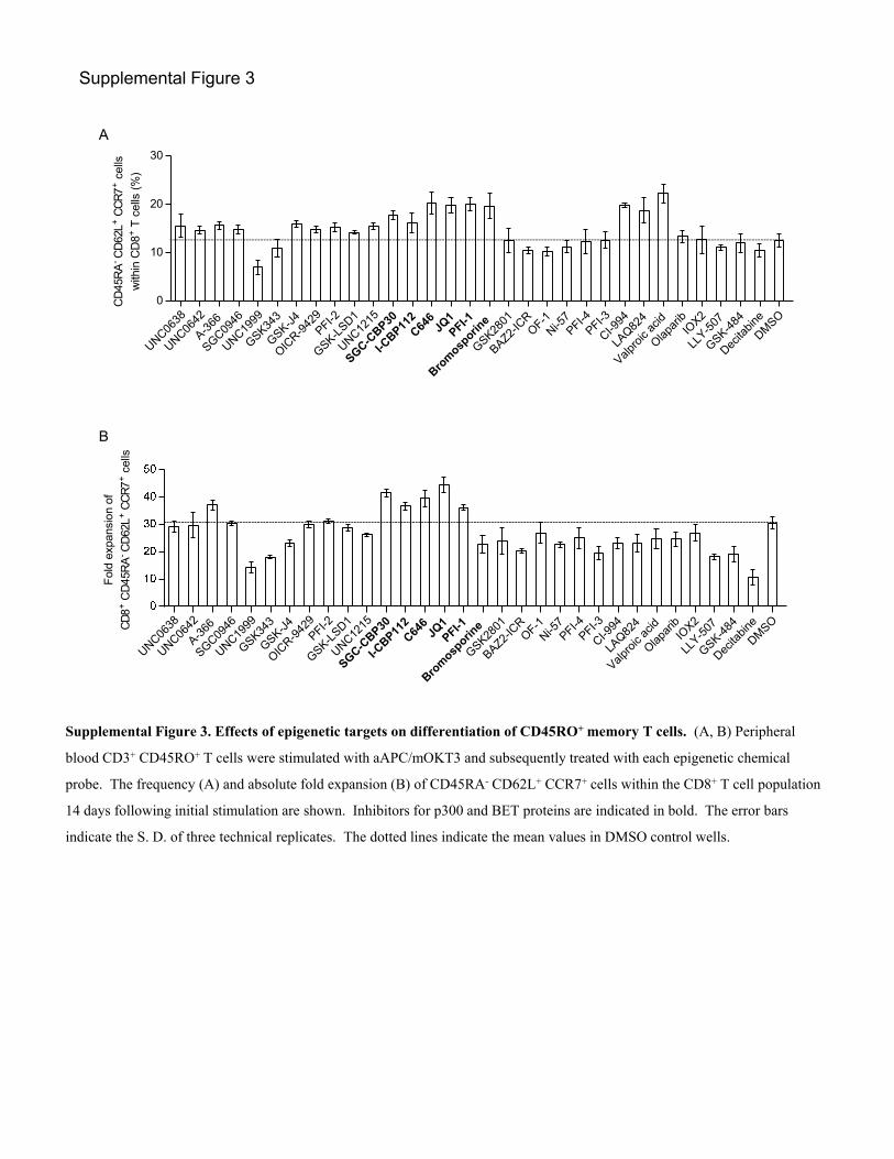
Supplemental Figure 3
Supplemental Figure 3. Effects of epigenetic targets on differentiation of CD45RO+ memory T cells. (A, B) Peripheral
blood CD3+ CD45RO+ T cells were stimulated with aAPC/mOKT3 and subsequently treated with each epigenetic chemical
probe. The frequency (A) and absolute fold expansion (B) of CD45RA- CD62L+ CCR7+ cells within the CD8+ T cell population
14 days following initial stimulation are shown. Inhibitors for p300 and BET proteins are indicated in bold. The error bars
indicate the S. D. of three technical replicates. The dotted lines indicate the mean values in DMSO control wells.
UNC0638
UNC0642
A-366
SGC0946
UNC1999
GSK343
GSK-J4
OICR-9
429PFI-2
GSK-LSD1
UNC1215
SGC-CBP30
I-CBP112C646 JQ
1PFI-1
Bromosporine
GSK2801
BAZ2-IC
ROF-1
Ni-57PFI-4
PFI-3
CI-994
LAQ82
4
Valpro
ic ac
id
Olapar
ibIO
X2
LLY-5
07
GSK-484
Decita
bine
DMSO
0
10
20
30
CD
45R
A-C
D62
L+ C
CR
7+ c
ells
with
in C
D8+
T c
ells
(%
)A
UNC0638
UNC0642
A-366
SGC0946
UNC1999
GSK343
GSK-J4
OICR-9
429PFI-2
GSK-LSD1
UNC1215
SGC-CBP30
I-CBP112C646 JQ
1PFI-1
Bromosporine
GSK2801
BAZ2-IC
ROF-1
Ni-57PFI-4
PFI-3
CI-994
LAQ82
4
Valpro
ic ac
id
Olapar
ibIO
X2
LLY-5
07
GSK-484
Decita
bine
DMSO
Fol
d ex
pans
ion
of
CD
8+ C
D45
RA
-C
D62
L+ C
CR
7+ c
ells
B

JQ1
(-)-J
Q1JQ
1
(-)-J
Q1
0
200
400
600
A
CD4+ CD8+
B
JQ1
(-)-J
Q1
0
2
4
6
8
10
Dea
d ce
lls (
%)
n.s.n.s.
n.s.
Supplemental Figure 4
Supplemental Figure 4. Effects of JQ1 treatment on T cell division rate and viability. (A, B) CD3+ T cells labeled
with CFSE were stimulated with aAPC/mOKT3 and cultured in the presence of JQ1 or (-)-JQ1 for 3 days. The average
mean fluorescence intensity of CFSE in CD4+ and CD8+ T cells (A) as well as the frequency of dead cells (B) were
evaluated by flow cytometry (n=5). Error bars depict the S. D.
C
JQ1
Contro
l0
20
40
60
80 P<0.05
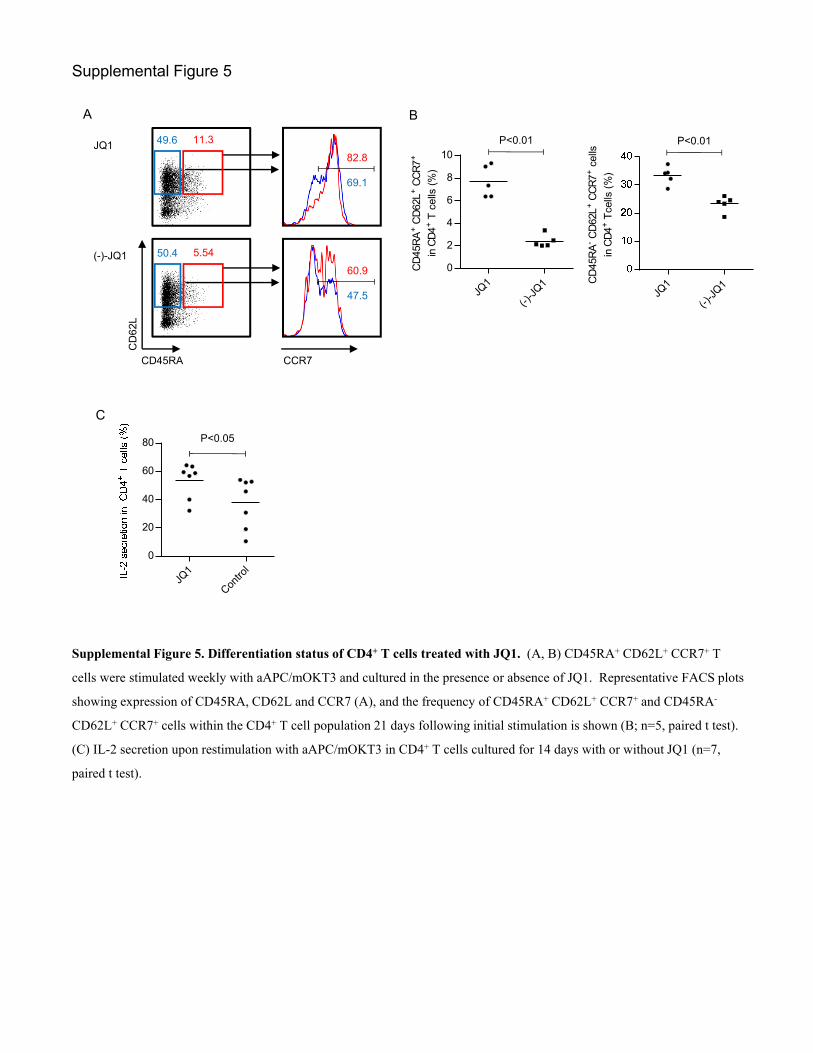
Supplemental Figure 5. Differentiation status of CD4+ T cells treated with JQ1. (A, B) CD45RA+ CD62L+ CCR7+ T
cells were stimulated weekly with aAPC/mOKT3 and cultured in the presence or absence of JQ1. Representative FACS plots
showing expression of CD45RA, CD62L and CCR7 (A), and the frequency of CD45RA+ CD62L+ CCR7+ and CD45RA-
CD62L+ CCR7+ cells within the CD4+ T cell population 21 days following initial stimulation is shown (B; n=5, paired t test).
(C) IL-2 secretion upon restimulation with aAPC/mOKT3 in CD4+ T cells cultured for 14 days with or without JQ1 (n=7,
paired t test).
Supplemental Figure 5
B
P<0.01
JQ1
(-)-J
Q1CD
45R
A- C
D62
L+ C
CR
7+ c
ells
in C
D4+
Tce
lls (
%)
P<0.01
JQ1
(-)-J
Q1
0
2
4
6
8
10
CD
45R
A+ C
D62
L+ C
CR
7+
in C
D4+
T c
ells
(%
)
CD45RA
CD
62L
CCR7
49.6 11.3
82.8
69.1
50.4 5.54
60.9
47.5
JQ1
(-)-JQ1
A

CM CM CM CM EM EM EM EM
Nor
mal
ized
inte
nsity
val
ues
CM CM CM CM EM EM EM EM
Nor
mal
ized
inte
nsity
val
ues
CM CM CM CM EM EM EM EM
Nor
mal
ized
inte
nsity
val
ues
CM CM CM CM EM EM EM EM
Nor
mal
ized
inte
nsity
val
ues
CM CM CM CM EM EM EM EM
Nor
mal
ized
inte
nsity
val
ues
CM CM CM CM EM EM EM EM
Nor
mal
ized
inte
nsity
val
ues
SCMSCM
SCM CM CM CM EM EM EM
Nor
mal
ized
inte
nsity
val
ues
SCMSCM
SCM CM CM CM EM EM EM
Nor
mal
ized
inte
nsity
val
ues
SCMSCM
SCM CM CM CM EM EM EM
Nor
mal
ized
inte
nsity
val
ues
SCMSCM
SCM CM CM CM EM EM EM
Nor
mal
ized
inte
nsity
val
ues
SCMSCM
SCM CM CM CM EM EM EM
Nor
mal
ized
inte
nsity
val
ues
SCMSCM
SCM CM CM CM EM EM EM
Nor
mal
ized
inte
nsity
val
ues
GSE11057 GSE23321
P<0.01
P<0.01
P<0.05
P<0.01
P<0.05
P<0.01
P<0.01
P<0.01
P<0.01
P<0.01
P<0.01
P<0.05
ACTN1
SELL
TCF7
GZMA
KLRG1
PLEK
Supplemental Figure 6. Differentially expressed genes between the TSCM/TCM and TEM phenotypes. Normalized intensity values of the indicated genes from the published microarray data (GSE11057 and GSE23321) are shown.
Supplemental Figure 6

JQ1
(-)-J
Q1
0
10
20
30
40
B
JQ1
Contro
l0
20
40
60
80
100
JQ1
DMSO
0
20
40
60P<0.05 P<0.01n.s.
ACTN1SELL
TCF7
GZMA
KLRG1PLEK
Rel
ativ
e ex
pres
sion
(JQ
1-tr
eate
d / c
ontr
ol)
D
IL-2
+ IF
N-
+ T
NF-
+ c
ells
with
in C
D8
+ T
cel
ls (
%)
C
P<0.05
Supplemental Figure 7. Comparison of the functional properties of CD45RO+ memory T cells treated with JQ1 or (-)-JQ1.
(A) CD3+ CD45RO+ cells were stimulated with aAPC/mOKT3 and cultured with JQ1 or (-)-JQ1 for 14 days. The frequency of
CD45RA- CD62L+ CCR7+ cells within the CD8+ T cell population is shown (n=8, paired t test). (B, C) CD45RO+ T cells treated
with JQ1 or (-)-JQ1 for 14 days were restimulated with aAPC/mOKT3, and the production of IL-2, IFN- and TNF- in CD8+ T
cells was assessed with intracellular flow cytometry. The frequency of individual cytokine-secreting cells (B) and those
producing all three cytokines (C) is shown (n=4, paired t test). (D) Expression profiles of representative genes with differential
expression between the TCM and TEM phenotypes. The average expression levels in the JQ1-treated CD8+ T cells relative to those
in (-)-JQ1-treated CD8+ T cells are shown (n=4). Error bars indicate the S. D.
Supplemental Figure 7
JQ1
(-)-J
Q1
Fre
quen
cy o
f CD
45R
A-
CD
62L+
CC
R7+
cel
ls (
%)
AP<0.01

Supplemental Figure 8
Supplemental Figure 8. Effects of JQ1 on CD8+ T cell differentiation in the absence of costimulatory signals. (A, B)
CD3+ T cells were stimulated every week with plate-coated anti-CD3 mAb (clone OKT3) and cultured in the presence or
absence of JQ1. Representative FACS plots (A) and the frequencies of CD45RA+ CD62L+ CCR7+ and CD45RA- CD62L+
CCR7+ cells (B) within the CD8+ T cell population 14 days following the initial stimulation (n=7, paired t test). (C, D)
Secretion levels of IL-2, IFN-, and TNF- were evaluated by intracellular flow cytometry in JQ1- and (-)-JQ1-treated
CD8+ T cells. The frequencies of each type of cytokine-secreting cells (C) and cells producing all three cytokines (D) are
shown (n=5, paired t test).
JQ1
(-)-J
Q1
0
2
4
6
8
IL-2
sec
retio
n (%
)
JQ1
(-)-J
Q1
20
30
40
50
60
JQ1
(-)-J
Q1
0
10
20
30
C P<0.01n.s.
n.s.
JQ1
(-)-J
Q1IL-2
+ IF
N-
+ T
NF-
+ c
ells
(%
)
P<0.05
D
JQ1
(-)-J
Q1CD
45R
A+ C
D62
L+ C
CR
7+ c
ells
(%
)
JQ1
(-)-J
Q1CD
45R
A- C
D62
L+ C
CR
7+ c
ells
(%
)
P<0.01 P<0.01
BA JQ1 (-)-JQ1
27.6 11.651.7 14.6
35.3
34.2
23.9
22.6
CD45RA
CD
62L
CCR7

Supplemental Figure 9. Differentiation of CD8+ T cells upon antigen-specific T cell stimulation. CD3+ T cells were
stimulated weekly with aAPC/A2 loaded with mutant MART127–35 peptide in the presence of JQ1 or (-)-JQ1. The CD8+
A2/MART1 multimer+ cells were analyzed for CD45RA, CD62L and CCR7 expression 21 days after initial stimulation.
Representative FACS plots of four independent experiments are shown.
Supplemental Figure 9
JQ1
(-)-JQ1
CD8
MA
RT
1 m
ultim
er
6.01
8.64
CD45RA
CD
62L
10.532.4
5.7221.6
CCR7
8.2
5.3
17.1
9.97

Supplemental Figure 10
Supplemental Figure 10. Cytokine secretion by CAR-transduced T cells following restimulation with K562-CD19. JQ1- or
(-)-JQ1-treated CAR-T cells were stimulated with K562-CD19 in the absence of drugs. Five days later, they were restimulated
with K562-CD19, and cytokine secretion was evaluated by intracellular flow cytometry. Frequencies of each type of cytokine-
producing cell and cells secreting all three cytokines are shown (n=5, paired t test).
JQ1
(-)-J
Q1
0
10
20
30
IL-2
sec
retio
n (%
)
JQ1
(-)-J
Q1
0
20
40
60
80
IFN
- s
ecre
tion
(%
)
JQ1
(-)-J
Q1
0
10
20
30
40
50
TNF-
sec
retio
n (
%)
JQ1
(-)-J
Q1
IL-2
+ IF
N-
+ T
NF-
+ce
lls (
%)P<0.05 P<0.05
n.s.
P<0.05

Supplemental Figure 11
Supplemental Figure 11. Treatment of CD19+ acute lymphoblastic cell line NALM-6 with anti-CD19 CAR-transduced T cells. (A) NSG mice were intravenously injected with NALM6-GL and, 14 days later, treated with CAR-transduced T cells treated with JQ1 or (-)-JQ1. In vivo bioluminescent imaging of luciferase activity at the indicated time points following T cell infusion is shown. (B) Phenotypic analysis for persistent CD8+ CAR-T cells in the peripheral blood. Representative FACS plots at day 7 following T cell infusion are shown.
Protein L / Streptavidin-APC
CD8
FS
C
CD45RA
CD
62L
CCR7
18.7
(-)-JQ1
74.440.1 14.3
37.1
32.4
35.9
JQ1
51.840.2 15.2
31.6
24.0
B
CAR-negative control T cells
Gated CD8+ T cells
A(-)-JQ1
Days after T cell infusion
0
7
14
21
28
35
42
No T cells
Days after T cell infusion
0
7
JQ1
0
7
14
21
28
35
42
49
56
63
70
77

Supplemental Figure 12
Supplemental Figure 12. Autopsy analysis of mice transplanted with JQ1- or (-)-JQ1-CAR-T cells. (A) Representative
FACS plots evaluating the persistence of NALM6-GL and CD19 expression in the spleen and bone marrow. (B)
Representative FACS plots showing the persistence of CAR-T cells in the spleen.
A
In vitro cultured NALM6-GL
CD19
JQ1-CAR-T cell transplanted mice
Spleen Bone marrow
(-)-JQ1-CAR-T cell transplanted mice
Spleen Bone marrow
GFP
61.8 66.0 38.4 55.8
GFP+ cells
K562
B
CD45
FS
C
CD8
CD
4
3.2475.6
Protein L / Streptavidin-PE
0.65(-)-JQ1
0.5760.5
5.76JQ1 Residual CD8+ T cells
Control T cells

Supplemental Figure 13
Supplemental Figure 13. Cytokine secretion by A2/MART1-T cells following restimulation with aAPC/A2. JQ1- or (-)-
JQ1-treated A2/MART1-T cells were stimulated with aAPC/A2 loaded with MART127-35 peptide in the absence of drugs.
Five days later, the T cells were restimulated with aAPC/A2 with MART127-35, and cytokine secretion was evaluated by
intracellular flow cytometry. Frequencies of each type of cytokine-producing cell and cells secreting all three cytokines are
shown (n=4; paired t test).
JQ1
(-)-J
Q1
0
10
20
30
40
IL-2
sec
retio
n (%
)
JQ1
(-)-J
Q1
50
55
60
65
70
75
IFN
- s
ecre
tion
(%
)
JQ1
(-)-J
Q1
36
38
40
42
44
46
TNF-
sec
retio
n (
%)
P<0.01 n.s.
n.s.
P<0.05
JQ1
(-)-J
Q1
IL-2
+ IF
N-
+ T
NF-
+ce
lls (
%)

Supplemental Figure 14
Supplemental Figure 14. Surface marker phenotypes of CD8+ T cells with BATF knockdown. Surface expression of CD27,
CD28, and CD127 in the CD8+ NGFR+ T cells transduced with control or siBATF 14 days following initial stimulation with
aAPC/mOKT3. Representative plots of the samples in Figure 7C are shown.
CD27
CD28
Control siBATF-1 siBATF-2
CD127
CD45RA+ CD62L+ CCR7+
CD45RA- CD62L+ CCR7+
CD45RA- CD62L- CCR7-

Supplemental Figure 15
Supplemental Figure 15. BATF knockdown by lentiviral shRNA. (A, B) CD3+ T cells were transduced with lentiviral
shRNAs against BATF and stimulated with aAPC/mOKT3. Protein levels of BATF in the CD8+ ZsGreen+ T cell population
were analyzed by intracellular flow cytometry three days following stimulation. Representative FACS plots (A) and average
relative mean fluorescence intensity normalized to the control (B) (n=4; one-way ANOVA). Error bars indicate the S.D.
(C) Naïve T cells were purified by magnetic selection, pre-treated with IL-7, and transduced with lentiviral shRNA targeting
BATF at day 5-8. Representative FACS plots of CD45RA, CD62L and CCR7 expression in the purified naïve T cells and
after shRNA transduction are shown.
Contrl
shBATF
-1
shBATF
-2
0.0
0.5
1.0
1.5
P<0.01
P<0.01B
BATF
Controlsh-BATF
shBATF-1 shBATF-2A
C
Purified naïve T
Cultured naïve T
CD45RA
CD
62L
CCR7
99.7
GFP
48.455.751.3
96.6 96.2 96.1
99.6 99.6
Control shBATF-1 shBATF-2
99.8
shRNA-transduced T cells
Untransduced control T cells
100.0

Supplemental Figure 16
Supplemental Figure 16. Memory T cell formation from naïve T cells transduced with lentiviral shRNA against BATF. (A)
Naïve T cells were transduced with shRNA against BATF following pretreatment with IL-7. They were subsequently stimulated
with aAPC/mOKT3, and the expression profiles of CD45RA, CD62L and CCR7 within the CD8+ ZsGreen+ T cell population were
analyzed 10 days following stimulation. Representative FACS plots of the five experiments are shown. (B) Naïve T cells
transduced with shRNA against BATF were transplanted into irradiated NSG mice. Mice were sacrificed 11 days following T cell
infusion, and the CD8+ ZsGreen+ cells engrafted in the spleen were analyzed. Representative FACS plots analyzing expression of
CD45RA, CD62L and CCR7 within the CD45+ CD8+ ZsGreen+ population are shown. (C) Naïve T cells with shRNA against
BATF were transplanted into irradiated NSG mice, as shown in Figure 7H. Mice were sacrificed 16 days following T cell
infusion, and the frequency of CD45RA+/- CD62L+ CCR7+ cells within the ZsGreen+ CD8+ T cell population in the spleen was
analyzed (n=6, one-way ANOVA).
B
29.445.816.3
CD45RA
CD
62L
Control shBATF-1 shBATF-2
67.0 31.3 34.0
96.8
81.0
93.4
61.3
91.8
66.6
CCR7
CD8
ZsG
reen
3.736.324.68
C
P<0.01P<0.01
n.s.
Control
shBATF-1
shBATF-2C
D45
RA
+ C
D62
L+ C
CR
7+ c
ells
with
in C
D8+
ZsG
reen
+ T
cel
ls (
%)
Control
shBATF-1
shBATF-2
0
5
10
15
20
CD
45R
A- C
D62
L+ C
CR
7+ c
ells
with
in C
D8
+ Z
sGre
en+ T
cel
ls (
%)
CD45RA
CD
62L
Control shBATF-1 shBATF-2
CCR7
35.0 25.737.2 23.740.8 15.1
51.6
19.6
75.6
37.8
79.7
44.5
A

Supplemental Figure 17
Supplemental Figure 17. Knockdown effects of genes suppressed by JQ1 on memory T cell differentiation. (A) Expression
of the candidate genes targeted by JQ1 was assessed by quantitative real-time PCR. Relative expression levels of the indicated
genes in JQ1-treated T cells relative to (-)-JQ1-treated cells (normalized to UBC) three days after stimulation with aAPC/mOKT3
are shown (n=4, one-sample test compared to one). Error bars indicate the S.D. ND, not detected within 45 cycles of qPCR. *
P<0.05, ** P<0.01. (B) CD3+ T cells were retrovirally transduced with the control, siFOSL2, siID2, or siPRDM1 and NGFR.
Expression of each target gene compared with the control was assessed by quantitative PCR (n=4, one sample t test compared to
one). Error bars indicate the S.D. * P<0.05, ** P<0.01. (C) CD3+ T cells were stimulated with aAPC/mOKT3 and transduced
with control, siFOSL2, siID2, or siPRDM1, and NGFR. The frequency of CD45RA+/- CD62L+ CCR7+ cells within the NGFR+
CD8+ T cell population 14 days after initial stimulation is shown (n=5, paired ANOVA).
Contro
l
siFOSL2
siID2
siPRDM1
0
5
10
15
CD
45R
A+ C
D62
L+ C
CR
7+ c
ells
with
in C
D8
+ T
cel
ls (
%) n.s.
Contro
l
siFOSL2
siID2
siPRDM1
0
2
4
6
8
10
CD
45R
A- C
D62
L+ C
CR
7+ c
ells
with
in C
D8
+ T
cel
ls (
%)
n.s.C
siFOSL2
siID2
siPRDM1
Rel
ativ
e ex
pres
sion
B
*
**
**
AHR
CREMEGR2
FOSL1
FOSL2ID2
NFIL3
PLAGL2
STAT3
TRIP13
UHRF
PRDM1
RUNX3
STAT4
TBX21
0.0
0.5
1.0
1.5
2.0
2.5
A
**
**
ND

Supplemental Figure 18
Supplemental Figure 18. Phosphorylation of ribosomal protein S6 kinase (S6K) is decreased in JQ1-treated T cells. (A,
B) CD3+ T cells were stimulated with aAPC/mOKT3 at an E:T ratio of 3:1 and treated with JQ1 or (-)-JQ1 in the presence of
IL-2 and IL-15. Phosphorylation of the indicated proteins in CD8+ T cell population was analyzed 5 days after stimulation.
Representative FACS plots (A) and relative mean fluorescence intensity normalized to freshly isolated CD8+ T cells (B) (n=4;
unpaired t test). (C, D) CD3+ T cells were stimulated with aAPC/mOKT3 and transduced with lentiviral shRNA against
BATF. The shRNA-transduced T cells were rested in cytokine-free media and then restimulated with aAPC/mOKT3.
Phosphorylation of S6K in the ZsGreen+ CD8+ T cell population was quantified by intracellular flow cytometry.
Representative FACS plots (C) and relative mean fluorescence intensity normalized to the control plasmid-transduced cells
(D) (n=4; one-way ANOVA).
n.s.
Day 0
JQ
1
(-)-J
Q1
Rel
ativ
e m
ean
fluor
esce
nce
inte
nsity
Day 0
JQ
1
(-)-J
Q1
0
5
10
15n.s.
Day 0
JQ
1
(-)-J
Q1
0
1
2
3n.s.
Day 0
JQ
1
(-)-J
Q1
0
5
10
15 n.s.P<0.01
pSTAT5 p-p38 MAPK pERK pAKT pS6KB
Day 0
JQ1
(-)-J
Q1
0
10
20
30
40
Rel
ativ
e m
ean
fluor
esce
nce
inte
nsity
CControl
shBATF-1
shBATF-2
Ctsh
shBATF
-1
shBATF
-2
0.0
0.5
1.0
1.5
Rel
ativ
e m
ean
fluor
esce
nce
inte
nsity
of p
S6k
D
P<0.01
P<0.01
pS6K
pSTAT5 p-p38 MAPK pERK pAKT pS6K
JQ1
(-)-JQ1
Day 0 CD8+ T cells
A

Supplemental Figure 19
Supplemental Figure 19. Phenotypic analysis of CAR-T cells after adoptive transfer. C646- or DMSO-treated CAR-T cells were infused into NSG mice transplanted with NALM6-GL. The frequency of CD45RA+/- CD62L+ CCR7+ T cells within the CD8+ CAR+ T cell population was analyzed (n=10, unpaired t test).
Fre
qunc
y w
ithin
CD
8+ C
AR
+ T
cel
ls (
%)
CD45RA+
CD62L+ CCR7+CD45RA-
CD62L+ CCR7+
n.s.
n.s. n.s.
n.s.n.s.
n.s.

Supplemental Table 1. Epigenetic chemical probes with defined targets.
Probe Target Tested dose (M) References Probe Target Tested dose (M) References
Histone-modifying enzymes Histone readers
UNC0638 G9a/GLP 0.1 1 JQ1 BET Bromodomain 0.15 15
UNC0642 G9a/GLP 0.1 2 PFI-1 BET Bromodomain 1 16
A-366 G9a/GLP 0.5 3 Bromosporine pan-Bromodomain 1
SGC0946 DOT1L 0.5 4 GSK2801 BAZ2A/B 1 17
UNC1999 EZH2 0.2 5 BAZ2-ICR BAZ2B/A 1 18
GSK343 EZH2 0.5 6 OF-1 BRPF1-3 1
GSK-J4 JMJD3/UTX 1 7 Ni-57 BRPF1-3 0.5
OICR-9429 WDR5 1 8 PFI-4 BRPF1B 0.25
PFI-2 SETD7 1 9 PFI-3 SMARCA4 1 19
GSK-LSD1 LSD1 1 10 CI-994 HDAC 1
UNC1215 L3MBTL3 1 11 LAQ824 HDAC 0.01 20
SGC-CBP30 CREBBP/EP300 0.2 12 VPA HDAC 400
I-CBP112 CREBBP/EP300 1 13 Other targets
C646 EP300 10 14 Olaparib PARP 1
IOX2 HIF1a 50 21
GSK484 PAD-4 0.5 22
LLY-507 SMYD2 1 23
Decitabine DNMT 0.05

Supplemental Table 2. Detailed information of the mice transplanted with CAR-T cells.
Probe Overall survival
NALM6-GL at autopsy (%)
CAR-T cell at autopsy (%)
Signs of GVHD Bone
marrow Spleen
T cells in the bone marrow T cells in the spleen
CD4+ CD8+ CAR+ in
CD4+ T cells CAR+ in
CD8+ T cells CD4+ CD8+
CAR+ in CD4+ T cells
CAR+ in CD8+ T cells
1
JQ1
85 35.6 25.2 2.02 0.46 0.53 1.56 7.25 0.93 0.41 2 fur loss and
red skin
2 64 67.8 42.8 0.56 0.08 3.60 6.56 0.38 0.08 1.23 6.92 none
3 65 66 61.8 0.06 0.06 2.60 7.55 0.11 0.35 2.34 5.76 none
4 59 24.6 14.1 1.05 0.06 3.07 8.7 2.35 0.03 0.00 2.13 none
5 91
(alive) NA
fur loss and red skin
6 91
(alive) NA none
7 45 66.1 21.6 0.40 0.02 1.36 4 0.35 0.02 0.76 0 none
8 91
(alive) NA none
9 33 73.2 35.2 0.05 0.02 6.12 0.00 0.06 0.05 2.72 5.17 none
10 46 45.1 33 0.01 0.06 0.00 1.79 0.00 0.01 0.00 0 none
1
(-)-JQ1
40 82.3 22.4 0.02 0.05 0.00 0 0.53 1.78 0.89 0.484 none
2 41 84.2 48.5 0.06 0.23 2.33 6.44 0.30 2.45 0.86 0.649 none
3 37 83.4 48.6 0.04 0.23 4.00 2.76 0.10 1.00 0.00 0.813 none
4 54 55.8 38.4 0.00 0.00 NA NA 0.00 0.02 NA 0 none
5 61 28.4 16.8 0.07 0.02 2.71 12.3 0.79 0.21 0.89 3.39 none
6 46 82.6 45.3 0.21 0.27 1.32 2.11 0.28 1.54 0.63 0.794 none
7 36 72.9 22.2 0.02 0.00 5.88 NA 0.13 0.01 7.32 0 none
8 33 68.3 21.5 0.01 0.02 0.00 8.7 0.13 0.12 2.20 6.71 none
9 40 60.2 21.1 0.03 0.04 0.00 0 0.59 0.49 1.26 2.76 none
10 34 84.5 37.7 0.07 0.19 2.94 2.25 0.13 1.08 0.00 1.21 none
References
1. Vedadi M, Barsyte-Lovejoy D, Liu F, Rival-Gervier S, Allali-Hassani A, Labrie V, Wigle TJ,
Dimaggio PA, Wasney GA, Siarheyeva A, et al. A chemical probe selectively inhibits G9a and
GLP methyltransferase activity in cells. Nat Chem Biol. 2011;7(8):566-74.
2. Liu F, Barsyte-Lovejoy D, Li F, Xiong Y, Korboukh V, Huang XP, Allali-Hassani A, Janzen
WP, Roth BL, Frye SV, et al. Discovery of an in vivo chemical probe of the lysine
methyltransferases G9a and GLP. J Med Chem. 2013;56(21):8931-42.

3. Sweis RF, Pliushchev M, Brown PJ, Guo J, Li F, Maag D, Petros AM, Soni NB, Tse C, Vedadi
M, et al. Discovery and development of potent and selective inhibitors of histone
methyltransferase g9a. ACS Med Chem Lett. 2014;5(2):205-9.
4. Yu W, Chory EJ, Wernimont AK, Tempel W, Scopton A, Federation A, Marineau JJ, Qi J,
Barsyte-Lovejoy D, Yi J, et al. Catalytic site remodelling of the DOT1L methyltransferase by
selective inhibitors. Nat Commun. 2012;3(1288.
5. Konze KD, Ma A, Li F, Barsyte-Lovejoy D, Parton T, Macnevin CJ, Liu F, Gao C, Huang XP,
Kuznetsova E, et al. An orally bioavailable chemical probe of the Lysine Methyltransferases
EZH2 and EZH1. ACS Chem Biol. 2013;8(6):1324-34.
6. Verma SK, Tian X, LaFrance LV, Duquenne C, Suarez DP, Newlander KA, Romeril SP,
Burgess JL, Grant SW, Brackley JA, et al. Identification of Potent, Selective, Cell-Active
Inhibitors of the Histone Lysine Methyltransferase EZH2. ACS Med Chem Lett.
2012;3(12):1091-6.
7. Kruidenier L, Chung CW, Cheng Z, Liddle J, Che K, Joberty G, Bantscheff M, Bountra C,
Bridges A, Diallo H, et al. A selective jumonji H3K27 demethylase inhibitor modulates the
proinflammatory macrophage response. Nature. 2012;488(7411):404-8.
8. Grebien F, Vedadi M, Getlik M, Giambruno R, Grover A, Avellino R, Skucha A, Vittori S,
Kuznetsova E, Smil D, et al. Pharmacological targeting of the Wdr5-MLL interaction in
C/EBPalpha N-terminal leukemia. Nat Chem Biol. 2015;11(8):571-8.
9. Barsyte-Lovejoy D, Li F, Oudhoff MJ, Tatlock JH, Dong A, Zeng H, Wu H, Freeman SA,
Schapira M, Senisterra GA, et al. (R)-PFI-2 is a potent and selective inhibitor of SETD7
methyltransferase activity in cells. Proc Natl Acad Sci U S A. 2014;111(35):12853-8.
10. Mohammad HP, Smitheman KN, Kamat CD, Soong D, Federowicz KE, Van Aller GS,
Schneck JL, Carson JD, Liu Y, Butticello M, et al. A DNA Hypomethylation Signature Predicts
Antitumor Activity of LSD1 Inhibitors in SCLC. Cancer Cell. 2015;28(1):57-69.
11. James LI, Barsyte-Lovejoy D, Zhong N, Krichevsky L, Korboukh VK, Herold JM, MacNevin
CJ, Norris JL, Sagum CA, Tempel W, et al. Discovery of a chemical probe for the L3MBTL3
methyllysine reader domain. Nat Chem Biol. 2013;9(3):184-91.
12. Hay DA, Fedorov O, Martin S, Singleton DC, Tallant C, Wells C, Picaud S, Philpott M,
Monteiro OP, Rogers CM, et al. Discovery and optimization of small-molecule ligands for the
CBP/p300 bromodomains. J Am Chem Soc. 2014;136(26):9308-19.
13. Picaud S, Fedorov O, Thanasopoulou A, Leonards K, Jones K, Meier J, Olzscha H, Monteiro O,
Martin S, Philpott M, et al. Generation of a Selective Small Molecule Inhibitor of the CBP/p300
Bromodomain for Leukemia Therapy. Cancer Res. 2015;75(23):5106-19.
14. Bowers EM, Yan G, Mukherjee C, Orry A, Wang L, Holbert MA, Crump NT, Hazzalin CA,
Liszczak G, Yuan H, et al. Virtual ligand screening of the p300/CBP histone acetyltransferase:
identification of a selective small molecule inhibitor. Chem Biol. 2010;17(5):471-82.
15. Filippakopoulos P, Qi J, Picaud S, Shen Y, Smith WB, Fedorov O, Morse EM, Keates T,
Hickman TT, Felletar I, et al. Selective inhibition of BET bromodomains. Nature.
2010;468(7327):1067-73.
16. Picaud S, Da Costa D, Thanasopoulou A, Filippakopoulos P, Fish PV, Philpott M, Fedorov O,
Brennan P, Bunnage ME, Owen DR, et al. PFI-1, a highly selective protein interaction inhibitor,
targeting BET Bromodomains. Cancer Res. 2013;73(11):3336-46.
17. Chen P, Chaikuad A, Bamborough P, Bantscheff M, Bountra C, Chung CW, Fedorov O, Grandi
P, Jung D, Lesniak R, et al. Discovery and Characterization of GSK2801, a Selective Chemical
Probe for the Bromodomains BAZ2A and BAZ2B. J Med Chem. 2015.

18. Drouin L, McGrath S, Vidler LR, Chaikuad A, Monteiro O, Tallant C, Philpott M, Rogers C,
Fedorov O, Liu M, et al. Structure enabled design of BAZ2-ICR, a chemical probe targeting the
bromodomains of BAZ2A and BAZ2B. J Med Chem. 2015;58(5):2553-9.
19. Vangamudi B, Paul TA, Shah PK, Kost-Alimova M, Nottebaum L, Shi X, Zhan Y, Leo E,
Mahadeshwar HS, Protopopov A, et al. The SMARCA2/4 ATPase Domain Surpasses the
Bromodomain as a Drug Target in SWI/SNF-Mutant Cancers: Insights from cDNA Rescue and
PFI-3 Inhibitor Studies. Cancer Res. 2015;75(18):3865-78.
20. Catley L, Weisberg E, Tai YT, Atadja P, Remiszewski S, Hideshima T, Mitsiades N,
Shringarpure R, LeBlanc R, Chauhan D, et al. NVP-LAQ824 is a potent novel histone
deacetylase inhibitor with significant activity against multiple myeloma. Blood.
2003;102(7):2615-22.
21. Chowdhury R, Candela-Lena JI, Chan MC, Greenald DJ, Yeoh KK, Tian YM, McDonough
MA, Tumber A, Rose NR, Conejo-Garcia A, et al. Selective small molecule probes for the
hypoxia inducible factor (HIF) prolyl hydroxylases. ACS Chem Biol. 2013;8(7):1488-96.
22. Lewis HD, Liddle J, Coote JE, Atkinson SJ, Barker MD, Bax BD, Bicker KL, Bingham RP,
Campbell M, Chen YH, et al. Inhibition of PAD4 activity is sufficient to disrupt mouse and
human NET formation. Nat Chem Biol. 2015;11(3):189-91.
23. Nguyen H, Allali-Hassani A, Antonysamy S, Chang S, Chen LH, Curtis C, Emtage S, Fan L,
Gheyi T, Li F, et al. LLY-507, a Cell-active, Potent, and Selective Inhibitor of Protein-lysine
Methyltransferase SMYD2. J Biol Chem. 2015;290(22):13641-53.