superior and posterior mediastinum - md connect · netter, f.h. interactive atlas of human anatomy....
TRANSCRIPT

Superior and Posterior Mediastinum
Assoc. Prof. Jenny Hayes

WARNING This material has been provided to you pursuant
to section 49 of the Copyright Act 1968 (the Act) for the purposes of research or study.
The contents of the material may be subject to copyright protection under the Act.
Further dealings by you with this material may be
a copyright infringement. To determine whether such a communication would be an
infringement, it is necessary to have regard to the criteria set out in Part 3, Division 3 of the
Act.

Objectives • The layout and contents of the superior and posterior
mediastinum • The course of the ascending aorta, arch of aorta, descending
thoracic aorta and the major branches of each • The origin, course and relations of the jugular, subclavian,
brachiocephalic veins and SVC (and be aware of the azygous system of veins)
• The course and major relationships of trachea, oesophagus, thoracic duct, vagus and phrenic nerves
Suggested pre-reading Gray’s Anatomy for Students: Regional anatomy of Superior and Posterior mediastinum, Chapter 3, Thorax, p. 204-223, 2nd ed. OR Clinicall y Orientated Anatomy, Moore: Superior mediastinum and great vessels, posterior mediastinum, Chapter 1, Thorax, p.160-170, 6th ed.

Netter, F.H. Interactive Atlas of Human Anatomy. 3rd ed. New Jersey, Icon Learning Systems, 2003, ISBN: 1-929007-15-9, Plate #186
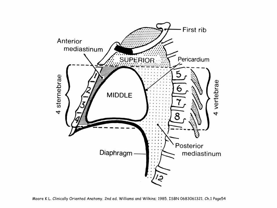
Moore K L. Clinically Oriented Anatomy. 2nd ed. Williams and Wilkins; 1985. ISBN 0683061321. Ch.1 Page54
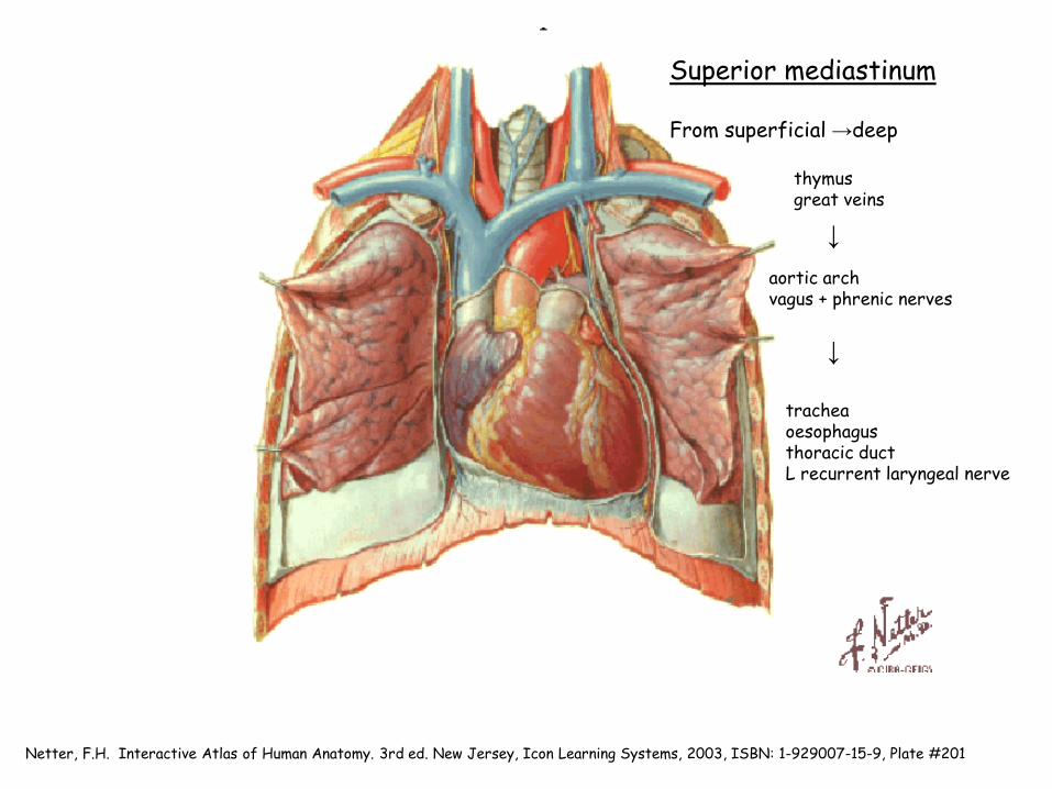
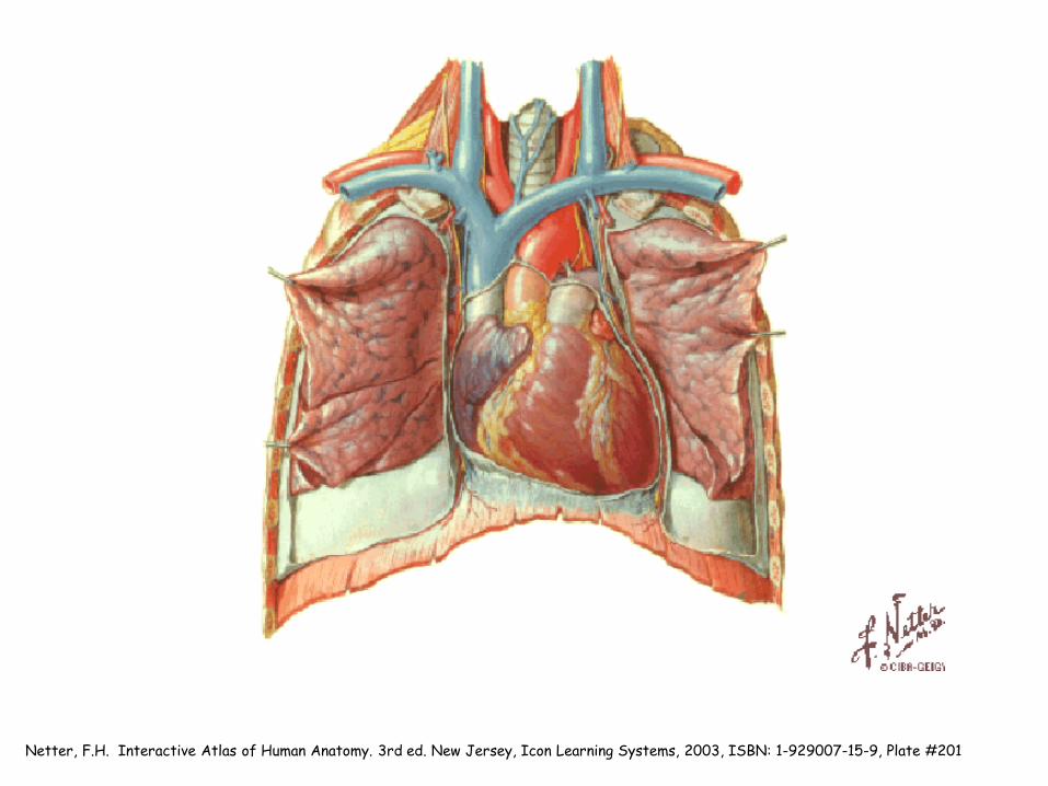
Netter, F.H. Interactive Atlas of Human Anatomy. 3rd ed. New Jersey, Icon Learning Systems, 2003, ISBN: 1-929007-15-9, Plate #201
thymus great veins
↓ aortic arch vagus + phrenic nerves
↓
trachea oesophagus thoracic duct L recurrent laryngeal nerve
Superior mediastinum From superficial →deep
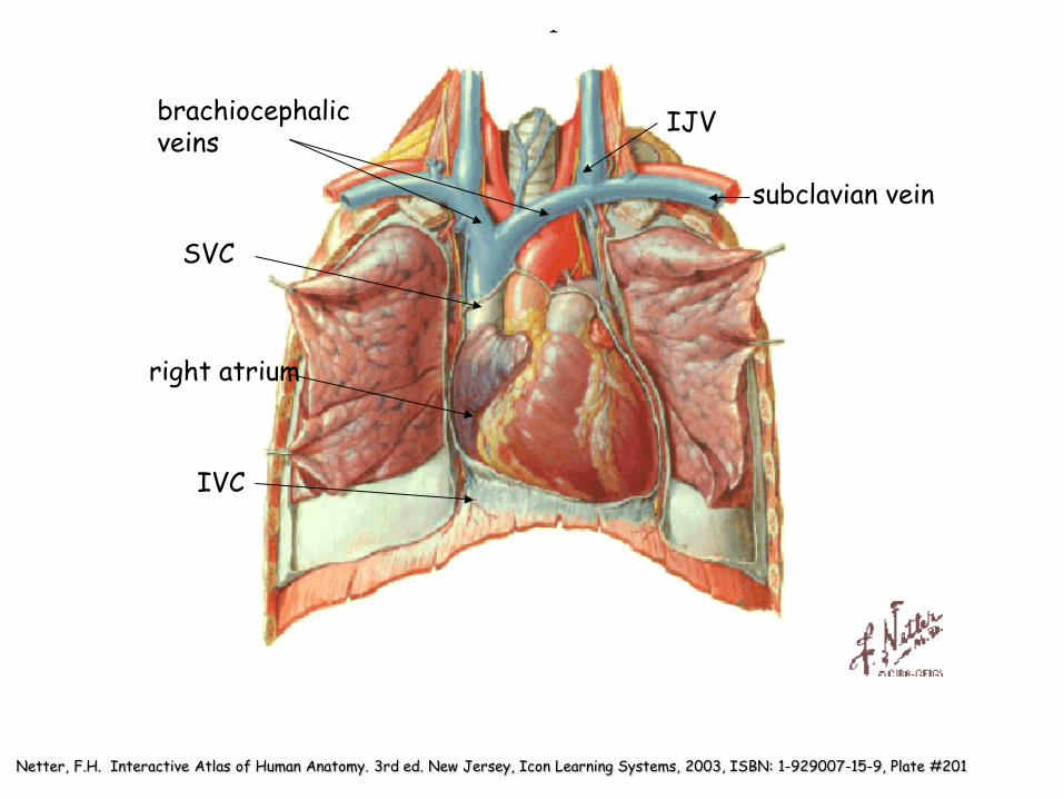
Netter, F.H. Interactive Atlas of Human Anatomy. 3rd ed. New Jersey, Icon Learning Systems, 2003, ISBN: 1-929007-15-9, Plate #201
brachiocephalic veins
SVC
right atrium
IJV
subclavian vein
IVC
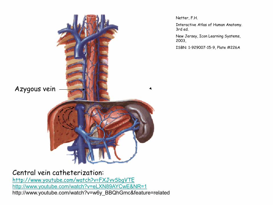
Netter, F.H.
Interactive Atlas of Human Anatomy. 3rd ed.
New Jersey, Icon Learning Systems, 2003,
ISBN: 1-929007-15-9, Plate #226A
Azygous vein
Central vein catheterization: http://www.youtube.com/watch?v=FXJvvSbgVTE http://www.youtube.com/watch?v=eLXN89AYCwE&NR=1 http://www.youtube.com/watch?v=w6y_BBQhGmc&feature=related

Netter, F.H. Interactive Atlas of Human Anatomy. 3rd ed. New Jersey, Icon Learning Systems, 2003, ISBN: 1-929007-15-9, Plate #219
Arch of aorta
Left pulmonary artery
Left main bronchus
Pulmonary veins Descending
thoracic aorta

Netter, F.H. Interactive Atlas of Human Anatomy. 3rd ed. New Jersey, Icon Learning Systems, 2003, ISBN: 1-929007-15-9, Plate #201
Ligamentum arteriosum

Netter, F.H. Interactive Atlas of Human Anatomy. 3rd ed. New Jersey, Icon Learning Systems, 2003, ISBN: 1-929007-15-9, Plate #182A
Brachiocephalic trunk
Right subclavian
Right common carotid Left common carotid
Left subclavian
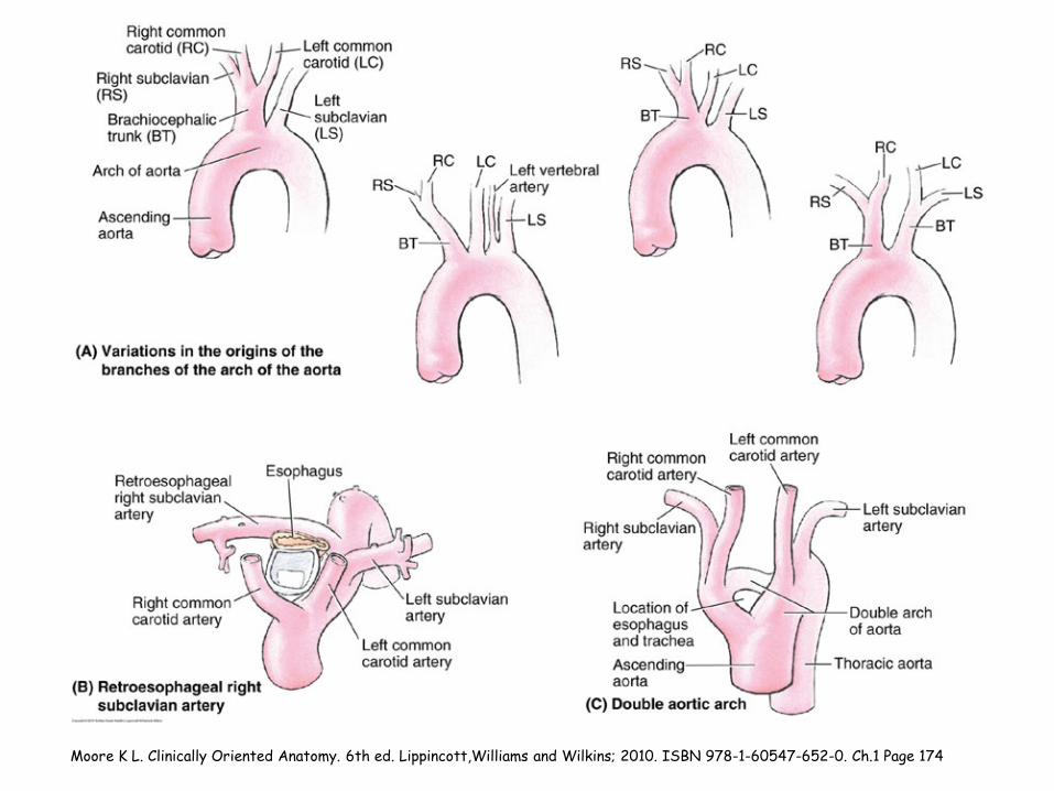
Moore K L. Clinically Oriented Anatomy. 6th ed. Lippincott,Williams and Wilkins; 2010. ISBN 978-1-60547-652-0. Ch.1 Page 174
Netter, F.H. Interactive Atlas of Human Anatomy. 3rd ed. New Jersey, Icon Learning Systems, 2003, ISBN: 1-929007-15-9, Plate #201
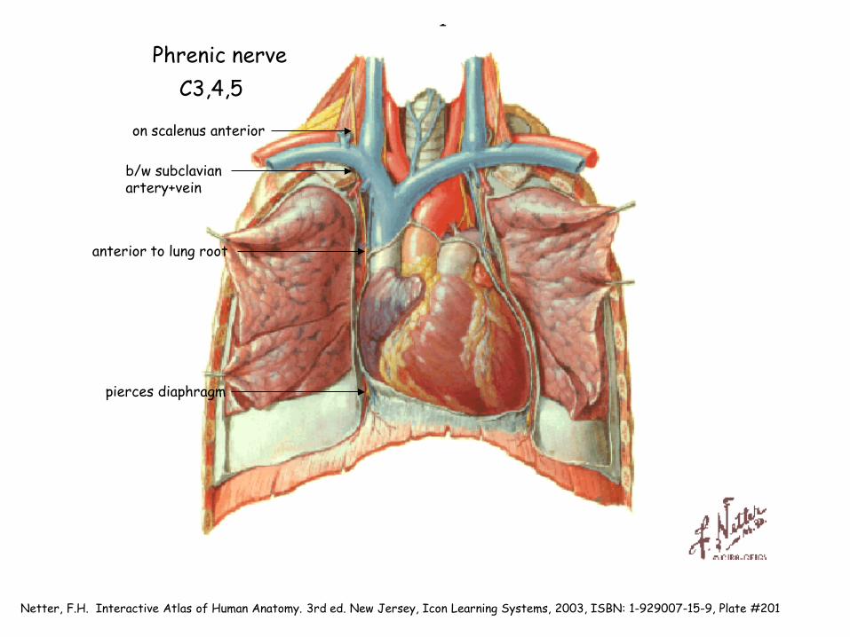
on scalenus anterior
b/w subclavian artery+vein
anterior to lung root
pierces diaphragm
C3,4,5 Phrenic nerve

Netter, F.H. Interactive Atlas of Human Anatomy. 3rd ed. New Jersey, Icon Learning Systems, 2003, ISBN: 1-929007-15-9, Plate #182A
Right phrenic nerve is lateral to venous structures: SVC,R atrium, IVC
Left phrenic nerve is lateral to arterial structures: Aortic arch and L ventricle
Netter, F.H. Interactive Atlas of Human Anatomy. 3rd ed. New Jersey, Icon Learning Systems, 2003, ISBN: 1-929007-15-9, Plate #181
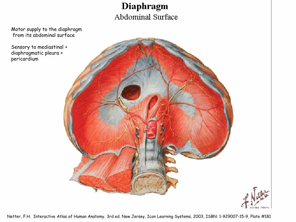
Motor supply to the diaphragm from its abdominal surface Sensory to mediastinal + diaphragmatic pleura + pericardium

Netter, F.H. Interactive Atlas of Human Anatomy. 3rd ed. New Jersey, Icon Learning Systems, 2003, ISBN: 1-929007-15-9, Plate #182A
Vagus nerve Cranial nerve Posterolateral to CCA
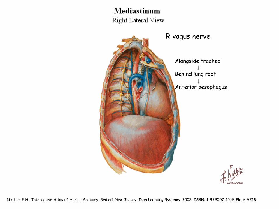
Netter, F.H. Interactive Atlas of Human Anatomy. 3rd ed. New Jersey, Icon Learning Systems, 2003, ISBN: 1-929007-15-9, Plate #218
Alongside trachea ↓ Behind lung root ↓ Anterior oesophagus
R vagus nerve
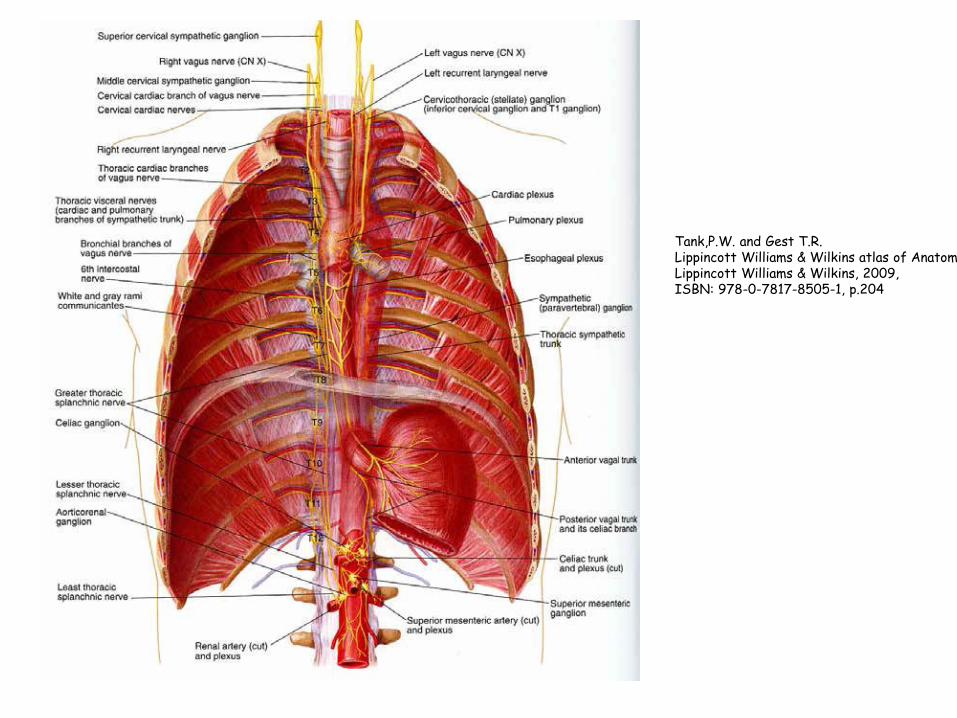
Tank,P.W. and Gest T.R. Lippincott Williams & Wilkins atlas of Anatomy Lippincott Williams & Wilkins, 2009, ISBN: 978-0-7817-8505-1, p.204

Netter, F.H. Interactive Atlas of Human Anatomy. 3rd ed. New Jersey, Icon Learning Systems, 2003, ISBN: 1-929007-15-9, Plate #219
Lateral to aortic arch* *posterior to phrenic n *crossed by superior IC vein *gives of L recurrent laryngeal nerve ↓ Behind lung root ↓ Anterior oesophagus
phrenic nerve
superior IC vein
L recurrent laryngeal nerve
L vagus nerve
Netter, F.H. Interactive Atlas of Human Anatomy. 3rd ed. New Jersey, Icon Learning Systems, 2003, ISBN: 1-929007-15-9, Plate #201

Netter, F.H. Interactive Atlas of Human Anatomy. 3rd ed. New Jersey, Icon Learning Systems, 2003, ISBN: 1-929007-15-9, Plate #220

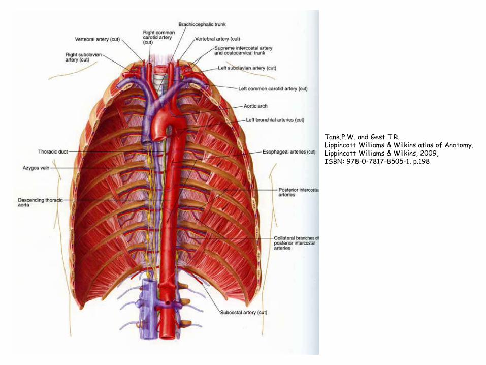
Tank,P.W. and Gest T.R. Lippincott Williams & Wilkins atlas of Anatomy. Lippincott Williams & Wilkins, 2009, ISBN: 978-0-7817-8505-1, p.198

Netter, F.H. Interactive Atlas of Human Anatomy. 3rd ed. New Jersey, Icon Learning Systems, 2003, ISBN: 1-929007-15-9, Plate #220
Left of vertebral column
Approaching midline
Midline
Descending thoracic aorta
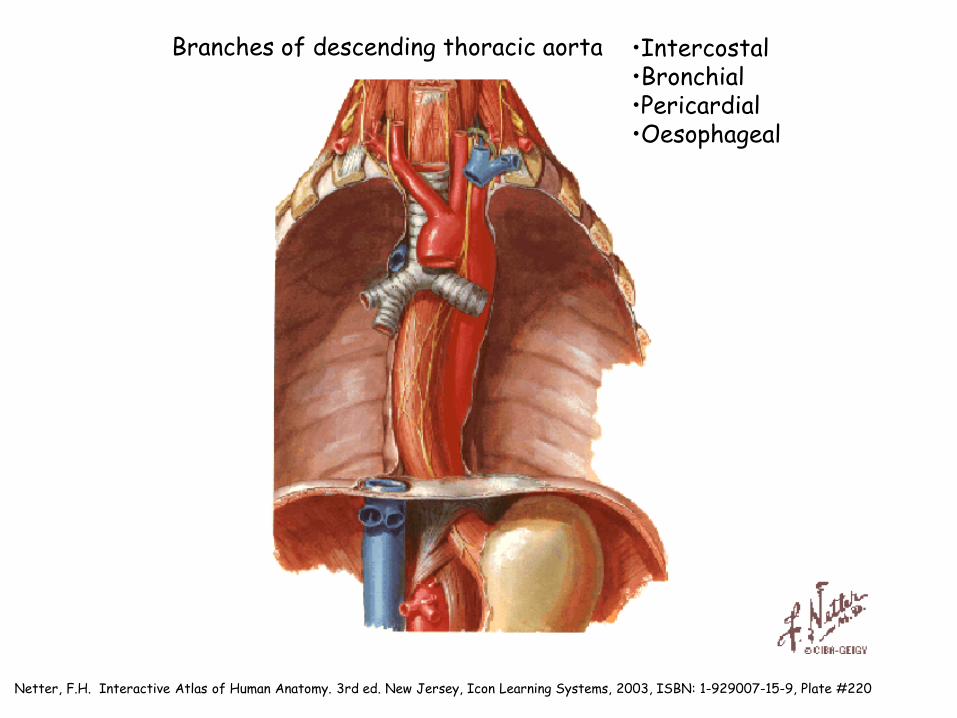
Netter, F.H. Interactive Atlas of Human Anatomy. 3rd ed. New Jersey, Icon Learning Systems, 2003, ISBN: 1-929007-15-9, Plate #220
•Intercostal •Bronchial •Pericardial •Oesophageal
Branches of descending thoracic aorta

Tank,P.W. and Gest T.R. Lippincott Williams & Wilkins atlas of Anatomy. Lippincott Williams & Wilkins, 2009, ISBN: 978-0-7817-8505-1, p.198

Hansen,J.T. Netter’s Clinical Anatomy. 2nd ed. Elsevier, 2010, ISBN: 978-1-4377-0272-9, p.108
Relevant museum specimens displayed (beneath windows) Head & Neck Bay: • 516-100084 is a a dissection of the root of the neck and the superior
mediastinum. On the right side, part of the anterior wall of the thorax and some superficial muscles have been removed.
Thorax Bay: • 516-100208 is a dissection of the mediastinal contents showing the relations of
the trachea, the oesophagus, the thoracic duct and the left recurrent laryngeal nerve; the relations of the aortic arch and its branches to the vagus and phrenic nerves. In addition, it shows the relations of the great vessels to the pericardium and of the thyroid gland to the carotid sheath.
Images and information on these specimens can also be accessed via the museum
online database: http://mdhs.unimelb.edu.au/harrybrookesallenmuseum/catalogue An excellent pre-workshop and pre-dissection resource is Acland’s anatomy:
http://www.aclandanatomy.com.ezp.lib.unimelb.edu.au/