superficially porous particles for peptide and protein analysis · • proteins were fraction...
TRANSCRIPT

Superficially Porous Particles for Peptide and Protein Analysis
Barry E. Boyes, Joseph J. Kirkland, Stephanie A. Schuster, Brian M. Wagner, and
Joseph J. DeStefano
Advanced Materials Technology, Inc., 3521 Silverside Rd., Wilmington, DE 19810

The original 2.7 µm superficially porous particles (SPP) introduced in 2006 were created with an average pore size of 90 Å, which was suitable for small molecule analytical separations. This SPP particle technology now has been expanded to include wider pore sizes and larger particle sizes that are specifically designed for larger biomolecules. Novel particle designs with specially selected bonded phases for peptide and protein separations are described. This presentation includes fast separations of peptides and intact protein mixtures, as well as examples of very high resolution separations of larger proteins and associated variants and contaminants. Columns with specially engineered bonded phases for these particles demonstrate high temperature stability, which is ideally suited for the conditions that are often used for analytical and small scale preparative biomolecular separations. Protein recovery and sample loading investigations are included. The optimized shell thickness of the new 400 Å SPP particles represents a compromise between a short diffusion path versus adequate retention and mass load tolerance. Examples of high molecular weight protein separations highlight the advantages of using columns of superficially porous particles with wider pores. Some comparisons with conventional totally porous particles are also shown.
Objective
Abstract
The objective of this work is to demonstrate fast, efficient separations of proteins using superficially porous particles that have been optimized for such applications.

Sample Loading Study for Halo 400 C4Columns: 4.6 x 100 mm; Gradient: 39 - 49% in 10 min.;
Mobile phase: A - 0.1% aqueous trifluoroacetic acid;B - acetonitrile with 0.1% trifluoroacetic acid; Temperature: 60 oC;
Flow rate: 0.5 mL/min; Injection volume: 5 mL
mg Apomyoglobin
0.01 0.1 1
Peak
wid
th, m
in.
0.04
0.06
0.08
0.10
0.12
0.14
0.16
0.18
Shell thickness, 0.15 mmShell thickness, 0.20 mmShell thickness, 0.25 mm
Apomyoglobin – 17 kDa
The particle with 0.20 µm shell thickness offers a compromise between sample loadability and retention, as well as an optimized diffusion path for large MW biomolecules.
Sample Loading Study for HALO Protein C4
Columns: 4.6 x 100 mm; Gradient: 39-49% B in 10 min. Mobile phase: A – 0.1% trifluoroacetic acid
B – acetonitrile with 0.1% trifluoroacetic acid; Temperature: 60 oC Flow rate: 0.5 mL/min.; Injection volume: 5 µL
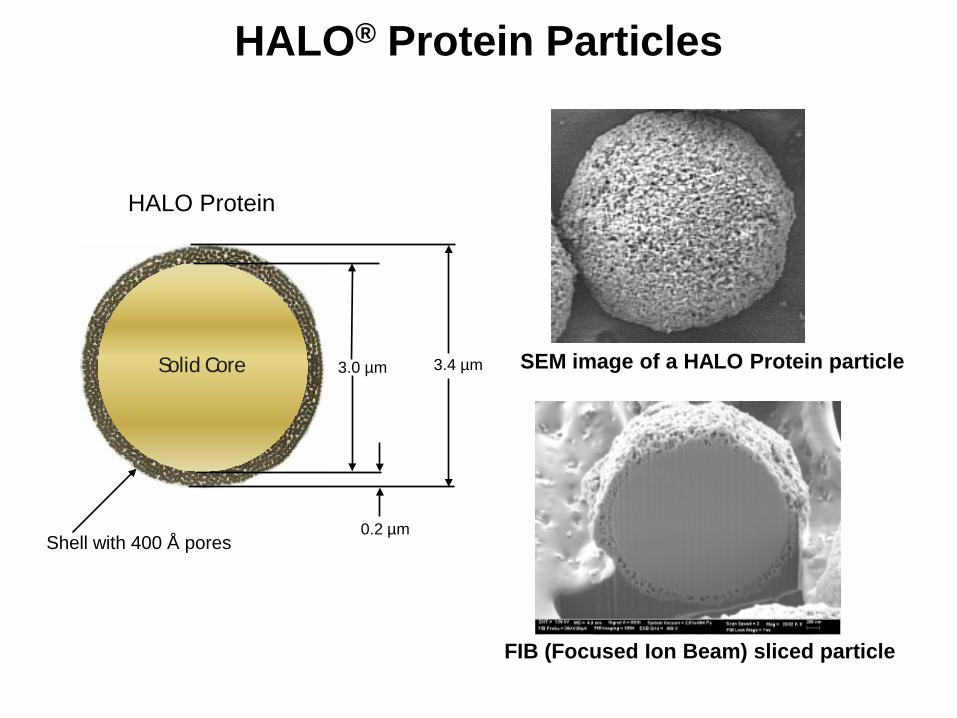
0.2 µm
3.0 µm 3.4 µm
Shell with 400 Å pores
Solid Core
HALO Protein
HALO® Protein Particles
FIB (Focused Ion Beam) sliced particle
SEM image of a HALO Protein particle
0
1
2
3
4
5
6
7
8
9
0 5000 10000 15000
Ret
entio
n Ti
me
(min
.)
Column Volumes
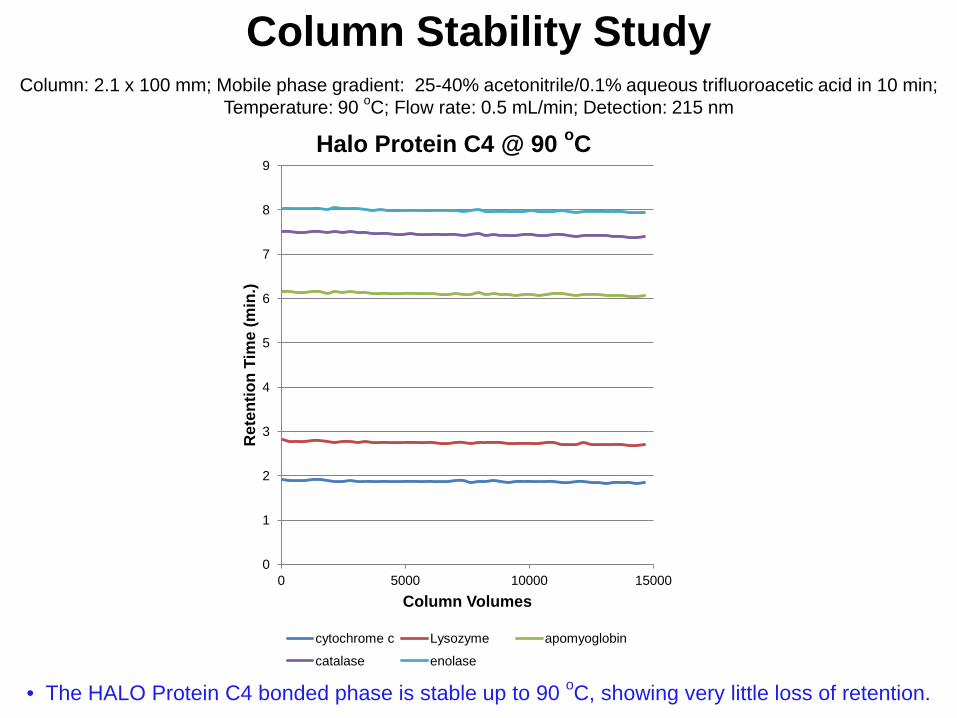
Halo Protein C4 @ 90 oC
cytochrome c Lysozyme apomyoglobin
catalase enolase
Column Stability Study
Column: 2.1 x 100 mm; Mobile phase gradient: 25-40% acetonitrile/0.1% aqueous trifluoroacetic acid in 10 min; Temperature: 90 oC; Flow rate: 0.5 mL/min; Detection: 215 nm
• The HALO Protein C4 bonded phase is stable up to 90 oC, showing very little loss of retention.

Column: 2.1 x 100 mm HALO Protein C4 Instrument: Agilent 1200 SL Injection Volume: 2 µL Detection: 215 nm Temperature: as indicated
Mobile Phase A: water/0.1% TFA Mobile Phase B: acetonitrile/0.1% TFA Gradient: 28-58% B in 10 min. Flow rate: 0.45 mL/min
Peak Identities (in order): 1. Lysozyme 14.3 kDa 2. BSA 66.4 kDa 3. α-Chymotrypsinogen A 25.0 kDa 4. Enolase 46.7 kDa 5. Ovalbumin 44.0 kDa
0 1 2 3 4 5 6 7 8 9 10
Time, min
30 oC
60 oC
90 oC
1
2
3
4
5
1
2 3 4 5
1 2 3
4 5
• Protein peak shape and recovery improve with increased temperature of analysis.
Protein Separations: Effect of Temperature

-20
0
20
40
60
80
100
120
140
160
0 1 2 3 4 5 6 7 8 9 10 11 12 13 14 15
Abso
rban
ce, m
AU
Time, min
-20
0
20
40
60
80
100
120
140
160
0 5 10 15 20 25 30 35 40 45
Abso
rban
ce, m
AU
Time, min
Totally porous, 300 Å, C4,1.7 µm P = 134 bar Flow rate = 0.2 mL/min Gradient = 20-60% B in 45 min. A = water/0.1% TFA B = Acetonitrile/0.1% TFA Temp = 60 oC
• Separation is 3 times faster at the same back pressure on the HALO Protein column compared to the same sample run on a sub-2-µm totally porous particle column
Peak Identities: 1. Ribonuclease A 2. Cytochrome c 3. BSA 4. Apomyoglobin 5. Enolase 6. Phosphorylase b
1
2 3
4
5
6
1
2 3
4
5
6
HALO Protein, 400 Å, C4, 3.4 µm P = 132 bar Flow rate = 0.6 mL/min Gradient = 20-60% B in 15 min. A = water/0.1% TFA B = Acetonitrile/0.1% TFA Temp = 60 oC
Protein Separations: Fused-Core compared to Totally Porous

• Proteins were fraction collected from a 4.6 x 100 mm HALO Protein C4 column run at 60 oC under gradient conditions with water/ACN/0.1% TFA mobile phase. Blanks were obtained by replacing the column with a union
• Lyophilized proteins were reconstituted using 3 M Urea/1% Triton X-100/0.25% acetic acid
• Protein recoveries were measured using QuantiPro™ BCA Assay Kit for 0.5-30 μg/mL protein (Sigma-Aldrich, St. Louis, MO)
• Samples were incubated at 37 oC for 100 min. • Each sample was run in duplicate • Absorbance values were measured at 562 nm • HALO Protein C4 shows good recovery of proteins
Protein Recovery Cytochrome c 100 (5.8 SD)
Catalase 92 (18 SD)
Protein Recovery Studies
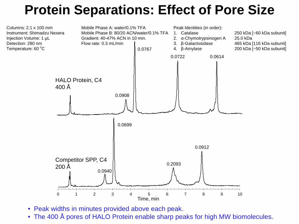
Columns: 2.1 x 100 mm Instrument: Shimadzu Nexera Injection Volume: 1 µL Detection: 280 nm Temperature: 60 oC
Mobile Phase A: water/0.1% TFA Mobile Phase B: 80/20 ACN/water/0.1% TFA Gradient: 40-47% ACN in 10 min. Flow rate: 0.3 mL/min
Peak Identities (in order): 1. Catalase 250 kDa [~60 kDa subunit] 2. α-Chymotrypsinogen A 25.0 kDa 3. β-Galactosidase 465 kDa [116 kDa subunit] 4. β-Amylase 200 kDa [~50 kDa subunit]
Time, min 0 1 2 3 4 5 6 7 8 9 10
HALO Protein, C4 400 Å
Competitor SPP, C4 200 Å
0.0908
0.0767 0.0722 0.0614
0.0940
0.0699
0.2093
0.0912
Protein Separations: Effect of Pore Size
• Peak widths in minutes provided above each peak. • The 400 Å pores of HALO Protein enable sharp peaks for high MW biomolecules.

-5
45
95
145
195
245
295
0 5 10 15 20 25 30 35 40 45
Abso
rban
ce @
215
nm
Time, min
Column: 4.6 x 100 mm, HALO-5 Peptide ES-C18 Instrument: Agilent 1100 Injection Volume: 10 µL Detection: 215 nm Temperature: 45 oC Pressure: 54 bar initial
Mobile Phase A: water/0.1% TFA Mobile Phase B: ACN/0.1% TFA Gradient: 5-40% B in 45 min. Flow rate: 1.0 mL/min Sample: Apomyoglobin Tryptic Digest [2 mg/mL]
The extremely low back pressure of the HALO-5 Peptide ES-C18 column enables fast, efficient proteomic separations with a low potential for plugging.
Tryptic Digest using HALO-5 Peptide ES-C18
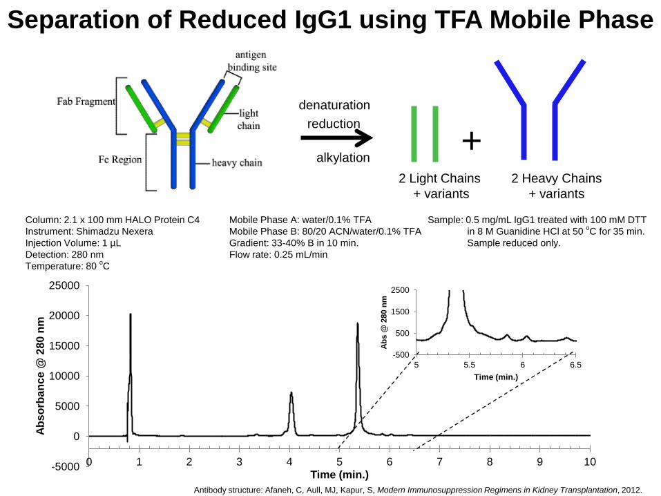
reduction
alkylation +
Column: 2.1 x 100 mm HALO Protein C4 Instrument: Shimadzu Nexera Injection Volume: 1 µL Detection: 280 nm Temperature: 80 oC
Mobile Phase A: water/0.1% TFA Mobile Phase B: 80/20 ACN/water/0.1% TFA Gradient: 33-40% B in 10 min. Flow rate: 0.25 mL/min
Sample: 0.5 mg/mL IgG1 treated with 100 mM DTT in 8 M Guanidine HCl at 50 oC for 35 min. Sample reduced only.
denaturation
2 Light Chains + variants
2 Heavy Chains + variants
Antibody structure: Afaneh, C, Aull, MJ, Kapur, S, Modern Immunosuppression Regimens in Kidney Transplantation, 2012.
-5000
0
5000
10000
15000
20000
25000
0 1 2 3 4 5 6 7 8 9 10
Abso
rban
ce @
280
nm
Time (min.)
-500
500
1500
2500
5 5.5 6 6.5
Abs
@ 2
80 n
m
Time (min.)
Separation of Reduced IgG1 using TFA Mobile Phase

ID Mass (Da)
LC1 23,204
LC2 23,192
LC3 23,203
HC1 50,539
HC2 50,424
HC3 50,668
HC4 50,680
HC5 28,862
0.0 2.5 5.0 7.5 10.0 12.5 15.0 17.5 min
0.0
1.0
2.0
3.0
4.0
5.0
6.0
7.0
LC1
LC2
LC3
HC
1
HC
2 H
C3
HC
4
HC
5
2.1 mm ID x 100 mm HALO Protein C4; 0.4 mL/min.; A: 0.5 % formic acid with 20 mM Ammonium Formate B: 45% AcN/45% IPA/ 0.5 % formic acid with 20 mM Ammonium Formate; Gradient: 29-32% B in 20 min.; 80 oC
Detection: 280 nm Abs; Shimadzu LCMS-2020, ESI +4.5 kV, 2 pps, 500-2000 m/z
500 750 1000 1250 1500 1750 m/z 0.00
0.25
0.50
0.75
1.00
1.25
1.50
1.75
2.00
2.25
2.50
2.75
3.00
3.25
3.50
Inten. (x100,000)
1366.0
1290.0
1221.9
1161.4
1105.9 1451.6
1655.6 1546.7
1010.1 1783.1
1931.5 930.2
859.4 1604.6 773.6 1859.1
LC = light chain HC = heavy chain
Masses deconvoluted
using MagTran
Abs
orba
nce
@ 2
80 n
m
High Resolution Analysis of mAb IgG1 Light and Heavy Chains with LC/MS

• Fused-core particles with 400 Å pores are effective for efficiently
separating proteins without restricted diffusion • Protein separations can be run approximately 3 times faster on columns
of Fused-core particles compared to columns of sub-2-µm particles at the same back pressure
• Fused-core particles have performance advantages over totally porous particles for separating peptides and proteins
• Columns of 400 Å particles are both efficient and stable up to 90 oC • With the low back pressure afforded by 5-µm 160 Å Fused-core
particles, columns of these particles are less prone to overpressurizing due to plugging and longer columns can be run for high resolution separations of proteomic samples
• With the correct choice of mobile phase, high resolution LC-MS data can be obtained for mAb separations using 400 Å Fused-core particles
Conclusions
Special thanks to Robert Moran for assistance with chromatographic measurements.
Acknowledgment