superficial subcutaneous leiomyosarcoma on the face of a ......lms is extremely rare in patients...
TRANSCRIPT
Vol. 42 / No. 6 / November 2015
795
Superficial Subcutaneous Leiomyosarcoma on the Face of a Pediatric PatientByung Mi Lee1, Seok Joo Kang1, Seong Pin Jeon1, Hook Sun1, Bomi Kim2
Departments of 1Plastic and Reconstructive Surgery and 2Pathology, Busan Paik Hospital, Inje University College of Medicine, Busan, Korea
Correspondence: Hook SunDepartment of Plastic and Reconstructive Surgery, Busan Paik Hospital, Inje University College of Medicine, 75 Bokji-ro, Busanjin-gu, Busan 47392, KoreaTel: +82-51-890-6136, Fax: +82-51-894-7976, E-mail: [email protected]
No potential conflict of interest relevant to this article was reported.
Received: 30 Apr 2015 • Revised: 26 May 2015 • Accepted: 29 May 2015 pISSN: 2234-6163 • eISSN: 2234-6171 http://dx.doi.org/10.5999/aps.2015.42.6.795 • Arch Plast Surg 2015;42:795-798
Copyright 2015 The Korean Society of Plastic and Reconstructive SurgeonsThis is an Open Access article distributed under the terms of the Creative Commons Attribution Non-Commercial License (http://creativecommons.org/licenses/by-nc/3.0/) which permits unrestricted non-commercial use, distribution, and reproduction in any medium, provided the original work is properly cited.
Images
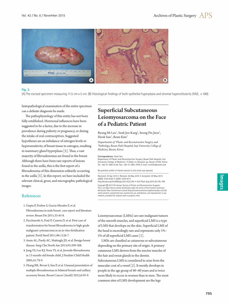
Fig. 3. (A) The excised specimen measuring 11.5 cm×5 cm. (B) Histological findings of both epithelial hyperplasia and stromal hypercellularity (H&E, ×100).
A B
histopathological examination of the entire specimen can a definite diagnosis be made. The pathophysiology of this entity has not been fully established. Hormonal influences have been suggested to be a factor, due to the increase in prevalence during puberty or pregnancy, or during the intake of oral contraceptives. Suggested hypotheses are an imbalance of estrogen levels or hypersensitivity of breast tissue to estrogen, resulting in mammary gland hyperplasia [5]. Thus, a vast majority of fibroadenomas are found in the breast. Although there have been rare reports of lesions found in the axilla, this is the first report of a fibroadenoma of this dimension solitarily occurring in the axilla [5]. In this report, we have included the relevant clinical, gross, and micrographic pathological images.
References
1. Gupta P, Foshee S, Garcia-Morales F, et al. Fibroadenoma in male breast: case report and literature review. Breast Dis 2011;33:45-8.
2. Pacchiarotti A, Frati P, Caserta D, et al. First case of transformation for breast fibroadenoma to high-grade malignant cystosarcoma in an in vitro fertilization patient. Fertil Steril 2011;96:1126-7.
3. Amin AL, Purdy AC, Mattingly JD, et al. Benign breast disease. Surg Clin North Am 2013;93:299-308.
4. Jung YS, Lee KJ, Yoon TI, et al. Juvenile fibroadenoma in 13-month-old female child. J Paediatr Child Health 2005;41:78-9.
5. Zhang RR, Bevan S, Sun P, et al. Unusual presentation of multiple fibroadenomas in bilateral breasts and axillary accessory breast. Breast Cancer (Auckl) 2012;6:95-9.
Leiomyosarcomas (LMSs) are rare malignant tumors of the smooth muscles, and superficial LMS is a type of LMS that develops on the skin. Superficial LMS of the head is exceedingly rare and represents only 1%–5% of all superficial LMS cases [1]. LMSs are classified as cutaneous or subcutaneous depending on the primary site of origin. A primary cutaneous LMS derives from the erector muscles of the hair and sweat glands in the dermis. Subcutaneous LMS is considered to arise from the muscular coat of a vessel [2]. It mostly develops in people in the age group of 40–60 years and is twice more likely to occur in women than in men. The most common sites of LMS development are the legs
796
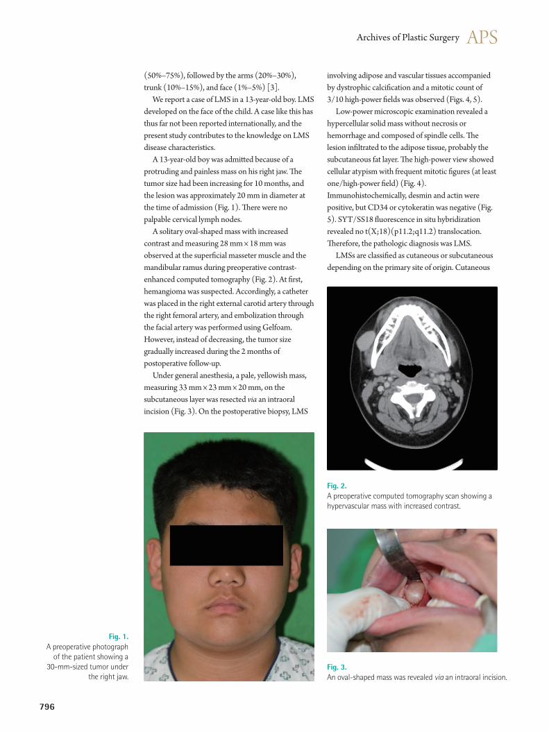
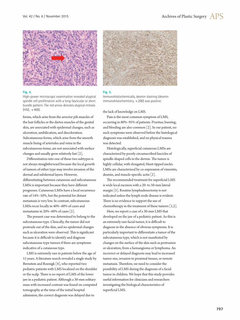
(50%–75%), followed by the arms (20%–30%), trunk (10%–15%), and face (1%–5%) [3]. We report a case of LMS in a 13-year-old boy. LMS developed on the face of the child. A case like this has thus far not been reported internationally, and the present study contributes to the knowledge on LMS disease characteristics. A 13-year-old boy was admitted because of a protruding and painless mass on his right jaw. The tumor size had been increasing for 10 months, and the lesion was approximately 20 mm in diameter at the time of admission (Fig. 1). There were no palpable cervical lymph nodes. A solitary oval-shaped mass with increased contrast and measuring 28 mm × 18 mm was observed at the superficial masseter muscle and the mandibular ramus during preoperative contrast-enhanced computed tomography (Fig. 2). At first, hemangioma was suspected. Accordingly, a catheter was placed in the right external carotid artery through the right femoral artery, and embolization through the facial artery was performed using Gelfoam. However, instead of decreasing, the tumor size gradually increased during the 2 months of postoperative follow-up. Under general anesthesia, a pale, yellowish mass, measuring 33 mm × 23 mm × 20 mm, on the subcutaneous layer was resected via an intraoral incision (Fig. 3). On the postoperative biopsy, LMS
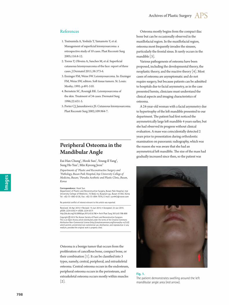
involving adipose and vascular tissues accompanied by dystrophic calcification and a mitotic count of 3/10 high-power fields was observed (Figs. 4, 5). Low-power microscopic examination revealed a hypercellular solid mass without necrosis or hemorrhage and composed of spindle cells. The lesion infiltrated to the adipose tissue, probably the subcutaneous fat layer. The high-power view showed cellular atypism with frequent mitotic figures (at least one/high-power field) (Fig. 4). Immunohistochemically, desmin and actin were positive, but CD34 or cytokeratin was negative (Fig. 5). SYT/SS18 fluorescence in situ hybridization revealed no t(X;18)(p11.2;q11.2) translocation. Therefore, the pathologic diagnosis was LMS. LMSs are classified as cutaneous or subcutaneous depending on the primary site of origin. Cutaneous
Fig. 1. A preoperative photograph
of the patient showing a 30-mm-sized tumor under
the right jaw.
Fig. 2. A preoperative computed tomography scan showing a hypervascular mass with increased contrast.
Fig. 3. An oval-shaped mass was revealed via an intraoral incision.
Vol. 42 / No. 6 / November 2015
797
forms, which arise from the arrector pili muscles of the hair follicles or the dartos muscles of the genital skin, are associated with epidermal changes, such as ulceration, umbilication, and discoloration. Subcutaneous forms, which arise from the smooth muscle lining of arterioles and veins in the subcutaneous tissue, are not associated with surface changes and usually grow relatively fast [2]. Differentiation into one of these two subtypes is not always straightforward because the local growth of tumors of either type may involve invasion of the dermal and subdermal layers. However, differentiating between cutaneous and subcutaneous LMSs is important because they have different prognoses. Cutaneous LMSs have a local recurrence rate of 14%–50%, but the potential for distant metastasis is very low. In contrast, subcutaneous LMSs recur locally in 40%–60% of cases and metastasize in 20%–60% of cases [2]. The present case was determined to belong to the subcutaneous type. Clinically, the tumor did not protrude out of the skin, and no epidermal changes such as ulceration were observed. This is significant because it is difficult to identify and diagnose subcutaneous-type tumors if there are symptoms indicative of a cutaneous type. LMS is extremely rare in patients below the age of 15 years. A literature search revealed a single study by Bernstein and Roenigk [4], who reported two pediatric patients with LMS localized on the shoulder or the scalp. There is no report of LMS of the lower jaw in a pediatric patient. Although a 30-mm solitary mass with increased contrast was found on computed tomography at the time of the initial hospital admission, the correct diagnosis was delayed due to
Fig. 4. High-power microscopic examination revealed atypical spindle cell proliferation with a long fascicular or short bundle pattern. The red arrow denotes atypical mitosis (H&E, ×400).
Fig. 5. Immunohistochemically, desmin staining (desmin immunohistochemistry, ×200) was positive.
the lack of knowledge on LMS. Pain is the most common symptom of LMS, occurring in 80%–95% of patients. Pruritus, burning, and bleeding are also common [2]. In our patient, no such symptoms were observed before the histological diagnosis was established, and no physical trauma was detected. Histologically, superficial cutaneous LMSs are characterized by poorly circumscribed fascicles of spindle-shaped cells in the dermis. The tumor is highly cellular, with elongated, blunt-tipped nuclei. LMSs are characterized by co-expression of vimentin, desmin, and muscle-specific actin [2]. The recommended treatment for superficial LMS is wide local excision with a 20- to 50-mm lateral margin [5]. Routine lymphadenectomy is not indicated unless the lymph node disease is evident. There is no evidence to support the use of chemotherapy in the treatment of these tumors [1,2]. Here, we report a case of a 30-mm LMS that developed on the jaw of a pediatric patient. As this is an extremely rare facial tumor, it is difficult to diagnose in the absence of obvious symptoms. It is particularly important to differentiate a tumor of the subcutaneous type, which is not manifested by changes on the surface of the skin such as protrusion or ulceration, from a hemangioma or lymphoma. An incorrect or delayed diagnosis may lead to increased tumor size, invasion to proximal tissues, or remote metastasis. Therefore, we need to consider the possibility of LMS during the diagnosis of a facial tumor in children. We hope that this study provides useful information for clinicians and researchers investigating the biological characteristics of superficial LMS.
798
Osteoma mostly begins from the compact iliac bone but can be occasionally observed in the maxillofacial region. In the maxillofacial region, osteoma most frequently invades the sinuses, particularly the frontal sinus. It rarely occurs in the mandible [3]. Various pathogenesis of osteoma have been proposed, including the developmental theory, the neoplastic theory, and the reactive theory [4]. Most cases of osteoma are asymptomatic and do not require surgery, but because patients can be admitted to hospitals due to facial asymmetry, as in the case presented herein, clinicians must understand the clinical aspects and imaging characteristics of osteoma. A 24-year-old woman with a facial asymmetry due to hypertrophy of the left mandible presented to our department. The patient had first noticed the asymmetrically large left mandible 4 years earlier, but she had observed its progress without clinical evaluation. A mass was coincidentally detected 2 years prior to presentation during orthodontic examination on panoramic radiography, which was the reason she was aware that she had an asymmetrical left mandible. The size of the mass had gradually increased since then, so the patient was
References
1. Tsutsumida A, Yoshida T, Yamamoto Y, et al. Management of superficial leiomyosarcoma: a retrospective study of 10 cases. Plast Reconstr Surg 2005;116:8-12.
2. Torres T, Oliveira A, Sanches M, et al. Superficial cutaneous leiomyosarcoma of the face: report of three cases. J Dermatol 2011;38:373-6.
3. Enzinger FM, Weiss SW. Leiomyosarcoma. In: Enzinger FM, Weiss SW, editors. Soft tissue tumors. St. Louis: Mosby; 1995. p.491-510.
4. Bernstein SC, Roenigk RK. Leiomyosarcoma of the skin. Treatment of 34 cases. Dermatol Surg 1996;22:631-5.
5. Porter CJ, Januszkiewicz JS. Cutaneous leiomyosarcoma. Plast Reconstr Surg 2002;109:964-7.
Peripheral Osteoma in the Mandibular AngleEui Han Chung1, Hook Sun1, Young-Il Yang2, Sung Ho Yun3, Min Kyeong Jeon3
Departments of 1Plastic and Reconstructive Surgery and 2Pathology, Busan Paik Hospital, Inje University College of Medicine, Busan; 3Paradia Aesthetic and Plastic Clinic, Busan, Korea
Correspondence: Hook SunDepartment of Plastic and Reconstructive Surgery, Busan Paik Hospital, Inje University College of Medicine, 75 Bokji-ro, Busanjin-gu, Busan 47392, KoreaTel: +82-51-890-6136, Fax: +82-51-894-7976, E-mail: [email protected]
No potential conflict of interest relevant to this article was reported.
Received: 30 Apr 2015 • Revised: 15 Jun 2015 • Accepted: 23 Jun 2015 pISSN: 2234-6163 • eISSN: 2234-6171 http://dx.doi.org/10.5999/aps.2015.42.6.798 • Arch Plast Surg 2015;42:798-800
Copyright 2015 The Korean Society of Plastic and Reconstructive SurgeonsThis is an Open Access article distributed under the terms of the Creative Commons Attribution Non-Commercial License (http://creativecommons.org/licenses/by-nc/3.0/) which permits unrestricted non-commercial use, distribution, and reproduction in any medium, provided the original work is properly cited.
Imag
es
Osteoma is a benign tumor that occurs from the proliferation of cancellous bone, compact bone, or their combination [1]. It can be classified into 3 types, namely, central, peripheral, and extraskeletal osteoma. Central osteoma occurs in the endosteum, peripheral osteoma occurs in the periosteum, and extraskeletal osteoma occurs mostly within muscles [2].
Fig. 1. The patient demonstrates swelling around the left mandibular angle area (red arrow).