summary of findings introduction
TRANSCRIPT

SURVEILLANCE FOR HIGHLY PATHOGENIC AVIAN INFLUENZA IN MINNESOTA’S MIGRATORY BIRDS
Erik Hildebrand1, Michelle Carstensen, and Erika Butler
SUMMARY OF FINDINGS
As part of a national strategy for early detection of highly pathogenic avian influenza (HPAI) in North America, the Minnesota Department of Natural Resources (MNDNR) and the United States Department of Agriculture (USDA) conducted surveillance for the virus in waterfowl in the state. A combined total of 1,409 birds were sampled for HPAI in Minnesota during 2009. Testing did not result in any positive cases of HPAI, however nearly 200 did test positive for a low pathogenic strain of avian influenza. Approximately 44,374 wild birds were sampled throughout the United States in 2009, and no positive cases of HPAI were detected. Minnesota will continue surveillance for the virus in the state’s waterfowl in 2010, in cooperation with the Mississippi Flyway Council of the U.S. Fish and Wildlife Service and the USDA.
INTRODUCTION Recent worldwide attention on the spread of a highly pathogenic strain of avian influenza, subtype H5N1, from Asia to Europe and Africa in 2006 has led to the development of a coordinated National Strategic Plan for early detection of HPAI-H5N1 introduction into North America by wild birds. Although movements of domestic poultry or contaminated poultry products, both legally and illegally, are believed to be the major driving force in the spread of HPAI-H5N1, migratory birds are thought to be a contributing factor.
Avian Influenza is a viral infection that occurs naturally in wild birds, especially waterfowl, gulls, and shorebirds. It is caused by type A influenza viruses that have 2 important surface antigens, hemagglutinin (H) and nuraminidase (N), that give rise to 144 possible virus subtypes. Influenza viruses vary widely in pathogenicity and ability to spread among birds. The emergence of an Asian strain HPAI-H5N1 virus in 1996 and subsequent spread of the virus in Asia, Africa, and Europe has killed thousands of wild birds and millions of domestic poultry. In 1997, HPAI-H5N1 became zoonotic in Hong Kong and to-date has infected at least 496 humans in Eurasia and Africa, resulting in over 293 deaths. The National Plan outlined a surveillance strategy that focused on sampling of wild bird species in North America that have the highest risk of being exposed to or infected with HPAI-H5N1 because of their migratory movement patterns. Currently, these include birds that migrate directly between Asia and North America, birds that may be in contact with species from areas in Asia with reported outbreaks, or birds that are known to be reservoirs of AI. A step-down plan was developed by the Mississippi Flyway Council in 2006 identifying Minnesota as a key flyway state needed to participate in regional sampling for early detection of HPAI-H5N1 in migratory ducks, geese, and shorebirds. In July 2009, the MNDNR entered into a $70,000 cooperative agreement with the United States Department of Agriculture’s Wildlife Services (USDA-WS) to sample 600 wild birds (either live-caught or hunter-harvested) in Minnesota for HPAI-H5N1 during 2009. In addition to the 600 samples to be collected by MNDNR, USDA-WS was also planning to collect a similar number of samples in the state during the same period. Bird species that were targeted include those listed as priority species in the National Strategic Plan or approved for sampling in Minnesota by the Mississippi Flyway Council. There have been surveillance efforts for the past 4 years with nearly 6,600 samples from MN, submitted for HPAI-H5N1 testing. __________________________________________
1 Corresponding author e-mail: [email protected]

METHODS
The MNDNR planned to sample 50 common goldeneye (Bucephala clangula), 50 ring-neck ducks (Aythya collaris), 50 mallards (Anas platyrhynchos), and 30 blue-winged teal (Anas discors) during the summer months, primarily in conjunction with planned banding activities. In the fall, through hunter-harvested surveillance, sampling targets were as follows: 80 Northern pintails (Anas acuta), 80 mallards, 80 American green-winged teal (anas crecca), 80 American blue-winged teal (Anas discors), 50 Northern shovelers (Anas clypeata), and 50 American wigeon (Anas Americana). USDA-WS planned to sample a similar number of the duck species mentioned above or others from their functional group (e.g., dabblers, divers, shorebirds), as well as 50 Canada geese. If sampling goals per species could not be met, other targeted waterfowl species within the same functional group could be sampled and counted toward the state’s total. Sampling strategies were coordinated between the MNDNR and USDA-WS to maximize access to targeted birds species through existing banding operations and fall hunter-harvested surveillance. Cloacal and oral-pharyngeal swabs were used to collect samples and they were submitted to the Veterinary Diagnostic Laboratory in St. Paul, MN for initial screening for the virus. If positive for avian influenza virus, samples were forwarded to the National Veterinary Services Laboratories in Ames, IA for strain-typing. RESULTS AND DISCUSSION
From April 1, 2009 through March 31, 2010 MNDNR and USDA collected a total of 1,409 samples from wild-caught live birds (n=310), hunter-harvested birds (n=1,016), agency (USDA-WS) harvested (n=73), and mortality/morbidity events (n=10) (Figure 1). Testing did not result in any positive cases of HPAI-H5N1; however 8 different duck species tested positive for a low pathogenic strain of avian influenza with the subtype H5, and only 1 tested positive for a N1 subtype. The testing protocol was limited to the screening for H5, H7, and N1 subtypes only; however in some cases other subtypes were identified and reported elsewhere (Table 1, Figure 2). According to the latest numbers of the United States Geologic Survey’s website (http://wildlifedisease.nbii.gov/ai/), approximately 44,374 birds have been sampled for HPAI-H5N1 in the U.S. in 2009. No positive cases of HPAI-H5N1 have been found anywhere in North America to date. Since the majority of H5 positives (low pathogenic forms only) detected by USDA-WS in the United States since 2006 have been found in dabbling ducks, the primary focus of future sampling will be on these species (Genus Anas, Aix, Cairina, and Dendrocygna). Surveillance for HPAI-H5N1 will likely continue in Minnesota and other parts of the U.S. next year. The USDA has banked all samples taken from 2006 to 2009, and is currently accepting proposals from state agencies and universities for further avian influenza research. Minnesota remains prepared to assist with future surveillance objectives if needed. In addition, the MNDNR has developed a surveillance and response plan for HPAI in wild birds, which includes increased vigilance of mortality and morbidity events within the state.
ACKNOWLEDGEMENTS
This project would not have been possible without the valuable contribution of the waterfowl research group, including Jeff Lawrence, Steve Cordts, Jim Berdeen, and Jim’s group of banding interns. Other MNDNR staff that provided valuable assistance to this project included Joel Huener, Dawn Torrison, Randy Pracher, Perry Loegering, Jeff Hines, Joel Anderson, Dave Trauba, Kevin Kotts, Martha Minchak, Dan Rhode, Bob Welsh, Bryan Lueth, David Pauly, Judy Markl, Jon Cole, Shelly Gorham, and Blane Klemek. I would also like to recognize our USDA-WS partner on the project, Paul Wolf, for his efforts to ensure that we met our overall sampling goals. Lastly, much of the hunter-harvested sampling was accomplished
through assistance from Pat Reddig, University of Minnesota, and numerous students from both the Natural Resources program and the veterinary college.
REFERENCES
Halvorson, D. A., C. J. Kelleher, and D.A. Senne. 1985. Epizootiology of avian influenza: effect of season on incidence in sentinel ducks and domestic turkeys in Minnesota. Applied and Environmental Microbiology 49: 914-919.
Hanson, B. A., D. E. Stallknecht, D.E. Swayne, L. A. Lewis, and D. A. Senne. 2003. Avian influenza viruses in Minnesota ducks during 1998-2000. Avian Diseases 47: 867-871.
Interagency Asian H5N1 Early Detection Working Group. 2006. An early detection system for Asian H5N1 highly pathogenic avian influenza in wild migratory birds: U.S. Interagency Strategic Plan. Unpubl. Rept. Report to the Department of Homeland Security, Policy Coordinating Committee for Pandemic Influenza Preparedness.
Michigan Department of Natural Resources, Wildlife Division. 2006. Michigan surveillance and response plan for highly pathogenic avian influenza in free-ranging wildlife. Unpubl. Rept.
Mississippi Flyway Council. 2006. Surveillance for early detection of highly pathogenic avian influenza H5N1 in wild migratory birds: a strategy for the Mississippi Flyway. Unpubl. Rept.
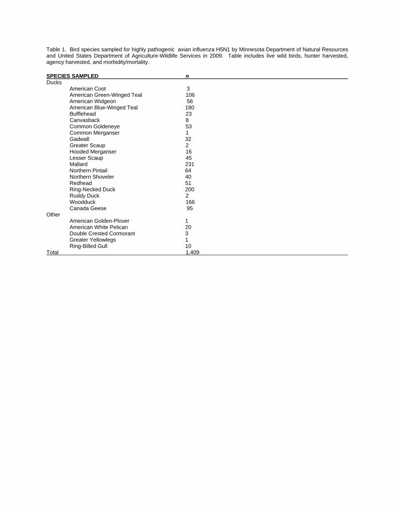
Table 1. Bird species sampled for highly pathogenic avian influenza H5N1 by Minnesota Department of Natural Resources and United States Department of Agriculture-Wildlife Services in 2009. Table includes live wild birds, hunter harvested, agency harvested, and morbidity/mortality.
SPECIES SAMPLED n Ducks American Coot 3 American Green-Winged Teal 106 American Widgeon 56 American Blue-Winged Teal 180 Bufflehead 23 Canvasback 8 Common Goldeneye 53 Common Merganser 1 Gadwall 32 Greater Scaup 2 Hooded Merganser 16 Lesser Scaup 45 Mallard 231 Northern Pintail 64 Northern Shoveler 40 Redhead 51 Ring-Necked Duck 200 Ruddy Duck 2 Woodduck 166 Canada Geese 95 Other American Golden-Plover 1 American White Pelican 20 Double Crested Cormorant 3 Greater Yellowlegs 1 Ring-Billed Gull 10 Total 1,409
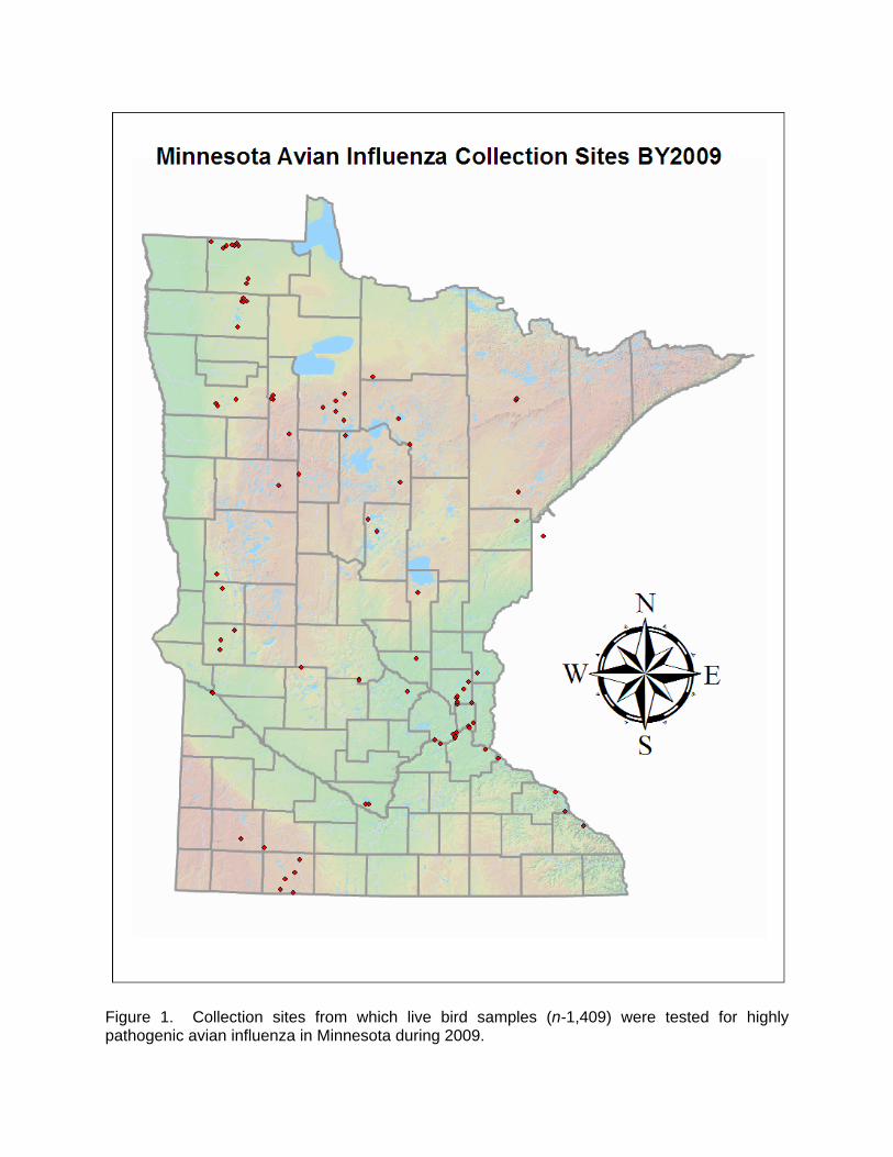
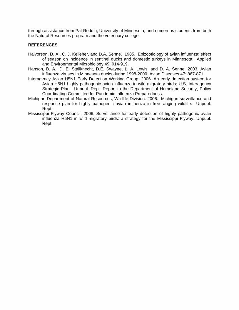
Figure 1. Collection sites from which live bird samples (n-1,409) were tested for highly pathogenic avian influenza in Minnesota during 2009.
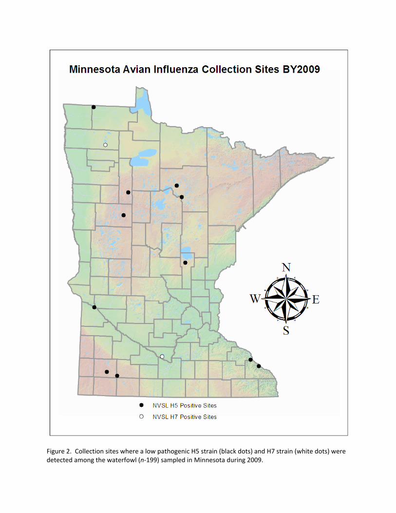
Figure 2. Collection sites where a low pathogenic H5 strain (black dots) and H7 strain (white dots) were detected among the waterfowl (n‐199) sampled in Minnesota during 2009.

MINNESOTA DEPARTMENT OF NATURAL RESOURCES CWD SURVEILLANCE PROGRAM 2009 Michelle Carstensen1, David Pauly, Erika Butler, Erik Hildebrand, and Lou Cornicelli SUMMARY OF FINDINGS
In fall 2009, the Minnesota Department of Natural Resources (MNDNR) sampled 2,685 hunter-harvested white-tailed deer (Odocoileus virginianus) for chronic wasting disease (CWD) in southeastern Minnesota. The surveillance effort was initiated primarily on the discovery of a CWD-positive captive elk facility in Olmsted county, and secondarily to monitor the ongoing risk of disease spread from CWD-infected wild deer from Wisconsin. All of the samples were negative for CWD. In addition, MNDNR submitted samples from 28 deer through statewide targeted surveillance, which included sick animals, escaped captive cervids, and roadkills; these samples were also negative for the disease. MNDNR plans to conduct hunter-harvested surveillance in southeastern MN in fall 2010, with efforts limited to a 15-mile radius around the CWD-infected captive elk facility in Olmsted county. INTRODUCTION
Chronic wasting disease is a transmissible spongiform encephalopathy (TSE) that affects elk (Cervus elaphus), mule deer (Odocoileus hemionus), white-tailed deer, and moose (Alces alces). TSEs are infectious diseases that alter the morphology of the central nervous system, resulting in a “sponge-like” appearance of this tissue. The etiological agent of CWD is an infectious protein, called a prion. Precise mechanisms and rates of CWD transmission remain unclear, although recent studies support animal-to-animal contact and environmental contamination as mechanisms that promote the spread of the disease. For example, one recent study has proven prions are shed in feces of infected deer 7-9 months before the onset of clinical signs, further supporting the high rate of horizontal transmission in infected populations. Incubation time of the disease, from infection to clinical signs, averages 16 months but can range from a few months to nearly 3 years. There is a limited distribution of infection in the body (primarily brain, spinal column, spleen, and lymph nodes) although a recent study demonstrated that prions can also be found in muscle. Clinical signs may include a loss of body condition and weight, excessive salivation, ataxia, and behavioral changes. Currently, there is no known treatment for the disease and it is always fatal. There is also no documented evidence of transmission of CWD to other species, including humans. To date, CWD has been diagnosed in 3 captive elk herds and 1 captive white-tailed deer herd within the state of Minnesota. Two of the elk herds (Stearns and Aitkin counties) were discovered in 2002 and depopulated; no additional CWD positive animals were found. In spring 2006, a captive white-tailed deer was found infected with CWD from a mixed deer/elk herd in Lac Qui Parle county. That herd was also depopulated without additional infection being detected. In all of these cases, the original source of the CWD has not been identified. In early 2009, a third captive elk herd (Olmsted county) was found infected with CWD. An 8-year old female was found CWD-positive at slaughter and records indicated she was born on the farm in 2001, thus suggesting that she was exposed to another CWD positive animal(s) on the farm. This herd was indemnified by the United States Department of Agriculture (USDA) and 558 adult elk were depopulated in September. An additional 3 adult elk were found infected with CWD (2 females, 1 male). Further, a management plan as enacted following the elk herd depopulation to further prevent a spillover of CWD to wild deer outside this facility. This plan included the cleaning and disinfecting of all livestock barns and equipment, maintenance of perimeter fencing, and a ban of captive cervids being restocked for 5 years. Since the property of the former elk facility has been sold and plans for development of a ________________________ 1Corresponding author e-mail: [email protected]

biomedical research park are in place, MNDNR has been concerned about environmental contamination of prions should fencing been removed. Thus, the management plan included the requirement of the top 2-inches of topsoil to be removed and stored behind 96-inch fencing prior to the initiation of any land development projects. MNDNR and BAH are working cooperatively to address the impact of CWD in these captive facilities, as well as management options to control its spread. Over the past 8 years, MNDNR has tested in excess of 33,000 deer across the state for CWD, all of which have been negative. Consequently, in recent years, sampling has been scaled back to address 3 main components: 1. Sampling of animals exhibiting symptoms of CWD (targeted surveillance); 2. Sampling of animals in response to elevated risk factors (e.g., detection of positive animals
in captive cervid farms, or proximity of Minnesota to positive CWD cases in other states); and
3. Sampling of hunter-killed deer for CWD in conjunction with surveillance for bovine tuberculosis.
METHODS
Hunter-harvested surveillance occurs at deer registration stations during the regular firearm hunting season. Stations are staffed with MNDNR personnel and students (veterinary medicine and natural resources) that were trained in lymph node extraction. Hunters were asked to voluntarily submit retropharyngeal lymph node samples for CWD testing. Samples were submitted to the Veterinary Diagnostic Laboratory at the University of Minnesota for disease screening. Any presumptive positive samples would be submitted to the National Veterinary Services Laboratories (Ames, IA) for official confirmation of the disease. Hunter information was recorded, including the hunter’s name, address, telephone number, MNDNR number, and location of kill. Maps were provided to assist the hunters in identifying the location (Township, Range, and Section) of the kill. Cooperating hunters were given a cooperator’s patch and entered into a raffle to win a firearm donated by the Minnesota Deer Hunter’s Association.
During fall 2009, registration stations were selected based on deer volume and distribution through the surveillance zone to meet a sampling goal of 300 deer per sampling block (n = 10), or an overall sampling goal of 3,000 samples (Figure 1). Registration stations were also selected based on their proximity to the CWD-positive captive elk facility and along the MN-WI border, to maximize our sampling of deer from those high-risk areas.
MNDNR continues to sample deer exhibiting clinical symptoms consistent with CWD (targeted surveillance) statewide. Information has been disseminated to wildlife staff regarding what to look for regarding symptomatic deer. Staff were provided the necessary equipment and training for lymph node removal and data recording. The number of samples expected through targeted surveillance is estimated to be less than 100 animals annually, as few reports of sick deer are taken.
RESULTS AND DISCUSSION
From June 2009 to April 2010, MNDNR collected a total of 28 samples from targeted surveillance efforts. This includes samples from 11 escaped captive cervids, 14 free-ranging sick deer and 3 wild deer removed from within the perimeter fence of the CWD-positive elk facility; all samples were negative for CWD.
MNDNR collected a total of 2,685 samples from hunter-harvested deer for CWD screening during fall 2009 (Figure 2). All samples were also negative for CWD. The sampling distribution of 300 samples collected per block was met in 50% of the sampling units (Figure 2,

Tables 1,2). Even though the sampling goal fell short in half of the sampling units, we achieved 88% of our overall surveillance goal of 3,000 samples. Further, a high proportion of samples were obtained within a 15-mile radius of the CWD-positive captive elk facility (Figure 3), as well as along the WI-MN border, where the risks of CWD in existing in wild deer are the highest.
Since the agency has now collected in excess of 33,000 negative samples in statewide surveillance efforts, we feel that future resources for CWD surveillance, in addition to targeted surveillance, are better spent addressing changing risk factors. Specifically, it is important to monitor the CWD surveillance activities occurring in our bordering states, and conduct periodic surveillance in Minnesota in response to CWD status changes in these states. Additionally, periodic surveillance in the vicinity of previous cases of CWD in captive cervids in Minnesota may be prudent. Given the most recent case of a CWD-infected cervid farm in Olmsted county, MNDNR plans to repeat surveillance efforts within a 15-mile radius of that farm during the fall 2010 firearm hunting season. Targeted surveillance of suspect deer is expected to continue throughout the state.
SURVEILLANCE COSTS
Conducting a disease surveillance effort that spans a large area and encompasses numerous deer registration stations requires a large, trained work force and a significant amount of expenditures to support the effort. The CWD surveillance effort in fall 2009 spanned the entire regular firearms season, requiring 4 consecutive weekends of staffed registration stations to obtain samples. The number of stations staffed each weekend ranged from 21 to 29, and summed to 102 stations over the duration of the project. In total, 116 trained MNDNR staff and 113 student workers were needed in the effort. Costs associated to the surveillance effort are listed in Table 3. ACKNOWLEDGEMENTS
We would especially like to recognize the tremendous amount of work and commitment
by Dave Pauly, whom stepped into the role of CWD Coordinator to assist the Wildlife Health Program in this massive surveillance effort. We would like to thank the all the MNDNR staff, students, and faculty from the University of Minnesota, Colleges of Veterinary Medicine and Natural Resources, for assisting in our sampling efforts. Also, a special thanks to Julie Adams and Bob Wright for making our surveillance maps. REFERENCES Angers, R. C., Browing, S. R., Seward, T. S., Sigurdson, C. J., Miller, M. W., Hoover, E. A., and
G.C.Telling. 2006. Prions in skeletal muscle of deer with chronic wasting disease. Science 311 (5764) :1117.
Baeten, L. A, Powers, B. E., Jewell, J. E., Spraker, T. R., and M. W. Miller. 2007. A natural case of chronic wasting disease in free-ranging moose (Alces alces shirasi). Journal of Wildlife Diseases 43(2): 309–318.
Diefenbach, D. R., C. S. Rosenberry, and R. C. Boyd. 2004. From the Field: Efficacy of detecting Chronic Wasting Disease via sampling hunter-killed white-tailed deer. Wildlife Society Bulletin 32: 267–272.
Miller, M. W., H. M. Swanson, L. L. Wolfe, F. G. Quartarone, S. L. Huwer, C. H. Southwick, and P. M. Lukacs. 2008. Lions and prions and deer demise. PLoS One 3(12): e4019. Miller, M.W., E.S. Williams, N.T. Hobbs, and L.L. Wolfe. 2004. Environmental sources of
prion transmission in mule deer. Emerg. Infec. Dis. 10(6): 1003–1006. Miller, M.W., E. S. Williams, C. W. McCarty, T. R. Spraker, T. J. Kreeger, C. T. Larsen, and E.
T. Thorne. 2000. Epizootiology of chronic wasting disease in free-ranging cervids in Colorado and Wyoming. Journal of Wildlife Diseases 36: 676–690.
Spraker, T. R., M. W. Miller, E. S. Williams, D. M. Getzy, W. J. Adrian, G. G. Schoonveld, R. A. Spowart, K. I. O’Rourke, J. M. Miller, and P. A. Merz. 1997. Spongiform encephalopathy in free-ranging mule deer (Odocoileus hemionus), white-tailed deer

(Odocoileus virginianus) and Rocky Mountain elk (Cervus elaphus nelsoni) in northcentral Colorado. Journal of Wildlife Diseases 33: 1–6.
Tamguney, Gultekin, M.W. Miller, L. L. Wolfe, T. M. Sirochmann, D. V. Glidden, C. Palmer. A. Lemus, S. J. DeArmond, and S. B. Prusiner. 2009. Asymptomatic deer excrete infectious prions in feces. Nature 461: 529–532.
Williams, E. S., and S. Young. 1980. Chronic Wasting Disease of captive mule deer: a spongiform encephalopathy. Journal of Wildlife Diseases 16: 89–98.
Williams, E.S., and S. Young. 1993. Neuropathology of chronic wasting disease of mule deer (Odocoileus hemionus) and elk (Cervus elaphus nelsoni). Veterinary Pathology 30: 36–45.
Williams, E.S., M. W. Miller, and E.T. Thorne. 2002. Chronic wasting disease: implications and challenges for wildlife managers. Presented at the North American Wildlife and Natural Resources Conference, April 3–7, 2002, Dallas, Texas.
Table 1. Breakdown of hunter-harvested deer samples collected for chronic wasting disease surveillance in southeastern Minnesota by registration station and sampling block, fall 2009.
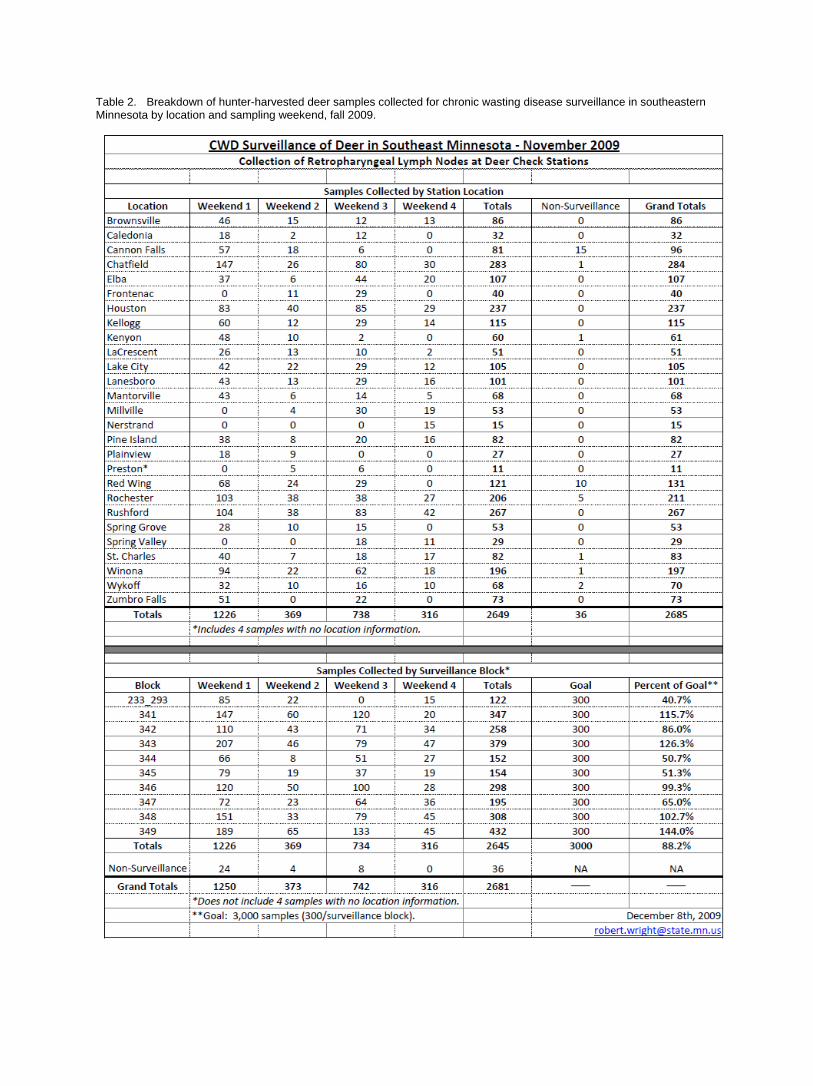
Table 2. Breakdown of hunter-harvested deer samples collected for chronic wasting disease surveillance in southeastern Minnesota by location and sampling weekend, fall 2009.

Table 3. Expenditure details for fall CWD surveillance program.
Expenditure Total cost
MNDNR Staff Salary $174,050 MNDNR Staff Travel Expenses $18,000 Veterinary Student Labor & Travel Expenses $42,815 Other Student Labor & Travel Expenses $52,000 Supplies $6,000 Fleet $28,025 Diagnostic Fees $67,825
Total $388,715
Figure 1. The Minnesota fall 2009 hunter-harvested surveillance program included 11 deer permit areas divided into 10 sampling blocks, with a total sampling goal of 3,000 samples.
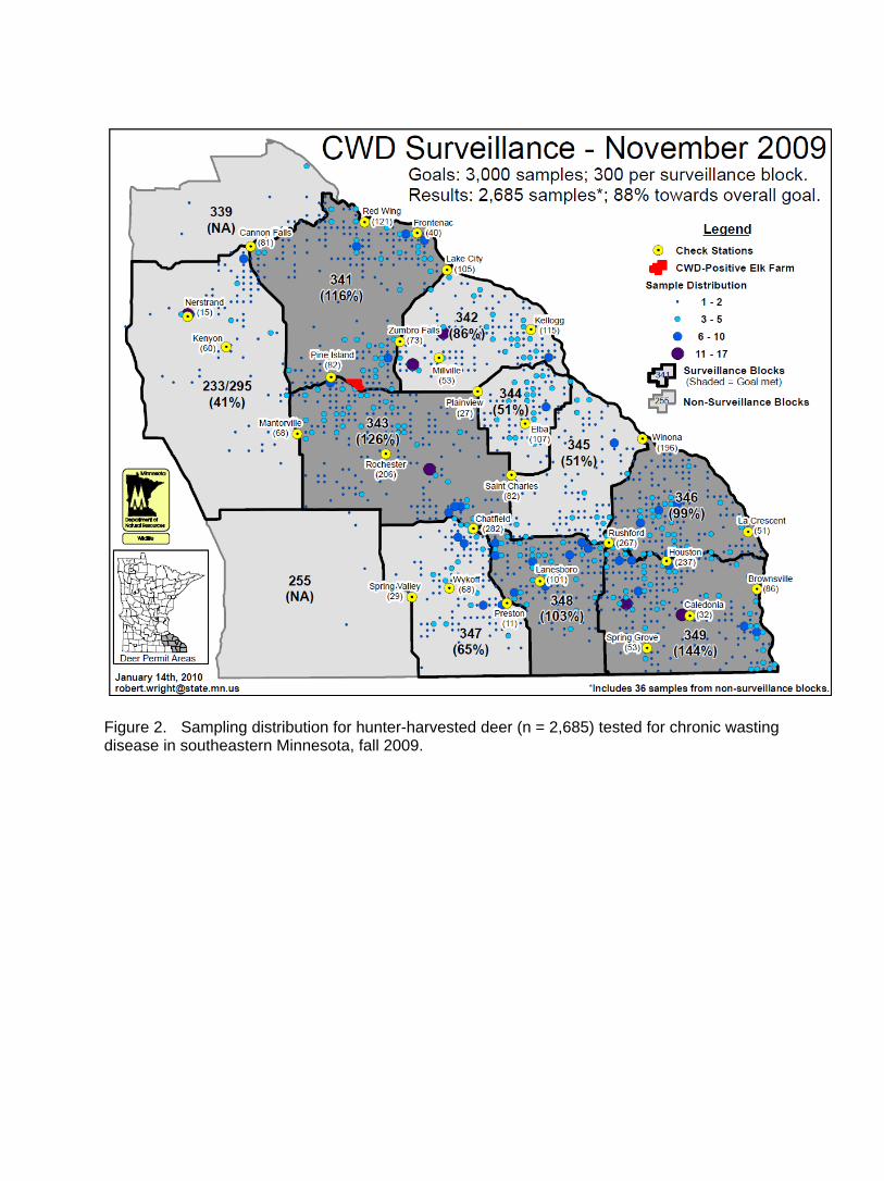
Figure 2. Sampling distribution for hunter-harvested deer (n = 2,685) tested for chronic wasting disease in southeastern Minnesota, fall 2009.
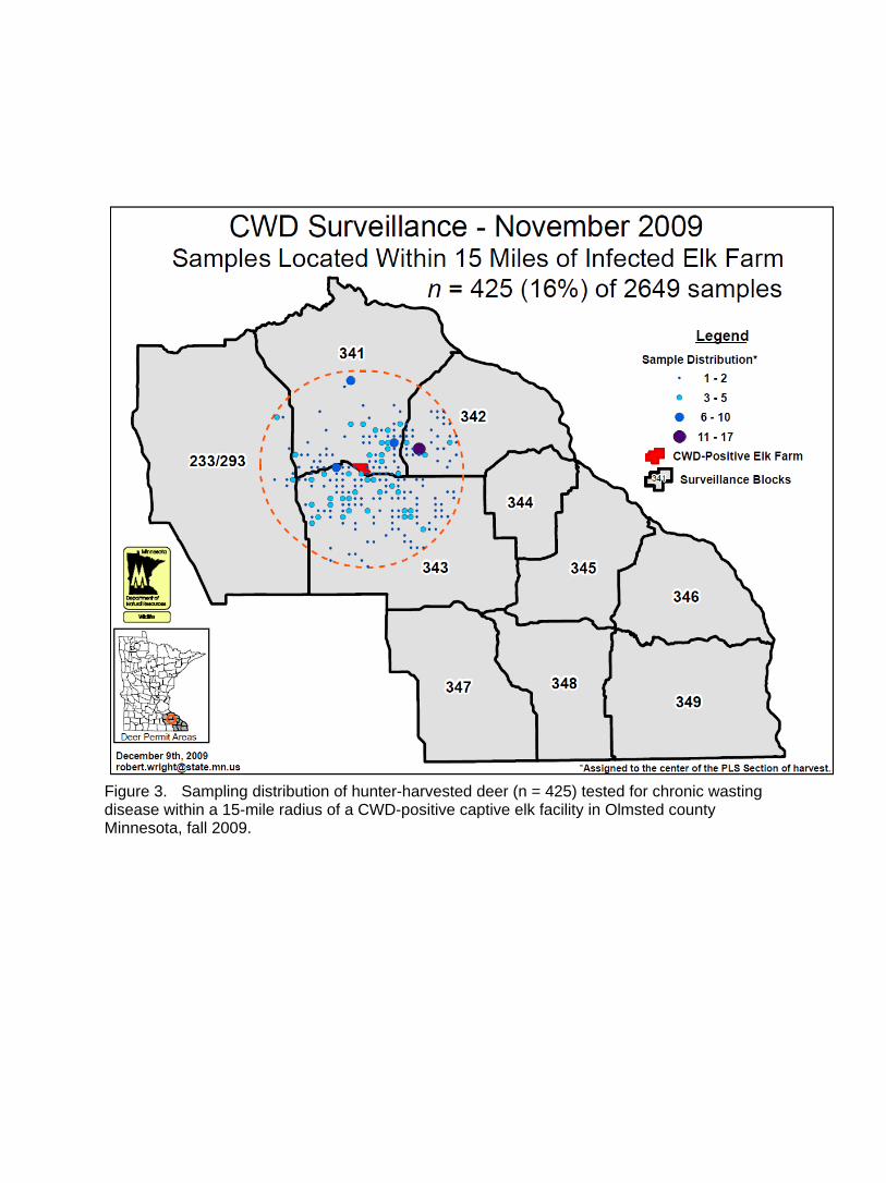
Figure 3. Sampling distribution of hunter-harvested deer (n = 425) tested for chronic wasting disease within a 15-mile radius of a CWD-positive captive elk facility in Olmsted county Minnesota, fall 2009.

PRELIMINARY RESULTS OF HERD HEALTH ASSESSMENT FOR NORTHWESTERN FREE-RANGING ELK FROM 2004-2009 Erik Hildebrand1, Michelle Carstensen, Erika Butler, and Lou Cornicelli SUMMARY OF FINDINGS The goal of this project was to assess the health of free-ranging elk (Cervus elaphus) from northwestern Minnesota (NW MN) by screening animals for a variety of diseases and parasites. Results indicate exposure to these pathogens, and not necessarily clinical illness. From the elk included in this study (n=86), we identified exposure to eastern equine encephalitis, West Nile Virus, malignant catarrhal fever, Neospora, anaplasmosis, borreliosis, bovine viral diarrhea virus 1 and 2, bovine herpes virus 1, Leptospira sp., and parainfluenza virus 3. A variety of fecal parasites were also identified (Coccidia, Strongyle-type ova, and Moniezia) on fecal examination. Lung and liver tissue were cultured for bacterial infection; Streptococcus sp. was isolated from the lung of 1 individual and no isolations were found in liver samples. All elk were negative for Mycobacterium paratuberculosis, blue tongue virus, epizootic hemorrhagic disease, brucellosis, chronic wasting disease, and bovine tuberculosis. Hepatic mineral levels were also evaluated. INTRODUCTION Elk are native to Minnesota and were formally protected from hunting in 1893. By the early 1900s, elk became scarce and the last native Minnesota elk was reportedly seen in the Northwest Angle in 1932. Reintroduction efforts were initiated in 1914 and 1915 which brought elk from Yellowstone National Park (WY) and Jackson (WY) to Minnesota’s Itasca State Park. The herd expanded to 25 animals by 1925 (MNDNR 2008). In 1935, 27 elk from Itasca State Park were moved to the Red Lake Game Preserve, which then expanded to nearly 100 animals by the 1940s. This herd, referred to as the Grygla herd, primarily occupies a 45 mi2 area north of Grygla, MN (Figure 1). In 1987, as complaints of elk causing crop damage increased, the Legislature created a compensation program for crop depredation and imposed limits on the elk herd size to pre-calving numbers of 20–30 animals. To accomplish the required reduction in elk numbers, the Minnesota Department of Natural Resources (MNDNR) instituted elk hunts in 1987, 1996, 1997, and 1998; yet, very few animals were taken each year (MNDNR 2008). The decision to hold a hunting season is based on herd size, and current policy requires a hunt if there are more than 30 in the herd before calving. Hunts have occurred since 2004 and the most recent aerial survey indicated the pre-calved Grygla herd is currently at approximately 40 animals, with the goal remaining at 30-38 elk.
A second herd of elk occurs in Kittson and Roseau Counties (Figure 1), and is termed the Kittson County herd. First noted along the Manitoba border in the early 1980s, these animals winter in Manitoba, while calving and spending the summers in MN. They were originally divided into 3 subgroups based on distinctive areas of use (Figure 2). These 3 subgroups were the Water Tower subgroup (north of Lancaster), the Lancaster subgroup (east of Lancaster) and the Caribou/Vita subgroup (located between Caribou, MN and Vita, Manitoba). The Caribou/Vita subgroup is known to occupy either side of the international border at any given time of year. The extent to which the other 2 subgroups cross into Canada is unknown. Little is also known regarding the extent of animal interchange between the Caribou/Vita subgroup and the other 2 subgroups (MNDNR 2009). Due to crop depredation issues, a hunting season was first held in 2008. The most recent elk survey estimated 27 (pre-calving) animals in the Kittson County herd excluding what might be in the Caribou-Vita subgroup. The current Elk Management Plan set a pre-calving population goal for the ____________ Corresponding author e-mail: [email protected]

Watertower and Lancaster subgroups at 20-30 each. The population goal for the Caribou-Vita subgroup is still under discussion with MNDNR and Manitoba Conservation. In 2010, the Water Tower and Lancaster subgroups were combined and are described as the Kittson Central Elk subgroup, thus we now begin to recognize both the Kittson Central Elk subgroup and Caribou-Vita subgroup. Expansion of elk in MN is limited by both habitat succession and reproduction rates within the herds, but the social factor of mandating the herd to a specified level is the main limiting factor. The purpose of this project was to screen NW MN elk for a variety of disease agents to determine which diseases they were being exposed to. Positive results are not diagnostic of clinical disease. While some of the test results may be all negative, this does not necessarily imply that the disease is not present or impacting the population. Some diseases cause death quickly without an immune response; thus, finding a positive in a seemingly healthy animal would be extremely rare. Discovery of bovine tuberculosis (TB) in cattle and free-ranging white-tailed deer (Odocoileus virginianus) has brought increased scrutiny as to the health status of the NW MN elk, particularly the Grygla Herd. While overlap in range between elk and known TB-infected deer or cattle farms is known to occur, there has been no evidence of TB-infection in MN’s elk herd. TB-infected cattle and deer in MN share the same strain, which is considered of Mexican or southwest US origin, and is not related to the strain of bovine TB found in elk in Manitoba’s Riding Mountain National Park. METHODS For this report, all elk sampled from NW MN were grouped as either harvested animals (including hunter-harvested, removed under depredation permits, agency sharpshooting, and illegally poached) or other (including road kills, sick, and found dead elk). All elk within the harvested category were assumed to be representative of healthy individuals within the population. For hunter-harvested elk, hunters were asked to collect samples of lung, liver, feces, blood, hair, and an incisor for aging. MNDNR provided a project overview, instructions for sample collection, and sampling kits at the mandatory elk hunter orientation sessions. Elk removed under depredation permits or other methods were sampled by trained MNDNR staff.
All equipment needed for sample collection/preservation was included in the sampling kit: soft-sided cooler; 1-60cc syringe for blood collection; 6-15cc serum tubes for blood storage; 3 whirlpaks for a sample of liver, lung, and feces; 2 specimen jars with formalin for liver and lung samples; 2 coin envelopes for hair and tooth; datasheet; protocol; Sharpie marker; 1 pair of large vinyl gloves; and 1 ice pack. Successful hunters dropped off their sampling kits when they registered their animal and also provided information on the location of their kill. Hunters collected blood from the chest cavity as soon after death as possible, using a 60 cc syringe. The blood was placed in serum tubes and kept cool until they were delivered to official MNDNR registration station. Liver and lung samples were collected and split, with half placed in a formalin jar, while the other half was frozen in whirlpak bags. If the hunter found anything unusual, such as a large abscess or tumor, those samples were also collected and split between the preservation methods (formalin fixation and freezing). Blood was centrifuged at the registration stations and serum was extracted and frozen. Cranial lymph nodes and obexes were removed by trained MNDNR staff at the registration stations to allow for chronic wasting and bovine tuberculosis testing. Where appropriate, MNDNR made arrangements with taxidermists to collect samples from trophy animals. All samples were submitted to the University of Minnesota Veterinary Diagnostic Laboratory (VDL), where the majority of the testing occurred; some tests were outsourced to the National Veterinary Services Laboratories (NVSL) in Ames, IA.

RESULTS AND DISCUSSION
A total of 86 elk were included in this health assessment project (Figure 3). Harvested elk accounted for 82 of the animals (61 hunter-harvested, 17 depredation permits, 1 sharpshooting, and 3 poached). In addition, 4 other animals were sampled (1 roadkill, 1 found dead, 1 shot by law enforcement due to possible injury/sickness, and 1 clinically ill elk (observed with neurological symptoms). The sick animal was dispatched by a local conservation officer and necropsy results indicate the observed clinical illness was likely due to P.tenuis infection, although it was also positive for L. interrogans serovar icterohaemorrhagicae.
Serologic results from harvested elk indicate exposure to eastern equine encephalitis, West Nile Virus, malignant catarrhal fever, Neospora, anaplasmosis, borreliosis, bovine viral diarrhea virus 1 and 2, bovine herpes virus 1, Leptospira sp., and parainfluenza virus 3 (Table 1). Liver samples from 65 harvested elk were evaluated for heavy metal and mineral status (Table 2). Though not included in Table 2, the sick elk’s hepatic mineral values fell within the means of all other harvested elk. Exact age was determined for 68 harvested elk (µ= 4.2 years; sd = 3.7 years; range 0.5 to 16 years old) (Figure 4). There were nearly twice as many females (n = 43) than males (n = 26) of known sex in the harvested elk category.
Complete sets of samples were not collected from all elk included in this project, as field conditions and sample quality varied; however, there were very few errors in tissue identification or insufficient sample quantities in those submitted. The following discussion provides an overview of the major findings from 86 elk included in this study (2004-2009). Samples from an additional 11 elk removed by sharpshooters from the Kittson County herd in spring 2010 are not included in this report, as results are pending. Mosquito-Borne Viruses Positive results were reported for 6 of 44 elk (13.6%) tested for eastern equine encephalitis (EEE) (Table 1, Figure 5). The positive results indicate that these animals were likely exposed to the EEE virus as the virus neutralization (VN) test prevents cross-reactivity with other viruses. Two harvested animals had titers ≥ 100. A titer that is greater than 100 is considered a VERY strong positive and indicates that the serum was able to neutralize nearly 100% of the virus. EEE is spread by mosquitoes and causes neurologic signs and often death. It poses a greater mortality threat for most species than West Nile Virus (WNV) does. Horses, deer, and other mammals are incidental, dead-end hosts of EEE virus. Under natural transmission conditions, they are only infected by bridge vectors, mosquito species that feed both on birds and large mammals (Schmitt et al. 2007). Positive results were reported for 32 of 45 elk (71.1%) tested for West Nile Virus (Table 1, Figure 5). Four elk had titers ≥ 100. Little is known about the effects of WNV in elk. In white-tailed deer it has been found that they often have a low titer and no clinical signs. However, the United States Department of Agriculture (USDA) has reported that reindeer (Rangifer tarandus) infected with WNV have high mortality rates and high titers, indicating that the virus may be more serious for some species than others.
Malignant Catarrhal Fever
Samples from 46 elk were submitted to NVSL for peroxidase-linked assay (PLA) testing for Malignant Catarrhal Fever (MCF) from 2004-2009. If the PLA test came back positive, the samples were further screened with a VN test. A total of 13 samples tested positive on the PLA test (28.9%) (Table 1, Figure 5); 11 with titers at 1:20, and 2 at 1:100. However, all elk were negative on VN. The PLA test is more sensitive than the virus isolation, meaning it is much better at identifying true positives. VN is more specific, which means it is better at identifying true negatives. There are a couple of issues with this testing. First, the PLA reacts with multiple Gammaherpes Viruses (such as the wildebeest strain, the sheep strain, the deer strain, etc). A

PLA positive does not indicate which strain has been found, it only indicates that one has. The higher the positive value with the PLA test, the stronger the positive in the sample. Second, the VN test only screens for the wildebeest strain (which is exotic to the U.S.) and would be negative if other strains are present. This means a sample that was positive on PLA and negative on VN was likely exposed to a gammaherpes virus, but not the wildebeest strain.
Gammaherpes viruses have been documented to cause serious illness and death in elk and other ruminants. The clinical symptoms can mimic P. tenuis infection as the animals often exhibit neurological deficits, go blind, and thrash on the ground prior to death. While infection with MCF frequently results in death, carrier status can occur and is identified with serology. Li et al. (1996) found small numbers of United States free-ranging elk were seropositive; these animals were once exposed to MCF viruses but whether they had recovered from a non-lethal disease is unknown.
Fecal Examination for Parasites
Fecal samples from 58 elk were screened for evidence of parasites from 2004-2009. Parasites were identified in 5 samples (8.6%), including Fascioloides magna, Coccidia sp., Strongyle-type ova, and Moniezia sp. Negative results do not necessarily mean the animal was parasite-free, only that it was not actively shedding at the time the feces were collected. Pulmonary Mycoplasma and Hepatic Salmonella Culture
From 2004-2009, a total of 18 lung samples were cultured for Mycoplasma and 19 liver samples were cultured for Salmonella. None was isolated.
Mycobacterium Paratuberculosis (Johne’s Disease) During this study, a total of 43 fecal samples were cultured for M. paratuberculosis and 57 fecal samples were genetically screened (polymerase chain reaction, PCR) from the bacterium. Additionally, a serological test (Biocor) was run on 52 samples. All culture, PCR, and Biocor results were negative for Johne’s disease. The negative fecal cultures and PCR results indicate that those elk were not actively shedding the bacterium. The negative Biocor results indicate that these animals had not been exposed to the bacterium.
All species of ruminants are believed to be susceptible to Johne’s and it is frequently diagnosed in cattle and sheep (Manning and Collins 2001). Elk infected with Johne’s may show non-specific clinical signs including poor weight gain and poor shedding of hair coat, and rapid weight loss and diarrhea may occur just prior to death (Barber-Meyer et al. 2007). Elk, mule deer (Odocoileus hemionus), white-tailed deer, bighorn x hybrid, and domestic sheep were susceptible to infection with M.paratuberculosis derived from paratuberculous bighorn sheep (Ovis Canadensis) (Williams et al. 1983). During the first year of exposure, only deer developed clinical paratuberculosis, characterized by poor body condition and diarrhea.
Anaplasmosis
A total of 46 samples were screened for Anaplasmosis (Anaplasma phagocytopila, formerly Ehrlichia phagocytophila) with the card test from 2004-2009. All animals were negative. In sheep, this disease produces significant effects on the immunological defense system, increasing their susceptibility to disease and secondary infections (Larson et al. 1994). Experimental studies have shown that elk can harbor asymptomatic infections with A. marginale and A. ovis, the causes of anaplasmosis in cattle and sheep, respectively. However, efforts to recover Anaplasma spp. from free-ranging elk populations have been unsuccessful, suggesting that even though these species are susceptible, they are probably not responsible for maintaining infections or acting as a source of infection for cattle (Corn and Nettles 2001).

Borreliosis (Lymes Disease) A total of 45 elk were screened for Lyme disease with an immunofluorescence assay
(IFA) from 2004-2009. Positive results were reported for 30 elk (66.7%) (Table 1, Figure 5). Borreliosis is a tick-borne bacterial disease that is maintained through a wildlife/tick cycle involving a variety of species, including mammals and birds. While evidence of natural infection in wildlife exists, there has been no documentation of clinical disease or lesions reported in wildlife species.
Brucellosis
A total of 49 elk were screened for Brucella with the card test. All results were negative, indicating that these animals were not likely exposed to the bacterium. Brucellosis has been a major disease issue among elk, bison and cattle in western states. The disease causes spontaneous abortions and is most likely spread through oral contact (e.g., licking or ingestion of contaminated materials) (Thorne et al. 1978).
Bovine Viral Diarrhea Virus (BVD) 1 and 2 A total of 56 elk were tested by serum neutralization (SN) for BVD 1 and 2 from 2004-
2009. Seven animals tested positive (12.5%) (Table 1, Figure 5). These results indicate that the elk population from NW MN was being exposed to BVD. Two animals had positive titer levels at 32/16, 3 were positive at 32/negative, and 2 had a titer level at 8/negative.
BVD is considered a major disease of cattle and is thought to be the most common infectious cause of reproductive failure in beef herds in the western U.S. BVD also causes enteritis, mucosal disease, infections, and respiratory disorders in cattle though experimentally inoculated non-pregnant elk showed no clinical signs and remained healthy for >50 days post inoculation (Barber-Meyer et al. 2007). Bovine Herpes Virus 1 (BHV)
A total of 57 elk were screened for BHV using a SN test from 2004-2009. Five animals were positive (7.1%) (Table 1, Figure 5). BHV is a disease of the respiratory tract. It is believed to infect all ruminant species and has been isolated from a large number of wild species. It is most commonly isolated in feedlot cattle.
Epizootic Hemorrhagic Disease (EHD) and Blue Tongue Virus (BTV) A total of 59 elk were screened for EHD using an Agar Gel Immuno Diffusion (AGID) test
and BTV using a Competitive Enzyme-Linked Immunoabsorbent Assay (cELISA) from 2004-2009. All results were negative. EHD and BTV are a hemorrhagic diseases transmitted by a biting midge that is known to cause illness and death in white-tailed deer. While it is known to be infective to a variety of domestic and wild ruminants, clinical disease is quite variable.
Leptospira sp.
A total of 59 elk were screened for 6 species of Leptospira, using a microscopic
agglutination test (MAT), from 2004-2009. Positive results are reported per Leptospira species below (Table 1, Figure 5):
L. bratislava: o 0/59
L. canicola: o 0/59

L. grippothyphosa: o 0/59
L. hardjo: o 1/59 (1.7%)
L. interrogans serovar icterohaemorrhagicae: o 7/59 (11.9%)
L. pomona: o 1/59 (1.7%)
The positive L. hardjo had a titer level of 200. Of the positives for L. interrogans serovar
icterohaemorrhagicae, 5 had a titer of 100 and 1 with a titer of 200. The positive L. Pomona had a titer of 800.
Leptospirosis is a bacterial disease that can infect a wide variety of mammals, both domestic and wild. Exposure usually occurs through direct contact with urine from carrier animals or indirectly by contact with a urine- contaminated environment (Bender and Hall 1996). Much of the landscape of NW MN contains environments where moist alkaline soils are present to house the bacteria, and it may survive for several weeks (Bender and Hall 1996).
Neospora sp.
A total of 51 elk were screened for Neospora with an ELISA test from 2004-2009. All samples tested negative. While clinical disease due to neospora infection is best described in domestic animals, reports of ill effects due to Neospora infection in wildlife do exist. Systemic neosporosis was diagnosed in a California black-tailed deer (Odocoileus hemionus) that was found dead (Woods et al. 1996). Neospora caninum causes abortion and serious clinical disease in livestock and companion animals, although dogs and coyotes are its only known definitive hosts that can shed oocysts (Dubey and Thulliez 2005). Recent study of neospora prevelance in white-tailed deer in northwestern MN reported 71% prevalence, indicating the parasite is present in elk range (Dubey et al. 2009). Parainfluenza Virus 3 (PI)
A total of 53 elk were screened for PI using a hemagglutination inhibition (HI) test from 2004-2009. Eighteen animals were positive (34%) (Table 1, Figure 5), with titers of 6 at 10, 4 at 20, 4 at 40, 3 at 80, and 1 at 160.
The positive results indicate that NW MN elk were exposed to PI. Domestic ruminants are considered the main source of infection for free-ranging ruminants. PI causes mild respiratory disorders in domestic cattle and sheep that serve as initiators for secondary infections of Pasteurella spp., which can result in bacterial pneumonia, but clinical symptoms in wild elk remain unknown (Barber-Meyer et al. 2007). Chronic Wasting Disease (CWD)
From 2004-2009, a total of 58 elk were screened for CWD using immunohistochemistry (IHC); including 42 animals with obex samples and 53 retropharyngeal lymph node samples. All results were negative. CWD is a transmissible spongiform encephalopathy that causes neurological disease in cervids. CWD is known to occur in elk, but has never been documented in wild cervids in MN. Bovine Tuberculosis
From 2004-2009, 77 sets of cranial lymph nodes (parotid, retropharyngeal, and submandibular) were collected and cultured for Mycobacterium bovis. All results were negative. Bovine tuberculosis is a chronic, progressive bacterial disease that infects a wide array of

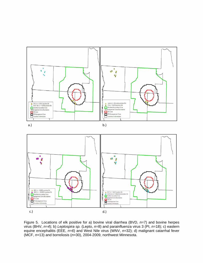
mammals. Bovine tuberculosis has been found in wild white-tailed deer in a small, localized area in NW MN which overlaps with the elk range, but has not been found in any wild elk in the state. Distribution of Positive Results for Select Diseases The geographic distribution of positive results for select disease agents was briefly evaluated (Figure 5). It was interesting to note that elk which tested positive for BVD all originated from the Kittson county herd while the elk which tested positive for BHV all originated from the Grygla herd. The significance of this finding is unknown. There was no clustering of positives observed for WNV, EEE, MCF, Borrelia, PI or Leptospirosis. ACKNOWLEDGMENTS This project would not have been possible without assistance from a number of MNDNR employees and volunteers. We would like to especially thank staff in NW MN who helped with collecting samples: Joel Huener, Donovan Pietruszewski, Christine Reisz, Randy Prachar, Dawn Torrison, Lou Cornicelli, and Marshall Deters. Finally we appreciate the help of all the volunteers including veterinary students Jonna Swanson, Macdonald Farnham, and Andrea Widdel who helped with this elk project. REFERENCES
Barber-Meyer, S., White, P., and Mech, D. 2007. Survey of selected pathogens and blood parameters of Northern Yellowstone elk: wolf sanitation effect implications. The American Midland Naturalist 158 (2): 369-381. Bender, L., and Hall, P. 1996. Leptospira interrogans exposure in free-ranging elk in Washington. Journal of Wildlife Diseases 32(1): 121-124. Corn, L. and Nettles, V. 2001. Health protocol for translocation of free-ranging elk. Journal of
Wildlife Diseases 37(3): 413-426. Dubey, J. P, M. C. Jenkins, O.C.H. Kwok, R. L. Zink, M. L. Michalski, V. Ulrich, J. Jill, M. Carstensen, and P. Thulliez. 2009. Seroprevalence of Neospora caninum and Toxoplasma gondii antibodies in white-tailed deer (Odocoileus virginianus) from Iowa and Minnesota using four serologic tests. Veterinary Parasitology 161: 330–334. Dubey, J. P., and Thulliez, P. 2005. Prevalence of antibodies to Neospora caninum in wild
animals. The Journal of Parasitol., 91(5): 1217-1218. Larsen, H. J. S., G. Overnes, H. Waldeland, and G.M. Johansen. 1994. Immunosuppression in
sheep experimentally infected with Ehrlichia phagocytophila. Research in Veterinary Science 56: 216-224.
Li, H., D. Shen, D. Jessup, D. Knowles, J. Gorham, T. Thorne, D. O’Toole, and T. Crawford. 1996. Prevalence of antibody to malignant catarrhal fever virus in wild and domestic
ruminants by competitive-inhibition ELISA. The Journal of Wildlife Diseases 32(3):437-443.
Manning, E. J. B., and M. T. Collins. 2001. Mycobacterium avium subsp. paratuberculosis: pathogen pathogenesis and diagnosis. In Mycobacterial infections in domestic and wild animals, E. J. B. Manning and M. T. Collins (eds.). Revue Scientifique et Technique Office International des Epizooties 20: 133-150
Minnesota Department of Natural Resources. 2008. Minnesota long range management plan for elk: Draft. Division of Fish and Wildlife, Minnesota Department of Natural Resources.19 pp.
Minnesota Department of Natural Resources. 2009. Strategic management plan for elk: Draft. Division of Fish and Wildlife, Minnesota Department of Natural Resources. 34pp.
Schmitt, M., S, Cooley, T., Fitzgerald, S., Bolin, S., Lim, A., Schaefer, S., Kiupel, M., Maes, R., Hogle, S., and D. O’Brien. 2007. An outbreak of eastern equine encephalitis virus in free-ranging white-tailed deer in Michigan. Journal of Wildlife Diseases 43(4): 635-644.
Thorne, T., Morton, J., Blunt, F., and Dawson, H. 1978. Brucellosis in elk. II. clinical effects and means of transmission as determined through artificial infections. Journal of Wildlife Diseases 14: 280-291.
Williams, E., Snyder, S., and Martin, K. 1983. Experimental infection of some north American wild ruminants and domestic sheep with Mycobacterium paratuberculosis: clinical and bacteriological findings. Journal of Wildlife Diseases 19(3): 185-191.
Woods, L. W., P. K. Swift, B. C. Barr, M. C. Horzinek, R. W. Nordhausen, M. N. Oliver, K. R. Jones, and N. J. Maclachlan. 1996. Systemic adenovirus infection associated with high mortality in mule deer (Odocoileus hemionus) in California. Veterinary Pathology 33: 125-132.

Table 1. Serological results from harvested elk in northwestern Minnesota, 2004-2009.
Disease n Apparent prevalence %
EEE 44 13.6 (n=6)
MCF 45 28.9 (n=13)
WNV 45 71.1 (n=32)
Anaplasmosis 46 0
Borreliosis 45 66.7 (n=30)
Brucellosis 49 0
BVD 1 and 2 56 12.5 (n=7)
BHV 56 7.1 (n=4)
BTV 58 0
EHD 58 0
L. bratislava 58 0
L. canicola 58 0
L. grippothyphosa 58 0
L. hardjo 58 1.7 (n=1)
L. interrogans serovar icterohaemorrhagicae 58 10.3 (n=6)
L. pomona 58 1.7 (n=1)
Neospora 51 0
PI 53 34 (n=18)
Mycobacterium paratuberculosis 52 0

Table 2. Hepatic mineral values of harvested elk in northwestern Minnesota, 2004-2009.
Element n Mean Standard deviation Minimum Maximum
Arsenic 64 0.71 0.37 0 1
Boron 25 0.50 0 0 0.50
Barium 25 0.03 0 0 0.025
Calcium 24 55.99 17.98 39.30 111
Cadmium 63 0.24 0.09 0 0.66
Cobalt 63 0.18 0.1 0 0.25
Chromium 25 0.10 0 0 0.10
Copper 65 12.54 11.93 1.30 63
Iron 65 199.65 162.69 31.30 946.3
Mercury 25 1.00 0 0 1
Potassium 24 2561.92 218.53 2108 2850
Magnesium 65 159.76 21.36 86 211.1
Manganese 65 2.62 0.95 0.28 5.60
Molybdenum 65 1.11 0.33 0.10 1.77
Sodium 24 947.21 193.24 626 1490
Phosphorous 24 4150.46 752.67 1650 5354
Lead 64 0.40 0.12 0 0.50
Antimony 25 0.50 0 0 0.50
Selenium 65 0.84 0.34 0 1.90
Thallium 25 1.25 0 0 1.25
Zinc 64 25.39 6.13 15.40 53

Figure 1. Current range of the 2 localized elk herds of northwest Minnesota in 2009.
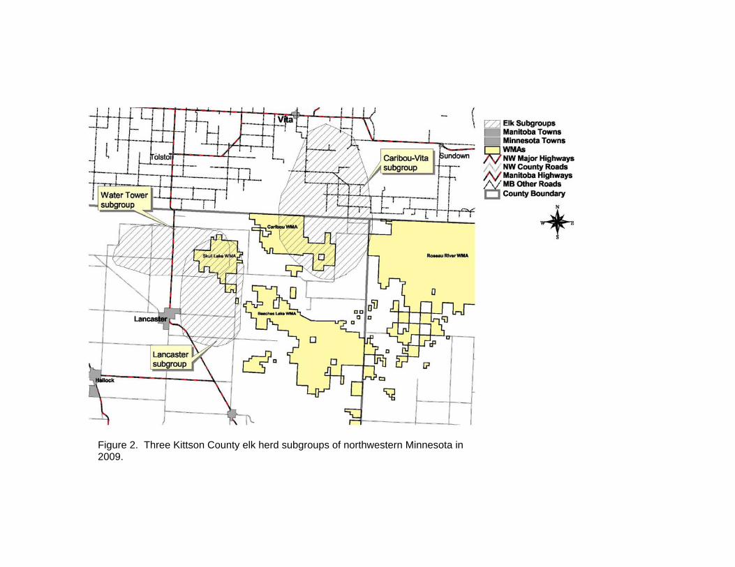
Figure 2. Three Kittson County elk herd subgroups of northwestern Minnesota in 2009.
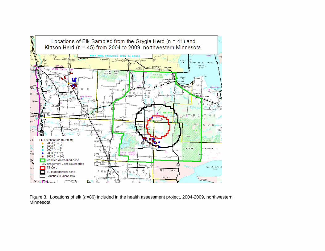
Figure 3. Locations of elk (n=86) included in the health assessment project, 2004-2009, northwestern
Minnesota.

0
2
4
6
8
10
12
14
16
0.5 1.0 2.0 3.0 4.0 5.0 6.0 7.0 8.0 11.0 12.0 14.0 16.0
Sample Size
Age (years)
Figure 4. Age distribution of harvested elk (n = 68) included in the 2004-2009 health assessment project, northwestern Minnesota.
a.) b.)
c.) d.)
Figure 5. Locations of elk positive for a) bovine viral diarrhea (BVD, n=7) and bovine herpes virus (BHV, n=4); b) Leptospira sp. (Lepto, n=8) and parainfluenza virus 3 (PI, n=18); c) eastern equine encephalitis (EEE, n=6) and West Nile virus (WNV, n=32); d) malignant catarrhal fever (MCF, n=13) and borreliosis (n=30), 2004-2009, northwest Minnesota.

PRELIMINARY RESULTS FROM THE 2007-2009 MOOSE HERD HEALTH ASSESSMENT PROJECT Erika Butler1, Michelle Carstensen, Erik Hildebrand, John Giudice, Robert Wright, and Mike Schrage SUMMARY OF FINDINGS
The purpose of this project was to screen 2007-2009 hunter-harvested (and presumably healthy) moose (Alces alces) for a variety of disease agents. Results were used to identify diseases the northeast Minnesota (NE MN) moose population have been exposed to as well served as a baseline for similar testing completed on non-hunting moose mortalities from the same population. Positive results confirmed that moose were exposed to, though not necessarily ill from, eastern equine encephalitis, West Nile Virus, malignant catarrhal fever, Neospora, anaplasmosis, bovine herpes virus 1, bovine viral diarrhea virus 1 and 2, borrelia, Leptospira sp, and parainfluenza virus 3. When possible, serological events were evaluated to determine whether there was any influence of age, location, or year of harvest. Additionally, a variety of fecal parasites were identified on fecal examination. All results were negative for Mycobacterium paratuberculosis, brucellosis, blue tongue virus, epizootic hemorrhagic disease, chronic wasting disease, and bovine tuberculosis. Hepatic mineral values were evaluated, whole livers were examined grossly and ranked according to the level of damage due to liver fluke infection, and histological examination of whole brains investigated how many apparently healthy moose have lesions consistent with migration tracts (presumably due to P. tenuis). 32BINTRODUCTION
Several lines of evidence suggest that the moose population in northeastern Minnesota is declining. Since 2002, annual survival and reproductive rates were substantially lower than documented elsewhere in North America (Lenarz et al. 2007) and modeling based on these vital rates indicated that the population is declining by approximately 15% per year since at least 2002 (Lenarz et al. 2010). Likewise, recruitment and twinning rates have steadily declined since 2002 (Lenarz 2009). In addition, hunter success rates have steadily decreased since 2001 (Lenarz 2009). Finally, anecdotal reports from local residents have reported a noticeable decline in moose numbers. Parasitic infection with Parelaphostrongylus tenuis, Echinococcus granulosus, Elaeophora schneideri, Sarcocystis spp., Fascloides magna, and Dermacenter albipictus has been documented in Minnesota’s moose. Copper deficiency has been reported in some moose. Poor antler development has also been noted in some bull mortalities. Many causes of mortality remain unknown with numerous prime-age animals dying, often during low stress periods of the year.
The purpose of this project was to screen presumably healthy moose for a variety of disease agents. Results were intended to indicate which diseases the NE MN moose population were exposed to. Exposure, itself, does not imply the animal was clinically ill with the disease. They also served as a baseline, allowing for comparisons between similar testing completed on non-hunting moose mortalities from the same population. While some of the test results may be all negative, this does not necessarily mean that the disease is not present or impacting the population. Some diseases cause death quickly and without an immune response; thus finding a positive in a seemingly healthy animal would be extremely rare.
___________________________
1 Corresponding author email: [email protected]

METHODS
In order to conduct this herd health assessment, hunters (both tribal and state) were asked to collect samples of lung, liver, blood, feces, hair, ticks, and an incisor for aging. The Wildlife Health Program provided a presentation and instructions relative to the moose herd health assessment project at the mandatory Minnesota Department of Natural Resources (MNDNR) Moose Hunt Orientation Sessions and tribal natural resource offices. Hunters were given a sampling kit with instructions at the sessions. Post-harvest, the sampling kits were dropped off at official registration stations by the hunters at the time of registration. Hunters were asked to locate their kill site on appropriate maps.
MNDNR provided hunters with all equipment needed for sample collection/preservation. Sampling kits included the following items: cooler; 1-60cc syringe for blood collection; 6-15cc serum tubes for blood storage; 3 whirlpaks for a sample of liver, lung and feces; 2 specimen jars with formalin for liver and lung samples; 2 coin envelopes for tooth and hair; datasheet; protocol; Sharpie marker; 1 pair of large vinyl gloves; and 1 icepack. In 2008, 1 -5-cc whole blood tube was added to the kits.
The hunters collected blood from the chest cavity as soon after death as possible, using a 60 cc syringe. The blood was placed in serum tubes and kept cool until they were delivered to official MNDNR registration stations or tribal natural resource offices. Liver and lung samples were collected and split, with half placed in a formalin jar, while the other half was frozen in whirlpak bags. In 2009, we asked hunters to begin collecting whole livers in addition to the formalin fixed sample. If the hunter found anything unusual, regardless of the location in the carcass, such as a large abscess or tumor, those samples were collected and split between the preservative methods (formalin fixation and freezing). Blood was centrifuged at the registration stations or tribal natural resource offices and serum was extracted and frozen. In 2008, we began collecting whole blood as well, from which blood smears were made and the remaining whole blood was frozen. Also, retropharyngeal lymph nodes, obexes, and whole brains (2008 and 2009 only) were removed by trained MNDNR staff, tribal staff, and volunteers at the registration stations with permission of the hunters. Portable refrigerators were located in advance at the registration stations to maintain the tissue samples. Samples were submitted to the University of Minnesota Veterinary Diagnostic Laboratory, where much of the testing occurred. A few of the tests were outsourced to the National Veterinary Services Laboratories (NVSL) in Ames, IA. RESULTS AND DISCUSSION
Samples from 368 moose were submitted for diagnostic screening from 2007-2009 (n = 128, 2007; n = 118, 2008; n = 122, 2009). Our samples originated from hunter-harvested animals and state hunters were only allowed to harvest males (some tribal hunters were allowed to take females); thus most of our samples were from male moose (Table 1). Samples were collected throughout our moose hunting zones (Figure 1.) and from three counties (Figure 2.). Precise ages were determined from moose in which hunters provided a central incisor. Ages of animals sampled ranged from <1 year to 14 years old (Figure 3.). A summary of serological testing results can be found in Table 2. 1BEastern Equine Encephalitis (EEE)
2B
Three hundred and thirty serum samples were submitted to NVSL for Virus Neutralization (VN) (2007-2008) or plaque reduction neutralization (PRNT) (2009) screening for EEE. Positive results were reported for 20 of the moose (6.1%) (Figure 4). The positive results indicated that these animals were exposed to the EEE virus. A titer that is greater than 100 is considered a VERY strong positive and means that the serum was able to neutralize nearly 100% of the virus. Multiple animals had titers ≥100.

MNDNR will be continuing EEE surveillance in hunter-harvested moose for the next 3 years (2010-2013) in an attempt to determine if there is a year effect in the prevalence rate. EEE is spread by mosquitoes and causes neurologic signs and often death. It poses a greater mortality threat for most species than West Nile Virus does, though the effects of EEE infection have not been studied in moose. West Nile Virus (WNV)
A total of 330 samples were submitted to NVSL for VN (2007 and 2008) and PRNT (2009) screening for WNV. Positive results were reported for 115 moose (34.8%) (Figure 5). Positive results indicated that animals were exposed to the WNV. A titer that is greater than 100 is considered a VERY strong positive and means that the serum was able to neutralize nearly 100% of the virus. Multiple animals had titers ≥ 100.
We found some evidence of a higher WNV exposure rate among adult males (36.4%, 95% CI: 30.8-42.5) compared to yearling males (21.4%, 95%CI: 9.0-41.0), but the estimate for yearlings was imprecise due to small sample sizes. For adult male moose, we found no evidence, or only very weak evidence, that the probability of testing positive for WNV was correlated with moose age (adult males only), year of harvest, or county.
MNDNR will be continuing WNV surveillance in hunter-harvested moose for the next 3 years (2010-2013) in an attempt to determine if there is a year affect in the prevalence rate. Little is known about the effects of WNV in moose. In white-tailed deer (Odocoileus virginianus) it has been found that they often have a low titer and no clinical signs. However, the USDA has found that reindeer (Rangifer tarandus) infected with WNV have high mortality rates and high titers, indicating that the virus is more serious for some species than others. Malignant Catarrhal Fever (MCF)
A total of 326 samples were submitted to NVSL for peroxidase-linked assay (PLA) testing for MCF. If the PLA test came back positive, the samples were screened with a VN test. A total of 114 samples tested positive on the PLA test (35%)( Figure 6). One of the 114 tested on VN came back positive (at 1:4). The PLA test is more sensitive than the virus isolation, meaning it is much better at identifying true positives. VN is more specific, which means it is better at identifying true negatives. There are a couple of issues with this testing. First, the PLA reacts with multiple Gammaherpes Viruses (such as the wildebeest strain, the sheep strain, the deer strain, etc). Second, a PLA positive does not indicate which strain has been found, only that one strain has been identified. The higher the positive value with the PLA test, the stronger the positive in the sample. The VN test only screens for the wildebeest strain (which is exotic to the U.S.) and would be negative if other strains are present. This means a sample that was positive on PLA and negative on VN was likely exposed to a gammaherpes virus, but not the wildebeest strain. The one positive result was a weak positive, and likely, a false positive.
Adult (92/255, 36.1%) and yearling males (11/27, 40.1%) had similar rates of exposure to MCF. Probability of MCF exposure was independent of age (adult males only) and we found only weak evidence that MCF varied by county. There were, however, large differences in estimated probability of MCF exposure by year (range: 3.7% in 2007 to 74.6% in 2008).
We have been collaborating with researchers to determine which strain of MCF the NE MN moose are being exposed to. To date, all attempts at strain-typing have been unsuccessful.
Gammaherpes viruses have been documented to cause serious illness and death in moose and other ruminants. The clinical symptoms can mimic P. tenuis infection as the animals often exhibit neurological deficits, go blind, and thrash on the ground prior to death. While infection with MCF frequently results in death, carrier status can occur and is identified with serology. Zarnke et al. 2002 found serologic evidence of exposure in numerous species across Alaska and reported 1% prevalence in moose.

3BFecal Examination for Parasites
4BA total of 318 fecal samples were screened for evidence of parasites on fecal floatation. While no ova, oocysts, or cysts were observed in 277 samples, 41 of the samples had evidence of parasitic infection (12.9%). Parasites identified include strongyle-type ova, Dictyocaulus, Moniezia, and Nematodirus. Negative results do not necessarily mean the animal was parasite free, only that it was not actively shedding at the time the feces were collected. 11BPulmonary Mycoplasma Culture
12BIn 2007, 119 lung samples were submitted for Mycoplasma culture. This bacterium was not isolated on any of the samples. Culture efforts were discontinued in 2008. 13BMycobacterium paratuberculosis (Johne’s Disease)
14BWe submitted 179 fecal samples for M. paratuberculosis culture in 2007 and 2008. All culture results were negative. This was discontinued in 2009. PCR was performed on 316 samples, with all results negative, and Biocor was run on 335 samples, with all of the results negative.
The negative fecal cultures and PCR results indicate that those moose were not actively shedding the bacterium. The negative Biocor results indicate that these animals had not been exposed to the bacterium.
All species of ruminants are believed to be susceptible to Johne’s and it is frequently diagnosed in cattle and sheep (Manning and Collins 2001). Clinical signs in wild ruminants are similar to those seen in sheep, and 1 moose with diarrhea, which resulted in death, was diagnosed with Johne’s (Soltys et al. 1967). Serologic evidence of exposure to Johne’s in moose has been documented, with 9/426 (2.1%) seropositive moose in Norway (Tryland et al. 2004). 15BAnaplasmosis
16BA total of 319 samples were screened for Anaplasmosis (Anaplasma phagocytopila, formerly Ehrlichia phagocytophila) with the card test. Only 1 of moose was positive (1/319, 0.3%); indicating that exposure to this bacterium is likely occurring, albeit at a low rate.
Moose are known to be susceptible to infection with A. phagocytophilum. In Norway, anaplasmosis was diagnosed in a moose calf, which displayed apathy and paralysis of the hind-quarters (Jenkins et al. 2001). This moose was concurrently infected with Klebseilla pneumonia, to which the calf’s death was attributed, though the Klebseilla infection was most likely secondary to and facilitated by the primary infection with A. phagocytophilum (Jenkins et al. 2001). In sheep, this disease produces significant effects on the immunological defense system, increasing their susceptibility to disease and secondary infections (Larsen et al. 1994).
A. phagocytophilum is known to occur in MN. In fact, from 1998-2005, 790 human cases were reported in MN and in recent years the MN Department of Health has documented an expansion in the areas in which MN residents are exposed to vector-borne diseases (MN Department of Health). The NE MN population of moose overlaps with the primary area of tick-borne disease risk determined by the MN Department of Health and NE MN. Borreliosis (Lyme disease)
17BA total of 319 samples were screened for lymes disease with an immunofluorescence assay (IFA). Positive results were reported for 73 of the samples (22.9%, 95% CI: 18.6-27.9) (Figure 7). We found evidence of higher Borrelia exposure among yearling males (44.4%, 95% CI: 26.4-63.9) compared to adult males (19.8%, 95% CI: 15.2-25.2), but the estimate for

yearlings was imprecise due to small sample size. The probability of Borrelia exposure among adult males was substantially lower in 2008 (2.2%, 95% CI: 0-5.2) compared to 2007 (34.8%, 95% CI: 23.5-46.0) or 2009 (28.8%, 95%CI: 18.8-38.7). Conversely, probability of exposure among adult males was independent of age, and there was only weak evidence of differences among counties. Borreliosis is a tick borne bacterial disease that is maintained in a wildlife/tick cycle involving a variety of species, including mammals and birds. While evidence of natural infection in wildlife exists, there has been no documentation of clinical disease or lesions reported in wildlife species. 18BBrucellosis
19BA total of 303 samples were submitted for Brucella screening with the card test. There was only 1 positive result, which was then forwarded for confirmatory testing using rivanol agglutination (RIV). The RIV result was negative, indicating that the positive result from the card test was likely a false positive. These negative results indicate that moose were not likely exposed to the bacterium. While naturally occurring fatal Brucella infections have been documented in free-ranging moose (Honour and Hickling 1993) and serologic evidence suggests that some moose populations are being exposed to Brucella sp. (Zarnke 1983), evidence suggests that the prevalence is low (Honour and Hickling 1993). 20BBovine Viral Diarrhea Virus (BVD) 1 & 2
21BA total of 333 samples were submitted for serum neutralization (SN) testing for BVD 1 & 2. Positive results were reported for 3 of the samples (1%); including 1 strong positive at a 1024/4096 titer. These results indicate that the moose population is being exposed to BVD at a very low rate.
BVD is considered a major disease of cattle and is thought to be the most common infectious cause of reproductive failure in beef herds in the western U.S. BVD is also considered a disease of wild ruminants such as moose, caribou (Rangifer tarandus), and deer. Some clinical signs of BVD include diarrhea, dehydration, fever, impaired vision and hearing, depression, abortions, and weakened neonates. Serologic evidence of BVD has been documented in 4 of 22 moose sampled in Alberta (Thorsen and Henderson 1971). Bovine Herpes Virus 1 (BHV)
22BA total of 333 samples were screened for BHV using a SN test. Only 1 moose was found positive (0.9%). BHV is a disease of the respiratory tract. It is believed to infect all ruminant species and has been isolated from a large number of wild species. It is most commonly isolated in feedlot cattle. Blue Tongue Virus (BTV)
23BA total of 334 samples were screened using a Competitive Enzyme-Linked Immunoabsorbent Assay (cELISA) for BTV. All results were negative. BTV is a hemorrhagic disease transmitted by a biting midge that is known to cause illness and death in white-tailed deer. While it is known to be infective to a variety of domestic and wild ruminants, clinical disease is quite variable.
Epizootic Hemorrhagic Disease (EHD)
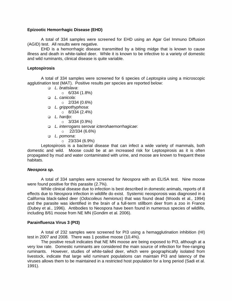
24BA total of 334 samples were screened for EHD using an Agar Gel Immuno Diffusion (AGID) test. All results were negative. EHD is a hemorrhagic disease transmitted by a biting midge that is known to cause illness and death in white-tailed deer. While it is known to be infective to a variety of domestic and wild ruminants, clinical disease is quite variable. 25BLeptospirosis
26BA total of 334 samples were screened for 6 species of Leptospira using a microscopic agglutination test (MAT). Positive results per species are reported below:
L. bratislava: o 6/334 (1.8%)
L. canicola: o 2/334 (0.6%)
L. grippothyphosa: o 8/334 (2.4%)
L. hardjo: o 3/334 (0.9%)
L. interrogans serovar icterohaemorrhagicae: o 22/334 (6.6%)
L. pomona: o 23/334 (6.9%)
Leptospirosis is a bacterial disease that can infect a wide variety of mammals, both domestic and wild. Moose could be at an increased risk for Leptospirosis as it is often propagated by mud and water contaminated with urine, and moose are known to frequent these habitats. Neospora sp.
27BA total of 334 samples were screened for Neospora with an ELISA test. Nine moose were found positive for this parasite (2.7%). While clinical disease due to infection is best described in domestic animals, reports of ill effects due to Neospora infection in wildlife do exist. Systemic neosporosis was diagnosed in a California black-tailed deer (Odocoileus hemionus) that was found dead (Woods et al., 1994) and the parasite was identified in the brain of a full-term stillborn deer from a zoo in France (Dubey et al., 1996). Antibodies to Neospora have been found in numerous species of wildlife, including 8/61 moose from NE MN (Gondim et al. 2006). Parainfluenza Virus 3 (PI3)
28BA total of 232 samples were screened for PI3 using a hemagglutination inhibition (HI) test in 2007 and 2008. There was 1 positive moose (10.4%).
The positive result indicates that NE MN moose are being exposed to PI3, although at a very low rate. Domestic ruminants are considered the main source of infection for free-ranging ruminants. However, studies of white-tailed deer, which were geographically isolated from livestock, indicate that large wild ruminant populations can maintain PI3 and latency of the viruses allows them to be maintained in a restricted host population for a long period (Sadi et al. 1991).

Chronic Wasting Disease (CWD)
A total of 87 obex samples and 88 retropharyngeal lymph nodes were screened for CWD using immunohistochemistry (IHC). All results were negative. CWD is a transmissible spongiform encephalopathy (TSE) that causes neurological disease in cervids. CWD is known to occur in moose, but has never been documented in wild cervids in MN. Bovine Tuberculosis
Cranial lymph nodes (parotid, retropharyngeal, and submandibular) from 88 moose were collected and cultured for Mycobacterium bovis. All results were negative.
Bovine tuberculosis is a chronic, progressive bacterial disease that infects a wide array of mammals. Bovine tuberculosis has been found in wild white tailed deer in a small, localized area in northwestern MN, but has not been found in any wild animals within the moose hunt permit areas. Brain Histopathology In 2008 and 2009, MNDNR collected whole brains from moose at registration stations. Brains were formalin-fixed and submitted for histological examination. A total of 47 whole brains were collected. Four complete coronary brain, cerebellum, and brain stem sections were processed for histological examination from each moose. An average of 25 histological slides per animal were examined. Areas examined included the frontal, temporal, parietal, and occipital lobes and the basal nuclei, thalamus, mesencephalon, and brain stem. This examination is meant to help identify lesions consistent with migration tracts (presumably due to P. tenuis) that may be present in brains of apparently healthy animals. No lesions were found in 41 of the brains, 5 had lymphocytic infiltration (unspecific chronic inflammatory lesion), and 1 had larval tracts present in the white matter (with mild to moderate meningitis, axonal degeneration, and secondary demyelination). MNDNR will continue the collection of whole brains of moose at registration stations in 2010-2013. Whole Liver Evaluation In 2009 only, hunters were asked to collect whole livers. A total of 57 livers were submitted for gross examination. The purpose of this is to develop a ranking system to evaluate liver fluke load and damage caused by liver flukes. The ranking system that was developed is as follows: no fluke induced lesions (no evidence of fluke migration), mild infection (approximately less than 15% of liver liver parenchyma is affected with mild prominence/fibrosis of bile ducts and few smaller nodules characterized by peripheral fibrosis and central presence of opaque brown pasty material), moderate infection (approximately 15-50% of the liver parenchyma affected by nodules and fibrosis), and marked infection (approximately 51-100% of the liver parenchyma affected with deformation of the entire liver by larger nodules with widespread fibrosis). Of the 57 livers examined, 34 had no fluke induced lesions, 15 had mild infection, 6 had moderate infection, and 2 had marked infection. Collection of whole livers will continue in 2010-2013. In addition, serum will be submitted for a serum chemistry profile in an attempt to correlate serum liver enzyme levels with the level of fluke induced damage. Hepatic Mineral Values Frozen liver samples were submitted for analysis of mineral values. A total of 293 samples were digested by wet ash and analyzed using inductively coupled plasma atomic emissions (ICPAES) spectroscopy. There was a change in diagnostic laboratory in 2009, thus some additional screening were performed on a subset of the sample. As a result, all 293

samples were analyzed for cadmium, arsenic, copper, iron, magnesium, manganese, molybdenum, lead, selenium, and zinc levels, while only 100 samples were analyzed for barium, calcium, boron, chromium, mercury, antimony, thallium, potassium, sodium, and phosphorus levels (Table 3). All results for arsenic, boron, chromium, mercury, antimony, selenium, and thallium were below the detectable threshold. While these results have not been fully evaluated, it is clear that some of the moose tested had deficient copper levels. 33BACKNOWLEDGMENTS
This project would not have been possible without assistance from a number of MNDNR employees, tribal biologists, and volunteers. We would like to especially thank our crews that worked moose registrations stations: Tom Rusch, Jeff Hines, Dave Ingebrigsten, Bob Kirsch, Nancy Gellerman, Walt Gessler, Dan Litchfield, David Pauly, Martha Minchak, Penny Backman, Kevin Carlisle, Margaret Dexter, and Julie Adams; as well as Julie Adams for making our area maps. We’d also like to thank tribal biologists Mike Schrage, Andy Edwards, Seth Moore, and Lance Overland for their help with the project. A big thanks to John Giudice for his assistance with the preliminary data analysis. Finally, we appreciate the help of volunteers Andrea Widdel, Melissa Wolfe, and MacDonald Farnham. REFERENCES Dubey, J. P., J. Rigoulet, P. Lagourette, C. George, L. Longeart, J. L. LeNet. 1996. Fatal
transplacental neosporosis in a deer (Cervus eldi siamensis). The Journal of Parasitology 82(2): 338-339.
Fischer, S., E. Weiland, and K. Froelich. 1998. Characterization of a bovine viral diarrhea virus isolated from roe deer in Germany. Journal of Wildlife Diseases 31:47-55.
Gondim, L. F. P. 2006. Neospora caninum in wildlife. Trends in Parasitology 22(6): 247-252. Honour, S., and K. M. H. Hickling. 1993. Naturally occurring Brucella suis biovar 4 infection in
a moose (Alces alces). Journal of Wildlife Diseases 29(4): 596-598. Jenkins, A., K. Handeland, S. Stuen, L. Schouls, I. Van de Pol, R. T., Meen, and B.E.
Kristiansen. 2001. Ehrlichiosis in a Moose Calf in Norway. Journal of Wildlife Diseases 37(1): 201-203.
Larsen, H. J. S., G. Overnes, H. Waldeland, and G.M. Johansen. 1994. Immunosuppression in sheep experimentally infected with Ehrlichia phagocytophila. Research in Veterinary Science 56: 216-224.
Lenarz, M. S. 2009. 2009 Aerial moose survey. Minnesota Department of Natural Resources, St. Paul, MN, USA <http://files.dnr.state.mn.us/recreation/hunting/moose/moose_survey_2009.pdf>
Lenarz, M. S., J. Fieberg, M. W. Schrage, and A. J. Edwards. 2010. Living on the edge: viability of moose in northeastern Minnesota. Journal of Wildlife Management 74(5):000-000.
Lenarz, M. S., M.W. Schrage, A.J. Edwards, and M.E. Nelson. 2007. Moose population dynamics in northeastern Minnesota. Pp. 346-348 in Summaries of wildlife research findings, 2005 (M.W. DonCarlos, R.O. Kimmel, J.S. Lawrence, and M.S. Lenarz, eds.). Minnesota Department of Natural Resources, St. Paul.
Manning, E. J. B., and M. T. Collins. 2001. Mycobacterium avium subsp. paratuberculosis: pathogen pathogenesis and diagnosis. In Mycobacterial infections in domestic and wild animals, E. J. B. Manning and M. T. Collins (eds.). Revue Scientifique et Technique Office International des Epizooties 20: 133-150.
Sadi, L., R. Loyel, M. St. George, and L. Lamontagu. 1991. Serologic survey of white-tailed deer on Anticosti Island, Quebec for bovine herpesvirus 1, bovine viral diarrhea, and parainfluenza 3. Journal of Wildlife Diseases 27:569-577.
Soltys, M. A., C. E. Andress, and A. L. Fletch. 1967. Johne’s disease in a moose (Alces alces). Bulletin of Wildlife Disease Association 3: 183-184.
Thorsen J. and J.P. Henderson. 1971. Survey for antibody to infectious bovine rhinotracheitis (IBR), bovine virus diarrhoea (BVD) and parainfluenza 3 (PI3) in moose sera. Journal of Wildlife Diseases 7: 93–95.
Tryland, M., I. Olsen, T. Vikoren, K. Handeland, J. M. Arnemo, J. Tharaldsen, B. Djonne,T. D. Josefsen, and L. J. Reitan. 2004. Serologic survey for antibodies against Mycobacterium avium subsp. paratuberculosis in free-ranging cervids from Norway. Journal of Wildlife Diseases 40(1): 32-41.
Woods, L. W., P. K. Swift, B. C. Barr, M. C. Horzinek, R. W. Nordhausen, M. N. Oliver, K. R. Jones, and N. J. Maclachlan. 1996. Systemic adenovirus infection associated with high mortality in mule deer (Odocoileus hemionus) in California. Veterinary Pathology 33: 125-132.
Zarnke, R. L. 1983. Serological survey for selected microbial pathogens in Alaskan wildlife. Journal of Wildlife Disease 19: 324-329.
Zarnke, R.L. 2000. Alaska wildlife serologic survey, 1975-2000. Alaska Department of Fish and Game. Federal Aid in Wildlife Restoration. Research Final Report. Grants W-24-5 and W-27-1 through W-27-4. Study 19.71. Juneau, Alaska.
Zarnke, R.L., H. Li, and T.B. Crawford. 2002. Serum antibody prevalence of malignant catarrhal fever viruses in seven wildlife species from Alaska. Journal of Wildlife Diseases 38(3):500-504.
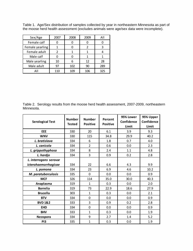
Table 1. Age/Sex distribution of samples collected by year in northeastern Minnesota as part of the moose herd health assessment (excludes animals were age/sex data were incomplete).
Sex/Age 2007 2008 2009 All
Female calf 0 0 0 0Female yearling 1 0 2 3
Female adult 2 1 1 4Male calf 0 0 1 1
Male yearling 10 6 12 28Male adult 97 102 90 289
All 110 109 106 325
Table 2. Serology results from the moose herd health assessment, 2007-2009, northeastern Minnesota.
Serological TestNumber Tested
Number Positive
Percent Positive
95% Lower Confidence
Limit
95% Upper Confidence
Limit EEE 330 20 6.1 3.9 9.3
WNV 330 115 34.8 29.9 40.2L. bratislava 334 6 1.8 0.7 4.0L. canicola 334 2 0.6 0.0 2.3
L. grippothyphosa 334 8 2.4 1.1 4.8L. hardjo 334 3 0.9 0.2 2.8
L. interrogans serovar icterohaemorrhagicae 334 22 6.6 4.3 9.9
L. pomona 334 23 6.9 4.6 10.2M. paratuberculosis 335 0 0.0 0.0 0.9
MCF 326 114 35.0 30.0 40.3Anaplasma 319 1 0.3 0.0 2.0
Borrelia 319 73 22.9 18.6 27.9Brucella 303 1 0.3 0.0 2.1
BTV 334 0 0.0 0.0 0.9BVD 1&2 333 3 0.9 0.2 2.8
EHD 334 0 0.0 0.0 0.9BHV 333 1 0.3 0.0 1.9
Neospora 334 9 2.7 1.4 5.2PI3 335 1 0.3 0.0 1.9
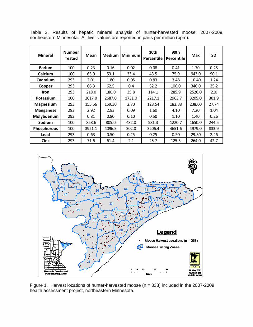
Table 3. Results of hepatic mineral analysis of hunter-harvested moose, 2007-2009, northeastern Minnesota. All liver values are reported in parts per million (ppm).
MineralNumber Tested
Mean Medium Minimum10th
Percentile90th
PercentileMax SD
Barium 100 0.23 0.16 0.02 0.08 0.41 1.70 0.25Calcium 100 65.9 53.1 33.4 43.5 75.9 943.0 90.1
Cadmium 293 2.01 1.80 0.05 0.83 3.48 10.40 1.24Copper 293 66.3 62.5 0.4 32.2 106.0 346.0 35.2
Iron 293 218.0 180.0 35.8 114.1 285.9 2526.0 210Potassium 100 2617.0 2687.0 1731.0 2217.1 2963.7 3205.0 301.9
Magnesium 293 155.56 159.30 2.70 128.54 182.88 238.60 27.74Manganese 293 2.92 2.93 0.09 1.60 4.10 7.20 1.04
Molybdenum 293 0.81 0.80 0.10 0.50 1.10 1.40 0.26Sodium 100 858.6 805.0 482.0 581.3 1220.7 1650.0 244.5
Phosphorous 100 3921.1 4096.5 302.0 3206.4 4651.6 4979.0 833.9Lead 293 0.63 0.50 0.25 0.25 0.50 29.30 2.26Zinc 293 71.6 61.4 2.1 25.7 125.3 264.0 42.7
Figure 1. Harvest locations of hunter-harvested moose (n = 338) included in the 2007-2009 health assessment project, northeastern Minnesota.

Figure 2. County of harvest location for hunter-harvested moose (n = 338) included in the 2007-2009 health assessment project, northeastern Minnesota.
Figure 3. Age distribution of hunter-harvested moose (n = 338) included in the 2007-2009 health assessment project, northeastern Minnesota.

Figure 4. Harvest locations of moose (n = 20) that tested positive for Eastern Equine Encephalitis from 2007-2009, northeastern Minnesota.
Figure 5. Harvest locations of moose (n = 115) that tested positive for West Nile Virus from 2007-2009, northeastern Minnesota.

Figure 6. Harvest locations of moose (n = 114) that tested positive for Malignant Catarrhal Fever from 2007-2009, northeastern Minnesota.
Figure 7. Harvest locations for moose that tested positive for Borrelia from 2007- 2009, northeastern Minnesota.

MANAGING BOVINE TUBERCULOSIS IN WHITE-TAILED DEER IN NORTHWESTERN MINNESOTA: A 2009 PROGRESS REPORT Michelle Carstensen1, Erika Butler, Erik Hildebrand, and Lou Cornicelli SUMMARY OF FINDINGS
Bovine tuberculosis (TB), first detected in Northwestern Minnesota in 2005, has since been found in 12 cattle operations and 27 free-ranging white-tailed deer (Odocoileus virginianus). Both deer and cattle have the same strain of bovine TB, which has been identified as one that is consistent with the disease found in cattle in the southwestern United States and Mexico. The Board of Animal Health (BAH) has been leading efforts to eradicate the disease in Minnesota’s cattle, which have included the depopulation of all infected herds, a buy-out program that removed 6,200 cattle from the affected area, and mandatory fencing of stored feeds on remaining farms. In response to the disease being detected in cattle, the Minnesota Department of Natural Resources (MNDNR) began surveillance efforts in free-ranging white-tailed deer within a 15-mile radius of the infected farms in fall 2005. To date, 27 deer have been found infected with bovine TB, and nearly all (n = 26) infected deer were sampled within a 164 mi2 area, called the Bovine TB Core, which is centered in Skime, Minnesota, and encompasses 8 of the previously infected cattle farms. The 27th case of bovine TB in deer was detected in November 2009, and was located 2.2 miles west of the original Core boundary. In total, 1,488 hunter-harvested deer were tested for bovine TB in northwest Minnesota during fall 2009, with only 1 positive case detected (apparent prevalence <0.07%). An aerial survey estimated the population of the Core to be 422 ± 126 deer in January 2010, and also detected a significant concentration of deer (>360 deer) in a 23mi2 area to the west of the Core boundary and within a 2mi radius of the most recent TB-positive deer. To further reduce deer numbers in the Core and this western expansion area, MNDNR conducted targeted removal operations using ground sharpshooting from February-April, 2010; 450 additional deer were removed. None of these deer had any clinical evidence of bovine TB infection at the time of sampling, yet final test results are pending. Further, a recreational feeding ban, instituted in November 2006 in a 4,000mi2 region in northwestern MN to help reduce the risk of deer to deer transmission of the disease, remains in effect. Under an agreement among the United States Department of Agriculture (USDA), BAH, and MNDNR, hunter-harvested deer surveillance will continue for at least the next 5 years to monitor infection in the local deer population, and any further aggressive management actions (e.g., sharpshooting deer in key locations) will be dependent on future surveillance results. INTRODUCTION
Bovine tuberculosis is an infectious disease that is caused by the bacterium
Mycobacterium bovis (M. bovis). Bovine TB primarily affects cattle; however, other mammals may become infected. Bovine TB was first discovered in 5 cattle operations in northwestern Minnesota in 2005. Since that time, 2 additional herds were found infected in 2006, 4 more in 2007, and 1 in 2008; resulting in further reduction of the state’s Bovine TB accreditation to Modified Accredited in early 2008. By fall 2008, Minnesota was granted a split-state status for TB accreditation that maintained only a small area (2,670mi2) in northwestern Minnesota as “Modified Accredited,” allowing the remainder of the state to advance to “Modified Accredited Advanced.” To date, 27 wild deer have been found infected with the disease in northwestern MN. Although Bovine TB was once relatively common in U.S cattle, it has historically been a very rare disease in wild deer. Prior to 1994, only 8 wild white-tailed and mule deer (O. hemionus) had been reported with Bovine TB in North America. In 1995, Bovine TB was detected in wild deer in Michigan. Though deer in Michigan do serve as a reservoir of Bovine ________________________ 1 Corresponding author e-mail: [email protected]
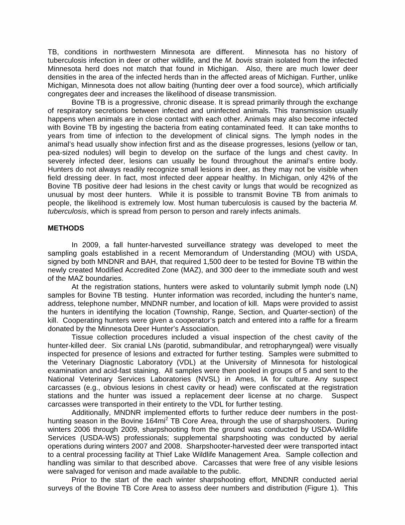
TB, conditions in northwestern Minnesota are different. Minnesota has no history of tuberculosis infection in deer or other wildlife, and the M. bovis strain isolated from the infected Minnesota herd does not match that found in Michigan. Also, there are much lower deer densities in the area of the infected herds than in the affected areas of Michigan. Further, unlike Michigan, Minnesota does not allow baiting (hunting deer over a food source), which artificially congregates deer and increases the likelihood of disease transmission.
Bovine TB is a progressive, chronic disease. It is spread primarily through the exchange of respiratory secretions between infected and uninfected animals. This transmission usually happens when animals are in close contact with each other. Animals may also become infected with Bovine TB by ingesting the bacteria from eating contaminated feed. It can take months to years from time of infection to the development of clinical signs. The lymph nodes in the animal’s head usually show infection first and as the disease progresses, lesions (yellow or tan, pea-sized nodules) will begin to develop on the surface of the lungs and chest cavity. In severely infected deer, lesions can usually be found throughout the animal’s entire body. Hunters do not always readily recognize small lesions in deer, as they may not be visible when field dressing deer. In fact, most infected deer appear healthy. In Michigan, only 42% of the Bovine TB positive deer had lesions in the chest cavity or lungs that would be recognized as unusual by most deer hunters. While it is possible to transmit Bovine TB from animals to people, the likelihood is extremely low. Most human tuberculosis is caused by the bacteria M. tuberculosis, which is spread from person to person and rarely infects animals. METHODS
In 2009, a fall hunter-harvested surveillance strategy was developed to meet the
sampling goals established in a recent Memorandum of Understanding (MOU) with USDA, signed by both MNDNR and BAH, that required 1,500 deer to be tested for Bovine TB within the newly created Modified Accredited Zone (MAZ), and 300 deer to the immediate south and west of the MAZ boundaries.
At the registration stations, hunters were asked to voluntarily submit lymph node (LN) samples for Bovine TB testing. Hunter information was recorded, including the hunter’s name, address, telephone number, MNDNR number, and location of kill. Maps were provided to assist the hunters in identifying the location (Township, Range, Section, and Quarter-section) of the kill. Cooperating hunters were given a cooperator’s patch and entered into a raffle for a firearm donated by the Minnesota Deer Hunter’s Association.
Tissue collection procedures included a visual inspection of the chest cavity of the hunter-killed deer. Six cranial LNs (parotid, submandibular, and retropharyngeal) were visually inspected for presence of lesions and extracted for further testing. Samples were submitted to the Veterinary Diagnostic Laboratory (VDL) at the University of Minnesota for histological examination and acid-fast staining. All samples were then pooled in groups of 5 and sent to the National Veterinary Services Laboratories (NVSL) in Ames, IA for culture. Any suspect carcasses (e.g., obvious lesions in chest cavity or head) were confiscated at the registration stations and the hunter was issued a replacement deer license at no charge. Suspect carcasses were transported in their entirety to the VDL for further testing.
Additionally, MNDNR implemented efforts to further reduce deer numbers in the post-hunting season in the Bovine 164mi2 TB Core Area, through the use of sharpshooters. During winters 2006 through 2009, sharpshooting from the ground was conducted by USDA-Wildlife Services (USDA-WS) professionals; supplemental sharpshooting was conducted by aerial operations during winters 2007 and 2008. Sharpshooter-harvested deer were transported intact to a central processing facility at Thief Lake Wildlife Management Area. Sample collection and handling was similar to that described above. Carcasses that were free of any visible lesions were salvaged for venison and made available to the public.
Prior to the start of the each winter sharpshooting effort, MNDNR conducted aerial surveys of the Bovine TB Core Area to assess deer numbers and distribution (Figure 1). This

information was used to guide sharpshooting activities and estimate the percentage of deer removed from the area.
RESULTS AND DISCUSSION
In fall 2009, we collected 1,488 samples from hunter-harvested deer; 947 samples from within the Modified Accredited Zone and 541 samples outside the zone (Figure 2). An enlarged and abscessed retropharyngeal lymph node was observed from 1 deer (3.5-yr-old male) while being sampled at a registration station; bovine TB was later confirmed by NVSL. This TB-positive male was harvested 2.2 miles west of the Core boundary, and marked the first incidence of the disease being detected in a new area. However, we do not believe this signifies movement of the disease, as this buck may have had a homerange that spanned the boundaries of the Core. Further, all the past TB-positive deer (n = 26) would have been born on or prior to 2005, when the disease was first detected in cattle, lending itself to our “Alive in ’05” theory of the spillover event. This newest case was born in 2006 and falls outside our spillover theory; however, additional cattle farms were detected TB-positive in 2006, 2007, and 2008, which could have been the exposure source for this individual deer or transmission may have occurred from another TB-infected deer.
Testing of remaining lymph node samples at NVSL has confirmed that there were no additional positive cases detected during the fall 2009 surveillance, resulting in an apparent prevalence rate of <0.07%. However, the fall sampling effort fell 553 samples (37%) short of its collection goal of 1,500 samples inside the MAZ; thus additional deer removal efforts in winter 2009–10 increased the sampling total by 450 deer, or 9% shy of the goal. Apparent prevalence of Bovine TB in the local deer population, sampled throughout a 1,730 to 2,670mi2 Surveillance Zone, indicates a significant decreasing trend from 2006–2009 (Table 1, Figure 3).
To supplement the number of samples collected through fall hunter-harvested surveillance and to further reduce deer density in the area where TB-positive deer had been confirmed, MNDNR contracted with USDA-Wildlife Services for ground sharpshooting in the Bovine TB Core Area, and a 23mi2 western expansion area, during February–April 2010. In total, 450 deer were removed from the TB Core Area, included approximately 150 from the western expansion area (Figure 4). No obvious clinical signs of bovine TB were observed on any of the deer; however, final test results are pending. Disease prevalence in the TB Core Area has decreased dramatically from 2007 to 2010 (Table 1, Figure 3). Although disease prevalence estimates in the TB Core Area are biased due to the limited geographic distribution of TB-positive deer and the increased probability of detecting a positive individual, the decreasing trend is consistent with the large-scale surveillance of the local deer populations in the fall.
Aerial survey results from January 2010 estimated that the deer population in the Bovine TB Core Area was a minimum of 422 ± 126 deer (Figure 1). This was significantly less than the February 2009 population estimate of 664 ± 87 (Figure 5, Table 2). It is apparent that aggressive deer removal in the TB Core Area through liberalized hunting, disease management permits, landowner shooting permits, and targeted sharpshooting has been able to reduce the deer population in this 164mi2 area by approximately 55% since 2006 (Figure 5, Table 2). It is likely that that the TB Core Area is home to both migratory and resident deer, some of which may move out of the zone to spring-summer-fall or winter ranges during the year. It is further likely that deer from the surrounding area are immigrating into the TB Core Area as deer numbers are reduced and habitat availability increases. The lack of severe winter weather condition in recent years has also allowed for good overwinter survival, increased reproduction, and recruitment into the local deer population.
The proximity of the TB-infected deer to infected cattle herds, the strain type, and the fact that disease prevalence (<0.1%) is low, supports our theory that this disease spilled-over from cattle to wild deer in this area of the state. To date, we have sampled 8,144 deer in the northwest since 2005, and a total of 27 confirmed culture-positive deer (Figure 6). Further, the

lack of infected yearlings or fawns and limited geographic distribution of infected adults further supports that this disease is not being spread efficiently in the local deer population. In November 2006, a ban on recreational feeding of deer and elk was instituted over a 4,000mi2 area to help reduce the risk of disease transmission among deer and between deer and livestock (Figure 7). Enforcement officers continue to enforce this rule and compliance is thought to be very high within the Bovine TB Management Zone. Further, the Minnesota State Legislature passed an initiative in 2008 that allocated funds to buy-out cattle herds located in the Bovine TB Management Zone, spending $3 million to remove 6,200 cattle from 46 farms by January 2009; resulting in the discovery of the 12th infected cattle herd. The remaining cattle farms in the TB-endemic area (n = 27) were required to erect deer-exclusion fencing to protect stored forage and winter feeding areas, costing an additional $690,000 in state funds.
As part of the requirements to regain TB-Free accreditation, USDA has required BAH to test all cattle herds within the Modified Accredited Zone annually, with additional movement restrictions for farms located within the Bovine TB Management Zone. BAH has submitted an application for status upgrade to USDA, and a decision is expected by October 2010. The MNDNR is committed to assisting BAH in regaining Minnesota’s TB-Free status as soon as possible. To accomplish this, the MNDNR will continue to conduct fall surveillance annually until 5 consecutive years with no TB-positive deer can be achieved, which would indicate that the disease was either eradicated or present in undetectable levels in the local deer population. SURVEILLANCE COSTS
Conducting a disease surveillance effort that spans a large area and encompasses numerous deer registration stations requires a large, trained work force and a significant amount of expenditures to support the effort. The bovine TB surveillance effort in fall 2009 spanned the first 10 days of regular firearms season, and included both an early antlerless (October 2009) and special late (January 2010) hunt (special hunt surveillance relied on head drop boxes for sample submission). The number of stations staffed each weekend ranged from 10 to 23, and summed to 41 stations over the duration of the project. In total, 44 trained MNDNR staff, 15 USDA disease biologists, and 46 student workers were needed in the effort. Costs associated to the surveillance effort are listed in Table 3. ACKNOWLEDGMENTS
There is no way to complete a project of this scale without the assistance and leadership from St. Paul and regional staff, including Ed Boggess, Dennis Simon, Dave Schad, Paul Telander, John Williams, and Mike Carroll. For all the help with field collections, we’d like to thank area staff from Thief Lake, Red Lake, Norris Camp, and Thief River Falls, Erik Hildebrand (Wildlife Health Specialist), Margaret Dexter (Fish and Wildlife Specialist), as well as students and faculty from the University of Minnesota, College of Veterinary Medicine. Randy Prachar provided excellent leadership through the sharpshooting operations, and we appreciate his efforts to support our carcass examination crews. Special thanks to Mary Reiswig for coordinating the venison donation program. Also thanks to Bob Wright, Erik Hildebrand, and the enforcement pilots (Tom Pfingsten and John Heineman) for conducting a deer survey within the Bovine TB Management Zone, as well as identifying illegal deer feeding activities. Also thanks to John Giudice for analyzing the survey data. We had an excellent team of GIS support including Steve Benson, Julie Adams, Bob Wright, and Chris Scharenbroich. We also want to recognize the tremendous amount of work conducted by USDA-Wildlife Services staff, led by John Hart (Grand Rapids) and disease biologist Paul Wolf (St. Paul). USDA-WS also loaned us disease biologists to assist with sample collections, including Darren Bruning (WA), Carl Betsill (NC), Tim White (IL), Barb Bodenstein (WI), Tom Hutton (MO), Jason Kloft (KS), and Tony Musante (NJ).
REFERENCES Miller, R., J. B. Kaneene, S. D. Fitzgerald, and S.M. Schmitt. 2003. Evaluation of the influence
of supplemental feeding of white-tailed deer (Odocoileus virginianus) on the prevalence of Bovine tuberculosis in the Michigan wild deer population. Journal of Wildlife Diseases 39: 84-95.
Minnesota Bovine Tuberculosis Management Plan. 2006. Minnesota Department of Agriculture, Minnesota Department of Natural Resources, Minnesota Board of Animal Health, United States Department of Agriculture, Unpubl. Rept.
O’Brien, D. J., S. D. Fitzgerald, T. J. Lyon, K. L. Butler, J. S. Fierke, K. R. Clarke, S. M. Schmitt, T. M. Cooley, and D. E. Berry. 2001. Tuberculous lesions in free-ranging white-tailed deer in Michigan. Journal of Wildlife Diseases 37: 608-613.
O’Brien, D. J., S. M. Schmitt, J. S. Fierke, S. A. Hogle, S. R. Winterstein, T. M. Cooley, W. E. Moritz, K. L. Diegel, S. D. Fitzgerald, D. E. Berry, and J. B. Kaneene. 2002. Epidemiology of Mycobacterium bovis in free-ranging whitetailed deer, Michigan, USA, 1995-2000. Preventive Veterinary Medicine 54: 47-63.
Schmitt, S. M., S. D. Fitzgerald, T. M. Cooley, C. S. Bruning-Fann, L. Sullivan, D. Berry, T. Carlson, R. B. Minnis, J. B. Payeur, and J. Sikarskie. 1997. Bovine tuberculosis in free- ranging white-tailed deer from Michigan. Journal of Wildlife Diseases 33: 749-758.

Table 1. Number of deer sampled for bovine TB and testing results listed by sampling strategy, fall 2005 to spring 2010, northwestern Minnesota.
Sampling strategy 2005 2006 2007 2008 2009 2010 Totals Hunter-harvested (Oct-Jan) # TB-positive Apparent Prevalence
474 1
0.21%
942 5
0.53%
1,166 5
0.43%
1,246 0
0.0%
1,488 1
0.07%
n/a 5,316
Sharpshooting (Feb-May) # TB-positive Apparent Prevalence
0 0 488 6
1.23%
937 6
0.64%
738 2
0.27%
450* 2,613
Landowner/Tenant # TB-positive
0 90 1
0 125 0
0 0 215
Total Deer Tested 474 1,032 1,654 2,308
738 450 8,144
Total # TB-positive 1 6 11 6 3 27 *Final culture results from winter 2010 sampling are still pending at NVSL. Table 2. Population estimates of deer within the Bovine TB Core Area, 2007–2010, northwest Minnesota.
Expenditure Total cost MNDNR staff salary $176,300 MNDNR staff travel expenses 30,950 Veterinary student labor & travel expenses $42,815 Other student labor & travel expenses $16,500 Supplies $18,775 Fleet $23,750 Diagnostic fees $52,350 USDA contract for sharpshooting $210,000 Total $571,440
Table 3. Expenditure details for 2009 Minnesota bovine tuberculosis surveillance program.

Figure 1. Results of aerial white-tailed deer survey of the Bovine TB Core Area in January 2010, northwestern Minnesota.

Figure 2. Locations of hunter-harvested deer (n=1,246) sampled for Bovine tuberculosis (TB) during fall 2009 in northwestern Minnesota.

Figure 3. Prevalence of Bovine TB in hunter-harvested deer from 2005–2009 in the Bovine TB Surveillance Zone and disease prevalence from sharpshooter removed deer from 2007– 2009 in the Bovine TB Core Area, northwestern Minnesota.

Figure 4. Locations of deer removed (n=450) by USDA ground sharpshooters during February-April 2010, in northwestern Minnesota.

Figure 5. Population estimate of deer within the Bovine TB Core, winters 2007–2010, northwestern Minnesota.
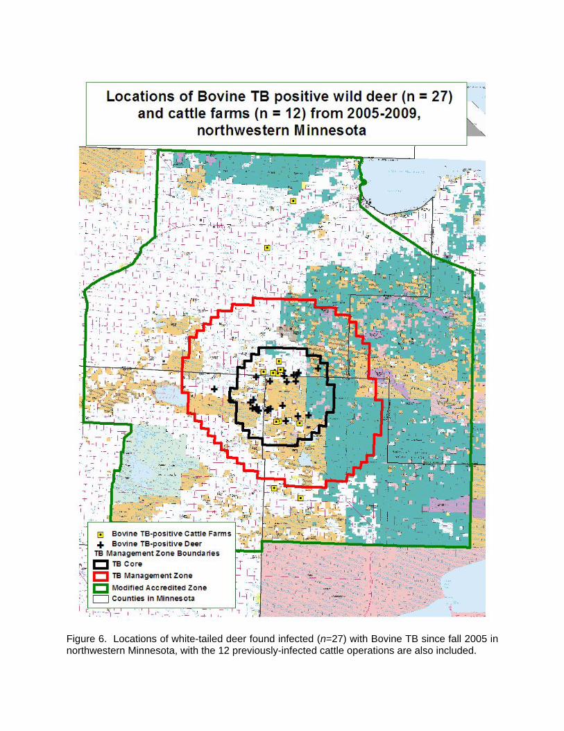
Figure 6. Locations of white-tailed deer found infected (n=27) with Bovine TB since fall 2005 in northwestern Minnesota, with the 12 previously-infected cattle operations are also included.
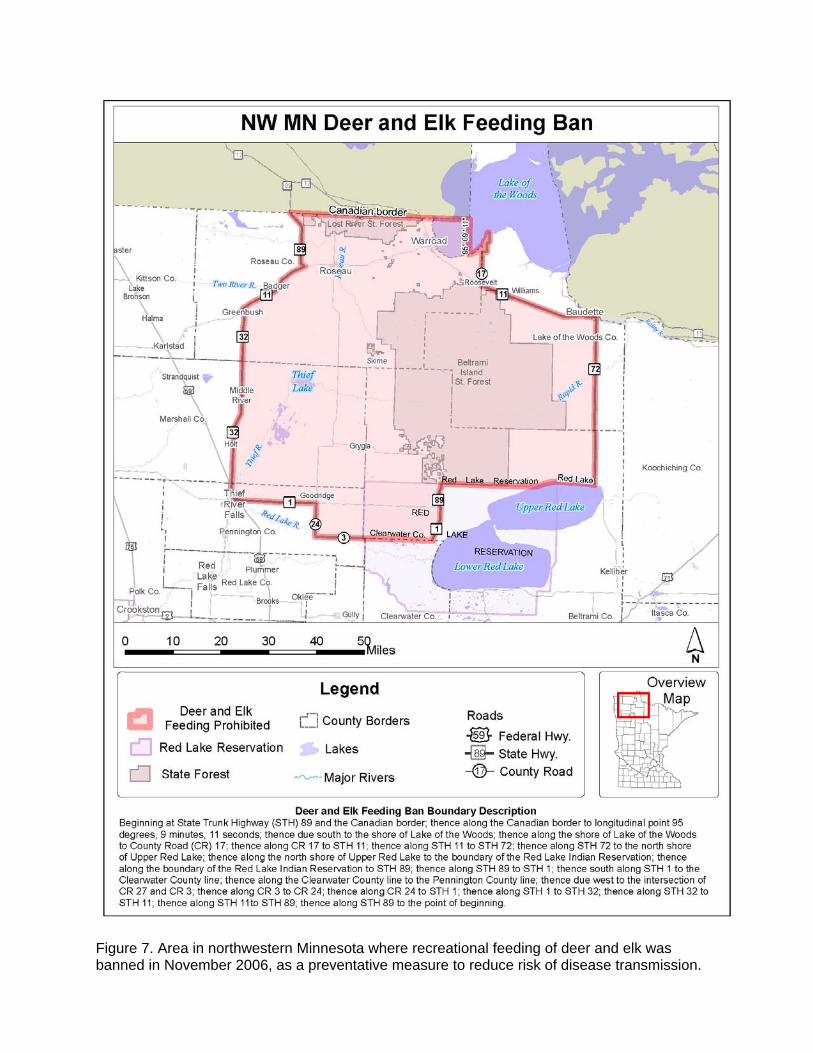
Figure 7. Area in northwestern Minnesota where recreational feeding of deer and elk was banned in November 2006, as a preventative measure to reduce risk of disease transmission.