sugar-responsive layer-by-layer film composed of
TRANSCRIPT

368 Vol. 66, No. 4
© 2018 The Pharmaceutical Society of Japan
Chem. Pharm. Bull. 66, 368–374 (2018)
Regular Article
Sugar-Responsive Layer-by-Layer Film Composed of Phenylboronic Acid-Appended Insulin and Poly(vinyl alcohol)
Chihiro Takei, Yui Ohno, Tomohiro Seki, Ryotaro Miki, Toshinobu Seki, and Yuya Egawa*Faculty of Pharmacy and Pharmaceutical Sciences, Josai University; 1–1 Keyakidai, Sakado, Saitama 350–0295, Japan.Received October 11, 2017; accepted January 19, 2018
Previous studies have shown that reversible chemical bond formation between phenylboronic acid (PBA) and 1,3-diol can be utilized as the driving force for the preparation of layer-by-layer (LbL) films. The LbL films composed of a PBA-appended polymer and poly(vinyl alcohol) (PVA) disintegrated in the presence of sugar. This type of LbL films has been recognized as a promising approach for sugar-responsive drug re-lease systems, but an issue preventing the practical application of LbL films is combining them with insulin. In this report, we have proposed a solution for this issue by using PBA-appended insulin as a component of the LbL film. We prepared two kinds of PBA-appended insulin derivatives and confirmed that they retained their hypoglycemic activity. The LbL films composed of PBA-appended insulin and PVA were successfully prepared through reversible chemical bond formation between the boronic acid moiety and the 1,3-diol of PVA. The LbL film disintegrated upon treatment with sugars. Based on the results presented herein, we dis-cuss the suitability of the PBA moiety with respect to hypoglycemic activity, binding ability, and selectivity for D-glucose.
Key words layer-by-layer; phenylboronic acid; insulin; sugar response; smart material
The layer-by-layer (LbL) deposition technique has attracted considerable interest for the preparation of nano-sized multi-layer films on solid surfaces.1,2) The LbL deposition technique usually leverages the electrostatic interactions between oppo-sitely charged molecules. This type of LbL films is stable on the solid support unless the pH of the surrounding solution is changed, which is advantageous for the construction of thin stable films. However, the good stability of the films, which need to be disintegrated for drug release, causes an issue for drug delivery applications.
An LbL film that disintegrates upon exposure to specific stimuli is desirable for drug delivery systems, but requires a reversible driving force for LbL film.3) To meet this demand, the incorporation of phenylboronic acid (PBA) is a promising approach.4–10) PBA derivatives show binding ability for the 1,2-diol, and 1,3-diol of sugars, which can result in the forma-tion of a cyclic ester even in aqueous solution.11,12) The cyclic ester formation is a reversible reaction, which is suitable for stimuli-responsive LbL films. For example, Anzai’s group has prepared LbL films composed of a PBA-modified amine dendrimer and poly(vinyl alcohol) (PVA) through interaction between PBA and the 1,3-diol of PVA with great success.7–9) This LbL film disintegrated when the film was immersed in sugar solutions, which is driven by the displacement of PVA by sugar on the PBA moiety.
The success of sugar-responsive LbL is significant, but fur-ther challenges must be overcome for the application of LbL films for sugar-responsive insulin release. One of the most important issues is combining the LbL film and insulin. For example, Sato et al. reported sugar-responsive microcapsules composed of insulin-loaded CaCO3 covered by an LbL film synthesized by leveraging the binding between concanavalin A and glycogen.13,14) Although D-glucose (Glc) accelerated the release rate of insulin, the microcapsules slowly released insu-lin even in the absence of Glc, which was likely due to the po-
rosity of the LbL film with concanavalin A and glycogen. The concept of an insulin-loaded CaCO3 core may be useful with an LbL film using a PBA moiety, but further improvement is needed to retain insulin in the absence of sugar.
Yoshida et al. fabricated an LbL film composed of insulin and a polyanion by using electrostatic interaction as a driv-ing force.15) The LbL film was made in a pH 3.0 solution, in which insulin has positive charges, which drives the interac-tion with the polyanion. The LbL film was stable in the acidic solution, but disintegrated in a neutral solution because the isoelectronic point of insulin is 5.4.16) Thus, insulin has an overall negative charge at pH 7.4, which results in the loss of the electrostatic interaction between insulin and the polyanion. This approach was effective for retaining insulin in the film, and the pH response was successfully achieved. However, the response was observed only for changes in pH but not in the presence of sugar.
Zhang et al. demonstrated a sugar-responsive LbL film composed of PVA-appended insulin and PBA-appended poly(acrylamide), which capitalized on the interaction between PVA and PBA.17) The LbL released PVA-appended insulin even in the absence of sugar, which indicated that the film was unstable in the buffer. Although the report described a small response in the LbL film in the presence of sugar at pH 8.5, the report did not discuss the hypoglycemic activity of the PVA-appended insulin. The activity of the PVA-appended insulin was likely lost because the molecular weight (MW) of PVA used in the study was too large (MW 1.59×10⁵).
In this study, we prepared PBA-appended insulin for use in the fabrication of an LbL film with PVA (Chart 1). The basic components of the film were PBA, insulin, and PVA, which are the same as the LbL film produced in the study by Zhang et al. However, the arrangement of these components is quite different. We expected the PBA-appended insulin to retain its hypoglycemic activity because PBA is a low-molecular-weight
* To whom correspondence should be addressed. e-mail: [email protected]

Vol. 66, No. 4 (2018) 369Chem. Pharm. Bull.
compound. There are some reports regarding the chemical modification of insulin, in which the relationship between chemical modification and its hypoglycemic activity was eval-uated.18,19) We appended 4-carboxyphenylboronic acid to insu-lin through the formation of an amide bond, which is termed PBA-Ins. In addition, we attached 3-fluoro-4-carboxyphenyl-boronic acid to insulin (FPBA-Ins) because PBA derivatives containing electron-withdrawing groups have been shown to have a higher binding constant for polyol molecules.20) We investigated the hypoglycemic activity of PBA-Ins and FPBA-Ins, and used them for preparation of LbL films through com-bination with PVA. By comparing the results of PBA-Ins and FPBA-Ins, we discuss the suitability of the PBA moiety with respect to hypoglycemic activity, binding ability and selectiv-ity for Glc.
ExperimentalMaterial Insulin (human, recombinant), sodium dodecyl
sulfate (SDS), PVA (average degree of polymerization 500, average MW 22000), acetic acid, Glc, D-fructose (Fru), and Alizarin Red S (ARS) were purchased from Wako Pure Chemical Industries, Ltd. (Osaka, Japan). Tributylamine (TBA), isobutyl chloroformate, PBA, and 3-fluorophenylbo-ronic acid were purchased from Tokyo Chemical Industry Co., Ltd. (Tokyo, Japan). 4-Carboxyphenylboronic acid, 2,4,6-trinitrobenzenesulfonic acid (TNBS, 5% in water), 4-((2-hydroxyethyl)-1-piperazineethanesulfonic acid) (HEPES) and N-cyclohexyl-2-aminoethanesulfonic acid (CHES) were purchased from Sigma-Aldrich Japan (Tokyo, Japan). 4-Car-boxy-3-fluorophenylboronic acid was purchased from Oak-wood Products Inc. (South Carolina, U.S.A.). All other chemi-cals were of reagent grade and were used as received.
Preparation of PBA-Ins and FPBA-Ins Insulin (300 mg, 51.7 µmol) was dissolved in a mixture of TBA (300 µL) and dimethyl sulfoxide (super dehydrated, 60 mL). Separately, 4-carboxyphenylboronic acid (43 mg, 259 µmol) was dissolved in a mixture of TBA (61.2 µL) and N,N-dimethylformamide (super dehydrated, 12 mL), and isobutyl chloroformate (33 µL, 252.6 µmol) was dissolved into the solution. This solution was then added to the insulin solution. The resulting mixed solution was stirred at room temperature under a nitrogen atmosphere. After 3 h, water (10 mL) was added to stop the reaction, and the solution was dialyzed against water using a dialysis tube (MWCO 3500). The resulting solution was lyophilized, and PBA-Ins was obtained. The same procedure
was followed for the preparation of FPBA-Ins with 4-carboxy-3-fluorophenylboronic acid.
Evaluation of the Number of Modified PBA For the preparation of the SDS solution, SDS was dissolved in a phosphate buffer (pH 9.0, 10 mM) for a final concentration of 0.15%. As a sample solution, the insulin derivative (PBA-Ins or FPBA-Ins) was dissolved to a final concentration of 172 µM in the SDS solution. Separately, TNBS was added to a final concentration of 0.03% with the SDS solution. The solutions were mixed in the following volumes, SDS solution (1.0 mL), sample solution (2.0 mL), and TNBS solution (1.0 mL). The mixture was kept at 50°C for 1 h to initiate the reaction. Then, HCl aqueous solution (0.20 M, 3.8 mL) was added to the vial, and was kept at room temperature for 30 min. To obtain the value of Asample, the absorbance of the solution was measured at the absorption maximum wavelength (JASCO V-560, ETC-505T, Tokyo, Japan). As a blank solution, the SDS (3.0 mL) and TNBS solutions (1.0 mL) were mixed and treated in the same way, and the ATNBS was obtained. Using unmodified insulin solution (172 µM) instead of the sample solution, Ainsulin was also obtained. A number of modifications of PBA or FPBA (n) were calculated using the following equation consid-ering that insulin has three amino groups that could possibly react.
sample TNBS
insulin TNBS1 3
− − − = ×
A AnA A
Preparation of LbL Films LbL films were prepared on the surface of a platinum film-coated quartz crystal micro-balance (QCM) resonator (9 MHz). The QCM resonator was mounted in a flow-through cell and monitored with a QCA922 instrument (SEIKO, EG&G, Tokyo, Japan). First, the QCM resonator was immersed in a pH 7.4 buffer (10 mM HEPES, containing 150 mM NaCl) for 5 min. Then, it was immersed in a 0.10 mg/mL PVA solution (10 mM HEPES buffer contain-ing 150 mM NaCl, pH 7.4) for 15 min and rinsed with HEPES buffer for 5 min. After the deposition of PVA, the QCM reso-nator was immersed in a 0.10 mg/mL PBA-Ins or FPBA-Ins solution (10 mM HEPES buffer containing 150 mM NaCl, pH 7.4) for 15 min and rinsed with HEPES buffer for 5 min. The LbL films were fabricated by repeating five cycles, which was shown in (PVA/PBA-Ins)5 film. A pH 5.0 buffer (10 mM acetic acid buffer, containing 150 mM NaCl) and a pH 9.0 buffer (10 mM CHES, containing 150 mM NaCl) were used to evaluate the effect of pH on the preparation of LbL films.
Chart 1. Schematic Illustration of the LbL Film Composed of PVA and PBA-Ins, and Its Sugar Induced Disintegration
370 Vol. 66, No. 4 (2018)Chem. Pharm. Bull.
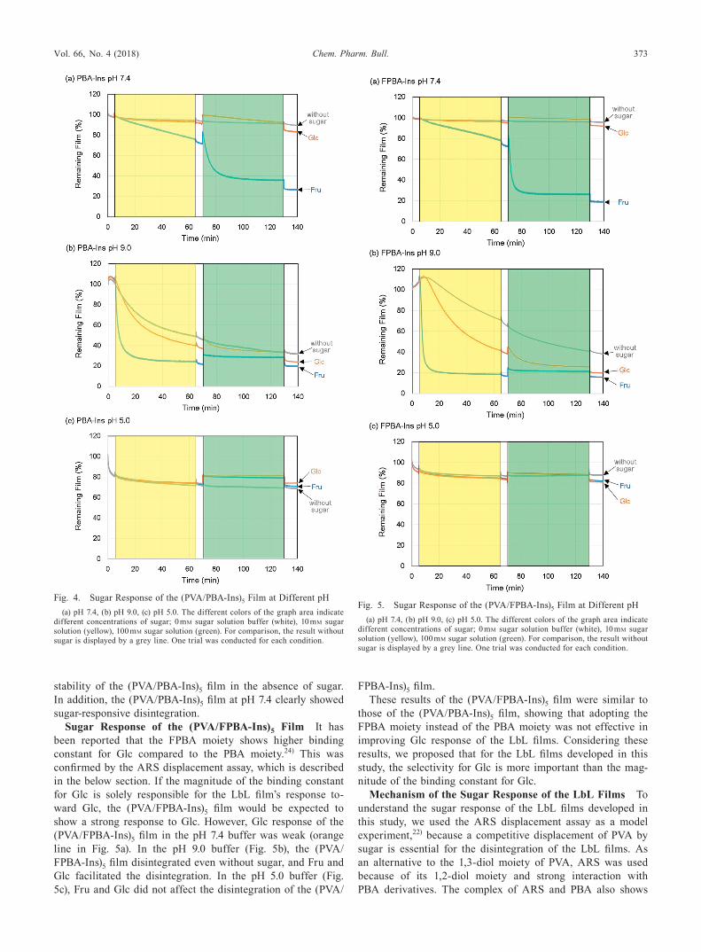
Sugar Response of the LbL Films The LbL films were prepared on the surface of QCM resonator at pH 7.4 with the above-mentioned method. Then, the QCM resonator was immersed in a 10 mM Glc solution (10 mM HEPES buffer con-taining 150 mM NaCl, pH 7.4) for 1 h. After rinsing with the pH 7.4 buffer (10 mM HEPES buffer containing 150 mM NaCl) for 5 min, the QCM resonator was immersed in a 100 mM Glc solution for 1 h. Finally, the films were rinsed in the pH 7.4 buffer. This procedure was also performed using Fru in place of Glc.
To investigate the effect of pH on the sugar response, the LbL films prepared in pH 7.4 buffer were immersed in a pH 9.0 buffer (10 mM CHES, containing 150 mM NaCl), and sugar (Glc or Fru) was added to the pH 9.0 buffer. Instead of the pH 9.0 buffer, a pH 5.0 buffer (10 mM acetic acid buffer, contain-ing 150 mM NaCl) was also used.
Measurement of Binding Constants The binding con-stants of the PBA derivative and ARS were calculated with a previously reported method.21) Binding constants of the PBA derivatives to Glc were evaluated with fluorescence measure-ments using ARS.20,22) A buffer solution (10 mM HEPES, pH 7.4) containing ARS (0.10 mM) and PBA (1.0 mM) was pre-pared. By changing the concentration of Glc, the fluorescent spectra (λex=476 nm, RF-5300PC, Shimadzu, Kyoto, Japan) were measured, and the fluorescence intensities at 568 nm were used to calculate the binding constants.
HPLC The lyophilized insulin derivatives were investi-gated with HPLC on a 5 µm, 150×4.6 mm i.d. octadecylsilyl column (HPLC Packed Column CAPCELL PAK C18, Shi-seido Fine Chemicals, Tokyo, Japan) by gradient elution at a flow rate of 1.0 mL/min. Mobile phase A was composed of acetonitrile–water–trifluoroacetic acid=300 : 700 : 1 (v/v/v), and mobile phase B was composed of acetonitrile–water–trifluo-roacetic acid=950 : 50 : 1 (v/v/v). HPLC (LC-20AT, SPD-20A, Shimadzu) was performed in linear gradient of 0 to 33.3% mobile phase B over 20 min. The effluent was monitored by absorption spectroscopy at 280 nm.
In Vivo Study Animal studies were performed accord-ing to the guidelines for animal use approved by the Life Science Research Center, Josai University. Male Wistar rats (220–270 g) were anesthetized with inhaled isoflurane, and their jugular vein was cannulated. After 4–7 d of recovery after surgery, the rats fasted for about 20 h before the experi-ments, and the insulin derivative was administered from the cannulated jugular vein. At a predetermined time, blood sam-ples were collected from the cannula, and the whole blood was centrifuged at 2000×g at 4°C for 2 min. The plasma Glc con-
centration was then determined by a glucose oxidase method (Glucose CII Test Wako Kit, Wako Pure Chemical Industries, Ltd.). The statistical significance of the results was analyzed using Dunnett’s test for multiple comparisons and Student’s t-test for comparison of two groups.
Results and DiscussionPreparation of PBA-Ins and FPBA-Ins The PBA moiety
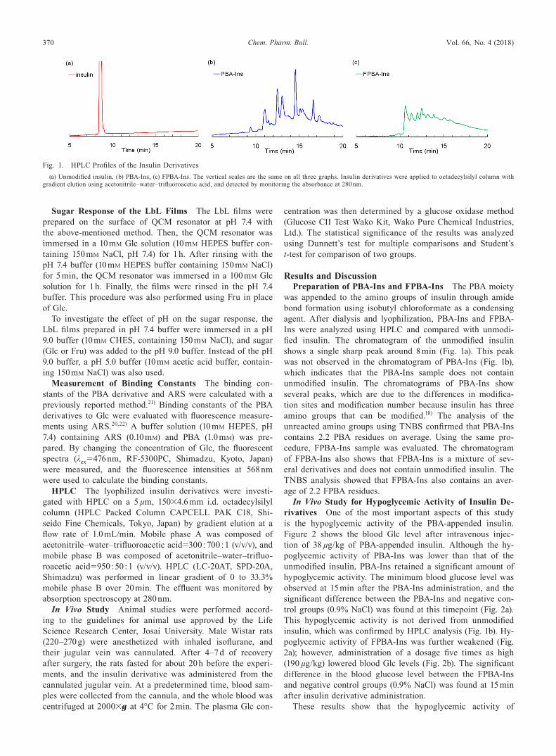
was appended to the amino groups of insulin through amide bond formation using isobutyl chloroformate as a condensing agent. After dialysis and lyophilization, PBA-Ins and FPBA-Ins were analyzed using HPLC and compared with unmodi-fied insulin. The chromatogram of the unmodified insulin shows a single sharp peak around 8 min (Fig. 1a). This peak was not observed in the chromatogram of PBA-Ins (Fig. 1b), which indicates that the PBA-Ins sample does not contain unmodified insulin. The chromatograms of PBA-Ins show several peaks, which are due to the differences in modifica-tion sites and modification number because insulin has three amino groups that can be modified.18) The analysis of the unreacted amino groups using TNBS confirmed that PBA-Ins contains 2.2 PBA residues on average. Using the same pro-cedure, FPBA-Ins sample was evaluated. The chromatogram of FPBA-Ins also shows that FPBA-Ins is a mixture of sev-eral derivatives and does not contain unmodified insulin. The TNBS analysis showed that FPBA-Ins also contains an aver-age of 2.2 FPBA residues.
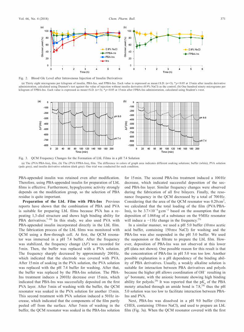
In Vivo Study for Hypoglycemic Activity of Insulin De-rivatives One of the most important aspects of this study is the hypoglycemic activity of the PBA-appended insulin. Figure 2 shows the blood Glc level after intravenous injec-tion of 38 µg/kg of PBA-appended insulin. Although the hy-poglycemic activity of PBA-Ins was lower than that of the unmodified insulin, PBA-Ins retained a significant amount of hypoglycemic activity. The minimum blood glucose level was observed at 15 min after the PBA-Ins administration, and the significant difference between the PBA-Ins and negative con-trol groups (0.9% NaCl) was found at this timepoint (Fig. 2a). This hypoglycemic activity is not derived from unmodified insulin, which was confirmed by HPLC analysis (Fig. 1b). Hy-poglycemic activity of FPBA-Ins was further weakened (Fig. 2a); however, administration of a dosage five times as high (190 µg/kg) lowered blood Glc levels (Fig. 2b). The significant difference in the blood glucose level between the FPBA-Ins and negative control groups (0.9% NaCl) was found at 15 min after insulin derivative administration.
These results show that the hypoglycemic activity of
Fig. 1. HPLC Profiles of the Insulin Derivatives(a) Unmodified insulin, (b) PBA-Ins, (c) FPBA-Ins. The vertical scales are the same on all three graphs. Insulin derivatives were applied to octadecylsilyl column with
gradient elution using acetonitrile–water–trifluoroacetic acid, and detected by monitoring the absorbance at 280 nm.
Vol. 66, No. 4 (2018) 371Chem. Pharm. Bull.
PBA-appended insulin was retained even after modification. Therefore, using PBA-appended insulin for preparation of LbL films is effective. Furthermore, hypoglycemic activity strongly depends on the modification group, so the selection of PBA residue is quite important.
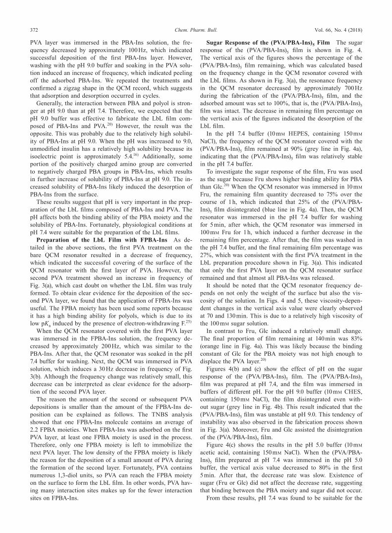
Preparation of the LbL Film with PBA-Ins Previous reports have shown that the combination of PBA and PVA is suitable for preparing LbL films because PVA has a re-peating 1,3-diol structure and shows high binding ability for PBA derivatives.7–10) In this study, we also used PVA with PBA-appended insulin incorporated directly in the LbL film. The fabrication process of the LbL films was monitored with QCM using a flow-through cell. At first, the QCM resona-tor was immersed in a pH 7.4 buffer. After the frequency was stabilized, the frequency change (ΔF) was recorded for 5 min. Then, the buffer was replaced with a PVA solution. The frequency sharply decreased by approximately 200 Hz, which indicated that the electrode was covered with PVA. After 15 min of soaking in the PVA solution, the PVA solution was replaced with the pH 7.4 buffer for washing. After that, the buffer was replaced by the PBA-Ins solution. The PBA-Ins treatment induces a 200 Hz decrease over 15 min, which indicated that PBA-Ins was successfully deposited on the first PVA layer. After 5 min of washing with the buffer, the QCM resonator was soaked in the PVA solution for another 15 min. This second treatment with PVA solution induced a 50 Hz in-crease, which indicated that the components of the film partly peeled off from the surface. After 5 min of washing in the buffer, the QCM resonator was soaked in the PBA-Ins solution
for 15 min. The second PBA-Ins treatment induced a 100 Hz decrease, which indicated successful deposition of the sec-ond PBA-Ins layer. Similar frequency changes were observed during the fabrication of all five bilayers. Finally, the reso-nance frequency in the QCM decreased by a total of 700 Hz. Considering that the area of the QCM resonator was 0.20 cm2, we calculated that the total loading of the film (PVA/PBA-Ins)5 to be 3.7×10⁻⁶ g cm−2 based on the assumption that the deposition of 1.068 ng of a substance on the 9 MHz resonator will induce a −1 Hz change in the frequency.23)
In a similar manner, we used a pH 5.0 buffer (10 mM acetic acid buffer, containing 150 mM NaCl) for washing and the PBA-Ins was also suspended in the pH 5.0 buffer. We used the suspension or the filtrate to prepare the LbL film. How-ever, deposition of PBA-Ins was not observed at this lower pH (data not shown). One possible reason for this result is that the concentration of PBA-Ins in pH 5.0 was too low. Another possible explanation is a pH dependency of the binding abil-ity of PBA derivatives. Usually, a weakly alkaline solution is suitable for interaction between PBA derivatives and polyols because the higher pH allows coordination of OH− resulting in sp3 boronate, with the anionic boronate showing high binding ability for polyols.20) It was reported that the pKa of the PBA moiety attached through an amide bond is 7.8,24) thus the pH 5.0 solution was too low to facilitate interaction between PBA-Ins and PVA.
Next, PBA-Ins was dissolved in a pH 9.0 buffer (10 mM CHES, containing 150 mM NaCl), and used to prepare an LbL film (Fig. 3a). When the QCM resonator covered with the first
Fig. 2. Blood Glc Level after Intravenous Injection of Insulin Derivatives(a) Thirty eight micrograms per kilogram of insulin, PBA-Ins, and FPBA-Ins. Each value is expressed as mean±S.D. (n=3); * p<0.05 at 15 min after insulin derivative
administration, calculated using Dunnett’s test against the value of injection without insulin derivative (0.9% NaCl) as the control; (b) One hundred ninety micrograms per kilogram of FPBA-Ins. Each value is expressed as mean±S.D. (n=3); * p<0.05 at 15 min after FPBA-Ins administration, calculated using Student’s t-test.
Fig. 3. QCM Frequency Changes for the Formation of LbL Films in a pH 7.4 Solution(a) The (PVA/PBA-Ins)5 film, (b) The (PVA/FPBA-Ins)5 film. The difference in colors of graph area indicates different soaking solutions; buffer (white), PVA solution
(pale grey), and insulin derivative solution (dark grey). One trial was conducted for each condition.
372 Vol. 66, No. 4 (2018)Chem. Pharm. Bull.
PVA layer was immersed in the PBA-Ins solution, the fre-quency decreased by approximately 100 Hz, which indicated successful deposition of the first PBA-Ins layer. However, washing with the pH 9.0 buffer and soaking in the PVA solu-tion induced an increase of frequency, which indicated peeling off the adsorbed PBA-Ins. We repeated the treatments and confirmed a zigzag shape in the QCM record, which suggests that adsorption and desorption occurred in cycles.
Generally, the interaction between PBA and polyol is stron-ger at pH 9.0 than at pH 7.4. Therefore, we expected that the pH 9.0 buffer was effective to fabricate the LbL film com-posed of PBA-Ins and PVA.20) However, the result was the opposite. This was probably due to the relatively high solubil-ity of PBA-Ins at pH 9.0. When the pH was increased to 9.0, unmodified insulin has a relatively high solubility because its isoelectric point is approximately 5.4.16) Additionally, some portion of the positively charged amino group are converted to negatively charged PBA groups in PBA-Ins, which results in further increase of solubility of PBA-Ins at pH 9.0. The in-creased solubility of PBA-Ins likely induced the desorption of PBA-Ins from the surface.
These results suggest that pH is very important in the prep-aration of the LbL films composed of PBA-Ins and PVA. The pH affects both the binding ability of the PBA moiety and the solubility of PBA-Ins. Fortunately, physiological conditions at pH 7.4 were suitable for the preparation of the LbL films.
Preparation of the LbL Film with FPBA-Ins As de-tailed in the above sections, the first PVA treatment on the bare QCM resonator resulted in a decrease of frequency, which indicated the successful covering of the surface of the QCM resonator with the first layer of PVA. However, the second PVA treatment showed an increase in frequency of Fig. 3(a), which cast doubt on whether the LbL film was truly formed. To obtain clear evidence for the deposition of the sec-ond PVA layer, we found that the application of FPBA-Ins was useful. The FPBA moiety has been used some reports because it has a high binding ability for polyols, which is due to its low pKa induced by the presence of electron-withdrawing F.25)
When the QCM resonator covered with the first PVA layer was immersed in the FPBA-Ins solution, the frequency de-creased by approximately 200 Hz, which was similar to the PBA-Ins. After that, the QCM resonator was soaked in the pH 7.4 buffer for washing. Next, the QCM was immersed in PVA solution, which induces a 30 Hz decrease in frequency of Fig. 3(b). Although the frequency change was relatively small, this decrease can be interpreted as clear evidence for the adsorp-tion of the second PVA layer.
The reason the amount of the second or subsequent PVA depositions is smaller than the amount of the FPBA-Ins de-position can be explained as follows. The TNBS analysis showed that one FPBA-Ins molecule contains an average of 2.2 FPBA moieties. When FPBA-Ins was adsorbed on the first PVA layer, at least one FPBA moiety is used in the process. Therefore, only one FPBA moiety is left to immobilize the next PVA layer. The low density of the FPBA moiety is likely the reason for the deposition of a small amount of PVA during the formation of the second layer. Fortunately, PVA contains numerous 1,3-diol units, so PVA can reach the FPBA moiety on the surface to form the LbL film. In other words, PVA hav-ing many interaction sites makes up for the fewer interaction sites on FPBA-Ins.
Sugar Response of the (PVA/PBA-Ins)5 Film The sugar response of the (PVA/PBA-Ins)5 film is shown in Fig. 4. The vertical axis of the figures shows the percentage of the (PVA/PBA-Ins)5 film remaining, which was calculated based on the frequency change in the QCM resonator covered with the LbL films. As shown in Fig. 3(a), the resonance frequency in the QCM resonator decreased by approximately 700 Hz during the fabrication of the (PVA/PBA-Ins)5 film, and the adsorbed amount was set to 100%, that is, the (PVA/PBA-Ins)5 film was intact. The decrease in remaining film percentage on the vertical axis of the figures indicated the desorption of the LbL film.
In the pH 7.4 buffer (10 mM HEPES, containing 150 mM NaCl), the frequency of the QCM resonator covered with the (PVA/PBA-Ins)5 film remained at 90% (grey line in Fig. 4a), indicating that the (PVA/PBA-Ins)5 film was relatively stable in the pH 7.4 buffer.
To investigate the sugar response of the film, Fru was used as the sugar because Fru shows higher binding ability for PBA than Glc.20) When the QCM resonator was immersed in 10 mM Fru, the remaining film quantity decreased to 75% over the course of 1 h, which indicated that 25% of the (PVA/PBA-Ins)5 film disintegrated (blue line in Fig. 4a). Then, the QCM resonator was immersed in the pH 7.4 buffer for washing for 5 min, after which, the QCM resonator was immersed in 100 mM Fru for 1 h, which induced a further decrease in the remaining film percentage. After that, the film was washed in the pH 7.4 buffer, and the final remaining film percentage was 27%, which was consistent with the first PVA treatment in the LbL preparation procedure shown in Fig. 3(a). This indicated that only the first PVA layer on the QCM resonator surface remained and that almost all PBA-Ins was released.
It should be noted that the QCM resonator frequency de-pends on not only the weight of the surface but also the vis-cosity of the solution. In Figs. 4 and 5, these viscosity-depen-dent changes in the vertical axis value were clearly observed at 70 and 130 min. This is due to a relatively high viscosity of the 100 mM sugar solution.
In contrast to Fru, Glc induced a relatively small change. The final proportion of film remaining at 140 min was 83% (orange line in Fig. 4a). This was likely because the binding constant of Glc for the PBA moiety was not high enough to displace the PVA layer.20)
Figures 4(b) and (c) show the effect of pH on the sugar response of the (PVA/PBA-Ins)5 film. The (PVA/PBA-Ins)5 film was prepared at pH 7.4, and the film was immersed in buffers of different pH. For the pH 9.0 buffer (10 mM CHES, containing 150 mM NaCl), the film disintegrated even with-out sugar (grey line in Fig. 4b). This result indicated that the (PVA/PBA-Ins)5 film was unstable at pH 9.0. This tendency of instability was also observed in the fabrication process shown in Fig. 3(a). Moreover, Fru and Glc assisted the disintegration of the (PVA/PBA-Ins)5 film.
Figure 4(c) shows the results in the pH 5.0 buffer (10 mM acetic acid, containing 150 mM NaCl). When the (PVA/PBA-Ins)5 film prepared at pH 7.4 was immersed in the pH 5.0 buffer, the vertical axis value decreased to 80% in the first 5 min. After that, the decrease rate was slow. Existence of sugar (Fru or Glc) did not affect the decrease rate, suggesting that binding between the PBA moiety and sugar did not occur.
From these results, pH 7.4 was found to be suitable for the
Vol. 66, No. 4 (2018) 373Chem. Pharm. Bull.
stability of the (PVA/PBA-Ins)5 film in the absence of sugar. In addition, the (PVA/PBA-Ins)5 film at pH 7.4 clearly showed sugar-responsive disintegration.
Sugar Response of the (PVA/FPBA-Ins)5 Film It has been reported that the FPBA moiety shows higher binding constant for Glc compared to the PBA moiety.24) This was confirmed by the ARS displacement assay, which is described in the below section. If the magnitude of the binding constant for Glc is solely responsible for the LbL film’s response to-ward Glc, the (PVA/FPBA-Ins)5 film would be expected to show a strong response to Glc. However, Glc response of the (PVA/FPBA-Ins)5 film in the pH 7.4 buffer was weak (orange line in Fig. 5a). In the pH 9.0 buffer (Fig. 5b), the (PVA/FPBA-Ins)5 film disintegrated even without sugar, and Fru and Glc facilitated the disintegration. In the pH 5.0 buffer (Fig. 5c), Fru and Glc did not affect the disintegration of the (PVA/
FPBA-Ins)5 film.These results of the (PVA/FPBA-Ins)5 film were similar to
those of the (PVA/PBA-Ins)5 film, showing that adopting the FPBA moiety instead of the PBA moiety was not effective in improving Glc response of the LbL films. Considering these results, we proposed that for the LbL films developed in this study, the selectivity for Glc is more important than the mag-nitude of the binding constant for Glc.
Mechanism of the Sugar Response of the LbL Films To understand the sugar response of the LbL films developed in this study, we used the ARS displacement assay as a model experiment,22) because a competitive displacement of PVA by sugar is essential for the disintegration of the LbL films. As an alternative to the 1,3-diol moiety of PVA, ARS was used because of its 1,2-diol moiety and strong interaction with PBA derivatives. The complex of ARS and PBA also shows
Fig. 4. Sugar Response of the (PVA/PBA-Ins)5 Film at Different pH(a) pH 7.4, (b) pH 9.0, (c) pH 5.0. The different colors of the graph area indicate
different concentrations of sugar; 0 mM sugar solution buffer (white), 10 mM sugar solution (yellow), 100 mM sugar solution (green). For comparison, the result without sugar is displayed by a grey line. One trial was conducted for each condition.
Fig. 5. Sugar Response of the (PVA/FPBA-Ins)5 Film at Different pH(a) pH 7.4, (b) pH 9.0, (c) pH 5.0. The different colors of the graph area indicate
different concentrations of sugar; 0 mM sugar solution buffer (white), 10 mM sugar solution (yellow), 100 mM sugar solution (green). For comparison, the result without sugar is displayed by a grey line. One trial was conducted for each condition.
374 Vol. 66, No. 4 (2018)Chem. Pharm. Bull.
high fluorescence intensity, which can be monitored in order to determine binding constant. When sugar is added to the solution, the complex disintegrates via competitive displace-ment of ARS, which results in a decrease of the fluorescence intensity. PBA and 3-fluorophenylboronic acid (FPBA) were used as a model of the modification residue of PBA-Ins and FPBA-Ins, respectively. When PBA was added to the solution of ARS, the fluorescence intensity increased and the binding constant between PBA and ARS (KPBA–ARS) was calculated to be 1.2×10³ M−1 according to a previously reported method.21) When Glc was added to the solution, the fluorescence in-tensity decreased and the binding constant between PBA and Glc (KPBA–Glc) was determined to be 3.2 M−1. The ratio of KPBA–ARS/KPBA–Glc was 3.8×10², which was similar to that of a previous report.22) In a similar manner, KFPBA–ARS and KFPBA–Glc were calculated to be 3.3×10³ and 8.1 M−1, respec-tively, and the ratio of KFPBA–ARS/KFPBA–Glc was determined to be 4.1×10². This result shows that there is little difference between PBA and FPBA in terms of Glc selectivity.
It should be noted that FPBA shows a higher binding con-stant for ARS; therefore, it is difficult to disintegrate by intro-duction of Glc even though FPBA has a relatively high bind-ing constant for Glc. We also recognized that FPBA-Ins shows a higher binding ability for PVA, which is the reason why it was difficult to disintegrate the (PVA/FPBA-Ins) film by Glc. Therefore, increase in the selectivity for Glc is quite important for Glc response of the LbL films developed in this study.
For future work, we propose that increasing the selectiv-ity not the magnitude of the binding constant for Glc, would improve the response of the LbL film toward Glc because displacement of PVA by Glc is essential for disintegration. There are some reports regarding Glc-selective sensors using this concept by incorporation of dimeric PBA derivatives.26–28) In addition, PBA-appended polymers show a relatively high selectivity for Glc compared to monomeric PBA derivatives.29) In these systems, two PBA moieties cooperatively bind one Glc molecule. Multiple arrangements of the PBA moiety may be effective in increasing the Glc response of the LbL films, but modification with large moieties tend to decrease hypogly-cemic activity.
ConclusionIn this study, PBA-Ins LbL films were prepared and their
hypoglycemia activity was confirmed by in vivo experiments. The (PVA/PBA-Ins)5 film was successfully prepared, and disintegrated in the presence of Fru; however, the response toward Glc was relatively low. Although introducing an electron-withdrawing group (F) was effective for increasing the binding constant toward Glc, the overall response of the (PVA/FPBA-Ins)5 film was similar to that of the (PVA/PBA-Ins)5 film. Furthermore, the hypoglycemic activity of FPBA-Ins decreased compared to that of PBA-Ins. For future work, optimization of the modification of the PBA moiety should be considered with respect to balance between Glc selectivity and
hypoglycemic activity.
Acknowledgment This work was supported by JSPS KAKENHI Grant Number JP16K08202.
Conflict of Interest The authors declare no conflict of interest.
References 1) Decher G., Eckle M., Schmitt J., Struth B., Curr. Opin. Colloid In-
terface Sci., 3, 32–39 (1998). 2) Sato K., Takahashi S., Anzai J., Anal. Sci., 28, 929–938 (2012). 3) Wohl B. M., Engbersen J. F. J., J. Control. Release, 158, 2–14 (2012). 4) De Geest B. G., Jonas A. M., Demeester J., De Smedt S. C., Lang-
muir, 22, 5070–5074 (2006). 5) Levy T., Déjugnat C., Sukhorukov G. B., Adv. Funct. Mater., 18,
1586–1594 (2008). 6) Hujaya S. D., Engbersen J. F. J., Paulusse J. M. J., Pharm. Res., 32,
3732–3745 (2015). 7) Watahiki R., Sato K., Suwa K., Niina S., Egawa Y., Seki T., Anzai
J., J. Mater. Chem. B, 2, 5809–5817 (2014). 8) Suwa K., Nagasaka M., Niina S., Egawa Y., Seki T., Anzai J., Col-
loid Polym. Sci., 293, 1043–1048 (2015). 9) Seno M., Yoshida K., Sato K., Anzai J., Mater. Sci. Eng. C, 62,
474–479 (2016).10) Yoshida K., Suwa K., Anzai J., Materials, 9, 425 (2016).11) Taylor M. S., Acc. Chem. Res., 48, 295–305 (2015).12) Anzai J., Mater. Sci. Eng. C, 67, 737–746 (2016).13) Sato K., Kodama D., Endo Y., Anzai J., J. Nanosci. Nanotechnol., 9,
386–390 (2009).14) Yoshida K., Hasebe Y., Takahashi S., Sato K., Anzai J., Mater. Sci.
Eng. C, 34, 384–392 (2014).15) Yoshida K., Sato K., Anzai J., J. Mater. Chem., 20, 1546–1552 (2010).16) Yki-Järvinen H., Dressler A., Ziemen M., HOE 901/300s Study
Group, Diabetes Care, 23, 1130–1136 (2000).17) Zhang X., Guan Y., Zhang Y., J. Mater. Chem., 22, 16299–16305
(2012).18) Lindsay D. G., Shall S., Biochem. J., 115, 587–595 (1969).19) Asada H., Douen T., Waki M., Adachi S., Fujita T., Yamamoto A.,
Muranishi S., J. Pharm. Sci., 84, 682–687 (1995).20) Yan J., Springsteen G., Deeter S., Wang B., Tetrahedron, 60, 11205–
11209 (2004).21) Ward C. J., Patel P., James T. D., J. Chem. Soc., Perkin Trans. 1,
2002, 462–470 (2002).22) Springsteen G., Wang B., Tetrahedron, 58, 5291–5300 (2002).23) Sauerbrey G., Z. Phys., 155, 206–222 (1959).24) Matsumoto A., Ishii T., Nishida J., Matsumoto H., Kataoka K., Mi-
yahara Y., Angew. Chem. Int. Ed., 51, 2124–2128 (2012).25) Matsumoto A., Matsumoto H., Maeda Y., Miyahara Y., Biochim.
Biophys. Acta Gen. Subj., 1830, 4359–4364 (2013).26) James T. D., Sandanayake K. R. A. S., Shinkai S., Angew. Chem.
Int. Ed. Engl., 35, 1910–1922 (1996).27) Arimori S., Ushiroda S., Peter L. M., Jenkins A. T. A., James T. D.,
Chem. Commun., 2002, 2368–2369 (2002).28) Arimori S., Bosch L. I., Ward C. J., James T. D., Tetrahedron Lett.,
43, 911–913 (2002).29) Egawa Y., Gotoh R., Seki T., Anzai J., Mater. Sci. Eng. C, 29,
115–118 (2009).