successful thrombolysis for acute myocardial infarction in type i von willebrand's disease...
TRANSCRIPT

LETTERS ANDCORRESPONDENCE
Letters and correspondence submitted for possible publication mustbe identified as such. Text length must not exceed 500 words andfive bibliographic references. A single concise figure or table may beincluded if it is essential to support the communication. Letters nottyped double-spaced will not be considered for publication. Letters notmeeting these specifications will not be returned to authors. Letters tothe Editor are utilized to communicate a single novel observation orfinding. Correspondence is to be used to supplement or constructivelycomment on the contents of a publication in the journal and cannotexceed the restrictions for Letters to the Editor. The Editor reservesthe right to shorten text, delete objectional comments, and makeother changes to comply with the style of the journal. Permission forpublication must be appended as a postscript. Submissions must besent to Marcel E. Conrad, M.D., Associate Editor, American Journalof Hematology, USA Cancer Center, Mobile, Alabama 36688 to permitrapid consideration for publication.
Post-Chemotherapy Sweet’s Syndrome in Three PatientsWith AML
To the Editor:Sweet’s syndrome (SS), called ‘‘Acute Febrile Dermatitis,’’has been associated with Acute Myeloid Leukemia (AML) [1]. The lowfrequency of this association (6%) is even rarer with neutropenia [2]. Herewe report three cases of AML with SS during the postchemotherapy period.
The skin lesions of SS are soft nodules, usually in the arms, legs, andtrunk [3]. The histology shows a perivascular neutrophil infiltrate in thedermis. It is associated with leukocytosis, fever, arthromyalgia, and some-times ocular and renal symptoms. The physiopathology of SS is still un-known. The use of ATRA, CSFs, and other cytokines has now been relatedto the appearance of SS. Half of the cases described were idiopathic.
Case 1 was a female, aged 64, with hematomas and malaise, white bloodcell count (WBC) 5.9 × 10⁹/L, with 40% blast cells. She was diagnosedwith AML-M1 and treated with a 3-7 regime, achieving complete remis-sion. She repeated treatment for consolidation and, on the second daypostreatment when granulocytes were 2.9 × 10⁹/L, she developed flu-likesymptoms, fever, and painful erythemato-violet confluent dermic lesions inthe legs and neck (Fig. 1). The skin biopsy showed a dense perivascularinflammatory infiltrate with frequent signs of leukocytoclasis, necrosis,and intense edema, not affecting the wall vessel.
Case 2 was a female, aged 27, complaining of fever, hematomas, gin-givitis, and gingivorragies, WBC 5.6 × 10⁹/L (28% blasts, 13% promy-elocytes). Coagulation tests confirmed a DIC picture. Bone marrow (BM)showed massive blastic infiltration, diagnosed with AML-M3. She wastreated with a 3-7 regime and ATRA. On the fourth day postreatment whenshe had granulocytes 0.7 × 10⁹/L, she developed fever, arthralgia, andmyalgia, with a cutaneous manifestation similar to the previous case in theface and forehead. The biopsy showed an intense edema in the dermis anda neutrophilic infiltrate among the collagen fibers.
Case 3 was a male, aged 65, with malaise, asthenia, anorexia, and weightloss, WBC 28 × 109/L. Blastic infiltration of AML-M4 cells was 90% inBM and 60% in peripheral blood. He was treated with the 3-7 regime. On
the fifth day postchemotherapy, he had arthromyalgia, fever, painful macu-lopapulomatous red-violet lesions on the skin of the arms and neck, andgranulocytes were 0.34 × 10⁹/L. The biopsy shows dermic edema withneutrophilic infiltrate without necrosis.
All three patients had fever and cutaneous lesions unresponsive to an-tibiotics, which rapidly disappeared with prednisone (1 mg/k/p.o./daily). Inthe cases reported of SS associated with AML, the SS exists at the time ofdiagnosis and is the reason for the consultation. This was not seen in anyof our three patients [1]. In these cases, lesions appeared between the firstand fifth day post-chemotherapy when two of the patients were neutrope-nic. In patients with AML, SS is usually more serious, but not in our cases.Two of our patients had lesions on the neck, although this localization israre [3]. Skin lesions are frequent in patients with AML and are oftenconfusing. We believe it is important to remember that they may corre-spond to SS. By keeping this possibility in mind, correct therapy can beinitiated and unnecessary additional treatments can be avoided.
VENANCIO CONESA
ALFONSO MORALES
MARIA J. MAJADO
CONSUELO GÓNZALEZ
RICARDO CANDEL
Service of Hematology, Hospital ‘‘Virgende la Arrixaca,’’ Murcia, Spain
REFERENCES
1. Cohen PR, Talpaz M, Kurzrock R: Malignancy associated Sweet’s syndrome:Review of the world literature. J Clin Oncol 6:1887–1897, 1988.
2. Torri O, Ruto F, Dierick A, Labeille B, Lok C, Desablens B, Denoeux JP: Der-matose aigue¨ febrile neutrophilique (syndrome de Sweet) en pe´rioded’agranulocytose the´rapeutique au cours d’une leuce´mie aiguemyeloblastique(LAM). Ann Dermatol Venereol 120:884–888, 1993.
3. Fett DL, Gibson LE, Su WPD: Sweet’s syndrome: Systemic signs and symptomsand associated disorders. Mayo Clin Proc 70:234–240, 1995.
Fig. 1. Skin lesions on neck and legs. Case 1.
American Journal of Hematology 57:179–185 (1998)
© 1998 Wiley-Liss, Inc.

Successful Thrombolysis for Acute Myocardial Infarctionin Type I von Willebrand’s Disease (vWD)
To the Editor:Antithrombotic drugs are contraindicated in patients withinherited coagulation disorders, thus making difficult the treatment whenthey present with acute thrombotic syndromes. Yet, several workers havereported thrombosis in various districts, including coronary arteries, inhaemophilia patients, contradicting the notion that congenital clotting de-ficiencies may have a protective effect on thrombosis [1]. We report thecase of a patient with a massive acute anterior myocardial infarction(AMI), who was successfully and uneventfully treated with recombinanttissue plasminogen activator (rtPA).
A 61-year-old man with vWD type I (ristocetin cofactor activity 20%)was admitted to the coronary care unit because of an anterior AMI. He wasknown to have three-vessel coronary artery disease that had been stable forthe last 2 months and was on a waiting list for surgical revascularization.All cardiac surgery theatres were operating and no space would have beenavailable for the next 3 hr. After consultation with the haemathologist onduty, front-loaded rtPA was started, followed by i.v. sodium heparin ti-trated to aPTT and continued for the following 48 hr. Mild gum bleedingstarted after 1 hr and continued for a further 6 hr. Epistaxis and haematuriaalso appeared after 5 hr and while the former was quickly arrested by nosepacking, the latter lasted for about 24 hr. Haemoglobin dropped from 14.7to 8.9 g/dl and haematocrit from 44 to 27% 48 hr after admission, requiringtransfusion of 2 U of concentrated red blood cells. Myocardial enzymerelease peaked at 10 hr from the beginning of rtPA administration (CK2838-MB 197 U/L). By the end of thrombolysis the patient became asymp-tomatic and the ECG evidenced a marked reduction of ST segment eleva-tion, small (<0.2 mV) Q waves, and poor progression of R waves inV2-V4. After pre-treatment with a total of 8,000 U of factor VIII concen-trate, on the eighth day of admission the patient underwent successfulsurgical myocardial revascularization while still receiving factor VIII con-centrate, without any major haemorrhagic or thrombotic complication. Hewas finally discharged well and asymptomatic on day 17, with a haemo-globin level of 12 g/dl and haematocrit 38%.
Most of the previously reported cases of AMI in patients with vWDdisease have been observed during factor replacement [2–4]. Once acutethrombosis has occurred in such patients, the use of antithrombotic therapyis usually avoided because of the danger of haemorrhagic complications.To our knowledge, this is the first report of myocardial infarction treatedwith r-tPA in a patient with vWD. The use of an aggressive thrombolyticstrategy resulted in the prompt disappearance of chest pain and ST segmentchanges, moderate release of cardiac enzymes, and, most importantly, onlyminimal bleeding complications. This suggests that, at least in patients withmild forms of haemophilic syndromes and acute major thrombotic events,the use of thrombolytic drugs is reasonable and safe.
GABRIELE FRAGASSO
LIONELLO CAMBA
GIUSEPPE PIZZETTI
PAOLO PAGNOTTA
SERGIO L. CHIERCHIA
Divisions of Cardiology andHematology, Instituto Scientifico H SanRaffaele, Milan, Italy
REFERENCES
1. Goodnough LT, Saito H, Ratnoff OD: Thrombosis or myocardial infarction incongenital clotting factor abnormalities and chronic thrombocytopenias: A reportof 21 patients and a review of 50 previously reported cases. Medicine 62:248,1983.
2. Shimpf KL, Zeltsch CH, Zeltsch P: Myocardial infarction complicating activatedprothrombin complex concentrate substitution in patient with haemophilia A (let-ter). Lancet 2:1043, 1982.
3. Chavin SI, Siegel DM, Rocco TA, Olson JP: Acute myocardial infarction duringtreatment with an activated prothrombin complex concentrate in a patient withfactor VIII deficiency and a factor VIII inhibitor. Am J Med 85:245, 1988.
4. Bond L, Bevan D. Myocardial infarction in a patient with haemophilia treated withDDAVP (letter). N Engl J Med 2:121, 1988.
Occurrence of a Myocardial Infarction in a 31-Year-OldWoman With Severe Hypercholesterolemia (Type IIAHyperlipidemia) Three Years After Mantle Irradiation forStage IIA Hodgkin’s Disease
To the Editor:Complications of mantle field radiotherapy for Hodgkin’sdisease include acute pericarditis, late constrictive pericarditis, coronaryartery disease, valvular heart disease, and myocardial fibrosis [1–3]. Therehas not been an adequate appraisal of the impact of the well-establishedcoronary risk factors (cigarette smoking, diabetes, hypercholesterolemia,and family history of coronary disease) on the risk for developing heartdisease following mantle radiotherapy. A 28-year-old woman presented inMay 1989, complaining of a right neck lump. She denied fever, chills,sweats, or weight loss. She had no other significant past medical history.There was a slightly tender 3 × 4 cmright supraclavicular lymph node aswell as two right axillary lymph nodes. The remainder of her examinationwas normal. An excisional biopsy of the supraclavicular lymph node re-vealed Hodgkin’s disease, nodular sclerosis type. She was staged as IIA.The serum cholesterol was found to be 519 mg/dl. The patient was referredfor extended field radiotherapy in January 1990. The treatment field wasextended mantle. The total irradiation exposure was 4,000 cGy. The nextrecorded lipid profile was in August 1990 at which time the cholesterol was544 mg/dl, triglycerides 102 mg/dl, HDL-C 46 mg/dl, and LDL-C 478mg/dl. Dietary interventions were initiated and Lovastatin therapy wasbegun.
In November 1992, she presented with an episode of left-sided chestpain radiating to the left arm. An electrocardiogram demonstrated T-waveinversions in leads III and aVF and there were elevations of CPK andCPK-MB, in a typical ‘‘rise and fall’’ pattern. A diagnosis of myocardialinfarction was made.
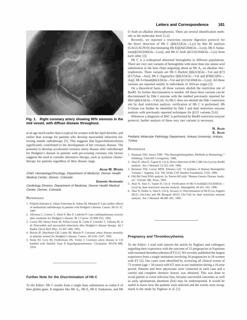
On the fifth hospital day, the patient underwent cardiac catheterization(Fig. 1). There was diffuse atherosclerosis throughout the coronary treewith sequential 75 and 90% stenoses in the mid-left anterior descendingartery and a 90% stenosis in the distal right coronary artery. She underwentsuccessful 2-vessel angioplasty. She did well until April 1993, when rap-idly progressive exertional angina developed. Repeat coronary angiogra-phy demonstrated re-stenosis of both LAD lesions. She underwent repeatangioplasty to both of these lesions with a successful result. Since thattime, she has had stable Class II angina which has been managed medi-cally.
The age-adjusted relative risk of acute myocardial infarction was 2.56 inpatients whose treatment included mediastinal irradiation [4]. Hyperten-sion, but not cigarette smoking or diabetes, was identified as a significantrisk factor for death from acute myocardial infarction. The existence ofhypercholesterolemia as a possible risk factor was not reported indepen-dently.
This patient had significant hypercholesterolemia and developed symp-tomatic coronary artery disease less than 3 years after the completion ofradiotherapy for Hodgkin’s disease. Her coronary artery disease developed
180 Letters and Correspondence
at an age much earlier than is typical for women with her lipid disorder, andearlier than average for patients who develop myocardial infarction fol-lowing mantle radiotherapy [5]. This suggests that hypercholesterolemiasignificantly contributed to the development of her coronary disease. Thepotential to develop accelerated coronary artery disease after radiotherapyfor Hodgkin’s disease in patients with pre-existing coronary risk factorssuggests the need to consider alternative therapy, such as systemic chemo-therapy for patients regardless of their disease stage.
ADAM M. MYERS
Chief, Hematology/Oncology, Department of Medicine, Denver HealthMedical Center, Denver, Colorado
EDWARD HAVRANEK
Cardiology Division, Department of Medicine, Denver Health MedicalCenter, Denver, Colorado
REFERENCES
1. Pohjola-Sintonen S, Johan Totterman K, Salmo M, Siltanen P: Late cardiac effectsof mediastinal radiotherapy in patients with Hodgkin’s disease. Cancer. 60:31–37,1987.
2. Allavena C, Conroy T, Aletti P, Bey P, Lederlin P: Late cardiopulmonary toxicityafter treatment for Hodgkin’s disease. Br J Cancer. 65:908–912, 1992.
3. Cosset JM, Henry-Amar M, Pellae-Cosset B, Carde P, Girinski T, Tubiana M, etal: Pericarditis and myocardial infarctions after Hodgkin’s disease therapy. Int JRadiat Oncol Biol Phys. 21:447–449, 1991.
4. Boivin JF, Hutchison GB, Lubin JH, Mauch P: Coronary artery disease mortalityin patients treated for Hodgkin’s disease. Cancer. 69:1241–1247, 1992.
5. Stone NJ, Levy RI, Fredrickson DS, Verter J: Coronary artery disease in 116kindred with familial Type II hyperlipoproteinemia. Circulation. 49:476–488,1974.
Further Note for the Discrimination of Hb C
To the Editor:Hb C results from a single base substitution at codon 6 ofbeta globin gene. It migrates like Hb A2, Hb E, Hb E Saskatoon, and Hb
O Arab on alkaline electrophoresis. There are several identification meth-ods at the molecular level [1,2].
Recently, we reported a restriction enzyme digestion protocol forthe direct detection of Hb C (b6(A3)Glu→Lys) by Bse RI analysis(GAGGAGN10) discriminating Hb E(b26(GH4)Glu→Lys)), Hb E Saska-toon(b22(GH4)Glu→Lys)), and Hb O Arab (b121(GH4)Glu→Lys) fromeach other [2].
Hb C is a widespread abnormal hemoglobin in different populations.There are very rare variants of hemoglobin with more than one amino acidsubstitution in the beta chain migrating about as Hb A2 on alkaline elec-trophoresis. There variants are Hb C-Harlem [b6(A3)Glu→Val and b73(E17)Asp→Asn]; Hb C-Ziguinchor [b6(A3)Glu→Val andb58(E2)Pro→Arg]; Hb S-Oman[b6(A3)Glu→Val andb121(GH4)Glu→Lys)]. All thesevariants are reported mainly in individuals of African origin [3].
On a theoretical basis, all these variants abolish the restriction site ofBseRI. So further discrimination is needed. All these three variants can bediscriminated by Dde I enzyme with the method previously reported forHbS (b6(A3)Glu→Val) [4]. As Hb C does not abolish the Dde l restrictionsite by dual restriction analysis, verification of Hb C is performed. HbS-Oman can further be identified by Dde I and dual restriction enzymeanalysis with previously reported techniques forb121 variants [5,6].
Whenever a diagnosis of HbC is performed by BseRI restriction enzymeprotocol, further analysis of these very rare variants is necessary.
N. AKAR
E. AKAR
Pediatric Molecular Pathology Department, Ankara University, Ankara,Turkey
REFERENCES
1. Huisman THJ, Jonxis THP: “The Hemoglobinopathies, Methods in Hematology.”Edinburg: Churchill Livingstone, 1986.
2. Akar N, Akar E, Tas¸tan H, Cin S; Direct detection of Hb C (B6 Glu-Lys) by BseRIanalysis. Am J Hematol 52:325–326, 1996.
3. Huisman THJ, Carver MFH, Efremov GD: “A Syllabus of Human HemoglobinVariants.” Augusta, GA: The Sickle Cell Anemia Foundation, USA, 1996.
4. Old JM: Fetal DNA analysis. In: Davies KE (ed): “Human Genetic Disease Analy-sis.” Oxford: IRL Press, 1993.
5. Akar N, Akar E, Tas¸tan H, Cin S: Verification of Hb O-Arab[(b121(GH4)Glu→Lys)] by dual restriction enzyme analysis. Hemoglobin 20:161–163, 1996.
6. Akar N, Ozden A, Akar E, Cin S¸ , Arcasoy A: Discrimination of Hb D Los Angeles(B121 Glu-Gln) and Hb Beograd (B121 Glu-Val) by dual restriction enzymeanalysis. Am J Hematol 48:280–281, 1995.
Pregnancy and Thrombocythemia
To the Editor:I read with interest the article by Pagliaro and colleaguesregarding their experience with the outcome of 15 pregnancies in 9 patientswith essential thrombocythemia (ET) [1]. We recently published the largestexperience from a single institution involving 34 pregnancies in 18 womenwith ET [2]. Our cases were identified by reviewing all clinical events of73 women (age < 50 years) with ET seen at our institution during a 16-yearperiod. Patients and their physicians were contacted in each case and acareful and complete obstetric history was obtained. This was done toavoid patient or event selection bias, because uneventful outcomes as wellas early spontaneous abortions (SA) may be underreported. It would beuseful to know how the patients were selected and the events were recog-nized in the study by Pagliaro et al. [1].
Fig. 1. Right coronary artery showing 90% stenosis in themid vessel, with diffuse disease throughout.
Letters and Correspondence 181

Both studies [1,2] report an increased rate of nonelective fetal loss. Inour study, this was due primarily to first-trimester SA. Similar to ourobservation, a recent review of the literature discloses an overall miscar-riage rate of 43%, a first-trimester SA rate of 36%, and an intrauterinedeath (IUD) rate of 5% [3]. The SA rate of 13% in the Pagliaro et al. studywas unusually low and the IUD rate of 20% unusually high. Nevertheless,the clinically relevant question is whether specific therapy influences out-come. In our study, the use of acetylsalicylic acid (ASA) did not affect thefrequency of SA. It is implied in the Pagliaro et al. study [1] that the useof ASA in association with heparin may result in a better outcome. Itshould be noted, however, that heparin treatment of their patients wasstarted during the second trimester and that the clinical course during thelast two trimesters in pregnant women with ET is usually uneventful, evenwithout any form of therapy [2,3].
Therefore, we do not recommend any form of therapy in ‘‘low-risk forthrombosis’’ patients with ET who are or may become pregnant [4]. Aprevious history of SA may be the best and only predictor of a similarsubsequent event [2], and in such patients, a systematic study in the thera-peutic use of ASA or a platelet-lowering agent may be considered. Specifictherapy with a platelet-lowering agent is recommended for those patientswith a previous history of thrombosis [4,5]. Hydroxyurea, interferon alfa,and anagrelide are the platelet-lowering agents usually considered in ET[4]. Because of the concern about teratogenicity, the use of hydroxyurea isdiscouraged during the first trimester. Currently, anagrelide is not advisedfor use during pregnancy. Because interferon alfa lacks mutagenicity, doesnot cross the placenta, and has been used successfully in pregnant womenwith ET, it may be the best possible option given the limited data [3].
AYALEW TEFFERI
Division of Hematology and Internal Medicine, Mayo Clinic and MayoFoundation, Rochester, Minnesota
REFERENCES
1. Pagliaro P, Arrigoni L, Muggiasca ML, Poggio M, Russo U, Rossi E: Primarythrombocythemia and pregnancy: Treatment and outcome in fifteen cases. Am JHematol 53:6, 1996.
2. Beressi AH, Tefferi A, Silverstein MN, Petitt RM, Hoagland HC: Outcome analy-sis of 34 pregnancies in women with essential thrombocythemia. Arch Intern Med155:1217, 1995.
3. Griesshammer M, Heimpel H, Pearson TC: Essential thrombocythemia and preg-nancy. Leuk Lymphoma 22(Suppl 1):57, 1996.
4. Tefferi A, Silverstein MN, Hoagland HC: Primary thrombocythemia. Semin Oncol22:334, 1995.
5. Cortelazzo S, Finazzi G, Ruggeri M, Vestri O, Galli M, Rodeghiero F, Barbui T:Hydroxyurea for patients with essential thrombocythemia and a high risk of throm-bosis. N Engl J Med 332:1132, 1995.
Arterial and Venous Thrombosis Associated to CombinedDeficiency of Protein C and Antithrombin III
To the Editor:Congenital deficiency of natural anticoagulants causes re-current thromboembolism [1], however, combined deficiency of these pro-teins has rarely been described [2–5]. We report a 52-year-old male with athrombosis of the right humeral artery at age 40 years; as a consequence helost the limb. He was evaluated for thrombophilia the first time in 1983because of an arterial occlusion of the right popliteal artery requiringamputation. His medical history was also significant for a right hydrocele,
mild venous insufficiency of the left leg, and chronic obstructive pulmo-nary disease. Antiplatelet agents were initiated and before the etiology ofhis thrombophilia was established, he was lost to follow-up until 1990when returned because of a pneumonia and an acute venous syndrome ofthe left leg characterized by swelling, warmth, and erythema. Venographyand Doppler ultrasound showed complete obstruction of the femoro-popliteal system and other findings suggesting chronic thromboembolism.Once discharged, he was lost again to follow-up without a diagnosis. Hereturned in 1994 because of sudden pain, coldness, and numbness of theanterior half of the left foot without previous intermittent claudication.Before starting heparin (20 UI/kg/h), plasma samples were obtained. Theresults of antithrombin III (AT-III), protein C (PC), as well as other he-mostatic parameters are shown in Table I. Despite receiving heparin up to60 UI/kg/h, no therapeutic effect on the partial thromboplastin time test(PTT) was achieved. Twelve hours later his condition worsened and he hadabsence of pedal, tibial, and popliteal pulses, cyanosis, decreased skintemperature, and gangrene up to the knee. The arteriography demonstratedtotal occlusion of the superficial femoral artery and the left leg was am-putated. Anatomopathological studies showed recent and complete ob-struction of the artery. Evolution post-surgery was unremarkable and nosource of emboli was found in the heart or aorta. Three weeks after surgery,AT-III and PC assays were repeated showing the same deficiency patternand heparin and antiplatelet agents were restarted. Again, despite thistherapy PTT remained within normal limits. Four weeks after surgery, hesuddenly developed dyspnea and chest pain with abnormal arterial gases.A pulmonary thromboembolism was diagnosed and he died 48 h laterbecause of multi-organ failure.
This patient had a combined deficiency of AT-III and PC. Few caseswith this association have been reported [2–4]. Unfortunately, no relativeswere studied. He suffered multiple venous and arterial thromboses, al-though the latter are infrequent complications in PC or AT-III deficientpatients. Cases reported with multiple deficiencies of natural anticoagu-lants [2–5] have been primarily young patients without arterial thromboticevents. In this case, there may have been a contributory effect of thesecondary polycythemia with its subsequent increase in the blood viscosity.However, it seems that the deficiency of the two most important naturalanticoagulants had a main role in his prothrombotic state. This casestrongly suggests that the risk of arterial and venous thrombosis increases
TABLE I. Hemostatic Features in a Combined PC andAT-III Deficiency*
AssayBeforeheparin
After lastamputation
Functional testPC 52% 40%AT-III 38% 30%AT-III heparin cofactor 289 289
Protein S (free) 102% 110%Plasminogen 97%C1-inhibitor 95%
Immunological testsPC 46% 40%AT-III 0.09 mg/ml 0.07 mg/mltPA 10 ng/ml
Coagulometric testsFactor II 88% 92%Factor X 110% 99%Prothrombin time 13.19 13.29
*Normal values: Functional PC: 75–129%; functional AT-III: 75–120%;AT-III heparin cofactor: >1009 in the presence of heparin; antigenic PC:80–120%; antigenic AT-III: 0.22–0.39 mg/ml.
182 Letters and Correspondence

in patients with multiple deficiencies of natural anticoagulants as otherage-related thrombotic risk actors appear in the patient.
LETICIA SANSORES-GARCIA
Department of Hematology, University of Texas Health Science Centerat Houston
ABRAHAM MAJLUF-CRUZ
Departamento de Hematologıa, HGR Gabriel Mancera, IMSS, MexicoCity, Mexico
REFERENCES
1. Bick RL, Ucar K: Hypercoagulability and thrombosis. Hematol Oncol Clin N Am6:1421, 1992.
2. Bowen D, Dasani H, Yung B, Bloom A: Deep venous thrombosis and pulmonaryembolism in a patient with type III von Willebrand’s disease, protein C andantithrombin III deficiency. Br J Haematol 81:446, 1992.
3. Gouault-Heilmann M, Quentin P, Dreyfus M, Gandrille S, Emmerich J, Leroy-Matheron C, Guesnu M: Massive thrombosis of venous cerebral sinuses in a2-year-old boy with a combined inherited deficiency of antithrombin III and pro-tein C. Thromb Haemost 72:782, 1994.
4. Jobin J, Vu L, Lessard M: Two cases of inherited triple deficiency in a largekindred with thrombotic diathesis and deficiencies of antithrombin III, heparincofactor II, protein C, and protein S. Thromb Haemost 66:295, 1991.
5. Jobin F, Vu L, Bigonesse JM. Follow-up: A young man with three deficiencies ofantithrombotic proteins, asymptomatic until now, spontaneously develops pulmo-nary embolism. Thromb Haemost 67:730, 1992.
Haemostatic Abnormalities in Dengue Haemorrhagic Feverin the New Delhi Outbreak, India
To the Editor:The recent epidemic of dengue haemorrhagic fever (DHF)in New Delhi, India (from August–November 1996) enabled us to gain aninsight into some of the mechanisms responsible for bleeding in DHF.
One hundred and twenty-seven patients (122 adults, median age 25years) were diagnosed to have dengue haemorrhagic fever by the WHOcriteria [1,2]. There was a male preponderence (male:female4 87:40).Haemorrhagic manifestations were present in all patients at admission orthey appeared within the first 3 days of the illness. Platelet counts per-
formed approximately 4.5 days after hospitalisation revealed normalisationof the initial thrombocytopenia in 71 patients. Thrombocytopenia persistedto be severe in 12 (21.4%), moderate in 36 (64.28%), and mild in 8(14.28%) cases. There was no correlation between the platelet count andclinical bleeding. In 4 cases with severe persistent thrombocytopenia, bonemarrow aspirates at 7 to 10 days of illness revealed reactive bone marrowwith an adequate number of megakaryocytes as has been reported byBierman et al. At 4 days, however, they had found the bone marrow to behypocellular [3].
Platelet aggregation was studied in 6 patients who were bleeding but hadreached platelet counts of more than 90,000 (90–156 × 103)/cu. mm. WithADP, the degree of aggregation was reduced in 5 (17.5 ± 10%) and withAdrenalin in 4 (11 ± 9%) cases, respectively. In contrast, earlier studieshave shown normal platelet aggregation with collagen, ADP, and adrenalin[3]. Immunofluorescent studies to detect anti-platelet antibodies were per-formed in 3 patients (platelet count between 40–60 × 103/cu mm). Theirpresence (+++) was demonstrated in all of them, suggesting an immune-mediated etiology for thrombocytopenia. This, along with associated plate-let dysfunction in some cases, may contribute to the observed absence ofcorrelation between the platelet count and bleeding in DHF. Platelet trans-fusions did not affect the ultimate outcome of patients. Their role in themanagement of bleeding in these patients, therefore, remains controversial.
Coagulation studies in 76 patients revealed thrombocytopenia with mildprolongation of activated partial thromboplastin time (APTT) to be thecommonest abnormality, seen in 29/76 (38.15%) cases (Table I). TheAPTT values ranged between 48 and 62 sec. In 5 such patients with APTTof 54, 50, 50, 52, and 56 sec, respectively, on whom factor VIII: C wasassayed, it was reduced to 33–42% of normal pooled plasma. Screening forFVIII inhibitors performed in 3 of these 5 patients was negative. ProlongedAPTT in these cases may have been due to reduced F VIII:C levels con-sequent upon mild consumptive coagulopathy or secondary to endothelialdamage adversely affecting the F VIII synthesis.
In 10 cases (13.15%), both APTT and PT were prolonged with a normalthrombin time (TT). The APTT values ranged between 48–65 sec and PTvalues between 14.5–18 sec. Fibrinogen degradation products (FDP) testedin 3 such patients were mildly elevated (between 10 and 40mg/ml). Al-though this may have been secondary to liver derangement, possibility ofa mild consumptive coagulopathy cannot be excluded. An earlier study hasalso demonstrated existence of an underlying mild consumptive coagu-lopathy in DHF without any patient showing acute DIC [4]. We, however,observed marked prolongations of PT, APTT, and TT, thrombocytopeniaand microangiopathic anaemia suggesting acute DIC in 4 (5.2%) cases, allof whom expired on account of dengue septicemic shock syndrome. Theremaining 72 patients, managed by intravenous fluid infusions, survived.
TABLE I. Haemostatic Abnormalities in Dengue Haemorrhagic Fever*
S. no. Abnormality
No ofpatients
(%) FDPFVIIIlevels
FVIIIinhibitor Outcome Probable cause
1. Isolatedthrombocytopenia
14 (18.9%) ND ND ND S Viral injury Ab mediated liverdamage
2. ↑ APTT, N PPT,↓ PC 29 (38.15%) ND 5/5(33–42%)
Negative3/3
S ?Consumptive coagulopathy
3. ↑ APTT, ↑ PPT, N TT,↓ PC
10 (13.15%) 3/3 (10–40mg/ml)
ND ND S ? Consumptive coagulopathy? Liver derangement
4. ↑ PPT,↑ APTT, ↑ TT,↓ PC
4 (5%) NDa ND ND Expired Acute DIC
5. Normal coagulation testsand PC
19 (25%) ND ND ND S NA
Total 76
*S: survived; PC: platelet count; N: normal; Ab: antibody; ND: not done; NA: not applicable.aPeripheral smear showed microangiopathic hemolytic anemia.
Letters and Correspondence 183

The current studies highlight some of the unusual features encounteredin the New Delhi Dengue outbreak. One hundred and twenty-two of 127were adults (>14 years) as against the reported high frequency in children.That the platelet transfusions should not have helped is also unusual. Whileimmune mediated platelet destruction may be a possible mechanism un-derlying thrombocytopenia in DHF, this needs elucidation in a larger groupof patients by antiplatelet antibody demonstration and platelet survivalstudies [5]. The mechanisms underlying isolated prologation of APTT aswell as other coagulation abnormalities also need to be studied further,since these may be important additional factors contributing to haemor-rhage.
R. SAXENA
M. BHARGAVA
J.P. WALI
D.K. MISHRA
S. MOHANTY
Departments of Haematology and Medicine, All IndiaInstitute of Medical Sciences, New Delhi, India
REFERENCES
1. Halstead SB: Dengue: Haematologic aspects. Semin Haematol 19:116–31, 1982.
2. WHO: Dengue haemorrhagic fever: Diagnosis, treatment and control. Bull.Geneva WHO, 2nd ed. 1996.
3. Bierman HR, Nelson ER: Hematodepressive virus diseases of Thailand. Ann In-tern Med 62:867–84, 1965.
4. Suvatte V, Pongpipat D, Tuchinda S, et al: Studies on serum complement C3 andfibrin degradation products in Thai hemorrhagic fever. J Med Assoc Thai 56:24,1973.
5. Mitrakul C, Poshy Achiuda M, Futrakul P, et al: Haemostatic and platelet kineticstudies in dengue hemorrhagic fever. Am J Trop Med Hyg 26:975–984, 1977.
Use of Erythropoietin (EPO) in Peripheral StemCell Transplantation
To the Editor:Estrin et al.’s recent letter describing the use of Erythro-poietin (EPO) in peripheral stem cell transplantation is an advance in themanagement of Jehovah’s Witnesses who cannot accept blood products[1]. From our experience as primary care physicians and haematologistsworking closely with this community, we highlight two issues that maypreclude the widespread acceptance of this strategy.
First, recombinant EPO is often stabilized in human serum albumin(HSA), a component of blood. Freeze-dried EPO (Boehringer Mannheim,Indianapolis, IN) is preferable to some patients because it does not containhuman or animal blood products. This may not be a major area of conten-tion as members of Jehovah’s Witnesses Hospital Liaison Committeesdistribute publications to their medical consultants on the use of erythro-poietin in accelerating post-surgical haematocrit recovery [2].
A more serious issue relates to the use of ‘‘shed’’ blood, which is not atall times in continuity with the circulation and as a matter of consciencemay be unacceptable to some patients. Pre-deposit autologous blood pro-grams to permit major orthopaedic and vascular surgery have not beenaccepted by this community and peripheral blood stem cell collectionscould similarly be rejected as they contain normal peripheral blood ele-ments.
Haematologists caring for Jehovah’s Witnesses should be aware of theseissues and the need to discuss them fully.
ALISON M. STREET
Haematology Unit, Pathology Service, Alfred Hospital, Melbourne,Victoria, Australia
LEO G. POPP
Hughesdale, Victoria, Australia
REFERENCES
1. Estrin JT, Ford PA, Henry DH, Stradden AP, Mason BA: Erythropoietin permitshigh-dose chemotherapy with peripheral blood stem-cell transplant for a Jehovah’sWitness. Am J Hematol 55:51, 1997.
2. Atabek U, Alvarez R, Pello MJ, Alexander JB, Camishion RC, Curry C, SpenceRK: Erythropoietin accelerates hematocrit recovery in post-surgical anemia. AmSurg 61:74–77, 1995.
Sezary Syndrome in an HTLV-I-Seronegative,Genome-Positive Japanese
To the Editor:Human T-cell lymphotropic virus type I (HTLV-I) is thecausative agent of adult T-cell leukemia (ATL) and tropical spastic para-paresis/HTLV-I-associated myelopathy. The vast majority of patients withthese diseases as well as asymptomatic HTLV-I carriers have antibodies toHTLV-I. Whether mycosis fungoides and Se´zary syndrome are associatedwith HTLV-I infection remains controversial. Some investigators foundHTLV-I sequences in these conditions, whereas others failed to confirmthis observation [1]. Here we report a seronegative HTLV-I carrier whodeveloped Se´zary syndrome not associated with HTLV-I.
The patient was a 58-year-old man who was admitted with generalizederythroderma in May 1990. Lymph nodes, up to 1.5 cm in diameter, werepalpable in the axillary and inguinal regions. The leukocyte count was12,700/ml with 20% abnormal lymphoid cells having cerebriform nuclei.Peripheral blood mononuclear cells (PBMC) contained 86.9% CD4 cellsand 5.9% CD8 cells. Serological studies were consistently negative forHTLV-I by particle agglutination (Fujirebio, Tokyo), ELISA (Eisai, To-kyo), indirect immunofluorescence, and Western blot (Eisai). Antibody toHTLV-I p40tax was also negative by ELISA. A skin biopsy specimenshowed Pautrier’s microabscesses and perivascular infiltration of abnormal
TABLE I. Primers and Probes Used for PCR Analysis
Regionamplified
Primer (probe)designation
Primer (probe)position
Productsize (bp)
1,301–1,320gag 1,420–1,401 120
(1,359–1,378)SK54 3,365–3,384
pol SK55 3,483–3,465 119(SK56) (3,426–3,460)E30 5,627–5,648
env E34 5,792–5,771 166(E33) (5,713–5,735)
7,341–7,360pX 7,460–7,441 120
(7,364–7,383)23–42
LTR 426–407 404(331–351)
184 Letters and Correspondence

lymphoid cells in the upper and mid dermis. A biopsied inguinal lymphnode was also infiltrated by abnormal lymphoid cells. We performedpolymerase chain reaction (PCR) analysis using five sets of primers andprobes specific for HTLV-Igag, pol, env, pX,and LTR regions (Table I).PBMC DNA was positive for all five sequences, while lymph nodeDNA was positive forpol and pX sequences only (Fig. 1). However,Southern blot hybridization revealed no evidence of HTLV-I integration inPBMC DNA after digestion withEcoRI or PstI. Furthermore, no mono-clonal integration of HTLV-I was found in PBMC DNA by inverse PCR,which is highly sensitive in detecting monoclonal integration of HTLV-I[2].
Thus, our patient was considered to be a seronegative HTLV-I carrierwho developed HTLV-I genome-negative Se´zary syndrome. Kikuchi et al.[3] also reported two seropositive patients with cutaneous T-cell lymphomain which no monoclonal integration of HTLV-I was detected by Southernblot hybridization and inverse PCR despite PCR positivity for three or allof the gag, pol, env,andpX sequences.
Previously, we described a seronegative patient with ATL whose leu-kemic cells harbored the full genome of HTLV-I [4]. The prevalence ofseronegative carriers of HTLV-I is poorly understood. In a survey in Oki-nawa where HTLV-I is endemic, Miyata et al. [5] found 17 HTLV-Icarriers among 1,015 high school students and one of them was a sero-negative carrier. To resolve the controversy regarding the association ofHTLV-I with mycosis fungoides and Se´zary syndrome, patients should beevaluated first for HTLV-I infection by serology and PCR, and then formonoclonal integration of HTLV-I by Southern blot hybridization and/orinverse PCR. PCR positivity alone with or without seropositivity is con-
sistent with polyclonal integration of complete or defective HTLV-I andmakes its causal role less likely in the pathogenesis of these diseases.
ISAO MIYOSHI
NOBUO HATAKEYAMA
KAZUO MURAKAMI
Department of Medicine, Kochi Medical School, Kochi, JapanTAKASHI SAWADA
Tsukuba Research Laboratories, Eisai Co., Ltd., Tsukuba, JapanYASUO TAKIMOTO
Hiroshima City Asa Hospital, Hiroshima, Japan
REFERENCES
1. Hall WW: Human T cell lymphotropic virus type I and cutaneous T cell leukemia/lymphoma. J Exp Med 180:1581–1585, 1994.
2. Takemoto S, Matsuoka M, Yamaguchi K, Takatsuki K: A novel diagnostic methodof adult T-cell leukemia: Monoclonal integration of human T-cell lymphotropicvirus type I proviral DNA detected by inverse polymerase chain reaction. Blood84:3080–3085, 1994.
3. Kikuchi A, Ohata Y, Matsumoto H, Sugiura M, Nishikawa T: Anti-HTLV-1antibody positive cutaneous T-cell lymphoma. Cancer 79:269–274, 1997.
4. Kubota T, Ikezoe T, Hakoda E, Sawada T, Taguchi H, Miyoshi I: HTLV-I-seronegative, genome-positive adult T-cell leukemia: Report of a case. Am JHematol 53:133–136, 1996.
5. Miyata H, Kamahora T, Iha S, Katamine S, Miyamoto T, Hino S: Dependency ofantibody titer on provirus load in human T lymphotropic virus type I carriers: Aninterpretation for the minor population of seronegative carriers. J Infect Dis 171:1455–1460, 1995.
Fig. 1. PCR analysis, showinggag, pol, env, pX, and LTR se-quences in DNA from peripheralblood (PB) and pol and pX se-quences in DNA from lymph node(LN). P is a positive control.
Letters and Correspondence 185