successful strategies to improve surgical icu documentation · successful strategies to improve...
TRANSCRIPT

7th AnnualAssociation for Clinical Documentation
Improvement SpecialistsConference
2
Successful Strategies to Improve Surgical ICU Documentation
Brent Hadder, MD
Assistant Professor, Department of Anesthesia
University of Iowa Hospitals & Clinics
Iowa City, Iowa

3
Learning Objectives
• At the completion of this educational activity, the learner will be able to:– Explain the types of surgical ICU patients and OR
documentation challenges– Define respiratory failure and shock – Describe typical documentation challenges in
rapidly changing ICU patients while working with trainees
– Recognize strategies to improve communication between coders and physicians
4
University of Iowa Hospitals and Clinics
• 711-bed hospital, including 190-bed children’s hospital– Only academic medical center in Iowa
• Hospital statistics– Over 32,000 inpatient admissions– Over 59,889 emergency-trauma visits– 157 intensive care beds – 27,875 major surgical operations– Level I trauma center– Comprehensive certified stroke center
• FY 2013 surgical/neurosciences ICU- 36 beds- 890 admissions- 2,907 transfers into SNICU- 80% occupancy
5
Operating Room
6
OR Documentation Challenges
• Dynamic environment in OR– Problems arise and are treated/resolved
quickly by anesthesia– Primarily flow sheet documentation without
diagnoses
• Anesthesia provider often diagnoses and treats common problems:– Anemia – Hypotension– Lactic acidosis– Hypothermia– Atelectasis– Low urine output
7
Anemia
• Diagnosis through lab test or visual
• Treat through transfusion
• Anemia can cause coagulopathy, and this is treated with FFP, cryo, or platelets
• Due to flow sheet documentation you will find lab and blood loss tallies and treatments
• Anesthesia relies on surgeons to document actual diagnoses
• Various causes– Anemic prior to surgery due to
trauma or chronic disease– Due to blood loss during surgery
8
Hypotension
• Various causes:– Secondary to certain anesthetic medications– Hypovolemia– Shock
• Diagnosis through hemodynamic monitors or lab results.
• May treat quickly with a fluid bolus or medication. It may require a drip.
• Hypotension can cause poor perfusion of end organs:– Acute kidney injury– MI– Stroke
• Due to flow sheet documentation it might be hard to identify boluses or other treatment.
9
Lactic Acidosis
• Various causes– Hypovolemia/under resuscitation – Anemia– Low cardiac output– Various forms of shock
• Diagnosis through lab tests
• Treat the cause of the acidosis
• Severe acidosis can impair how the body uses the medications (i.e., pressors will not work)
• Due to flow sheet documentation you may only find a lab test and a fluid bolus
• This condition can usually be diagnosed and treated quickly, so the surgeon may not be aware to document the diagnosis
10
Hypothermia
• Various causes– Trauma– Cold ORs or fluids– Patients come in cold
• Diagnosis through temperature probe
• Warm patient through fluids or heating devices
• May increase the risk of surgical site infections, delays awakening, and impacts the metabolism of drugs
• Due to flow sheet documentation you will find low temps and documentation of warming device
11
Atelectasis
• Various causes – Occurs in 90% of all anesthetized patients – Longer OR cases– Major cause of postoperative hypoxia is atelectasis
• Diagnosis through a slow drop in O2 sat– Chest x-ray but this isn’t routinely done in the OR
• Adjust the vent, recruitment maneuvers, or increase PEEP
• May increase the work of breathing or risk of reintubation
• This is a major concern with morbidly obese patients
• Atelectasis combined with partial neuromuscular blockade and opioids can lead to acute respiratory failure
Neligan, Patrick. Anesthesiology Clin 2013:30; 495–511.Duggan M., et al. Anesthesiology 2005:102; 838–854.
12
Low Urine Output
• Various causes: – Catheter occlusion– Renal or pre-renal conditions
• Diagnosis through low urine output. You will not see an increase in creatinine that soon.
• Identify the cause – check catheter, determine patient volume status.
• Could potentially increase risk for acute kidney injury.
13
Intensive Care Unit
14
Main Types of Surgical ICU Patients
• Wide variety of patients that require surgical/medical intensive care– Level I trauma center
• Blunt force trauma
• MVA
• Farm accidents
• Self-inflicted GSW
– Certified comprehensive stroke center/neurosurgery• Biggest population
• All types of strokes
• Subarachnoid hemorrhage
• Postop neurosurgical care
– Postoperative care for complex surgical patients• Ventilatory support
• Hemodynamic monitoring
• Vasoactive agent management
• Transplant management
– Other• ECMO
• Overdoses
• Overflow medical ICU patients
• Goals of care/end-of-life care
15
Trauma Patient Admission Challenges
• Present on admission – Many patients are unable to communicate on admission – Family unavailable– Resuscitation is needed to correct:
• Hypovolemic shock • Lactic acidosis• Hypothermia • Coagulopathy
– Be resolved prior to ICU admission – Return depending on patient’s injuries
– Comorbid conditions are unknown
• Glasgow Coma Scale of less than 8– Quick indicator of neurological status– Intubate patient due to increased risk of aspiration or decreased drive
to breathe– Increased intracranial pressure could result in brain herniation,
compression, or brain death
16
Stroke Patient Challenges
• Patient neurological or medical status can frequently change– Fully awake to obtunded (coma)– Loss of airway (acute respiratory failure)
• tPA can cause further bleeding or intracranial hemorrhage – CT scan 24 hours after tPA to evaluate for hemorrhage
• Tend to let the blood pressure run higher without treatment – Goal is to perfuse the injured brain
• Will allow sodium to run higher to aid with cerebral edema
17
Shock 101
18
Understanding Shock
• Shock and resuscitation is something that is treated frequently in the ICU or OR setting
• Signs of shock in intubated/sedated patients– Lactic acidosis– Hypotension– Low urine output– Tachycardia
• There are different types of shock, and it may be difficult to diagnose the type
• Main types are hypovolemic, cardiogenic, obstructive, distributive
19
Understanding Shock
• State of low perfusion is essentially shock– Pump = heart
– Tubes = blood vessels
– Fluid = blood
• Perfusion to the organs is impacted if the pump, tubes, or fluid are impacted
20
Hypovolemic Shock
• Loss of intravascular volume– Hemorrhage or dehydration– Involves the blood or fluid, and without this blood
pressure drops– Cardiac output drops
• Common in bleeding trauma patient
• Treat the cause by replacing the volume
• Blood loss anemia with lactic acidosis following a trauma – query about hypovolemic shock

21
Cardiogenic Shock
• Loss or damage of pump (i.e., heart)– Myocardial infarction (MI)– Cardiac contusion– After heart surgery– Infections– Overdose on certain medications
• Treat the cause by inotropes, balloon pumps, ECMO
• MI with a lactic acidosis and hypotension – query for cardiogenic shock
22
Obstructive Shock
• Blood is not circulating or outflow is obstructed– Pulmonary embolus– Tension pneumothorax– Pericardial tamponade
• Diagnosed by CT or echocardiogram
• Treated by draining the tamponade, chest tube for pneumothorax, anticoagulation for the pulmonary embolus
• Shock unspecified code can be used in these instances
23
Distributive Shock
• This involves the vessels themselves. The vessels are dilated and bigger, so it takes more fluid to fill them.– Septic and anaphylactic shock
• Caused by infection or serious allergic reaction.
• Treat the cause: – Infection – antibiotics, resuscitation, surgery
– Allergic reaction – epinephrine
24
Septic Shock
• Life-threatening drop in blood pressure that can lead to lung, kidney, or liver failure.
• Sometimes difficult to find the source of the infection, or to culture an organism.
• Presumed to be septic shock due to elevated white count and fever.
• Cardiac output is high in this instance. This is different from other types of shock.
25
Sepsis
26
Definitions
• Bacteremia– Bacteria in the blood
• SIRS – Systemic Inflammatory Response Syndrome– Inflammatory response to anything
– 2 or more of the following:• Temp > 38ºC or < 36ºC
• Heart rate > 90
• Resp rate > 20 or PaCo2 < 32
• White blood cell count > 1200 or < 4000, or 10% bands
Bone Roger C., et al. Crit. Care Med. 20(6):864–874. 1992.
27
Sepsis Definition
• Overwhelming immune response to infection:– Commonly caused by bacterial infections– Other causes appendicitis, pneumonia, meningitis, or
a urinary tract infection
• Chemicals released into the blood to fight the infection or chemical released by bacteria cause widespread inflammation.
• Inflammation may cause organ damage. Blood clotting during sepsis can reduce blood flow to organs or extremities.
28
Sepsis Definition
• Sepsis
– The body’s response to an INFECTION
– 2 or more of the following:
• Temp > 38ºC or < 36ºC
• Heart rate > 90
• Resp rate > 20 or PaCo2 < 32
• White blood cell count > 1200 or < 4000, or 10% bands
Bone Roger C., et al. Crit. Care Med. 20(6):864–874. 1992.
29
Severe Definitions
• Severe sepsis
– Sepsis with organ dysfunction
• Poor perfusion
• Lactic acidosis
• Low urine output
• Altered mental status
Bone Roger C., et al. Crit. Care Med. 20(6):864–874. 1992.
30
Septic Shock Definition
• Septic shock
– Hypotension from sepsis despite adequate volume resuscitation with perfusion problems
• Lactic acidosis
• Low urine output
• Altered mental status
• May require pressors
Bone Roger C., et al. Crit. Care Med. 20(6):864–874. 1992.
31
Other Definitions
• Sepsis-induced hypotension
– A systolic blood pressure < 90 mm Hg or a less than 40 mm Hg from baseline when there is no other cause for the hypotension
Bone Roger C., et al. Crit. Care Med. 20(6):864–874. 1992.
32
Case Study
• 63 y.o. male with DM Type II comes to ED with infection on his arm. Pt. has a fever and “feels terrible.” RR 35, BP 95/67, HR 130, temp 38.9. Initial lactate is elevated with a metabolic acidosis and an elevated WBC. Pt. is to go to the OR for debridement of necrotizing fasciitis.
What is the diagnosis?
33
Respiratory Failure
34
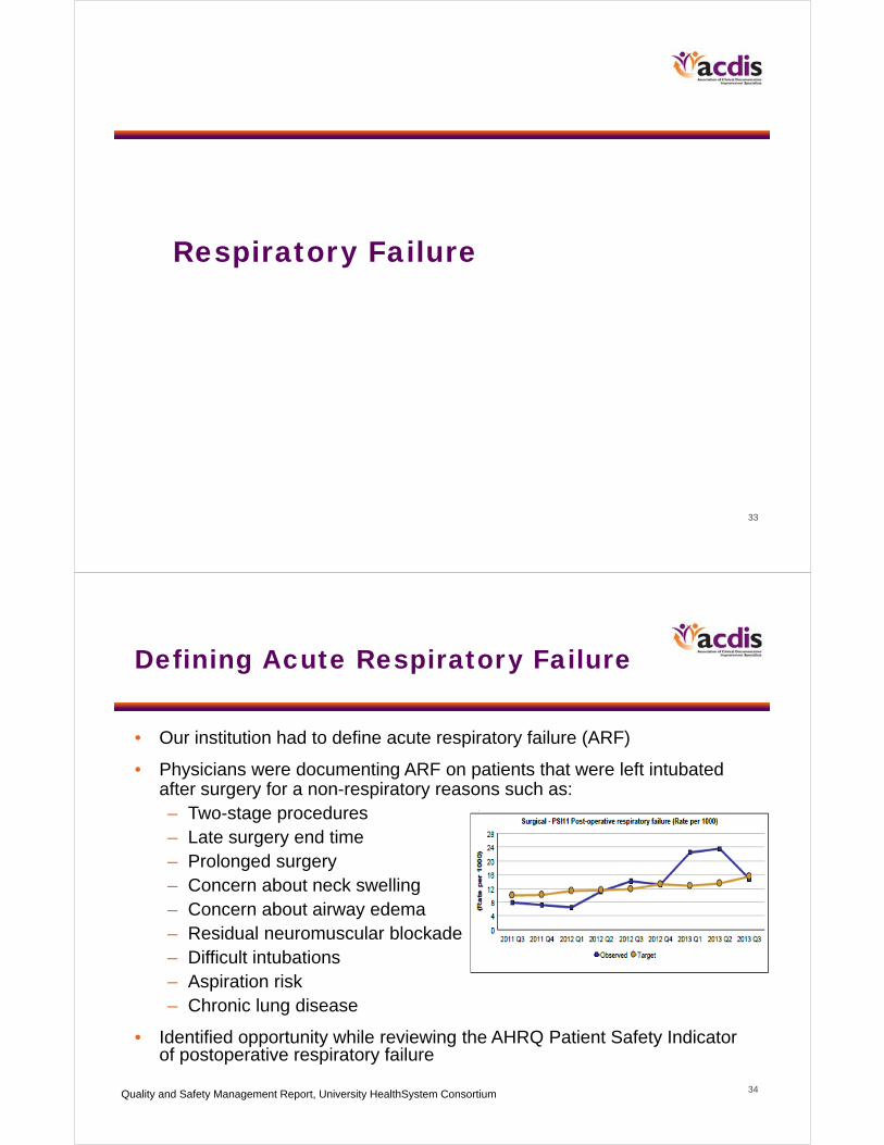
Defining Acute Respiratory Failure
• Our institution had to define acute respiratory failure (ARF)
• Physicians were documenting ARF on patients that were left intubated after surgery for a non-respiratory reasons such as:– Two-stage procedures– Late surgery end time – Prolonged surgery– Concern about neck swelling– Concern about airway edema– Residual neuromuscular blockade– Difficult intubations– Aspiration risk– Chronic lung disease
• Identified opportunity while reviewing the AHRQ Patient Safety Indicator of postoperative respiratory failure
Quality and Safety Management Report, University HealthSystem Consortium

35
Documentation Needs for ARF
• Is the respiratory failure acute or acute on chronic?• Was the respiratory failure present on admission
(POA)?• What caused the respiratory failure? Note any acute
conditions in relation to chronic disease. • Note the presence of hypoxemic, hypercapnic, or mixed
respiratory failure.• Note clinical signs, symptoms, or any lab findings that
support the diagnosis.• Tip: ARF frequently coexists with severe
CHF, pneumonia, COPD, or asthma.
36
Acute Respiratory Failure
Adults
• Shortness of breath, dyspnea
• Unable to speak in complete sentences
• Respiratory rate > 24
• Use of accessory muscles to breathe, labored breathing at rest, tripod position
• Hypoxemia, cyanosis
• Diagnosed airway edema
• Increased oxygen requirements (mask or nasal cannula)
• Need for continuous nebs, Bi-PAP/C-PAP or control ventilation or for intubation
• Confusion/altered mental status/obtunded, consider if Glasgow Coma Scale < 8
• Inability to protect airway (i.e., overdose, neurological conditions), unable to extubate
Arterial blood gas (ABG) parameters for acute respiratory failure
• pH of < 7.30 or > 7.50
• pCO2 of > 50
• pO2 of < 60 or pulse ox < 88%
• In patients with preexisting lung disease:
– pH < 7.35
– pCO2 markedly elevated from baseline or pO2 lower than baseline
37
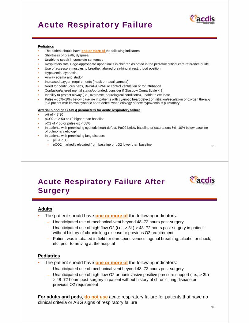
Acute Respiratory Failure
Pediatrics• The patient should have one or more of the following indicators• Shortness of breath, dyspnea• Unable to speak in complete sentences• Respiratory rate > age-appropriate upper limits in children as noted in the pediatric critical care reference guide• Use of accessory muscles to breathe, labored breathing at rest, tripod position• Hypoxemia, cyanosis• Airway edema and stridor• Increased oxygen requirements (mask or nasal cannula)• Need for continuous nebs, Bi-PAP/C-PAP or control ventilation or for intubation • Confusion/altered mental status/obtunded, consider if Glasgow Coma Scale < 8 • Inability to protect airway (i.e., overdose, neurological conditions), unable to extubate• Pulse ox 5%–10% below baseline in patients with cyanotic heart defect or initiation/escalation of oxygen therapy
in a patient with known cyanotic heart defect when etiology of new hypoxemia is pulmonary
Arterial blood gas (ABG) parameters for acute respiratory failure• pH of < 7.30 • pCO2 of > 50 or 10 higher than baseline• pO2 of < 60 or pulse ox < 88% • In patients with preexisting cyanotic heart defect, PaO2 below baseline or saturations 5%–10% below baseline
of pulmonary etiology• In patients with preexisting lung disease:
– pH < 7.35– pCO2 markedly elevated from baseline or pO2 lower than baseline
38
Acute Respiratory Failure After Surgery
Adults
• The patient should have one or more of the following indicators:– Unanticipated use of mechanical vent beyond 48–72 hours post-surgery
– Unanticipated use of high-flow O2 (i.e., > 3L) > 48–72 hours post-surgery in patient without history of chronic lung disease or previous O2 requirement
– Patient was intubated in field for unresponsiveness, agonal breathing, alcohol or shock, etc. prior to arriving at the hospital
Pediatrics
• The patient should have one or more of the following indicators:– Unanticipated use of mechanical vent beyond 48–72 hours post-surgery
– Unanticipated use of high-flow O2 or noninvasive positive pressure support (i.e., > 3L) > 48–72 hours post-surgery in patient without history of chronic lung disease or previous O2 requirement
For adults and peds, do not use acute respiratory failure for patients that have no clinical criteria or ABG signs of respiratory failure
39
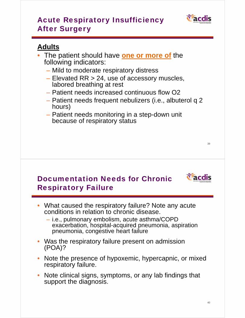
Acute Respiratory Insufficiency After Surgery
Adults• The patient should have one or more of the
following indicators:– Mild to moderate respiratory distress– Elevated RR > 24, use of accessory muscles,
labored breathing at rest– Patient needs increased continuous flow O2– Patient needs frequent nebulizers (i.e., albuterol q 2
hours)– Patient needs monitoring in a step-down unit
because of respiratory status
40
Documentation Needs for Chronic Respiratory Failure
• What caused the respiratory failure? Note any acute conditions in relation to chronic disease. – i.e., pulmonary embolism, acute asthma/COPD
exacerbation, hospital-acquired pneumonia, aspiration pneumonia, congestive heart failure
• Was the respiratory failure present on admission (POA)?
• Note the presence of hypoxemic, hypercapnic, or mixed respiratory failure.
• Note clinical signs, symptoms, or any lab findings that support the diagnosis.
41
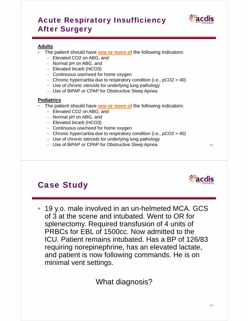
Acute Respiratory Insufficiency After Surgery
Adults• The patient should have one or more of the following indicators:
– Elevated CO2 on ABG, and– Normal pH on ABG, and– Elevated bicarb (HCO3)– Continuous use/need for home oxygen– Chronic hypercarbia due to respiratory condition (i.e., pCO2 > 40)– Use of chronic steroids for underlying lung pathology– Use of BiPAP or CPAP for Obstructive Sleep Apnea
Pediatrics• The patient should have one or more of the following indicators:
– Elevated CO2 on ABG, and– Normal pH on ABG, and– Elevated bicarb (HCO3)– Continuous use/need for home oxygen– Chronic hypercarbia due to respiratory condition (i.e., pCO2 > 40)– Use of chronic steroids for underlying lung pathology– Use of BiPAP or CPAP for Obstructive Sleep Apnea
42
Case Study
• 19 y.o. male involved in an un-helmeted MCA. GCS of 3 at the scene and intubated. Went to OR for splenectomy. Required transfusion of 4 units of PRBCs for EBL of 1500cc. Now admitted to the ICU. Patient remains intubated. Has a BP of 126/83 requiring norepinephrine, has an elevated lactate, and patient is now following commands. He is on minimal vent settings.
What diagnosis?
43
End-of-Life Considerations
44
End-of-Life Care
• In the ICU setting goals of care can change to comfort measures
• Our goals are for patient comfort– We are no longer focused on cure– Some diseases that we are not curing have symptoms that can
be treated in the comfort stage• Secretions• Pain• Anxiety• Delirium• Nausea/vomiting
• Palliative care consults may be a good source of documentation due to focused health history and patient’s story

45
Documentation Challenges With New Trainees
46
Challenges
• Learning how to take care of patients• Communicating with multiple services• Lack of continuity in rotation schedules• Selecting diagnoses from the EMR do not match what terms
you were trained• Lack of training in medical school on documentation and coding • Busy schedules with restricted hours• Varying backgrounds• Trying to learn how to write notes• Copy and pasting with EMR• “What’s in it for me?” or “It’s not my problem”
47
Strategies to Improve Communication
48
Communicating With the Physicians
• DRG nurses routinely attend bed huddles, physician rounds, and unit-based team meetings– Improves teamwork and communication
• Multidisciplinary team members learn how each role impacts healthcare
• Encourages ancillary services to assist in documentation improvement
– Respiratory therapy, dietitians, social workers
– Provides opportunity to clarify patient condition and treatments
– Reinforces importance of accurate clinical documentation
49
Communicating With the Physicians
• Morbidity and mortality conference attendance– Quality/safety improvement staff
listen for:• Documentation opportunities• Potential AHRQ patient safety metric
triggers– Postop respiratory failure– Accidental puncture laceration
• ARNPs and ICU fellows increased continuity for documentation and problem list creation
50
Technology
• Electronic medical record– Specific inbox called “Doc Query” where
all queries go from coding staff, quality office, and DRG nurses
– Create service-specific preference lists to aid in problem list builds
– Run data mining reports that look for query opportunities based on patient vitals, labs, and problem list entries
• Ex: alkalosis, acidosis, chronic kidney failure, altered mental status, pancytopenia, shock
• Voalte phones– iPhones that only work in the hospital– Face time or text queries that are difficult
to communicate via email
51
Teamwork
• Physicians, hospital coders, DRG nurses, IT, and quality office started talking!– Established multidisciplinary hospital
clinical documentation improvement committee• Meets every other week • Forum to educate, communicate, and
debate• Everyone has an equal voice
• Developed intranet site for documentation improvement – Performance metrics are updated
weekly• Transparency encourages competition
– References are readily available
52
Thank you. Questions?
In order to receive your continuing education certificate(s) for this program, you must complete the online evaluation. The link can be found in the continuing education section at the front of the workbook.