subunit stoichiometry and arrangement in a … · subunit stoichiometry and arrangement in a...
TRANSCRIPT

Subunit stoichiometry and arrangement in aheteromeric glutamate-gated chloride channelNurit Degani-Katzav, Revital Gortler, Lilach Gorodetzki, and Yoav Paas1
Laboratory of Ion Channels, The Mina and Everard Goodman Faculty of Life Sciences and The Institute of Nanotechnology and Advanced Materials, Bar-IlanUniversity, Ramat Gan 52900, Israel
Edited by Arthur Karlin, Columbia University, College of Physicians and Surgeons, New York, NY, and approved December 2, 2015 (received for reviewDecember 16, 2014)
The invertebrate glutamate-gated chloride-selective receptors(GluClRs) are ion channels serving as targets for ivermectin (IVM), abroad-spectrum anthelmintic drug used to treat human parasiticdiseases like river blindness and lymphatic filariasis. The native GluClRis a heteropentamer consisting of α and β subunit types, with yetunknown subunit stoichiometry and arrangement. Based on the re-cent crystal structure of a homomeric GluClαR, we introduced muta-tions at the intersubunit interfaces where Glu (the neurotransmitter)binds. By electrophysiological characterization of these mutants, wefound heteromeric assemblies with two equivalent Glu-binding sitesat β/α intersubunit interfaces, where the GluClβ and GluClα subunits,respectively, contribute the “principal” and “complementary” com-ponents of the putative Glu-binding pockets. We identified a muta-tion in the IVM-binding site (far away from the Glu-binding sites),which significantly increased the sensitivity of the heteromeric mu-tant receptor to both Glu and IVM, and improved the receptor sub-units’ cooperativity. We further characterized this heteromeric GluClRmutant as a receptor having a third Glu-binding site at an α/α inter-subunit interface. Altogether, our data unveil heteromeric GluClRassemblies having three α and two β subunits arranged in a counter-clockwise β-α-β-α-α fashion, as viewed from the extracellular side,with either two or three Glu-binding site interfaces.
allostery | Cys-loop receptors | ion channels | ivermectin |neurotransmitters
Glutamate-gated chloride-selective receptors (GluClRs) arepentameric glutamate-gated chloride channels unique to
invertebrates. They belong to the Cys-loop receptor superfamilyof transmembrane oligomers that open an intrinsic cationic oranionic channel pore upon binding of neurotransmitters, such asACh, serotonin, GABA, Gly, histamine, or Glu (1–9). GluClRs arespecific targets for ivermectin (IVM), a broad-spectrum anthel-mintic drug used to treat filarial diseases like onchocerciasis (riverblindness) and elephantiasis (lymphatic filariasis) that afflict hun-dreds of millions of people worldwide (10, 11). IVM is also broadlyused in cattle, swine, and pets to kill gastrointestinal roundworms,lungworms, grubs, sucking lice, and mange mites (12). The highefficiency of IVM stems from its capacity to act as an agonist thatkeeps the receptor’s ion channel continuously open (13–18). Be-cause the GluClR is chloride-selective, IVM causes sustained hy-perpolarization across postsynaptic membranes in the parasiticnematodes. This long-term hyperpolarization leads to suppressionof excitation in motor neurons and inhibition of locomotion (19);inhibition of the pharyngeal muscle activity, which interrupts withfeeding behavior (20); and interruption of secretion processes thatare crucial for evading the host immune system (21).Genes encoding two GluClR homologous subunits, GluClα
and GluClβ (glc-1 and glc-2, respectively), were first cloned fromCaenorhabditis elegans (13). When expressed in Xenopus oocytes,homomeric GluClαRs respond to IVM but not to Glu and, incontrast, homomeric GluClβRs respond to Glu but not to IVM(13, 16, 17, 22). A recent 3D crystal structure of a truncatedhomomeric GluClαR (GluClαcrystR; Protein Data Bank ID code3RIF) shows that when IVM is bound at the five α/α intersubunit
interfaces in the ion-channel pore periphery, Glu is lodged at thefive α/α intersubunit interfaces in the ligand-binding domain(LigBD) (23) (Fig. 1A). These Glu-binding sites are homologousto the neurotransmitter/agonist-binding sites of other Cys-loopreceptors (1, 2, 24), bacterial homologs of Cys-loop receptors(25–30), and ACh-binding proteins (31–34).Importantly, the naturally occurring GluClR robustly responds
to both Glu and IVM independently; therefore, it is considered toconsist of both GluClα and GluClβ subunit types (13–18). How-ever, little is known about the stoichiometry and molecular ar-rangement of the subunits in heteromeric GluClRs. Furthermore,the aforementioned crystallographic observations (23) are consis-tent with earlier studies showing that Glu elicits current responsesin homomeric GluClαRs only when applied after activation byIVM (14), which gives rise to the following question: Could an α/αintersubunit interface be formed in a heteromeric assembly, bindGlu, and functionally participate in the activation process evenwithout IVM preassociation? To resolve this question, we clarifiedhere the stoichiometry and positions of the α and β subunits inGluClα/βR heteromeric assemblies that carry mutations in boththe putative Glu- and IVM-binding pockets.
ResultsCan the Coupling Loops of the GluClα Subunit Mediate Channel Openingupon Glu Binding? Based on the capability of the WT homomericGluClα (GluClαWT) receptor to respond to Glu only followingexposure to IVM, it was suggested that IVM binding induces aconformational change that enables coupling of Glu binding atα/α intersubunit interfaces to the opening of the ion-channel gate(14, 23). To explore this suggestion further, we used a strategy ofmicrochimerism that is based on previous studies showing that invarious Cys-loop receptors, the β1β2, Cys, and β8β9 loops of the
Significance
Cys-loop receptors (CLRs) are transmembrane ion channels acti-vated by neurotransmitters to mediate chemoelectric excitationor inhibition throughout the nervous system. Hence, CLRs play akey role in our day-to-day life, from coordination of motions tocognition. Impairment of CLRs’ activity leads to various patho-physiological conditions. The CLR studied here is a glutamate-gated chloride-selective receptor (GluClR). GluClRs are unique toinvertebrates, yet they are pharmacologically important becausethey serve as targets for ivermectin, an anthelmintic drug usedto treat humans suffering from filarial diseases. This study pro-vides better understanding of the subunit arrangement andstoichiometry of Glu-binding sites in GluClRs.
Author contributions: N.D.-K. and Y.P. designed research; N.D.-K., R.G., L.G., and Y.P.performed research; N.D.-K., R.G., L.G., and Y.P. analyzed data; and N.D.-K. and Y.P. wrotethe paper.
The authors declare no conflict of interest.
This article is a PNAS Direct Submission.1To whom correspondence should be addressed. Email: [email protected].
This article contains supporting information online at www.pnas.org/lookup/suppl/doi:10.1073/pnas.1423753113/-/DCSupplemental.
E644–E653 | PNAS | Published online January 20, 2016 www.pnas.org/cgi/doi/10.1073/pnas.1423753113

LigBD interact with the M2–M3 loop of the pore domain tocouple neurotransmitter binding to channel gating (23, 35–44)(e.g., Fig. 1 A and C). These four loops are termed the couplingloops. Fig. 1D shows schemes of the WT GluClα and GluClβsubunits, as well as three microchimeric GluClβ subunits wherewe replaced the Cys loop, β8β9 loop, or both loops with the ho-mologous loops of the GluClα subunit. These microchimeric sub-units are termed GluClβα-CysL, GluClβα-β8β9L, and GluClβα-Loops,respectively. Note that the C. elegans GluClα and GluClβ subunitsshare an identical β1β2 loop sequence, whereas their M2–M3 loopsequence is almost identical (Fig. S1).CHO cells transfected with the GluClαWT subunit alone
showed very weak responses to 10 mM Glu (135 ± 27 pA in eightcells; mean ± SEM), but responded well to 500 nM IVM (Fig.S2B; 14 cells). This observation is in line with the findingsof Frazier et al. (45), who reported that HEK cells expressing
GluClα homomers are responsive to IVM but not to Glu. CHOcells transfected with the GluClβWT subunit alone showed veryweak, rare responses to 10 mM Glu (less than 230 pA in eightcells; Fig. S2A), in line with results obtained in HEK cells (45).No responses to 500 nM IVM in CHO cells transfected with theGluClβWT subunit alone were observed (10 cells), in agreementwith the same observations in HEK cells (18, 22). In contrastto these differential responses, cells cotransfected with bothGluClαWT and GluClβWT subunits displayed robust responsesto 1.5 mM Glu (EC50 concentration) and 500 nM IVM (Fig.S2C). We therefore deduce that robust responses to Glu andIVM (independently) in a cell cotransfected with the GluClαWTand microchimeric GluClβ subunits (Fig. S2 D–F) reflect thefunction of heteromeric GluClα/βR complexes. This deductionalso applies for the site-specific mutants discussed further below.Fig. 2 shows representative current traces elicited by in-
creasing Glu concentrations (Fig. 2A) and the correspondingdose–response curves (Fig. 2B) for the heteromeric WT andmicrochimeric GluClRs. The Glu-EC50 values specified in TableS1 indicate that the apparent affinities of the GluClαWT/βmicrochimericreceptors for Glu were very close to the apparent affinity of theGluClαWT/βWT receptor. The Hill coefficients of all four re-ceptors (Table S1) were >1, indicating their activation withpositive cooperativity. Note that the Glu-EC50 and the Hill co-efficient determined here for the GluClαWT/βWT receptor(Table S1) are very close to those values determined in Xenopusoocytes [Glu-EC50 = 1.36 ± 0.05 mM and Hill coefficient (nH) =1.7 ± 0.1] (13). Remarkably, Glu readily activates the GluClαWT/βα-Loops receptor, all of whose LigBD’s coupling loops are of theGluClα subunit (Fig. 2 and Table S1). We thus conclude that theβ1β2, Cys, and β8β9 loops of the GluClα subunit are inherentlycapable of coupling Glu binding to pore gating, with no need forIVM prebinding.Fig. 1. Structural characteristics of GluClRs. (A) Two neighboring subunits of
the homopentameric GluClαcrystR [Protein Data Bank (PDB) ID code 3RIF] areshown from the side in light and dark gray colors. Wide gray horizontal linesmark the putative membrane borders. The four coupling loops are coloredas shown in C and the upper row of D. Glu and IVM are shown as space-filling models with carbon, oxygen, and nitrogen atoms colored in yellow,red, and blue, respectively. They are bound at the α(+)/α(−) intersubunitinterface far away from each other: Glu in the extracellular LigBD and IVM inthe upper part of the pore-domain periphery, between M1 (of the light graysubunit) and M3 (of the dark gray subunit). Note that in Cys-loop receptors,the principal and complementary faces of a neurotransmitter-binding pocketare formed by the (+) and (−) sides of two adjacent subunits, respectively.(B) Top view of the GluClαcryst pentamer showing five identical subunits, whichare colored differently to highlight the intersubunit interfaces located be-tween the (+) and (−) sides. (C) Space-filling models of residues belonging tothe coupling loops, which create an extensive bond network at the interfacebetween the LigBD and the ion-channel pore domain. (D) Schemes of GluClRsubunits used in this study. The M1–M4 transmembrane segments are num-bered 1–4. Different colors reflect differences in amino acid sequences (Fig. S1).
Fig. 2. Glu-activation properties of WT and microchimeric GluClRs. (A) Rep-resentative Glu-elicited currents measured in cells cotransfected with the in-dicated subunits. In all cases, Glu was applied for 3 s. Glu concentrations:0.1 mM, 0.3 mM, 0.6 mM, 1 mM, 3 mM, 10 mM, and 30 mM in the upper rowand 0.1 mM, 0.3 mM, 0.6 mM, 1 mM, 10 mM, and 30 mM in the lower row.Measurements were performed at +60 mV. (B) Dose–response curves plottedfor responses measured in cells cotransfected with the GluClαWT subunit andthe GluClβ subunits indicated in the figure. Curves were fitted to the averageddata points with a nonlinear regression using the Hill equation (Eq. 1) (r2 > 0.99).Error bars correspond to SEM.
Degani-Katzav et al. PNAS | Published online January 20, 2016 | E645
PHYS
IOLO
GY
PNASPL
US

Contribution of the GluClα Subunit (−) Side to Glu Accommodation inHeteromeric GluClR Mutants. The aforementioned observationsbrought us to the recognition that a thorough study of how theGluClα subunit contributes to Glu binding in heteromericGluClRs is necessary. Therefore, we first introduced mutations inthe (−) side of the GluClα subunit based on the crystal structure ofthe homomeric GluClαcrystR (23) [the (−) and (+) subunit sidesare defined in Fig. 1 A and B]. This structure indicates that theδ-guanidino groups of α(−)R98 and α(−)R117 are at an appro-priate distance to form ion pairing with the α- and γ-carboxylgroups of Glu, respectively (Fig. 3D). A mutation that eliminatedthe charge and drastically reduced the side-chain size of α(−)R117,but kept hydrophilicity at this position (i.e., R→S), did notprovide a functional GluClαR117S/βWT receptor. We thereforereplaced the two Args (one at a time) with a more conservativeand bulkier hydrophilic residue, Asn or Gln, which can function ashydrogen bond donor (or acceptor) with no capability to form saltbridges. A mutant having an αR98N substitution (GluClαR98N/βWT receptor) provided robust responses, but very high Gluconcentrations were necessary to reach saturation [Fig. 3A, traces(Right) and brownish dose–response curve (Left)]. Note that todissolve Glu, it was titrated with equimolar concentrations ofNaOH; therefore, we did not change the Nernst potential for Cl−
during Glu application. However, the osmolarity and negativecharge of the external solution drastically increased during theapplication of high Glu concentrations (for 0.6 s). Even so, weassume that these factors did not affect the responses, as discussedin SI Text, section 1, in conjunction with Fig. S3.In the case of the GluClαR117N/βWT receptor, the current
responses did not allow us to analyze the dose–response relationreliably because they were very low (∼300 pA at 1 M Glu) anddid not reach saturation, unlike in the case of the GluClαR98N/βWT receptor. In contrast, introducing Q at position α(−)117,which has a longer side chain than N, created a responsive
GluClαR117Q/βWT receptor that enabled us to determine itsGlu-EC50 and Hill coefficient (Fig. 3A and Table S1).The crystal structure also indicates that α(−)S182 forms a hy-
drogen bond with the γ-carboxyl group of Glu (23) (Fig. 3D). Pre-venting this hydrogen bonding in the heteromeric GluClαS182A/βWT receptor produced an effect similar to the effect observed withthe α(−)R98N and α(−)R117Q substitutions (Fig. 3A and TableS1). The drastic effects exerted by mutations in the GluClα(−) sideraised the question of whether mutations at the homologous po-sitions in GluClβ would exert the same effects.
Contribution of the GluClβ Subunit to Glu Accommodation in HeteromericGluClR Mutants. Sequence alignments (17, 23) indicate that theGluClβ subunit has identical residues at positions homologousto GluCl α(−)R98, α(−)R117, and α(−)S182. These residues areβ(−)R66, β(−)R85, and β(−)S152, respectively (Fig. 3E). A3D homology model built here for the α(+)/β(−) intersubunitinterface (SI Materials and Methods and Fig. S4) predicts thatthese three β-subunit residues are sufficiently close to interact withGlu (Fig. 3E). However, heteromeric mutant receptors assem-bled of the αWT subunit, together with β(−)R66N, β(−)R85N, orβ(−)S152A, did not display the drastic increase in Glu-EC50typical of the homologous α-subunit mutants (Fig. 3B andTable S1). Most recently, Daeffler et al. (22) published a studywhere they investigated homomeric GluClβRs carrying a βT283Smutation in the pore-lining segments (see sequence with entry codeQ17328 in the UniProtKB database). The latter mutation, per se,caused a dramatic improvement in the response to Glu (70-folddecrease in Glu-EC50). Interestingly, when the βT283S mutationwas combined with a β(−)S152A mutation (no. 126 in ref. 22), theGlu-EC50 relatively increased by 590-fold (22). Clearly, if theβ-subunit (−) side were to contribute the “complementary” Glu-binding components in our heteromeric GluClαWT/βS152A re-ceptor, we would have observed a much larger rightward shift of
Fig. 3. Sensitivity to Glu of heteromeric GluClRscarrying single-site mutations within the putativeGlu-binding pockets. (A–C) Dose–response curvesplotted for the activation of various heteromericreceptors by Glu. Curves were fitted as described inFig. 2 (r2 > 0.99). Error bars correspond to SEM. “X”corresponds to mutations that were introduced inthe GluClα(−) (A), GluClβ(−) (B), and GluClβ(+) (C)subunit sides. (A, Right) Representative currenttraces evoked by increasing Glu concentrations of 10mM, 30 mM, 60 mM, 100 mM, 250 mM, 600 mM, and1,000 mM in cells coexpressing the indicated GluClRsubunits. Currents were measured at +60 mV. (D andE) Three-dimensional homology models of potentialβ(+)/α(−) and α(+)/β(−) intersubunit interfaces. Glu isshown with yellow-colored carbon atoms, whereasthe carbons of receptor residues are colored in grayor gold for the α or β subunit, respectively. Oxygenand nitrogen atoms are colored in red and blue,respectively. Hydrogen atoms are not shown. Dottedgreen lines correspond to potential ion pairingor hydrogen bonding, whereas dotted black linesindicate distances compatible with van der Waals orcation–pi interactions. Loop C caps the putative Glu-binding pocket. (F) Representative current traceelicited by 15 mM Glu (saturating concentration) in cells expressing the GluClαWT/βWT receptor, followed by inhibition of the leak current by 200 μM pic-rotoxin (PTX). (Inset) Magnification (7.5-fold) of the trace observed upon PTX application. Measurements were performed at +80 mV. (G) Representativesingle-channel current recorded from a cell-attached patch containing the GluClαWT/βWT receptor. The pipette solution included 15 mM Glu (saturatingconcentration). The voltage command was −90 mV. The closed and open state levels are indicated by c and o, respectively. The single-channel Po-max is 0.64.(H, Left) Curve fitted to an event amplitude histogram by two Gaussian functions for the single-channel currents exemplified in G. The mean amplitude of theopen state is 1.9 pA. (H, Right) Histogram of distribution of open times whose best-fit decay constant corresponds to the mean channel open time (τ0).(I) Averaged dose–response data points of the GluClαWT/βWT receptor (purple squares) normalized to obtain the estimated Popen values that are plotted as afunction of varying Glu concentrations. The purple line is the Hill curve. The dashed black line is the curve fitted based on an MWC allosteric model with twoequivalent Glu-binding sites (n = 2) (Eq. 2). (J) Plausible subunit arrangement of a heteromeric GluClRαWT/βWT receptor with two equivalent Glu-binding sites(black triangles), as viewed from the extracellular side.
E646 | www.pnas.org/cgi/doi/10.1073/pnas.1423753113 Degani-Katzav et al.

the dose–response curve (complementary is defined in Fig. 1). Takenthe results of the previous and current sections together, we inferthat in the recombinant heteromeric receptors studied here, theGluClα(−) side, rather than the GluClβ(−) side, contributes thecomplementary components to Glu binding. Hence, we hypothe-sized that the GluClβ(+) side contributes the “principal” Glu-bind-ing components in heteromeric GluClRs (principal is defined inFig. 1).To examine this hypothesis, we mutated residues β(+)F122,
β(+)T229, and β(+)Y232 that might contact Glu, as predicted bysequence alignments (17, 23) and our 3D homology model (Fig.3D). We then coexpressed the mutated β subunits (one at atime), together with the αWT subunit, and found that theyshifted the dose–response curves rightward (Fig. 3C). Table S1shows the extent of increase in the Glu-EC50 values, with themost prominent shift in the GluClαWT/βT229N and GluClαWT/βT229W receptors (by approximately eightfold compared withthe GluClαWT/βWT receptor). We infer that the GluClβ(+)side in the heteromeric assemblies generated here contributesthe principal Glu-binding components. Daeffler et al. (22)added to the homomeric GluClβT283S receptor a β(+)T229Amutation (no. 203 in ref. 22), which increased the Glu-EC50 to amuch larger extent than observed here for the heteromericGluClαWT/βT229N or GluClαWT/βT229W receptor. This differ-ence can be attributed to the nature of the replacing amino acids.In the current study, we did not wish to change the chemicalproperties of the amino acids too much. This approach wasadopted because the GluClαWT/βWT receptor inherently displayslow affinity for Glu, which would probably make a dramatic in-crease in Glu-EC50 difficult to probe. Hence, we kept the capa-bility of the replacing residues at position β(+)T229 to act ashydrogen bond donors (Asn, Trp) or a hydrogen bond acceptor(Asn). We expected that the greater size of the replacing residueswould interfere with, but not abolish, Glu accommodation. Thisexpectation emerged because position β(+)229 is located on loopC, which caps the putative Glu-binding pocket but, on the otherhand, is considered to be flexible and mobile (46) (Fig. 3D and E).As to the β(+)Y232S substitution, we probably eliminated a cation–pi interaction that was recently suggested to be formed in ahomomeric GluClβR, between the β(+)Y232 aromatic ringand the α-amino nitrogen of Glu (22). Still, one cannot ex-clude hydrogen bonding between the hydroxyl group of the Serwe introduced at this position [β(+)232] and the α-amino ni-trogen of Glu, which could explain the moderate effect of theβ(+)Y232S mutation.
Stoichiometry of the Glu-Binding Sites in a Heteromeric WT GluClα/βR.The results presented in the previous sections suggest that aβ(+)/α(−) intersubunit interface is involved in Glu accommoda-tion; so, how many such functional interfaces exist per hetero-pentamer? The various single-site mutant receptors discussed sofar share with the GluClαWT/βWT receptor Hill coefficientssmaller than 2 but clearly larger than 1 (Table S1). This propertysuggests that there is more than one Glu-binding site per heter-opentamer. To determine the number of functional sites andtheir microscopic equilibrium dissociation constants for Glubinding in the heteromeric GluClαWT/βWT receptor, we usedan allosteric model based on the Monod–Wyman–Changeux(MWC) theory (47), as applied also by Karlin (48) to the nicotinicACh receptor (nAChR) (reviewed in refs. 49 and 50). Because theGluClαWT/βWT receptor displays very slow and weak desensiti-zation, we simplified the allosteric model by focusing on two ma-jor states as previously performed for weakly or nondesensitizingCys-loop receptors such as: homomeric α7-nAChR mutants (51),homomeric α7-5HT3AR chimeras (52), and heteromeric GABAreceptors (53, 54). If the GluClαWT/βWT receptor has two equiva-lent (identical) Glu-binding sites, then Scheme I describes itsMWC allosteric activation mechanism as follows:
where R and R* are resting (closed) and active (open) receptorconformational states, respectively; A is an agonist molecule (Glu)that can complex with the receptor; Kd,R and Kd,R* are the mi-croscopic equilibrium dissociation constants for agonist binding tothe closed and open receptor states, respectively; and L is theequilibrium constant of the two receptor states (closed and open)in the absence of ligands. L is calculated by R/R* based on quan-titative determinations, as follows.Unoccupied R* corresponds to spontaneously open channels.
Spontaneous activity (Ispont) was measured as the fraction of theleak current that could be blocked by picrotoxin, an ion-channelpore blocker of GluClRs (55) (e.g., Fig. 3F, indicated by “a”;elaborated in SI Text, section 2). Unoccupied R is estimatedbased on the current elicited by saturating Glu concentrations[maximal current response (Imax)]. That is, Imax represents theactivatable receptor population, which is at rest in the absence ofGlu (Fig. 3F, indicated by “b”). However, Imax might not rep-resent all of the activatable channels because not all of them arenecessarily open at saturating Glu concentrations. Therefore, wedetermined the maximum open probability (Po-max) of the ionchannel by single-channel recordings at a saturating Glu con-centration (Fig. 3 G and H) and then calculated R by Imax/Po-max.Thus, L = (Imax/Po-max)·(1/Ispont). Experimental Po-max and Lvalues of three receptors are specified in Table S2 (footnotes).Ispont and Po-max (0.64) were also used to normalize the dose–
response data points of the GluClαWT/βWT receptor to esti-mate its open probability (Popen) at varying Glu concentrationsby [(I + Ispont)/(Imax + Ispont)]·Po-max (Fig. 3I). Then, to assess theapplicability of Scheme I to the WT receptor activation mode, acurve was fitted to the normalized data points using an MWCallosteric model with two equivalent Glu-binding sites (n = 2)and the experimental mean L value (85) (Fig. 3I, dashed blackcurve and Eq. 2). Table S2 provides the resulting Kd values (inbold). At very high Glu concentrations, the theoretical maximumopen probability Po-max* = 1/(1 + cnL), where c = Kd,R*/Kd,R(54). So, when n = 2, the theoretical Po-max* = 0.65 for theGluClαWT/βWT receptor, which closely predicts the experi-mental Po-max (0.64). In contrast, fitting curves using an MWCmodel with other n values (one or equivalent three, four, or fiveGlu-binding sites; Eq. 2) resulted in a theoretical Po-max* ≥ 0.68(Table S2). Moreover, analysis of the second-order Akaike in-formation criterion difference (ΔAICc) (56) (SI Materials andMethods) selected the allosteric model with n = 2 as the mostsuitable MWC model for curve fitting in the GluClαWT/βWTreceptor case (Table S2). Hence, we infer that the GluClαWT/βWT receptor has two functional equivalent Glu-binding sites.Taken together with the results shown in Fig. 3 A–C, we suggestthat these two Glu-binding sites likely lie at two β(+)/α(−)intersubunit interfaces (Fig. 3J). Although one cannot absolutelyexclude the possibility of a change in subunit stoichiometry dueto mutations, we argue that such a change is unlikely to occurhere (SI Text, section 3, Fig. S5, and Table S3).
Mutation in the IVM-Binding Pocket Gives Rise to a Third Glu-BindingSite. During our research, we identified a mutation in the putativeIVM-binding site (αL279W; position α(−)L218 in GluClαcrystR)that decreased the Glu-EC50 of the GluClαL279W/βWT receptorby ∼25-fold, compared with the GluClαWT/βWT receptor
Degani-Katzav et al. PNAS | Published online January 20, 2016 | E647
PHYS
IOLO
GY
PNASPL
US
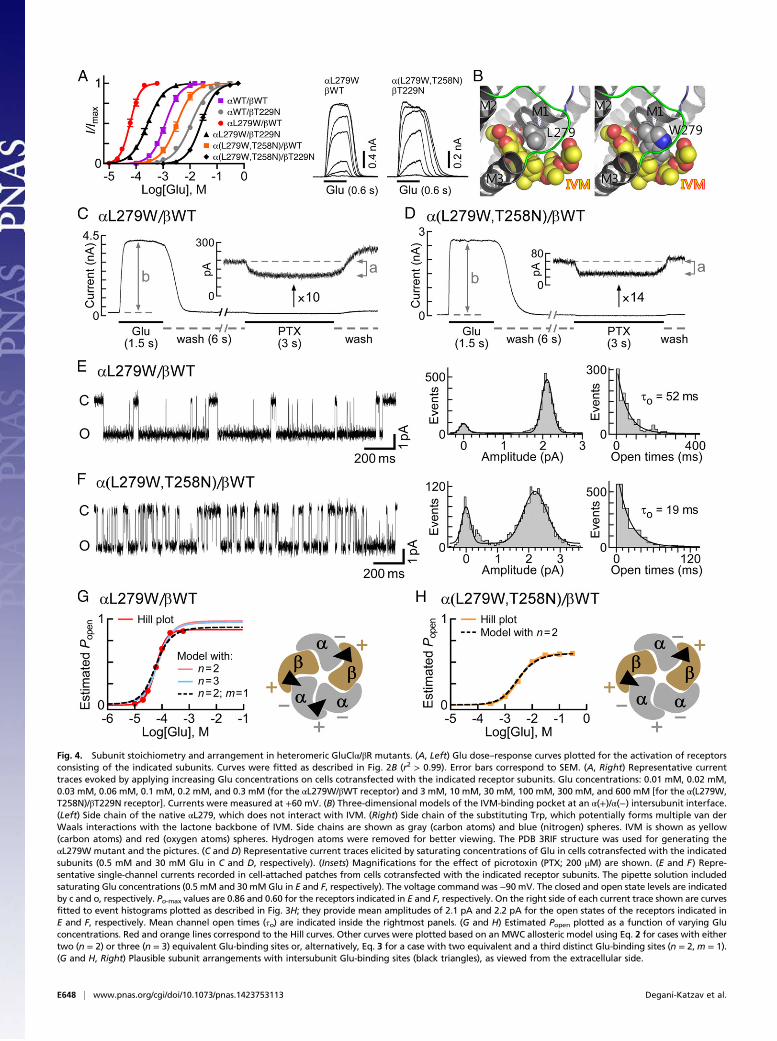
Fig. 4. Subunit stoichiometry and arrangement in heteromeric GluClα/βR mutants. (A, Left) Glu dose–response curves plotted for the activation of receptorsconsisting of the indicated subunits. Curves were fitted as described in Fig. 2B (r2 > 0.99). Error bars correspond to SEM. (A, Right) Representative currenttraces evoked by applying increasing Glu concentrations on cells cotransfected with the indicated receptor subunits. Glu concentrations: 0.01 mM, 0.02 mM,0.03 mM, 0.06 mM, 0.1 mM, 0.2 mM, and 0.3 mM (for the αL279W/βWT receptor) and 3 mM, 10 mM, 30 mM, 100 mM, 300 mM, and 600 mM [for the α(L279W,T258N)/βT229N receptor]. Currents were measured at +60 mV. (B) Three-dimensional models of the IVM-binding pocket at an α(+)/α(−) intersubunit interface.(Left) Side chain of the native αL279, which does not interact with IVM. (Right) Side chain of the substituting Trp, which potentially forms multiple van derWaals interactions with the lactone backbone of IVM. Side chains are shown as gray (carbon atoms) and blue (nitrogen) spheres. IVM is shown as yellow(carbon atoms) and red (oxygen atoms) spheres. Hydrogen atoms were removed for better viewing. The PDB 3RIF structure was used for generating theαL279W mutant and the pictures. (C and D) Representative current traces elicited by saturating concentrations of Glu in cells cotransfected with the indicatedsubunits (0.5 mM and 30 mM Glu in C and D, respectively). (Insets) Magnifications for the effect of picrotoxin (PTX; 200 μM) are shown. (E and F) Repre-sentative single-channel currents recorded in cell-attached patches from cells cotransfected with the indicated receptor subunits. The pipette solution includedsaturating Glu concentrations (0.5 mM and 30 mMGlu in E and F, respectively). The voltage command was −90 mV. The closed and open state levels are indicatedby c and o, respectively. Po-max values are 0.86 and 0.60 for the receptors indicated in E and F, respectively. On the right side of each current trace shown are curvesfitted to event histograms plotted as described in Fig. 3H; they provide mean amplitudes of 2.1 pA and 2.2 pA for the open states of the receptors indicated inE and F, respectively. Mean channel open times (τo) are indicated inside the rightmost panels. (G and H) Estimated Popen plotted as a function of varying Gluconcentrations. Red and orange lines correspond to the Hill curves. Other curves were plotted based on an MWC allosteric model using Eq. 2 for cases with eithertwo (n = 2) or three (n = 3) equivalent Glu-binding sites or, alternatively, Eq. 3 for a case with two equivalent and a third distinct Glu-binding sites (n = 2,m = 1).(G and H, Right) Plausible subunit arrangements with intersubunit Glu-binding sites (black triangles), as viewed from the extracellular side.
E648 | www.pnas.org/cgi/doi/10.1073/pnas.1423753113 Degani-Katzav et al.

(Fig. 4A and Table S1). This mutation increased the Hill co-efficient to 2.6, suggesting that the number of occupiable Glu-binding sites in the receptor mutant is probably not less than three.Intrigued by this possibility, we initially examined an MWC allo-steric model with either two or three equivalent Glu-binding sites.To this end, we determined the values of Ispont, Imax, Po-max, and Lfor the GluClαL279W/βWT receptor [Fig. 4 C and E and Table S2(footnotes)] and estimated its Popen at varying Glu concentrations,all as described above for the GluClαWT/βWT receptor. Then, acurve was fitted to the normalized dose–response data points usingan MWC allosteric model with n = 2 and the experimental meanL value (81) (Fig. 4G, salmon-colored curve and Eq. 2). Theresulting Kd values (Table S2, same line of “2, 0”) were applied tocalculate the theoretical Po-max* by 1/(1 + c2L) = 0.98, whichturned out to be much higher than the experimental Po-max (0.86).Extrapolating the salmon-colored curve in Fig. 4G (model withn = 2) until the theoretical Po-max* is reached indicates a strongdeviation of this curve from the Hill plot at high Glu concentra-tions. Alternatively, a curve was fitted to the normalized dose–response data points using an MWC allosteric model with threeequivalent Glu-binding sites (n = 3) and the same L value (81)(Fig. 4G, cyan-colored curve and Eq. 2). The resulting Kd values(Table S2, same line of “3, 0”) were used to calculate the theo-retical Po-max* by 1/(1 + c3L) = 0.96, which is also much higherthan the experimental Po-max (0.86). Extrapolation of the cyan-colored curve in Fig. 4G (model with n = 3) until the theoreticalPo-max* is reached indicates a strong deviation of this curve fromthe Hill plot at high Glu concentrations. Curve fitting with othervalues for n (one, or equivalent four or five Glu-binding sites)resulted in a theoretical Po-max* ≥ 0.95 (Table S2). We thereforeapplied an MWC allosteric model with two equivalent and a thirddistinct Glu-binding sites (n = 2, m = 1), using the same L value(81) (Fig. 4G, dashed black curve and Eq. 3). In this case, Kd,R andKd,R* characterize the two equivalent Glu-binding sites in theclosed and open states, respectively; and K′d,R and K′d,R* char-acterize the third Glu-binding site in the closed and open states,respectively [Table S2 (in bold)]. Scheme II describes the MWCallosteric mechanism that corresponds to the GluClαL279W/βWTreceptor:
where c = Kd,R*/Kd,R and c′ = K′d,R*/K′d,R. In this case, thetheoretical Po-max* = 1/(1 + cnc′mL) = 1/(1 + c2c′1L) = 0.89,which is much closer to the experimental Po-max (0.86) than incases of curve fitting with other numbers of Glu-binding sites(Table S2). Analysis of the ΔAICc selected the allosteric modelwith two equivalent and a third distinct Glu-binding sites (n = 2,m = 1) as the most appropriate MWC model for curve fitting inthe GluClαL279W/βWT receptor case (Table S2).The allosteric mechanism suggested above does not provide
details regarding the subunit types that form the third Glu-binding site interface in the GluClαL279W/βWT receptor,however. If the fifth subunit is GluClβ, then it will give rise toα(+)/β(−) and β(+)/β(−) intersubunit interfaces (envisioned inFig. 3J); however, based on the aforementioned results, the
GluClβ(−) side is less likely to contribute to Glu binding. If thefifth subunit is GluClα, then it will give rise to α(+)/α(−) andα(+)/β(−) intersubunit interfaces (envisioned in Fig. 4G, Right);so, the α(+)/α(−) intersubunit interface remains a reasonablecandidate to form the third Glu-binding site. However, thisworking hypothesis required further experimental investigation.Because the GluClα(−) side was inferred to line the two Glu-binding pockets (Fig. 3 and main text), we introduced an α(+)T258Nmutation (in loop C), in addition to the αL279W mutation. Thehomologous mutation [β(+)T229N] in the GluClαWT/βT229Nreceptor was shown to increase the Glu-EC50 by approximatelyeightfold, compared with the GluClαWT/βWT receptor (Fig. 3Cand Table S1; presented again in Fig. 4A in gray for conve-nience). Hence, an α(+)T258N mutation was anticipated to af-fect a potential α(+)/α(−) intersubunit Glu-binding site, withoutdirectly interfering with Glu binding at the two β(+)/α(−) sites.Fig. 4A shows that the dose–response curve of the GluClα(L279W,T258N)/βWT receptor is significantly shifted to theright relative to the curve of the GluClαL279W/βWT receptor,with an ∼57-fold increase in the Glu-EC50 and a decrease of theHill coefficient to nH = 1.6 (Table S1). These macroscopicproperties resemble the properties displayed by the GluClαWT/βWT receptor, which has two equivalent Glu-binding sites.To quantify the effect of the α(+)T258N mutation further, we
determined the values of Ispont, Imax, Po-max, and L for the GluClα(L279W,T258N)/βWT receptor [Fig. 4 D and F and Table S2(footnotes)] and estimated its Popen at varying Glu concentra-tions, all as described above for the GluClαWT/βWT receptor.Then, a curve was fitted to the normalized dose–response datapoints using an MWC allosteric model with two equivalent Glu-binding sites (n = 2) and the experimental mean L value (203)(Fig. 4H, dashed black curve and Eq. 2). The resulting Kd valuesare provided in Table S2 (in bold). The theoretical and experi-mental maximum open probabilities were found to be equal(0.60), whereas other values for n (one, or equivalent three, four,or five Glu-binding sites) resulted in higher theoretical Po-max*values (Table S2). In addition, the analysis of the ΔAICc selectedthe allosteric model with n = 2 as the most suitable MWC modelfor curve fitting in the GluClα(L279W,T258N)/βWT receptorcase (Table S2). Hence, the results imply that this double-mutantreceptor lost the third Glu-binding site, and its remaining twoequivalent Glu-binding sites display slightly lower affinity for Gluthan the GluClαWT/βWT receptor [Table S2 (in bold)]. Providedthat the mutations have not changed the subunit stoichiometry(as argued in SI Text, section 3), the two Glu-binding sites of theGluClα(L279W,T258N)/βWT receptor likely lie at β(+)/α(−)intersubunit interfaces (Fig. 4H, Right). As discussed above, theGluClβ(−) side is less likely to contribute to Glu binding, and so isan α(+)/β(−) intersubunit interface. We therefore infer that theα(+)T258N mutation is likely located at an α(+)/α(−) inter-subunit interface. Taken together, our results suggest that in theGluClαL279W/βWT receptor, an α(+)/α(−) intersubunit interfacelikely forms a third Glu-binding site (Fig. 4G, Right), whereas Glubinding to this interface is impaired by adding the α(+)T258Nmutation (Fig. 4H, Right).β(+)T229N is the homologous mutation of α(+)T258N. Com-
bining the αL279W mutation with the β(+)T229N mutation, togive a GluClαL279W/βT229N receptor, led to a fivefold rightwardshift of the dose–response curve relative to the GluClαL279W/βWT receptor (Fig. 4A and Table S1). This shift is much smallerthan the 57-fold rightward shift observed in the GluClα(L279W,T258N)/βWT receptor relative to the GluClαL279W/βWT re-ceptor (Fig. 4A and Table S1). This difference is in line with theabove conclusion that an α(+)/α(−) intersubunit interface formsthe third Glu-binding site in the GluClαL279W/βWT receptor.Interestingly, the α(+)T258N mutation in the GluClα(L279W,
T258N)/βWT receptor has not only eliminated the third α(+)/α(−)intersubunit Glu-binding site but also considerably decreased the
Degani-Katzav et al. PNAS | Published online January 20, 2016 | E649
PHYS
IOLO
GY
PNASPL
US
Glu-binding affinity of the two equivalent β(+)/α(−) interfacesrelative to the GluClαL279W/βWT receptor [Table S2 (in bold)].We suggest that the mutation in the (+) side of the plausibleα(+)/α(−) Glu-binding site interface could allosterically affect theother Glu-binding site interfaces. Combining all three mutations toproduce a GluClα(L279W,T258N)/βT229N receptor shifted thedose–response curve by 455-fold rightward relative to theGluClαL279W/βWT receptor (Fig. 4A and Table S1). This right-ward shift is larger by ∼90-fold than the fivefold rightward shiftobserved for the GluClαL279W/βT229N receptor, which suggeststhat also in the triple mutant, the α(+)/α(−) intersubunit interfacehas a strong allosteric relationship with the β(+)/α(−) Glu-bindingsite interfaces. Notably, the Hill coefficient decreased from nH =2.6 in the GluClαL279W/βWT receptor to nH = 1.5 in theGluClαL279W/βT229N receptor (Table S1), suggesting that theβ(+)T229N mutation exerts a reciprocal allosteric effect onthe third α(+)/α(−) intersubunit interface.
Effect of the αL279WMutation on the Responsiveness of the HeteromericGluClαL279W/βWT Receptor to IVM. The crystal structure of thehomomeric GluClαcrystR indicates that the backbone carbonyloxygen of αL279 (L218 in GluClαcrystR) forms a hydrogen bondwith hydroxyl O13-H of IVM, whereas the αL279 side chain does
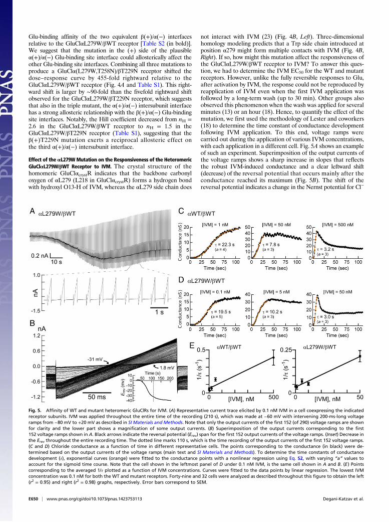
not interact with IVM (23) (Fig. 4B, Left). Three-dimensionalhomology modeling predicts that a Trp side chain introduced atposition α279 might form multiple contacts with IVM (Fig. 4B,Right). If so, how might this mutation affect the responsiveness ofthe GluClαL279W/βWT receptor to IVM? To answer this ques-tion, we had to determine the IVM EC50 for the WT and mutantreceptors. However, unlike the fully reversible responses to Glu,after activation by IVM, the response could not be reproduced byreapplication of IVM even when the first IVM application wasfollowed by a long-term wash (up to 30 min). Other groups alsoobserved this phenomenon when the wash was applied for severalminutes (13) or an hour (18). Hence, to quantify the effect of themutation, we first used the methodology of Lester and coworkers(18) to determine the time constant of conductance developmentfollowing IVM application. To this end, voltage ramps werecarried out during the application of various IVM concentrations,with each application in a different cell. Fig. 5A shows an exampleof such an experiment. Superimposition of the output currents ofthe voltage ramps shows a sharp increase in slopes that reflectsthe robust IVM-induced conductance and a clear leftward shift(decrease) of the reversal potential that occurs mainly after theconductance reached its maximum (Fig. 5B). The shift of thereversal potential indicates a change in the Nernst potential for Cl−
Fig. 5. Affinity of WT and mutant heteromeric GluClRs for IVM. (A) Representative current trace elicited by 0.1 nM IVM in a cell coexpressing the indicatedreceptor subunits. IVM was applied throughout the entire time of the recording (210 s), which was made at −60 mV with intervening 200-ms-long voltageramps from −80 mV to +20 mV as described in SI Materials and Methods. Note that only the output currents of the first 152 (of 290) voltage ramps are shownfor clarity and the lower part shows a magnification of some output currents. (B) Superimposition of the output currents corresponding to the first152 voltage ramps shown in A. Black arrows indicate the reversal potential (Erev) span for the first 152 output currents of the voltage ramps. (Inset) Decrease inthe Erev throughout the entire recording time. The dotted line marks 110 s, which is the time recording of the output currents of the first 152 voltage ramps.(C and D) Chloride conductance as a function of time in different representative cells. The points corresponding to the conductance (in black) were de-termined based on the output currents of the voltage ramps (main text and SI Materials and Methods). To determine the time constants of conductancedevelopment (τ), exponential curves (orange) were fitted to the conductance points with a nonlinear regression using Eq. S2, with varying “a” values toaccount for the sigmoid time course. Note that the cell shown in the leftmost panel of D under 0.1 nM IVM, is the same cell shown in A and B. (E) Pointscorresponding to the averaged 1/τ plotted as a function of IVM concentrations. Curves were fitted to the data points by linear regression. The lowest IVMconcentration was 0.1 nM for both the WT and mutant receptors. Forty-nine and 32 cells were analyzed as described throughout this figure to obtain the left(r2 = 0.95) and right (r2 = 0.98) graphs, respectively. Error bars correspond to SEM.
E650 | www.pnas.org/cgi/doi/10.1073/pnas.1423753113 Degani-Katzav et al.

and in the electrochemical driving force acting on Cl− ions. Thechloride conductance is defined by the slope of the current–voltage(I/V) relations extracted from the output currents of the voltageramps, and could be determined at several membrane voltagespans. Fig. S6A shows the slope conductance determined between−75 mV and −65 mV, around the reversal potential, and between+10 mV and +20 mV, as a function of time. The rise time of theconductance increment was found to be similar for all of the threeaforementioned voltage spans (Fig. S6A). Notably, during theapplications of high IVM concentrations, the conductance risewas followed by a decrease in the conductance to a steady state inall voltage spans and in both the WT and indicated mutant re-ceptors (Fig. S6A). Because the current decay under high IVMconcentrations was faster at −65 mV than at +20 mV (Fig. S6B),and because the exponential fits of the conductance rise time werevery similar at the different membrane voltage spans, we chose toanalyze the conductance development further between +10 mVand +20 mV. Fig. 5 C and D shows the development of the con-ductance under the application of different IVM concentrations indifferent representative cells.The exponential fits of the conductance rise time (e.g., Fig. 5 C
and D, orange curves) provide the time constant of conductancedevelopment (τ), whose reciprocal (1/τ) increased linearly withthe increase in IVM concentration (Fig. 5E). Because IVM doesnot readily dissociate from the receptor (13, 18) and the numberof possible intermediate IVM-bound closed states is not known,the simplest possible kinetic model that could describe the ac-tivation mechanism by IVM would be one in which the channelopens when IVM binds and closes after a very long time whenIVM dissociates. Scheme III describes this kinetic model:
where R is the unoccupied closed receptor, IVM·R* is the IVM-bound open receptor, and 1/τ = kf [IVM] + kb. The slope of thecurves in Fig. 5E corresponds to the IVM association rate constant(k forward, kf). The IVM dissociation rate constant (k backward,kb) is the extrapolated intercept of the linear curve with the y axis inFig. 5E. The apparent Kd would be kb/kf, giving 73 × 10−9 M forIVM binding to the GluClαWT/βWT receptor (kb = 5.3 × 10⁻² s−1
and kf = 7.3 × 10⁵ s−1·M−1). In contrast, the apparent Kd for IVMbinding to the GluClαL279W/βWT receptor was 9.7 × 10⁻⁹ M(kb = 3.4 × 10⁻² s−1 and kf = 3.5 × 10⁶ s−1·M−1), which indicatesthat the affinity of the mutant receptor for IVM is 7.5-foldhigher than the affinity of the WT receptor for IVM. Note thatbecause no experiments revealed that IVM could be washed outof the receptor (13, 18), the kb values are expected to be on theorder of <10−4 s−1. However, the values here were found to beon the order of 10−2 s−1, implying that IVM should be remov-able. We therefore cannot exclude the possibility that afteropening of the GluClR ion channel by IVM, a subsequentconformational change leads to trapping of IVM between thetransmembrane helices irreversibly.
DiscussionTo determine unequivocally the subunit stoichiometry and ar-rangement in native GluClα/βRs, high-resolution X-ray crystal-lography of heteropentameric receptors purified from theorganisms that naturally express them is necessary. To the best ofour knowledge, such a determination is yet out of reach. Hence,an alternative methodology must be considered. In Cys-loopreceptors, the neurotransmitter-binding pockets lie at the in-terface between adjacent subunits (1–9). One could therefore usesite-specific mutagenesis and biophysical characterization of acti-
vation mechanisms in recombinant receptors to find the types ofsubunits that line the agonist-binding pockets. By working withrecombinant receptors, however, one cannot exclude the possibilitythat the ratio of subunit cDNA transfected, the type of theexpressing cell, or a mutation might influence the receptor’s sub-unit composition (e.g., 45, 57, 58). We nevertheless argue that thespecific mutations we introduced are less likely to change thesubunit stoichiometry of the recombinant receptors studied here(SI Text, section 3).In various Cys-loop receptors, the β1β2, Cys, and β8β9 loops
were shown to play a key role in transducing the agonist-bindingenergy into ion-channel gating force (35–44). Here, we firstdemonstrated that although the homomeric GluClαR is not re-sponsive to Glu, the β1β2, Cys, and β8β9 loops of the GluClαsubunit are fully capable of coupling Glu binding to channelgating in a heteromeric GluClα/β microchimera that has the se-quences of the α-subunit loops. Subsequently, we undertook toidentify the intersubunit interfaces involved in Glu accommoda-tion by heteromeric GluClα/βRs. Taking advantage of the crystalstructure of a truncated homomeric GluClαcrystR as a template,we built a 3D homology model for the GluClβ subunit. Then,based on the two structures, we introduced single-site mutationsin the (−) side of either the GluClα subunit or the GluClβ subunitat positions carrying residues that putatively interact with Glu.Characterization of the effects of these mutations on the receptorfunction allowed us to suggest that in the heteromeric GluClα/βRs studied here, the (−) side of the α subunit, rather than the(−) side of the β subunit, contributes complementary componentsto Glu binding. Single-site mutations and functional analysis ofheteromeric GluClα/βRs carrying mutations in the (+) side of theβ subunit imply that this side contributes principal components toGlu binding.When considering the GluClαWT/βWT receptor in terms of
the MWC allosteric mechanism, we infer that a maximum of twoequivalent binding sites can be occupied by Glu [Fig. 3I, Scheme I,and Table S2 (in bold)]. Provided that the aforementioned single-site mutations introduced at the intersubunit interfaces have notchanged the subunit stoichiometry (as argued in SI Text, section 3),Glu binding likely takes place at two β(+)/α(−) intersubunit in-terfaces. Hence, one can envision a subunit arrangement asillustrated in Fig. 3J for a recombinant GluClαWT/βWT receptorexpressed in CHO cells, with no information regarding the typeof the fifth subunit.When considering the GluClαL279W/βWT receptor in terms
of the MWC allosteric mechanism, we infer that Glu can occupythree sites (Fig. 4G and Scheme II). These sites are (i) two equiv-alent Glu-binding sites that are likely located at β(+)/α(−) inter-subunit interfaces and display considerably higher affinity for Gluthan their homologous binding sites in the GluClαWT/βWT re-ceptor and (ii) a third distinct site with slightly lower Glu-bindingaffinity, in both the resting (closed) and active (open) receptorstates [Table S2 (in bold)]. We argue that the third Glu-bindingsite is formed between two adjacent α subunits; the arguments forthat conclusion are as follows:
i) In CHO cells, a WT GluClβ subunit does not assemble intoa homopentamer capable of responding to Glu or IVM, whichindicates that the β subunit has difficulties in creating Glu-bindingβ(+)/β(−) intersubunit interfaces (Fig. S2A and Table S1).
ii) In the heteromeric GluClα/βRs studied here, three single-site mutations in the β(−) side did not lead to drastic effectson the receptor activation by Glu, unlike the case of thesame mutations introduced at the homologous positions inthe α(−) side.
iii) The homomeric GluClαL279W receptor responds to veryhigh Glu concentrations (Fig. S7), indicating the capabilityof an α(+)/α(−) intersubunit interface to accommodate Glu(with no need for IVM prebinding).
Degani-Katzav et al. PNAS | Published online January 20, 2016 | E651
PHYS
IOLO
GY
PNASPL
US
iv) Adding a mutation in loop C (+ side) of the αL279W sub-unit gave rise to an α(L279W,T258N)/βWT receptor thatlost the third Glu-binding site (Fig. 4H), whereas theremaining two equivalent Glu-binding sites display micro-scopic equilibrium dissociation constants slightly higher thanthe microscopic equilibrium dissociation constants of theGluClαWT/βWT receptor [Table S2 (in bold)].
A third Glu-binding site located at an α(+)/α(−) intersubunitinterface requires that the fifth subunit would be a GluClα sub-unit. We therefore suggest that the subunits of the recombinantheteromeric GluClα/βRs studied here assemble in an anticlock-wise β-α-β-α-α fashion, as viewed from the extracellular side (Fig.4 G and H, Right). Notably, previous studies show that expressingthe heteromeric α4β2 nAChR under conditions that favor an(α4β2)2α4 stoichiometry (three α4 and two β2 subunits) results in areceptor having two α4(+)/β2(−) interfaces with high agonistsensitivity and a third binding site at the α4(+)/α4(−) interface thatdisplays low agonist sensitivity (59–61).As discussed in Results, the function of heteromeric receptors
containing the αL279W mutation, together with a Thr→Asn sub-stitution in loop C of the α subunit, β subunit, or both subunits,suggests that the two intersubunit interface types, α(+)/α(−) andβ(+)/α(−), likely affect each other allosterically. Possible struc-tural reasons for this mutual allosteric influence are provided inSI Text, section 4. Interestingly, an allosteric relationship betweendifferent extracellular intersubunit interfaces was proposed forthe heteromeric α1β2γ2 GABAA receptor (62). In the latter case,conformational movements induced by benzodiazepine binding atthe α/γ extracellular interface were suggested to propagate acrossthe α1 subunit to the β/α GABA-binding site interface (62).In the GluClαcrystR, L218 (αL279 in the full-length subunit
used here) is part of the IVM-binding pocket located betweenM1 and M3 of adjacent subunits (23) (Fig. 4B, Left). The clearincrease in the affinity of the GluClαL279W/βWT receptor forIVM (Fig. 5E; 7.5-fold) implies that the IVM-binding pockets ofthe heteromeric receptor are homologous to the IVM-bindingpockets of the homomeric GluClαcrystR. The structural mecha-nism underlying the effect of the αL279W mutation in the IVM-binding site is not clear. However, the microscopic equilibriumdissociation constants for Glu binding determined here implythat the conformational change induced by this mutation in theIVM-binding pocket propagates to the Glu-binding pockets andaffects their affinity for Glu. It is not known whether Glu andIVM induce the same conformational change in the couplingloops. In the heteromeric α1β2γ2 GABAA receptor, for example,it was demonstrated that positive benzodiazepine modulatorsinduce movements in loop F (β8β9 loop) of the γ2 subunit nearthe transmembrane channel domain (63). Such movements were
not triggered by the binding of GABA, the allosteric modulatorpentobarbital, or the inverse agonist methyl-6,7-dimethoxy-4-ethyl-β-carboline-3-carboxylate (63).In conclusion, our study provides evidence that the C. elegans
heteromeric GluClR contains three α subunits and two β sub-units arranged in an anticlockwise β-α-β-α-α fashion, as viewedfrom the extracellular side, with two Glu-binding sites located atthe β(+)/α(−) intersubunit interfaces. The α(+)/α(−) inter-subunit interface creates a third “dormant” Glu-binding site thatbecomes functional upon a conformational change induced by amutation in the IVM-binding pocket.
Materials and MethodsAdditional experimental procedures and data analyses are described inSI Materials and Methods.
Data analysis and mathematical modeling were performed using theClampfit 10 program implemented in pClamp 10, and GraphPad Prismsoftware.
Dose–response curves were fitted to the data points by a nonlinear re-gression using the Hill equation (Eq. 1):
IImax
=1
1+ 10ðlogEC50−log½Glu�ÞnH
, [1]
where I is the current response, Imax is the maximal current response, EC50 isthe agonist effective concentration that elicits 50% of the maximal currentresponse, [Glu] is the concentration of Glu, and nH is the Hill coefficient.
For the allosteric modeling, Eq. 2 was used:
Popen =1
1+ L
(1+ ½Glu�=Kd,R
1+ ½Glu��Kd,R*
)n, [2]
where Popen is the open probability estimated at varying Glu concentrations(54) (main text). [Glu] is the concentration of the agonist (Glu) for whichthere are n equivalent binding sites, each with a microscopic equilibriumdissociation constant of Kd,R in the resting (closed) state and Kd,R* in theactive (open) state. L is the equilibrium constant of the two states in theabsence of ligands. The L values were determined by functional experi-ments, as described in the main text.
For a receptor phenotype that does not behave as a receptor having only nequivalent Glu-binding sites, Eq. 3 [cf. Karlin (48)] was used:
Popen =1
1+ L
(1+ ½Glu�=Kd,R
1+ ½Glu��Kd,R*
)n(1+ ½Glu�
�K′d,R
1+ ½Glu��K′d,R*
)m, [3]
where m is the number of sites that Glu binds with microscopic equilibriumdissociation constants, K′d,R in the closed state and K′d,R* in the open state.
ACKNOWLEDGMENTS. We thank H. A. Lester for providing us with the initialGluClα subunit construct.
1. Karlin A (2002) Emerging structure of the nicotinic acetylcholine receptors. Nat RevNeurosci 3(2):102–114.
2. Lester HA, Dibas MI, Dahan DS, Leite JF, Dougherty DA (2004) Cys-loop receptors: Newtwists and turns. Trends Neurosci 27(6):329–336.
3. Betz H, Laube B (2006) Glycine receptors: Recent insights into their structural orga-nization and functional diversity. J Neurochem 97(6):1600–1610.
4. Sine SM, Engel AG (2006) Recent advances in Cys-loop receptor structure and func-tion. Nature 440(7083):448–455.
5. Taylor P, et al. (2007) Structure-guided drug design: Conferring selectivity amongneuronal nicotinic receptor and acetylcholine-binding protein subtypes. BiochemPharmacol 74(8):1164–1171.
6. Olsen RW, Sieghart W (2008) International Union of Pharmacology. LXX. Subtypes ofgamma-aminobutyric acid(A) receptors: Classification on the basis of subunit com-position, pharmacology, and function. Update. Pharmacol Rev 60(3):243–260.
7. Changeux JP (2010) Allosteric receptors: From electric organ to cognition. Annu RevPharmacol Toxicol 50:1–38.
8. Thompson AJ, Lester HA, Lummis SC (2010) The structural basis of function in Cys-loopreceptors. Q Rev Biophys 43(4):449–499.
9. Lynagh T, Lynch JW (2012) Molecular mechanisms of Cys-loop ion channel receptormodulation by ivermectin. Front Mol Neurosci 5:60.
10. Crump A, �Omura S (2011) Ivermectin, ‘wonder drug’ from Japan: The human useperspective. Proc Jpn Acad, Ser B, Phys Biol Sci 87(2):13–28.
11. Campbell WC (2012) History of avermectin and ivermectin, with notes on the historyof other macrocyclic lactone antiparasitic agents. Curr Pharm Biotechnol 13(6):853–865.
12. Geary TG (2005) Ivermectin 20 years on: Maturation of a wonder drug. Trends Parasitol21(11):530–532.
13. Cully DF, et al. (1994) Cloning of an avermectin-sensitive glutamate-gated chloridechannel from Caenorhabditis elegans. Nature 371(6499):707–711.
14. Etter A, Cully DF, Schaeffer JM, Liu KK, Arena JP (1996) An amino acid substitution inthe pore region of a glutamate-gated chloride channel enables the coupling of ligandbinding to channel gating. J Biol Chem 271(27):16035–16039.
15. Dent JA, Davis MW, Avery L (1997) avr-15 encodes a chloride channel subunit thatmediates inhibitory glutamatergic neurotransmission and ivermectin sensitivity inCaenorhabditis elegans. EMBO J 16(19):5867–5879.
16. Vassilatis DK, et al. (1997) Genetic and biochemical evidence for a novel avermectin-sensitive chloride channel in Caenorhabditis elegans. Isolation and characterization.J Biol Chem 272(52):33167–33174.
17. Li P, Slimko EM, Lester HA (2002) Selective elimination of glutamate activation andintroduction of fluorescent proteins into a Caenorhabditis elegans chloride channel.FEBS Lett 528(1-3):77–82.
18. Slimko EM, McKinney S, Anderson DJ, Davidson N, Lester HA (2002) Selective elec-trical silencing of mammalian neurons in vitro by the use of invertebrate ligand-gatedchloride channels. J Neurosci 22(17):7373–7379.
E652 | www.pnas.org/cgi/doi/10.1073/pnas.1423753113 Degani-Katzav et al.
19. Cook A, et al. (2006) Caenorhabditis elegans ivermectin receptors regulate locomotorbehaviour and are functional orthologues of Haemonchus contortus receptors. MolBiochem Parasitol 147(1):118–125.
20. Brownlee DJ, Holden-Dye L, Walker RJ (1997) Actions of the anthelmintic ivermectinon the pharyngeal muscle of the parasitic nematode, Ascaris suum. Parasitology 115(Pt 5):553–561.
21. Moreno Y, Nabhan JF, Solomon J, Mackenzie CD, Geary TG (2010) Ivermectin disruptsthe function of the excretory-secretory apparatus in microfilariae of Brugia malayi.Proc Natl Acad Sci USA 107(46):20120–20125.
22. Daeffler KN, Lester HA, Dougherty DA (2014) Functional evaluation of key interac-tions evident in the structure of the eukaryotic Cys-loop receptor GluCl. ACS ChemBiol 9(10):2283–2290.
23. Hibbs RE, Gouaux E (2011) Principles of activation and permeation in an anion-selective Cys-loop receptor. Nature 474(7349):54–60.
24. Miller PS, Aricescu AR (2014) Crystal structure of a human GABAA receptor. Nature512(7514):270–275.
25. Bocquet N, et al. (2009) X-ray structure of a pentameric ligand-gated ion channel inan apparently open conformation. Nature 457(7225):111–114.
26. Hilf RJ, Dutzler R (2009) Structure of a potentially open state of a proton-activatedpentameric ligand-gated ion channel. Nature 457(7225):115–118.
27. Pan J, et al. (2012) Structure of the pentameric ligand-gated ion channel ELIC coc-rystallized with its competitive antagonist acetylcholine. Nat Commun 3:714.
28. Gonzalez-Gutierrez G, et al. (2012) Mutations that stabilize the open state of theErwinia chrisanthemi ligand-gated ion channel fail to change the conformation of thepore domain in crystals. Proc Natl Acad Sci USA 109(16):6331–6336.
29. Howard RJ, et al. (2011) Structural basis for alcohol modulation of a pentamericligand-gated ion channel. Proc Natl Acad Sci USA 108(29):12149–12154.
30. Spurny R, et al. (2012) Pentameric ligand-gated ion channel ELIC is activated by GABAand modulated by benzodiazepines. Proc Natl Acad Sci USA 109(44):E3028–E3034.
31. Brejc K, et al. (2001) Crystal structure of an ACh-binding protein reveals the ligand-binding domain of nicotinic receptors. Nature 411(6835):269–276.
32. Celie PH, et al. (2004) Nicotine and carbamylcholine binding to nicotinic acetylcholinereceptors as studied in AChBP crystal structures. Neuron 41(6):907–914.
33. Hansen SB, et al. (2005) Structures of Aplysia AChBP complexes with nicotinic agonistsand antagonists reveal distinctive binding interfaces and conformations. EMBO J24(20):3635–3646.
34. Hibbs RE, et al. (2009) Structural determinants for interaction of partial agonists withacetylcholine binding protein and neuronal alpha7 nicotinic acetylcholine receptor.EMBO J 28(19):3040–3051.
35. Grosman C, Salamone FN, Sine SM, Auerbach A (2000) The extracellular linker ofmuscle acetylcholine receptor channels is a gating control element. J Gen Physiol116(3):327–340.
36. Kash TL, Jenkins A, Kelley JC, Trudell JR, Harrison NL (2003) Coupling of agonistbinding to channel gating in the GABA(A) receptor. Nature 421(6920):272–275.
37. Bouzat C, et al. (2004) Coupling of agonist binding to channel gating in an ACh-binding protein linked to an ion channel. Nature 430(7002):896–900.
38. Reeves DC, Jansen M, Bali M, Lemster T, Akabas MH (2005) A role for the beta 1-beta2 loop in the gating of 5-HT3 receptors. J Neurosci 25(41):9358–9366.
39. Jha A, Cadugan DJ, Purohit P, Auerbach A (2007) Acetylcholine receptor gating atextracellular transmembrane domain interface: the cys-loop and M2-M3 linker. J GenPhysiol 130(6):547–558.
40. Bouzat C, Bartos M, Corradi J, Sine SM (2008) The interface between extracellular andtransmembrane domains of homomeric Cys-loop receptors governs open-channellifetime and rate of desensitization. J Neurosci 28(31):7808–7819.
41. LeeWY, Free CR, Sine SM (2009) Binding to gating transduction in nicotinic receptors: Cys-loop energetically couples to pre-M1 and M2-M3 regions. J Neurosci 29(10):3189–3199.
42. Perkins DI, et al. (2009) Loop 2 structure in glycine and GABA(A) receptors plays a keyrole in determining ethanol sensitivity. J Biol Chem 284(40):27304–27314.
43. Pless SA, Lynch JW (2009) Magnitude of a conformational change in the glycine re-ceptor beta1-beta2 loop is correlated with agonist efficacy. J Biol Chem 284(40):27370–27376.
44. Wiltfong RE, Jansen M (2009) Probing protein packing surrounding the residues inand flanking the nicotinic acetylcholine receptor M2M3 loop. J Neurosci 29(6):1626–1635.
45. Frazier SJ, Cohen BN, Lester HA (2013) An engineered glutamate-gated chloride(GluCl) channel for sensitive, consistent neuronal silencing by ivermectin. J Biol Chem288(29):21029–21042.
46. Yoluk O, Brömstrup T, Bertaccini EJ, Trudell JR, Lindahl E (2013) Stabilization of theGluCl ligand-gated ion channel in the presence and absence of ivermectin. Biophys J105(3):640–647.
47. Monod J, Wyman J, Changeux JP (1965) On the nature of allosteric transitions: Aplausible model. J Mol Biol 12:88–118.
48. Karlin A (1967) On the application of “a plausible model” of allosteric proteins to thereceptor for acetylcholine. J Theor Biol 16(2):306–320.
49. Edelstein SJ, Changeux JP (1996) Allosteric proteins after thirty years: The binding andstate functions of the neuronal alpha 7 nicotinic acetylcholine receptors. Experientia52(12):1083–1090.
50. Auerbach A (2012) Thinking in cycles: MWC is a good model for acetylcholinereceptor-channels. J Physiol 590(Pt 1):93–98.
51. Corringer PJ, et al. (1999) Mutational analysis of the charge selectivity filter of thealpha7 nicotinic acetylcholine receptor. Neuron 22(4):831–843.
52. Pittel I, Witt-Kehati D, Degani-Katzav N, Paas Y (2010) Probing pore constriction in aligand-gated ion channel by trapping a metal ion in the pore upon agonist dissoci-ation. J Biol Chem 285(34):26519–26531.
53. Chang Y, Weiss DS (1999) Allosteric activation mechanism of the alpha 1 beta 2gamma 2 gamma-aminobutyric acid type A receptor revealed by mutation of theconserved M2 leucine. Biophys J 77(5):2542–2551.
54. Forman SA (2012) Monod-Wyman-Changeux allosteric mechanisms of action and thepharmacology of etomidate. Curr Opin Anaesthesiol 25(4):411–418.
55. Etter A, et al. (1999) Picrotoxin blockade of invertebrate glutamate-gated chloridechannels: Subunit dependence and evidence for binding within the pore.J Neurochem 72(1):318–326.
56. Burnham KP, Anderson DR (2002) Model Selection and Multimodel Inference: APractical Information-Theoretic Approach (Springer, New York), 2nd Ed.
57. Wagoner KR, Czajkowski C (2010) Stoichiometry of expressed alpha(4)beta(2)deltagamma-aminobutyric acid type A receptors depends on the ratio of subunit cDNAtransfected. J Biol Chem 285(19):14187–14194.
58. Krashia P, et al. (2010) Human α3β4 neuronal nicotinic receptors show differentstoichiometry if they are expressed in Xenopus oocytes or mammalian HEK293 cells.PLoS One 5(10):e13611.
59. Harpsøe K, et al. (2011) Unraveling the high- and low-sensitivity agonist responses ofnicotinic acetylcholine receptors. J Neurosci 31(30):10759–10766.
60. Mazzaferro S, et al. (2011) Additional acetylcholine (ACh) binding site at alpha4/alpha4 interface of (alpha4beta2)2alpha4 nicotinic receptor influences agonistsensitivity. J Biol Chem 286(35):31043–31054.
61. Ahring PK, et al. (2015) Engineered α4β2 nicotinic acetylcholine receptors as modelsfor measuring agonist binding and effect at the orthosteric low-affinity α4-α4 inter-face. Neuropharmacology 92:135–145.
62. Sancar F, Czajkowski C (2011) Allosteric modulators induce distinct movements at theGABA-binding site interface of the GABA-A receptor. Neuropharmacology 60(2-3):520–528.
63. Hanson SM, Czajkowski C (2008) Structural mechanisms underlying benzodiazepinemodulation of the GABA(A) receptor. J Neurosci 28(13):3490–3499.
64. Wang DS, Mangin JM, Moonen G, Rigo JM, Legendre P (2006) Mechanisms for pic-rotoxin block of alpha2 homomeric glycine receptors. J Biol Chem 281(7):3841–3855.
65. Nelson ME, Kuryatov A, Choi CH, Zhou Y, Lindstrom J (2003) Alternate stoichiometriesof alpha4beta2 nicotinic acetylcholine receptors. Mol Pharmacol 63(2):332–341.
66. Plazas PV, Katz E, Gomez-Casati ME, Bouzat C, Elgoyhen AB (2005) Stoichiometry ofthe alpha9alpha10 nicotinic cholinergic receptor. J Neurosci 25(47):10905–10912.
67. Moroni M, Zwart R, Sher E, Cassels BK, Bermudez I (2006) alpha4beta2 nicotinic re-ceptors with high and low acetylcholine sensitivity: pharmacology, stoichiometry, andsensitivity to long-term exposure to nicotine. Mol Pharmacol 70(2):755–768.
68. Gu Y, Camacho P, Gardner P, Hall ZW (1991) Identification of two amino acid residuesin the epsilon subunit that promote mammalian muscle acetylcholine receptor as-sembly in COS cells. Neuron 6(6):879–887.
69. Kreienkamp HJ, Maeda RK, Sine SM, Taylor P (1995) Intersubunit contacts governingassembly of the mammalian nicotinic acetylcholine receptor. Neuron 14(3):635–644.
70. Green WN, Wanamaker CP (1997) The role of the cystine loop in acetylcholine re-ceptor assembly. J Biol Chem 272(33):20945–20953.
71. Bar-Lev DD, Degani-Katzav N, Perelman A, Paas Y (2011) Molecular dissection of Cl–selective Cys-loop receptor points to components that are dispensable or essential forchannel activity. J Biol Chem 286(51):43830–43841.
72. Adams DJ, Gage PW (1979) Characteristics of sodium and calcium conductancechanges produced by membrane depolarization in an Aplysia neurone. J Physiol 289:143–161.
73. Fenwick EM, Marty A, Neher E (1982) Sodium and calcium channels in bovine chro-maffin cells. J Physiol 331:599–635.
74. Hosoya Y, Yamada M, Ito H, Kurachi Y (1996) A functional model for G protein ac-tivation of the muscarinic K+ channel in guinea pig atrial myocytes. Spectral analysisof the effect of GTP on single-channel kinetics. J Gen Physiol 108(6):485–495.
75. Wang F, Zeltwanger S, Yang IC, Nairn AC, Hwang TC (1998) Actions of genistein oncystic fibrosis transmembrane conductance regulator channel gating. Evidence fortwo binding sites with opposite effects. J Gen Physiol 111(3):477–490.
76. Collier ML, Hume JR (1995) Unitary chloride channels activated by protein kinase C inguinea pig ventricular myocytes. Circ Res 76(2):317–324.
77. Kiefer F, Arnold K, Künzli M, Bordoli L, Schwede T (2009) The SWISS-MODEL Re-pository and associated resources. Nucleic Acids Res 37(Database issue):D387–D392.
Degani-Katzav et al. PNAS | Published online January 20, 2016 | E653
PHYS
IOLO
GY
PNASPL
US