subcutaneous, abdominal, and thoracic encapsulated fat
TRANSCRIPT

DISEASES OF AQUATIC ORGANISMSDis Aquat Org
Vol. 145: 159–164, 2021https://doi.org/10.3354/dao03605
Published online July 15
1. INTRODUCTION
Despite their extreme longevity, few disease condi-tions have been observed during post-mortem exam-ination of subsistence-harvested Bering−Chukchi−Beaufort Seas (BCB) bowhead whales Balaena mys-ticetus (Stimmelmayr et al. 2021). Here, we reportthe incidence, sex and age distribution, and grossand histopathological characteristics of encapsulatedfat necrosis that presented as subcutaneous (inter-face between muscle tissue and blubber), abdominal,and thoracic masses in 7 bowhead whales harvestedfor subsistence by Alaskan Inupiaq between 1996
and 2015 near the coastal communities of Utqiaġvik(71.29° N, 156.79° W), Kaktovik (70.13° N, 143.62° W),and Wainwright (70.63° N, 160.03° W), Alaska, USA.Encapsulated fat necrosis, with subcutaneous andabdominal locations, is a well-known adipose lesiondescribed in humans and cows (Burgdorf & Hurt2011). It is usually an incidental finding that needs tobe distinguished from neoplastic or inflammatorylesions. Given the nutritional importance of BCBbow head whales for the 11 Alaskan whaling commu-nities, continued health monitoring of landed bow-head whales is essential from a food safety and foodsecurity point of view.
© The authors 2021. Open Access under Creative Commons byAttribution Licence. Use, distribution and reproduction are un -restricted. Authors and original publication must be credited.
Publisher: Inter-Research · www.int-res.com
*Corresponding author: [email protected]
NOTE
Subcutaneous, abdominal, and thoracic encapsulated fat necrosis in bowhead whales
Balaena mysticetus from Alaska, USA
Raphaela Stimmelmayr1,2,*, David S. Rotstein3, Gay Sheffield4, John Craig George1
1Department of Wildlife Management, North Slope Borough, PO Box 69, Utqiagvik, Alaska 99723, USA2Institute of Arctic Biology, University of Alaska Fairbanks, PO Box 757000, Fairbanks, Alaska 99775-70, USA
3Marine Mammal Pathology Services, Olney, Maryland 20832, USA4Alaska Sea Grant, University of Alaska Fairbanks−Marine Advisory Program, Nome, Alaska 99762, USA
ABSTRACT: We describe a case series of encapsulated fat necrosis with subcutaneous, abdominal,and thoracic locations in 7 subsistence-harvested bowhead whales Balaena mysticetus. Masses hada variably-dense fibrous capsule surrounding necrotic adipocytes and calcium salts (saponi -fication). One animal also had prior concussive injury, pleural fibrosis, and hepatic lipoma; theother animals had no significant findings. The described condition is uncommon in bowheadwhales, with 7/575 (1.2%) observed from 1996 to 2015. The exact mechanisms of development ofencapsulated fat necrosis in bowhead whales remain to be determined. Encapsulated fat necrosishas been reported in other baleen whales, humans, and cows. It is usually an incidental findingduring post-mortem examination that needs to be differentiated from neoplastic and inflamma-tory lesions, as the latter may have public health implications. Assessment of further cases in bow-head whales and other baleen whales is warranted to better understand their pathogenesis.
KEY WORDS: Aboriginal whaling · Alaska · Balaena mysticetus · Bowhead whale · Encapsulatedfat necrosis · Subcutaneous · Abdominal · Thoracic · Husks
OPENPEN ACCESSCCESS
Dis Aquat Org 145: 159–164, 2021
2. MATERIALS AND METHODS
Seven whales landed between 1996 and 2015 weresexed and aged based on a combination of publishedcriteria including genital groove length and totalbody length (George et al. 1999). Whales were exam-ined within 5−10 h of death. Post-mortem exami -nation aligned with community-specific customarybow head whale butchering processes for food con-sumption (Stimmelmayr et al. 2017). Organs col-lected for histopathology varied, but typically in -cluded the encapsulated masses, lung, lymphoidtissue (spleen and/or lymph node), gonads, liver, kid-ney, heart, and skin. Masses only were collectedfrom 3 whales.
Tissue samples were fixed in 10% buffered forma-lin, processed routinely, sectioned at 5−7 μm, andstained with hematoxylin and eosin. Subcutaneous,abdominal, and thoracic masses were stained with ad -ditional stains including periodic acid-Schiff (PAS),Prussian blue, Fontana-Masson, von Kossa, and Hall’sbilirubin to identify deposited pigment. Masson’strichrome stain was used to highlight the capsule ofthe masses. For 1 whale (15KK1) with thoracicmasses initially suspected to be thoracic granulomas,additional stains including Gram, Grocott methena -mine silver (GMS), Fite’s acid-fast, and Ziehl-Neelsen stains were applied to screen for fungi andbacteria.
3. RESULTS
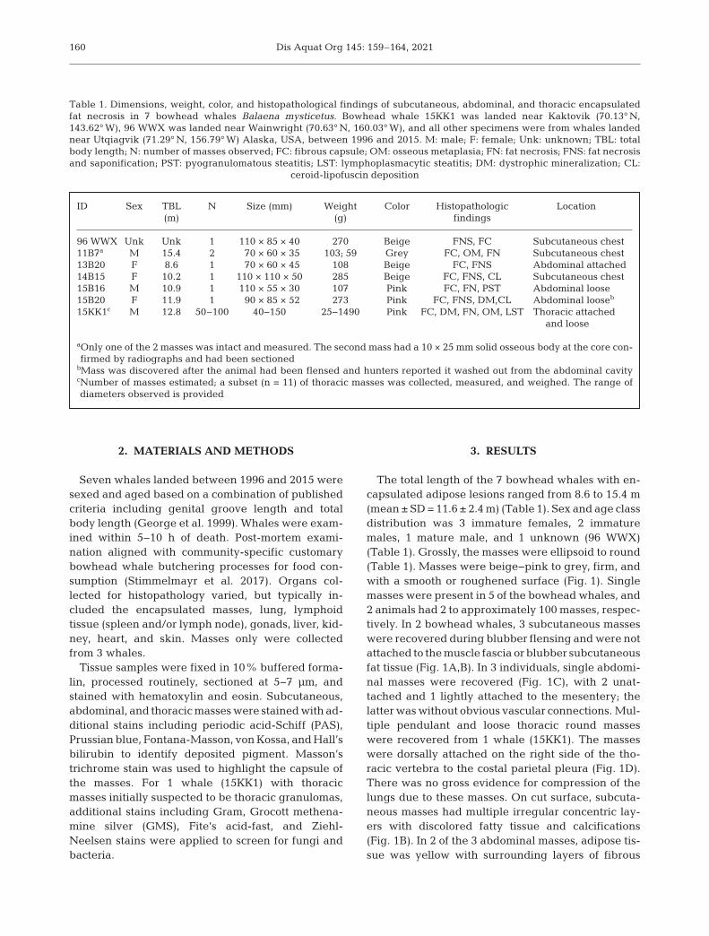
The total length of the 7 bowhead whales with en -capsulated adipose lesions ranged from 8.6 to 15.4 m(mean ± SD = 11.6 ± 2.4 m) (Table 1). Sex and age classdistribution was 3 immature females, 2 immaturemales, 1 mature male, and 1 unknown (96 WWX)(Table 1). Grossly, the masses were ellipsoid to round(Table 1). Masses were beige−pink to grey, firm, andwith a smooth or roughened surface (Fig. 1). Singlemasses were present in 5 of the bowhead whales, and2 animals had 2 to approximately 100 masses, respec-tively. In 2 bowhead whales, 3 subcutaneous masseswere recovered during blubber flensing and were notattached to the muscle fascia or blubber subcutan eousfat tissue (Fig. 1A,B). In 3 individuals, single abdomi-nal masses were recovered (Fig. 1C), with 2 unat-tached and 1 lightly attached to the mesentery; thelatter was without obvious vascular connections. Mul-tiple pendulant and loose thoracic round masseswere recovered from 1 whale (15KK1). The masseswere dorsally attached on the right side of the tho-racic vertebra to the costal parietal pleura (Fig. 1D).There was no gross evidence for compression of thelungs due to these masses. On cut surface, subcuta-neous masses had multiple irregular concentric lay-ers with discolored fatty tissue and calcifications(Fig. 1B). In 2 of the 3 abdominal masses, adipose tis-sue was yellow with surrounding layers of fibrous
160
ID Sex TBL N Size (mm) Weight Color Histopathologic Location (m) (g) findings
96 WWX Unk Unk 1 110 × 85 × 40 270 Beige FNS, FC Subcutaneous chest11B7a M 15.4 2 70 × 60 × 35 103; 59 Grey FC, OM, FN Subcutaneous chest13B20 F 8.6 1 70 × 60 × 45 108 Beige FC, FNS Abdominal attached14B15 F 10.2 1 110 × 110 × 50 285 Beige FC, FNS, CL Subcutaneous chest15B16 M 10.9 1 110 × 55 × 30 107 Pink FC, FN, PST Abdominal loose15B20 F 11.9 1 90 × 85 × 52 273 Pink FC, FNS, DM,CL Abdominal looseb
15KK1c M 12.8 50–100 40−150 25−1490 Pink FC, DM, FN, OM, LST Thoracic attachedand loose
aOnly one of the 2 masses was intact and measured. The second mass had a 10 × 25 mm solid osseous body at the core con-firmed by radiographs and had been sectioned
bMass was discovered after the animal had been flensed and hunters reported it washed out from the abdominal cavity cNumber of masses estimated; a subset (n = 11) of thoracic masses was collected, measured, and weighed. The range ofdiameters observed is provided
Table 1. Dimensions, weight, color, and histopathological findings of subcutaneous, abdominal, and thoracic encapsulatedfat necrosis in 7 bowhead whales Balaena mysticetus. Bowhead whale 15KK1 was landed near Kaktovik (70.13° N,143.62° W), 96 WWX was landed near Wainwright (70.63° N, 160.03° W), and all other specimens were from whales landednear Utqiagvik (71.29° N, 156.79° W) Alaska, USA, between 1996 and 2015. M: male; F: female; Unk: unknown; TBL: totalbody length; N: number of masses observed; FC: fibrous capsule; OM: osseous metaplasia; FN: fat necrosis; FNS: fat necrosisand saponification; PST: pyogranulomatous steatitis; LST: lymphoplasmacytic steatitis; DM: dystrophic mineralization; CL:
ceroid-lipofuscin deposition

Stimmelmayr et al.: Encapsulated fat necrosis in bowhead whales
connective tissue (Fig. 1C). Thoracic masses werecharacterized by thick fibrotic capsules, roughenedand smooth surfaces, and a white caseated dry core(Fig. 1E,F). Two masses had a green caseated drycore.
No significant gross findings were noted in 6 of thewhales. Additional gross findings in the whale withthoracic masses (15KK1), which was noted to exhibitabnormal ante-mortem swimming behavior, in cludedpleural fibrosis hepatic lipoma (previously re portedby Stimmelmayr et al. 2017), asymmetric testes (left:97 cm vs. right: 89 cm), and an old whale bomb-associated tailstock injury (left lateral). The tailstockinjury (213 cm anterior of flukes) consisted of a promi-nent penetration scar covered by whale lice (~200),and an underlying cylindrical cavity estimated to be100 cm long and 20 cm wide. The height was notmeasured. The cavity contained 5 separate com -ponents of an unexploded 1950s-style whale bomblance (Ebenezer Pierce type) (Sheffield 2016). Initialson the projectile were traced to a known whaling cap-tain (deceased) who had reported having struck andlost a bowhead whale during the 1960s.
On histopathological examination, all masses wereencapsulated by fibrous connective tissue of variablethickness, which was stained dark blue under Mas-
son’s trichrome staining (Fig. 2A,B). All masses werecomposed of numerous mature adi po cytes, character-ized by a large clear cytoplasm and a flattened or in-conspicuous nucleus at the peri phery. In 2 whales(14B15 and 15KK1), scattered throughout the masseswere low to moderate numbers of collagen fibers.Multifocally, there were areas of fat necrosis, charac-terized by the loss of cell outlines and replaced bypooled accumulation of an amorphous, deep eosino -philic to amphophilic substance (Fig. 2C,D). Further-more, basophilic, granular to amorphous, von Kossa-positive calcium salts (saponification) were observedwithin the necro tic adipose tissue and in the sur-rounding fibrous stroma (Fig. 2E,F). Granular baso -philic mineral deposition (dystrophic mineralization)was ob served in 1 whale, and formation of bone (os-seous metaplasia) was observed in 2 of the 7 whales.PAS-positive ceroid-lipofuscin pigments were presentin 14B15 and 15B20.
Based on the special stains, hemosiderin, hemato -idin, melanin, and bilirubin were not present. Inflam-mation was uncommon and included lymphoplasma-cytic and pyogranulomatous infiltrates in 15KK1, andbased on the special stains, no bacteria, includingacid-fast bacteria, metazoans, or fungi were present.The site of the bomb in 15KK1 was infiltrated by
161
Fig. 1. Variable gross presentations of encapsulated fat necrosis in bowhead whales Balaena mysticetus. (A) Subcutaneousoval mass (110 × 110 × 50 mm), yellow−white appearance with a firm smooth surface (whale ID: 14B15). (B) Cross section ofthe mass depicted in (A), showing multiple irregular concentric fibrous layers with discolored beige pasty material at the core.(C) Cross section of an abdominal elongated oval mass (110 × 55 × 40 mm), with yellow fatty tissue and surrounding concentricfibrous layers (15B16). (D) In situ pendulant thoracic masses (15KK1). (E) Variable gross presentation of thoracic masses de-picted in (D), pink−white appearance with firm smooth or roughened surfaces. (F) Cross section of one of the masses depicted
in (D), showing thick concentric fibrous layers and a white caseated dry core. Scale is in cm

Dis Aquat Org 145: 159–164, 2021
neutro phils with surrounding fibroblasts (fibroplasia)and mature collagen. Other histopathologic findingsfrom 15KK1 included pleural fibrosis, hepatic lipoma,mild hepatic hemosiderosis, and testicular atrophy.Focal and mild chronic interstitial nephritis wasfound in 15B20.
4. DISCUSSION
Masses of similar gross appearance and anatomicaldistribution (subcutaneous, abdominal, thoracic) butwith limited histopathological description have beenreported from post-mortem examination of landed
162
Fig. 2. Photomicrographs showing characteristic staining and physical properties of encapsulated fat necrosis in bowheadwhales Balaena mysticetus. (A) Dense capsule surrounding necrotic fat. Deposition of calcium salts (saponification) is evident.HE; bar = 200 μm. (B) Capsule composed of layers of dark blue-staining collagen. Masson’s trichrome; bar = 200 μm. (C)Necrosis and collapse of adipocytes. HE; bar = 500 μm. (D) Clusters of necrotic adipocytes. HE; bar = 200 μm. (E) Basophilic,
granular mineral within the mass. HE; bar = 200 μm. (F) Granular mineral stains. van Kossa; bar = 200 μm

Stimmelmayr et al.: Encapsulated fat necrosis in bowhead whales
baleen whales (blue whale Balaenoptera musculus,fin whale B. physalus, humpback whale Megapteranovae angliae, Bryde’s whale B. brydei/edeni, andsei whale B. borealis) during pelagic commercialwhaling in the Antarctic (1948/49) and South Africa(1962/ 1963) (Cockrill 1951, 1960, Uys & Best 1966).
In humans and cows, subcutaneous and abdominalencapsulated fat necrosis is described as firm, mobile,ivory-hued nodules with a distinct fibrotic capsule,absence of vascular connection, and yellow on cutsurface (Anonymous 1926, Przyjemski & Schuster1977, Herzog et al. 2010, Burgdorf & Hurt 2011). Theadipose tissue inside can be normal or with areas ofnecrosis, inflammation, fibrosis, and calcification. Le-sion growth over time is thought to occur by accumu-lation of lamellar units of fibrosis and acquisition ofnutrients by diffusion. Dimensions of encapsulatedfat necrosis in bowhead whales (Table 1) are compa-rable to what has been reported for sei whales (up to60 mm) and cows (60−80 mm), but are generallylarger than those reported for humans (Uys & Best1966, Burgdorf & Hurt 2011, Huang et al. 2011).
Depending on the anatomical location, differingetiologies have been proposed for encapsulated fatnecrosis. Subcutaneous encapsulated fat necrosiscase reports in humans have been associated withprior trauma (Hurt & Santa Cruz 1989, Burgdorf &Hurt 2011). In the absence of prior trauma history, ori-gin of encapsulated fat necrosis is linked to rapid vas-cular insufficiency and or tissue ischemia associatedwith underlying collagen and endocrine-metabolicdisturbances (Anonymous 1926, Herzog et al. 2010,Hanson et al. 2014, Ban 2016). Cockrill (1951, 1960)did not discuss the role of trauma in the etiology ofsubcutaneous encapsulated bodies in large whales,colloquially known as ‘husks’ among whaler men,and thought that the masses were of parasitic origin.However, he noted the preferred anatomical locationbeing within and surrounding the psoas muscula-ture, the latter being the commonest site for penetrat-ing trauma from harpoons and Discovery markingschemes (Rayner 1940). Uys & Best (1966) excluded aparasitic origin in their case material and proposedthat unknown trauma history was a likely cause forthese externally located encapsulated masses (associ-ated with blubber or muscle tissue) re sulting in local-ized ischemia and tissue necrosis. We cannot excludea possible role for an unknown trauma history forsubcutaneous encapsulated fat necrosis in bowheadwhales, but gross evidence suggesting trauma history(i.e. external scarring and fibrous blubber texture)were absent in both bowhead whales with subcuta-neous encapsulated fat necrosis. Although specula-
tive, excessive lipid re serves, characteristic of bow-head whales (George et al. 2015), could predisposethese whales to compromised adipose tissue per -fusion and resulting tissue ischemia. Tissue ischemiahas been observed in humans and experimental ani-mal models of obesity (e.g. obese Zucker rat) (Anony-mous 1926, Stapleton et al. 2008).
Abdominal encapsulated fat necrosis originates byinfarction of epiploic appendices caused by torsion orthrombosis with subsequent inflammatory cellularresponse and capsule formation (Dockerty et al. 1956).Bowhead whales per se do not have epiploic appen-dices, but torsion or thrombosis of abundantly presentinternal fat deposits is a possibility. Although specula-tive, rolling behavior of bowhead whales (Wür sig &Clark 1993) could facilitate accidental torsion of inter-nal fat depots. In dogs and cats, pancreatitis leadingto leakage of pancreatic enzymes and subsequentabdominal fat saponification has been identified as acommon process resulting in abdominal fat necrosis(Schwarz et al. 2000, Adamama-Moraitou et al. 2008).Pancreatic tissue was not collected from the 3 whaleswith abdominal masses, thus we cannot exclude thepresence of pancreatitis; however, pancreatic lesionsin cluding pancreatitis appear to be uncommon(~3.5%) inbowheadwhales (Stimmelmayretal. 2021).
Uys & Best (1966) speculated that decompressionstresses and gaseous emboli resulting in localizedischemia and tissue necrosis were likely mechanismsfor abdominal and thoracic encapsulated fat necrosisin sei and Bryde’s whales. An infectious etiology wasinitially considered for 15KK1 which presented withnumerous thoracic masses. There was no histopatho-logical evidence of infectious agents; however, thismay reflect the chronicity (post-infection). The multi-decadal embedment (~49 yr) of the projectile isremarkable; however, although rare, historic whaleharvest projectiles and/or research instrumentationanchored subcutaneously into whales have beenpreviously documented in Western Arctic bowheadwhales (George & Bockstoce 2008).
Independent of etiology, subcutaneous and ab dom -inal encapsulated fat necrosis described here is un-common in bowhead whales (1.2%; 7/575). For ex -amined sei and Bryde’s whales, Uys & Best (1966)reported a lower prevalence of 0.6% (13/2000) formuscle and fat necrosis bodies, respectively. Noquantitative assessment of prevalence is available forsubcutaneous and abdominal masses from examina-tion of landed Antarctic whales, but Cockrill (1960)noted them as being commonly observed. The detec-tion rate of these encapsulated masses in bowheadwhales, based on size, coloration, and marble-like ap -
163

Dis Aquat Org 145: 159–164, 2021
pearance, likely varies by anatomical location. Subcu-taneous encapsulated bodies are unlikely to be over-looked by aboriginal whaling crews during the flens-ing process. Loose abdominal and thoracic masses aremore likely to be missed during post-mortem exami-nation given the size of viscera and challenging fieldlogistics (Stimmelmayr et al. 2017). As was the casefor 15KK1, detection of pendulant thoracic masses ishighly likely. The biological be havior of these lesionsis unknown, but evidence for co-morbidity with theexception of 15KK1 (i.e. hepatic lipoma, pleural fibro-sis) was absent. Total body length of whales from thisstudy (8.6−15.4 m) falls within the reported range forlanded bowhead whales (Suydam & George 2018).
In conclusion, our small case series presents grossand comprehensive histopathological staining char-acterization of encapsulated fat necrosis in bowheadwhales. Although an incidental finding, encapsulatedfat necrosis needs to be distinguished from neoplasticand inflammatory lesions, as the latter may have pub-lic health implications. Assessment of additional case material including those from other baleen whale spe-cies will be helpful to better understand the etio logy ofencapsulated fat necrosis in large baleen whales.
Acknowledgements. We are particularly grateful to FreddieAishanna for graciously allowing us to examine the whalelance fragment (15KK1). Consultations with Alaska EskimoWhaling commission (AEWC) leadership and KaktovikWhaling Captains Association members were essential inassessing the food safety and public health concerns associ-ated with 15KK1. This study would have not been possiblewithout the long-term support by the Whaling CaptainsAssociations of Barrow (Utqiaġvik), Wainwright, and Kakto -vik, Alaska, of the bowhead harvest monitoring program andtheir continued interest in bowhead whale health re search.This study was funded by qualified outer continental shelf oiland gas revenues by a substantial grant (F12AF01265) fromthe Coastal Impact Assistance Program, US Fish and WildlifeService, the US Department of the Interior, and the NorthSlope Borough Department of Wildlife Management. Collec-tion of marine mammal tissues (2011− 2015) was conductedunder NMFS permits 17350-01 and -02.
LITERATURE CITED
Adamama-Moraitou KK, Prassinos NN, Galatos AD, TontisDK, Rallis TS (2008) Isolated abdominal fat tissue inflam-mation and necrosis in a cat. J Feline Med Surg 10: 192−197
Anonymous (1926) Localized saponification of fat. Edin-burgh Med J 33: 627−631
Ban M (2016) Nodular-cystic fat necrosis — a review of 147Japanese patients. J Dermatol Res 1: 65−68
Burgdorf WH, Hurt MA (2011) Mobile encapsulated adiposetissue (MEAT) of cows and humans: a distinct non- neoplastic entity. Int J Surg Pathol 19: 576−582
Cockrill WR (1951) Antarctic whaling, the role of the veteri-nary surgeon in the whaling industry, with special refer-
ence to standards of inspection in the production ofwhale meat for human consumption and some notes ofthe pathology of the baleen whales. Vet Rec 63: 111−125
Cockrill WR (1960) Pathology of Cetacea. A veterinary studyon whales. Part I and II. Br Vet J 116: 133−189
Dockerty MB, Lynn TE, Waugh JM (1956) A clinicopatho-logic study of the epiploic appendages. Surg GynecolObstet 103: 423−433
George JC, Bockstoce JR (2008) Two historical weapon frag-ments as an aid to estimating the longevity and move-ments of bowhead whales. Polar Biol 31: 751−754
George JC, Bada J, Zeh J, Scott L, Brown SE, O’Hara T, Suy-dam R (1999) Age and growth estimates of bowheadwhales (Balaena mysticetus) via aspartic acid racemiza-tion. Can J Zool 77: 571−580
George JC, Druckenmiller ML, Laidre KL, Suydam RS, Per-son BT (2015) Bowhead whale body condition and linksto summer sea ice and upwelling in the Beaufort Sea.Prog Oceanogr 136: 250−262
Hanson P, Pandit M, Menon V, Roberts S, Barber TM (2014)Painful fat necrosis resulting from insulin injections.Endo crinol Diabetes Metab Case Rep 2014: 140073
Herzog K, Burgdorf W, Hewicker-Trautwein M (2010)Mobile encapsulated bodies comprising fat necrosis andfibrous tissue in the abdominal cavity of cows. J CompPathol 143: 309−312
Huang CH, Lin SC, Chang KC, Chow NH (2011) Numerousperitoneal loose bodies with ileus. Histopathology 58: 318−319
Hurt MA, Santa Cruz DJ (1989) Nodular-cystic fat necrosis. Areevaluation of the so-called mobile encapsulated lipoma.J Am Acad Dermatol 21: 493−498
Przyjemski CJ, Schuster SR (1977) Nodular-cystic fat necro-sis. J Pediatr 91: 605−607
Rayner GW (1940) Whale marking. Progress and results toDecember 1939. Discov Rep 19: 245−284
Schwarz T, Morandi F, Gnudi G, Wisner E, Paterson C, Sulli-van M, Johnston P (2000) Nodular fat necrosis in the felineand canine abdomen. Vet Radiol Ultrasound 41: 335−339
Sheffield G (2016) Kaktovik — 2015 Season. UAF Alaska SeaGrant (Nome) Report to the Kaktovik Whaling CaptainsAssociation, Kaktovik, AK
Stapleton PA, James ME, Goodwill AG, Frisbee JC (2008)Obesity and vascular dysfunction. Pathophysiology 15: 79−89
Stimmelmayr R, Rotstein D, Seguel M, Gottdenker N (2017)Hepatic lipomas and myelolipomas in subsistence-harvested bowhead whales Balaena mysticetus, Alaska(USA): a case review 1980−2016. Dis Aquat Org 127: 71−74
Stimmelmayr R, Rotstein D, Sheffield G, Brower HK,George JC (2021) Diseases and parasites. In: George JC,Thewissen JGM (eds) The bowhead whale. Balaenamysticetus: biology and human interactions. AcademicPress, p 471−498
Suydam RS, George JC (2018) Subsistence harvest of bow-head whales (Balaena mysticetus) by Alaskan Eskimos,1974 to 2016. Paper SC/67b/AWMP6 presented to theScientific Committee of the International WhalingCommission
Uys CJ, Best PB (1966) Pathology of lesions observed inwhales flensed at Saldanha Bay, South Africa. J CompPathol 76: 407−412
Würsig B, Clark CW (1993) Behavior. In: Burns JJ, Monta -gue JJ, Cowles CJ (eds) Special Publication 2: the bow-head whale. The Society of Marine Mammalogy, MossLanding, CA, p 157−199
164
Editorial responsibility: Stephen Raverty, Abbotsford, British Columbia, Canada
Reviewed by: 3 anonymous referees
Submitted: April 12, 2020Accepted: April 7, 2021Proofs received from author(s): July 2, 2021