subacute toxicity in rats - iqb: medciclopedia · appendix v : experimental protocol ... kc-4a s...
TRANSCRIPT

REPORT NO. CD-98/6289T
FOUR-WEEK TOXICITY STUDY IN
RATS BY INTRAVENOUS ADMINISTRATION
WITH A TWO-WEEK RECOVERY PERIOD.
TEST SUBSTANCE: IQB-9302.HCl
VOLUME I


CD-98/6289T
IV.
CONTENTS
Volume I
page
IDENTIFICATION SHEET....................................................................................................... I
SIGNATURES.......................................................................................................................... II
QAU STATEMENT ................................................................................................................ III
CONTENTS.............................................................................................................................IV
SUMMARY ...............................................................................................................................1
CONCLUSIONS ........................................................................................................................3
INTRODUCTION......................................................................................................................4
1. ANIMALS ............................................................................................................................5
1.1. Supply ..........................................................................................................................5
1.2. Identification ................................................................................................................5
1.3. Housing ........................................................................................................................6
2. DIET AND WATER ............................................................................................................6
2.1. Diet...............................................................................................................................6
2.2. Water............................................................................................................................7
3. TEST SUBSTANCE ............................................................................................................7
3.1. Identification ................................................................................................................7
3.2. Formulation of the test substance.................................................................................7
3.3. Formulation analysis ....................................................................................................8
3.4. Administration route and procedure.............................................................................8
3.5. Administration volume ................................................................................................8
3.6. Frequency and duration of treatment ...........................................................................8
3.7. Dose levels and group sizes .........................................................................................9
3.8. Recovery period ...........................................................................................................9
4. OBSERVATIONS..............................................................................................................10
4.1. Clinical signs..............................................................................................................10
CONTENTS
CD-98/6289T
V.
page
4.2. Bodyweight ................................................................................................................10
4.3. Food intake.................................................................................................................10
4.4. Water intake ...............................................................................................................10
4.5. Ophthalmoscopy ........................................................................................................10
5. LABORATORY STUDIES................................................................................................11
5.1. Haematology ..............................................................................................................11
5.2. Biochemistry ..............................................................................................................12
5.3. Analysis of urine ........................................................................................................13
6. TERMINAL STUDIES ......................................................................................................14
6.1. Sacrifice and macroscopic examination.....................................................................14
6.2. Organ weights ............................................................................................................14
6.3. Taking of histological samples...................................................................................15
6.4. Histopathological examination ..................................................................................16
7. STATISTICAL EVALUATION ........................................................................................16
8. ARCHIVES ........................................................................................................................17
9. STUDY FACILITIES.........................................................................................................17
10. STUDY DATES.................................................................................................................18
11. EXPERIMENTAL PROTOCOL........................................................................................18
12. STANDARD OPERATING PROCEDURES....................................................................18
13. DIRECTIVES.....................................................................................................................18
14. RESULTS...........................................................................................................................19
14.1.Mortality.....................................................................................................................19
14.2.Clinical signs..............................................................................................................19
14.3.Bodyweight ................................................................................................................20
14.4.Food intake.................................................................................................................20
14.5.Water intake ...............................................................................................................20
14.6.Ophthalmoscopy ........................................................................................................21
CONTENTS
page

CD-98/6289T
VI.
15. LABORATORY STUDIES................................................................................................21
15.1.Haematology ..............................................................................................................21
15.2.Biochemistry ..............................................................................................................21
15.3.Analysis of urine ........................................................................................................22
16. TERMINAL STUDIES ......................................................................................................22
16.1.Organ weights ............................................................................................................22
16.2.Macroscopic observations..........................................................................................23
16.2.1. Animals sacrificed at the end of the treatment period ...................................... 23
16.2.2. Animals sacrificed at the end of the recovery period ....................................... 23
16.3.Microscopic observations ..........................................................................................24
16.3.1. Animals sacrificed at the end of the treatment period ...................................... 24
16.3.2. Animals sacrificed at the end of the recovery period ....................................... 26
16.4.Histopathological summary .......................................................................................26
FIGURES .................................................................................................................................28
TABLES...................................................................................................................................32
Volume II
HISTOPATHOLOGICAL REPORT (Animals sacrificed at the end of the
treatment period) ....................................................................................................................114
HISTOPATHOLOGICAL REPORT (Animals sacrificed at the end of the
recovery period)......................................................................................................................159
APPENDIX I : DIET ANALYSIS CERTIFICATE...............................................................180
APPENDIX II : WATER ANALYSIS CERTIFICATE ........................................................185
APPENDIX III : TEST SUBSTANCE ANALYSIS CERTIFICATE ...................................197
APPENDIX IV : FORMULATION ANALYSIS RESULTS ................................................199
APPENDIX V : EXPERIMENTAL PROTOCOL.................................................................202
APPENDIX VI : PROTOCOL AMENDMENT....................................................................223


CD-98/6289T
4.
The microscopic examination of the samples taken did no reveal any alterations related to the
administration of the test substance.

4.
REPORT NO. CD-98/6289T
FOUR-WEEK TOXICITY STUDY IN RATS BY INTRAVENOUS ADMINISTRATION
WITH A TWO-WEEK RECOVERY PERIOD.
TEST SUBSTANCE: IQB-9302.HCl
___________________________________________________________________________
INTRODUCTION
The aim of this Study is to evaluate the toxicity of the test substance IQB-9302.HCl, a local
anaesthetic, when administered intravenously to rats during a period of four consecutive weeks,
so as to provide a rational base for the evaluation of the toxicological risk to man and indicate
potential target organs.
This route has been chosen because it is the proposed route for administration to humans.

CD-98/6289T
5.
EXPERIMENTAL PROCEDURE
1. ANIMALS
1.1. Supply
A total of 120 rats (60 males and 60 females) of the Crl:CD® (SD) BR Sprague-
Dawley strain with an approximate age of 28 days and from CHARLES RIVER were
supplied by CRIFFA, S.A. (c/Paraires, 1-7, Nave 5, Polígono Industrial Santiga,
08130-STA. PERPÈTUA DE MOGODA, Barcelona, Spain) on 30th December 1998.
On their arrival a sample of animals was chosen at random and weighed to ensure
compliance with the age requested. The mean weights of males and females were 80g
and 82 g respectively.
The animals were housed in Makrolon cages (55 x 32.7 x 19 cm), with sawdust litter,
in such a way that each cage contained a maximum of 5 animals of the same sex.
All animals underwent a period of 20 days of observation and acclimatisation between
the date of arrival and the start of treatment. During the course of this period, the
animals were inspected by a veterinary surgeon to ensure that they fulfilled the health
requirements necessary for initiation of the Study.
During the acclimatisation period, 100 animals (50 males and 50 females) were
selected for the Study. They were distributed among the experimental groups using a
random distribution method. This procedure allows approximate equalisation of
initial bodyweights whilst allowing random allocation to experimental groups.
1.2. Identification
The rats were individually identified by numbers tattooed on the ears.
The marking of the animals was performed when the animals were distributed among
the study groups.

CD-98/6289T
6.
1.3. Housing
The rats were housed in Makrolon cages (55 x 32.7 x 19 cm), placed on racks. The
cages had sawdust on the floor (Ultrasorb, Panlab, S.L. Mejía Lequerica, 34,
Barcelona, Spain) as litter. From the week before initiation of the treatment, each cage
contained a maximum of 5 rats of the same sex and treatment group.
Each cage was identified by a card, colour coded according to the dose level. This card
stated the cage number, number and sex of the animals it contained, Study number,
test substance code, administration route, dose level and Study Director's name, date
of the arrival of the animals and initiation of treatment.
The temperature and relative humidity were continuously monitored. The temperature
was between 19ºC and 25ºC. The relative humidity was generally maintained at 40-
70%. Humidity indices lower than 40% and higher than 70% were avoided for
prolonged periods.
Lighting was controlled to supply 12 hours of light (7:00 to 19:00 hours) and 12 hours
of dark for each 24-hour period.
The cages corresponding to each experimental group were distributed on racks in such
a manner that external factors, such as environmental conditions, were balanced as far
as possible.
2. DIET AND WATER
2.1. Diet
All the rats had free access to a pelleted rat diet UAR A04C (Usine d'Alimentation
Rationnelle, 91360-Villemoisson sur Orge, France) batches no. 80507 and 80609.
The diet was analyzed by the manufacturer to check its composition and to detect
possible contaminants.


CD-98/6289T
10.
4. OBSERVATIONS
4.1. Clinical signs
All the rats were observed at least twice daily with the purpose of recording any
symptoms of ill-health or behavioural changes. These observations were also
performed on week-ends. The observations included but were not limited to changes
in skin and fur, in the eyes and mucous membranes, in the respiratory, circulatory,
central nervous and autonomous systems, somatomotor activity and behaviour.
4.2. Bodyweight
The bodyweight of each rat was recorded one week before the start of treatment, daily
during the course of the same and on the day of sacrifice. The rats selected for the
recovery period were weighed twice a week and on the day of sacrifice. The mean
weights for the different groups and sexes were calculated from the individual
weights.
4.3. Food intake
Prior to the beginning of treatment, and afterwards once a week, the food intake of
each cage was recorded and the mean weekly intake per rat was calculated.
4.4. Water intake
Water intake was checked by visual observation during the Study. In addition, the
water consumption in each cage was measured daily for a period of 5 days, during the
3rd week of treatment and, subsequently, during the 2nd week of the recovery period.
4.5. Ophthalmoscopy
Before treatment started, the eyes of all animals were examined. These examinations
included the cornea, the conjunctivae, the sclera, the iris and fundus.
The observations were made with the aid of an indirect ophthalmoscope.

CD-98/6289T
11.
Before the end of the treatment and before the end of the recovery period, additional
examinations of the eyes of the animals from the Control and high dose groups were
made.
Prior to each examination, the pupils of the rats were dilated by instillation of one
drop of cyclopentholate chlorhydrate eyedrops. (Colircusí Ciclopléjico®, Laboratorios
Cusí, S.A. Batches no. L09 and M08).
5. LABORATORY STUDIES
During the 4th week of treatment, samples of blood were withdrawn from the orbital sinus
of 10 males and 10 females from each group, under light ether anaesthesia after fasting for
16 hours.
The blood samples were taken from each animal approximately between 7:30 and 10:00
hours in order to reduce biological variation caused by circadian rhythms.
In addition, samples of the urine produced during 16 hours by 10 males and 10 females
were taken. To this end the rats were deprived of food for this period of time.
5.1. Haematology
The following determinations were performed:
Parameter Method/Instrumentation Units
Erythrocyte count Haematological counter.SYSMEX F-800
106/µL
Haemoglobin Haematological counter.SYSMEX F-800
g/100 mL
Haematocrit Haematological counter.SYSMEX F-800
%
Mean corpuscularvolume (MCV)
Calculation.SYSMEX F-800
fL
Mean corpuscularhaemoglobin (MCH)
Calculation.SYSMEX F-800
pg

CD-98/6289T
12.
Parameter Method/Instrumentation Units
Mean corpuscularhaemoglobin concentration (MCHC)
Calculation.SYSMEX F-800
g/100 mL
Reticulocyte count* New methylene blue stain.Microscope
%
Total leukocyte count Haematological counter.SYSMEX F-800
103/µL
Differential leukocyte count- Neutrophils- Lymphocytes- Eosinophils- Basophils- Monocytes
May Grünwald-Giemsa stain.Microscope
103/µL
Platelet count Haematological counter.SYSMEX F-800
103/µL
Prothrombin time Coagulometer. KC-4A s
*Slides were prepared.
5.2. Biochemistry
The following blood chemistry determinations were carried out:
Parameter Method/Instrumentation Units
Glucose Glucose dehydrogenase.COBAS MIRA
mg/100 mL
Urea Urease-GLDH. COBAS MIRA mg/100 mLCreatinine Jaffé. COBAS MIRA mg/100 mLTotal bilirubin Jendrassik-Grof reaction.
COBAS MIRAmg/100 mL
Aspartate aminotransferase(AST/GOT)
Malate dehydrogenase. DGKC.COBAS MIRA
U/L
Alanine aminotransferase(ALT/GPT)
Lactate dehydrogenase. DGKC.COBAS MIRA
U/L
Sorbitol dehydrogenase(SDH)
Reduction of fructose.COBAS MIRA
U/L
Alkaline phosphatase p-nitrophenylphosphate.DGKC. COBAS MIRA
U/L
Total cholesterol CHOD-PAP. COBAS MIRA mg/100 mL

CD-98/6289T
13.
Parameter Method/Instrumentation Units
Sodium Ion selective electrode. NOVA I
mmol/L
Potassium Ion selective electrode. NOVA I
mmol/L
Chloride Coulombimetric.CORNING 925
mmol/L
Calcium MTB. COBAS MIRA mg/100 mLInorganic phosphorus Phosphomolybdate without
deproteinization.COBAS MIRA
mg/100 mL
Total protein Biuret. COBAS MIRA g/100 mLAlbumin Bromocresol green.
COBAS MIRAg/100 mL
The albumin/globulin ratios were calculated from the total protein and albumin
values.
5.3. Analysis of urine
The following determinations were made:
Parameters MethodColourVolume
Macroscopic observation
Specific gravity RefractometrypHProteinsGlucoseBilirubinKetonesUrobilinogenHaemoglobin
Combur 8 test
The Combur 8 test is a diagnostic strip kit obtained from Boehringer Mannheim and it
is used as a qualitative indicator of the concentration of the different parameters. The
results are presented using the following scale:
0 = negative+ = small quantity of the parameter analyzed
++ = moderate quantity of the parameter analyzed
CD-98/6289T
14.
+++ = large quantity of the parameter analyzed
The urinary sediment was examined for the detection of:
− Epithelial cells
− Leukocytes
− Erythrocytes
− Organisms (bacteria, etc.)
− Crystals
− Other abnormal constituents (casts, sperm, etc.)
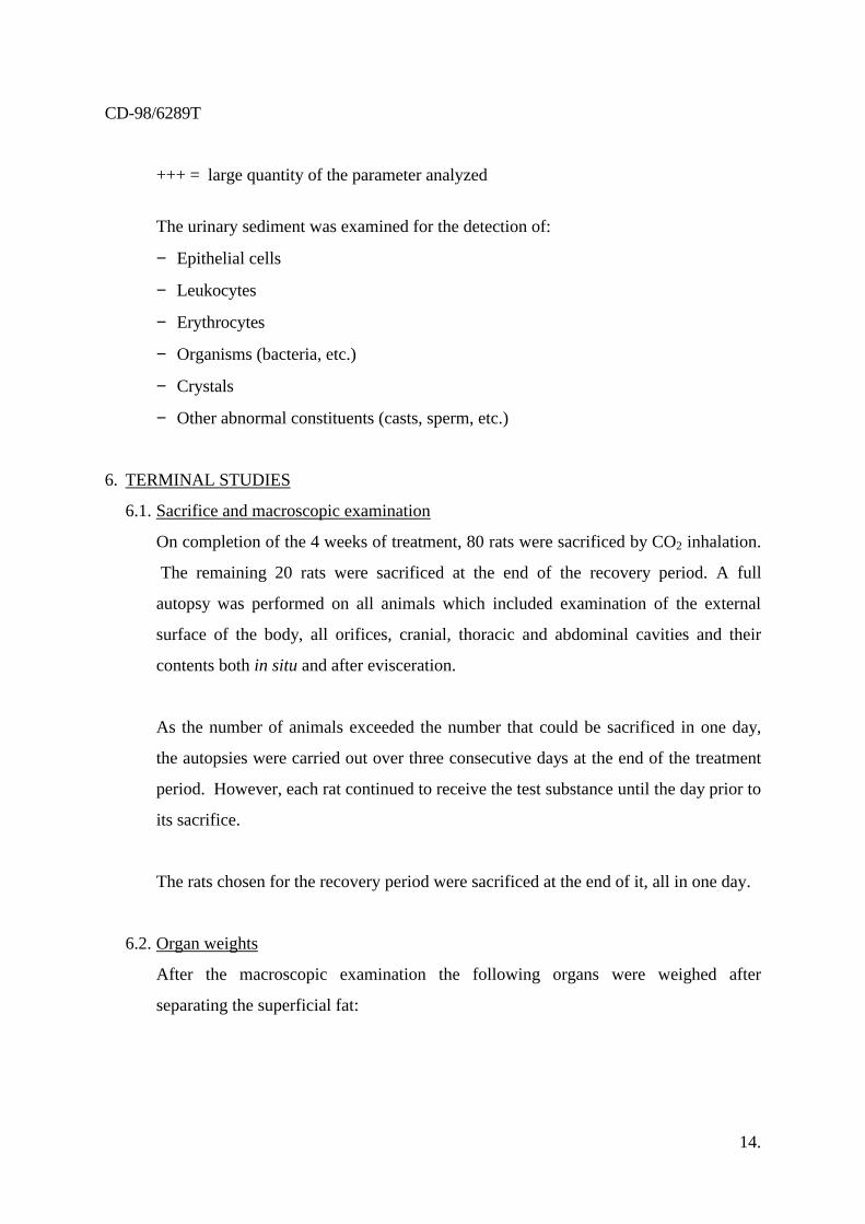
6. TERMINAL STUDIES
6.1. Sacrifice and macroscopic examination
On completion of the 4 weeks of treatment, 80 rats were sacrificed by CO2 inhalation.
The remaining 20 rats were sacrificed at the end of the recovery period. A full
autopsy was performed on all animals which included examination of the external
surface of the body, all orifices, cranial, thoracic and abdominal cavities and their
contents both in situ and after evisceration.
As the number of animals exceeded the number that could be sacrificed in one day,
the autopsies were carried out over three consecutive days at the end of the treatment
period. However, each rat continued to receive the test substance until the day prior to
its sacrifice.
The rats chosen for the recovery period were sacrificed at the end of it, all in one day.
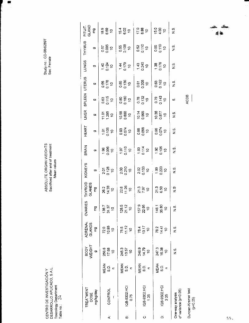
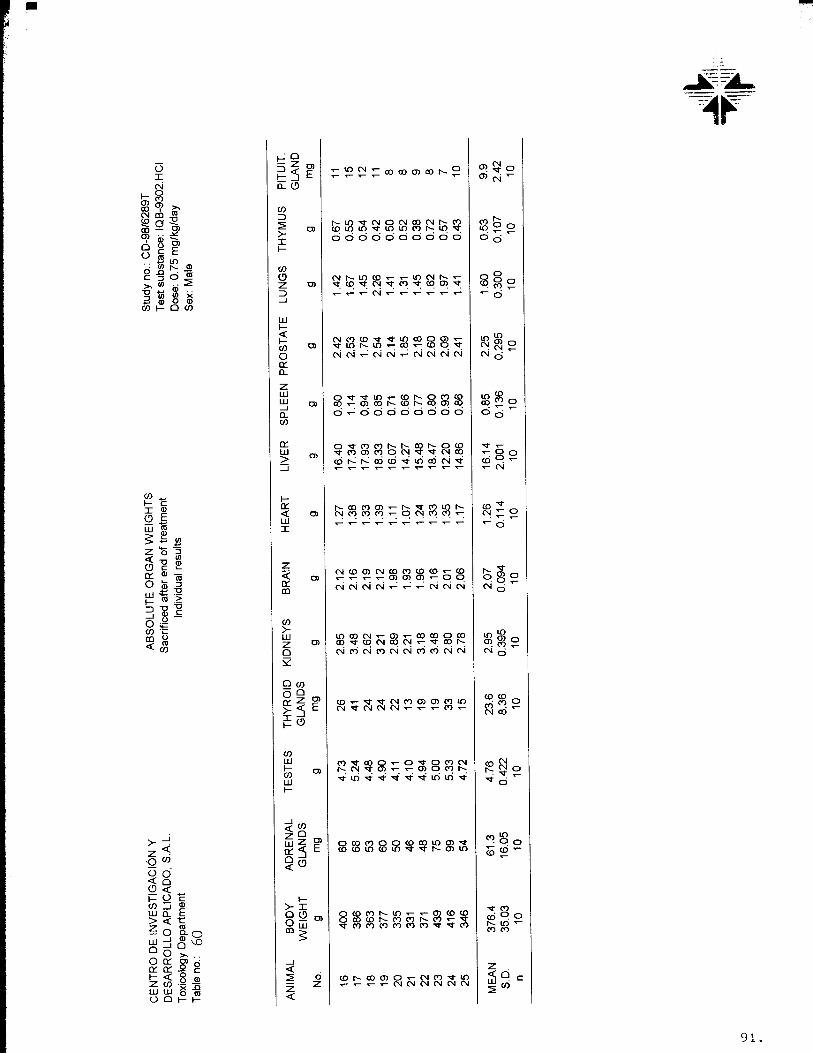
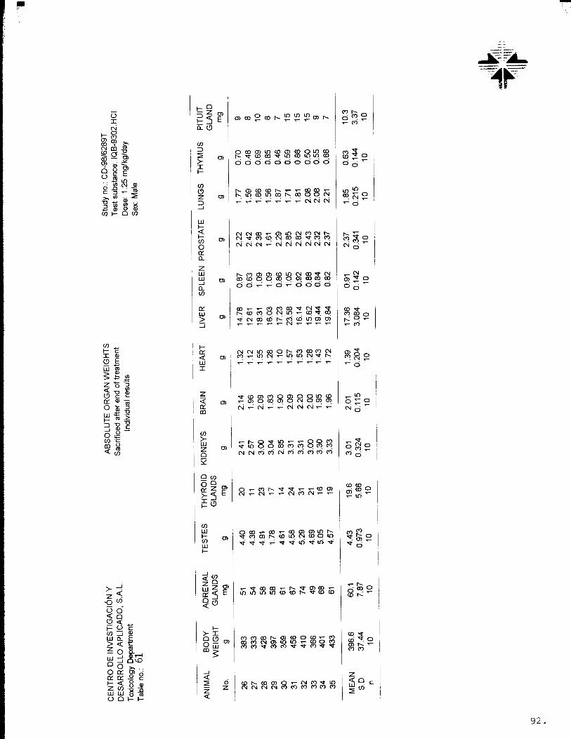
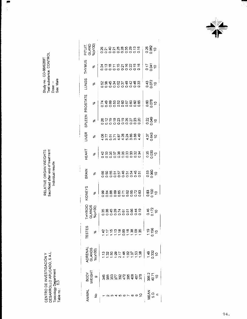
6.2. Organ weights
After the macroscopic examination the following organs were weighed after
separating the superficial fat:

CD-98/6289T
15.
Adrenals Pituitary gland
Brain Prostate and seminal vesicles
Heart Spleen
Kidneys Testes and epididymides
Liver Thymus
Lungs Thyroids
Ovaries Uterus
6.3. Taking of histological samples
Samples of the following organs and tissues were taken and fixed in 10% neutral
buffered formalin, with the exception of the eyes, which were preserved in Davidson's
fixative:
AdrenalsAortaBone (sternum)Brain (bulbar, cerebellar andcortical sections)CaecumColonEyes and optic nervesFemur (with joint)HeartInjection site (tail)KidneysLiverLungs and mainstem bronchiLymph nodes (submandibular andmesenteric)Mammary glandOesophagusOvariesPancreasPituitary glandProstateRectumSalivary glands
Sciatic nerveSeminal vesiclesSkeletal muscleSkin (abdominal)Small intestine (duodenum, ileum,jejunum)Spinal cord (cervical, thoracic andlumbar)SpleenStomachTestes and epididymidesThymusThyroid and parathyroidsTissue masses or tumours(including regional lymph nodes)TongueTracheaUrinary bladderUterus (corpus and cervix)VaginaWhatever other organ or tissuewith macroscopic alterations.


CD-98/6289T
17.
In the tables, the letters N.S. mean that, for the corresponding parameters, the differences
between mean values for the stated groups are not statistically significant.
In the tables statistical significance is represented by an S. (p<0.05) at the foot of the
corresponding column. The letters A, B, C and D represent the mean values for the
Control group and groups 2, 3 and 4 respectively.
The letters are placed in ascending order and may be interpreted statistically as follows:
− The difference between two means underlined by the same line is not statistically
significant, according to the Duncan-Kramer test (p < 0.05).
− The difference between two means not underlined by the same line is statistically
significant, according to the Duncan-Kramer test (p < 0.05).
The remainder of the urine parameters were evaluated statistically using the homogeneity
test (χ2 test p < 0.01)(2).
8. ARCHIVES
All the data pertaining to the Study will be kept for at least five years in the archives at
Centro de Investigación y Desarrollo Aplicado, S.A.L. All tissues preserved in formalin
will be stored for a period of two years after the completion of the Study.
No material relating to this Study will be destroyed without the prior written consent of the
Sponsor.
9. STUDY FACILITIES
This Study was conducted in the laboratories and animal housing of the Toxicology
Department of Centro de Investigación y Desarrollo Aplicado, S.A.L., Centro Industrial
Santiga, c/Argenters 6, 08130-SANTA PERPÈTUA DE MOGODA, Barcelona (Spain).
(2) Manual of Pharmacologic Calculations.
Ronald J. Tallarida and Rodney B. Murray.Springer-Verlag (1987)

CD-98/6289T
18.
The histopathological examination of the histological preparations was performed in the
Centro de Histopatología Veterinaria, c/Castellnou, 21, 08017-BARCELONA (Spain).
10. STUDY DATES
The duration of the Study was as follows:
Protocol signed: 9th October 1998
Protocol amendment no. 1 accepted: 18th January 1999
Protocol amendment no. 2 accepted: 3rd February 1999
Arrival date of animals: 30th December 1998
Treatment started: 18th January 1999
End of treatment: 16th February 1999
Recovery period: 15th February to 1st March 1999
Final Report: See Page I
11. EXPERIMENTAL PROTOCOL
Appendix V contains the experimental protocol.
The protocol amendments approved in the course of the Study are shown in Appendix
VI.
12. STANDARD OPERATING PROCEDURES
All procedures of this Study were carried out according to the Centro de Investigación y
Desarrollo Aplicado, S.A.L. Standard Operating Procedures.
13. DIRECTIVES
The Study procedures described in this Report are in accordance with Directive
91/507/EEC relating to analytical, pharmacotoxicological and clinical standards and
protocols in respect of testing of medicinal products (Annex, Part 3, referring to
Toxicological and Pharmacological testing) and Annex I of Recommendation
83/571/EEC.

CD-98/6289T
19.
14. RESULTS
14.1. Mortality
No mortalities were recorded among the animals treated with the substance
IQB-9302.HCl at the different doses administered nor among the Control group
animals.
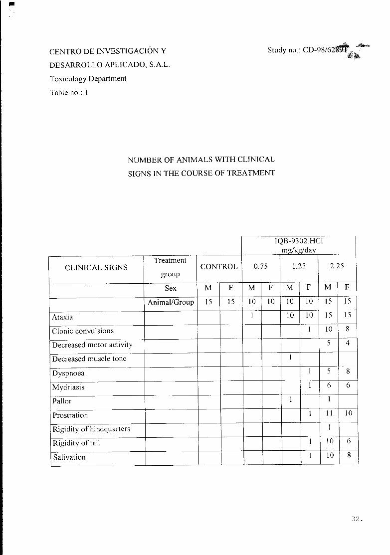
14.2. Clinical signs
The frequency of the clinical signs according to sex and treatment group is shown
in Table no. 1.
No clinical signs were recorded among the animals pertaining to the Control group.
One male administered with IQB-9302.HCl at the dose of 0.75 mg/kg/day
presented, on day 23 of the treatment, ataxia after the administration which
disappeared two minutes afterwards.
All of the animals treated at the dose of 1.25 mg/kg/day presented ataxia. This
alteration was accompanied occasionally, in one male, by decreased muscle tone
and pallor and in one female by prostration, dyspnoea, salivation, clonic
convulsions, mydriasis and rigidity of the tail. All the clinical signs were observed
immediately after administration and had disappeared two minutes after the
treatment.
All of the animals treated at the dose of 2.25 mg/kg/day presented ataxia. This
alteration was accompanied occasionally, in most of the animals, by clonic
convulsions, salivation and prostration. Similarly, some of the animals presented
mydriasis, rigidity of the tail and hindquarters, decreased motor activity and pallor.
All clinical signs started immediately after treatment and disappeared two minutes
afterwards.

CD-98/6289T
20.
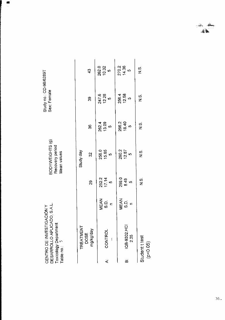
14.3. Bodyweight
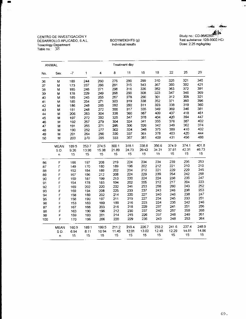
The bodyweight increase, according to sex and treatment group, during the
treatment and recovery period, is shown in Figures nos. 1 to 4 and Tables nos. 2 to
5.
The individual values for each animal are shown in Tables nos. 35 to 40.
The bodyweight increase in males and females treated with IQB-9302.HCl at the
three treatment doses was, in the course of the treatment and recover period, similar
to that recorded for the Control group animals and no statistically significant
differences were recorded.
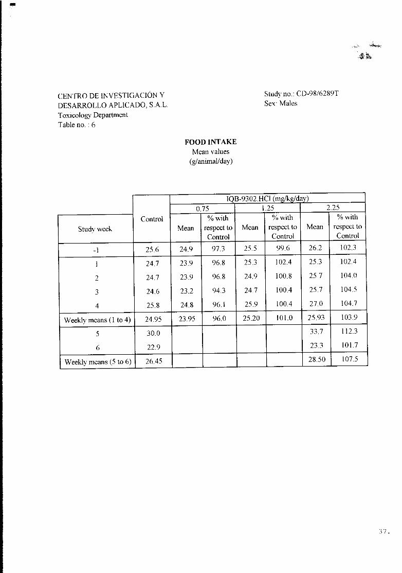
14.4. Food intake
Tables nos. 6 and 7 contain the weekly mean food intake of the males and females
pertaining to the different treatment groups.
During the treatment period, the food intake in males and females treated with the
test substance at the three doses administered was similar to that recorded in the
animals of the Control group.
During the recovery period, the food intake in the males and females treated at
2.25 mg/kg/day was similar to that recorded in the Control group.
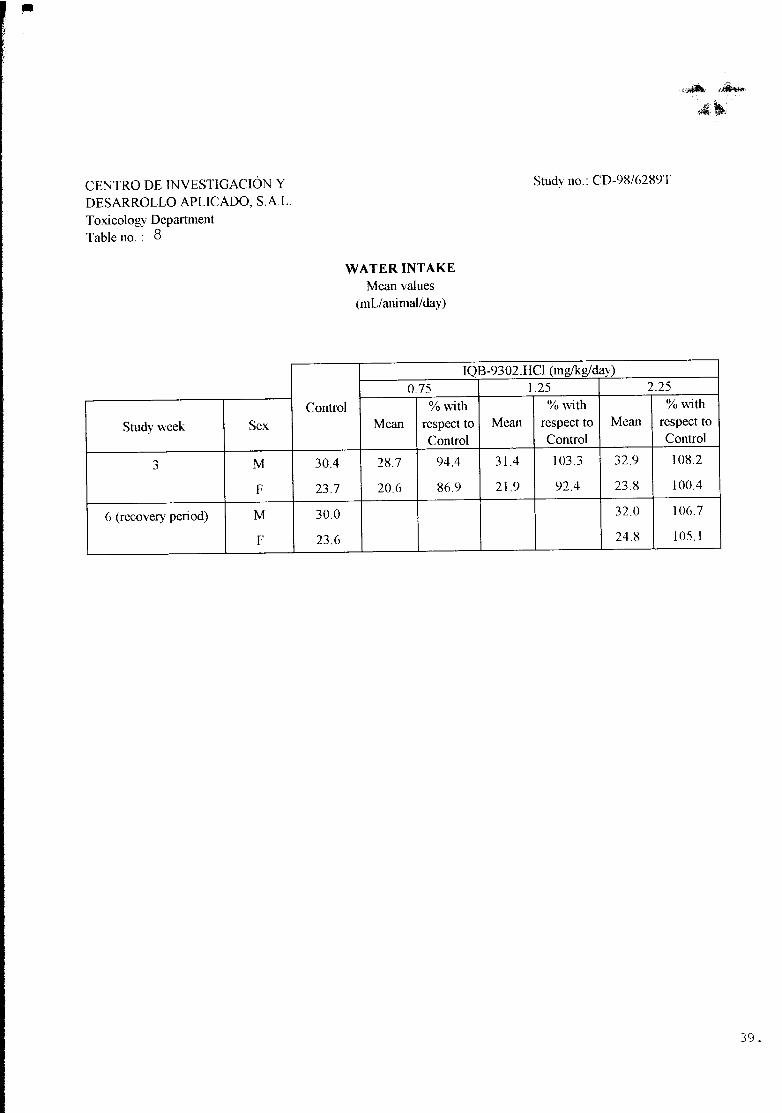
14.5. Water intake
The mean water intake by sex and treatment group during the course of the 3rd
week of treatment and during the second week of the recovery period is shown in
Table no. 8.
No noticeable alterations were recorded in the water intake during the third week of
treatment.


CD-98/6289T
23.
In the females, the absolute and relative weight of the lungs at 2.25 mg/kg/day was
statistically higher than that recorded in the Control group.
16.2. Macroscopic observations
16.2.1. Animals sacrificed at the end of the treatment period
Table no. 31 shows the frequency of the macroscopic observations by
organ, sex and treatment group of the animals sacrificed at the end of the
treatment.
The autopsies carried out revealed some renal alterations such as unilateral
dilation of the renal calices in one male belonging to the Control group and
one female treated at 1.25 mg/kg/day. A bilateral dilation of renal calices
was registered in one male pertaining to the Control group and one female,
one male and two females treated at the doses of 0.75, 1.25 and
2.25 mg/kg/day, respectively.
One female treated at the dose of 0.75 mg/kg/day and one male treated at
the dose of 2.25 mg/kg/day presented petechial areas in the thymus.
One male administered at the dose of 1.25 mg/kg/day presented both testes
decreased in size.
One female treated at the dose of 1.25 mg/kg/day presented a whitish
nodule of 0.5 cm in diameter in the spleen. Similarly, the spleen of one male
treated at 2.25 mg/kg/day presented a nodular surface.
16.2.2. Animals sacrificed at the end of the recovery period
Table no. 32 shows the frequency of the macroscopic observations by organ,
sex and treatment group.

CD-98/6289T
24.
In the autopsies carried out at the end of the recovery period, one male
belonging to the Control group and one female treated at the dose of
2.25 mg/kg/day presented unilateral dilation of renal calices.
16.3. Microscopic observations
16.3.1. Animals sacrificed at the end of the treatment period
The frequencies of the microscopic observations by organ, sex and treatment
group can be found in Table no. 33.
MICROSCOPIC ALTERATIONS NOT ASSOCIATED WITH THE
TREATMENT
SPLEEN
Lymphoid hyperplasia
IQB-9302.HCl (2.25 mg/kg/day): 44M
LIVER
Lymphocytary infiltrate, portal
IQB-9302.HCl (2.25 mg/kg/day): 92F
Microgranuloma
Control: 55F
IQB-9302.HCl (2.25 mg/kg/day): 91F
PITUITARY
Simple cyst
IQB-9302.HCl (2.25 mg/kg/day): 40M
EYES
Lymphocytary infiltrate in Harder’s gland, unilateral
IQB-9302.HCl (2.25 mg/kg/day): 94F

CD-98/6289T
25.
LUNGS
Intraalveolar histiocytosis, focal
Control: 7M, 54F
IQB-9302.HCl (2.25 mg/kg/day): 36M, 38M
KIDNEYS
Dilation of renal pelvis
Control: 8M, 10M
IQB-9302.HCl (0.75 mg/kg/day): 69F
(1.25 mg/kg/day): 29M, 85F
Interstitial nephritis, focal
IQB-9302.HCl (2.25 mg/kg/day): 43M
Pyelitis, acute, non-specific
IQB-9302.HCl (2.25 mg/kg/day): 94F, 95F
TESTES
Tubular atrophy
IQB-9302.HCl (1.25 mg/kg/day): 29M
THYMUS
Multifocal congestion
IQB-9302.HCl (2.25 mg/kg/day): 38M
URINARY BLADDER
Cystitis, acute, non-specific
IQB-9302.HCl (2.25 mg/kg/day): 94F

CD-98/6289T
26.
16.3.2. Animals sacrificed at the end of the recovery period
The frequencies of the microscopic observations by organ, sex and treatment
group can be found in Table no. 34.
MICROSCOPIC ALTERATIONS NOT ASSOCIATED WITH THE
TREATMENT
LIVER
Hepatocytary vacuolisation, centrolobular
Control: 15M
PITUITARY
Simple cyst
Control: 65F
LUNGS
Intraalveolar histiocytosis, focal
Control: 15M
IQB-9302.HCl (2.25 mg/kg/day): 50M
KIDNEYS
Dilation of renal pelvis
Control: 15M
IQB-9302.HCl (2.25 mg/kg/day): 99F
16.4. Histopathological summary
The microscopic observation of the samples corresponding to the animals
sacrificed at the end of the treatment period did not reveal any alteration
associated with the intravenous administration of the substance IQB-9302.HCl.

CD-98/6289T
27.
Similarly, the histopathological study of the samples belonging to the animals that underwent
the recovery period did not reveal any alteration associated with the administration of the test
substance.
The histopathological findings described are quite frequent in this type of laboratory animals.
No evident relation with the intravenous administration of the test substance IQB-9302.HCl is
observed.