subacute sclerosing panencephalitis manifesting as bell’s
TRANSCRIPT

BRIEF REPORT Open Access
Subacute Sclerosing Panencephalitismanifesting as Bell’s palsy and bilateralmacular necrotizing retinitis: an atypicalpresenting featureLagan Paul*, Tanya Jain, Manisha Agarwal and Shalini Singh
Abstract
Background: Subacute sclerosing panencephalitis (SSPE) is a potentially lethal complication of measles infection.Neurological complications take years to manifest after primary viral infection of brain and can lead to blindness insome individuals.
Findings: A 13-year-old female patient with history of Bell’s palsy 2 months prior, presented with rapidlyprogressing necrotizing retinitis in both eyes. Soon after, she was unable to walk, developed myoclonic jerks,altered sensorium and loss of bowel and bladder control. Her clinical history, CSF IgG measles antibody analysis,MRI brain and EEG findings confirmed the diagnosis of SSPE.
Conclusion: SSPE in our case presented as Bell’s palsy and sudden painless diminution of vision due to ocularinvolvement, and developed full blown disease within 2 months. SSPE can present as a diagnostic challenge andwarrants early identification and referral for timely diagnosis and management.
IntroductionSubacute sclerosing panencephalitis (SSPE) is a progressiveneurodegenerative disease caused by persistence of defect-ive measles infection commonly seen in children and youngadults. It usually occurs 7–10 years after measles infection[1]. Ocular manifestations usually precede the neurologicalmanifestations of the disease [1]. Our case presented withbell’s palsy as the first neurological sign and retinal involve-ment happened after 2months. This case highlights the im-portant role of an ophthalmologist in the diagnosis of thisfatal disease and aiding the neurologist in timely interven-tion and proper rehabilitation.
Case reportA 13-year-old Asian Indian female presented to ourclinic with complaint of diminution of vision in both
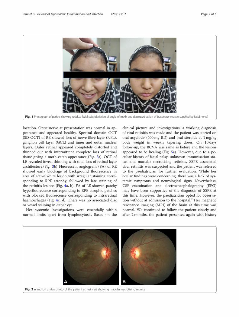
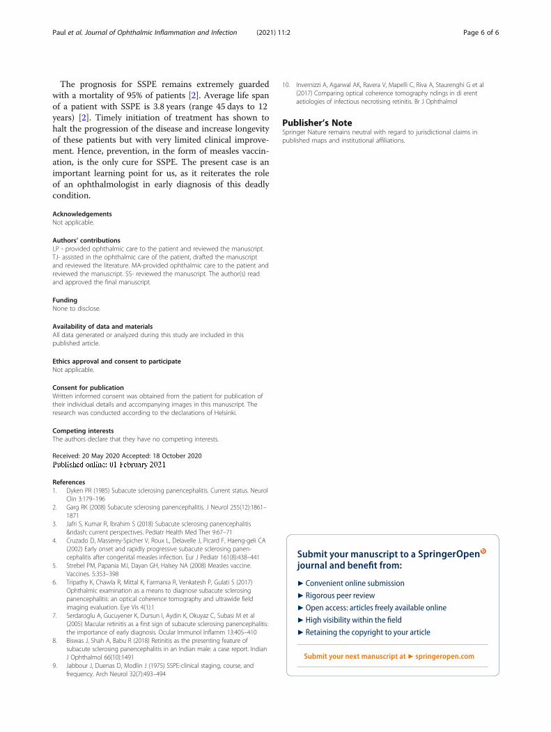
eyes for the last 20 days. There was a history of acuteonset left sided facial palsy 2 months back, which wastreated with oral acyclovir but no neurological workupwas performed then. Systemic history was unremarkable.She had no history of similar episode in the past and herimmunisation status was not known. Central nervoussystem (CNS) examination was within normal limitsapart from mild residual sequelae of facial palsy (Fig. 1).Her best corrected visual acuity (BCVA) in right eye(RE) was 1/60, Near <N/60 and 3/60, Near<N/36 in theleft eye (LE). There was no evidence of anterior segmentor vitreous inflammation. Her fundus examination re-vealed whitish ground glass retinitis lesions with ill-defined edges over the posterior pole with haemorrhagesaround it in RE. (Fig. 2) LE showed an area of thinningwith RPE mottling and haemorrhages in macular areawith no active retinal lesions. There was presence of as-sociated mild venous dilation and areas of vascular oc-clusion and few healed lesions in mid-peripheral
© The Author(s). 2021 Open Access This article is licensed under a Creative Commons Attribution 4.0 International License,which permits use, sharing, adaptation, distribution and reproduction in any medium or format, as long as you giveappropriate credit to the original author(s) and the source, provide a link to the Creative Commons licence, and indicate ifchanges were made. The images or other third party material in this article are included in the article's Creative Commonslicence, unless indicated otherwise in a credit line to the material. If material is not included in the article's Creative Commonslicence and your intended use is not permitted by statutory regulation or exceeds the permitted use, you will need to obtainpermission directly from the copyright holder. To view a copy of this licence, visit http://creativecommons.org/licenses/by/4.0/.
* Correspondence: [email protected] Department, Dr. Shroff’s Charity Eye Hospital, 5027, KedarnathRoad, Daryaganj, New Delhi 110002, India
Journal of OphthalmicInflammation and Infection
Paul et al. Journal of Ophthalmic Inflammation and Infection (2021) 11:2 https://doi.org/10.1186/s12348-020-00223-1
location. Optic nerve at presentation was normal in ap-pearance and appeared healthy. Spectral domain OCT(SD-OCT) of RE showed loss of nerve fibre layer (NFL),ganglion cell layer (GCL) and inner and outer nuclearlayers. Outer retinal appeared completely distorted andthinned out with intermittent complete loss of retinaltissue giving a moth-eaten appearance (Fig. 3a). OCT ofLE revealed foveal thinning with total loss of retinal layerarchitecture.(Fig. 3b) Fluorescein angiogram (FA) of REshowed early blockage of background fluorescence inarea of active white lesion with irregular staining corre-sponding to RPE atrophy, followed by late staining ofthe retinitis lesions (Fig. 4a, b). FA of LE showed patchyhyperfluorecence corresponding to RPE atrophic patcheswith blocked fluorescence corresponding to intraretinalhaemorrhages (Fig. 4c, d). There was no associated discor vessel staining in either eye.Her systemic investigations were essentially within
normal limits apart from lymphocytosis. Based on the
clinical picture and investigations, a working diagnosisof viral retinitis was made and the patient was started onoral acyclovir (400 mg BD) and oral steroids at 1 mg/kgbody weight in weekly tapering doses. On 10 daysfollow-up, the BCVA was same as before and the lesionsappeared to be healing (Fig. 5a). However, due to a pe-culiar history of facial palsy, unknown immunisation sta-tus and macular necrotising retinitis, SSPE associatedviral retinitis was suspected and the patient was referredto the paediatrician for further evaluation. While herocular findings were concerning, there was a lack of sys-temic symptoms and neurological signs. Nevertheless,CSF examination and electroencephalography (EEG)may have been supportive of the diagnosis of SSPE atthis time. However, the paediatrician opted for observa-tion without at admission to the hospital.” Her magneticresonance imaging (MRI) of the brain at this time wasnormal. We continued to follow the patient closely andafter 2 months, the patient presented again with history
Fig. 1 Photograph of patient showing residual facial palsy(deviation of angle of moth and decreased action of buccinator muscle supplied by facial nerve)
Fig. 2 a and b Fundus photo of the patient at first visit showing macular necrotising retinitis
Paul et al. Journal of Ophthalmic Inflammation and Infection (2021) 11:2 Page 2 of 6

of total loss of vision in both eyes with involuntary jerkymovement of the right upper limb, generalised loss ofsensorium and decreased awareness along with loss ofbladder and bowel control since 10 days duration. Thechild was conscious, but not oriented and uncooperativefor examination. On ocular examination, there wascomplete loss of light perception in both eyes with re-stricted ocular movements in all directions. Pupils weresluggishly reactive in both eyes and fundus examinationrevealed total disc pallor and healed retinitis lesions inthe both eyes (Fig. 5b). A referral to a neurologist re-vealed that higher mental functions, cranial nerves,motor and sensory examination could not be performedas the patient was uncooperative for examination.Muscle strength was 3/5 and, while superficial and deeptendon reflexes could be elicited, the plantar reflex wasequivocal or decreased. There was involuntary loss ofbladder and bowel control. CSF analysis was done whichrevealed an opening pressure of 8 cm of water, was acel-lular and negative for any infection on microbiologicalstains. IgG measles antibody in CSF (10.7 mg/dl) wasraised with increased CSF/Serum quotient (4.48). MRI ofthe brain bilateral T2/flair hyper-intensities with gyralswellings involving parieto-occipital lobes. EEG examin-ation revealed intermittent high voltage slow wave dis-charges suggestive of progressive myoclonic epilepsy(Fig. 6). The above clinical history and investigations sat-isfied the Dyken’s criteria and the diagnosis of SSPE wasmade [1]. The patient was put on oral anti-convulsant
for the epilepsy with supportive treatment and followedup by the neurologist. While interferon therapy can beused in such cases, our patient was not able to receivethis due to financial constraints.
DiscussionSSPE is a progressive neurodegenerative encephalopathycaused by the persistence of an aberrant mutated mea-sles virus [2, 3]. It usually occurs 7–10 years after mea-sles infection, but the latency varies from 1month to 27years [2–5]. Stringent immunization schedules in devel-oped world has led to significant decline in the preva-lence of this deadly disease to 1 per 100,000 cases [5].Measles virus tends to affect the neurons and oligoden-drocytes of brain at the time of primary systemic infec-tion and remains quiescent for years before manifestingclinically [2]. The relationship between retinal andneurologic involvement in SSPE is not clearly under-stood. Ocular involvement occurs in almost 50% of SSPEcases and may antedate the onset of neurologic symp-toms by several weeks or months [2]. The most charac-teristic ophthalmologic lesion is macular necrotizingretinitis, which spreads centrifugally to involve the pos-terior pole. Other protean ocular findings described withSSPE include papillitis, papilledema, optic atrophy,macular edema, serous macular detachment, or smallhemorrhages within the posterior pole [6–8].Diagnosis of SSPE is based on the Dyken’s criteria,
which include two major and four minor criteria [1].
Fig. 3 a SD-OCT of the RE showing loss of retinal architecture with moth eaten appearance of the retina. b SD-OCT of the LE showing loss ofretinal architecture with foveal thinning
Paul et al. Journal of Ophthalmic Inflammation and Infection (2021) 11:2 Page 3 of 6

Major criteria include 1) raised anti-measles antibody ti-ters in CSF and 2) typical or atypical clinical history.Minor criteria include 1) characteristic EEG findings thatinclude periodic, generalized, bilaterally synchronousand symmetrical high-amplitude slow waves that recurat regular intervals of 5–15 s called Radermecker com-plexes, 2) CSF globulin levels greater than 20% of thetotal CSF protein, 3) Characteristic histopathologicalfindings on brain biopsy,4) Molecular diagnostic test toidentify wild-type measles virus mutated genome. Usu-ally two major criteria plus one minor criterion are re-quired, our case satisfied 2 major and 2 minor criteriaand hence diagnosis of SSPE was confirmed.Bell’s palsy (lower motor nuclear) as the first present-
ing neurological sign in our case is unique and neverbeen described in literature. Residual right-sided facialnerve palsy could be appreciated at the time of appear-ance of retinal lesions which happened 2months later.The characteristic hemorrhagic necrotising retinitis le-sions at the macular area have been reported before [6,7, 9]. Such retinitis lesions have been reported to be thepresenting features and act as a clue to early diagnosis ofthis disease. The timing of appearance of these lesions
have been different in most studies ranging from 6 weeksby Tripathi et al. [6] to 18 months by Shah et al. [8]. Inour patient it was 8 weeks. On spectral domain opticalcoherence tomography (SD-OCT), there is a predomin-ant involvement of the nuclear layers of the retina is de-scribed which tends to spread from the inner layers tothe outer retinal layers. The necrosis affects first theNFL, the GCL, and then the inner nuclear layer. Theouter nuclear layer becomes involved secondarily givingthe entire retina disorganized moth-eaten appearance.Retinal pigment epithelium and the choroid appear un-involved. Indeed our patient exhibited all the OCT fea-tures of SSPE [10]. FA in SSPE showed early blockedfluorescence of retinitis lesions, followed by late stainingof these lesions with blocked fluorescence correspondingto the area of haemorrhages, similar to our case [7].Fullblown neurological features developed about 2 monthsafter ophthalmic lesions in the present case when shewas referred to the neurologist for diagnosis and furthermanagement. Involuntary loss of bladder and bowel con-trol occurs in these patients due to inability of the brainto control the autonomic nervous system. MRI brain ofthe present case showed gyral swelling involving the
Fig. 4 a and b FA of the RE showing early blockage of background fluorescence in area of active lesion with irregular staining corresponding toRPE atrophy, followed by late staining of the retinitis lesions. c and d FA of LE showed patchy hyperfluorecence corresponding to RPE atrophicpatches with blocked fluorescence corresponding to intraretinal haemorrhages
Paul et al. Journal of Ophthalmic Inflammation and Infection (2021) 11:2 Page 4 of 6

parieto-occipital regions, thereby explaining loss of lightperception and disc pallor. Due to late diagnosis of SSPEand non-availability of isoprinosine, palliative treatmentwas advised by the neurologist. Supportive therapy withanti-convulsant and antispasmodic drugs and neurotonicagents were started for her.One of the most important limitations in treatment of
SSPE is difficulty in recognising early manifestations ofdisease, when the inflammatory changes are, possibly,still reversible. Supportive treatment including
management of seizures and other complications is themainstay of treatment. No standard treatment protocolsfor the treatment of SSPE have been described. Antiviraldrugs and immunomodulators are used in the treatmentof SSPE. Isoprinosine, interferon alfa, ribavirin, and lami-vudine are the most commonly used drugs in routineclinical practice but with limited role. At present, effect-ive measles vaccination seems to be the only beneficialand cost-effective treatment strategy to prevent thisdreaded neurological disorder.
Fig. 5 a Fundus photograph taken 10 days after commencement of treatment with oral steroids and oral antivirals showing healing retinitispatches. b Fundus photography taken after 2 months showing disc palor in both eyes and scarred retinitis patches
Fig. 6 EEG report examination revealed intermittent high voltage slow wave discharges (red arrow)
Paul et al. Journal of Ophthalmic Inflammation and Infection (2021) 11:2 Page 5 of 6
The prognosis for SSPE remains extremely guardedwith a mortality of 95% of patients [2]. Average life spanof a patient with SSPE is 3.8 years (range 45 days to 12years) [2]. Timely initiation of treatment has shown tohalt the progression of the disease and increase longevityof these patients but with very limited clinical improve-ment. Hence, prevention, in the form of measles vaccin-ation, is the only cure for SSPE. The present case is animportant learning point for us, as it reiterates the roleof an ophthalmologist in early diagnosis of this deadlycondition.
AcknowledgementsNot applicable.
Authors’ contributionsLP - provided ophthalmic care to the patient and reviewed the manuscript.TJ- assisted in the ophthalmic care of the patient, drafted the manuscriptand reviewed the literature. MA-provided ophthalmic care to the patient andreviewed the manuscript. SS- reviewed the manuscript. The author(s) readand approved the final manuscript.
FundingNone to disclose.
Availability of data and materialsAll data generated or analyzed during this study are included in thispublished article.
Ethics approval and consent to participateNot applicable.
Consent for publicationWritten informed consent was obtained from the patient for publication oftheir individual details and accompanying images in this manuscript. Theresearch was conducted according to the declarations of Helsinki.
Competing interestsThe authors declare that they have no competing interests.
Received: 20 May 2020 Accepted: 18 October 2020
References1. Dyken PR (1985) Subacute sclerosing panencephalitis. Current status. Neurol
Clin 3:179–1962. Garg RK (2008) Subacute sclerosing panencephalitis. J Neurol 255(12):1861–
18713. Jafri S, Kumar R, Ibrahim S (2018) Subacute sclerosing panencephalitis
– current perspectives. Pediatr Health Med Ther 9:67–714. Cruzado D, Masserey-Spicher V, Roux L, Delavelle J, Picard F, Haeng-geli CA
(2002) Early onset and rapidly progressive subacute sclerosing panen-cephalitis after congenital measles infection. Eur J Pediatr 161(8):438–441
5. Strebel PM, Papania MJ, Dayan GH, Halsey NA (2008) Measles vaccine.Vaccines. 5:353–398
6. Tripathy K, Chawla R, Mittal K, Farmania R, Venkatesh P, Gulati S (2017)Ophthalmic examination as a means to diagnose subacute sclerosingpanencephalitis: an optical coherence tomography and ultrawide fieldimaging evaluation. Eye Vis 4(1):1
7. Serdaroglu A, Gucuyener K, Dursun I, Aydin K, Okuyaz C, Subasi M et al(2005) Macular retinitis as a first sign of subacute sclerosing panencephalitis:the importance of early diagnosis. Ocular Immunol Inflamm 13:405–410
8. Biswas J, Shah A, Babu R (2018) Retinitis as the presenting feature ofsubacute sclerosing panencephalitis in an Indian male: a case report. IndianJ Ophthalmol 66(10):1491
9. Jabbour J, Duenas D, Modlin J (1975) SSPE-clinical staging, course, andfrequency. Arch Neurol 32(7):493–494
10. Invernizzi A, Agarwal AK, Ravera V, Mapelli C, Riva A, Staurenghi G et al(2017) Comparing optical coherence tomography ndings in di erentaetiologies of infectious necrotising retinitis. Br J Ophthalmol
Publisher’s NoteSpringer Nature remains neutral with regard to jurisdictional claims inpublished maps and institutional affiliations.
Paul et al. Journal of Ophthalmic Inflammation and Infection (2021) 11:2 Page 6 of 6