study on role of excitatory amino acid input on...
TRANSCRIPT
STUDY ON ROLE OF EXCITATORY AMINO ACID
INPUT ON ROSTRAL VENTROLATERAL
MEDULLA NEURONS AND ASSESSMENT OF
PERIPHERAL VASCULAR REACTIVITY IN
OBESITY-INDUCED HYPERTENSION
FARAH WAHIDA BINTI SUHAIMI
UNIVERSITI SAINS MALAYSIA
2010

STUDY ON ROLE OF EXCITATORY AMINO ACID
INPUT ON ROSTRAL VENTROLATERAL
MEDULLA NEURONS AND ASSESSMENT OF
PERIPHERAL VASCULAR REACTIVITY IN
OBESITY-INDUCED HYPERTENSION
by
FARAH WAHIDA BINTI SUHAIMI
Thesis submitted in the fulfillment of the requirement
for the degree of
Master of Science (Physiology)
October 2010

KAJIAN KE ATAS PERANAN INPUT ASID AMINO
PERANGSANG PADA NEURON ROSTRAL
VENTROLATERAL MEDULA DAN PENILAIAN
TINDAK BALAS VASKULAR PERIFERAL DALAM
HIPERTENSI TERARUH OBESITI
oleh
FARAH WAHIDA BINTI SUHAIMI
Tesis yang diserahkan untuk
memenuhi keperluan bagi
Ijazah Sarjana Sains (Fisiologi)
Oktober 2010

ii
ACKNOWLEDGEMENT
First of all, I would like to express my gratitude to my supervisor Dr
Aidiahmad Dewa, who has been very helpful and supportive throughout this study
especially during the thesis writing. His constructive comments and valuable insights
have guided me towards the completion of my study. My utmost gratitude also goes
to Professor Ahmad Pauzi Md Yusof who has inspired me a lot with his brilliant
ideas and skillful techniques.
Special thanks to our Dean, Associate Professor Dr. Syed Azhar Syed
Sulaiman for the facilities provided during the course of this study.
I would like to extend my appreciation to Professor Emeritus John Coote, a
great physiologist from Birmingham University who had taught me the technique in
brain microinjection. Not forgotten, all technical and non-technical staffs of the
School of Pharmaceutical Sciences, Universiti Sains Malaysia especially Mrs Yong
Mee Nyok, Mr Wan Teow Seng, Mr. Mohamad Hasan Haji Ramli, Mr Roseli Hassan
and Mr Yusuf for their assistance.
I am indebted to my beloved father and mother, Suhaimi bin Zulkafli and
Zaiton binti Md Jamil, my brothers and sisters and all my family members for their
unconditional love, support and encouragement throughout my study in Universiti
Sains Malaysia.
A lot of thanks to my best friend, Nurul Hasnida Mohammad Yusoff for
being with me from the beginning of my study and to my colleagues, Zurina Hassan,
Zainiharyati Mohd Zain and Dr Farook Ahmad for the discussions of scientific, life
and personal affairs. I really enjoy the moment we spent together.

iii
Last, but not least, I am grateful to the Institute of Postgraduate Studies for
awarding me the USM Fellowship, and Research Creativity and Management Office,
USM for the Research University grant entitled ‘Influence of excitatory amino acid
(EAA) input to rostral ventrolateral medulla (RVLM) neurons in obesity-induced
hypertension’ (1001/PFARMASI/811053).

iv
TABLE OF CONTENTS
PAGE
ACKNOWLEDGEMENTS ii
TABLE OF CONTENTS iv
LIST OF TABLES viii
LIST OF FIGURES viii
LIST OF ABBREVIATIONS x
LIST OF SYMBOLS xiii
ABSTRAK xiv
ABSTRACT xvi
CHAPTER ONE: INTRODUCTION 1
1.1 Obesity 1
1.2 Characteristics of obesity 1
1.3 Obesity-related hypertension 3
1.3.1 Characteristics of obesity-related hypertension 3
1.3.2 Sympathetic nervous system in obesity 4
1.3.3 Aetiology of obesity-related hypertension 5
1.4 Rat’s model of obesity-induced hypertension 11
1.5 Central baroreceptors pathway 12
1.5.1 Central baroreceptor pathway in obesity-induced
hypertension
14
1.5.2 Rostral ventrolateral medulla 15

v
1.5.2.1 Neurotransmitters and neuromodulators in
RVLM
17
1.5.2.2 Tonic glutamatergic input to RVLM
presympathetic neurons
20
1.5.2.3 Tonic GABAergic input to RVLM
presympathetic neurons
20
1.5.2.4 Intrinsic pacemaker of RVLM 21
1.5.2.5 RVLM in obesity-induced hypertension 22
1.6 Study on peripheral vascular reactivity 24
1.6.1 Endothelial dysfunction in obesity-induced hypertension 24
1.7 Objectives 26
1.8 Brief outline of the study 26
CHAPTER TWO: MATERIALS & METHODS 28
2.1 Animal preparation 28
2.2 Development of obesity-induced hypertensive rats 28
2.3 Measurement of food intake and non invasive blood pressure 29
2.4 Measurement of invasive blood pressure 29
2.5 Study on the role of EAA input in RVLM
2.5.1 Determination of the functional pressor site of the RVLM 30
2.5.2 Experimental protocol 32
2.6 Study of peripheral vascular reactivity 33
2.7 Histological study 34
2.7.1 Fixation of the tissue 34

vi
2.7.2 Rehydration 34
2.8 Preparation of drugs 36
2.9 Statistical analysis 38
CHAPTER THREE: RESULTS 39
3.1 Haemodynamic and metabolic changes in OP, OR and LF rats
(n=36)
39
3.2 Determination of the functional pressor site of RVLM 44
3.3 Effects of L-glutamate injection into the RVLM 51
3.4 Effects of KYN injection into the RVLM 53
3.5 Effects of intravenous hexamethonium 55
3.6 Histological verification of RVLM site 57
3.7 Study of peripheral vascular reactivity 58
CHAPTER FOUR: DISCUSSION 64
4.1 Development of obesity-induced hypertensive rats 64
4.2 Drug of choices 65
4.3 Study on role of EAA input to the RVLM neurons in obesity-
induced hypertensive rats
66
4.4 Peripheral vascular reactivity 74
4.5 Afferent signals in the activation of sympathetic nervous system in
obesity-induced hypertension
78

vii
CHAPTER FIVE: CONCLUSION 80
REFERENCES 82
APPENDICES
LIST OF PUBLICATIONS
viii
LIST OF TABLES
PAGE
Table 2.1 List of drugs and chemicals 37
Table 3.1 Measured variables after 16 weeks of diet for LF, OR and
OP rats.
40
LIST OF FIGURES
PAGE
Figure 1.1 General scheme of possible mechanisms involved in
obesity-induced hypertension.
10
Figure 1.2 Schematic sagittal view of the medulla oblongata
depicting neural pathways involved in neurogenic
hypertension.
13
Figure 1.3 General outline of the study on obesity-induced
hypertension.
27
Figure 2.1 Skull surface with reference line. 31
Figure 3.1 Body weight gain in LF, OR and OP rats. 41
Figure 3.2 Average weekly food intake in LF, OR and OP rats. 42
Figure 3.3 Non invasive SBP in LF, OR and OP rats. 43
Figure 3.4a Schematic representation for the coronal section of
medulla.
45
Figure 3.4b Photomicrograph of coronal section through the medulla
stained with neutral red.
45
ix
Figure 3.5a Drawing of coronal section through the medulla for the
coordinate C=11.7 mm, L=1.7 mm, 1.9 mm, 2.1 mm, 2.3
mm, V=8.5 mm, 9.0 mm, 9.5 mm, 10 mm, 10.5 mm.
46
Figure 3.5b Drawing of coronal section through the medulla for the
coordinate C=12.0 mm, L=1.7 mm, 1.9 mm, 2.1 mm, 2.3
mm, V=8.5 mm, 9.0 mm, 9.5 mm, 10 mm, 10.5 mm.
47
Figure 3.5c Drawing of coronal section through the medulla for the
coordinate C=12.3 mm, L=1.7 mm, 1.9 mm, 2.1 mm, 2.3
mm, V=8.5 mm, 9.0 mm, 9.5 mm, 10 mm, 10.5 mm.
48
Figure 3.5d Drawing of coronal section through the medulla for the
coordinate C=12.6 mm, L=1.7 mm, 1.9 mm, 2.1 mm, 2.3
mm, V=8.5 mm, 9.0 mm, 9.5 mm, 10 mm, 10.5 mm.
49
Figure 3.5e Drawing of coronal section through the medulla for the
coordinate C=13.0 mm, L=1.7 mm, 1.9 mm, 2.1 mm, 2.3
mm, V=8.5 mm, 9.0 mm, 9.5 mm, 10 mm, 10.5 mm.
50
Figure 3.6 Effects of L-glutamate (1 nmol) microinjection into the
RVLM on MAP of LF, OR and OP rats.
52
Figure 3.7 Effects of different concentrations of KYN microinjection
into the RVLM on MAP of LF, OR and OP rats.
54
Figure 3.8 Effects of administration of intravenous hexamethonium
on MAP of LF, OR and OP rats.
56
Figure 3.9 Schematic representation of coronal section through the
medulla.
57
Figure 3.10 Effects of intravenous noradrenaline on MAP. 60
Figure 3.11 Effects of intravenous phenylephrine on MAP. 61
Figure 3.12 Effects of intravenous angiotensin II on MAP. 62
Figure 3.13 Effects of intravenous noradrenaline on HR. 63
x
LIST OF ABBREVIATIONS
AGT angiotensinogen
Amb nucleus ambiguus
AMPA a-amino-3-hydroxy-5-methyl-4-isoxazolepropionic acid
ANOVA analysis of variance
ANP atrial natriuretic peptide
AT1 angiotensin type 1 receptor
ATPase adenosine triphosphatase
bpm beats per minute
C caudal from bregma
Ca2+
calcium ion
CVLM caudal ventrolateral medulla
EAA excitatory amino acids
EPSP excitatory postsynaptic potential
et al. et alii, others
FFA free fatty acid
G gauge
g gram
GABA gamma-aminobutyric acid
GluRs glutamate receptors
g/kg gram per kilogram
HDL high density lipid
HR heart rate
IL interleukin
iNOS inducible nitric oxide synthase

xi
IO inferior olive
kDa kilodalton
kg kilogram
KYN kynurenic acid
L lateral from midline
LHA lateral hypothalamus area
LF low fat diet-treated rats
M molar
MAP mean arterial blood pressure
mg/kg milligram per kilogram
Mg2+
magnesium ion
min minutes
mm millimeter
mmHg millimeter mercury
MSNA muscle sympathetic nerve activity
n number of animals
NADPH nicotinamide adenine dinucleotide phosphate
ng nanogram
nL nanoliter
NMDA N-methyl-D-aspartate
nM nanomolar
NO nitric oxide
NOS nitric oxide synthase
Npr-C natriuretic peptide receptor type C
NTS nucleus tractus solitarius
OP obesity-prone rats
xii
OR obesity-resistant rats
OSA obstructive sleep apnea
O2- superoxide ion
PAG periaqueaductal gray
PGI2 prostacyclin
PVN paraventricular nucleus
py pyramidal tract
RAS renin angiotensin system
ROb raphe obscurus
RVLM rostral ventrolateral medulla
s second
SEM standard error of mean
SBP systolic blood pressure
SHR spontaneously hypertensive rats
SNS sympathetic nervous system
SP5I spinal trigeminal nucleus;
sp5 spinal trigeminal tract
TNF tumor necrosis factor
TXA2 thromboxane A2
µg microgram
µM micromolar
µm micrometer
µm2
micrometer square
V ventral from the skull surface
w/v weight over volume

xiii
LIST OF SYMBOLS
α alpha
β beta
°C degree Celsius
% percentage
< less than
> more than

xiv
KAJIAN KE ATAS PERANAN INPUT ASID AMINO PERANGSANG PADA
NEURON ROSTRAL VENTROLATERAL MEDULA DAN PENILAIAN
TINDAK BALAS VASKULAR PERIFERAL DALAM HIPERTENSI
TERARUH OBESITI
ABSTRAK
Rostral ventrolateral medula (RVLM) memainkan peranan penting dalam
pengawalan tonik dan berfasa tekanan darah. Kajian ini dijalankan bagi menentukan
sama ada input asid amino perangsang (EAA) ke RVLM menyumbang kepada
tekanan darah yang tinggi dan untuk menilai tindak balas vaskular periferal dalam
hipertensi teraruh obesiti. Tikus jantan Sprague-Dawley diberi makan diet rendah-
lemak atau diet sederhana tinggi-lemak (MHF) selama 16 minggu. Kemudian, tikus
yang diberi makan diet MHF dibahagikan kepada tikus cenderung obes (OP) dan
tikus rintang obes (OR) berdasarkan taburan berat badan. Tikus OR didefinisikan
sebagai tikus dengan berat badan yang sama atau kurang dari tikus kawalan (LF)
manakala tikus OP didefinisikan sebagai tikus dengan berat badan yang paling besar.
Pada minggu ke-16, tikus digunakan untuk mengkaji peranan EAA ke atas neuron
RVLM atau untuk mengkaji tindak balas vaskular periferal dalam tikus hipertensi
teraruh obesiti. Dalam kajian yang pertama, L-glutamat (1 nmol) dan asid kainurenik
(KYN) pada kepekatan 4 nM, 40 nM dan 4 µM disuntik ke dalam RVLM dan
heksametonium (20 mg/kg) diberi secara intravena. Batang otak dikeluarkan untuk
pengesahan histologi dan indeks adipositi dikira. Tikus OP menunjukkan perolehan
berat badan yang lebih ketara, indeks adipositi yang lebih tinggi dan peningkatan
tekanan darah. Tindakbalas presor terhadap L-glutamat adalah lebih besar dalam
tikus OP berbanding tikus OR dan LF, dan keputusan ini mencadangkan peningkatan

xv
tindak balas RVLM terhadap EAA dan peningkatan peranan input EAA kepada
neuron vasomotor RVLM. Suntikan KYN pada 40 nM menurunkan tekanan darah
arteri purata (MAP) dalam tikus OP sahaja manakala pada 4 nM, tiada sebarang
perubahan yang signifikan pada MAP diperhatikan pada semua tikus. Suntikan KYN
pada kepekatan 4 µM meningkatkan MAP dalam semua kumpulan tikus. Keputusan
yang diperolehi mencadangkan terdapat ketidakseimbangan input EAA kepada
RVLM dalam tikus yang obes, di mana, keseimbangan disesarkan ke arah
perangsang yang menjurus kepada peningkatan tekanan darah. Yang kedua,
pertambahan perencatan kepada laluan glutamat perangsang oleh KYN
berkemungkinan mendedahkan laluan perangsang yang lain yang memerlukan
penyelidikan lanjutan. Heksametonium menghasilkan penurunan MAP yang lebih
besar dalam tikus OP berbanding tikus OR dan LF dan keputusan ini mencadangkan
peningkatan tonik simpatetik vasomotor dalam tikus OP. Untuk kajian tindak balas
vaskular periferal, pelbagai dos intravena noradrenalina (0.15-5 µg/kg), fenilefrina
(1-32 µg/kg) dan angiotensin II (3.75-120 ng/kg) menghasilkan tindak balas presor
bergantungan dos yang lebih besar pada MAP dalam tikus OP yang ternyahsaraf
berbanding tikus OR dan LF yang ternyahsaraf. Keputusan ini menggambarkan
peningkatan dalam tindak balas vaskular periferal terhadap agen-agen vasoaktif yang
mungkin telah menyumbang sebahagiannya kepada tekanan darah yang tinggi dalam
tikus hipertensi teraruh obesiti.

xvi
STUDY ON ROLE OF EXCITATORY AMINO ACID INPUT ON ROSTRAL
VENTROLATERAL MEDULLA NEURONS AND ASSESSMENT OF
PERIPHERAL VASCULAR REACTIVITY IN OBESITY-INDUCED
HYPERTENSION
ABSTRACT
Rostral ventrolateral medulla (RVLM) plays a pivotal role in the tonic and phasic
regulation of blood pressure. The present study was carried out to determine whether
the excitatory amino acid (EAA) input to the RVLM contributed to elevated blood
pressure and to assess the peripheral vascular reactivity in obesity-induced
hypertension. Male Sprague-Dawley rats were fed with low-fat diet or moderately
high-fat (MHF) diet for 16 weeks. Then, rats on MHF diet were segregated into
obesity-prone (OP) and obesity-resistant (OR) rats based on the body weight
distribution. OR rats were defined as rats with body weight similar or less than
control rats (LF rats) while OP rats were defined as rats with greatest body weight.
At week 16, rats were subjected to study the role of EAA on RVLM neurons or to
study the peripheral vascular reactivity in obesity-induced hypertension. In the
former study, L-glutamate (1 nmol) and kynurenic acid (KYN) at concentrations of 4
nM, 40 nM and 4 µM were microinjected into RVLM and hexamethonium (20
mg/kg) was administered intravenously. Brain stem was removed for histological
verification and adiposity index was calculated. OP rats exhibited significantly larger
weight gain, higher adiposity index and also elevated blood pressure. Pressor
response to L-glutamate was greater in OP rats than in OR and LF rats, suggesting an
increased responsiveness of the RVLM towards EAA and increased role of EAA
input to RVLM vasomotor neurons. KYN injection at 40 nM reduced mean arterial
xvii
blood pressure (MAP) in OP rats only while at 4 nM no significant change in MAP
was observed in any of the groups. KYN injection at 4 µM increased MAP in all
groups. Results obtained suggest an imbalance of EAA input to the RVLM of obese
rats, whereby, the balance has shifted towards the excitation leading to elevation of
blood pressure. Secondly, increased inhibition of glutamate excitatory pathway by
KYN may unmask other excitatory pathway that awaits further investigation.
Hexamethonium produced a greater decrease in MAP in OP rats than OR and LF rats
thus suggesting the presence of elevated sympathetic vasomotor tone in OP rats. For
peripheral vascular reactivity study, different doses of intravenous noradrenaline
(0.15-5 µg/kg), phenylephrine (1-32 µg/kg) and angiotensin II (3.75-120 ng/kg)
produced greater dose-dependent pressor responses in MAP of pithed OP rats than in
pithed OR and LF rats, reflecting an enhancement in peripheral vascular
responsiveness to vasoactive agents that might have partly contributed to the elevated
blood pressure in obesity-induced hypertensive rats.
1
CHAPTER ONE
INTRODUCTION
1.1 Obesity
Obesity is a major health problem which is associated with poor quality of
life and decreased life expectancy especially in the developed countries (Doll et al.,
2002; Davy and Orr, 2009), and worryingly in developing countries, the prevalence
of obesity is increasing (Antic et al., 2003). In human, obesity is defined as having
body mass index (BMI = body weight (kg) / height (m)2
) greater than 30 (Barton et
al., 2003; Caballero, 2003; Stapleton et al., 2008).
1.2 Characteristics of obesity
Obesity develops when a person’s energy intake exceeds the expenditure, and
body fat accumulates to an extent that can negatively affect health (Bruce-Keller et
al., 2008; Gooren, 2008). This happens when there is lack of physical activity in
lifestyle but excess calories intake which plays a dominant role in the development of
obesity (Sowers, 2003; Eikelis and Esler, 2005).
There is a body of evidence that shows obesity as a result of inherited gene
that confers susceptibility, and also the environment that is becoming more
obesogenic (Diaz, 2002; Sowers, 2003; Mercer and Archer, 2008). Cigarettes
smoking, nutritional factors, such as dietary fat and psychological stress also
contribute to the development of the obesity (Barton et al., 2003).

2
Adipose tissue particularly the visceral adipose tissues express predominantly
β3-adrenergic receptors which play a pivotal role in the thermogenesis. Studies in
human have shown that β3-adrenergic receptor polymorphism affects the regulation
of lipolysis and energy expenditure which eventually can act as a precursor to the
pathology of obesity (Yasuda et al., 2006).
In obesity-prone individuals, the thresholds for sensing hormonal and
metabolic signals are genetically greater. These signals are important in inhibiting
weight gain through their action on the network of metabolic sensing neurons that
regulate the energy homeostasis. Thus, the raised threshold will diminish the effects
of inhibitory signals which convey the message to the brain in the situation of excess
of energy stores (Levin, 2005).
Obesity has been associated with a number of diseases including
hypertension, type II diabetes mellitus, atherosclerosis, focal-segmented
glomerulosclerosis, albuminuria, dyslipidemia, insulin resistance and
proinflammatory states (Diaz, 2002; Barton et al., 2003; Sowers, 2003; Grundy,
2004). Obstructive sleep apnea also presents in obese hypertensive patients, and
obesity is also a predisposing factor for the psychological problem such as anger,
anxiety and depression (Naderali, 2008).

3
1.3 Obesity-related hypertension
Obesity is intimately linked with hypertension in a way that an increase in
blood pressure is closely related to the magnitude of weight gain (Landsberg, 1986;
1989; Rocchini et al., 1989; Hall et al., 1993). However, the exact mechanism
linking both obesity and hypertension remains poorly understood. Previous studies
have reported that approximately 75% of cases of hypertension can be attributed to
obesity. For an increase of 10 kg of body weight, there is a parallel increase of 3.0
mmHg systolic and 2.3 mmHg of diastolic blood pressure. This minor increase in
blood pressure can portend a 12% increase in coronary heart disease risk and a 24%
increase in stroke risk (Mark et al., 1999; Aneja et al., 2004; Rahmouni et al., 2005a;
Francischetti and Genelhu, 2007; Mittendorfer and Peterson, 2008).
1.3.1 Characteristics of obesity-related hypertension
Some alterations in hemodynamic parameters have been observed in obesity-
related hypertension. Cardiac output and blood flow are significantly increased with
weight gain (Hall et al., 1993; 1999). Expanded blood volume and elevated
ventricular filling pressure lead to the increase in stroke volume whereas; diminished
cardiac vagal tone leads to an increase in resting heart rate. Increases in these two
parameters thereby, contributing to the higher resting cardiac output. Blood flow
tends to increase in obesity-induced hypertension in order to perfuse the excess
adipose tissue mass as well as other regions, and the summation raises the cardiac
output (Hall et al., 2000; Davy and Hall, 2004).

4
Obese hypertensive human has been shown to exhibit activated renin-
angiotensin system (RAS) and the sympathetic nervous system, elevated circulating
leptin, and reduced growth hormone concentration (Montani et al., 2002). In
addition, the levels of triglycerides and dyslipidemia are elevated that indicate low
levels of HDL-cholesterol. Glomerulosclerosis in the kidney may occur as a result of
hyperlipidemia which eventually leads to alteration in kidney function (Hall, 2003).
Increased oxidative stress is also observed in obesity-related hypertension that
predisposes to atherosclerosis (Dobrian et al., 2000).
1.3.2 Sympathetic nervous system in obesity
Sympathetic nervous system (SNS) plays a crucial role in the regulation of
metabolic and cardiovascular homeostasis. Several lines of evidence have shown that
there is an elevated sympathetic nervous activity in obesity (Esler, 2000; Montani et
al., 2002; El Atat et al., 2003). This long-term overactivity of SNS causes a rise in
blood pressure via peripheral vasoconstriction and increased in renal tubular sodium
reabsorption (Kassab et al., 1995; Rahmouni et al., 2005a). It is further supported by
the observation that there is an increased muscle sympathetic nerve activity (MSNA)
in obese normotensive subjects and lean hypertensive subjects. This study also
demonstrates a greater increase in the MSNA of obese hypertensive subjects (Grassi
et al., 2000; Alvarez et al., 2002). Elevated level of SNS can be stimulated by
increasing the caloric intake whereas fasting can reduce the sympathetic nervous
activity (Antic et al., 2003; Davy and Hall, 2004; Rahmouni et al., 2005a).

5
1.3.3 Aetiology of obesity-related hypertension
There are several potential mechanisms that contribute to the SNS activation
in obese human. One of these is through RAS which seems to be activated in obesity
(Boustany et al., 2004). In normotensive humans, infusion of angiotensin II increases
the MSNA while inhibition of angiotensin converting enzyme decreases the activity
(Davy and Orr, 2009). Treatment with angiotensin-converting enzyme inhibitor in
obese dogs and obese hypertensive humans attenuates sodium retention and also
decreases blood pressure thus supporting the significant role of angiotensin II in
stimulating the renal sodium retention which then contributes to the elevation of
blood pressure in obesity (Francischetti and Genelhu, 2007). Furthermore, it is well
known that adipose tissues can release angiotensinogen (AGT) which is then
converted to the active form of angiotensin II (Dandona et al., 2005; Rahmouni et
al., 2005a; Mittendorfer and Peterson, 2008). Frederich and his colleagues had
demonstrated that food intake plays a pivotal role in the expression of AGT in rat’s
adipose tissue. They reported that level of AGT expression was significantly reduced
when the rat was fasting while refeeding caused a marked increase in AGT
expression of adipose tissue. This phenomenon is accompanied with parallel changes
in blood pressure whereby, fasting leads to a fall and refeeding causes a rise in blood
pressure, respectively. Thus, this finding suggests that changes in AGT expression in
adipose tissue might contribute to the changes in systemic blood pressure associated
with fasting and refeeding as the level of plasma AGT and hepatic AGT expression
had not changed. It has been proposed that the presence of tumor necrosis factor
(TNF) α might be the possible stimulator for the AGT gene expression, as there was
an overexpression of this TNF-α mRNA and protein in adipose tissue of obese

6
subjects and animals. In addition, stimulation of AGT expression by TNF-α has been
suggested in rat liver (Frederich et al., 1992; Engeli et al., 2000).
It has also been reported that there is an upregulation of renin activity and
aldosterone in obesity which lead to an increase in plasma volume and contraction of
vascular smooth muscle, and in the end may result in increased blood pressure.
Furthermore, previous study has also proven that weight loss is accompanied with a
reduction in plasma renin activity and aldosterone which lead to a fall in blood
pressure. Obesity may also be associated with an imbalance in the sympathovagal
system. This was shown by heart rate variability studies and renal noradrenaline
spillover studies. Obesity may increase the blood pressure as a result of increased
sympathetic tone which leads to α1-adrenoceptor stimulation (El Atat et al., 2003;
Mittendorfer and Peterson, 2008).
Leptin, a 164-amino acid protein derived from adipocytes, promotes weight
loss by reducing the food intake and increasing energy expenditure through an action
on the arcuate nucleus of the hypothalamus. Plasma leptin level is elevated in
obesity due to resistance to its anorectic and weight-reducing effects (Haynes, 2005).
However, elevated level of leptin in obesity does preserve the activation of the renal
sympathetic nerve activity and exerts its effects through activation of the
ventromedial and dorsomedial hypothalamus. This condition is referred as selective
leptin resistance (Aizawa-Abe et al., 2000; Zhang and Reisin, 2000; Rahmouni et al.,
2005b; Francischetti and Genelhu, 2007). Administration of chronic leptin infusion
in rats had significantly increased arterial blood pressure and heart rate as a result of
increased sympathetic activity. This was confirmed by blockade of the adrenergic
pathway which abolishes the effect (Monassier et al., 2006; Tentolouris et al., 2006).

7
Obese subjects are always accompanied with elevated insulin levels. Insulin
acts on regions of the brain to promote reduction of food intake and increase of
energy expenditure via sympathetically mediated thermogenesis. However, there is
often a resistance to the actions of insulin in peripheral tissues. It has been reported
that high levels of insulin in obese human and dogs do not have any effect on the salt
excretion, sympathetic activity and blood pressure. Moreover, aspirin therapy to
improve insulin resistance does not prevent the development of hypertension in high-
fat diet treated dogs, thus indicating that insulin resistance is not responsible for
obesity-associated hypertension. Additionally, SNS activity and blood pressure are
not elevated in patients with insulinoma, eventhough there is a presence of high
fasting insulin plasma levels compared to lean subjects (Francischetti and Genelhu,
2007). In dogs fed with high fat diet, clonidine pretreatment, a centrally active α2 –
agonist which inhibits central nervous sympathetic activation prevents the
development of insulin resistance and hypertension (Rocchini et al., 1999). Further
study by Rocchini et al. (2004) showed that peripheral α1– and β-blockade did not
prevent the obesity-induced insulin resistance, indicating that hypertension and
insulin resistance in obesity are not directly related. In contrast, high levels of insulin
in rats do elevate blood pressure that may be mediated via complex interaction
between insulin, RAS and thromboxane (TXA2) activity (El Atat et al., 2003;
Rahmouni et al., 2005a).
Obstructive sleep apnea (OSA) is another potential mechanism that can
contribute to the activation of SNS in obesity-induced hypertension. OSA which is
common in obesity is believed to evolve from episodic nocturnal sympathetic
stimulation into ongoing, daytime sympathetic nervous activation. This is supported

8
by study on lean subjects with OSA which shows that there is some elevation of
sympathetic tone. In addition, study on obese subjects with OSA shows a greater
elevation of sympathetic tone (Esler et al., 2006).
Obesity also shows a characteristic of low-grade systemic inflammatory state
as the adipocytes itself can release pro-inflammatory cytokines, interleukin-6 (IL-6)
and TNF-α. Emerging data has shown the link between increased inflammation with
a rise in blood pressure (Grundy, 2004; Mittendorfer and Peterson, 2008).
Elevated free fatty acids (FFA) in obesity may cause a rise in blood pressure
through two major mechanisms, namely endothelial dysfunction and sympathetic
activation. High levels of FFA may inhibit the nitric oxide synthase activity thus
blunts the vasodilatory response. FFA also has the capability to stimulate generation
of oxygen radical from vascular endothelial and smooth muscle cells via activation
of NADPH oxidase, thereby adding to the decreased nitric oxide bioavailability
(Montani et al., 2002). It also can interact with the SNS by enhancing the vascular
response to α-adrenergic agonists (Antic et al., 2003). Engeli and Sharma (2001)
have also proposed that adipocyte-derived fatty acid can boost the release of a
hepatic stimulator of aldosterone synthesis.
Obesity has also been associated with several alterations in renal structure
and functions. Adipose tissue which surrounds the kidney may increase the intrarenal
pressure leading to impairment of pressure natriuresis, hence, contributing to the
development of hypertension in obesity. Alterations in the histology of the renal
medulla, such as increased interstitial cells and extracellular matrix mainly by
glycosamino-glycan and hyaluran component, cause a rise in renal interstitial fluid
pressure and tissue oedema. Consequently, the vasa recta blood flow is reduced
9
while tubular reabsorption is increased due to compression of the thin Henle’s loops
(Sharma, 2004; Francischetti and Genelhu, 2007).
Natriuretic peptides act on its specific receptors to exert its effects on the
plasma volume, renal sodium handling and blood pressure. These peptides can
dampen the baroreceptor function and lower the activation threshold of vagal
efferents which results in suppression of reflex tachycardia and vasoconstriction due
to decreased extracellular volume. In obese subjects, there is an overexpression of
the natriuretic peptide receptor type C (Npr-C) in adipose tissue which can
negatively affect the systemic activity and actions of these peptides. As a result, these
will lead to sodium retention and development of hypertension. Studies have shown
that in obese subjects, urinary sodium excretion has been delayed whilst response of
plasma ANP to saline load is blunted (Zhang and Reisin, 2000; El Atat et al., 2003;
Aneja et al., 2004).
Adiponectin, a 30 kDa protein which is also secreted by white fat, is known
to enhance insulin sensitivity and also can stimulate oxidation of fatty acid in muscle.
An in vitro study has shown that adiponectin can relax the aorta and mesenteric
arteries. Adiponectin also can exert its effects on blood vessels whereby, its plasma
levels have been correlated to vasodilator responses in human. Obesity has been
associated with low plasma level of adiponectin while weight loss is associated with
high plasma level of adiponectin (Sharma, 2004; Morris, 2008).
Sympathetic activity can affect the baroreflex sensitivity. It has been shown
that baroreflex sensitivity is reduced in obese man as compared to lean where men
with higher visceral fat had lower baroreflex sensitivity than those with lower
visceral fat (Grassi et al., 2000; Tentolouris et al., 2006).
10
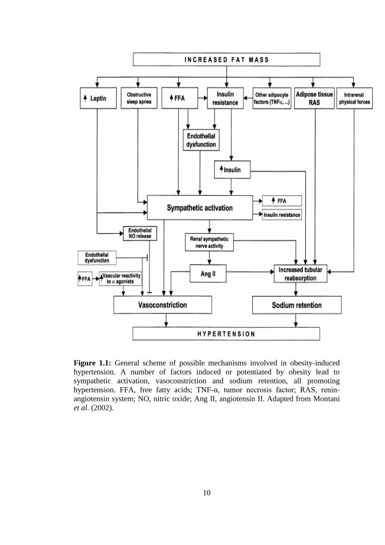
Figure 1.1: General scheme of possible mechanisms involved in obesity-induced
hypertension. A number of factors induced or potentiated by obesity lead to
sympathetic activation, vasoconstriction and sodium retention, all promoting
hypertension. FFA, free fatty acids; TNF-α, tumor necrosis factor; RAS, renin-
angiotensin system; NO, nitric oxide; Ang II, angiotensin II. Adapted from Montani
et al. (2002).

11
1.4 Rat’s model of obesity-induced hypertension
Animal models have been widely used as a tool to study the susceptibility to
obesity in human. It includes genetically obese animals (Zucker rats), surgically
induced obese rats (ventromedial hypothalamus-lesioned rat), spontaneously obese
animals (rhesus monkeys) and diet-induced obese rat and mouse. Among these
models, the dietary model developed by Levin in 1983 is the most relevant with
regard to human obesity as it allows one to examine resistance and susceptibility to
obesity. It also represents the genetic and environmental factors that may contribute
to the development of obesity (Lauterio et al., 1994; 1998; Rahmouni et al., 2005b;
Reuter, 2007) and also allows one to dissociate between the factors related to diet
and obesity per se (Dobrian et al., 2000). However, that particular study by Levin
(1983) did not measure any cardiovascular variables.
In this model, rats on moderately high-fat diet (32% kcal as fat) for a certain
period of time will be segregated into different groups based on the body weight
distribution. One third of the rats whose body weight gain is greater, is considered as
obesity-prone rats while one third lower is considered as obesity-resistant rats and
the remainder is excluded from the study (Lauterio et al., 1998). A number of studies
demonstrated the presence of elevated level of blood pressure, cholesterol and
insulin, activation of RAS and increase in renal oxidative stress in obesity-prone rats
(Carroll et al., 2006). Other than that, obesity-prone rats also exhibit characteristics
that mimic the obese hypertensive patients, such as increased plasma noradrenaline
response to intravenous glucose, increased plasma leptin concentration and decreased
growth hormone secretion and synthesis (Dobrian et al., 2000).

12
1.5 Central baroreceptors pathway
Baroreceptor reflex is important in maintaining the perfusion pressure in the
face of disturbances of circulatory homeostasis through a number of neuronal and
humoral regulatory adjustments. Alteration in the pressure load at the specialized
pressure sensors which are located on the walls of aortic arc and carotid sinuses
initiates these adjustments (Stauss, 2002).
Baroreceptor afferent fibers that run from glossopharyngeal and vagal nerves
provide tonic, excitatory input to neurons of the nucleus of the solitary tract (NTS)
which is subjacent to the area postrema in the dorsomedial medulla during the resting
blood pressure levels. Then, second-order NTS neurons that convey the baroreceptor
signals project to caudal and intermediate part of the ventrolateral medulla (VLM)
and excite the neurons. Neurons from the caudal part of the VLM (CVLM) project to
rostral part of the VLM (RVLM) and inhibit the sympathoexcitatory neurons. It has
been demonstrated that blockade of the inhibitory pathway to RVLM eliminate the
baroreflex (Dampney et al., 2001; Alzamora et al., 2006; Kawabe et al., 2008).
The neurons from the RVLM further project to sympathetic preganglionic
neurons in the intermediolateral column of the spinal cord which is important in
controlling the sympathetic vasomotor outflow (Bergamaschi et al., 1995; Gordon
and Sved, 2002; Morrison, 2003; Iigaya et al., 2007). Baroreceptor sympathetic
efferents regulate the heart, kidneys and also noradrenaline release (Guyenet, 2006).
Meanwhile, alteration in the cardiac parasympathetic nerve activity which is evoked
by baroreceptor is mediated by a direct excitatory projection from second order
neurons in the NTS to the preganglionic parasympathetic neurons in the nucleus
ambiguus (Sved et al., 2001).

13
Figure 1.2: Schematic sagittal view of the medulla oblongata depicting neural
pathways involved in neurogenic hypertension. Premotor neurons of the RVLM
provide excitatory drive to preganglionic neurons in the intermediolateral cell
column (IML), which provide sympathetic output to target organs. RVLM neurons
receive excitatory (▲) inputs from (1) forebrain structures, (2) commissural NTS,
and (3) area postrema, and inhibitory (∆) inputs from the CVLM. The intermediate
and commissural NTS represent the primary site of projection of afferent fibers
arising from baro- and chemoreceptors. Increased excitatory inputs and/or decrease
of inhibitory inputs result in enhancement of RVLM activity, thus increasing
sympathetic output, a common feature of different forms of hypertension. Adapted
from Colombari et al. (2001).
14
1.5.1 Central baroreceptor pathway in obesity-induced hypertension
Baroreflex play a pivotal role in the acute and chronic regulation of body
fluid volumes and arterial pressure but in chronic hypertension, baroreflex function is
often impaired. There is an intriguing finding that shows the baroreflexes rapidly
adapt and reset towards the existing level of the arterial pressure, but the magnitude
and the time course of resetting remain unclear. Recently, a number of studies have
suggested that baroreflexes resetting is incomplete in hypertension (Lohmeier et al.,
2002; 2003; 2007).
Study using Fos-Li immunoreactivity in model of obesity-induced
hypertension has shown that there is a chronic activation of the neurons subserving
the baroreflexes. In this study, increased Fos staining in NTS and CVLM were
observed thus indicating important roles of these regions in mediating baroreflex
inhibition of sympathoexcitatory cells in RVLM. This finding is supported by the
observation in animals with intact baroreflexes which shows similar response during
acute elevations in arterial pressure, and also study in animals without baroreflexes
which shows no increase in Fos staining in NTS and CVLM. Therefore, these
findings indicate that activation of baroreflex is sustained during obesity
hypertension and thus baroreflex-mediated sympathoinhibition is a long-term
compensatory response in this model of hypertension. Eventhough activation of
central baroreflex pathway is sustained in obesity hypertension, it has been shown
that Fos-Li expression is significantly increased in the RVLM of obese dogs
(Lohmeier et al., 2003), thereby supports other studies that showed increased
sympathetic activity to the kidney and other vascular beds in obesity hypertension
(Esler, 2000; Hall et al., 1999). Additionally, the presence of increased number of

15
Fos-Li cells in obese dogs may reflect the dominant action of excitatory inputs over
chronic inhibitory effects of the baroreflex to the RVLM (Lohmeier et al., 2003).
Another study in obesity-induced hypertensive rats with intact baroreflex
showed that reflex bradycardia was reduced during pressor responses to
phenylephrine but no difference was observed in reflex tachycardia during depressor
responses to sodium nitroprusside in both obese hypertensive rats and control.
Similar inhibition of reflex tachycardia was seen in both groups after either
cholinergic or β-adrenergic blockade by methylatropine or propranolol. Meanwhile,
reflex bradycardia was reduced more in obese-hypertensive rats than in control rats
but equally reduced after β-adrenergic and cholinergic blockade respectively. Hence,
it is postulated that selective impairment of reflex bradycardia might be due to
deficient of parasympathetic activity since blockade of β-adrenergic would leave
only residual reflex responses by parasympathetic activity (Bunag et al., 1990).
1.5.2 Rostral ventrolateral medulla
Rostral ventrolateral medulla (RVLM) plays a pivotal role in the tonic and
phasic regulation of cardiovascular function (Lipski et al., 1996; Araujo et al., 1999;
Campos and McAllen, 1999; Ito et al., 2001; 2002; Mayorov and Head, 2002;
Granata and Cohen, 2004; Oshima et al., 2006; Menezes and Fontes, 2007). RVLM
bulbospinal neurons that project directly to sympathetic preganglionic neurons in the
spinal cord, thus called ‘presympathetic’ neurons are a major source of basal tonic
sympathetic vasomotor activity (Yang and Coote, 1998; Tagawa and Dampney,
1999; Potts et al., 2000; Hu et al., 2002; Horiuchi and Dampney, 2002; Dampney et

16
al., 2003; Oshima et al., 2006;). These tonic excitations of sympathetic vasomotor
outflow are important in maintaining normal arterial blood pressure (Brooks et al.,
2004; Horiuchi et al., 2004). Therefore, any neuronal inhibition of RVLM will
markedly result in decrease in arterial pressure. These RVLM neurons receive both
excitatory and inhibitory synaptic inputs resulted from stimulation of peripheral
receptors and also higher center in the brain (Horiuchi et al., 2004).
Numerous studies have shown that RVLM neurons are tonically active but
the tonic excitatory drive to the neurons remains unclear (Horiuchi et al., 2004;
Stocker et al., 2007). However, it is believed that the tonic excitatory input arise
from one or combination of afferents to the RVLM. Studies on neuroanatomical has
documented some brain regions which project to the vasomotor area of the RVLM.
In rats, some regions have been identified including CVLM, NTS, area postrema,
raphe nuclei, A5 neurons, hypothalamic paraventricular neurons (PVN), central
amygdaloid nucleus, periaqueductal gray (PAG), parabrachial nucleus and lateral
hypothalamic area (LHA). Inhibition of these brain regions individually did not lead
to marked decrease in arterial pressure indicating that there might be two or more
regions that drive tonic excitatory input to the RVLM. Thus, marked decrease in
arterial pressure would only be observed after inhibition of correct combination of
these regions (Sved et al., 2001; Pilowsky and Goodchild, 2002; Dampney et al.,
2003).
17
1.5.2.1 Neurotransmitters and neuromodulators in RVLM
RVLM neurons consist of heterogenous cell population whereby, different
genes, different complement of receptors, transmitters, enzymes and other proteins
are found in different subpopulations of bulbospinal RVLM neurons. These includes
glutamate, catecholamines, preproenkephalin/enkephalin, substance P, neuropeptide
Y, cocaine- and amphetamine-regulated transcript, preprogalanin, calbindin, GABAA
and GABAB receptors, P2X receptors, alpha2A-adrenergic receptors, angiotensin1A
receptors, and mu opioid receptors. Among these, the key for neurochemical within
RVLM are those involved glutamate and GABA transmission (Pilowsky and
Goodchild, 2002).
Glutamate is an excitatory amino acid (EAA) which acts as a principle
neurotransmitter in the central nervous system (Goren et al., 2000; Gonzalez and
Robinson, 2004; Maragakis and Rothstein, 2004; Shigeri et al., 2004; Bridges and
Esslinger, 2005). Glutamate is released into the synaptic cleft and bind to glutamate
receptors resulted in propagation of action potential. Apart from glutamate receptors,
glutamate transporters also play a role in modulation of the synaptic activity by
removing the glutamate from the synaptic cleft. Studies have proven the importance
of glutamate in normal central nervous system synaptic function. However, excess
accumulation of glutamate in the synaptic cleft can lead to neurotoxicity. Defects in
the function of glutamate transporters lead to a rise in extracellular glutamate which
then results in the development of neurologic disease. Glutamate transporters
dysfunction might be a result of alteration in transcription or splicing, increased
turnover of the transporters, altered trafficking of glutamate transporter, abnormal

18
protein phosphorylation and reduced transport capacity (Maragakis and Rothstein,
2004; Boeck et al., 2005).
Glutamate can act on two main classes of receptors which are ionotropic (ion-
channel linked) and metabotropic receptors (G-protein linked) (Tsuchihashi et al.,
1994). The latter receptors couple to the intracellular second messenger cascades via
G-proteins (Tsuchihashi et al., 2000; Herlenius and Lagercrantz, 2004;
Vandenberghe and Bredt, 2004; Galik et al., 2008). Ionotropic glutamate receptors
can be subdivided into two types which are N-methyl-D-aspartate (NMDA) and non-
NMDA receptors based on their sensitivity to agonists and differing molecular
structures. Non-NMDA receptors can be further classified into kainate and AMPA
receptors. Each class of glutamate receptors (GluRs) has their own subunits whereby,
NMDA receptors consist of NR1and NR2A-NR2D. Meanwhile, GluR1-GluR4
represents the AMPA-sensitive family and GluR5-GluR7, KA1 and KA2 belong to
kainate family (Smart, 1997; Pamidimukkala et al., 2002).
Gamma-aminobutyric acid (GABA) is a dominant inhibitory neurotransmitter
in the central nervous system of the mature animal. GABA can acts on two types of
receptors namely, GABAA and GABAB receptor. GABAA receptor is a
transmembrane protein wherein, other substances like barbiturate and
benzodiazepines can bind to specific sites and thus modulating the opening
properties of the chloride channel. These receptors have different pharmacological
and electrophysiological properties depending on their subunit composition. GABAB
receptor (G protein-coupled) which is present in lower level in the central nervous
system, performs its function late in the development of central nervous system
(Herlenius and Lagercrantz, 2004).
19
Nerve cells contain high concentration of chloride during the early
development of the central nervous system. Therefore, opening of the chloride
channel by GABA leads to depolarization (i.e., excitation) to occur. By contrast, the
decrease in chloride concentration during maturation led to an opposite effect of
GABA whereby, chloride ions are pumped out and cell hyperpolarization occurs.
Hence, GABA switches its role from excitatory to inhibitory neurotransmitter in the
mature animal (Herlenius and Lagercrantz, 2004).
Kynurenic acid (KYN), an endogenous tryptophan metabolite is present in
the mammalian central nervous system. Increase level of KYN in the brain is related
with sedation, analgesia and reduction of post-ischaemic or post-inflammatory
damage. KYN has the capability to reduce the neurotoxic effects of glutamate by
acting on the glutamate ionotropic receptors (Stone and Addae, 2002; Prescott et al.,
2006; Moroni et al., 2007).
Nitric oxide (NO) is present with varying amounts in most of the brain
regions. NO is synthesized from the amino acid L-arginine by nitric oxide synthase
(NOS) (Raevskii, 1997). NO plays a vital role in many functions of the nervous
system including memory, learning, vision and cardiovascular regulation. NO can
act centrally, decreasing the sympathetic nerve activity and blood pressure
(Dominiczak and Bohr, 1995; Zhang and Patel., 1998; Patel et al., 2001). Recent
study reported that high levels of NO can reduce glutamate presynaptic release via
inhibition of presynaptic N-type Ca2+
-channel activity thereby, depressing the
RVLM neurons (Tai et al., 2005).

20
1.5.2.2 Tonic glutamatergic input to RVLM presympathetic neurons
Numerous studies have demonstrated that RVLM presympathetic neurons
receive excitatory glutamatergic synaptic input which originate from both medullary
and supramedullary regions (Dampney et al., 2003). There are some possible sources
of this glutamatergic synaptic input such as input from pontine reticular formation,
lateral tegmental field and also from the PVN of the hypothalamus. Blockade of
ionotropic glutamate receptors in the RVLM of cat and rabbit caused a profound
reduction in blood pressure indicating that glutamatergic synaptic input is the main
source of tonic excitatory drive to RVLM (Barman et al., 2000; Hourichi and
Dampney, 2002; Sved et al., 2002).
1.5.2.3 Tonic GABAergic input to RVLM presympathetic neurons
Studies have reported that there is a presence of tonic GABAergic input to the
RVLM neurons. This GABAergic input to RVLM is mediated partly via neurons of
the caudal aspect of the VLM (CVLM) (Sved and Gordon, 1994). Furthermore, tonic
GABAergic input to the RVLM presympathetic neurons are also partly engaged in
baroreceptor-independent regulatory mechanism. Inhibitory action of GABA on
RVLM neurons is mediated preferentially through activation of GABAA receptor.
Blockade of this GABAA receptor in the sympathoexcitatory regions of the RVLM
eliminates the action of inhibitory vasomotor neurons from the caudal part thus
results in large increase in sympathetic nerve activity and blood pressure (Moreira et
al., 2005; Menezes and Fontes, 2007).

21
1.5.2.4 Intrinsic pacemaker of RVLM
Apart from synaptic input which is driven by neurons located elsewhere in
the brain, it has been speculated that tonic activity of RVLM neurons can also be
generated by intrinsic pacemaker activity which is capable of generating action
potentials (autoactivity) (Lewis and Coote, 1993; Dampney et al., 2003; Coote,
2006). This theory arises due to failure of blockade of glutamate receptors to cause
significant reduction of tonic discharge of RVLM neurons. In addition, removal of
synaptic input by blockade of excitatory or inhibitory EAA receptors or during
perfusion of low-Ca2+
high-Mg2+
solution did not affect the regular and irregular
discharge of RVLM neurons in brain slice. This theory is further supported by the
fact that action potential in RVLM neurons was not initiated by EPSPs, the regular
discharge was reset by an experimentally given stimulus and the discharge was
eliminated by membrane hyperpolarization with no residual EPSPs. Besides, other
membrane properties which were exhibited by the cells could also contribute to the
intrinsic oscillating depolarization (Lewis and Coote, 1993; Coote, 2006).
Nevertheless, such intrinsic activity of ‘pacemaker’ neurons was only
recorded in vitro in isolated medulla and spinal cord but not in vivo. Intracellular
recordings of identified RVLM presympathetic neurons in vivo showed irregular
low-frequency action potentials which triggered by EPSPs (Lipski et al., 1996;
Coote, 2006) and in accordance with the pattern of discharge from synaptic drive
observed in sympathetic preganglionic neurons (Coote, 2006). This theory also failed
to explain the basic rhythmic oscillations of activity in RVLM presympathetic
neurons which are related with similar activity in postganglionic sympathetic nerves.

22
It also cannot explain the non-uniformity of sympathetic activity in different
vasomotor nerves.
Therefore, synaptic drive is the most probably source of tonic activity in
RVLM neurons in unchallenged adult animal. However, some considerable condition
should be taken into account. For instance, specific slow synaptic input due to its
removal could induce activation of selective inward currents which has been shown
to trigger beating activity in RVLM neurons in vitro as a response to challenging
environmental stimuli (Coote, 2006).
1.5.2.5 RVLM in obesity-induced hypertension
Sved and his colleagues had proposed that sympathetic vasomotor outflow
from RVLM neurons is maintained by balance of excitatory and inhibitory input
(Sved et al., 2001). In normotensive rats, administration of KYN, an EAA antagonist
into RVLM did not produce any effect on the arterial blood pressure (Ito et al.,
2002). This indicated that under resting conditions, direct excitatory and indirect
inhibitory influences of EAA are in perfect balance whereby, activation of the
CVLM inhibitory mechanisms counterbalances the excitatory drive to the RVLM
neurons. Blockade of the EAA receptors in the RVLM simultaneously removed both
of the influence of tonic excitation and inhibition which led to no net change in
sympathetic vasomotor activity and arterial blood pressure (Sved et al., 2002;
Horiuchi et al., 2004). However, blockade of EAA receptors in RVLM in
combination with CVLM inhibition using muscimol (GABA agonist) did cause a
profound fall in arterial blood pressure indicating that RVLM does receive important

23
tonic excitatory drive. In contrast, injection of muscimol into CVLM alone results in
increase of both sympathetic activity and arterial blood pressure due to release of
excitatory mechanisms to the RVLM (Moreira et al., 2005).
However, in models of experimental hypertension, the excitatory and
inhibitory balance is altered (Ito et al., 2002). In Dahl-sensitive model, the balance of
excitatory amino acid input was disrupted, whereby; the balance is shifted towards
excitation in Dahl-sensitive rats as compared with Dahl-resistant rats. A significant
drop in mean arterial pressure was recorded after microinjection of KYN into the
RVLM (Ito et al., 2001). In addition, disruption in the balance of excitatory and
inhibitory input was also observed in spontaneously hypertensive rat (SHR) (Ito et
al., 2000).
In addition, recent study has proven that the RVLM neurons do contribute to
obesity-induced hypertension in rats. However, the finding only indicates that the
tonic activity of RVLM neurons is necessary for the elevated blood pressure in
obesity-hypertension rats but it does not demonstrate that the tonic activity of RVLM
neurons is elevated (Stocker et al., 2007).
Thus, this present study aims to investigate the neural pathways that underlie
the activation of RVLM neurons by determining whether EAA input particularly L-
glutamate within the RVLM neurons do contribute to elevated blood pressure in
obesity. For this purpose, we investigate further if the microinjections of EAA-
receptor antagonist into RVLM vicinity result in a reduction of arterial pressure in
obesity-induced hypertensive rats.

24
1.6 Study on peripheral vascular reactivity
Previous study has demonstrated that elevated blood pressure in diet-induced
obesity rats is associated with an enhanced vascular contractility (Boustany-Kari et
al., 2007). However, such finding was observed in in vitro preparation of the
mesenteric arteries of the rats, thus not presenting the whole animal system.
Numerous studies has been carried out using pithed rats (Fozard and Leach, 1968;
Barret, 1971; Dong et al., 1995; Gavin and Docherty, 1996; Zhou and Vargas, 1996;
Dendorfer et al., 1999; Elayan et al., 2002; 2008; Cobos-Puc et al., 2007) as pithing
eliminates the central reflex responses to the pressor effects upon peripheral
stimulation. Therefore, availability of this pithing technique enables us to assess in
vivo peripheral vascular reactivity in the rat’s model of obesity-induced hypertension.
1.6.1 Endothelial dysfunction in obesity-induced hypertension
Obesity is closely related with increased risk for hypertension whereby,
inflammation, vascular remodeling and changes in vascular reactivity are the leading
role in the pathogenesis of this disease (Mundy et al., 2007). Abnormalities of
arterial function and structures have been observed in obese human and in certain
animal models of obesity including the changes in release of vasoactive substances
from the endothelial cells and also elasticity of the arterial wall. Naderali and his
colleagues have reported that endothelial dysfunction can occur as a result of acute
consumption of a palatable diet long before development of any significant obesity.
Short term withdrawal of this palatable diet incompletely corrected the endothelial
dysfunction. However, endothelial dysfunction can be corrected completely by

25
chronic removal of palatable diet which indicates that diet per se plays an important
role in the development of vascular abnormalities in animal models of obesity
(Naderali et al., 2004).
Endothelial dysfunction in obesity is due to alteration in the synthesis/release
of prostanoids and nitric oxide. These substances play a vital role in the regulation of
the vascular tone (Mascher et al., 2006). Prostacyclin (PGI2) is mainly generated in
endothelial cells from arachidonic acid. PGI2 can cause vasodilation through
activation of protein kinase A which reduce the myosin light chain kinase activity
and also can inhibit platelet aggregation (Khazaei et al., 2008).
Experimental studies in animal model of obesity has shown the presence of
oxidative stress which can lead to the endothelial dysfunction. Oxidative stress can
block the nitric oxide synthase and also inactivate the action of nitric oxide, thus
impairing the NO-mediated vasorelaxation. It can also cause an increase in
generation of endothelin-1 and increase effects of the superoxide anion and hydrogen
peroxide on vascular smooth muscle cells (Wolk et al., 2003).
Insulin acts as a vasodilator that can affect the peripheral resistance in normal
vasculature. Insulin acts by inhibiting the voltage-gated Ca2+
influx and stimulating
the glucose transport and glucose phosphorylation, thus leading to activation of Ca2+
ATPase transcription and also increase in cellular Ca2+
efflux. As a result, there is
decrease in vascular resistance due to net decrease of intracellular Ca2+
. However, the
presence of insulin resistance in obesity blunt these effects, thereby increases the
vascular resistance (Zhang and Reisin, 2000).