study on biological effect of la3+ on escherichia coli by atomic force microscopy
TRANSCRIPT

JOURNAL OFInorganicBiochemistry
Journal of Inorganic Biochemistry 98 (2004) 68–72
www.elsevier.com/locate/jinorgbio
Study on biological effect of La3þ on Escherichia coli by atomicforce microscopy
Liu Peng a,*, Liu Yi a,*, Lu Zhexue a, Zhu Juncheng a, Dong Jiaxin a, Pang Daiwen a,Shen Ping b, Qu Songsheng a
a Department of Chemistry, College of Chemistry and Molecular Sciences, Wuhan University, Wuhan 430072, PR Chinab School of Life Sciences, Wuhan University, Wuhan 430072, PR China
Received 13 May 2003; received in revised form 21 August 2003; accepted 27 August 2003
Abstract
The biological effects of rare-earth metal ions on the organism have been studied using La3þ as a probe ion and Escherichia coli
cell as a target organism. Atomic force microscopy (AFM) studies reveal that La3þ substantially changes the structure of the outer
cell membrane responsible for the cell permeability. Significant damages of the outer cell membrane are observed using scanning
electron microscopy (SEM) after the introduction of La3þ. In result, the cell becomes easily attacked by lysozyme. Moreover, in-
ductively coupled plasma-mass spectrometry (ICP-MS) measurements show considerable amount of Ca2þ and Mg2þ in the su-
pernatant from the La3þ exposed cells. It is proposed that La3þ can replace Ca2þ from the binding sites because of their close ionic
radii and similar ligand specificities. Lipopolysaccharide (LPS), which forms the outer membrane of Gram-negative bacteria, could
not serve as the cellular envelope steadily after Ca2þ and Mg2þ released from their binding sites on the LPS patches.
� 2003 Elsevier Inc. All rights reserved.
Keywords: La3þ; Lipopolysaccharide; Permeability; Biological effect; Lysozyme
1. Introduction
The physiological functions and biological effects of
the rare-earth elements have been well studied [1–3].
Recently some fertilizers with rare-earth elements addi-
tive have been used in China to improve the yields and
qualities of crops. It has been found out that the rare-
earth elements can accumulate in the soil, bioaccumu-
late in crops and finally transfer to the food chain.
Increasing attention has been paid to the influence ofrare-earth elements on the environment and human
health [4]. Although the toxic action of La3þ as a probe
ion on organisms has been reported [5], the mechanism
of its biological effects is still unclear.
Escherichia coli (E. coli) is a kind of Gram-negative
bacteria with a cell membrane and wall model shown
* Corresponding authors. Tel.: +86-27-87218284; fax: +86-27-
87647617.
E-mail addresses: [email protected] (L. Peng), [email protected].
edu.cn, [email protected] (L. Yi).
0162-0134/$ - see front matter � 2003 Elsevier Inc. All rights reserved.
doi:10.1016/j.jinorgbio.2003.08.012
in Fig. 1 [6–8]. The inner membrane is composed of
phospholipid chains and proteins, and the outermembrane consists of lipopolysaccharide (LPS), pep-
tidoglycan and periplasm. LPS forms the surface of
Gram-negative bacteria and acts as a selective per-
meability barrier for E. coli and other Gram-negative
bacteria. Molecular structure of LPS is shown in Fig. 1
[8]. It is composed of lipid A, an inner core, an outer
core and the O-antigen units. Loss or damages of LPS
molecules would lead to an increase in the perme-ability of the cell wall. It is presumed that the
biological effects of rare-earth elements on the or-
ganisms contribute to the change in the membrane
permeability [2,3]. However, no molecule-/nano-level
experiment has been performed to confirm this per-
meability alteration caused by the rare-earth elements
owing to the resolution limit of optical instrumentals.
Atomic force microscopy (AFM) techniques can pro-vide nano-level image of cell surface under physio-
logical conditions [9]. It has been used to widely study
the structure details of biological systems since its

Fig. 1. Schematic molecular representation of E. coli envelop. The envelop of Gram-positive bacterium consists of the outer membrane, the inner
membrane and peptidoglycan layer. MDO is membrane-derived oligosaccharides.
L. Peng et al. / Journal of Inorganic Biochemistry 98 (2004) 68–72 69
invention in 1986 [10]. Three-dimensional (3D) high-
resolution topographs can be in situ available.
In this paper, we choose the E. coli (HB101) cell as a
target organism and La3þ as a probe ion to study the
biological effects of rare earth elements using the AFM
technique. Inductively Coupled plasma-mass spectrom-
etry (ICP-MS) and scanning electron microscopy (SEM)
have been used to further explore the biological effectmechanism of La3þ.
2. Materials and methods
2.1. Cells and reagents
The strain of E. coli (HB101), which was wild type incell wall synthesis, was provided by the Chinese Center
for Type Culture Collections of Wuhan University. The
peptone culture contained NaCl 5 g, peptone 5 g, beef
extract 5 g, Yeast extract 6 g per 1000 ml (pH¼ 7.2). The
culture was sterilized in high-pressure steam at 120 �Cfor 30 min.
Analytical grade reagent Lanthanum nitrate was
obtained from China Medicine (Group) ShanghaiChemical Reagent Corporation. Lysozyme (20 000 U/
mg) extracted from chicken egg albumin was provided
by Amresco. The lysozyme solution was diluted to
100 000 U/ml with TE buffer.
2.2. Sample preparation
When E. coli was grown to primary-log phase at37 �C in a peptone culture, two of 4 ml bacteria sus-
pensions were centrifuged at 5000 r/min for 2 min. The
precipitate was re-suspended in 4 ml ultra-pure water
and was then washed and recovered. This procedure was
repeated 5 times to thoroughly remove the culture me-
dium. 400 lg/ml La3þ was then added into one of the
washed suspensions. After the two cell suspensions were
incubated in the culture media at 37 �C for 1 h with
continuous shaking, the solutions were centrifuged at5000 r/min for 2 min. The supernatant and the bacteria
were used for the following analysis.
2.3. Analysis of Ca2þ and Mg2þ
Inductively Coupled Plasma Mass Spectrometer
(ICP-MS) (Agilent 7500a) was used in this experiment to
determine the amount of Mg2þ and Ca2þ. ICP-MS is amethod for rapid and sensitive determination of all
considered elements and the method is sufficiently sen-
sitive to detect 1 ng/l of elements [11,12]. The concen-
trations of Ca2þ and Mg2þ in the supernatant obtained
from the above experiment were analyzed by ICP-MS.
2.4. Imaging cells
AFM measurements (PicoScan system, Molecular
Imaging Inc, Tempe, AZ) were performed in the air
using a Molecular Imaging PicoScan system (Molecular
Imaging Inc, Tempe, AZ) operating in the Magnetic AC
(MAC) mode. Special coated AFM tips (type II MAC
tips, Molecular Imaging, Inc) were employed. These tips
were coated with a magnetically conductive material and
possess a lower spring constant range (1.2–5.5 N/m)than common tapping mode models due to the lower

Fig. 2. AFM images of the native E. coli (HB101) and cells treated by 400 lg/ml La3þ. The first column (A1, A2, A3) shows images of the native
E. coli in the different scanning area. The second column (B1, B2, B3) presents images of cells treated by La3þ in the different scanning area.
The scanning area of the 1st, 2nd, 3rd row are 14� 21 lm2, 4� 4 lm2, 0.5� 0.5 lm2, respectively. The 4th row presents the 3D images of graph A3
and B3.
70 L. Peng et al. / Journal of Inorganic Biochemistry 98 (2004) 68–72
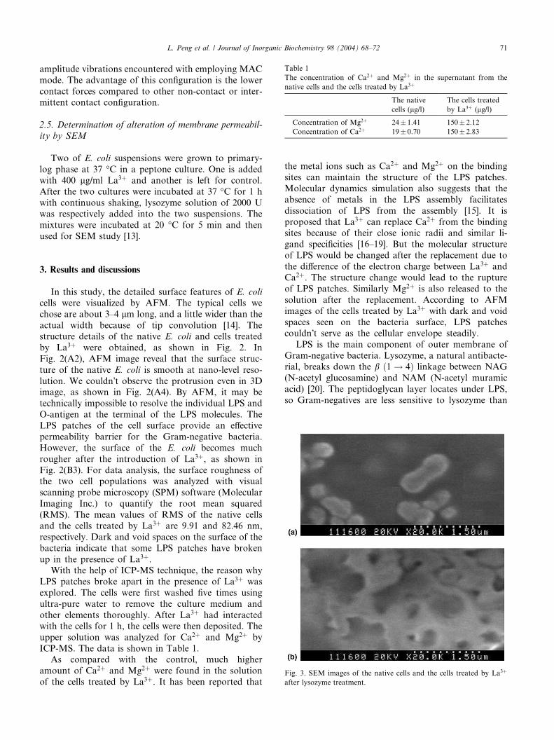
Table 1
The concentration of Ca2þ and Mg2þ in the supernatant from the
native cells and the cells treated by La3þ
The native
cells (lg/l)The cells treated
by La3þ (lg/l)
Concentration of Mg2þ 24� 1.41 150� 2.12
Concentration of Ca2þ 19� 0.70 150� 2.83
L. Peng et al. / Journal of Inorganic Biochemistry 98 (2004) 68–72 71
amplitude vibrations encountered with employing MAC
mode. The advantage of this configuration is the lower
contact forces compared to other non-contact or inter-
mittent contact configuration.
2.5. Determination of alteration of membrane permeabil-
ity by SEM
Two of E. coli suspensions were grown to primary-
log phase at 37 �C in a peptone culture. One is added
with 400 lg/ml La3þ and another is left for control.
After the two cultures were incubated at 37 �C for 1 h
with continuous shaking, lysozyme solution of 2000 Uwas respectively added into the two suspensions. The
mixtures were incubated at 20 �C for 5 min and then
used for SEM study [13].
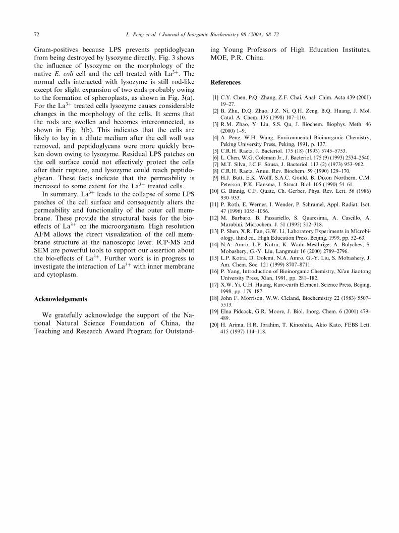
Fig. 3. SEM images of the native cells and the cells treated by La3þ
after lysozyme treatment.
3. Results and discussions
In this study, the detailed surface features of E. colicells were visualized by AFM. The typical cells we
chose are about 3–4 lm long, and a little wider than the
actual width because of tip convolution [14]. The
structure details of the native E. coli and cells treated
by La3þ were obtained, as shown in Fig. 2. In
Fig. 2(A2), AFM image reveal that the surface struc-
ture of the native E. coli is smooth at nano-level reso-
lution. We couldn�t observe the protrusion even in 3Dimage, as shown in Fig. 2(A4). By AFM, it may be
technically impossible to resolve the individual LPS and
O-antigen at the terminal of the LPS molecules. The
LPS patches of the cell surface provide an effective
permeability barrier for the Gram-negative bacteria.
However, the surface of the E. coli becomes much
rougher after the introduction of La3þ, as shown in
Fig. 2(B3). For data analysis, the surface roughness ofthe two cell populations was analyzed with visual
scanning probe microscopy (SPM) software (Molecular
Imaging Inc.) to quantify the root mean squared
(RMS). The mean values of RMS of the native cells
and the cells treated by La3þ are 9.91 and 82.46 nm,
respectively. Dark and void spaces on the surface of the
bacteria indicate that some LPS patches have broken
up in the presence of La3þ.With the help of ICP-MS technique, the reason why
LPS patches broke apart in the presence of La3þ was
explored. The cells were first washed five times using
ultra-pure water to remove the culture medium and
other elements thoroughly. After La3þ had interacted
with the cells for 1 h, the cells were then deposited. The
upper solution was analyzed for Ca2þ and Mg2þ by
ICP-MS. The data is shown in Table 1.As compared with the control, much higher
amount of Ca2þ and Mg2þ were found in the solution
of the cells treated by La3þ. It has been reported that
the metal ions such as Ca2þ and Mg2þ on the binding
sites can maintain the structure of the LPS patches.
Molecular dynamics simulation also suggests that the
absence of metals in the LPS assembly facilitates
dissociation of LPS from the assembly [15]. It is
proposed that La3þ can replace Ca2þ from the binding
sites because of their close ionic radii and similar li-
gand specificities [16–19]. But the molecular structureof LPS would be changed after the replacement due to
the difference of the electron charge between La3þ and
Ca2þ. The structure change would lead to the rupture
of LPS patches. Similarly Mg2þ is also released to the
solution after the replacement. According to AFM
images of the cells treated by La3þ with dark and void
spaces seen on the bacteria surface, LPS patches
couldn�t serve as the cellular envelope steadily.LPS is the main component of outer membrane of
Gram-negative bacteria. Lysozyme, a natural antibacte-
rial, breaks down the b ð1 ! 4Þ linkage between NAG
(N-acetyl glucosamine) and NAM (N-acetyl muramic
acid) [20]. The peptidoglycan layer locates under LPS,
so Gram-negatives are less sensitive to lysozyme than
72 L. Peng et al. / Journal of Inorganic Biochemistry 98 (2004) 68–72
Gram-positives because LPS prevents peptidoglycan
from being destroyed by lysozyme directly. Fig. 3 shows
the influence of lysozyme on the morphology of the
native E. coli cell and the cell treated with La3þ. Thenormal cells interacted with lysozyme is still rod-likeexcept for slight expansion of two ends probably owing
to the formation of spheroplasts, as shown in Fig. 3(a).
For the La3þ treated cells lysozyme causes considerable
changes in the morphology of the cells. It seems that
the rods are swollen and becomes interconnected, as
shown in Fig. 3(b). This indicates that the cells are
likely to lay in a dilute medium after the cell wall was
removed, and peptidoglycans were more quickly bro-ken down owing to lysozyme. Residual LPS patches on
the cell surface could not effectively protect the cells
after their rupture, and lysozyme could reach peptido-
glycan. These facts indicate that the permeability is
increased to some extent for the La3þ treated cells.
In summary, La3þ leads to the collapse of some LPS
patches of the cell surface and consequently alters the
permeability and functionality of the outer cell mem-brane. These provide the structural basis for the bio-
effects of La3þ on the microorganism. High resolution
AFM allows the direct visualization of the cell mem-
brane structure at the nanoscopic lever. ICP-MS and
SEM are powerful tools to support our assertion about
the bio-effects of La3þ. Further work is in progress to
investigate the interaction of La3þ with inner membrane
and cytoplasm.
Acknowledgements
We gratefully acknowledge the support of the Na-
tional Natural Science Foundation of China, the
Teaching and Research Award Program for Outstand-
ing Young Professors of High Education Institutes,
MOE, P.R. China.
References
[1] C.Y. Chen, P.Q. Zhang, Z.F. Chai, Anal. Chim. Acta 439 (2001)
19–27.
[2] B. Zhu, D.Q. Zhao, J.Z. Ni, Q.H. Zeng, B.Q. Huang, J. Mol.
Catal. A: Chem. 135 (1998) 107–110.
[3] R.M. Zhao, Y. Liu, S.S. Qu, J. Biochem. Biophys. Meth. 46
(2000) 1–9.
[4] A. Peng, W.H. Wang, Environmental Bioinorganic Chemistry,
Peking University Press, Peking, 1991, p. 137.
[5] C.R.H. Raetz, J. Bacteriol. 175 (18) (1993) 5745–5753.
[6] L. Chen, W.G. Coleman Jr., J. Bacteriol. 175 (9) (1993) 2534–2540.
[7] M.T. Silva, J.C.F. Sousa, J. Bacteriol. 113 (2) (1973) 953–962.
[8] C.R.H. Raetz, Anuu. Rev. Biochem. 59 (1990) 129–170.
[9] H.J. Butt, E.K. Wolff, S.A.C. Gould, B. Dixon Northern, C.M.
Peterson, P.K. Hansma, J. Struct. Biol. 105 (1990) 54–61.
[10] G. Binnig, C.F. Quate, Ch. Gerber, Phys. Rev. Lett. 56 (1986)
930–933.
[11] P. Roth, E. Werner, I. Wender, P. Schramel, Appl. Radiat. Isot.
47 (1996) 1055–1056.
[12] M. Barbaro, B. Passariello, S. Quaresima, A. Cascillo, A.
Marabini, Microchem. J. 51 (1995) 312–318.
[13] P. Shen, X.R. Fan, G.W. Li, Laboratory Experiments in Microbi-
ology, third ed., High Education Press, Beijing, 1999, pp. 52–63.
[14] N.A. Amro, L.P. Kotra, K. Wadu-Mesthrige, A. Bulychev, S.
Mobashery, G.-Y. Liu, Langmuir 16 (2000) 2789–2796.
[15] L.P. Kotra, D. Golemi, N.A. Amro, G.-Y. Liu, S. Mobashery, J.
Am. Chem. Soc. 121 (1999) 8707–8711.
[16] P. Yang, Introduction of Bioinorganic Chemistry, Xi�an Jiaotong
University Press, Xian, 1991, pp. 281–182.
[17] X.W. Yi, C.H. Huang, Rare-earth Element, Science Press, Beijing,
1998, pp. 179–187.
[18] John F. Morrison, W.W. Cleland, Biochemistry 22 (1983) 5507–
5513.
[19] Elna Pidcock, G.R. Moore, J. Biol. Inorg. Chem. 6 (2001) 479–
489.
[20] H. Arima, H.R. Ibrahim, T. Kinoshita, Akio Kato, FEBS Lett.
415 (1997) 114–118.