study and case compilation · · 2018-04-05study and case compilation. ... class ii clinical case...
TRANSCRIPT
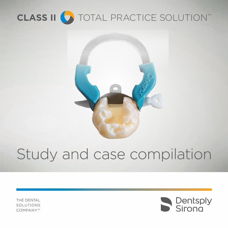
Study and case compilation
That’s why Dentsply Sirona developed the Class II Solution™. It is a proven approach to achieving a reliable outcome, addressing key challenges with innovative product solutions at each step of the procedure.
With a solution for each step of the procedure, the Class II Solution™ provides you with tools to achieve procedure success and patient satisfaction.
1 American Dental Association Procedure Recap Report (2006).
45%
of direct restorations are Class II procedures.1
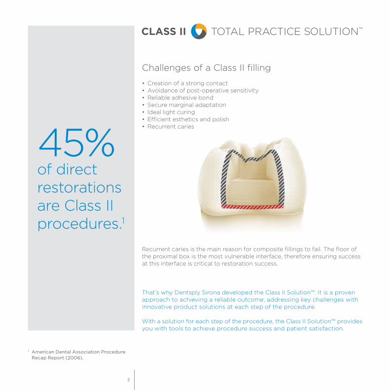
Challenges of a Class II filling
• Creation of a strong contact • Avoidance of post-operative sensitivity• Reliable adhesive bond• Secure marginal adaptation• Ideal light curing• Efficient esthetics and polish• Recurrent caries
Recurrent caries is the main reason for composite fillings to fail. The floor of the proximal box is the most vulnerable interface, therefore ensuring success at this interface is critical to restoration success.
2

Content
Class II clinical case ................................................................. 4
........................................................................... 6
...................................................................... 9
......................................................................................... 14
........................................................ 19
.................................................................... 24
........................................................................................... 28
Palodent® PlusSectional Matrix System
Prime&Bond elect®Universal Dental Adhesive
SDR® flow+Bulk Fill Flowable
TPH Spectra® STUniversal Composite Restorative
SmartLite® Focus®Pen-Style LED Curing Light
Enhance®Finishing System
3
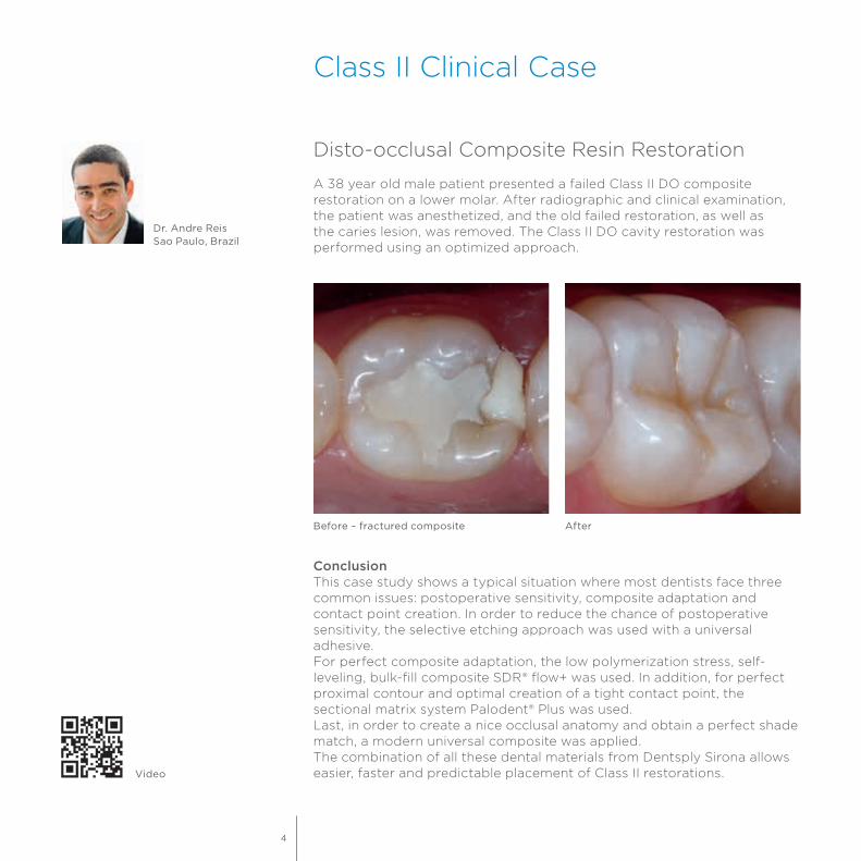
Disto-occlusal Composite Resin Restoration
A 38 year old male patient presented a failed Class II DO composite restoration on a lower molar. After radiographic and clinical examination, the patient was anesthetized, and the old failed restoration, as well as the caries lesion, was removed. The Class II DO cavity restoration was performed using an optimized approach.
Dr. Andre ReisSao Paulo, Brazil
Before – fractured composite After
ConclusionThis case study shows a typical situation where most dentists face three common issues: postoperative sensitivity, composite adaptation and contact point creation. In order to reduce the chance of postoperative sensitivity, the selective etching approach was used with a universal adhesive.For perfect composite adaptation, the low polymerization stress, self-leveling, bulk-fill composite SDR® flow+ was used. In addition, for perfect proximal contour and optimal creation of a tight contact point, the sectional matrix system Palodent® Plus was used.Last, in order to create a nice occlusal anatomy and obtain a perfect shade match, a modern universal composite was applied.The combination of all these dental materials from Dentsply Sirona allows easier, faster and predictable placement of Class II restorations.Video
Class II Clinical Case
4

2. The Palodent® Plus sectional matrix system was placed using the Universal Ni-Ti ring and the 6.5 mm matrix to prevent gaps in gingival-axial corner.
1. The old composite restoration and caries were removed. Note that the distobuccal cusp was also compromised.
3. The hollow underside of the wedge allows placement of a second wedge from the opposite side for a tight gingival margin.
4. Enamel margins were selectively etched with phosphoric acid.
5. Note the chalky white appearance after rinsing off the phosphoric acid and air-drying.
6. Prime&Bond elect® adhesive was applied in the selective etching mode.
7. In one single increment of up to 4 mm, SDR® flow+ bulkfill composite was inserted to replace the dentin structure. Light activation was performed for 20 sec with SmartLite® Focus® curing light.
8. Afterwards, TPH Spectra® composite (shade A1), was used for the occlusal surface. First, a composite increment was inserted for reconstruction of distal proximal ridge.
9. Appearance after placement of the composite. Note the characterization stains.
5

Adjacent teeth damaged by dental bur
ObjectiveCutting and finishing approximal preparations with conventional instrumentation and methods may produce iatrogenic damage in adjacent tooth surfaces which subsequently requires restoration. The objective of this investigation was to determine the occurrence of iatrogenic damage and whether, under everyday working conditions in dental practice, such damage could be reduced significantly by using an alternative method and instrumentation designed especially for the purpose.
MethodDental practitioners were asked to take impressions of teeth scheduled for Class II amalgam restorations. One group (control) prepared the teeth with conventional rotary instrumentation (n = 71), while the test group used a new method and instrumentation (n = 63). These comprised a set of files, a right-angle handpiece with reduced stroke, 36 fixed (rotation-locked) positions for the files and a cylindrical bur with a recessed front-end cutting surface. Damage to the adjacent teeth was assessed under a stereomicroscope.
ResultsUsing conventional methods, all adjacent tooth surfaces showed damage, often exposing deep layers of dental tissues. There was a clinical and statistically significant reduction of incidence and severity of iatrogenic preparation trauma in the test group.
ConclusionIt appears that conventional approximal box preparation results in significant damage to adjacent tooth surfaces. With the system tested, damage to adjacent tooth surfaces during preparation of proximal boxes can be significantly reduced. This should have an impact on the subsequent rate of restoration for the adjacent surfaces.
Source: Iatrogenic damage to adjacent teeth during classical approximal box preparation (Lussi A, Gygax M, J Dent 1998; 26: 435-441).
Prof. Dr. A. LussiBern, Switzerland
Palodent® PlusSectional Matrix System
6
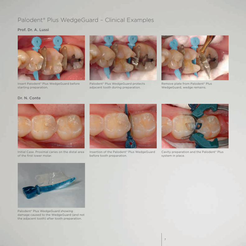
Insert Palodent® Plus WedgeGuard before starting preparation.
Palodent® Plus WedgeGuard protects adjacent tooth during preparation.
Remove plate from Palodent® Plus WedgeGuard, wedge remains.
Prof. Dr. A. Lussi
Palodent® Plus WedgeGuard – Clinical Examples
Initial Case. Proximal caries on the distal area of the first lower molar.
Insertion of the Palodent® Plus WedgeGuard before tooth preparation.
Cavity preparation and the Palodent® Plus system in place.
Palodent® Plus WedgeGuard showing damage caused to the WedgeGuard (and not the adjacent tooth) after tooth preparation.
Dr. N. Conte
7

1. Matrix band is burnished with the round – ball end of the pin twizzer.
Cusp missing. Ring still holds firmly. Provided by Dr. Dao.
Back to back restoration. Provided by Dr. Dias.
3. Final restoration with a very natural contact point.
Stacked wedges in periodontal case. Provided by Dr. Hugenberg.
2. Matrix band is in place, providing a good seal at the margins. NiTi ring creates the necessary separation. All is ready for the restoration.
MOD. Rings position allows full visibility in the cavity. Provided by Dr. Kurtzmann.
Interactive rings and wedges. Provided by Dr. De La Peña.
Perfect marginal adaptation and proximal seal. Provided by Dr. Ayad Mouayad Al-Obaidi.
Dr. W. Dias
Predictable tight contacts with Palodent® Plus sectional matrix system
Versatile Clinical Uses
8
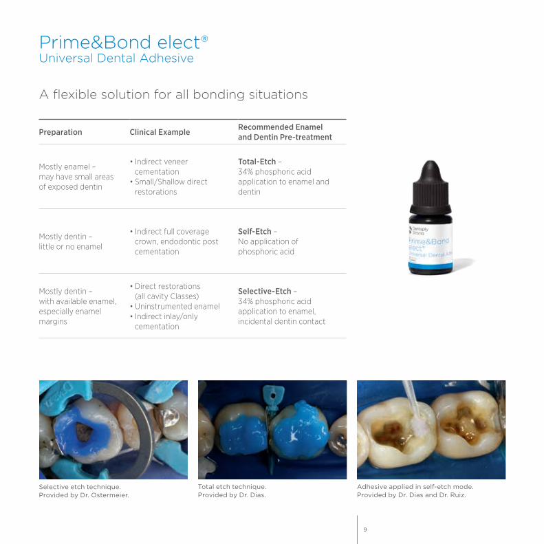
Prime&Bond elect®Universal Dental Adhesive
A flexible solution for all bonding situations
Preparation Clinical Example Recommended Enamel and Dentin Pre-treatment
Mostly enamel – may have small areas of exposed dentin
• Indirect veneer cementation
• Small/Shallow direct restorations
Total-Etch – 34% phosphoric acid application to enamel and dentin
Mostly dentin – little or no enamel
• Indirect full coverage crown, endodontic post cementation
Self-Etch – No application of phosphoric acid
Mostly dentin – with available enamel, especially enamel margins
• Direct restorations (all cavity Classes)
• Uninstrumented enamel• Indirect inlay/only
cementation
Selective-Etch – 34% phosphoric acid application to enamel, incidental dentin contact
Adhesive applied in self-etch mode.Provided by Dr. Dias and Dr. Ruiz.
Total etch technique.Provided by Dr. Dias.
Selective etch technique. Provided by Dr. Ostermeier.
9
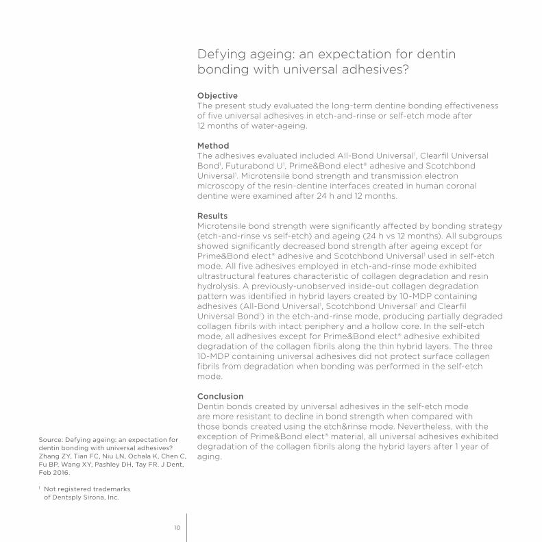
Defying ageing: an expectation for dentin bonding with universal adhesives?
ObjectiveThe present study evaluated the long-term dentine bonding effectiveness of five universal adhesives in etch-and-rinse or self-etch mode after 12 months of water-ageing.
MethodThe adhesives evaluated included All-Bond Universal1, Clearfil Universal Bond1, Futurabond U1, Prime&Bond elect® adhesive and Scotchbond Universal1. Microtensile bond strength and transmission electron microscopy of the resin-dentine interfaces created in human coronal dentine were examined after 24 h and 12 months.
ResultsMicrotensile bond strength were significantly affected by bonding strategy (etch-and-rinse vs self-etch) and ageing (24 h vs 12 months). All subgroups showed significantly decreased bond strength after ageing except for Prime&Bond elect® adhesive and Scotchbond Universal1 used in self-etch mode. All five adhesives employed in etch-and-rinse mode exhibited ultrastructural features characteristic of collagen degradation and resin hydrolysis. A previously-unobserved inside-out collagen degradation pattern was identified in hybrid layers created by 10-MDP containing adhesives (All-Bond Universal1, Scotchbond Universal1 and Clearfil Universal Bond1) in the etch-and-rinse mode, producing partially degraded collagen fibrils with intact periphery and a hollow core. In the self-etch mode, all adhesives except for Prime&Bond elect® adhesive exhibited degradation of the collagen fibrils along the thin hybrid layers. The three 10-MDP containing universal adhesives did not protect surface collagen fibrils from degradation when bonding was performed in the self-etch mode.
ConclusionDentin bonds created by universal adhesives in the self-etch mode are more resistant to decline in bond strength when compared with those bonds created using the etch&rinse mode. Nevertheless, with the exception of Prime&Bond elect® material, all universal adhesives exhibited degradation of the collagen fibrils along the hybrid layers after 1 year of aging.
Source: Defying ageing: an expectation for dentin bonding with universal adhesives?Zhang ZY, Tian FC, Niu LN, Ochala K, Chen C, Fu BP, Wang XY, Pashley DH, Tay FR. J Dent, Feb 2016.
1 Not registered trademarks of Dentsply Sirona, Inc.
10

All-Bond Universal1Microtensile bond strength [MPa]
Etch-and-rinse Self-etch
30
50
70
20
40
60
10
0
Clearfil Universal Bond1
Microtensile bond strength [MPa]
Etch-and-rinse Self-etch
30
50
70
20
40
60
10
0
Prime&Bond elect®Microtensile bond strength [MPa]
Etch-and-rinse Self-etch
30
50
70
20
40
60
10
0
Futurabond U1
Microtensile bond strength [MPa]
Etch-and-rinse Self-etch
30
50
70
20
40
60
10
0
Scotchbond Universal1Microtensile bond strength [MPa]
Etch-and-rinse Self-etch
30
50
70
20
40
60
10
0
24 hours 12 months
11
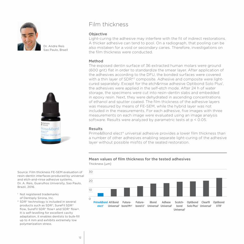
Film thickness
ObjectiveLight-curing the adhesive may interfere with the fit of indirect restorations. A thicker adhesive can tend to pool. On a radiograph, that pooling can be also mistaken for a void or secondary caries. Therefore, investigations on the film thickness were conducted.
MethodThe exposed dentin surface of 36 extracted human molars were ground (600 grit) flat in order to standardize the smear layer. After application of the adhesives according to the DFU, the bonded surfaces were covered with a thin layer of SDR®2 composite. Adhesive and composite were light-cured separately. Except for the etch&rinse adhesive Optibond Solo Plus1, the adhesives were applied in the self-etch mode. After 24 h of water storage, the specimens were cut into resin-dentin slabs and embedded in epoxy resin. Next, they were dehydrated in ascending concentrations of ethanol and sputter coated. The film thickness of the adhesive layers was measured by means of FE-SEM, while the hybrid layer was not included in the measurements. For each adhesive, five images with three measurements on each image were evaluated using an image analysis software. Results were analyzed by parametric tests at p < 0.05.
ResultsPrime&Bond elect® universal adhesive provides a lower film thickness than a number of other adhesives enabling separate light-curing of the adhesive layer without possible misfits of the seated restoration.
Mean values of film thickness for the tested adhesivesThickness [µm]
Prime&Bond elect®
All Bond Universal1
Futura- bond M+1
Adhese Universal1
Clearfil Universal1
Scotch- bond
Universal1
Optibond XTR1
Futura- bond U1
iBond Universal1
Optibond Solo Plus1
30
20
10
0
Source: Film thickness FE-SEM evaluation of resin-dentin interfaces produced by universal and etch-and-rinse adhesive systems, Dr. A. Reis, Guarulhos University, Sao Paulo, Brazil, 2016.
1 Not registered trademarks of Dentsply Sirona, Inc.
2 SDR® technology is included in several products such as SDR®, SureFil SDR® flow, SureFil SDR® flow+ and SDR® flow+. It is self-levelling for excellent cavity adaptation, it enables dentists to bulk-fill up to 4 mm and exhibits extremely low polymerization stress.
Dr. Andre ReisSao Paulo, Brazil
12
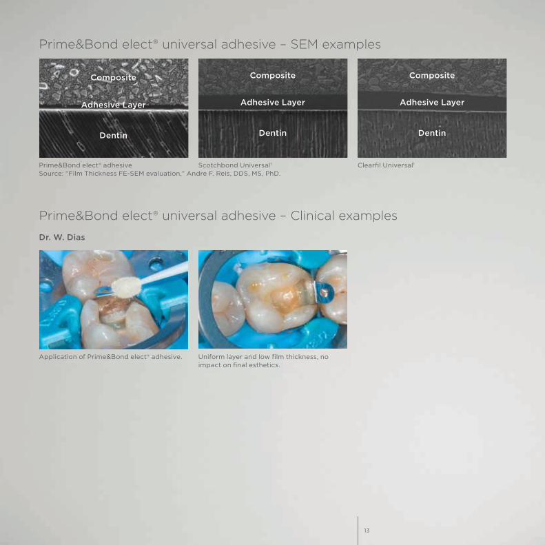
Application of Prime&Bond elect® adhesive. Uniform layer and low film thickness, no impact on final esthetics.
Dr. W. Dias
Prime&Bond elect® adhesiveSource: “Film Thickness FE-SEM evaluation,” Andre F. Reis, DDS, MS, PhD.
Clearfil Universal1
Prime&Bond elect® universal adhesive – SEM examples
Prime&Bond elect® universal adhesive – Clinical examples
Composite
Adhesive Layer
Dentin
Composite
Adhesive Layer
Dentin
Composite
Adhesive Layer
Dentin
Scotchbond Universal1
13
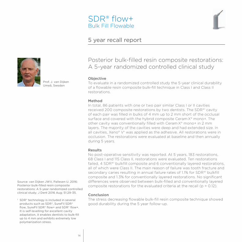
Posterior bulk-filled resin composite restorations: A 5-year randomized controlled clinical study
ObjectiveTo evaluate in a randomized controlled study the 5-year clinical durability of a flowable resin composite bulk-fill technique in Class I and Class II restorations.
MethodIn total, 86 patients with one or two pair similar Class I or II cavities received 200 composite restorations by two dentists. The SDR®1 cavity of each pair was filled in bulks of 4 mm up to 2 mm short of the occlusal surface and covered with the hybrid composite Ceram·X® mono+. The other cavity was conventionally filled with Ceram·X® mono+ in 2 mm layers. The majority of the cavities were deep and had extended size. In all cavities, Xeno® V+ was applied as the adhesive. All restorations were in occlusion. The restorations were evaluated at baseline and then annually during 5 years.
ResultsNo post-operative sensitivity was reported. At 5 years, 183 restorations, 68 Class I and 115 Class II, restorations were evaluated. Ten restorations failed, 4 SDR®1 bulkfill composite and 6 conventionally layered restorations, all of which were Class II. The main reason of failure was tooth fracture and secondary caries resulting in annual failure rates of 1.1% for SDR®1 bulkfill composite and 1.3% for conventionally layered restorations. No significant differences were observed between bulk-filled and conventionally layered composite restorations for the evaluated criteria at the recall (p = 0.12).
ConclusionThe stress decreasing flowable bulk-fill resin composite technique showed good durability during the 5 year follow-up.
5 year recall report
Source: van Dijken JWV, Pallesen U. 2016: Posterior bulk-filled resin composite restorations: A 5-year randomized controlled clinical study; J Dent 2016 Aug; 51:29-35.
1 SDR® technology is included in several products such as SDR®, SureFil SDR® flow, SureFil SDR® flow+ and SDR® flow+. It is self-levelling for excellent cavity adaptation, it enables dentists to bulk-fill up to 4 mm and exhibits extremely low polymerization stress.
Prof. J. van DijkenUmeå, Sweden
SDR® flow+Bulk Fill Flowable
14
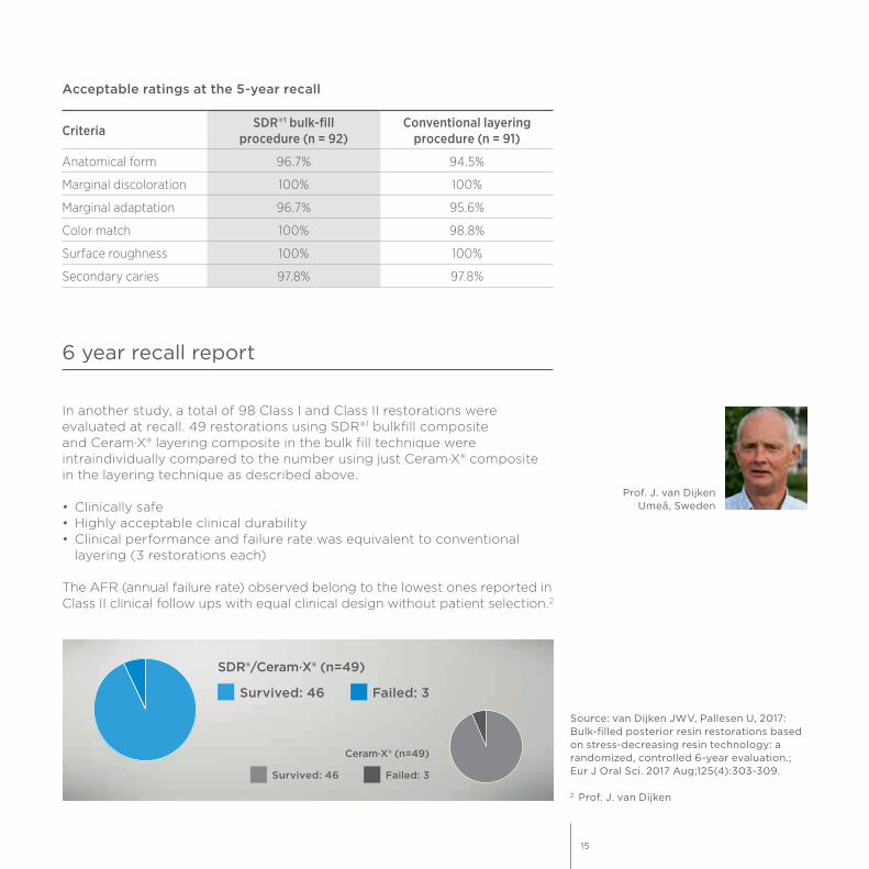
In another study, a total of 98 Class I and Class II restorations were evaluated at recall. 49 restorations using SDR®1 bulkfill composite and Ceram·X® layering composite in the bulk fill technique were intraindividually compared to the number using just Ceram·X® composite in the layering technique as described above.
• Clinically safe• Highly acceptable clinical durability• Clinical performance and failure rate was equivalent to conventional
layering (3 restorations each)
The AFR (annual failure rate) observed belong to the lowest ones reported in Class II clinical follow ups with equal clinical design without patient selection.2
6 year recall report
Prof. J. van DijkenUmeå, Sweden
Criteria SDR®1 bulk-fill procedure (n = 92)
Conventional layering procedure (n = 91)
Anatomical form 96.7% 94.5%
Marginal discoloration 100% 100%
Marginal adaptation 96.7% 95.6%
Color match 100% 98.8%
Surface roughness 100% 100%
Secondary caries 97.8% 97.8%
Acceptable ratings at the 5-year recall
Source: van Dijken JWV, Pallesen U, 2017: Bulk-filled posterior resin restorations based on stress-decreasing resin technology: a randomized, controlled 6-year evaluation.; Eur J Oral Sci. 2017 Aug;125(4):303-309.
2 Prof. J. van Dijken
Failed: 3Survived: 46
SDR®/Ceram·X® (n=49)
Failed: 3Survived: 46
Ceram·X® (n=49)
15
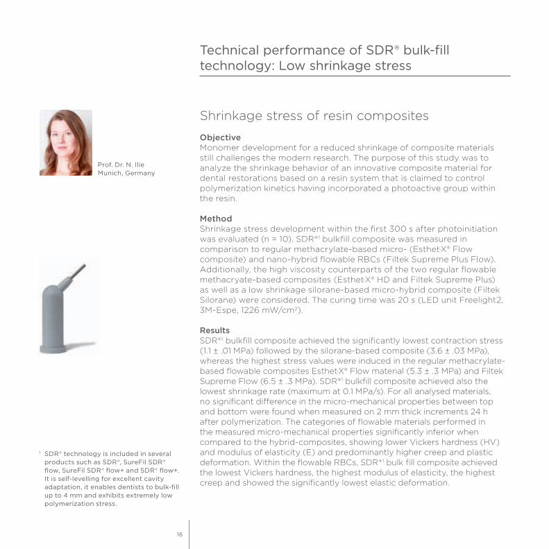
Shrinkage stress of resin composites
ObjectiveMonomer development for a reduced shrinkage of composite materials still challenges the modern research. The purpose of this study was to analyze the shrinkage behavior of an innovative composite material for dental restorations based on a resin system that is claimed to control polymerization kinetics having incorporated a photoactive group within the resin.
MethodShrinkage stress development within the first 300 s after photoinitiation was evaluated (n = 10). SDR®1 bulkfill composite was measured in comparison to regular methacrylate-based micro- (Esthet·X® Flow composite) and nano-hybrid flowable RBCs (Filtek Supreme Plus Flow). Additionally, the high viscosity counterparts of the two regular flowable methacryate-based composites (Esthet·X® HD and Filtek Supreme Plus) as well as a low shrinkage silorane-based micro-hybrid composite (Filtek Silorane) were considered. The curing time was 20 s (LED unit Freelight2, 3M-Espe, 1226 mW/cm2).
ResultsSDR®1 bulkfill composite achieved the significantly lowest contraction stress (1.1 ± .01 MPa) followed by the silorane-based composite (3.6 ± .03 MPa), whereas the highest stress values were induced in the regular methacrylate-based flowable composites Esthet·X® Flow material (5.3 ± .3 MPa) and Filtek Supreme Flow (6.5 ± .3 MPa). SDR®1 bulkfill composite achieved also the lowest shrinkage rate (maximum at 0.1 MPa/s). For all analysed materials, no significant difference in the micro-mechanical properties between top and bottom were found when measured on 2 mm thick increments 24 h after polymerization. The categories of flowable materials performed in the measured micro-mechanical properties significantly inferior when compared to the hybrid-composites, showing lower Vickers hardness (HV) and modulus of elasticity (E) and predominantly higher creep and plastic deformation. Within the flowable RBCs, SDR®1 bulk fill composite achieved the lowest Vickers hardness, the highest modulus of elasticity, the highest creep and showed the significantly lowest elastic deformation.
Technical performance of SDR® bulk-fill technology: Low shrinkage stress
1 SDR® technology is included in several products such as SDR®, SureFil SDR® flow, SureFil SDR® flow+ and SDR® flow+. It is self-levelling for excellent cavity adaptation, it enables dentists to bulk-fill up to 4 mm and exhibits extremely low polymerization stress.
Prof. Dr. N. IlieMunich, Germany
16

Source: Investigations on a methacrylate- based flowable composite-based on the SDR® technology (Ilie N, Hickel R, Dental Materials 27 (2011), 348-355)
* Not registered trademarks of Dentsply Sirona, Inc.
ConclusionSDR®1 bulkfill composite revealed the lowest shrinkage stress and shrinkage-rate values in comparison to regular methacrylate composites but intermediate micro-mechanical properties. Being at the same time more rigid (higher modulus of elasticity) and more plastic (low We/Wtot and high creep values) as the regular flowable materials, its effect on interfacial stress build-up cannot be easily predicted.
Shrinkage stress
[MPa]
7
6
3
2
1
0 20 40 60 Time [sec]
5
4
Filtek Supreme Plus Flow*
Esthet·X® flow
Esthet·X® HDFiltek Supreme Plus*
Filtek Silorane*
SDR®1
Comparison of the shrinkage stress development (averaged curves, n = 10) as a function of time, for SDR®1 with controlled polymerization, the silorane-based microhybrid composite and four regular methacrylate-based composites.
17
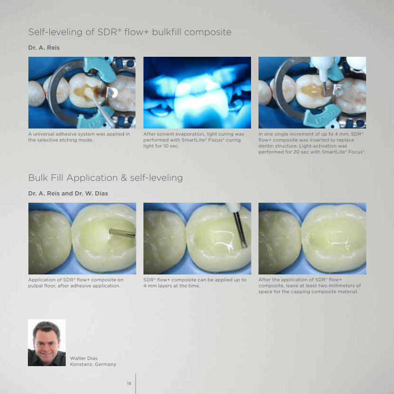
In one single increment of up to 4 mm, SDR® flow+ composite was inserted to replace dentin structure. Light-activation was performed for 20 sec with SmartLite® Focus®.
After solvent evaporation, light curing was performed with SmartLite® Focus® curing light for 10 sec.
A universal adhesive system was applied in the selective etching mode.
Application of SDR® flow+ composite on pulpal floor, after adhesive application.
After the application of SDR® flow+ composite, leave at least two millimeters of space for the capping composite material.
SDR® flow+ composite can be applied up to 4 mm layers at the time.
Dr. A. Reis and Dr. W. Dias
Dr. A. Reis
Self-leveling of SDR® flow+ bulkfill composite
Bulk Fill Application & self-leveling
Walter DiasKonstanz, Germany
18

TPH Spectra® STUniversal Composite Restorative
Flexural strength
ObjectivesThe objective of this study was to investigate different composites regarding their flexural strength.
MethodFifteen specimens (2 x 2 x 25 mm) were made following ISO 4049 and stored in distilled water at 37 °C for 14 days. Flexural strength was tested with a crosshead speed of 1 mm/min in a four-point bending test with 10 and 20 mm distance between the upper and lower support, respectively. Four-point bending allows challenging a larger portion of the bending beam compared to three-point bending described in the ISO 4049. Therefore, the resulting values are typically lower.
ResultsHigh flexural strength is considered by international standards to be an important mechanical property for posterior restorations bearing occlusal stress.Mean flexural strength of TPH Spectra® ST composite surpasses 100 MPa – the threshold for indirect restorations according to ISO 4049 – even under four-point bending.
Prof. Dr. Ing. U. LohbauerErlangen, Germany
Dr. R. BelliErlangen, Germany
Source: Measurement of fracture toughness and fatigue resistance of 4 restorative ma-terials, U. Lohbauer, R. Belli, 2015, University of Erlangen. In vitro study, report # 14.1524, 2015.
1 Not registered trademarks of Dentsply Sirona, Inc.
Bars with different letters are statistically significantly different.
4-point bending testMean flexural strength [MPa]
TPH Spectra® ST Filtek Supreme Ultra1 Tetric Evo Ceram1
150
100
50
0
abb
19

Fracture toughness
ObjectiveFracture toughness describes the resistance to catastrophic failure of an existing crack in a material. This study aims at evaluating the fracture toughness of three different composites.
MethodFifteen specimens of three different composites were prepared in a mold with an integrated V-shaped notch and stored dry at 37 °C for 14 days. The notch was further sharpened using razor blades in a custome made device to control load and depth of sharpening. Specimens were loaded at a crosshead speed of 10 mm/min in a three-point bending test. The crack length was measured under light microscope.
ResultsHigh fracture toughness is needed to resist propagation of cracks in the material and improves longevity of a restoration. TPH Spectra® ST composite shows a good fracture toughness comparable to other control materials.
Prof. Dr. Ing. U. LohbauerErlangen, Germany
Dr. R. BelliErlangen, Germany
Source: Measurement of fracture toughness and fatigue resistance of 4 restorative ma-terials: U. Lohbauer, R. Belli, 2015, University of Erlangen. In vitro study, report #14.1524, 2015.
1 Not registered trademarks of Dentsply Sirona, Inc.
Fracture toughnessMean fracture toughness [MPa m0.5]
TPH Spectra® ST
Filtek Supreme Ultra1
Tetric Evo Ceram1
3.0
2.0
1.0
0
a ab bBars with different letters are statistically
significantly different.
20

Wear
ObjectiveWear in the oral cavity is a multifactorial process. Besides abrasion during grinding movements different wear patterns are generated during forceful occlusal contacts. Furthermore, localized wear in the occlusal contact area (OCA) might be different from generalized wear induced by chewing the food bolus without direct contact to the antagonist. Therefore, we used the so called “Leinfelder Wear Machine” to test both situations – localized and generalized wear.
MethodTwo protocols were applied to test generalized and localized wear, respectively. Both protocols include loading the specimens for 400,000 cycles at 1 Hz with 80 N with a stylus that additionally rotates for 30°. To mimic the food bolus a slurry of about 44 micron acrylic glass beads surrounded the specimens in both protocols. To stimulate generalized wear, the stylus was pressed through the slurry onto the specimen without touching it. To simulate localized wear, a steel bearing was mounted to their stylus so that it contacted the specimens.
ResultsLow loss of material is beneficial in occusal areas under chewing forces for a stable occlusion. TPH Spectra® ST composite showed very good resistance to generalized wear. Under the harsh conditions of localized wear TPH Spectra® ST composite showed very high resistance to loss of height resulting in a low depth of the wear facet.
Prof. M. A. LattaOmaha, USA
Source: A Laboratory evaluation of local-ized wear and generalized wear of dental restorative materials, Mark A. latta, Creighton university. 2015 Report #14.1523, 2015.
1 Not registered trademarks of Dentsply Sirona, Inc.
Bars with different letters are statistically significantly different.
Volume loss under generalized wearMean volume loss [mm3]
TPH Spectra® ST
Filtek Supreme Ultra1
Tetric Evo Ceram!
0.6
0.4
0.2
0
a ab
Maximum depth of wear facet under localized wearMean maximum depth [µm]
TPH Spectra® ST
Filtek Supreme Ultra1
Tetric Evo Ceram1
150
100
50
0
a ab
21

Dr. Ayad Al-Obaidi
TPH Spectra® ST composite – Clinical challenge with advanced layering
Initial case showing defective and leaking old amalgam restorations.
Rubber dam isolation, caries removal, tooth preparation and finishing of the margins with fine-grit diamond burs in slow speed.
Excellent proximal adaptation and contact area of Palodent® Plus matrix. Intimate contact and contour on the vestibular and palatal embrasure areas.
Modeling step by step. TPH Spectra® ST composite is carefully placed on the distal-vestibular cusp using the oblique incremental technique. 20 second cure.
Modeling step by step. After application of SDR® flow+ material and proximal ridge build-up, TPH Spectra® ST composite is carefully placed using the oblique incremental technique.
Modeling step by step. Placement and modeling of the second increment (A2) on the mesial-vestibular cusp. This increment is cured for 20 seconds before the next step.
Final basic anatomy and contour. Well defined pits and fissures as well as cusp ridges help prevent excessive occlusal adjustment. 20 second cure.
Advanced modeling for the enhancement natural anatomy and contour of the cusp morphology using TPH Spectra® ST composite BW (Bleach White).
Two-week recall. Final restoration and excellent gloss retention with TPH Spectra® ST universal composite A2 and BW giving the tooth back its lost anatomy and natural esthetic appearance.
22

Excellence in class II with SDR® flow+ bulkfill composite and TPH Spectra® ST layering composite
Initial case showing defective and fractured old amalgam restoration.
Rubber dam isolation, caries removal, tooth preparation and finishing of the margins with fine-grit diamond burs in slow speed.
Excellent proximal adaptation and contact area of Palodent® Plus matrix. Intimate contact and contour on the vestibular and palatal embrasure areas.
Appearance after composite placement. Note the deep pits and fissures obtained and the robust proximal contact, with nearly no vestibular and palatal excess.
Magnified view of the excellent proximal adap-tation and contact area of Palodent® Plus matrix. The proximal contour may be enhanced with Teflon tape for additional compression effect.
In one single increment of up to 4 mm, SDR® flow+ bulkfill composite was inserted to replace the dentin structure. Light activation was performed for 20 sec with SmartLite® Focus®.
Occlusal view. Appearance after composite placement. Note the natural contour and engaging appearance obtained with Palodent® Plus sectional matrix and TPH Spectra® ST composite A2 and BW.
3-month recall. Occlusal-palatal view depicting excellent gloss retention. Note the characterization and natural appearance obtained with TPH Spectra® ST universal composite A2 and BW.
Dr. Ayad Al-ObaidiBaghdad, Iraq
23

Irradiance over distance and curing efficiency
Typically, irradiance of curing lights is measured while the device is held as close to the photometer sensor as possible – “near-to-tip” irradiance. Due to different designs of the components influencing the overall optic performance (LED dome, reflector, lens, light guide), devices show completely different pattern when irradiance is measured over distance.
SmartLite® Focus® curing light was optimized to allow a lower “near- to-tip” irradiance (measured at 0 mm distance) but to maintain high irradiance over distance – which is opposite to the behavior of a number of competitive lights. This is quite important to note as it stands in contrast to the current approach promoting curing devices via the “near-to-tip” irradiance.
1 Source: Report by Bluelight Anlaytics™ Inc., 2012.2 Source: Data on file.* Not registered trademarks of Dentsply Sirona, Inc.
Mr. C. FelixBluelight Analytics™, Canada
Irradiance over distance1
Irradiance [mW/cm2]
2400
2000
800
400
1600
1200
0 104 6 82 93 5 71 Distance [mm]
SmartLite® Focus® Elipar*
Demi Plus*Valo Cordless*
Bluephase Style*Radii Plus*
Bluephase G2*
SmartLite® Focus®Pen-Style LED Curing Light
24
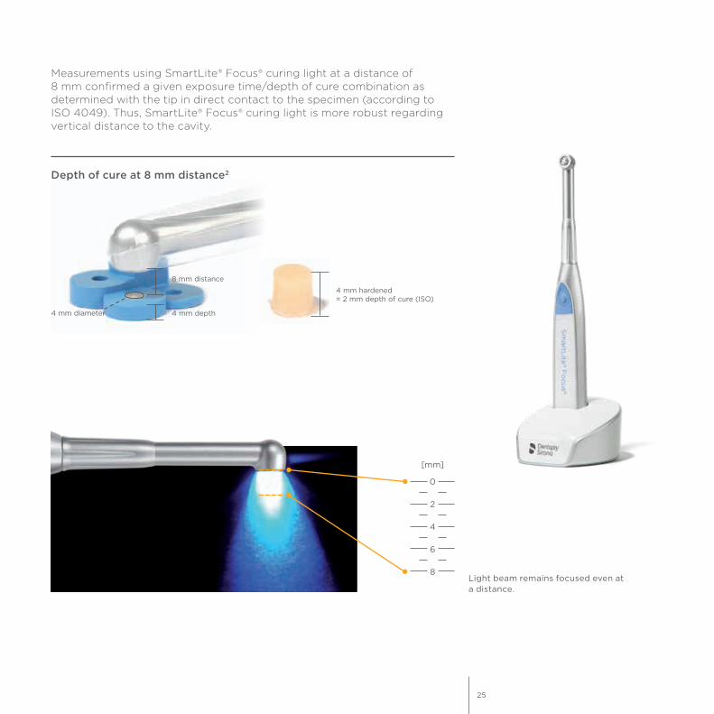
[mm]
8
0
2
4
6
Measurements using SmartLite® Focus® curing light at a distance of 8 mm confirmed a given exposure time/depth of cure combination as determined with the tip in direct contact to the specimen (according to ISO 4049). Thus, SmartLite® Focus® curing light is more robust regarding vertical distance to the cavity.
Light beam remains focused even at a distance.
4 mm depth
8 mm distance
4 mm diameter
4 mm hardened = 2 mm depth of cure (ISO)
Depth of cure at 8 mm distance2
25
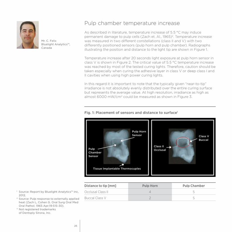
Pulp chamber temperature increase
As described in literature, temperature increase of 5.5 °C may induce permanent damage to pulp cells (Zach et. Al., 1965)2. Temperature increase was measured in two different constellations (class II and V) with two differently positioned sensors (pulp horn and pulp chamber). Radiographs illustrating the position and distance to the light tip are shown in Figure 1.
Temperature increase after 20 seconds light exposure at pulp horn sensor in class V is shown in Figure 2. The critical value of 5.5 °C temperature increase was reached by most of the tested curing lights. Therefore, caution should be taken especially when curing the adhesive layer in class V or deep class I and II cavities when using high power curing lights.
In this regard it is important to note that the typically given “near-to-tip” irradiance is not absolutely evenly distributed over the entire curing surface but represents the average value. At high resolution, irradiance as high as almost 6000 mW/cm2 could be measured as shown in Figure 3.
Mr. C. FelixBluelight Analytics™, Canada
1 Source: Report by Bluelight Analytics™ Inc, 2012.2 Source: Pulp response to externally applied heat (Zach L, Cohen G. Oral Surg Oral Med Oral Pathol. 1965 Apr;19:515-30).* Not registered trademarks of Dentsply Sirona, Inc.
Fig. 1: Placement of sensors and distance to surface1
Pulp Chamber Sensor
Pulp Horn Sensor
Class II Occlusal
Class V Buccal
Tissue Implantable Thermocuples
Distance to tip [mm] Pulp Horn Pulp Chamber
Occlusal Class II 4 5
Buccal Class V 2 5
26

1 Source: Report by Bluelight Analytics™ Inc, 2012.2 Source: Pulp response to externally applied heat (Zach L, Cohen G. Oral Surg Oral Med Oral Pathol. 1965 Apr;19:515-30).* Not registered trademarks of Dentsply Sirona, Inc.
Fig. 2: Temperature increase in Class V1
[°C]
SmartLite® Focus®
Elipar S10* Valo Cordless*
Bluephase Style*
Bluephase G2*
Radii Plus* Demi Plus*
65.5
4
2
0
8
10
12
Fig 3: Maximum irradiance at 0 mm distance at high resolution1
[mW/cm2]
SmartLite® Focus®
Elipar S10* Valo Cordless*
Bluephase Style*
Bluephase G2*
Radii Plus* Demi Plus*
3000
2000
1000
0
4000
5000
6000
Low Standard ExtraHigh
27
Enhance®Finishing System
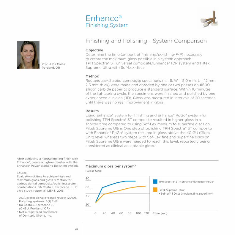
Finishing and Polishing - System Comparison
ObjectiveDetermine the time (amount of finishing/polishing-F/P) necessary to create the maximum gloss possible in a system approach – TPH Spectra® ST universal composite/Enhance® F/P system and Filtek Supreme Ultra with Sof-Lex discs.
MethodRectangular-shaped composite specimens (n = 5; W = 5.0 mm, L = 12 mm, 2.5 mm thick) were made and abraded by one or two passes on #600 silicon carbide paper to produce a standard surface. Within 10 minutes of the lightcuring cycle, the specimens were finished and polished by one experienced clinician (JD). Gloss was measured in intervals of 20 seconds until there was no real improvement in gloss.
ResultsUsing Enhance® system for finishing and Enhance® PoGo® system for polishing TPH Spectra® ST composite resulted in higher gloss in a shorter time compared to using Sof-Lex medium to superfine discs on Filtek Supreme Ultra. One step of polishing TPH Spectra® ST composite with Enhance® PoGo® system resulted in gloss above the 40 GU (Gloss Unit) level whereas two steps with Sof-Lex fine and superfine discs on Filtek Supreme Ultra were needed to reach this level, reportedly being considered as clinical acceptable gloss.1
Prof. J. Da CostaPortland, OR
After achieving a natural looking finish with Enhance®, create a high-end luster with the Enhance® PoGo® diamond polishing system.
Source:Evaluation of time to achieve high and maximum gloss and gloss retention for various dental composite/polishing system combinations. DA Costa J, Ferracane JL. In vitro study, report #14.1543, 2016.
1 ADA professional product review (2010). Polishing systems. 5(1) 2-16.2 Da Costa J, Ferracane JL (OHSU, Portland, OR)3 Not a registered trademark of Dentsply Sirona, Inc.
Maximum gloss per system2
[Gloss Unit]
40
20
80
60
0 1208040 60 10020 Time [sec]
TPH Spectra® ST + Enhance®/Enhance® PoGo®
Filtek Supreme Ultra3 + Sof-lex™ 3 Discs (medium, fine, superfine)3
28

Initial case. After restoration placement, contouring and finishing is done using Enhance® mini cups.
After restoration placement, contouring and finishing is done using Enhance® mini points.
Final result.
Class II DO restoration with TPH Spectra® ST composite after finishing with Enhance® system.
Enhance® mini cusp, point and flame.
Class II DO restoration with TPH Spectra® ST composite, final result after polishing with Enhance® PoGo® system.
Enhance® Enhance® PoGo® finishing system polishing system
Dr. A. Ferrando
Finishing and polishing with Enhance® system
Dr. W. Dias
29
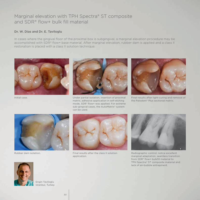
Initial case. Under partial isolation, insertion of proximal matrix, adhesive application in self-etching mode, SDR® flow+ was applied. For extreme sub-gingival cases, the AutoMatrix® system can be used.
Final results after light-curing and removal of the Palodent® Plus sectional matrix.
Rubber dam isolation. Final results after the class II solution application.
Radiographic control, notice excellent marginal adaptation, seamless transition from SDR® flow+ bulkfill material to TPH Spectra® ST composite material and lack of air-bubble entrapment.
Dr. W. Dias and Dr. E. Taviloglu
In cases where the gingival floor of the proximal box is subgingival, a marginal elevation procedure may be accomplished with SDR® flow+ base material. After marginal elevation, rubber dam is applied and a class II restoration is placed with a class II solution technique.
Engin TavilogluIstanbul, Turkey
Marginal elevation with TPH Spectra® ST composite and SDR® flow+ bulk fill material
30

The Class II Solution™
Dr. W. Dias and Dr. A. Ferrando
Before
After
Alvaro FerrandoMurcia, Spain
31
K79200272-02© Dentsply Sirona 03/2018
For more information visit www.dentsplysirona.com.
www.facebook.com/dentsplysirona.restorative
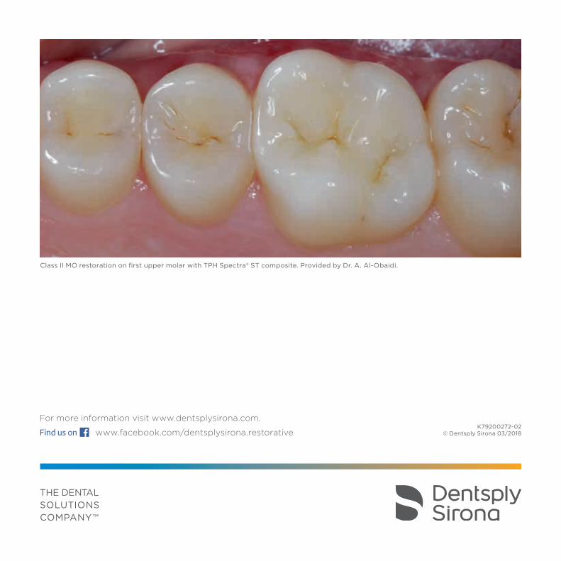
Class II MO restoration on first upper molar with TPH Spectra® ST composite. Provided by Dr. A. Al-Obaidi.