studies on the metabolism of β,β′-iminodipropionitrile in the rat
TRANSCRIPT
Biochemical Pharmacology, Vol. 19, pp. 2277-2287. Pergamon Press. 1970. Printed in Great Britain
STUDIES ON THE METABOLISM OF &fK IMINODIPROPIONITRILE IN THE RAT
SIAN WILLIAMS, E. K. BR~WNLOW and II. HEATH
Department of Chemical Pathology, University College Hospital Medical School, London, England
(Received 19 November 1969; accepted 10 February 1970)
Abstract-Liver, brain and urine extracts of normal rats and of rats treated with /J&Y- iminodipropionitrile (IDPN) have been anaIysed by separation on Dowex 50-H’ resin and by paper electrophoresis and chromatography. The possibility that IDPN could be exerting its toxic effects by metabolism into 3,3’-diaminopropylamine (DAPA) with alteration in the physiological activity of spermine and spermidine was investigated. It has been shown that DAPA does not accumulate in the liver or brain following IDPN administration, and that gross alteration in the polyamine content of these tissues does not occur. The presence of an unknown ninhydrin-reacting substance was detected in the IDPN-treated rat brains. Three metabolites of IDPN were identified in the urine following IDPN treatment, namely cyanoacetic acid, /3-alanine and &aminopropionitrile. Prevention of the symptoms of IDPN poisoning with ethionine did not inhibit the formation of these metabolites or of thiocyanate.
ADM~~IS~ATION of ~,~-iminodipropionitrile (IDPN} to a variety of mammalian species results in the development of both a central nervous system disorde&s and a retinal angiopathy similar to that seen in human diabetes mellituss-5 The neurological syndrome has been referred to as the ECC syndrome, i.e. excitation with choreiform and circling movements.2 Numerous biochemicaV7 and histologicall*- investi- gations into the action of IDPN on cerebral tissue have been carried out but the mode of action of this iminonitrile has not yet been elucidated.
The neurological symptoms so characteristic of IDPN poisoning do not become evident until the third or fourth day after the administration of a large dose of the drug. It seemed possible that the toxic agent might be a metabolite of IDPN, and it was decided to study the metabolism of this iminonitrile in the rat. Reduction of IDPN would lead to the formation of 3,3’-dia~nopropylamine (DAPA), a compound chemi- cally related to the polyamines spermine and spermidine .21*aa The possibility that IDPN could be exerting its toxic effect by conversion into DAPA with resulting alteration in the polyamine activity was investigated. This study reports on the analysis of brain and liver extracts for the presence of DAPA and the estimation of the spermidine and spermine contents of these tissues.
IDPN administration has been shown to result in the urinary excretion of cyano- acetic acid (C!AA).s3 This is also known to be a metabolite of #&amino-propionitrile BAPN), ethylenecyanohydrin, succinonitrile and valeronitrileJ3 It was thus decided to analyse the urine of IDPN-treated rats for the presence of CAA and other possible metabolites of IDPN. Ethionine has been showd2 to protect rats against IDPN-toxicity
BP-7G 2277

2278 SIKN WILLIAMS, E. K. BROWNLOW and H. HEATH
and we therefore investigated whether pretreatment with ethionine inhibited the forma- tion of IDPN metabolites.
MATERIALS AND METHODS
Chemicals. /3,/Y-Iminodipropionitrile was obtained from Kodak Ltd., Kirkby, Liverpool, and was recrystallised in the hydrochloride form from 95 % (v/v) ethanol. Dowex SO-H+ resin (2% cross linked, 100-200 mesh) and 2,4-dinitrofluorobenzene were obtained from Sigma Chemical Company, London. All other reagents used were of analytical grade.
Animals. Male albino rats (SO-120 g) were maintained on M.R.C. diet 41 and water ad lib. IDPN was administered by subcutaneous injection of a 200 mg/ml aqueous solution of IDPN hydrochloride (200 mg/lOO g body wt.) neutralised with 5 N NaOH. SKF-525A was administered by intraperitoneal injection of 5 mg/lOO g body wt. 1 hr before administration of 100 mg IDPN/l~ g body wt. and 4 hr after IDPN treatment. Rats maintained on a low protein diet (3 per cent casein) for 2 months were generously provided by Dr. A. McLean of the Department of Experimental Pathology. These rats were treated with 100 mg IDPN/lOO g body wt. Rats were protected against the toxic action of IDPN by daily intraperitoneal injection of 24 mg ethionine/lOO g body wt. for 5 days; IDPN was administered on day 3 (100 mg IDPN/lOO g body wt.). The rats were examined for the development of the E.C.C. syndrome by the landing and swim tests.2
Preparation of extracts. Liver: The rat was killed by cervical dislocation and the liver was rapidly dissected out and weighed. Homogenisation was carried out in O-1 N HC124 and protein was precipitated by addition of an equal volume of 10% (w/v) TCA. After centrifugation at 3ooO g for 10 min the precipitate was washed with an equal volume of 10% TCA and the mixture was again centrifuged. The TCA was removed by shaking the combined supernatants three times with ether (x3 vol.). This extract was concentrated over P2O5 in uacuo and the equivalent of 08 g liver was loaded onto the ion-exchange column.
Brain: The animals were killed by cervical dislocation and the brain was rapidly dissected, washed in 0.9% ~w/v) sodium chloride solution, dried on filter paper and weighed. A TCA/ether extract was prepared as above
Urine: The urine was collected every 24 hr and treated with TCA and ether as described above. The extract was concentrated to 4 or 5 ml over P&5 in DQCUO and a known volume (about 3 ml) was loaded onto the ion-exchange column.
Separation of tisslte components by ion-exchange chromatography. The method used was adapted from the procedures described by Tabor, Rosenthal and Tabor and Stevens.26 Dowex 50-H’ resin was packed into a column (0.6 x 8.0 cm). The sample was loaded onto the column and the first 3 ml effluent was discarded. Gradient elution was carried out with 2.5 N HCl and fractionation was achieved by means of an L.K.B. 3400B RadiRac Fraction Collector. At the start of the elution, the mixing vessel contained 275 ml water and the dropping funnel contained 250 ml 2.5 N HCl. 5 x 5-ml fractions were collected before the acid gradient was started; 80 x 5-ml fractions were collected in the liver and brain experiments, and 35 x S-ml fractions were collected in the urine experiments. One-ml portions (or a smaller known volume) of each fraction were taken to dryness over PsOs and NaOH flake in uacuo, and the dried fractions were analysed by the fluorodinitrobenzene (DFB) assay.27
Metabolism of &,5’-iminodipropionitrile 2279
Analysis of fractions by paper electrophoresis Separation of DAPA and spermidine was achieved by paper electrophoresis in 1 M
formate buffer pH 3.2 at 2500 V for 90 min. Portions (0.5 ml) of fractions 44-48 of the liver extracts and 3 ml portions of fractions 44-48 of the brain extracts were analysed by paper electrophoresis under the above conditions. Paper electrophoresis of 05 ml portions of fractions 27-30 and 61-66 of the liver extracts, 2 ml portions of the same fractions of the brain extracts and 0.3 ml portions of fractions 24-29 of the urine extracts was carried out in O-1 M citrate buffer pH 3.2 at 500 V for 105 min. Analyses of 0.3 ml portions of a mixture of fractions 12-18 of the urine samples were carried out in 1 M formic/acetic buffer pH 1.9 at 400 V for 90 min. Portions (0.3 ml) of frac- tions l-3 of the urine samples were analysed by paper electrophoresis in 1 M potassium acetate buffer pH 5.3 at 500 V for 90 min. The strips were dried at 100” and developed with ninhydrin reagent.28 Diazotised sulphanilic acids* was also used for the analysis of the urine fractions.
Analysis of fractions by paper chromatography Portions (0.3 ml) of fractions 12-18 of brain and liver extracts were analysed by
ascending 2-way paper chromatography using n-butanol-acetic acid-water (120 :30 : 50) and phenol-ammonia as the solvent systems .as Paper chromatography of fractions 1 and 2 of the urine samples was carried out in methylethylketone-propionic acid water (174: 57: 69) run for 3 hr and developed with ninhydrin or diazotised sulphanilic acid reagents. These fractions were also analysed by paper chromatography in the butanol-acetic acid-water system.
Detection of IDPN (a) Hydrolysis of IDPN followed by the formation of a DNP derivative: 50 ~1
0.5 N NaOH was added to a known amount of dried eluate and the mixture was hydrolysed for 60 min at 87”. Blank estimations were carried out at the same time. A small amount of solid CO2 was added and after evaporation of excess COs, the DFB assay was carried out by the method of Dubin, except that the ratio of DFB- borax was changed from 1: 10 to 1: 20. IDPN was found to be eluted from the column in fractions 12-18 and so portions of fractions 10-20 of each extract were analysed before and after hydrolysis.
(b) Detection of IDPN with pentacyanoquoferrate (PCF) reagent: 50 ~1 portions of fractions 10-20 of each extract were tested with the PCF reagent.28
Recovery of unknown compound The compound which gave a green colour with ninhydrin was separated from other
urinary components in the following manner: Urine collected for 48 hr from three rats treated with 200 mg IDPN/lOO g body wt. was concentrated and the concentrate was streaked onto electrophoresis strips (about 0.3 ml on each). Paper electrophoresis was carried out in 1 M formic-acetic buffer pH 1.9 at 400 volts for 90 min. The band corresponding to the unknown compound was cut out and eluted off the paper with distilled water; the eluate was concentrated and the process of electrophoresis, location and elution was repeated. The concentrated eluate was loaded onto a Dowex 50-H+ column and gradient elution with 2.5 N HCl was carried out as described previously. The “green spot” compound was eluted in fractions 14-16 and was extracted from the concentrated eluate with ethanol. The residue remaining after evaporation of the alcohol was dissolved in 2 ml water and the solution was used for identification tests.
2280 SI.~N WILLIAMS, E. K. BROWNLOW and H. HEATH
Estimation of thiocyanate The amount of thiocyanate present in the urine samples was determined coiori-
metrically.29 A 2 ml portion of ferric nitrate solution (5 g Fe(N0&.9H20 and 5 ml concentrated nitric acid made up to 100 ml) was added to a 0.508 ml urine sample in a final volume of 5 ml. The amount of chromogen formed was measured within 5 min in a Unicam SP700 spectrophotometer at 450 rnp against a urine blank. The content of thiocyanate in the urine sampIes was calculated from a standard curve prepared by substituting known amounts of thiocyanate in the urine samples.
RESULTS
Analysis of normal rat liver and rat her 24 hr qfler IDPN administration Separation of amines was achieved by column chromatography. The elution patterns
were similar except for an increase in the amino acid peak (fractions 12-18) following IDPN administration.
Paper eiectrophoresis of fractions: No DAPA was detectable in fractions 45 and 47 and there were no differences in the electrophoresis patterns obtained. The amount of amine present in fractions 42-50 was calculated from a calibration curve for amines (DFB assay) and this was taken as an estimate of the amount of spermidine present in the liver, since no other ninhydrin positive spot was detectable in these fractions. Normal rat liver was found to contain approximately 350 pg free base/g wet wt., while approximately 330 pg free base/g wet wt. was calculated to be present in the IDPN-treated rat liver. Paper electrophoresis of fractions 42-64 showed only one spot, corresponding to authentic spermine. The approximate amount of amine present in fractions 59-69 was 82 pg free base/g wet wt. in the normal rat liver and 98 pg free base/g wet wt. in the IDF~-treated rat liver.
Paper chromatography of fractions 12-18: No difference in the distribution or concentration of ninhydrin-positive substances was observed followii?g IDPN admini- stration.
Identification of IDPN: DFB estimations of fractions 12-18 of both normal and IDPN-treated rat liver showed a decrease in extinction after hydrolysis. If IDPN was present in the liver, an increase in extinction values, or at least a smaller decrease in the drop which occurred after hydrolysis, would have been observed. Spot tests OR fractions 13-16 using the PCF reagent showed no difference between normal and IDPN-treated rat liver fractions.
Analysis of coronal rat brain and rat brain 3 ai?d 8 days after IlX’_W adi~l~~~stration Separation of amines by ion exchange chromatography showed an increase in the
amino acid peak (fractions 12-l 8) following IDPN administration. Paper electrophoresis of fractions: DAPA was not detectable in fractions 43-47
following IDPN administration, Normal rat brain was found to contain approximately 82 IJ-g spermidine (free base)/g wet wt. while the 3-day IDPN-treated rat contained 41 rg and the S-day rat contained 57 pg free base/g wet wt. Spermine was detected in fractions 61-65; normal and 3-day IDPN-treated rat brain contained approxi- mateIy 40 pg free base/g wet wt., while the 8-day treated rat brain contained 55 pg free base/g wet wt.
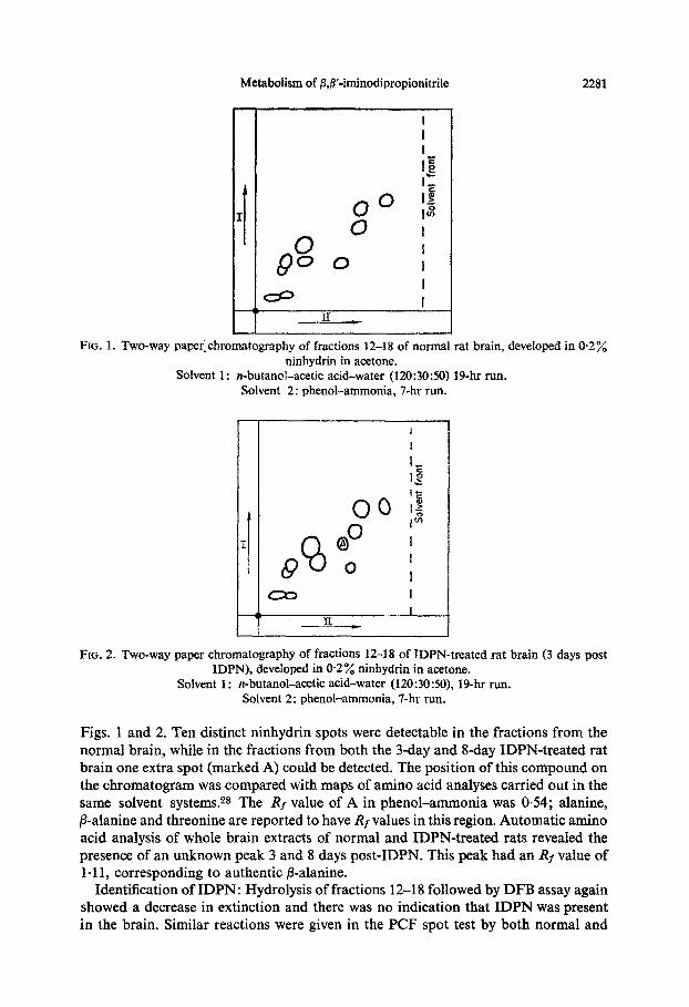
Paper chromatography of fractions 12-18 : The separation of amines of normal and 3-day IDPN-treated rat brains achieved by 2-way paper chromatography is shown in
Metabolism of @,p-iminodipropionitrile 2281
Solvent 1:
FIG. 1. Two-way pa~r_~hromatography of fractions 12-18 of normal rat brain, deveioped in 0.2% ninhydrin in acetone.
n-butanol-acetic acid-water (120:3050) 19-hr run. Solvent 2: phenol-ammonia, 7-hr run.
FOG. 2. Two-way paper ~hromato~aphy of fractions 12-18 of IDPN-treated rat brain (3 days post XDPN), developed in @2 % ninhydrin in acetone.
Solvent 1: n-butanol-acetic acid-water (120:30:50), 19-hr run. Solvent 2: phenol-ammonia, 7-hr run.
Figs. 1 and 2. Ten distinct ninhydrin spots were detectable in the fractions from the normal brain, while in the fractions from both the 3-day and f&day IDPN-treated rat brain one extra spot (marked A) could be detected. The position of this compound on the chromatogram was compared with maps of amino acid analyses carried out in the same solvent systems.28 The Rf value of A in phenol-ammonia was 0.54; alanine, ,&alanine and threonine are reported to have Rf values in this region. Automatic amino acid analysis of whole brain extracts of normal and IDPN-treated rats reveaIed the presence of an unknown peak 3 and 8 days post-IDPN. This peak had an _RI value of 1.11, corresponding to authentic @alanine.
Identification of IDPN: Hydrolysis of fractions 12-18 followed by DFB assay again showed a decrease in extinction and there was no indication that IDPN was present in the brain. Similar reactions were given in the PCF spot test by both normal and
2282 SIAN WILLIAMS, E. K. BROWNLOW and H. HEATH
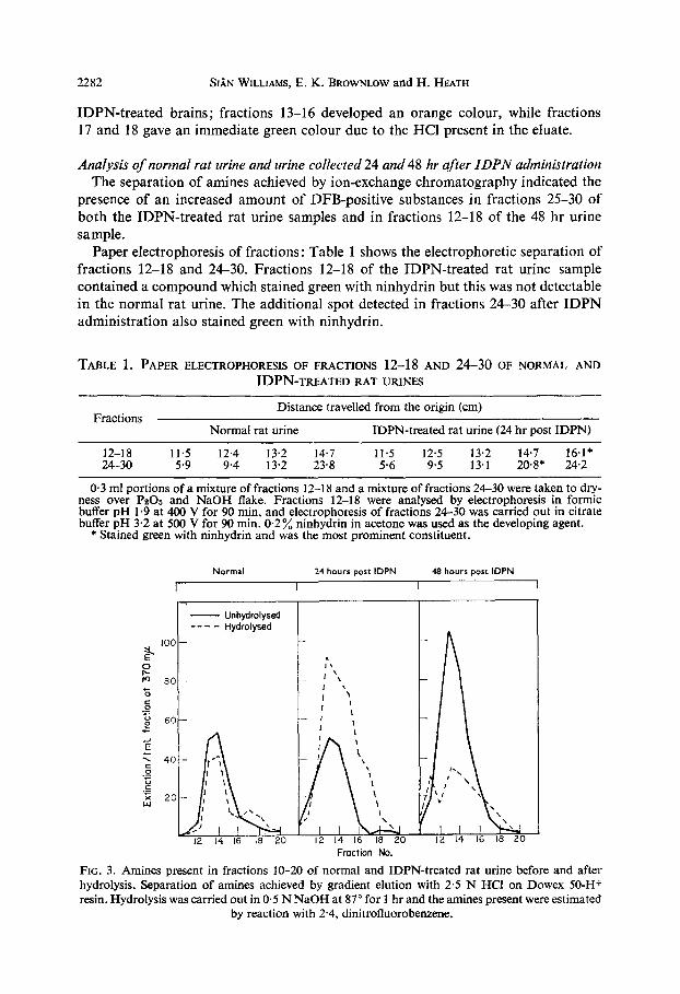
IDPN-treated brains; fractions 13-16 developed an orange colour, while fractions 17 and 18 gave an immediate green colour due to the HCl present in the eluate.
Analysis of normal rat urine and urine collected 24 and 48 hr after IDPN administration The separation of amines achieved by ion-exchange chromatography indicated the
presence of an increased amount of DFB-positive substances in fractions 25-30 of both the IDPN-treated rat urine samples and in fractions 12-18 of the 48 hr urine sample.
Paper electrophoresis of fractions: Table 1 shows the electrophoretic separation of fractions 12-18 and 24-30. Fractions 12-18 of the IDPN-treated rat urine sample contained a compound which stained green with ninhydrin but this was not detectable in the normal rat urine. The additional spot detected in fractions 24-30 after IDPN administration also stained green with ninhydrin.
TABLE 1. PAPER ELECTROPHORESIS OF FRACTIONS 12-l 8 AND 24-30 OF NORMAL AND
IDPN-TREATED RAT URINES
Fractions Distance travelled from the origin (cm)
Normal rat urine IDPN-treated rat urine (24 hr post IDPN)
12-18 11.5 12.4 13.2 14.7 11.5 12.5 13.2 14.7 16.1* 24-30 5.9 9.4 13.2 23.8 5.6 9.5 13.1 2o*s* 24.2
0.3 ml portions of a mixture of fractions 12-18 and a mixture of fractions 24-30 were taken to dry- ness over PZOS and NaOH flake. Fractions 12-18 were analysed by electrophoresis in formic buffer pH 1.9 at 400 V for 90 min, and electrophoresis of fractions 24-30 was carried out in citrate buffer pH 3.2 at 500 V for 90 min. 0.2% ninhydrin in acetone was used as the developing agent.
* Stained green with ninhydrin and was the most prominent constituent.
Normal 24 hours post IDPN 48 hours port IDPN
I I I I
T
Fraction No.
FIG. 3. Amines present in fractions 10-20 of normal and IDPN-treated rat urine before and after hydrolysis. Separation of amines achieved by gradient elution with 2.5 N HCl on Dowex 50-H+ resin. Hydrolysis was carried out in 0.5 N NaOH at 87” for 1 hr and the amines present were estimated
by reaction with 2.4, dinitrofluorobenzene.

Metabolism of /I$‘-iminodipropionitrile 2283
Identification of IDPN : Figure 3 shows the elution patterns obtained by determina- tion of amines present in fractions 10-20 of normal and IDPN-treated rat urines before and after hydrolysis. A decrease in extinction was observed in both normal rat urine and the 48 hr IDPN-treated rat urine after hydrolysis, but a large increase in extinction was observed after hydrolysis of the fractions from the 24 hr IDPN- treated rat urine. The PCF spot test also indicated the presence of unchanged IDPN in the urine 24 hr after IDPN administration (see Table 2). The stable green colour produced by fractions 17-18 was due to the HCl present, but the green colour obtained in fractions 13-16 of the IDPN-treated rat urine turned white on leaving, as did the IDPN standard.
TABLE 2. PCF SPOT TEST ON FRACTIONS 12-18 OF NORMAL AND 24 hr IDPN-TREATED RAT URINES
Colour reaction Fraction
Normal rat urine IDPN-treated rat urine (24 hr post IDPN)
12 Pale orange Pale orange 13
;:
zl;;;zge, darker on heating Pale orange, green on heating > white
As above z;eeeo$ heating z whtte
3; “,;s > orange on heating As above
Green 18 Green Green
Fifty ~1 portions of fractions 12-18 were tested with the PCF reagent described by Smith.2B
Detection of CAA: Analysis of fractions 1 and 2 by paper electrophoresis using diazotised sulphanilic acid as the developing agent showed the presence of a compound giving a bright orange colour in both the IDPN-treated rat urine samples but not in the normal urine. The distance travelled from the origin (13.0 cm) corresponded to authentic CAA. The presence of CAA was confirmed by paper chromatography in methylethylketone-propionic acid-water; the orange spot in the urine sample had an Rf value of 0.74 and a mixture of fraction 1 and authentic CAA developed as 1 spot with an Rf value of 0.74. CAA had an Rf value of 0.72 in n-butanol-acetic acid-water, while both fraction 1 and a mixture of fraction 1 and authentic CAA had an Rf value of 0.73.
Detection of p-alanine: The presence of /3-alanine in the IDPN-treated rat urine sample was shown by paper electrophoresis. A blue spot (9.9 cm from the origin) was detected with ninhydrin in the IDPN-treated rat urine but not in the normal rat urine; authentic fl-alanine had similar electrophoretic properties (9.8 cm from origin). A compound giving a bright green colour with ninhydrin (18.2 cm from origin) was also present in the IDPN-treated rat urine. Paper chromatography of p-alanine in methylethylketone gave a blue spot with an Rf value of 0.45; a mixture of fraction 1 and authentic @alanine had an Rf value of 0.46.
Detection of BAPN: When the compound giving a green colour with ninhydrin was separated from other urinary components by electrophoresis, it was eluted from the ion-exchange column in fractions 14-16. This compound was identified as BAPN as follows: electrophoresis in sodium acetate, pH 5.6, 500 V for 120 min (17.6 cm
2284 SI;ZN WILLIAMS, E. K. BROWNLOW and IX. HEATH
from origin); paper chromatography in n-butanol-acetic acid-water (& 0.41) and phenot-ammonia (& 0.65); acid hydrolysis gave @alanine; melting point of DNP- derivative of unknown compound was 129” and the DNP-derivative of BAPN had a melting point of 128”.
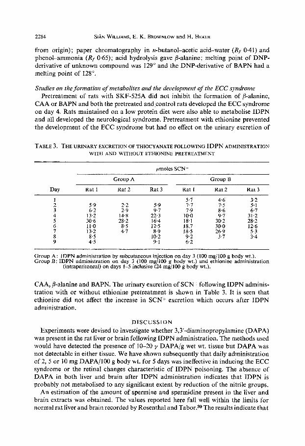
Studies on the formation of metabolites and the development of the ECC syndrome Pretreatment of rats with SKF-525A did not inhibit the formation of /3-alanine,
CAA or BAPN and both the pretreated and.control rats developed the ECC syndrome on day 4. Rats maintained on a low protein diet were also able to metabolise IDPN and all developed the neurological syndrome. Pretreatment with ethionine prevented the development of the ECC syndrome but had no effect on the urinary excretion of
TABLE 3, THE URINARY EXCRETION OF THIOCYANATE FOLLOWING IDPN AD~MINISTRATION WITH AND WITHOUT ET~IONINE PRETREATMENT
pmoles SCN-
Day
:.
:
ii
3 9
Rat 1
5-9
13.2 6.2
30.6 11.0
13.2 8.5 4.5
Group A
Rat 2
2.2
14.8 2.9
28.2
i:;
Rat 3
5-9
22.3 9.7
12.5 16.4
,i.; 9.1
Rat I
5‘7
10.0 ;:;
18.7 18.1
18.5 ;::
Group B
Rat 2
4.6 7.5
8.6
30.0 3z
26.9 3.7
Rat 3
3-2
31.2 ;::
28.2 12.6
:::
Group A: IDPN administration by subcutaneous injection on day 3 (100 mg/lOO g body wt.). Group B: IDPN administration on day 3 (100 mg/lOO g body wt.) and ethionine administration
(intraperitoneal) on days l-5 inclusive (24 mg/lOO g body wt.).
CAA, /3-alanine and BAPN. The urinary excretion of SCNf following IDPN adminis- tration with or without ethionine pretreatment is shown in Table 3. It is seen that ethionine did not affect the increase in SCN+ excretion which occurs after IDPN administration.
DISCUSSION
Experiments were devised to investigate whether 3,3’-diaminopropy~amine (DAPA) was present in the rat liver or brain following IDPN administration. The methods used would have detected the presence of IO-20 y DAPA/g wet wt. tissue but DAPA was not detectable in either tissue. We have shown subsequently that daily administration of 2, 5 or IO mg DAPA/lOO g body wt. for 5 days was ineffective in inducing the ECC syndrome or the retinal changes characteristic of IDPN poisoning. The absence of DAPA in both liver and brain after IDPN administration indicates that IDPN is probably not metabolised to any significant extent by reduction of the nitrile groups.
An estimation of the amount of spermine and spermidine present in the liver and brain extracts was obtained. The values reported here fall well within the limits for normal rat liver and brain recorded by Rosenthal and Tabor.30The results indicate that

Metabolism of /3,/3’-iminodipropionitrile 2285
no gross changes in spermine and spermidine contents occur as a result of IDPN administration, and since no DAPA was detected, it seems unlikely that IDPN exerts its toxic effects by metabolism into DAPA.
IDPN administration resulted in an increase in the amino acid peak (fractions 12-18) of both liver and brain extracts. It has been suggested that IDPN may be acting by interfering with amino acid metabolism .sr Significant increases in brain y-amino- butyrate, glutamate and aspartate were reported by Vivanco et al.9 but other workers found no significant alteration in these amino acids after IDPN administration.iOJi Analysis of the amino acid peak of normal and IDPN-treated rat liver eluates by paper chromatography showed no variation in the distribution of ninhydrin spots, but IDPN-treated rat brain was found to contain one ninhydrin spot which was not present in the normal brain fractions. Both paper chromatography and automatic amino acid analysis showed that this compound had similar Rf values to /3-alanine, but the fraction has not been further identified.
The detection of IDPN is extremely difficult due to the fact that it does not react with the normal reagents for the detection of imino groups. Hydrolysis of IDPN would result in the formation of iminodiopropionic acid, and since this was found to react with DFB, it was decided to develop a method for the estimation of IDPN by hydrolysis followed by the formation of a DNP derivative. IDPN could not be detected in the brain or liver using this method or the PCF spot test. This test was very in- sensitive (test using 50 ~1 urine would have detected 50 y IDPN) and IDPN would only be detected in the tissue extracts if present in very high concentration (about 5 mg/g wet wt. tissue). A positive PCF reaction was, however, given by the urine sample col- lected 24 hr after IDPN administration. An increase in extinction was observed after hydrolysis of the fractions from the urine extract as compared to the decrease observed in the brain and liver extracts, showing that unchanged IDPN was excreted in the urine. The fact that IDPN could not be detected using the methods described in the brain or liver following IDPN administration shows the necessity of finding a sensitive identification test for IDPN, since it is not known whether the iminonitrile can cross the blood-brain barrier or whether it is metabolised before reaching the brain. It is interesting to note that after administration of a-y-diaminobutyric acid (DABA) to rats, this compound could be detected in the brain.9 Thyroxine pretreatment protects against the toxicity of both DABA and IDPN, and since no DABA could be detected in the brains of thyroxine-pretreated animals, it was suggested that thyroxine prevented the entry of DABA into the brain by increasing the rate of its metabolism.
Three metabolites of IDPN, namely cyanoacetic acid, p-alanine and BAPN, were identified in the urine of IDPN-treated rats. Cyanoacetic acid, identified by electro- phoresis and paper chromatography, would be formed from IDPN by oxidative cleavage. No evidence was obtained that CAA had lathyrogenic properties32 and it was suggested that the formation of CAA from BAPN was a method of detoxication. It thus seems probable that IDPN is detoxified in the body to form CAA.
Analysis of IDPN-treated rat urine showed the presence of a compound with similar electrophoretic and chromatographic properties to /3-alanine. The presence of /3- alanine was indicated in chick embryo following BAPN administration.33 Since BAPN was identified as a metabolite of IDPN, it is probable that /3-alanine would be formed by hydrolysis of the BAPN. It was stated that /Lalanine was not lathyrogenic;m it thus seems that its formation in the IDPN-treated rat is a process of detoxication.

2286 SIAN WILLIAMS, E. K. BROWNLOW and H. HEATH
The compound giving a bright green colour with ninhydrin was identified as the lathyrogen BAPN, but no symptoms of BAPN toxicity are observed in the IDPN- treated rats. It is possible that the concentration of BAPN present at any one time is not high enough to produce any lathyrogenic effects, or that, if the toxicity of IDPN and BAPN resides in the nitrile groups, the IDPN may block the nitrile-reacting sites, thus blocking the effect of any BAPN formed. It has been stated that the toxicity of organic nitriles is in almost every case dependent on whether they can be metabolised to CN- and on the rate of excretion of SCN-.s4 A slight increase in SCN- excretion was recorded, but the amount of SCN- excreted was never equivalent to more than 2 per cent of the dose administered. It thus seems that the major pathway of IDPN metabolism is via oxidative cleavage to form CAA and BAPN, which is hydrolysed to give /Lalanine.
It was decided to investigate whether it might be possible to inhibit or slow down the process of metabolism to see if this had any effect on the onset and severity of the ECC syndrome. SKF-525A is known to be an inhibitor of drug metabolisms5 but pretreat- ment of rats with this compound had no effect on the metabolism of IDPN or on the time of onsetof the ECC syndrome, probably due to the fact that SKF-525A stimulates drug metabolism after a primary inhibitory effect. 3s Rats maintained on a low protein diet were also capable of metabolising IDPN and developed the neurological symptoms. Pretreatment with ethionine, however, was successful in preventing the development of the ECC syndrome, but did not inhibit the formation of the three metabolites and had no effect on the increased level of SCN- excreted after IDPN administration. It has been suggested12 that ethionine may exert its protective effect by inhibiting the metabo- lism of IDPN, but this study indicates that this is not so.
Acknowledgement-This research was supported by the Medical Research Council and the British Foundation for Research into the Prevention of Blindness. We wish to thank Miss H. Making and Mrs. G. Drake for their assistance. We are grateful to Mr. P. R. E. Wallace for his help with the amino acid analyses.
REFERENCES
1. J. DELAY, P. PICHOT, J. THUILLIER and J.-P. MARQUISET, C. r. Sot. Biol. (Paris) 146, 533 (1952). 2. H. SELYE, Rev. Can. Biol. 16, 1 (1957). 3. H. HEATH and A. C. R~ER, Aspects of Comparative Ophthalmology (Ed. 0. GRAHAM-JONES)
p. 317. Pergamon Press, Oxford (1966). 4. H. HEATH and A. C. RU’ITER, Br. J. exp. Path. 47, 116 (1966). 5. J. FORGACS and J. BABEL, Experientiu 24, 1208 (1968). 6. S. HARTH and P. MANDEL, C. r. Sot. Biol. (Paris) 152, 1566 (1958). 7. P. MANDEL, S. HARTH and G. REBEL, ChemicalPathology of the Nervous System (Ed. J. FOLCH-PI)
p. 551. Pergamon Press, London (1961). 8. S. WILLIAMS, R. A. PATERSON and H. HEATH, J. Neurochem. 15, 227 (1968). 9. F. VIVANCO, F. RAMOS and C. JIMENEZ-DIAZ, J. Neurochem. 13, 1461 (1966).
10. I. M. CHAK and N. N. DE, J. scient. ind. Res. 2OC, 98 (1961). 11. J. K. TEWS, G. M. KOPF and W. E. STONE, ht. J. Neurophurmuc. 7,29 (1968). 12. E. K. BROWNLOW and H. HYEATH, J. Neurochem. 16, 567 (1969). 13. H. A. HARTMANN and D. SCOBIE, Fedn Proc. 24,493 (1965).
14. M. BEAUVALLET and J. FUGAZZA, Biochem. Phurmuc. 8, 10 (1961). 15. D. E. SLAGEL, H. A. HARTMANN and M. K. EDSTROM, J. Neuropath. exp. Neural. 25, 244 (1966).
16. R. A. PATERSON and H. HEATH, Expl Eye Res. 6,233 (1967). 17. H. HEATH, R. A. PATERSON and J. C. D. HART, DiabetoIogia 3, 515 (1967). 18. H. A. HARTMANN and J. J. LALICH, Am. J. Path. 32, 660 (1956).
Metabolism of /Ql’-iminodipropionitrile 2281
19. S. M, CHOU and H. A. HARTMANN, Aefa Neuropath. 3,428 (1964). 20. D. E. SJ.AGEL and H. A. HARTMANN, .i. Nearopath. exp. Neural. 24, 599 (1965). 21. H. TABOR end C. W. TABOR, Pharmac. Rw. 16,245 (1964). 22. M. Sm, Acfaphysioi. scand. Suppl. 298. 7 (1967) 23. L. P. MERKOW, S. H. LIPTON, J. J. LALICH and F. M. STRONG, Proc. Sot. exp. Biol. Med. 102,
728 (1958). 24. A. RAINA, Acfaphysiol. wand. 60, Suppl. 218, 7 (1963). 25. H. TABOR, S. M. ROSE- and C. W. TABOR, J. biol. Chem. 233, PO7 (1958). 26. L. STEVENS, Biochem. J. 99,llP (1966), and personal communication. 27. D. T. D~JB~N, J. bioL Chem. 235,783 (1960). 28. I. SMITH, Chromafographic and Electrophorefic TechnQues. Vol. I. Chromatography. Heinmann,
New York (1960). 29. J. R. ELKINGTON and M. TAFFEL, Am. J. Physiof. 138, 126 (1943). 30. S. M. ROSENTHAL and C. W. TABOR, J. Pharmac. exp. Ther. 116, 131 (1956). 31. P. SACRA and J. D. MCCOLL, Archs inf. Pharmacodyn. Ther. 122,94 (1959). 32. S. H. LIPTON, J. J. LALIC~ and F. M. STRONG, J. Am. Chem. Sot. 80,2022 (1959, 33. S. D. ORLOFF and J. Gross, J. exp. Med. 117,lOOP (1963). 34. R. T. Wt~r..t~m, in Detoxicafion Mechanisms, 2nd edition, p. 390. Chapman and Hall, London
(1959). 35. G. J. ~~ANNERING, in SelectedPharmacological Testing Methods, Vol. III, p. 105. (Ed. A. BURGER)
E. Arnold, London (1968). 36. R. KATO, E. CHI~RA and P. VAIANELLI, Biochem. Pharmac. 13,69 (1964).