studies on the epidemiology and histopathology of ... · studies on the epidemiology and...
TRANSCRIPT
Studies on the epidemiology and histopathology ofEuclinostomum heterostomum (Trematoda; Digenea) infectionin Channa punctata from North India
P. A. Ahammed Shareef, Syed M. A. Abidi
Received – 01 May 2015/Accepted – 08 September 2015. Published online: 31 October 2015; ©Inland Fisheries Institute in Olsztyn, Poland
Citation: Shareef P.A.A., Abidi S.M.A. 2015 – Studies on the epidemiology and histopathology of Euclinostomum heterostomum (Trematoda;Digenea) infection in Channa punctata from North India – Arch. Pol. Fish. 23: 133-140.
Abstract. A survey on the occurrence and epidemiology of theencysted progenetic metacercariae of Euclinostomum
heterostomum infection in Channa punctata in the Aligarhregion of North India revealed a mean prevalence, intensity,and abundance of 18.61, 1.52, and 0.38%, respectively,during the period from April 2011 to March 2012. Liver,kidney, peritoneum, muscle, and ovary were found to beinfected with this parasite, and the later three are reported forthe first time in this fish species. The histopathology of theinfected tissues indicated the following at the host-parasiteinterface: tissue damage, infiltration of immune cells into thecyst wall, chronic inflammatory responses, andgranulomatous lesions. The infected liver showeddegeneration of hepatocytes, cytoplasmic vacuolation, nuclearalterations, mallory body formation, fibrosis, and necrosis.The pathology of the infected kidney included distortion anddilation of renal tubules, vacuolar degeneration, hypertrophyand hyperplasia of tubular epithelial cells, occlusion oftubules, fibrosis, hemorrhage, and congestion of glomeruli.The infected muscle demonstrated comparatively fewerpathological changes confined only to the circumference ofthe cyst wall. The ovary displayed the least changes. Theconclusions drawn from the study are that the largemetacercarial cysts formed by E. heterostomum in the vitalorgans of the economically important fish C. punctata couldresult in the impairment of fish physiology and health, thereby
affecting their productivity and quality for humanconsumption.
Keywords: incidence, Euclinostomum heterostomum,Channa punctata, histopathology, epidemiology, parasiticinfection
Introduction
Aquaculture is a rapidly growing sector worldwide,and India it is the second largest producer of thesame with an annual production of 312.8 milliontonnes (FAO 2008). However, health-related prob-lems are an important limiting factor, and parasiticinfections are one of the leading constraints ofaquaculture production, both qualitatively as well asquantitatively (Scholz 1999). The snake heads, alsoknown as murrels, belonging to the familychannidae, constitute the dominant group of airbreathing fresh water fishes in terms of both cultureand capture fisheries in south and southeast Asiancountries where they are regarded as high demandfood fish (Wee and Tacon 1982, Haniffa and Mydeen2011). The spotted murrel, Channa punctata
(Bloch), is regarded as a delicious food fish in India(Shareef and Abidi 2012).
Arch. Pol. Fish. (2015) 23: 133-140DOI 10.1515/aopf-2015-0015
RESEARCH PAPER
© Copyright by Stanis³aw Sakowicz Inland Fisheries Institute in Olsztyn.
© 2015 Author(s). This is an open access article licensed under the Creative Commons Attribution-NonCommercial-NoDerivs License(http://creativecommons.org/licenses/by-nc-nd/3.0/).
P.A.A. Shareef [�], S.M.A. AbidiSection of Parasitology, Department of ZoologyFaculty of Life Sciences, Aligarh Muslim UniversityAligarh, 202 002, U. P., Indiae-mail: [email protected]
Among the parasites, trematodes are the domi-nant group that causes retarded growth, morbidity,and mortality especially in juvenile fishes. It has beenestimated that about 30000 species of helminths areparasites of fishes (Williams and Jones 1994). In ad-dition to the huge economic loss they cause, at least 50species of these helminths are potentially zoonotic(Deardorff 1991). The hemophagic clinostomidtrematode Euclinostomum heterostomum (Rudolphi1809) is a common parasite of piscivorous birds inmany regions of Asia, Africa, and Europe (Yamaguti1971). In the Indian subcontinent, the larval stage ofthis parasite usually infects the liver and kidney of thesecond intermediate host fish C. punctata as encystedprogenetic metacercariae (Jhansilakshmibai andMadhavi 1997). The extent and seasonal variability oftheir infection have not been reported from North In-dia or elsewhere. Therefore, aim of the present studywas to survey the incidence and epidemiology of thisparasite in the Aligarh region of North India.
Histological studies of various pathogenic agents indifferent fish species have been reported and proposedas an efficient method to asses fish health, such as chem-icals and pesticides (Altinok and Capkin 2007,Butchiram et al. 2009, Camargo and Martinez 2007,Kelly and Janz 2009, Troncoso et al. 2011), bacteria
(Gudmundsd�ttir et al. 2006), and helminth parasites(Sanil et al. 2011, Shareef and Abidi 2012). Apart fromthe lifecycle and morphometry elucidated by some re-searchers, no study has been found that reports on thehost-parasite interaction and the pathogenicity and dam-age exerted on the tissues of C. punctata during infec-tion with E. heterostomum. However, Shareef and Abidi(2012) reported on the histopathological alterations in-duced by another clinostomid trematode, Clinostomum
complanatum, in the same fish. Therefore, the presentstudy attempted to identify the histopathological changesexerted by the parasite on its fish host. We hypothesizedthat the encystment of such a big parasite in the liver andkidney would result in histopathological lesions as previ-ously reported for pesticides and other chemicals in thesame fish (Butchiram et al. 2009).
Materials and Methods
Examination of fishes for parasitic infection
Live C. punctata from the size group 7-16 cm werebrought from the local fish market of Aligarh (latitude:27°54’ N; longitude: 78°05’ E), North India andtransported to the lab in water containers monthly forone year between April 2011 to March 2012. Theywere maintained in aquaria, but dissected withinthree days, and the body surface, gills, peritoneal cav-ity, and all internal organs were examined for E.
heterostomum infection. The organ/tissue infectionwas systematically recorded and the prevalence, in-tensity, and abundance were calculated using stan-dard formula separately for each month throughoutthe study period. The encysted progeneticmetacercariae of E. heterostomum were freed from thehost tissue and mechanically excysted in 0.74% NaClby carefully tearing up the cyst wall using forceps andneedle to liberate the worms, which were processed toprepare permanent slides after staining in carmine formicroscopic observations.
Histopathology
Liver, kidney, muscle, and ovary from C. punctata in-fected with the encysted progenetic metacercariae ofE. heterostomum and uninfected tissue samples werewashed quickly in 0.74% NaCl and immediatelyfixed in 10% neutral buffered formalin for 48 hours,then dehydrated in ascending grades of ethanol,cleared in xylene, and embedded in paraffin wax tocut 5-7 μm thick sections using a rotatory microtome.The sections were stained with hematoxylin andeosin, and permanently mounted slides were pre-pared. The gross pathology was studied and photo-graphed under a light microscope (Zeiss AxioscopeA1), and photographs of the whole mounts weretaken with a zoom stereomicroscope (Nikon SMZ1500).
134 P. A. Ahammed Shareef, Syed M. A. Abidi
Results
Epidemiology
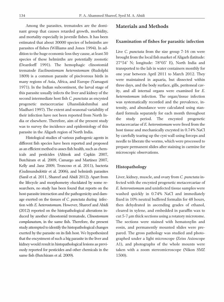
The examination of the infected fish viscera showedwhite or brown round cysts (2-4 mm) present in liver(Fig. 1A), kidney (Fig. 1B), peritoneal membrane,ovary, and muscles. The later three sites are being re-ported for the first time in this fish species. The mi-croscopic observation of the prepared permanent
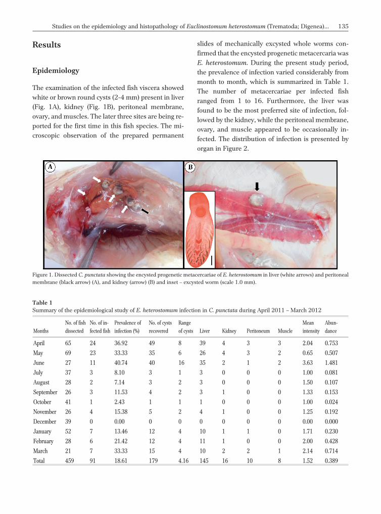
slides of mechanically excysted whole worms con-firmed that the encysted progenetic metacercaria wasE. heterostomum. During the present study period,the prevalence of infection varied considerably frommonth to month, which is summarized in Table 1.The number of metacercariae per infected fishranged from 1 to 16. Furthermore, the liver wasfound to be the most preferred site of infection, fol-lowed by the kidney, while the peritoneal membrane,ovary, and muscle appeared to be occasionally in-fected. The distribution of infection is presented byorgan in Figure 2.
Studies on the epidemiology and histopathology of Euclinostomum heterostomum (Trematoda; Digenea)... 135
Table 1Summary of the epidemiological study of E. heterostomum infection in C. punctata during April 2011 – March 2012
Months
No. of fish
dissected
No. of in-
fected fish
Prevalence of
infection (%)
No. of cysts
recovered
Range
of cysts Liver Kidney Peritoneum Muscle
Mean
intensity
Abun-
dance
April 65 24 36.92 49 8 39 4 3 3 2.04 0.753
May 69 23 33.33 35 6 26 4 3 2 0.65 0.507
June 27 11 40.74 40 16 35 2 1 2 3.63 1.481
July 37 3 8.10 3 1 3 0 0 0 1.00 0.081
August 28 2 7.14 3 2 3 0 0 0 1.50 0.107
September 26 3 11.53 4 2 3 1 0 0 1.33 0.153
October 41 1 2.43 1 1 1 0 0 0 1.00 0.024
November 26 4 15.38 5 2 4 1 0 0 1.25 0.192
December 39 0 0.00 0 0 0 0 0 0 0.00 0.000
January 52 7 13.46 12 4 10 1 1 0 1.71 0.230
February 28 6 21.42 12 4 11 1 0 0 2.00 0.428
March 21 7 33.33 15 4 10 2 2 1 2.14 0.714
Total 459 91 18.61 179 4.16 145 16 10 8 1.52 0.389
Figure 1. Dissected C. punctata showing the encysted progenetic metacercariae of E. heterostomum in liver (white arrows) and peritonealmembrane (black arrow) (A), and kidney (arrow) (B) and inset – excysted worm (scale 1.0 mm).
Histopathology
The histology of the infected organs showed that theparasite was surrounded by a thick fibrous capsule ofcyst wall. This resulted in the formation ofgranulomas and the loss of a large functional part ofthese vital organs. The presence of extracorporeallydigested host blood found inside the cyst and withinthe caecae of the worms is a clear indication of thehemophagic nature of this parasite regardless of thetissue/organ of infection.
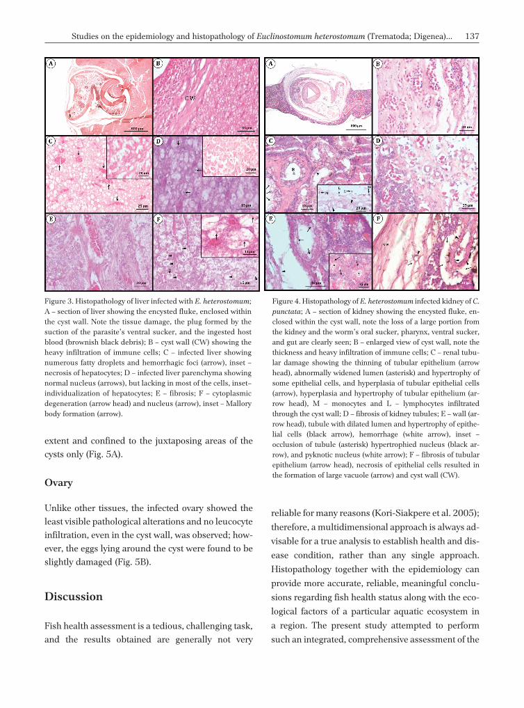
Liver
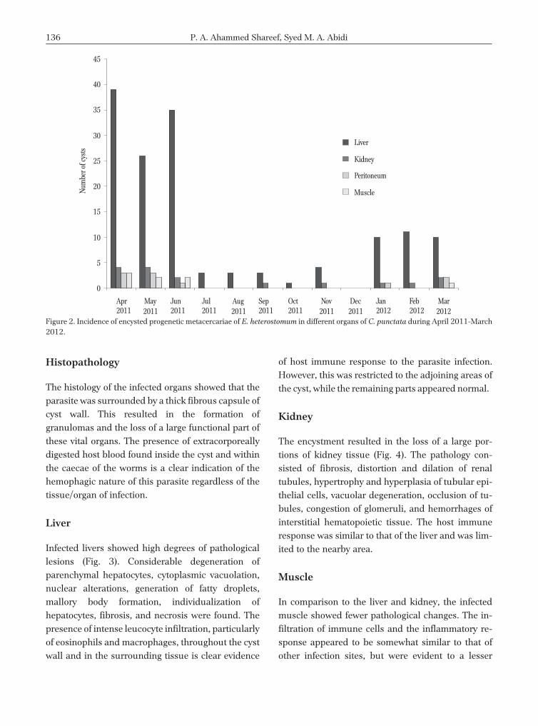
Infected livers showed high degrees of pathologicallesions (Fig. 3). Considerable degeneration ofparenchymal hepatocytes, cytoplasmic vacuolation,nuclear alterations, generation of fatty droplets,mallory body formation, individualization ofhepatocytes, fibrosis, and necrosis were found. Thepresence of intense leucocyte infiltration, particularlyof eosinophils and macrophages, throughout the cystwall and in the surrounding tissue is clear evidence
of host immune response to the parasite infection.However, this was restricted to the adjoining areas ofthe cyst, while the remaining parts appeared normal.
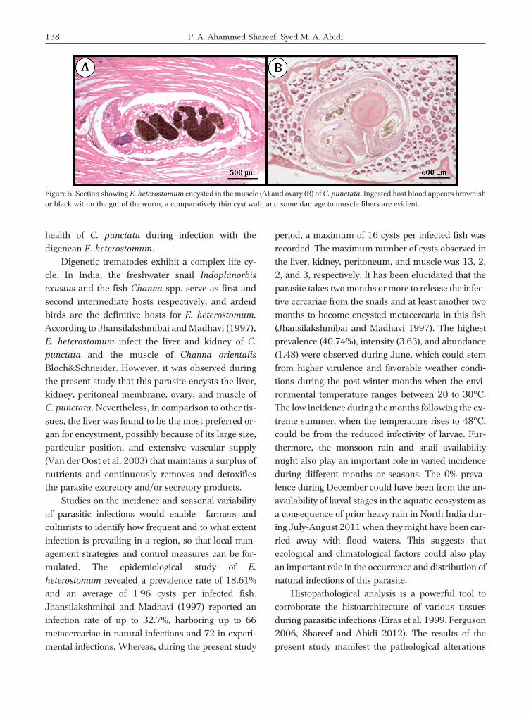
Kidney
The encystment resulted in the loss of a large por-tions of kidney tissue (Fig. 4). The pathology con-sisted of fibrosis, distortion and dilation of renaltubules, hypertrophy and hyperplasia of tubular epi-thelial cells, vacuolar degeneration, occlusion of tu-bules, congestion of glomeruli, and hemorrhages ofinterstitial hematopoietic tissue. The host immuneresponse was similar to that of the liver and was lim-ited to the nearby area.
Muscle
In comparison to the liver and kidney, the infectedmuscle showed fewer pathological changes. The in-filtration of immune cells and the inflammatory re-sponse appeared to be somewhat similar to that ofother infection sites, but were evident to a lesser
136 P. A. Ahammed Shareef, Syed M. A. Abidi
45
0
5
10
15
20
25
30
35
40
Apr2011
May2011
Jun2011
Jul2011
Aug2011
Sep2011
Oct2011
Nov2011
Dec2011
Jan2012
Feb2012
Mar2012
Liver
Kidney
Peritoneum
MuscleNum
bero
fcys
ts
Figure 2. Incidence of encysted progenetic metacercariae of E. heterostomum in different organs of C. punctata during April 2011-March2012.
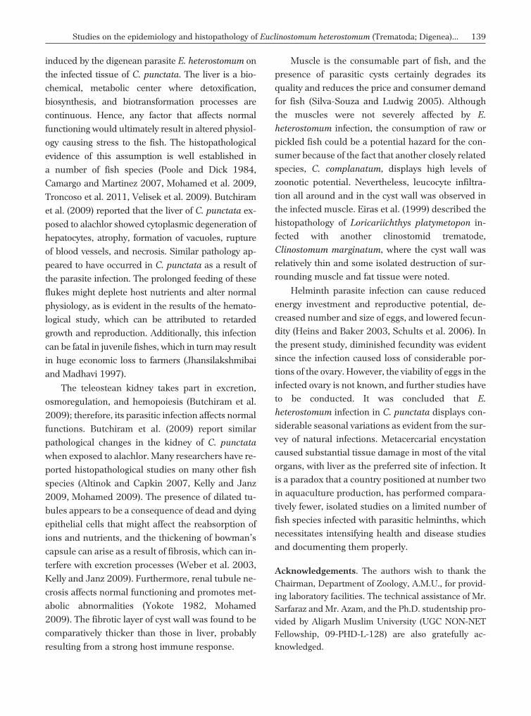
extent and confined to the juxtaposing areas of the
cysts only (Fig. 5A).
Ovary
Unlike other tissues, the infected ovary showed the
least visible pathological alterations and no leucocyte
infiltration, even in the cyst wall, was observed; how-
ever, the eggs lying around the cyst were found to be
slightly damaged (Fig. 5B).
Discussion
Fish health assessment is a tedious, challenging task,
and the results obtained are generally not very
reliable for many reasons (Kori-Siakpere et al. 2005);
therefore, a multidimensional approach is always ad-
visable for a true analysis to establish health and dis-
ease condition, rather than any single approach.
Histopathology together with the epidemiology can
provide more accurate, reliable, meaningful conclu-
sions regarding fish health status along with the eco-
logical factors of a particular aquatic ecosystem in
a region. The present study attempted to perform
such an integrated, comprehensive assessment of the
Studies on the epidemiology and histopathology of Euclinostomum heterostomum (Trematoda; Digenea)... 137
Figure 3. Histopathology of liver infected with E. heterostomum;A – section of liver showing the encysted fluke, enclosed withinthe cyst wall. Note the tissue damage, the plug formed by thesuction of the parasite’s ventral sucker, and the ingested hostblood (brownish black debris); B – cyst wall (CW) showing theheavy infiltration of immune cells; C – infected liver showingnumerous fatty droplets and hemorrhagic foci (arrow), inset –necrosis of hepatocytes; D – infected liver parenchyma showingnormal nucleus (arrows), but lacking in most of the cells, inset–individualization of hepatocytes; E – fibrosis; F – cytoplasmicdegeneration (arrow head) and nucleus (arrow), inset – Mallorybody formation (arrow).
Figure 4. Histopathology of E. heterostomum infected kidney of C.
punctata; A – section of kidney showing the encysted fluke, en-closed within the cyst wall, note the loss of a large portion fromthe kidney and the worm’s oral sucker, pharynx, ventral sucker,and gut are clearly seen; B – enlarged view of cyst wall, note thethickness and heavy infiltration of immune cells; C – renal tubu-lar damage showing the thinning of tubular epithelium (arrowhead), abnormally widened lumen (asterisk) and hypertrophy ofsome epithelial cells, and hyperplasia of tubular epithelial cells(arrow), hyperplasia and hypertrophy of tubular epithelium (ar-row head), M – monocytes and L – lymphocytes infiltratedthrough the cyst wall; D – fibrosis of kidney tubules; E – wall (ar-row head), tubule with dilated lumen and hypertrophy of epithe-lial cells (black arrow), hemorrhage (white arrow), inset –occlusion of tubule (asterisk) hypertrophied nucleus (black ar-row), and pyknotic nucleus (white arrow); F – fibrosis of tubularepithelium (arrow head), necrosis of epithelial cells resulted inthe formation of large vacuole (arrow) and cyst wall (CW).
health of C. punctata during infection with thedigenean E. heterostomum.
Digenetic trematodes exhibit a complex life cy-cle. In India, the freshwater snail Indoplanorbis
exustus and the fish Channa spp. serve as first andsecond intermediate hosts respectively, and ardeidbirds are the definitive hosts for E. heterostomum.
According to Jhansilakshmibai and Madhavi (1997),E. heterostomum infect the liver and kidney of C.
punctata and the muscle of Channa orientalis
Bloch&Schneider. However, it was observed duringthe present study that this parasite encysts the liver,kidney, peritoneal membrane, ovary, and muscle ofC. punctata. Nevertheless, in comparison to other tis-sues, the liver was found to be the most preferred or-gan for encystment, possibly because of its large size,particular position, and extensive vascular supply(Van der Oost et al. 2003) that maintains a surplus ofnutrients and continuously removes and detoxifiesthe parasite excretory and/or secretory products.
Studies on the incidence and seasonal variabilityof parasitic infections would enable farmers andculturists to identify how frequent and to what extentinfection is prevailing in a region, so that local man-agement strategies and control measures can be for-mulated. The epidemiological study of E.
heterostomum revealed a prevalence rate of 18.61%and an average of 1.96 cysts per infected fish.Jhansilakshmibai and Madhavi (1997) reported aninfection rate of up to 32.7%, harboring up to 66metacercariae in natural infections and 72 in experi-mental infections. Whereas, during the present study
period, a maximum of 16 cysts per infected fish wasrecorded. The maximum number of cysts observed inthe liver, kidney, peritoneum, and muscle was 13, 2,2, and 3, respectively. It has been elucidated that theparasite takes two months or more to release the infec-tive cercariae from the snails and at least another twomonths to become encysted metacercaria in this fish(Jhansilakshmibai and Madhavi 1997). The highestprevalence (40.74%), intensity (3.63), and abundance(1.48) were observed during June, which could stemfrom higher virulence and favorable weather condi-tions during the post-winter months when the envi-ronmental temperature ranges between 20 to 30°C.The low incidence during the months following the ex-treme summer, when the temperature rises to 48°C,could be from the reduced infectivity of larvae. Fur-thermore, the monsoon rain and snail availabilitymight also play an important role in varied incidenceduring different months or seasons. The 0% preva-lence during December could have been from the un-availability of larval stages in the aquatic ecosystem asa consequence of prior heavy rain in North India dur-ing July-August 2011 when they might have been car-ried away with flood waters. This suggests thatecological and climatological factors could also playan important role in the occurrence and distribution ofnatural infections of this parasite.
Histopathological analysis is a powerful tool tocorroborate the histoarchitecture of various tissuesduring parasitic infections (Eiras et al. 1999, Ferguson2006, Shareef and Abidi 2012). The results of thepresent study manifest the pathological alterations
138 P. A. Ahammed Shareef, Syed M. A. Abidi
Figure 5. Section showing E. heterostomum encysted in the muscle (A) and ovary (B) of C. punctata. Ingested host blood appears brownishor black within the gut of the worm, a comparatively thin cyst wall, and some damage to muscle fibers are evident.
induced by the digenean parasite E. heterostomum on
the infected tissue of C. punctata. The liver is a bio-
chemical, metabolic center where detoxification,
biosynthesis, and biotransformation processes are
continuous. Hence, any factor that affects normal
functioning would ultimately result in altered physiol-
ogy causing stress to the fish. The histopathological
evidence of this assumption is well established in
a number of fish species (Poole and Dick 1984,
Camargo and Martinez 2007, Mohamed et al. 2009,
Troncoso et al. 2011, Velisek et al. 2009). Butchiram
et al. (2009) reported that the liver of C. punctata ex-
posed to alachlor showed cytoplasmic degeneration of
hepatocytes, atrophy, formation of vacuoles, rupture
of blood vessels, and necrosis. Similar pathology ap-
peared to have occurred in C. punctata as a result of
the parasite infection. The prolonged feeding of these
flukes might deplete host nutrients and alter normal
physiology, as is evident in the results of the hemato-
logical study, which can be attributed to retarded
growth and reproduction. Additionally, this infection
can be fatal in juvenile fishes, which in turn may result
in huge economic loss to farmers (Jhansilakshmibai
and Madhavi 1997).
The teleostean kidney takes part in excretion,
osmoregulation, and hemopoiesis (Butchiram et al.
2009); therefore, its parasitic infection affects normal
functions. Butchiram et al. (2009) report similar
pathological changes in the kidney of C. punctata
when exposed to alachlor. Many researchers have re-
ported histopathological studies on many other fish
species (Altinok and Capkin 2007, Kelly and Janz
2009, Mohamed 2009). The presence of dilated tu-
bules appears to be a consequence of dead and dying
epithelial cells that might affect the reabsorption of
ions and nutrients, and the thickening of bowman’s
capsule can arise as a result of fibrosis, which can in-
terfere with excretion processes (Weber et al. 2003,
Kelly and Janz 2009). Furthermore, renal tubule ne-
crosis affects normal functioning and promotes met-
abolic abnormalities (Yokote 1982, Mohamed
2009). The fibrotic layer of cyst wall was found to be
comparatively thicker than those in liver, probably
resulting from a strong host immune response.
Muscle is the consumable part of fish, and the
presence of parasitic cysts certainly degrades its
quality and reduces the price and consumer demand
for fish (Silva-Souza and Ludwig 2005). Although
the muscles were not severely affected by E.
heterostomum infection, the consumption of raw or
pickled fish could be a potential hazard for the con-
sumer because of the fact that another closely related
species, C. complanatum, displays high levels of
zoonotic potential. Nevertheless, leucocyte infiltra-
tion all around and in the cyst wall was observed in
the infected muscle. Eiras et al. (1999) described the
histopathology of Loricariichthys platymetopon in-
fected with another clinostomid trematode,
Clinostomum marginatum, where the cyst wall was
relatively thin and some isolated destruction of sur-
rounding muscle and fat tissue were noted.
Helminth parasite infection can cause reduced
energy investment and reproductive potential, de-
creased number and size of eggs, and lowered fecun-
dity (Heins and Baker 2003, Schults et al. 2006). In
the present study, diminished fecundity was evident
since the infection caused loss of considerable por-
tions of the ovary. However, the viability of eggs in the
infected ovary is not known, and further studies have
to be conducted. It was concluded that E.
heterostomum infection in C. punctata displays con-
siderable seasonal variations as evident from the sur-
vey of natural infections. Metacercarial encystation
caused substantial tissue damage in most of the vital
organs, with liver as the preferred site of infection. It
is a paradox that a country positioned at number two
in aquaculture production, has performed compara-
tively fewer, isolated studies on a limited number of
fish species infected with parasitic helminths, which
necessitates intensifying health and disease studies
and documenting them properly.
Acknowledgements. The authors wish to thank theChairman, Department of Zoology, A.M.U., for provid-ing laboratory facilities. The technical assistance of Mr.Sarfaraz and Mr. Azam, and the Ph.D. studentship pro-vided by Aligarh Muslim University (UGC NON-NETFellowship, 09-PHD-L-128) are also gratefully ac-knowledged.
Studies on the epidemiology and histopathology of Euclinostomum heterostomum (Trematoda; Digenea)... 139
Author contribution. P.A.A.S. and S.M.A.A. designed
research, P.A.A.S. performed research, P.A.A.S. and
S.M.A.A. wrote the paper.
References
Altinok I., Capkin E. 2007 – Histopathology of rainbow troutexposed to sublethal concentrations of methiocarb orendosulfan – Toxicol. Pathol. 35: 405-410.
Butchiram M.S., Tilak K.S., Raju P.W. 2009 – Studies onhistopathological changes in the gill, liver and kidney ofChanna punctatus (Bloch) exposed to Alachlor – J. Envi-ron. Biol. 30: 303-306.
Camargo M.M.P., Martinez C.B.R. 2007 – Histopathology ofgills, kidney and liver of a neotropical fish caged in anurban stream – Neotrop. Ichthyol. 5: 327-336.
Deardoeff T.L. 1991 – Epidemiology of marine fish-borne par-asitic zoonoses – Southeast Asian J. Trop. Med. PublicHealth. 22: 146-149.
Eiras J.C., Dias M.L.G.G., Pavanelli G.C., Machado M.H.1999 – Histological studies on the effects of Clinostomum
marginatum (Digenea, Clinostomidae) in its second inter-mediate host Loricariichthys platymetopon
(Osteichthyes, Loricariidae) of the upper Paraná River,Brazil – Acta Scientiarum 21: 237-241.
Ferguson H.W. 2006 – Systemic pathology of fish. A text andatlas of normal tissues in teleosts and their responses indisease, second ed. – Scotian Press, UK.
FAO 2008 – FAOSTAT Online Statistical Service – UnitedNations Food and Agriculture Organization (FAO),Rome, available from: http://faostat.fao.org.
Gudmundsd�ttir B.K., Björnsd�ttir B., Gudmundsd�ttir S.,Bambir S.H. 2006 – A comparative study of susceptibilityand induced pathology of cod, Gadus morhua (L.) and hali-but, Hippoglossus hippoglossus (L.), following experimentalinfection with Moritella viscosa – J. Fish Dis. 29: 481-487.
Haniffa M.A., Mydeen A.K. 2011 – Hematological Changes inChanna striatus Experimentally Infected by Aeromonas
hydrophila – Biores. Bull. 4: 246-253.Heins D.C., Baker J.A. 2003 – Reduction of egg size in natural
populations of threespine stickleback infected witha cestode macroparasite – J. Parasitol. 89: 1-6.
Jhansilakshmibai K., Madhavi R. 1997 – Euclinostomum
heterostomum (Rudolphi, 1809) (Trematoda): life-cycle,growth and development of the metacercaria and adult –Syst. Parasitol. 38: 51-64.
Kelly J.M., Janz D.M. 2009 – Assessment of oxidative stressand histopathology in juvenile northern pike (Esox
lucius) inhabiting lakes downstream of a uranium mill –Aquat. Toxicol. 92: 240-249.
Kori-Siakpere O., Ake J.E.G., Idoge E. 2005 – Haematologicalcharacteristics of the African snakehead, Parachanna
obscura – Afr. J. Biotechnol. 4: 527-530.
Mohamed F.A.S. 2009 – Histopathological Studies on Tilapia
zillii and Solea vulgaris from Lake Qarun, Egypt – World.J. Fish. Mar. Sci. 1: 29-39.
Poole B.C., Dick T.A. 1984 – Liver pathology of yellow perch,Perca flavescens (Mitchill), infected with larvae of thenematode Raphidascaris acus (Bloch, 1979) – J. Wildl.Dis. 20: 303-307.
Sanil N., Asokan P.K., John L., Vijayan K.K. 2011 – Patholog-ical manifestations of the acanthocephalan parasite,Tenuiproboscis sp. in the mangrove red snapper(Lutjanus argentimaculatus) (Forssk�l, 1775), a candi-date species for aquaculture from Southern India –Aquaculture 310: 259-266.
Scholz T. 1999 – Parasites in cultured and feral fish – Vet.Parasitol. 84: 317-335.
Schultz E.T., Topper M., Heins D.C. 2006 – Decreased repro-ductive investment of female threespine sticklebackGasterosteus aculeatus infected with the cestodeSchistocephalus solidus: parasite adaptation, host adap-tation, or side effect? – Oikos. 114: 303-310.
Shareef P.A.A., Abidi S.M.A. 2012 – Incidence andhistopathology of encysted progenetic metacercaria ofClinostomum complanatum (Digenea: Clinostomidae) inChanna punctatus and its development in experimentalhost – Asian. Pac. J. Trop. Biomed. 2: 421-426.
Silva-Souza A.T., Ludwig G. 2005 – Parasitism of Cichlasoma
paranaense Kullander, 1983 and Gymnotus carapo
Linnaeus, 1814 by Clinostomum complanatum
(Rudolphi, 1814) metacercariae in the Taquari River –Braz. J. Biol. 65: 513-519.
Troncoso I.C., Cazenave J., Bacchetta C., Bistoni M.A. 2012 –Histopathological changes in the gills and liver ofProchilodus lineatus from the Salado River basin (SantaFe, Argentina) – Fish Physiol. Biochem. 38: 693-702.
Van der Oost R.V., Beyer J., Vermeulen N.P.E. 2003 – Fishbioaccumulation and biomarkers in environmental riskassessment: a review – Environ. Toxicol. Pharmacol. 13:57-149.
Velisek J., Svobodova Z., Machova J. 2009 – Effects ofbifenthrinu on some haematological, biochemical andhistopathological parameters of common carp (Cyprinus
carpio L.) – Fish Physiol. Biochem. 35: 583-590.Weber L.P., Higgins P.S., Carlson R.I., Janz D.M. 2003 –
Development and validation of methods for measuringmultiple biochemical indices of condition in juvenilefishes – J. Fish Biol. 63: 637-658.
Wee K.L., Tacon A.G.J. 1982 – A preliminary study on thedietary protein requirement of juvenile snakehead – Bull.Jpn. Soc. Sci. Fish. 48: 1463-1468.
Williams H., Jones A. 1994 – Parasitic worms of fish – Taylor& Francis, London.
Yamaguti S. 1971 – Synopsis of digenetic trematodes of vertebrates.Vol. 1 – Keigaku Publishing Company, Tokyo, p. 1074.
Yokote M. 1982 – Digestive system – In: An Atlas of Fish His-tology (Ed.) T. Hibiya, Normal and Pathological Feature,Kodanshan Ltd, Tokyo, 74-93.
140 P. A. Ahammed Shareef, Syed M. A. Abidi