studies on the development of gametophytes and … · · 2017-02-05in clove oil as counterstain....
TRANSCRIPT
S. Afr. J. Bol.. 1996. 62(2) 99
Studies on the development of gametophytes and endosperm in lamesbrittenia microphylla (Scrophulariaceae) with reference to its systematic position
H.G.v.G. Urs and C.R. Nagendran*
Department of Botany, University of Mysore, Manasa Gangothri, Mysore 570006, India *Department of Biology, National University of Lesotho, PO. Roma 180, Lesotho
Received 21 September 1995: reviJed 29 November 1995
In Jamesbrittenia microphylla (L.r.) Hilliard, the anther is bilobed, bilocular and bisporangiate. The microsporangium wall comprises epidermis, endothecium, middle layer and tapetum. Cytokinesis of pollen mother cells is simultaneous. Microspore tetrads are tetrahedral. The triaperturate pollen grains are shed at the 2-celled stage. Ovules are anatropous, tenuinucellate and unHegmic. The development of the embryo sac is of the Polygonum type. An endothelium surrounds the mature embryo sac. Endosperm development is ab initio cellular. Both chalaza I and micropylar haustoria are well developed. The chalazal haustorium is 2-celled and the micropylar haustorium is 4-celled. These results are compared with earlier work in the Manuleae tribe and the close relationship between Manuleae and Selagineae is emphasized.
Keywords: Embryology, Jamesbrittenia, Manuleae, megasporogenesis, microsporogenesis, Scrophulariaceae, Selagineae, systematic position.
'"To whom correspondence should be addressed.
Introduction
The tribe Manuleae of the Scrophulariaceae has recently been revised taxonomically; it includes 17 genera and 344 species (Hilliard 1994). Embryological investigation on such a large and heterogeneous group is limited to three species: Lyperia violacea (Jarosz) Benth. (as Sutera antirrhinoides auct. non (L.f.) Hiem], Siltemfoetida Roth [as Chaellostomafoetidum Benth.], and ZallIziamkya capellsis (L.) Walp. Crete (1948) studied the development of endosperm and embryo in S. foetida; he recorded a 2-celJed chalazal haustorium, a 4-celled micropylar haustorium in the endosperm and the Onagrad type of embryo development. In a subsequent article (Crete 1949), he examined the endosperm development in L. violacea and noted that both the chalazal and the micropylar haustoria have four uninucleate cells. Later, Juoett (1962) studied the embryology of L. viofacea, S. foetida and Z. capensis and noted a 2-celled chalazal haustorium in all three taxa. Mestre and Guignard (1969) recorded the Onagrad (= Crucifer) type of embryo ontogeny in Z. capellsis. Recent embryological information pertaining to the family is available in Nagendran and Dinesh (1989), Johri et af. (1992) and Urs (1992).
It is clear from the foregoing that detailed embryological investigations in the tribe are lacking and the earlier studies are too fragmentary to allow proper phylogenetic analysis. An investigation of unstudied genera is therefore highly desirable for a bellcr understanding of the embryology of this diverse and interest ing tribe. The present study deals with the development of the mkrosporangium, male gametophyte, megasporangium, fema le gametophyte and endosperm in lamesbrittenia microphylla (L.f.) Hilliard.
Materials and Methods
The observalions are based on formalin-acetic acid-alcohol (FAA) tixcd material of J. microphylla obtained from Prof. B.L. Robertson and Dr M.e. Olivier of the University of Port Elizabeth, South Africa. The material was processed for microtomy by the methods of dehydration, infiltration and embedding in paraffin wax (Nagendran et al. 1977). Sections were cut between 8 and 13 11m in thickness
and stained in Heidenhain's iron alum- haematoxylin with crythrosin in clove oil as counterstain. The voucher specimen has been deposited in the herbarium of the University of Mysore. Manasa Gangothri campus. India.
Results
Microsporangium and male gametophyte
The anther is bilobed, bilocular and bisporangiate. Each anther bears only two elongate microsporangia. Quite early during deve lopment, a hypodermal archesporium of dense protoplasmic and large nucleated cells is organized at two sites in the young anther (Figures 1 & 2). Periclinal divisions in the archesporium cells lead to the outcr primary parietal and the inner primary sporogenous layers, respectively (Figure 3). While the former, by further periclinal divisions, contributes to the wall layers of the microsporangium, the latter builds up the sporogenous tissue (Figure 4). The microsporangium wall comprises the epidermis. an endothecium, a middle layer and the secretory type of tapetum (Figures 6 & 7). During anther wall development, the endothecium and the middle layer arise as sister layers (Figure 4). The tapetal layer is of dual origin. During the organization of the tapetum, in the parietal layer, a group of cells adjacent to the connective of the anther become richly protoplasmic and conspicuous. These cells ultimately align with the tapetal strip organized by the parietal layer, thus forming a continuous sheath of tapelum enclosing the sporogenous tissue (Figures 5 & 6). Meanwhile, the pollen mother ce lls derived from the sporogenous tissue divide meiotically to produce tetrahedral micros pore tetrads (Figures 7-14). Cytokioesis of the pollen mother cells is simultaneous. The individual microspores of a tetrad separate as their nucleus mitotically divides to form a small generative cell and a large vegetative cell (Figures 15 & 16). By about this stage, the tapetum is completely absorbed by the developing microspores . The cells of the middle layer become crushed and obliterated quite early during the wall development , and the endothecial cells, on the other hand, become enlarged and acquire fibrous bands on their tangential and transverse walls. The epidermal layer of cells also enlarges and becomes conspicuous (Figure 9).

100
The mature pollen grains are 2-celled and triaperturatc (Figure 16). The exine is smooth and thicker than the intine. The pollen grains arc shed through :l common opening due to the breakdown of ce lls separating adjacent microsporangia (Figure 8).
Megasporangium and female gametophyte
In each lacule of the bi locular ovary. there arise small finger~shaped ovular primordia on the axile placenta. Each of
2
i) .... , .. , ! "
4" Jo. • . ' . "~
.. ..... . ,. <ot'/:!'
s. Arr. 1. BoL. 1996. 62(2)
these develops into a unitegmic, tenuinucellate, and analTopouS ovule. A hypodermal archesporia] cell differentiates in the young ovular primordium even before the integumentary primordium appears (Figure 17). Following enlargement, it directly functions as the megaspore mother cell (Figure 18). Cytokinesis in the megaspore mother cell accompanies meiosis and the chalaza I megaspore of a linear tetrad gives ri se to the Polygonum ty pe of embryo sac which is 8-nucleate and bipolar (Figures 19- 23).
3
6 7
10 II 12 13
14 15 16
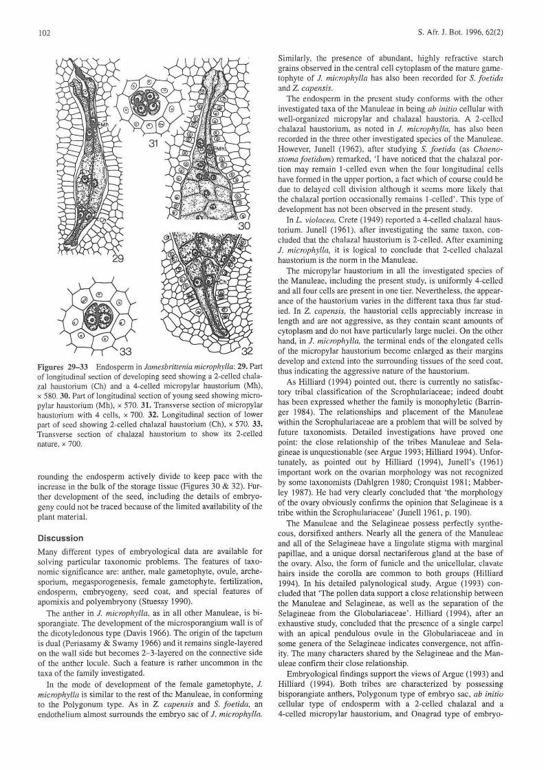
Figures 1- 16 Microsporangium and male gametophyte in iamesbrittenia microphylla: 1. Out li ne of young anther in transverse section showing sires of microsporangia, x 310. 2. Part of young anther in transverse sec tion showing hypodermal archesporial cells at the sporangial sites , x 710. 3. Transverse section of part of developing anther with primary parietal and primary sporogenous layers, x 7 10. 4. Transverse section of microsporangium to show development of wall layers and sporogenous ti ssue. x 710. 5. Transverse section diagram of anther with two microsporangia at microspore mother cell stage, x 60. 6. Part marked X in Figure 5 enlarged. Note 2- to 3-layered tapetum on the connective side. x 475 . 7. Transverse section of part of microsporangium at microspore tetrad stage showing binucleate tapetal cell s, x 710. 8. Transverse section diagram of mature anther, x 60. 9. Portion marked X in Figure 8 enlarged to show details of wall layers, x 7 10. 10-14. Meiotic stages in microspore mother cells and organization of tetrads. x 890. 15. Uninucleate pollen grain. x 890. 16. Mature 2-celled pollen grain, x 890.

S. Arr. J. Bot.. 1996.62(2)
Figures ] 7-24 Female gametophyte development in lamesbrille· Ilia microphy lfa : 17. Ovular primordium in longitudinal section showing archesporial cell , x 765. 18. Part of longitudinal section of developing ovule showing megaspore mother cell. x 765. 19. Part of longitudinal section of ovu le with linear tetrad of megaspores. x 765. 20. Functional megaspore in nucellus, x 765. 21. A 2-nucleale embryo sec. (Note breakdown of nucellar envelope) x 765. 22. A 4· nucleate embryo sac, x 765. 23. An 8-nucleate embryo sac, x 765. 24. A mature embryo sac. x 765.
26
101
The egg apparatus in the organized embryo sac consists of (wo prominent pyriform, posteriorly vacuolate synergids and a pear-shaped anteriorly vacuolate egg. The antipodal cells are qui te conspicuous and have a definite arrangement. The second~ ary nucleus. formed as a result of the fusion of two polar nuclei, lies in the centre of the mature gametophyte. At maturity, an endothelium of richly protoplasmic cells surrounds the embryo sac except at the extreme micropylar and chalazal ends (Figure 24).
Endospe rm
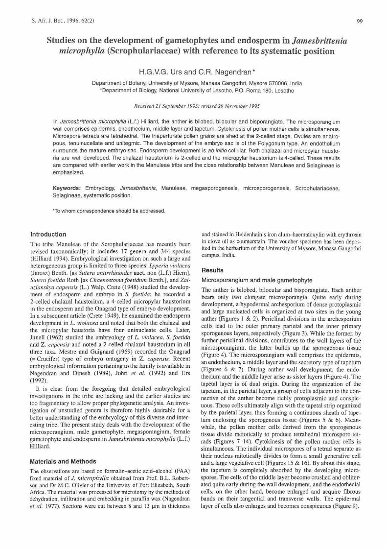
Fertili zation is porogamous. After double fertili zation. the embryo sac has the pear-shaped zygote at its micropylar end and the degenerating remains of the antipodal cells at the chalazal end. In the central cell, the conspicuously large primary endosperm nucleus lies at the centre of the dense protoplasmic endosperm mother cell (Figurc 25). The first divisi on of the endosperm mother cell is transverse and occurs much earlier than that of the zygote. Consequent ly, two primary endosperm chambers are organized (Figure 26). The next division in both chambers is vertical. This results in two juxtaposed cells in each chamber (Figure 27). However, the two cells of the primary chalaza I chamber do not divide further; they directly form the chalazal haustorium. A vertical wall is soon laid down in the primary micropylar chamber, producing four circumaxial cells (Figure 28). Further, a transverse division in these cells delimits an upper tier of four ce lls, constituting the micropylar haustorium. from a lower tier of four cells, which function as the initials of the endosperm lissue proper (Figure 29).
The four cells of the single-tier micropy lar haustorium elongate and acquire dense contents (Figures 30. 31). The terminal ends of the haustorial cells become swollen as their margins devclop fringed lobation, which makes the haustorium an efficient absorptive organ. Meanwhile , the two uninucleate cells of the young chalazal haustorium elongate and extend towards the terminal end of the chalazal vasculature of the seed. The hausto· ri al cells accumulate rich cytoplasmic contents and their nuclei acquire deep staining capacity (Figures 32. 33).
The initials of the endosperm proper divide a number of times to finally build up the bulk of the endosperm tissue in which the developing embryo becomes lodged. The endothelial cel ls sur-
Figures 25-28 Endosperm development in lamesbrittenia microphylla: 25. Fertilized embryo sac, x 670. 26. A 2-ceJled endosperm, x 670. 27. A 4-celled endosperm, x 670. 28. A 6-celled endospenn, x 670. (Note remains of antipodal cell s in aJl Figures).
102
Figures 29-33 Endosperm in Jamesbritrenia microplr.vl/a; 29. ParI of longitudinal sect ion of developing seed showing a 2·celled chalazal haustorium (Ch) and a 4-celled micropylar haustorium (Mh), x 580.30. Part of longitudinal section of young seed showing micropylar haustorium (Mh), x 570. 31. Transverse section of micropylar haustorium with 4 ce ll s, x 700. 32. Longitudinal section of lower part of seed showing 2-celled chalazal haustorium (Ch), x 570. 33. Transverse section of chalazal haustorium to show its 2-celled nature. x 700.
rounding the endosperm actively divide [Q keep pace with the increase in the bulk of the storage ti ssue (Figures 30 & 32). Further development of the seed, including the detail s of embryogeny could not be traced because of the limited availability of the plant materiaL
Discussion
Many different types of embryological data are available for solvi ng particular taxonomic problems. The features of taxonomic significance are: anther, male gametophyte, ovule, archesporium, megasporogenesis, female garnetophyte, fertilization, endosperm, embryogeny, seed coat, and special features of apomixis and polyembryony (Stuessy 1990).
The anther in J. microphylla, as in all other Manuleae, is bisporangiatc. The development of the microsporangiurn wall is of the dicotyledonous type (Davis 1966). The origin of the tapetum is dual (Pcriasarny & Swamy 1966) and it remains single-layered on the wall side but becomes 2- 3-layered on the connective side of the anther lacule. Such a feature is rather uncommon in the taxa of the family investigated.
In the mode of development of the female gametophyte. 1. microphylla is similar to the rest of the Manuleae. in conforming to the Polygonum type. As in Z. capensis and S. joetida, an endothelium almost surrounds the embryo sac of J . microphylla.
S. Afr. I. Bol. 1996.62(2)
Similarly, the presence of abundant, highly refractive starch grains observed in the central ce ll cytoplasm of the mature gametophyte of J. microphylla has also been recorded for S. foetida and Z. capellsis.
The endosperm in the present study conforms wi th the other investigated taxa of the Manuleae in being ab initio cellular with well-organized micropylar and chalazal haustoria. A 2-celled chalaza l haustorium, as noted in J. microphylia, has also been recorded in the three other investigated species of the Manuleae. However. lunell (1962), afte r studying S. foetida (as Chaeno· stoma foetidum) remarked, 'I have noticed that the chalazal portion may remain I-celled even when the four longitudinal cells have formed in the upper port ion, a fact which of course could be due to delayed cell division although it seems more likely that the chalazal portion occasionally remains l -celJed'. This type of development has not been observed in the present study.
In L violacea, Crete (1949) reported a 4·celled chalaza I haus· torium. Junell (1961), after inves tigating the same taxon, concluded that the chalaza! haustorium is 2-celled. After examining J. microphylla, it is logical to conclude that 2-ccllcd chalazal haustorium is the norm in the Manuleae.
The micropylar haustorium in all the investigated species of the Manuleae, including the present study. is uniformly 4-celled and all four cells arc present in one tier. Nevertheless, the appearance of the haustorium varies in the different taxa thus far studied. In Z. capeTlSis, the haustoria I cells appreciably increase in length and are not aggressive, as they contain scant amounts of cytoplasm and do not have particularly large nuclei. On the other hand. in 1. microphylla, the terminal ends of the elongated ce lls of the micropylar haustorium become enlarged as their margins develop and extend into the surrounding tissues of the seed coat. thus indicating the aggressive nature of the haustorium.
As Hilliard ( 1994) pointed out, there is curremly no satisfactory tribal classification of the Scrophulariaceae; indeed doubt has been expressed whether the family is monophyletic (Barringer 1984). The relationships and placement of the Manuleae within the Scrophulariaceae are a problem that will be solved by future taxonomists. Detailed investigations have proved one point: the close relationship of the tribes Manull!ae and Selagineae is unquestionable (see Argue 1993; Hilliard 1994). Unfor· tunately. as pointed out by Hilliard (1994). lunell's (1961) important work on the ovarian morphology was not recognized by some taxonomists (Dahlgren 1980; Cronquist 1981; Mabber· ley 1987). He had very clearly concluded that ' the morphology of the ovary obviously confirms the opinion that Selagincac is a tribe within the Scrophulariaceae' (Junell1961, p. 190).
The Manuleae and the Selagineae possess perfectly synthccous. dorsifixed anthers. Nearly all the genera of the Manuleae and all of the Selagineae have a lingulate stigma with marginal papillae, and a unique dorsal nectariferous gland at the base of the ovary. Also, the fonn of funicle and the unicellular. clavate hairs inside the corolla are common to both groups (Hilliard 1994). In his detailed palynological study. Argue (1993) con· cluded that 'The pollen data support a close relationship between the Manuleae and Selagineae. as well as the separation of the Selagineae from the Globulariaceae·. Hilliard (1 994). after an exhaustive stud y, concluded that the presence of a single carpel with an apical pendulous ovule in the Globulariaceae and in some genera of the Selagineae indicates convergence. not affinity. The many characters shared by the Selagineae and the Manuleae confirm their close relationship.
Embryological findings support the views of Argue (1993) and Hilliard (1994). Both tribes are characterized by possessing bisporangiatc anthers, Polygonum type of embryo sac, ab initio cellular type of endosperm with a 2-celled chalazal and a 4-celled micropylar haus(Qrium, and Onagrad type of embryo-
S. Afr. J. Bol.. 1996. 62(2)
geny. Moreover, the sequence of divisions during early endosperm formation and the subsequem delimitation of haustoria is essen ti ally the same in both the tribes (Raj u 1973; Johri el al. 1992).
Acknowledgements Th~ assis tance of Professor Govindappa D. Arekal is much appreciated. The research was funded by the UniversilY Grants Commission. New Delhi. India.
References ARGUE. C.L. 1993. Pollen morphology in the Seiagineae, Manuleae
(Scrophulariaceac), and selected Globu lariaccae. and its taxonomic
sigmficancc. Am. 1. Bot. 80: 723-733. BARRINGER, K. A. 1984. Seed morphology and ciass ificaion of the
Scropllulariaceae. Am. 1. Bo{. 71 (Abstracts): 156.
CRETE. P. 194R. Recherc hes emhryo logiques chez Ics scrofu lariacees. Dcvcloppement c..Ie I'albumen et de J'embryon chez Ie Chaenostoma·
fuerie/ttl/! Oaeg.) Bcnlh. Bull. Soc. Bol. Fr. 95: 142-146.
CRETE. P. 1949. Recherehe.~ embryologiques chez les scrofulari acees. Dcvcloppe ment c..Ie l'albumen chez Ie Lyperia vio/m.:ea Benth . BIlII. Soc Bor. Fr. 96: 186- 188.
CRONQUIST. A. 198 1. An integrated system of c lass ification of flowering plants. Columbia University Press, New York.
DAHLGREN. R.M.T. 1980. A revised system of classification of the
angiosperms. Bm. 1. Linn. Soc. 8: 9J - 124.
DAVIS. G.L. 1966. Systematic embryology of the angiosperms. Wiley, New York.
\03
HIL LI ARD, O.M. 1994. The ManuJeae: A lr ibe of Scrophu lariaceae. pp.
1-579. Edinburgh University Press. Ed inburgh.
JOHRI. B.M .. AM BEGAOKAR. K. B. & SRIVASTAVA. P. S. 1992. Comparative embryology of angiosperms. Vol. 2. Springer-Verlag, Berlin.
JUNELL. S. 1961. Ovarian morphology and taxonomical position of
Selagineae. Svensk bol. Tidskr. 55: 168- 192.
JUNELL, S. 1962. Embryology of Haben.'iTreiria. Dischi.Hna. Sutera and
Zafuzial1JJ..}·a (Scrophulariaceae). Acta Horti Go(eburg 25: 91-10 I.
MABBERLEY. DJ. 1987. The plant-book. Cambridge University Press. Cambridge.
MESTRE. J .e. & GUIGNARD. J.L. 1969. Embryogenic des Scrofulariace'ecs. Le developpement de I'embryon chez Ie Zalazianskya cap
emi.{ Walp. C. r. hebel. Seam:. Acad. Sci .. Paris 268: 3 l70-3 172.
NAGENDRAN. C.R .. AREKAL. G.D. & SUBRAMANYAM. K. 1977. Embryo sac studies in Lhree Indian species of p/J/ypleurwn (Podo
stemaceae). PI. Sy.H. Evol!a. 128: 2 15-226.
NAGEN DRAN. C. R. & DINESH. M.S. 1989. Embryology of .og;o
sperms: a classified bibliography ( 1965- 1985). Indira Pub!. House. Michigan.
PERIASAMY. K. & SWAMY. B.G.L. 1966. Morphology of the amher
tapetum of angiosperms . Curr. Sci. 35: 420-430.
RAJU, D. 1973. Embryological studies in the tribe Sc lagineae: Aga/h
e/pis dubia. Phytomorph%gy 23: 194-205.
STUESSY. TF. 1990. Plant taxonomy: the systematic evaluation of comparative data. Columbia University Press. New York.
URS. H.G.V.G. 1992. Relationships of Scrophulariaceae based on
embryology. J. M)'sore Vniv. Sec;/. B 32: 475-482.