studies on blood cell metabolism. · studies on blood cell metabolism. ... the hexose molecule into...
TRANSCRIPT
STUDIES ON BLOOD CELL METABOLISM.
II. THE EFFECT OF METHYLENE BLUE AND OTHER DYES UPON THE GLYCOLYSIS AND LACTIC ACID FORMATION OF
MAMMALIAN AND AVIAN ERYTHROCYTES.
BY E. S. GUZMAN BARRON AND GEORGE A. HARROP, JR.
(From the Chemical Division of the Medical Clinic, the Johns Hopkins Hospital and University, Baltimore.)
(Received for publication, June 19, 1928.)
In the preceding paper (1) we have shown that the oxygen con- sumption of mammalian erythrocytes is enormously accelerated in the presence of minute amounts of methylene blue and certain other dyes. This effect upon the respiration of the mammalian cells differs from the effect of methylene blue upon the erythrocytes of avian blood. In the present paper we present the results of a continuation of this study; namely, a comparison of the influence of methylene blue upon glycolysis and lactic acid formation in mammalian erythrocytes, and in the actively respiring cells of avian blood.
The extensive literature upon glycolysis in the blood has been reviewed by other observers. Lepine (2) first introduced the term and showed that the glycolytic process in blood required the pres- ence of a ferment. Since glycolysis did not occur in serum he stated that it was in some way connected with the blood cells and suggested that the ferment was located in the leucocytes. The important work of Levene and Meyer (3) demonstrated the role of leucocytes in glycolysis. Rona and Arnheim (4), as well as others, have shown that glycolysis is effected by erythrocytes as well.
The studies of recent years have indicated that the disappear- ance of glucose from the blood (and other tissues) occurs in two different ways. In the first, an actual oxidation with considerable liberation of energy occurs; the second is effected by a splitting of the hexose molecule into 2 molecules of lactic acid, but without a
65
by guest on June 1, 2018http://w
ww
.jbc.org/D
ownloaded from

66 Studies on Blood Cell Metabolism. II
coincident utilization of oxygen. The latter process has rather striking analogy to the fermentation of yeast cells. It is possible, indeed probable, that some of the sugar first converted into lactic acid is further broken up by oxidation, in which case it falls in the first category.
In order to determine the relative amounts of glucose which disappear by these two paths, a knowledge of the amount of lactic acid produced is required as well as of the amount of glucose which is lost. Since 2 mols of lactic acid are formed from the glycolysis of each mol of sugar, the glucose which is split up in excess of this simple proportion must then disappear by the other route. It is convenient to express this relation by means of what we may call the glycolytic quotient, as follows:
Millimols lactic acid produced 2 X millimols glucose degraded
When this quotient is unity, it indicates that all of the glucose has been converted into lactic acid. When the ratio is less than unity, the fraction expresses the proportion of the total glucose converted to lactic acid, the remainder having disappeared by another channel or by further splitting of lactic acid. A study of this glycolytic quo~tient as found in adult mammalian erythrocytes in blood prepared and freed from leucocytes by the methods de- tailed in Paper I (I), and incubated as previously described, demonstrated the fact (Tables I and II) first predicted by Claude Bernard’ that the sugar loss can usually be accounted for in very
1 It is of interest that the relation between the disappearance of sugar and the formation of lactic acid was first postulated by Claude Bernardwho considered that it was a fermentation.
“Ce ferment lactique se rencontre dans le sang, dans les muscles, dans le foie lui-m&me; car, j’ai constate que les muscles et divers tissus ne deviennent acides apres la mort qu’autant qu’ils renferment du sucre ou de la mat&e glycogene qui subit t&s-rapidement une fermentation lac- tique. J’ai reconnu autrefois [Cours de physiologie, au College de France, t. I, pp. 379-392, 18551 cette fermentation lactique de la matiere glyco- gene, d’abord dans les muscles du fcetus, oh elle presente son summum d’in- tensite; je l’ai constatee plus tard chez l’homme et les animaux adultes. J’ai vu egalement la fermentation lactique dans le sang. Quand on prend sur un animal qu’on vient de sacrifier le sang le plus sucre, celui des veines sus-hepatiques, on constate que sous l’influence d’une chaleur moderee
by guest on June 1, 2018http://w
ww
.jbc.org/D
ownloaded from

E. S. G. Barron and G. A. Harrop, Jr. 67
large part by new formation of lactic acid. This glycolysis, which requires no oxygen, is in harmony with the observation that adult mammalian erythrocytes have a scarcely measurable oxygen con- sumption. The carbohydrate metabolism of the mammalian erythrocyte thus appears to occur without oxygen under normal conditions. In contrast to the glycolytic quotient of mammalian blood, that of avian blood is always less than 1, indicating that some oxidative process plays a share in the fate of the sugar which disappears, a fact in agreement with the active oxygen consumption found in nucleated red blood cells.
Action of Methylene Blue and Similar Dyes upon the Glycolysis and Lactic Acid Production of Erythrocytes.
The investigations of Loeb (5) and of Melvin (6) showed that ox and sheep blood under aerobic conditions have very slight glycolysis in vitro. A similar behavior on the part of avian blood was later shown by Riiter (7). Glycolysis is more marked in the blood of the dog and of man.
(a) Mammalian Blood.-The addition of methylene blue2 produces a lowering of the glycolytic quotient in mammalian blood. It increases the amount of sugar destruction, but its chief effect
le sucre disparatt du sang en lui donnant une reaction acide, au lieu de la reaction alcaline normale. . . . . J’admets dorm, quant A moi, que la destruction du sucre a lieu par fermentation et non par l’influence directe des alcalis du sang, qui favorisent seulement cette reaction. . . . . Si l’on prend deux parties Bgales du m&me sang sucre et si I’on fait cuire l’une d’elles de fapon a coaguler ou a detruire le ferment lactique, on voit le sang rester alcalin et le sucre persister pendant t&s-longtemps dans la liqueur.” (Bernard, C., Lepons sur le diabhte, Paris, 1877, 328.)
2 The concentration of methylene blue and of the other dyes used, as in the previous communication, was 0.005 per cent unless otherwise indi- cated. The same specimens of dyes were employed. Blood sugar deter- minations were made by Benedict’s modification of the Folin-Wu tech- nique. The lactic acid was determined by the modification of Clausen’s method introduced by Friedemann, Cotonio, and Shaffer. For each experiment 10 cc. of blood were used. Triplicate blood sugar estimations and duplicate lactic acid estimations were made. The limits of error of the analytic procedures are estimated to be per liter 0.2 mM for lactic acid and 0.25 mM for sugar, under the conditions of these experiments.
by guest on June 1, 2018http://w
ww
.jbc.org/D
ownloaded from

68 Studies on Blood Cell Metabolism. II
is to lessen the coincident lactic acid production (Table I).” We interpret the meaning of these results to be that methylene blue causes a shift of the course of carbohydrate catabolism away from the (anaerobic) formation of lactic acid and toward the oxidative
TABLE I.
Mammalian Erythrocytes. Comparative Study of Glycolysis and Lactic Acid Formation as InfEuenced by the Oxidative Action of Methylene Blue
(M.B.) (0.005 Per Cent Concentration).
1
2
3*
4*
5*
Dog blood. Control. ............ Added M.B ......... Control. ........... Added M.B. .......
Human blood. Control ........... Added M.B. ...... Control. .......... Added M.B ....... Control. .......... Added M.B. ......
-
--
.3
.4
.4
.4
.5
-
Blood glw KIM pm.
.962.1! 2.01
.502.0( 1.61
.282.OZ 0.94
,222.X 1.38
.98 3.41 2.0:
1.772.19 5.73 3.54 1.95 4.472.25 1.503.878.664.7s
.-
L
i
I
1
,
I
i : -
3.81
1.77
1.65
1.04
1.41
/
,
I
I
I -
1.00 0.58 0.96 0.37
0.91 0.46 0.99 0.63 0.99 0.64
* The blood used in Experiment 3 was from a patient with heart disease, in Experiment 4 from a patient with polycythemia, and in Experiment 5 from a patient with hyperthyroidism.
path. The action of methylene blue is most striking in the experi- ments upon blood with low glycolytic power (Table II). In these
* For example, in Table I, Experiment 1, the glycolytic quotient reached unity in the control sample, indicating that no oxidation had taken place. In the sample containing methylene blue, however, the glycolytic quotient was reduced to 0.58. In this case the lactic acid increase was 2.28 milli- mols, an amount corresponding to a sugar decrease of 1.14 millimols. As the glucose decrease found however was 1.95 millimols, we may conclude
by guest on June 1, 2018http://w
ww
.jbc.org/D
ownloaded from

E. S. G. Barron and G. A. Harrop, Jr. 69
experiments the amount of sugar which disappears in the presence of methylene blue is quite as large as it is in control specimens, which contain no dye, but there is practically no new formation of lactic acid.4
(b) Avian Blood.-The glycolytic quotient in the case of avian blood is in general lower than in the case of mammalian blood, a fact in accord with its higher respiratory metabolism. The experiments on chicken blood, however, examined twice from dif- ferent birds showed neither disappearance of glucose nor methyl- ene blue action (Table II). It is stated by Bornstein and Ascher (8) that although normal goose blood shows no glycolysis, anemic goose blood has some glycoIytic power. In our experiments, no glycolysis was found in blood taken from one goose until after the bird had been bled repeatedly and thus rendered anemic. Material from a second normal goose, however, showed glycolysis even in the first sample taken. The effect of methylene blue upon the glycolytic quotient in goose blood is similar to that in mammalian blood.
that 1.14 millimols disappeared by glycolysis and the remainder (0.81 millimols) by oxidation.
Where the glycolytic quotient is less than 1 in the control, the correspond- ing amount of sugar has disappeared by a path other than that of lactic acid formation. The amount of glucose which is consumed coincident with the new lactic acid formation in the methylene blue experiment is then determined on the basis of that produced in the control. This amount deducted from the total amount of glucose which disappears in the methy- lene blue experiment gives the amount oxidized by reason of the presence of the methylene blue itself. For example, in Table II, Experiment 3, the glycolytic quotient is 0.59, indicating that a very large proportion (41 per cent) of the glucose has disappeared by the oxidative path. This must be taken into account in estimating the methylene blue effect, as follows: Since 1.01 millimols of lactic acid were produced in the control during the disappearance of 0.86 millimols of sugar, the production of the 0.27 milli- mols of lactic acid found in the specimen to which methylene blue was added was associated on this basis with the loss of 0.23 millimols of sugar. The difference between this amount and the total glucose which dis- appeared (0.89 millimols - 0.23 millimols = 0.66 millimols) is that oxidized by the methylene blue.
4 For example, in Experiment I, Table II, the glycolytic quotient of the control blood is equal to unity, while in the methylene blue sample the quotient has fallen to 0.01. This fall is due to the large amount of sugar (0.73 millimols) which has disappeared by oxidation and without coin- cident increase of lactic acid.
by guest on June 1, 2018http://w
ww
.jbc.org/D
ownloaded from

70 Studies on Blood Cell Metabolism. II
(c) E$ect of Other Dyes.--In accordance with our findings in the preceding paper on the respiratory metabolism of erythrocytes, the oxidative action of methylene blue upon glucose in the blood is shared by other dyes which Clark (9) has shown to possess similar
TABLE II.
Mammalian and Avian Blood with Low Glycolytic Power. Comparative Study of Glycolysis and Lactic Acid Formation. Oxidative Action of
Methylene Blue (M.B.) (0.005 Per Cent Concentration).
Ox blood. Control. . . M.B.. . . . . . .
Sheep blood. Control. . . . . M.B.. . . . . . .
Goose blood. Control.. . . . M.B.. . . . . . .
Chicken blood Control. . .
M.B.. . . . . .
-
-
--
.l
-
3 hrs. incubation.
3.61 2.94O.G 2.780.8:
2.34 1.600.71 1.520.8:
6.39 5.530.8f 5.500.8!
4.72 14.680.0(
14.72 O.O(
Lactic mid, nud per 1.
7.50 8.85 1.3! 7.52 0.0:
$.165.581.4: 4.21 O.O!
1.187.19 1.0: 6.450.2
3.253.270.0(
3.260.01
-
_ -
3
-
0.73
0.79
0.66
No oxi d&ion
by M.B.
-
--
-
1.00 0.01
0.96 0.03
0.59 0.17
No gly- coly- sis.
oxidation-reduction potentials. Studies have been made of the effect of toluylene blue, phenol indophenol, and Bindschedler’s green (double zinc salt) in equimolar concentration. In each case there is an oxidizing action on the carbohydrates, as the glycolytic quotient and calculated glycolysis demonstrate (Table III). The
by guest on June 1, 2018http://w
ww
.jbc.org/D
ownloaded from

cn
* w
N -
I Expe
rimen
t No
. %
33
-0 .ta
- 0
. .
. .
. .
. .
. .
. .
. .
. p-
s: .
. .
. .
. .
. .
. .
. .
. .
P -
;u f
. .
. .
. .
. .
. .
. .
. .
. rt
L
, 00
0 00
000rr
0000
0 :=
“a
inioJ
;o q-
34,
b&&l
~bbb
~~rn
~~
Glyd
ytic
quoti
ent.
3s
rWQP
OOOW
wruJ
r 2
by guest on June 1, 2018http://www.jbc.org/Downloaded from
72 Studies on Blood Cell Metabolism. II
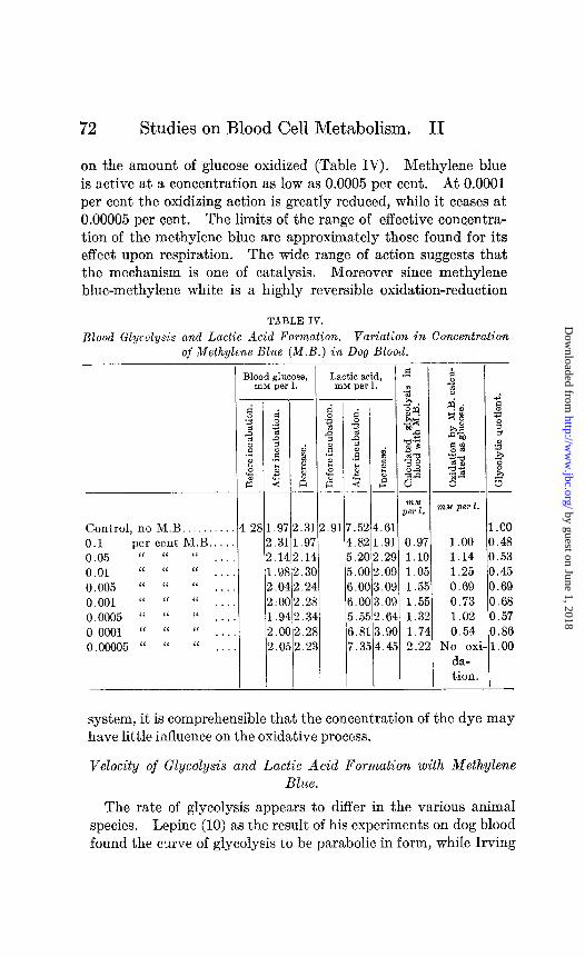
on the amount of glucose oxidized (Table IV). Methylene blue is active at a concentration as low as 0.0005 per cent. At 0.0001 per cent the oxidizing action is greatIy reduced, while it ceases at 0.00005 per cent. The limits of the range of effective concentra- tion of the methylene blue are approximately those found for its effect upon respiration. The wide range of action suggests that the mechanism is one of catalysis. Moreover since methylene blue-methylene white is a highly reversible oxidation-reduction
TABLE IV.
Blood Glycolysis and Lactic Acid Formation. Variation in Concentration _ _ of Methylene Blue (M.B.) in Dog Blood.
I
Control, no M.B. . . 0.1 per cent M.B.. 0.05 “ “ “ . . . 0.01 “ “ “ . . . . 0.005 “ “ “ . . . 0.001 “ “ “ ..,. 0.0005 “ “ “ . . . . 0.0001 “ “ “ . . . . 0.00005 “ “ “ . . .
3lood glucose, Lactic acid, m&l per 1. InM per 1. -
31 2 2 1 2 2 1 2 2
-
-
f: 3 2 2
.5 $ 2
.9
.3 .I .9 .O :O .9
!. C i. C
-
-
d 2 z i -
.3 .9
1.1, :. 31 !. 2 !.2 !.3 !.2 2.2
_-
14 21 32 35 0: 0: 5: 1: 51
:.6 1 .9 1
!.2 9 !.O 9 1.0 9 i.0 9 3.6 84 s.9 0 k.4 :5
- -
nzM per 1.
1.00 1.14 1.25 0.69 0.73 1.02 0.54
No ox da- tion.
- I
1 a a C C C C (
i- 1
-
-
.oo I. 48 1.53 I. 45 I.69 ).68 j.57 I.86 1.00
system, it is comprehensible that the concentration of the dye may have little influence on the oxidative process.
Velocity of GlycoJysis and Lactic Acid Formation with Methylene Blue.
The rate of glycolysis appears to differ in the various animal species. Lepine (10) as the result of his experiments on dog blood found the curve of glycolysis to be parabolic in form, while Irving
by guest on June 1, 2018http://w
ww
.jbc.org/D
ownloaded from

E. S. G. Barron and G. A. Harrop, Jr. 73
(11) working with rabbit blood maintains that the rate of glycoly- sis is linear “over a wide range of values, the rate being about 18 mg. per cent per hour at 38°C.” The initial concentrations of glucose and of lactic acid present undoubtedly may have influence upon the velocity of the process.
In some experiments in which the rate of glycolysis only was followed in human blood, the curve was more or less linear in type, while in experiments with dog blood the curve described by
TABLE V.
Rate of Glycolysis and Lactic Acid Formation in Dog Blood with and without Methylene Blue (M.B.), c&u&g the First 3 Hours of
Incubation (W’).
1 hr. incubation, control.. . . . 1 “ “ M.B.. . . . .
After 2 hrs. incubation. Control................... Added M.B. . . . . . . . . . . . . .
After 3 hrs. incubation. Control................... Added M.B . . . . . . . . . . .
. .
Blood sugar, xnhl per 1.
Lactic acid, InM per 1.
--~ 3.08 1.20 2.91
2.70 1.58
2.43 1.85 2.20 2.08
6.42 3.51 0.95 5.46 2.55 0.61
1.97 2.31 7.56 4.65 1.00 1.62 2.66 5.78 2.87 0.53
-
.
1
--__ 5.13 2.22 0.93 4.14 1.23 0.39
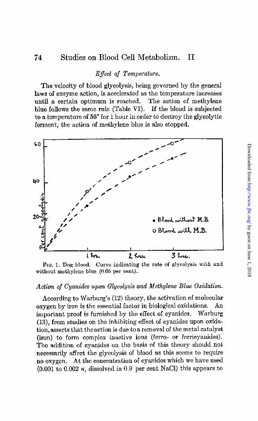
Lepine was found. Table V presents the data from an experiment with dog blood while Fig. 1 represents the glycolytic curve in the control sample and in the sample containing methylene blue. The curves are similar, the chief difference being that methylene blue has accelerated the process. The lactic acid production in the control blood increases in proportion to the glycolysis as may be seen from the glycolytic quotients. In the sample containing methylene blue the glycolytic quotient is reduced.
by guest on June 1, 2018http://w
ww
.jbc.org/D
ownloaded from
74 Studies on Blood Cell Metabolism. II
E$ect of Temperature.
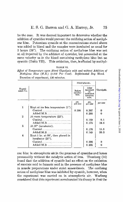
The velocity of blood glycolysis, being governed by the general laws of enzyme action, is accelerated as the temperature increases until a certain optimum is reached. The action of methylene blue follows the same rule (Table VI). If the blood is subjected to a temperature of 56” for 1 hour in order to destroy the glycolytic ferment, the action of methylene blue is also stopped.
Fro. 1. Dog blood. Curve indicating the rate of glycolysis with and without methylene blue (0.05 per cent).
Action of Cyanides upon Glycolysis and Methylene Blue Oxidation.
According to Warburg’s (12) theory, the activation of molecular oxygen by iron is the essential factor in biological oxidations. An important proof is furnished by the effect of cyanides. Warburg (13), from studies on the inhibiting effect of cyanides upon oxida- tion, asserts that theaction is due toa removal of the metal catalyst (iron) to form complex inactive ions (ferro- or ferricyanides). The addition of cyanides on the basis of this theory should not necessarily affect the glycolysis of blood as this seems to require no oxygen. At the concentration of cyanides which we have used (0.001 to 0.002 N, dissolved in 0.9 per cent NaCl) this appears to
by guest on June 1, 2018http://w
ww
.jbc.org/D
ownloaded from
E. S. G. Barron and G. A. Harrop, Jr. 75
be the case. It was deemed important to determine whether the addition of cyanides would prevent the oxidizing action of methyl- ene blue. Potassium cyanide at the concentrations stated above was added to blood and the samples were incubated as usual for 3 hours (38”). The oxidizing action of methylene blue was not at all impaired by the addition of cyanides, but proceeded at the same velocity as in the blood containing methylene blue but no cyanide (Table VII). This oxidation, then, is effected by methyl-
TABLE VI.
Effect of Temperature upon Blood Glycolysis with and without Addition of Methylene Blue (M.B.) (0.005 Per Cent). DeJibtinated Dog Blood,
Duration of experiment, 150 minutes.
Experi- ment No.
-
- - gm.
per cent per cent
Kept at ice box temperature (1”). Control.. . . . . . . . . . . . . . . . . . . . 0.208 0 Added M.B. . . . . . . . . . . . . . . . . 0
At room temperature (22”).
0.207 0.208
Control........................ Added M.B.. . . . . . . . . .
At 37” (incubator). Control........................ Added M.B.. . . . . . . . . .
Kept 1 hr. at 56”, then placed in incubator (37”). Control. . . . . . . . . . . . . . . . . . Added M.B.. . . . . . . . . . . . . . . .
-
0.189 0.173
0.176 0.145
0.206 0.206
9.1 16.3
15.5 30.3
0 0
Blood glucose.
Before inruba-
tion.
After exposure
ia indicated teIllpeB,-
ture.
:1yco1yeia
ene blue in atmospheric air in the presence of cyanides and hence presumably without the catalytic action of iron. Thunberg (14) found that the addition of cyanide had no effect on the oxidation of succinic acid to fumaric acid in the presence of methylene blue in muscle preparations under strict anaerobiosis. The oxidizing action of methylene blue was inhibited by cyanide, however, when the experiment was carried on in atmospheric air. Warburg considered that this experiment corroborated his theory in that the
by guest on June 1, 2018http://w
ww
.jbc.org/D
ownloaded from

76 Studies on Blood Cell Metabolism. II
succinic acid and washed muscle in the presence of cyanide form a system which does not take up oxygen. In the glycolysis of blood, however, the oxidation of some intermediary carbohydrate seems to occur with methylene blue in the presence of air without the aid of the iron catalyst. The fact that the oxidizing action of methylene blue upon glucose is not disturbed by the presence of cyanides is in accordance with the lack of influence of cyanides upon methylene blue oxidation noted in the preceding paper.
TABLE VII.
Action of Potassium Cyanide upon Blood Glycolysis and Lactic Acid Forma- tion, with and without Methylene Blue (M.B.) (0.005 Per Cent).
Dejibinated Human Blood. Incubation at SY” for 3 Hours.
Blood glucose, InM per 1. - -
d .z 2 2 .5 3 2
81 .3 1 .2 1 .3
1 .3 1 .I3
1 .3 - -
Control. . . . . . . . . . 3.31 Added MB..
‘I 0.001 N KCN.. “ 0.001 “ “ +
M.B.. _. . . , . Added 0.002 N KCN.
“ 0.002 “ “ + M.B., . . _.
Lactic acid, InM per 1.
- I
f
:: F4
-
52 !.9! 21 ..71 13 1.9(
a1 ..61 3: 1.1:
11 ..6( - -
pz.
0.92
1.17
1.21
0.72 0.40 0.98
0.40 0.78
0.40
Action of Phosphates.
The effect of methylene blue upon blood glycolysis in the pres- ence of added phosphate was studied. Two kinds of experiments were performed. In one Sorensen’s phosphate mixture (pH 7.4) and glucose (d-glucose Pfanstiehl) were added to blood prepared in the usual way, and the glycolysis and lactic acid formation were determined after 3 hours incubation. In the other, phosphates alone were added and the sugar decrease and lactic acid formation were compared in the samples with and without phosphates and
by guest on June 1, 2018http://w
ww
.jbc.org/D
ownloaded from
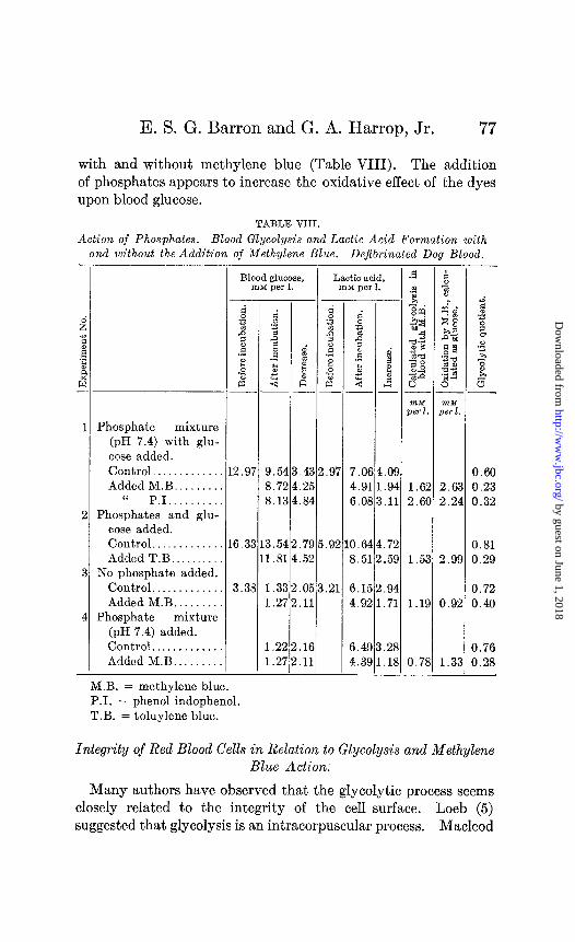
E. S. G. Barron and G. A. Harrop, Jr. 77
with and without methylene blue (Table VIII). The addition of phosphates appears to increase the oxidative effect of the dyes upon blood glucose.
TABLE VIII.
Action of Phosphates. Blood Glycolysis and Lactic Acid Formation with and without the Addition of Methylene Blue. Dejibrinated Dog Blood. -
I
Phosphate mixture (pH 7.4) with glu- cose added. Control. . . . . Added M.B. . . . . .
‘I P.I.. . . . Phosphates and glu-
cose added. Control. . . Added T.B..
No phosphate added. Control. . . . . . . Added M.B. .
Phosphate mixture (pH 7.4) added. Control.. . . . . . . . . . Added M.B. . . .
Blood glucose, rnM per 1.
Lactic acid, rnM per 1.
-
--
j.54 3.72 3.13
.43 2.97 7.0f
.25 4.91
.84 6.OE
6.3 3.54 1.81
.79 5.92 0.64 .52 8.51
8 1 1
3.3
-
1.33 1.27
.05 3.21 6.1:
.11 4.92
I 1
-
1.22 .16 1.27 .11
6.4s 4.3E -
-
f t z -E
L.O! 1.92 i.l:
1.7: !.5!
Z.9f 1.73
1.2j ..lI -
1.6; 2.6I
1.5:
1.1s
0.78
0.60 2.63 0.23 2.24 0.32
0.81 2.99 0.29
0.72 0.92 0.40
0.76 1.33 0.28
M.B. = methylene blue. PI. = phenol indophenol. T.B. = toluylene blue.
Integrity of Red Blood Cells in Relation to Glycolysis and Methylene Blue Action.
Many authors have observed that the glycolytic process seems closely related to the integrity of the cell surface. Loeb (5) suggested that glycolysis is an intracorpuscular process. Macleod
by guest on June 1, 2018http://w
ww
.jbc.org/D
ownloaded from

78 Studies on Blood Cell Metabolism. II
(15) repeated the suggestion, which correlates his observation that glycolysis is diminished in blood cells washed with saline solution, with the observations of Rona and Doblin (16) that washing ren- dered the corpuscles incapable of absorbing sugar. We have repeated Macleod’s (15) experiments in order to compare glycolysis under such conditions with the oxidative action of methylene blue. The red cells were washed with Locke’s mammalian solution instead of saline. It was found that washing of the blood cells decidedly impairs both normal glycolysis and methylene blue action (Table IX).
TABLE IX.
E$ect of Alteration or Destruction of Erythrocyte Surface upon Glycolysis with and without Methylene Blue (M.B.). (Washing of Red Blood Cells
and Hemolysis.)
lhpri- merit No.
Blood glucose, InM p*r 1.
- Before
incuba- tion.
After in- cubation (3 hrs.).
Xycolysis.
Control blood.. . . . . . . . . . . . . . . . . 6.39 Added M.B.. . . . . . . . . . . . . .
Blood washed twice with Locke’s solution. . . . . . . . . . . . . . . 11 .oo Added M.B.. . . . . .
Control blood.. . . . . . . . . . 5.55 Added M.B... . . . . . . . . . .
Washed blood cells. _. . ., . . . . 5.05 Added M.B.. . . . . . . . . . .
Hemolysis.
- per cent
5.00 21.7 4.36 31.7
10.10 8.2 9.45 14.0 2.00 64.0 1.55 72.0 3.33 34.0 2.27 55.0
Ox blood hemolyzed with water, control......................... 10.00 Added M.B.. . . . . . .
10.00 9.94
0 0
Rona and Arnheim (4) in 1913 showed that hemolysis with distilled water stops glycolysis. These authors hemolyzed the blood with large amounts of water (20 cc. of blood were mixed with 180 cc. of water). We have compared the glycolysis in nor- ma1 human blood with that of blood hemolyzed in two ways: (a) repeated freezing and thawing, and (b) hemolysis with distilled water. In the latter case the serum was removed by strong cen- trifugation and then replaced by an equivalent amount of water.
by guest on June 1, 2018http://w
ww
.jbc.org/D
ownloaded from

E. S. G. Barron and G. A. Harrop, Jr. 79
Glycolysis was impaired in the hemolyzed samples, but not stopped, as the following example shows:
Glycolysia. Human blood. per cent
(a) Glycolysis after 3 hrs., 37”. Control..................... 52 (b) Hemolysis by freezing and thawing.. . . . . . . . . . . . . . . . . . . 36 (c) Hemolysis with distilled water.. . . . . . . . . . . . . . . . . . . . . . . . . . . 18
In order to observe the action of methylene blue in a sample of blood in which glycolysis was entirely lacking, ox blood was employed. Here (Table IX, Experiment 3) hemolysis with dis- tilled water stopped glycolysis, and methylene blue as well had no action.
TABLE X.
Effect of Hemolysis by Freezing and Thawing upon DeJibrinated Goose Blood.
Glnrose, IKIM per 1.
Before After incuba- inouba-
tion. tion.
Sample A, clear super- Control. 5.25 5.22 natant fluid. Added methylene blue. 5.25
Sample B, thick sus- Control. 4.25 3.85 pension from bot- Added methylene blue. 3.33 tom.
Glycol- ysis.
per cent
0 0
9.4 21.6
Warburg (17) in his studies on the respiration of goose erythro- cytes produced hemolysis by repeated freezing and thawing. After prolonged centrifuging of this cytolyzed cell suspension at high speed two layers were formed-the upper containing the hemoglobin and fluid protoplasm, and the lower the solid cell particles, chiefly the nuclei. Respiration took place entirely in this lower layer. We have used Warburg’s method to study glycolysis and methylene blue action in anemic goose blood hemo- lyzed by freezing and thawing. The clear supernatant fluid showed neither glycolysis nor oxidation by methylene blue, while the thick bottom layer showed both normal glycolysis and an in- crease of this process in the presence of methylene blue. The results of one such experiment are shown in Table X.
by guest on June 1, 2018http://w
ww
.jbc.org/D
ownloaded from

80 Studies on Blood Cell Metabolism. II
Glycolysis and the action of methylene blue therefore seem to be intimately connected with the integrity of the cell structures. When the surface is damaged or destroyed the glycolytic process is slowed or ceases altogether, and with it, the oxidizing action of methylene blue.
Action of Methylene Blue upon Blood Serum and Glucose Solutions.
It has been known for a long time that glucose in alkaline solu- tion can be oxidized by methylene blue. Spoehr (18) observed the
TABLE XI.
Action of Methylene Blue upon Glucose Solutions in Sfrensen’s Phosphate Mixtures (~/15) at pH 7.40 after Different Times of
Incubation (37”).
4 hrs. incubation.. . . . . . 6 “ “ . . . . . . . . . . .
12 “ “ .
Glucose, nxv per 1.
Observations. Before After
incubation. incubation.
9.26 9.23 No oxidation. 8.26 8.26 “ “
10.91 10.94 “ lL
TABLE XII.
Action of Methylene Blue upon Glucose and Lactic Acid Content of Blood Serum. Incubation at 37” for 3 Hours.
Glucose, rn~ per 1.
Control.. . . . . . . . . . . . . . . . . . . . . . . 8.77 8.72 0 Added methylene blue.. . . . 8.77 0
Lactic acid, nm per 1.
oxidation of sugar solutions with methylene blue in the presence of disodium phosphate in a continuous stream of air. The catalysis with methylene blue alone occurred only in alkaline solution, disodium phosphate being sufficiently alkaline for this purpose. It could be increased threefold when iron was added, which led him to assume that iron acted as a catalyst. The process described in the present paper is not related to Spoehr’s observa- tions as is indicated by the following experiments. Sorensen’s phosphate mixture at pH 7.4, containing glucose, was incubated
by guest on June 1, 2018http://w
ww
.jbc.org/D
ownloaded from

E. S. G. Barron and G. A. Harrop, Jr. 81
at 37” for different lengths of time (4,6, and 12 hours), methylene blue being added in the same concentration as was used in the experiments with blood. The amount of glucose after incubation was unchanged (see Table XI).
The action of methylene blue on serum containing glucose and lactic acid was then examined. Samples of serum were in- cubated for 3 hours at 37” with and without methylene blue. The glucose and the lactic acid remained unchanged (see Table XII). We conclude that methylene blue has no action upon glucose or lactic acid in the absence of the cells under these conditions.
Effect of Anaerobiosis in Relation to Glycolysis and Methylene Blue Action.
All of the experiments which are reported above have been performed under aerobic conditions. Owing to the method employed for defibrinating the blood and freeing it from leucocytes the material was completely saturated with oxygen just prior to its incubation. In the experiments in which dyes were added, it is true, the oxygen content of the material fell to a low level at the end of the experiment, owing to the increased oxygen con- sumption of the erythrocytes under such conditions, as was previously reported (1). In the following experiments, a com- parison of glycolysis and lactic acid formation was attempted between completely reduced blood and blood which was kept com- pletely and continuously oxygenated. A sample of blood was divided into four flasks, which were arranged in pairs connected with tubing in the thermostat at 38”. In one flask of each set methylene blue was added (0.005 per cent). The stoppers were sealed and a continuous slow stream of nitrogen was passed through one pair of flasks, while oxygen was passed through the other. The flasks were continuously agitated in a shaking device for 3 hours, at the end of which period the glucose and lactic acid contents were redetermined. (Table XIII.)
A constant stream of oxygen passing through blood undergoing glycolysis does not appear to affect this process appreciably as no difference was found between the samples exposed to the gas stream and control samples kept in stoppered flasks. On the other hand a stream of nitrogen which produced relative anaerobiosis increased glycolysis as well as lactic acid formation. In these
by guest on June 1, 2018http://w
ww
.jbc.org/D
ownloaded from

82 Studies on Blood Cell Metabolism. II
TABLE XIII.
Action of Methylene Blue (M.B.) upon Blood Glycolysis and Lactic Acid Formation in Oxygenated and Reduced Blood. Incubation at 38”
for 3 Hours.
Goose blood. Oxygenated blood.
Control. ............ Added M.B. ........
Reduced blood. Control. ............ Added M.B .........
Oxygenated blood. Control. ............. Added M.B. ........
Reduced blood. Control. ............ Added M.B. ........
Oxygenated blood. Control. ............ Added M.B .........
Reduced blood. Control ............. Added M.B .........
Human blood. Oxygenated blood.
Control ............ Added M.B. ........
Reduced blood. Control ............ Added M.B .........
Dog blood. Reduced blood.
Control ............. Added M.B. ........
3.486.112.37 i.87 7.07 1.2( 5.343.14 6.52O.Q
4.27 4.21 11.365.45 4.214.27 10.684.81
3.72 6.612.11 ‘.44 8.340.9( 4.963.76 8.030.5:
4.903.82 12.004.5( 4.87 3.8: 11.72 4.25
7.556.001.5; 4..20 3.3:
L.66 5.370.71 4.66
4.45 3.X 3.83 3.72
8.43 3.7; 9.12 4.5t
5.17 3.962.21 !.70 7.104.4( 2.783.39 5.853.X
3.612.56 7.805.N 3.17 3.00 7.805.1C
3.166.162.00 !.78 6.22 3.4 5.802.36 6.453.65
Lactic acid, mx per 1.
.-
) i
) )
L
-
1.8f j
0.5; r
2.3f 3
5
5
0.2:
3.3:
1.81
0.44
0.22
I.26 I.10
1.65 1.55
I.21 1.08
1.60 I.55
I.23
I.61 I.61
1.00 I.46
1.00 I.85
I.85 I.77
by guest on June 1, 2018http://w
ww
.jbc.org/D
ownloaded from

E. S. G. Barron and G. A. Harrop, Jr. 83
experiments it will be seen that the glycolytic quotient in the re- duced blood, although higher than in the oxygenated blood, never reached unity, a fact which indicates that some oxidative process was going on while the blood was being reduced. It is known that muscle tissue in the presence of oxygen does not show a marked increase in lactic acid as this substance is in part oxidized and in part resynthesized to glycogen; under anaerobic conditions the amount of glucose destroyed corresponds to the lactic acid increase. In the present experiments the lactic acid formed in oxygenated goose blood is so low as to bring the glycolytic quotient down to 0.20, while in reduced blood (relative anaerobiosis) there is a rise of the lactic acid content which brings the glycolytic quotient up to 0.50 or 0.60. The similarity to the mechanism observed in muscle is evident.
Methylene blue, which produced its usual effect in the presence of oxygen, showed scarcely any oxidizing action upon the reduced blood. In one instance (Experiment 3, Table XIII) where nearly one-half of the glucose was oxidized in the presence of oxygen (no lactic acid increase), none was oxidized in the blood reduced by the nitrogen stream. It seems probable that all of the experiments would have shown similar results had strict anaerobiosis been attained. It is evident however that such a condition was not attained because the nitrogen used, while of high purity, contained on analysis traces of oxygen, which were also found in the blood on analysis at the end of the experiments.
In similar experiments with mammalian blood (Experiments 4 and 5, Table XIII) the flasks containing the samples were kept for 1 hour at 38” in a shaking device in a stream of pure nitrogen.b When the color of the blood indicated complete reduction, methyl- ene blue was added. The flasks were then maintained under the same conditions for 2 or 3 hours. Even with these precautions oxidation by methylene blue was not completely stopped, but it was extremely small.
The failure of methyIene blue to produce oxidation in blood undergoing glycolysis in the absence of oxygen is of some signifi- cance. A strong argument in favor of Wieland’s theory is
6 The highly purified nitrogen used in these experiments was prepared in an apparatus to be described shortly by Professor Michaelis to whose kindness we are indebted for the use of this material.
by guest on June 1, 2018http://w
ww
.jbc.org/D
ownloaded from

84 Studies on Blood Cell Metabolism. II
based upon Thunberg’s (14) observation that methylene blue oxidizes succinic acid under anaerobic conditions. The presence of the oxyhemoglobin makes strict anaerobiosis difficult in material containing erythrocytes. The question is being studied with reference to the glycolysis produced by leucocytes. Of interest in relation to the effect of methylene blue upon erythrocyte oxida- tion under anaerobic conditions is the recent communication of Baumberger (19), who found that methylene blue at a concentra- tion of 1 to l,OOO,OOO had a marked effect in postponing the onset of fatigue in frog muscle under anaerobic conditions. The oxygen equivalent of the methylene blue was far too small to account for the increased output of work produced. The changes in the lactic acid produced were not reported.
DISCUSSION.
It has been shown above that minute quantities of methylene blue in isotonic saline solution, when added to blood undergoing glycolysis, alter the carbohydrate metabolism in such a way that the velocity of sugar transformation is increased, while at the same time less lactic acid is formed. Coincident with this shift in the path of the metabolism there is a marked increase in oxygen con- sumption and carbon dioxide production, as shown in Paper I. We suggest that methylene blue produces its effect so far as car- bohydrate is concerned by increasing the oxidation of some degra- dation product of glucose. This effect is not produced in blood in which normal glycolysis is absent, nor does it occur either in blood serum or in glucose solutions at pH 7.4. It is therefore not analo- gous to the oxidation of glucose by methylene blue in alkaline solutions as reported by other observers.
The point in the glycolytic process at which methylene blue exerts its action cannot be determined from our data. The chain of transformations which has been suggested by Shaffer (20) is well known: glucose ---f hexosephosphate --$ glyceric aldehyde + methylglyoxal + lactic acid.
Of these steps, the transformation of glucose to hexosephosphate, which does not require the presence of oxygen, and the splitting of glyceric aldehyde and methylglyoxal, which are transformed at once to lactic acid, seem to be excluded. Methylene blue has no effect on the oxidation of lactic acid in serum in which there is no
by guest on June 1, 2018http://w
ww
.jbc.org/D
ownloaded from

E. S. G. Barron and G. A. Harrop, Jr. 85
glycolysis. Where glycolysia does not occur in avian blood the addition of methylene blue produces no change in the concentra- tion of lactic acid. Ftirth and Lieben (21), however, have shown that lactic acid is consumed by horse erythrocytes, and Ray (22) working, it is true, at the unphysiological pH value of 6.9 has demonstrated the oxidation of lactic acid by dog erythrocytes. One cannot exclude the possibility that the observed methylene blue effect, at least in part, is due to oxidation of lactic acid.
In favor of the possibility that the principal point at which the methylene blue acts is upon the oxidation of hexosephosphate, is the fact that the action of methylene blue is increased by increasing the inorganic phosphate concentration, either alone, or together with added glucose. As to the exact nature of the methylene blue effect little may be said. It is conceivable that it acts as a coenzyme or catalyst, rendering the substrate (hexosephosphate?) more sensitive to the action of molecular oxygen. On the other hand one might consider that methylene blue plays in this system the role ascribed to iron in the oxidations produced by Warburg with his charcoal model.
CONCLUSIONS.
The relationship between glycolysis and the oxidative processes in blood cells may be expressed by the ratio
Millimols of lactic acid increase 2 X millimols glucose decrease
which is termed the glycolytic quotient. Normal adult mammalian red blood cells with almost no oxida-
tive processes show a high glycolytic quotient. Nucleated erythro- cytes (avian blood cells) with appreciable oxidative processes have a low glycolytic quotient.
The addition of methylene blue (or dyes with similar oxidation- reduction potentials) to the blood undergoing glycolysis produces an increased sugar degradation and a diminished formation of lactic acid. This is probably due in large part to the oxidation of some intermediary product. The glycolytic quotient is lowered.
The process begins only after the glucose molecule is acted upon by the glycolytic enzyme and is converted into other unstable and oxidizable substances. It is suggested that oxidation may
by guest on June 1, 2018http://w
ww
.jbc.org/D
ownloaded from
86 Studies on Blood Cell Metabolism. II
take place in the early stages of dissociation, before the glucose molecule is split into three carbon chain fragments, probably when the glucose molecule is esterified to hexosediphosphoric acid.
This reaction probably requires the presence of oxygen but the addition of cyanides does not impair the oxidative process. It is suggested that methylene blue acts as a catalyst, rendering the hexosephosphate molecule more sensitive to oxidation by molecu- lar oxygen.
Our thanks are due to Professor Michaelis for his suggestions concerning a number of questions which have arisen in the course of this study.
BIBLIOGRAPHY.
1. Harrop, G. A., Jr., and Bsrron, E. S. G., Studies on blood cell metab- olism. I. The effect of methylene blue and other dyes upon the oxygen consumption of mammalian and avian erythrocytes, J. Exp. Med., 1928, xlviii, 207.
2. Lepine, Les sucres du sang, Compt. rend. Acad., 1890, cxii, 65. 3. Levene, P. A., and Meyer, G. M., The action of leucocytes on glucose,
J. Biol. Chem., 1912, xi, 361. 4. Rona, P., and Amheim, F., BeitrSlge zur Frage der Glykolyse. III,
Biochem. Z., 1913, xlviii, 35. 5. Loeb, A., Beziehungen zwischen Zuckergehalt der Erythrocyten und
Glykolyse, Biochem. Z., 1913, xlix, 413. 6. Melvin, G. S., On glycolysis in blood, Biochem. J., 1912, vi, 422. 7. Riiter, E., giber Glykolyse und MilchsSiurebildung im Vogelblut, 2.
ges. exp. Med., 1923, xxxvii, 1.51. 8. Bornstein, A., and Ascher, O., uber Glykolyse im Vogelblut unter dem
Einfluss oxydationshemmender Gift.e, 2. ges. exp. Med., 1926, lii, 607. 9. Clark, W. M., et al., Studies on oxidation-reduction. Papers I to XI,
Pub. Health Rep., U. S. P. H. S., 1923-27. 10. Lepine, Glycolyse du sucre du sang, J. physiol. et path. gkn., 1918,
xvii, 555. 11. Irving, J. T., Degradation of glucose by the blood corpuscle of the
rabbit, Biochem. J., 1926, xx, 613, 1320. 12. Warburg, O., uber Eisen den sauerstofftibertragenden Bestandteil des
Atmungsferments, Biochem. Z., 1924, clii, 479. 13. Warburg, O., Physikalische Chemie der Zellatmung, Biochem. Z.,
1921, cxix, 134; ‘iiber die antikatilytische Wirkung der Blausaiire, ibid., 1923, cxxvi, 266.
14. Thunberg, T., Zur Kenntnis der vitalen Bernsteins&ureoxydation, Skand. Arch. Physiol., 1916, xxxiii, 233; iiber die vital Dehydrierung der Bernsteinsgure bei Abwesenheit von Sauerstoff, Zentr. Physiol., 1917, xxxi, 91.
by guest on June 1, 2018http://w
ww
.jbc.org/D
ownloaded from
E. S. G. Barron and G. A. Harrop, Jr.
15. Macleod, J. J. R., Blood glycolysis: its extent and significance in carbo- hydrate metabolism. The supposed existence of “sucre virtuel” in freshly drawn blood, J. Biol. Chem., 1913, xv, 497.
16. Rona, P., and Doblin, A., Beitrage zur Frage der Glykolyse. II, Biochem. Z., 1911, xxxii, 489.
17. Warburg, O., tfber Beeinflussung der Sauerstoffatmung, Z. physiol. Chem., 1911, lxx, 413; Beitriige zur Physiologie der Zelle, Ergebn. Physiol., 1914, xiv, 253.
18. Spoehr, H. A., The oxidation of carbohydrates with air, J. Am. Chem. Sot., 1924, xlvi, 1494.
19. Baumberger, J. P., The effect of methylene blue on the anaerobic con- traction of muscle, Skand. Arch. Physiol., 1926, xlix, 88.
20. Shaffer, P. A., Intermediary metabolism of carbohydrates, Physiol. Rev., 1923, iii, 395.
21. Fiirth, O., and Lieben, F., uber Milchsliurezerstorung durch Hefe und durch Blutzellen, Biochem. Z., 1922, cxxviii, 144.
22. Ray, G. B., Oxidation of sodium lactate by red blood cells, Am. J. Physiol., 1927, Ixxxii, 465.
by guest on June 1, 2018http://w
ww
.jbc.org/D
ownloaded from

Jr.E. S. Guzman Barron and George A. Harrop,
AND AVIAN ERYTHROCYTESACID FORMATION OF MAMMALIAN
UPON THE GLYCOLYSIS AND LACTIC METHYLENE BLUE AND OTHER DYES
METABOLISM: II. THE EFFECT OF STUDIES ON BLOOD CELL
1928, 79:65-87.J. Biol. Chem.
http://www.jbc.org/content/79/1/65.citation
Access the most updated version of this article at
Alerts:
When a correction for this article is posted•
When this article is cited•
to choose from all of JBC's e-mail alertsClick here
#ref-list-1
http://www.jbc.org/content/79/1/65.citation.full.htmlaccessed free atThis article cites 0 references, 0 of which can be
by guest on June 1, 2018http://w
ww
.jbc.org/D
ownloaded from