students corner case report
TRANSCRIPT

AbstractParathyroid carcinoma is a rare malignant neoplasm ofthe parathyroid glands which results in enlargement andexcessive production of parathyroid hormone (PTH)responsible for pathologically raising calcium levels in theblood resulting in bone pain/fractures, renal stones andother signs of hypercalcaemia.
A 37 year old woman presented with sudden,spontaneous bone pain and fracture of the right femoralshaft. This unusual presentation was explained byextremely high PTH levels and hypercalcaemia in theblood and a hard, solitary mass palpable in the neck.During surgical excision of this mass, finding of severaladhesions, possible capsular invasion and lymph nodeenlargement led to a diagnosis of parathyroid carcinoma.
The neoplasm proliferates via adenoma-carcinomasequence so early diagnosis and prompt surgical excisionwith post-operative care may provide palliation and keeprecurrences in check. Pancreas and pituitary evaluation isalso necessary as this presentation may be a part ofWermer (MEN-1) syndrome.
Keywords: Parathyroid, Carcinoma, Fractures, Neoplasia,Brown's tumour.
IntroductionParathyroid cancer forms in one or more of theparathyroid glands and is one of the rarest of all humancancers. Its estimated incidence is 0.015 per 100,000people and estimated prevalence is 0.005% in the UnitedStates.1,2
Parathyroid carcinoma is the rarest cause (<1%) ofprimary hyperparathyroidism, the most common onebeing parathyroid adenoma (85-95%) followed byparathyroid hyperplasia (5-10%). Patients present withmarked hypercalcaemia and hyperparathyroidism. Raised
levels of calcium and parathyroid hormone (PTH) are theprimary cause for the morbidity and mortality associatedwith parathyroid cancer and not the infiltration of tissuesby malignant cells. Parathyroid cancer typically growsslowly and has a rather low malignant potential. It is morelikely to invade local structures (thyroid, strap muscles,esophagus, trachea, recurrent laryngeal nerve) than tometastasize to the lymph nodes.
Patients of parathyroid carcinoma usually have palpableneck masses as opposed to parathyroid adenomas whichare normally not palpable. The clinical features mayinclude: fragile bones that fracture easily (osteoporosis),kidney stones, polyuria, abdominal pain, easy fatigability,depression, arthralgia, nausea, vomiting, loss of appetite,and frequent complaints of illness with no apparentcause.3 Patients of parathyroid carcinoma have a muchhigher occurrence of bone disease than patients ofparathyroid adenoma.4
If the patient has any of the following clinical findings,parathyroid carcinoma should be suspected:1,4
u Severe hypercalcaemia: > 14 mg/dl.
u Serum PTH level: > twice that of normal.
u Cervical mass palpated in a hypercalcaemic patient.
u Hypercalcaemia with unilateral vocal cord paralysis.
u Simultaneous renal and skeletal disease in a patient withprimary hyperparathyroidism.
Surgically, parathyroid cancers may be differentiated fromadenomas by their hard feel and lobulation; adenomas areusually soft, round/ oval in shape and reddish-brown incolour.5 In most cases, parathyroid cancers have a maximumdiameter of 3.0-3.5 cm compared with approximately 1.5 cmfor adenomas.1 In about 50% of the patients, the carcinoma issurrounded by a dense, fibrous capsule that penetratessurrounding tissues. Histopathologically, it is difficult todifferentiate benign from malignant parathyroid tumours.1,5,6
Intraoperatively, when parathyroid carcinoma is
Vol. 67, No. 12, December 2017
1956
STUDENTS CORNERCASE REPORT
Pathological bone fractures in a patient with parathyroid carcinoma — A CaseReportAzib Shahid, Fatima Iftikhar
Services Institute of Medical Sciences, University of Health Sciences, Lahore,Pakistan.Correspondence: Azib Shahid. Email: [email protected]
suspected based on severity or invasion of surroundingtissues by a firm parathyroid tumour, aggressive excisionis performed, including the thyroid and surroundingtissues as necessary.7
Tumour recurrence occurs in nearly 40-60% of patients,usually within 2 to 5 years after the initial resection.8,9
Certain conditions appear to increase the risk of parathyroidcancer, including: multiple endocrine neoplasia type 1,autosomal dominant familial isolated hyperparathyroidismand hyperparathyroidism-jaw tumour syndrome. Parathyroidcancer has also been associated with external radiationexposure but most reports describe an association betweenradiation and the more common parathyroid adenoma.4
Case PresentationA 37 year old, rather emaciated, female presented at theemergency of services hospital, Lahore in July 2016 with ahistory of a road traffic accident and pain in the left thigh. Shegot a radiograph done which showed no fractures at thattime. She was discharged on analgesics, and advised forrestricting movements of left lower limb. However, mild painpersisted and 2 months later she presented again withsuddenly developed severe pain, swelling and deformity ofthe same thigh and inability to move the left lower limb.Radiograph showed complete spiral fracture of the upper
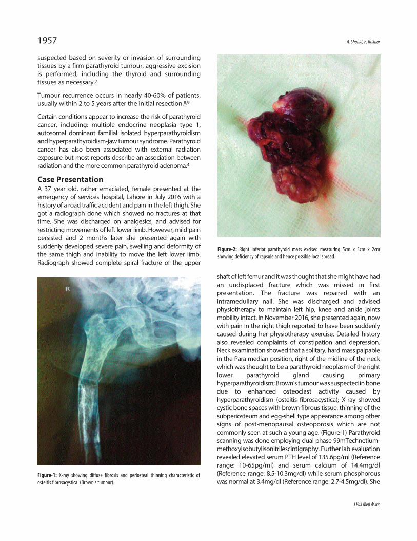
shaft of left femur and it was thought that she might have hadan undisplaced fracture which was missed in firstpresentation. The fracture was repaired with anintramedullary nail. She was discharged and advisedphysiotherapy to maintain left hip, knee and ankle jointsmobility intact. In November 2016, she presented again, nowwith pain in the right thigh reported to have been suddenlycaused during her physiotherapy exercise. Detailed historyalso revealed complaints of constipation and depression.Neck examination showed that a solitary, hard mass palpablein the Para median position, right of the midline of the neckwhich was thought to be a parathyroid neoplasm of the rightlower parathyroid gland causing primaryhyperparathyroidism; Brown's tumour was suspected in bonedue to enhanced osteoclast activity caused byhyperparathyroidism (osteitis fibrosacystica); X-ray showedcystic bone spaces with brown fibrous tissue, thinning of thesubperiosteum and egg-shell type appearance among othersigns of post-menopausal osteoporosis which are notcommonly seen at such a young age. (Figure-1) Parathyroidscanning was done employing dual phase 99mTechnetium-methoxyisobutylisonitrilescintigraphy. Further lab evaluationrevealed elevated serum PTH level of 135.6pg/ml (Referencerange: 10-65pg/ml) and serum calcium of 14.4mg/dl(Reference range: 8.5-10.3mg/dl) while serum phosphorouswas normal at 3.4mg/dl (Reference range: 2.7-4.5mg/dl). She
J Pak Med Assoc
1957 A. Shahid, F. Iftikhar
Figure-1: X-ray showing diffuse fibrosis and periosteal thinning characteristic ofosteitis fibrosacystica. (Brown's tumour).
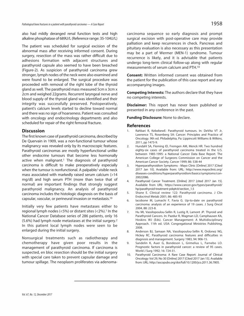
Figure-2: Right inferior parathyroid mass excised measuring 5cm x 3cm x 2cmshowing deficiency of capsule and hence possible local spread.
also had mildly deranged renal function tests and highalkaline phosphatase of 689U/L (Reference range: 35-104U/L)
The patient was scheduled for surgical excision of theabnormal mass after receiving informed consent. Duringsurgery, resection of the mass was rather difficult due toadhesions formation with adjacent structures andparathyroid capsule also seemed to have been breached(Figure-2). As suspicion of parathyroid carcinoma grewstronger, lymph nodes of the neck were also examined andwere found to be enlarged. The surgical procedure wasproceeded with removal of the right lobe of the thyroidgland as well. The parathyroid mass measured 5cm x 3cm x2cm and weighed 22grams. Recurrent laryngeal nerve andblood supply of the thyroid gland was identified and theirintegrity was successfully preserved. Postoperatively,patient's calcium levels started to decline toward normaland there was no sign of hoarseness. Patient was consultedwith oncology and endocrinology departments and alsoscheduled for repair of the right femoral fracture.
DiscussionThe first known case of parathyroid carcinoma, described byDe Quervain in 1909, was a non-functional tumour whosemalignancy was revealed only by its macroscopic features.Parathyroid carcinomas are mostly hyperfunctional unlikeother endocrine tumours that become less hormonallyactive when malignant.1 The diagnosis of parathyroidcarcinoma is difficult to make preoperatively especiallywhen the tumour is nonfunctional. A palpable/ visible neckmass associated with markedly raised serum calcium (>14mg/dl) and high serum PTH (more than twice that ofnormal) are important findings that strongly suggestparathyroid malignancy. An analysis of parathyroidcarcinoma includes the histologic diagnosis on the basis ofcapsular, vascular, or perineural invasion or metastasis.10
Initially very few patients have metastases either toregional lymph nodes (<5%) or distant sites (<2%).1 In theNational Cancer Database series of 286 patients, only 16(5.6%) had lymph node metastases at the initial surgery.2In this patient local lymph nodes were seen to beenlarged during the initial surgery.
Nonsurgical treatments such as radiotherapy andchemotherapy have given poor results in themanagement of parathyroid carcinoma. If carcinoma issuspected, en bloc resection should be the initial surgerywith special care taken to prevent capsular damage andtumour spillage. The neoplasm proliferates via adenoma-
carcinoma sequence so early diagnosis and promptsurgical excision with post-operative care may providepalliation and keep recurrences in check. Pancreas andpituitary evaluation is also necessary as this presentationmay be a part of Wermer (MEN-1) syndrome. Tumourrecurrence is likely, and it is advisable that patientsundergo long-term clinical follow-up along with regularmeasurements of serum calcium and PTH.10
Consent: Written informed consent was obtained fromthe patient for the publication of this case report and anyaccompanying images.
Competing Interests: The authors declare that they haveno competing interests.
Disclaimer: This report has never been published orpresented in any conference in the past.
Funding Disclosure: None to declare.
References1. Rahbari R, KebebewE: Parathyroid tumours. In: DeVita VT Jr,
Lawrence TS, Rosenberg SA: Cancer: Principles and Practice ofOncology. 9th ed. Philadelphia, Pa: Lippincott Williams & Wilkins,2011, pp 1473-9.
2. Hundahl SA, Fleming ID, Fremgen AM, Menck HR. Two hundredeighty-six cases of parathyroid carcinoma treated in the U.S.between 1985-1995: a National Cancer Data Base Report. TheAmerican College of Surgeons Commission on Cancer and theAmerican Cancer Society. Cancer 1999; 86: 538-44
3. Hyperparathyroidism Symptoms - Mayo Clinic [Online] 2017 [Cited2017 Jan 10]. Available from: URL: http://www.mayoclinic.org/diseases-conditions/hyperparathyroidism/basics/symptoms/con-20022086.
4. Parathyroid Cancer Treatment. [Online] 2017 [cited 2017 Jan 15].Available from URL: https://www.cancer.gov/types/parathyroid/hp/parathyroid-treatment-pdq#cit/section_1.5
5. Shane E. Clinical review 122: Parathyroid carcinoma. J ClinEndocrinol Metab 2001; 86: 485-93
6. Iacobone M, Lumachi F, Favia G. Up-to-date on parathyroidcarcinoma: analysis of an experience of 19 cases. J Surg Oncol2004, 88: 223-8.
7. Hu MI, Vassilopoulou-Sellin R, Lustig R, Lamont JP. Thyroid andParathyroid Cancers. In: Pazdur R, Wagman LD, Camphausen KA,Hoskins WJ (Eds). Cancer Management: A MultidisciplinaryApproach. 11th ed. USA: Congregational Ministries Publishing,2008.
8. Anderson BJ, Samaan NA, Vassilopoulou-Sellin R, Ordonez NG,Hickey RC. Parathyroid carcinoma: features and difficulties indiagnosis and management. Surgery 1983, 94: 906-15.
9. Sandelin K, Auer G, Bondeson L, Grimelius L, Farnebo LO.Prognostic factors in parathyroid cancer: a review of 95 cases.World J Surg 1992; 16: 724-31.
10. Parathyroid Carcinoma: A Rare Case Report: Journal of ClinicalOncology: Vol 29, No 30 [Online] 2017 [Cited 2017 Jan 15]. Availablefrom: URL: http://ascopubs.org/doi/full/10.1200/jco.2011.36.7805.
Vol. 67, No. 12, December 2017
Pathological bone fractures in a patient with parathyroid carcinoma — A Case Report 1958