structure of the rift valley fever virus nucleocapsid protein … · structure of the rift valley...
TRANSCRIPT

Structure of the Rift Valley fever virusnucleocapsid protein reveals anotherarchitecture for RNA encapsidationDonald D. Raymonda,b, Mary E. Piperc, Sonja R. Gerrardc,d, and Janet L. Smitha,b,1
aLife Sciences Institute, bDepartment of Biological Chemistry, cCellular and Molecular Biology Program, and dDepartment of Epidemiology, University ofMichigan, Ann Arbor, MI 48109
Edited by Robert A. Lamb, Northwestern University, Evanston, IL, and approved May 19, 2010 (received for review February 17, 2010)
Rift Valley fever virus (RVFV) is a negative-sense RNA virus (genusPhlebovirus, familyBunyaviridae) that infects livestock andhumansand is endemic to sub-Saharan Africa. Like all negative-senseviruses, the segmented RNA genome of RVFV is encapsidated bya nucleocapsid protein (N). The 1.93-Å crystal structure of RVFV Nand electron micrographs of ribonucleoprotein (RNP) reveal anencapsidated genome of substantially different organization thanin other negative-sense RNA virus families. The RNP polymer,viewed in electronmicrographs of both virus RNP and RNP reconsti-tuted frompurifiedNwithadefinedRNA,hasanextendedstructurewithout helical symmetry. N-RNA species of ∼100-kDa apparentmolecular weight and heterogeneous composition were obtainedby exhaustive ribonuclease treatment of virus RNP, by recombinantexpression of N, and by reconstitution from purified N and an RNAoligomer. RNA-freeN,obtainedbydenaturationand refolding,hasanovel all-helical fold that is compact andwell ordered at both the NandC termini. UnlikeNof other negative-sense RNAviruses, RVFVNhas no positively charged surface cleft for RNA binding and noprotruding termini or loops to stabilize a defined N-RNA oligomeror RNP helix. A potential protein interaction site was identified ina conserved hydrophobic pocket. The nonhelical appearance ofphlebovirus RNP, the heterogeneous ∼100-kDa N-RNA multimer,and the N fold differ substantially from the RNP and N of othernegative-sense RNA virus families and provide valuable insightsinto the structure of the encapsidated phlebovirus genome.
ribonucleoprotein ∣ viral protein ∣ segmented genome ∣negative-sense RNA virus
RNA viruses cause a myriad of human and animal diseases,including measles, poliomyelitis, rabies, the common cold,
dengue fever, and Rift Valley fever. Despite tremendous diversityamong RNA viruses, all must package a protected RNA genomeinto virus particles. RNA viruses protect their genome in one oftwo ways, providing either a protein shell or a protein coat for thegenome (1). The process is generally known by the term encap-sidation, but functionally and structurally encapsidation takes avariety of forms. Most positive-sense RNA and double-strandedRNA viruses place their genome within a protein shell, known asa capsid. By contrast, the negative-sense RNA viruses encapsi-date their genome by coating the length of the RNA with a nu-cleocapsid protein (N). Although capsid and N all bind RNA, theresulting RNA-protein complexes differ, and it is not possible tomake generalizations about the proteins involved in encapsida-tion across all RNA virus families.
Rift Valley fever is a mosquito- and aerosol-borne disease oflivestock and humans in sub-Saharan Africa and is caused by RiftValley fever virus (RVFV).Rift Valley fever in domestic ruminantsresults in abortion and high rates of mortality, especially amongyoung animals (2). In humans, Rift Valley fever is typically aself-limited febrile illness, although severe disease, such as hemor-rhagic fever and encephalitis, occurs in a small percentage of cases(2). No effective therapeutics exist for treating Rift Valley fever.RVFVhas amembrane envelope and a genomecomposedof three
negative-sense RNA segments, termed small (S), medium (M),and large (L) (2). It belongs to the Phlebovirus genus in the Bunya-viridae family. As with all negative-senseRNAviruses, the genomeis bound, or encapsidated, by N. The N of RVFV is a 27-kDa pro-tein encoded by the S segment. Encapsidation of RVFV genomicRNA, as with all negative-sense RNA viruses, plays an essentialrole in multiple steps within the replicative cycle including tran-scription and replication by the RNA-directed RNA polymerase(RdRp) and packaging of genome into virions (3). RVFV N isthought to interact with the viral RdRp because N is essentialto replication and transcription (4). N also plays a role in virusassembly through interactions with the viral envelope glycopro-teins (GN and GC) (5).
Bunyavirus N binds single-stranded RNA (ssRNA) nonspeci-fically (6, 7), although some N may have a preference for specificviral RNA sequences (8–10). Studies on some animal viruseswithin the Bunyaviridae family found that encapsidated RNAis resistant to disruption by high salt and Ribonuclease treatment(8, 10, 11). Despite the common property of tight, nonspecificbinding to single-stranded RNA, homology of N within theBunyaviridae family is not apparent from sequence data, becauseN from different genera appear unrelated. However, within agenus, the N are clearly homologous. When RVFVN is comparedacross the Phlebovirus genus, the amino acid identity generallyranges from 50% to 59% and is 36% for Uukuniemi virus, themost divergent clade within the Phlebovirus genus. The highdegree of sequence identity indicates that the phlebovirus N havesimilar structures and form similar RNPs. Additionally, the phle-bovirus N are distantly related to the N of the Tenuivirus, a genusof negative-sense RNA viral plant pathogens with worldwidedistribution (12). Otherwise, the phlebovirus N appear unrelatedto N of other negative-sense RNA viruses.
Structures are available for N from several negative-senseRNA viruses, including influenza A virus (FLUA) (13), rabiesvirus (RABV) (14), human respiratory syncytial virus (HRSV)(15), vesicular stomatitis virus (VSV) (16), and Borna diseasevirus (BDV) (17). For some of these, ribonucleoprotein (RNP)complexes have been visualized by crystallography or electronmicroscopy (FLUA) (18), (RABV) (14), (HRSV) (15), (VSV)(16). The crystallized RNP oligomers are high-order ring struc-tures formed by specific contacts of loops or chain termini ofneighboring N subunits. In some viruses, the number of subunitsin the ring matches the helical repeat of the RNP polymers (15).
Author contributions: D.D.R., M.E.P., S.R.G., and J.L.S. designed research; D.D.R., M.E.P.,and S.R.G. performed research; D.D.R., M.E.P., S.R.G., and J.L.S. analyzed data; andD.D.R., M.E.P., S.R.G., and J.L.S. wrote the paper.
The authors declare no conflict of interest.
This article is a PNAS Direct Submission.
Data deposition: The coordinates and structure factors for the crystal structure have beendeposited in the Protein Data Bank, www.rcsb.org (PDB ID code 3LYF).1To whom correspondence should be addressed. E-mail: [email protected].
This article contains supporting information online at www.pnas.org/lookup/suppl/doi:10.1073/pnas.1001760107/-/DCSupplemental.
www.pnas.org/cgi/doi/10.1073/pnas.1001760107 PNAS ∣ June 29, 2010 ∣ vol. 107 ∣ no. 26 ∣ 11769–11774
BIOCH
EMISTR
Y

None of the structurally characterized N is from the Bunyaviridaefamily and none has detectable homology with the phlebovirus N.Early electron micrographs of encapsidated bunyavirus genomesreveal a noncondensed, macrocircular form that appears to lacksymmetry (19, 20). Nevertheless, negative-sense RNA viruses areassumed to have condensed helical structures based on micro-graphs of RNP from several virus families (15, 21–24).
Here we report the 1.93-Å crystal structure of recombinantRVFV N and views of two forms of RVFV RNP by electronmicroscopy. N has a novel protein fold that differs substantiallyfrom N of other negative-sense RNA viruses. The refolded,recombinant N forms stable multimeric N-RNA complexes ofsimilar appearance to N-RNA multimers released from virusRNP by exhaustive ribonuclease treatment. The N-RNA multi-mer is heterogeneous with 4–7 N subunits and has an apparentmolecular weight of 100 kDa. Authentic virus RNP and RNPreconstituted from refolded N and defined RNA have a similarnonhelical appearance and similar ribonuclease resistance.
ResultsProtein Oligomeric State in Solution. Purification of recombinant N(recN) under native conditions, including exhaustive ribonucleasetreatment, resulted in a discrete complex of the protein andEscherichia coli nucleic acid as determined by the ratio of absor-bances at 260 nm and 280 nm (Fig. S1A and B). The protein couldnot be separated from nucleic acid under native conditions usinghigh salt concentrations and pH extremes. The recombinantN-RNAmultimer had an apparent mass of 100 kDa by size-exclu-sion chromatography. Nucleic acid was extracted from the recom-binant RVFV N by denaturation and then treated with eitherdeoxyribonuclease or ribonuclease. The nucleic acid was sensitiveonly to ribonuclease treatment, demonstrating that N was boundtoRNA. The formation of a nonspecific ribonucleoprotein (RNP)complex between recombinant RNA-binding proteins and E. coliRNA is not uncommon (6, 15). Crystal structures of RABV (14),VSV (16), andHRSV (15)RNPswere solved fromRNPsbound toE. coliRNA, but no crystals were obtained using the recombinantRVFVRNPs.We therefore used denaturation to obtainRNA-freerecN for crystallization. After purification from RNA and refold-ing, N was predominantly a monomer of apparent molecular mass21 kDa, with about 10% as a dimer (Fig. S1C).
Cross-Linking of Authentic Virus RNPs and Recombinant N with RNA.We tested whether the refolded recN could interact with RNAsimilarly to N in viral RNPs. Purified viral RNPs, refolded recN,and reconstituted N-RNA multimer (recN-RNA) were cross-linked with a homobifunctional amine-reactive cross-linker andthen separated by electrophoresis under denaturing conditions(Fig. 1). In the absence of cross-linker (Fig. 1A, lane 1; Fig. 1B,lanes 4 and 9), N from all samples migrated as a monomer. Whenviral RNPs were exposed to increasing amounts of cross-linker,the monomer band decreased in intensity and four higher mole-cular weight complexes appeared (Fig. 1A, lanes 2 and 3). Cross-linking of RNA-free, refolded recN resulted in predominantmonomer and minor dimer species (Fig. 1B, lanes 5–8), consis-tent with the behavior of recN in size-exclusion chromatography(Fig. S1C). In contrast, when recN-RNA was cross-linked, manyspecies of higher molecular weight were observed (Fig. 1B, lanes10–13). The number of N within the dominant cross-linkedspecies created from recN-RNA and virus RNPs (Fig. 1B, lane11 and Fig. 1A, lane 3) was estimated to be 2, 4, 6, and 10 basedon an apparent molecular weight of 25 kDa for N. Thus, refoldedrecN behaves similarly to viral N in its ability to bind RNA and toform multimeric complexes. The cross-linked species formed byboth viral RNPs and recN-RNA appear primarily as multiples oftwo, consistent with a previously reported dimeric association ofN (25).
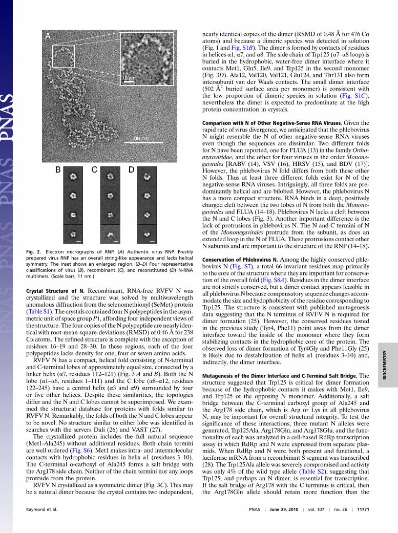
Electron Microscopy of Authentic Virus RNPs and Reconstituted RNPs.We compared RNPs isolated from virus with RNPs reconstitutedfrom refolded N and a large single-stranded RNA. Freshly pre-pared samples (Fig. S2) were viewed by negative-stain electronmicroscopy before and after overnight ribonuclease digestionat room temperature. Both authentic virus RNP and reconsti-tuted RNP were ribonuclease-resistant and had a remarkablysimilar string-like appearance (Fig. 2A and Fig. S3). No helicalsymmetry was apparent in either sample. The appearance of phle-bovirus RNP is strikingly different from images of similarly pre-pared RNP from other negative-sense RNA viruses, which haveobvious helical symmetry (15, 21–24).
Heterogenous Multimeric N-RNA Complexes. Aggressive ribonu-clease digestion (3 d at 37 °C) released a multimer from the virusRNP (Fig. 2B and Fig. S4A). In electron micrographs, this particleis similar in appearance to the ribonuclease-resistant, recombinantN-RNAmultimer purified from E. coli (Fig. 2C and Fig. S4B) andto multimers reconstituted from refolded N and defined RNAoligomers (Fig. 2D and Fig. S4 C and D). Remarkably, the multi-mers are of similar size distribution (10–12 nm diameter) regard-less of the source of RNA. The N-RNA multimers from allsources appear heterogeneous, with 4–7 bright objects per particle(Fig. 2 B–D). The heterogeneity of the recombinant N-RNAmultimer explains its inability to crystallize. The recombinantand reconstituted multimers have an apparent molecular weightof 90–100 kDa by size-exclusion chromatography (Fig. S5). Thus,we conclude that the N-RNA multimers contain 4–7 N subunitsand an unknown amount of RNA.
The stoichiometry of N and RNA in a reconstituted N-RNAmultimer was estimated using known N and RNA extinctioncoefficients at 260 nm and 280 nm.We used a short RNAdecamerin order to enhance the contribution of protein to the total absor-bance,whichwas dominatedbyRNA.The reconstitutedN-RNA10
multimer had similar behavior upon gel filtration (Fig. S5) andsimilar appearance in electron micrographs (Fig. S4) to theN-RNA25 multimer. The purified, reconstituted N-RNA10 multi-mer had anA260∕A280 ratio of 0.94, indicating an average stoichio-metry of 5∶1 N∶RNA10, in good agreement with the gel filtrationdata. This result indicates that eachbright spot onmultimer imagesis due to one N subunit.
RVFV RNPsDSP
28 kDa
36 kDa
55 kDa
72 kDa95 kDa130 kDa
250 kDa
***
A
*
BDSP
N N-RNA
26 kDa
34 kDa
55 kDa
72 kDa
95 kDa
130 kDa
43 kDa
250 kDa170 kDa
36 kDa
28 kDa
*
*
*
1 2 3 4 1098765 1211 13
*
*
Fig. 1. Similar multimer complexes of authentic virus RNPs and purifiedrecombinant N bound to RNA. (A) Viral RNP. Purified RVFV RNPs werecross-linked with 0.0, 5.0, or 20.0 mM DSP and analyzed by immuno-blot.Asterisks indicate predominant cross-linked species. Molecular weightmarkers are in the rightmost lane. (B) Recombinant N. N or N bound to U25
single-stranded RNA (N-RNA) was cross-linked using 0.0, 1.0, 5.0, 10.0, or20.0mMDSP, separated by SDS/PAGE, and visualizedwith colloidal Coomassiestain. The dominant cross-linked species are indicated by asterisks.
11770 ∣ www.pnas.org/cgi/doi/10.1073/pnas.1001760107 Raymond et al.
Crystal Structure of N. Recombinant, RNA-free RVFV N wascrystallized and the structure was solved by multiwavelengthanomalous diffraction from the selenomethionyl (SeMet) protein(Table S1). The crystals contained fourNpolypeptides in the asym-metric unit of space group P1, affording four independent views ofthe structure. The four copies of theNpolypeptide are nearly iden-tical with root-mean-square-deviations (RMSD) of 0.46 Å for 238Cα atoms. The refined structure is complete with the exception ofresidues 16–19 and 28–30. In these regions, each of the fourpolypeptides lacks density for one, four or seven amino acids.
RVFV N has a compact, helical fold consisting of N-terminaland C-terminal lobes of approximately equal size, connected by alinker helix (α7, residues 112–121) (Fig. 3 A and B). Both the Nlobe (α1–α6, residues 1–111) and the C lobe (α8–α12, residues122–245) have a central helix (α3 and α9) surrounded by fouror five other helices. Despite these similarities, the topologiesdiffer and the N and C lobes cannot be superimposed. We exam-ined the structural database for proteins with folds similar toRVFVN. Remarkably, the folds of both the N and C lobes appearto be novel. No structure similar to either lobe was identified insearches with the servers Dali (26) and VAST (27).
The crystallized protein includes the full natural sequence(Met1-Ala245) without additional residues. Both chain terminiare well ordered (Fig. S6). Met1 makes intra- and intermolecularcontacts with hydrophobic residues in helix α1 (residues 3–10).The C-terminal α-carboxyl of Ala245 forms a salt bridge withthe Arg178 side chain. Neither of the chain termini nor any loopsprotrude from the protein.
RVFV N crystallized as a symmetric dimer (Fig. 3C). This maybe a natural dimer because the crystal contains two independent,
nearly identical copies of the dimer (RSMD of 0.48 Å for 476 Cαatoms) and because a dimeric species was detected in solution(Fig. 1 and Fig. S1B). The dimer is formed by contacts of residuesin helices α1, α7, and α8. The side chain of Trp125 (α7–α8 loop) isburied in the hydrophobic, water-free dimer interface where itcontacts Met1, Gln5, Ile9, and Trp125 in the second monomer(Fig. 3D). Ala12, Val120, Val121, Glu124, and Thr131 also formintersubunit van der Waals contacts. The small dimer interface(502 Å2 buried surface area per monomer) is consistent withthe low proportion of dimeric species in solution (Fig. S1C),nevertheless the dimer is expected to predominate at the highprotein concentration in crystals.
Comparison with N of Other Negative-Sense RNA Viruses. Given therapid rate of virus divergence, we anticipated that the phlebovirusN might resemble the N of other negative-sense RNA viruseseven though the sequences are dissimilar. Two different foldsfor N have been reported, one for FLUA (13) in the familyOrtho-myxoviridae, and the other for four viruses in the order Monone-gavirales [RABV (14), VSV (16), HRSV (15), and BDV (17)].However, the phlebovirus N fold differs from both these otherN folds. Thus at least three different folds exist for N of thenegative-sense RNA viruses. Intriguingly, all three folds are pre-dominantly helical and are bilobed. However, the phlebovirus Nhas a more compact structure. RNA binds in a deep, positivelycharged cleft between the two lobes of N from both the Monone-gavirales and FLUA (14–18). Phlebovirus N lacks a cleft betweenthe N and C lobes (Fig. 3). Another important difference is thelack of protrusions in phlebovirus N. The N and C termini of Nof the Mononegavirales protrude from the subunit, as does anextended loop in the N of FLUA. These protrusions contact otherN subunits and are important to the structure of the RNP (14–18).
Conservation of Phlebovirus N. Among the highly conserved phle-bovirus N (Fig. S7), a total 66 invariant residues map primarilyto the core of the structure where they are important for conserva-tion of the overall fold (Fig. S8A). Residues in the dimer interfaceare not strictly conserved, but a dimer contact appears feasible inall phlebovirusNbecause compensatory sequence changes accom-modate the size and hydophobicity of the residue corresponding toTrp125. The structure is consistent with published mutagenesisdata suggesting that the N terminus of RVFV N is required fordimer formation (25). However, the conserved residues testedin the previous study (Tyr4, Phe11) point away from the dimerinterface toward the inside of the monomer where they formstabilizing contacts in the hydrophobic core of the protein. Theobserved loss of dimer formation of Tyr4Gly and Phe11Gly (25)is likely due to destabilization of helix α1 (residues 3–10) and,indirectly, the dimer interface.
Mutagenesis of the Dimer Interface and C-Terminal Salt Bridge. Thestructure suggested that Trp125 is critical for dimer formationbecause of the hydrophobic contacts it makes with Met1, Ile9,and Trp125 of the opposing N monomer. Additionally, a saltbridge between the C-terminal carboxyl group of Ala245 andthe Arg178 side chain, which is Arg or Lys in all phlebovirusN, may be important for overall structural integrity. To test thesignificance of these interactions, three mutant N alleles weregenerated, Trp125Ala, Arg178Gln, and Arg178Glu, and the func-tionality of each was analyzed in a cell-based RdRp transcriptionassay in which RdRp and N were expressed from separate plas-mids. When RdRp and N were both present and functional, aluciferase mRNA from a recombinant S segment was transcribed(28). The Trp125Ala allele was severely compromised and activitywas only 4% of the wild type allele (Table S2), suggesting thatTrp125, and perhaps an N dimer, is essential for transcription.If the salt bridge of Arg178 with the C terminus is critical, thenthe Arg178Gln allele should retain more function than the
Fig. 2. Electron micrographs of RNP. (A) Authentic virus RNP. Freshlyprepared virus RNP has an overall string-like appearance and lacks helicalsymmetry. The inset shows an enlarged region. (B–D) Four representativeclassifications of virus (B), recombinant (C), and reconstituted (D) N-RNAmultimers. (Scale bars, 11 nm.)
Raymond et al. PNAS ∣ June 29, 2010 ∣ vol. 107 ∣ no. 26 ∣ 11771
BIOCH
EMISTR
Y

Arg178Glu allele, and this was the observed result (Table S2).The activity of the Arg178Gln and Arg178Glu alleles was 25%and 7% of wild type, respectively. All alleles expressed proteinat a level similar to wild type and all appeared capable of forminghigher molecular weight complexes with RNA (Fig. S9).
Interaction Sites. We considered whether the RVFV N has anobvious RNA-binding surface. The N lobe has a higher calculatedisoelectric point (pI of 9.4 vs. 8.3) and a more positively chargedsurface (Fig. 4A) than the C lobe. We compared RVFV N withstructure-based homology models of N from other clades withinthe Phlebovirus genus. Based on the high sequence identity, Nfrom all phleboviruses are expected to bind RNA similarly. Thereis a general trend of greater positive charge on the N lobe than onthe C lobe, but the structures lack a common conserved basicsurface. We also mapped sequence conservation onto the RVFVN surface (Fig. 4B). The most strongly conserved surface is ahydrophobic pocket at the junction of the N and C lobes formedby a loop (residues 27–35) together with the C-terminal half ofα10 and the five succeeding amino acids (residues 198–210)(Fig. 4B and Fig. S8). Residues 27–35 are among the most mobileregions of the N structure (Fig. S8B). The combination of mobi-lity, conservation, and hydrophobicity suggest that this site may beinvolved in a conserved protein-protein interaction.
DiscussionThe structure of RVFV N reveals an addition to nature’s reper-toire of RNA-binding proteins (Fig. 3). High levels of sequence
identity (36–59%) assure that all phlebovirus N possess theRVFV N fold and also suggest that all phlebovirus N bindRNA similarly. The fold may also exist in N throughout theBunyaviridae family.
This work establishes that RNP organization in the Phlebo-viruses is different than in other negative-sense RNA viruses.Crystal structures or reconstructions from electron cryomicro-scopy have been reported for RNPs from four negative-senseRNA viruses (Mononegavirales and Orthomyxoviruses) (14–16,18). In all cases, RNA binds nonspecifically in an electropositivecleft between the lobes of the N subunit. RNP oligomers fromthese viruses have a similar architecture in which RNA bindsaround either the outside or inside of a ring of 9–11 N subunits.In all cases, protrusions from the N subunits make specific con-tacts with adjacent subunits to maintain the ring structure. Insome cases, the number of subunits in the oligomer ring matchesthe helical repeat of the polymeric RNP, which is apparent inelectron micrographs. For HRSV, each N subunit also interactswith other N subunits in the preceding or following turns of thehelical nucleocapsid (15).
In contrast to the RNPs of Mononegavirales and Orthomyxo-viruses, no helical structure was apparent in any electron micro-graphs of RVFV authentic or reconstituted RNP (Fig. 2A andFig. S3). Our results are similar to early electron micrographsof bunyavirus RNP, which lacked helical symmetry and also sug-gested that bunyavirus RNPs form large macrocircles (20), prob-ably due to pairing of 10–15 complementary bases at the 3′ and 5′ends of each genomic segment (29). RVFV RNP macrocircles
Fig. 3. Structure of RVFV N. (A) Polypeptide fold. The stereo ribbon diagram is colored as a rainbow from blue at the N terminus to red at the C terminus withloops in gray. Helix α7, (vertical) in the center of the image, links the N lobe at the left and the C lobe at the right. (B) Diagram of helical secondary structure inthe RVFN polypeptide. Colors are matched to A. (C) RVFV N dimer. In this view along the dimer axis, monomers are in green and cyan and the twofold axis isindicated by an ellipse. (D) Details of the dimer interface. The subunits are colored as in C, side chains with dimer contacts are shown in stick form in the stereoview. Hydrogen bonds are shown as dashed lines.
11772 ∣ www.pnas.org/cgi/doi/10.1073/pnas.1001760107 Raymond et al.

were seen in some of our images. Aggressive ribonuclease treat-ment of RVFV RNP released an N-RNA multimer that appearsheterogeneous in composition and has an average diameter of11 nm (Fig. 2B and Fig. S4A). Multimers of nearly identicalappearance to those from virus RNP were observed both forribonuclease-treated recombinant RNP and for RNP reconsti-tuted with small RNAs (Fig. 2 C and D and Fig. S4). The hetero-geneity of these minimal particles was surprising but is consistentwith the lack of helical symmetry in the RNP. The N crystal struc-ture is also consistent with the lack of helical symmetry. Thehighly compact phlebovirus N has no protruding loops or terminithat could link it to other N molecules in a superstructure like therings of 9–11 subunits observed for the Mononegavirales andOrthomyxoviruses (14–16, 18), although we cannot rule out thepossibility of major conformational changes to N upon RNAbinding. We observed no large superstructure for recombinantRVFV N in solution, unlike the recombinant rings purified forN from RABV (14), HRSV (15), VSV (16), and FLUA (18).All N-RNA multimers that we could test by gel filtration hada similar apparent molecular weight of ∼100 kDa (Fig. S5).
Our workingmodel for the structure of phlebovirus RNP is thatN binds cooperatively to RNA. However, the cooperativity islimited to 4–7N subunits based on the similar size and appearanceof multimers from viral, recombinant and reconstituted RNP andtheir similar behavior upon gel filtration (Fig. 2 and Figs. S4 andS5). The RNP lacks a strong helical structure, and N-N contactsare too weak for ribonuclease treatment to release a specificN-RNA oligomer from virus RNP. Instead of a tightly associated,symmetric N-RNA oligomer, limited protein–protein interactionsmay lead to oligomers of 4–7 Nmonomers bound to RNA, result-ing in the observed mixture of multimer species upon exhaustiveRNase treatment. If there is a weakly associated fundamental
oligomer, it may be a hexamer because our cross-linking datashowed a preponderance of species containing multiples of twoN subunits (Fig. 1). Overall, the organization of phlebovirusRNPappears less symmetric and with few specific protein–proteininteractions compared to the helical RNP of theMononegaviralesandOrthomyxoviruses. The evolutionary path to phlebovirus RNPis not common with the path to helical RNP of some othernegative-sense RNA viruses.
The unusual N-RNA multimer and perhaps the nonhelicalRNPmay be a general property of the Bunyaviridae. The observedRVFV N-RNA multimer species are similar to the reported109-kDa recombinant RNP fromBunyamwera virus (6). Bunyam-wera virus and RVFV belong to different genera within familyBunyaviridae and their N are not obviously similar at the aminoacid level.
The N lobe of RVFV N was identified as a potential RNAinteraction site because it is more positively charged than theC lobe in N from all phlebovirus clades. In whatever mannerN binds RNA, it is expected to engage the phosphate backbonebecause the multimer is so highly ribonuclease resistant. Theelectron micrographs do not permit assignment of RNA to aspecific location in the multimer particles.
The relevance of the N dimer to the RNP is unclear. Based onthe size of multimers in electron micrographs (Fig. 2 and Fig. S4)and their gel filtration profile (Fig. S5), each bright spot in themultimer images must be a monomer and not a dimer of N. Aprecedent for different oligomer species for RNA-free andRNA-bound N exists for FLUA where the free protein is a trimerand the RNP is a nonamer (13, 18). We probed the RVFV Ndimer interface by site-directed mutagenesis at Trp125 but foundcross-linked RNA complexes similar to the wild type (Fig. S9).However, the Ala substitution revealed an important role forTrp125 in replication (Table S2). This could be due to any numberof molecular interactions, most obviously with the RdRp.
The most highly conserved surface of phlebovirus N is a hydro-phobic pocket at the interface of the N- and C-terminal lobes(Fig. 4 and Fig. S8). The conservation in this region suggests animportant function that is common to phleboviruses, and thehydrophobicity of the surface suggests that it is not a site forRNA binding but rather is for interaction with a viral or (uniden-tified) host protein. Among potential viral protein partners, theRdRp is an obvious possibility because N is required for transcrip-tion and replication by the RdRp (4). However, several lines ofevidence suggest that an envelope glycoprotein may be the targetof the conserved hydrophobic pocket on N.
Packaging of RNPs into virions occurs at a site of virus assemblyon the Golgi membrane (30). The cytoplasmic tail of the RVFVenvelope glycoproteinGN was shown recently to recruit the encap-sidated genome to the Golgi membrane prior to virion assembly(31).Moreover, three regions within theGN tail of theUukuniemivirus were shown to be important for nucleoprotein binding to theglycoproteins (5). Genome recruitment is expected to be similar inall phleboviruses, thus the conserved hydrophobic pocket of N is acandidate GN binding site. This hypothesis is consistent with theability of bunyaviruses, both in nature and in vitro, to undergoreassortment in which progeny have genomic segments that derivefrommore thanone parental virus (32–34).Reassortment requirespromiscuity in the interaction of N with genomic RNAs fromheterologous viruses and in protein-protein interactions necessaryfor assembling virions. All characterized reassortant bunyavirusesisolated in nature are M segment reassortants (35, 36), demon-strating that the envelope glycoproteins, which are encoded bythe M segment, are capable of interacting with heterologousRNPs. The hydrophobic character of some regions of theGN-tail,aswell as Pro andTrp residueswithin it, are conserved amongphle-boviruses (37) and could function in protein–protein interactionswith N. Whether the conserved pocket of N interacts with theGN
A
180˚
ConservedVariable
B
N C C
Fig. 4. Properties of the RVFV N surface. (A) Electrostatic surface potential.The surface potential from −20 kT in red to þ20 kT in blue is shown forthe front and back of the RVFV N monomer. The ribbon diagrams belowshow the positions of the N-terminal (blue) and C-terminal (green) lobesand the linker (pink). The image at left is in the same orientation as (Fig. 3A).(B) Conserved hydrophobic pocket. The most conserved surface of N is at thetop relative to A. In this view, the N-terminal domain is at left and coloring isby conservation among phlebovirus N, as indicated.
Raymond et al. PNAS ∣ June 29, 2010 ∣ vol. 107 ∣ no. 26 ∣ 11773
BIOCH
EMISTR
Y

cytoplasmic tail, with the RdRp, or with a host protein, it haspotential as a drug target because it is conserved in phleboviruses.
The structure and characterization of phlebovirus N and RNPreveal another paradigm for encapsidation of the genome ofnegative sense RNA viruses, provide a platform for furtherstudies of virus pathogenicity, and suggest a potential site fordevelopment of effective antiviral therapeutics.
Materials and MethodsDetailed methods are given in SI Text.
Production and Purification of Recombinant N. The expression construct for Nencoded an N-terminal His6 tag fused to SUMO fused to N. The expressionplasmid was transformed into E. coli, and the protein was purified from thesoluble fraction of the cell lysate. His6-SUMO-N was bound to a metal-affinitycolumn, unfolded in 8 M urea, washed with several column volumes of 8 Murea to remove RNA, refolded in a linear gradient of 8 M to 0 M urea, andeluted with an imidazole gradient. The His6-SUMO was cleaved from therefolded protein with SUMO protease (38), a kind gift of C. Lima, MemorialSloan-Kettering Cancer Center. The cleavage product, N with its full naturalsequence, was purified by a second step of metal-affinity chromatographyfollowed by gel filtration (Fig. S1).
Crystallography. The crystal structure was solved by Se-MAD using the SeMetprotein. Crystals in space group P1 contain four N polypeptides per unit cellwith cell constants a ¼ 67.1 Å, b ¼ 69.6 Å, c ¼ 80.6 Å, α ¼ 78.4°, β ¼ 69.7°,γ ¼ 60.9° for the 1.93-Å dataset from a crystal of wild-type N. The final modelis of excellent crystallographic and stereochemical quality with R ¼ 0.211,Rfree ¼ 0.254, RMSDbonds ¼ 0.013 Å and 99.6% of residues in allowed regionsof the Ramachandran plot.
Cross-Linking. For cross-linking, a reconstitutedRNP (N-RNA)was generatedbyincubating refolded recombinant N (recN) with a 25-nucleotide polyU RNA
oligomer for 30 min at a ratio of 6∶1 recN∶RNA. The sample was then runon an S200 size-exclusion column to separate N-RNA from RNA-free recN.The recN andN-RNAwere dialyzed against PBS to remove the Tris storage buf-fer prior to incubation with DSP. Purified recN, N-RNA, or purified viral RNPs(vRNP) were cross-linked by incubating 30 μg of recN at a concentration of1 mg∕mL, or purified vRNPs with 0.0, 1.0, 5.0, 10.0, or 20.0 μM dithiobis[succi-nimidyl propionate] (Pierce) for 15min at roomtemperature. The cross-linkingwas quenched by addition of Tris pH 6.7 to a final concentration of 100 mM.Protein complexes were analyzed by SDS/PAGE followed by either colloidalCoomassie stain or western blot using a polyclonal rabbit anti-N antibody.
Electron Microscopy. All samples were absorbed onto a carbon-coated gridand stained with 0.75% uranyl formate using standard protocols. A Morgag-ni 268(D) transmission electron microscope equipped with a mounted OriusSC200W CCD camera was used for imaging at room temperature.
RVFV transcription assay. BSR-T7/5 cells were plated at 1 × 10⁵ cells/well in12-well culture plates. After 24 h, cells were transfected using 2 μg∕mLTransITLT1 (Mirus Corporation) and plasmids in the ratio 0.25 μg pSTrRVFV-SΔNΔNSs∶hRLuc∶0.50 μg pN: 0.75 μg pRdRp. At 48 h posttransfection, the cells wereharvested and analyzed by luciferase assay and western blot.
ACKNOWLEDGMENTS. We thank Georgios Skiniotis, Min Su, and JustinSchilling (Life Sciences Institute, Univ. Michigan) for advice and assistancewith electron microscopy, and Katrin Karbstein and Crystal Young (Dept.Chemistry, Univ. Michigan) for the 684-mer RNA sample. The research wassupported by National Institutes of Health (NIH) Grant P01-AI055672 toJ.L.S., by NIH Regional Center of Excellence for Bio-defense and EmergingInfectious Diseases Grant U54-AI057153 to S.R.G., by the Rackham GraduateSchool at the University of Michigan to S.R.G., and by NIH Training GrantsT32-GM008270 in Molecular Biophysics to D.D.R. and T32-GM007315 inCell and Molecular Biology to M.E.P. The GM/CA beamlines are supportedby the NIH Institute of General Medical Sciences and National CancerInstitute, and the APS by the United States Department of Energy.
1. Flint SJ, Enquist LW, Racaniello VR, Skalka AM (2004) Principles of virology : Molecularbiology, pathogenesis, and control of animal viruses (ASM Press, Washington),pp 83–125.
2. Nichol ST (2001) Fields Virology, eds BN Fields, DM Knipe, PM Howley, and DE Griffin(Lippincott, Williams & Wilkins, Philadelphia), pp 1603–1633.
3. Schmaljohn CS, Hooper JW (2001) Fields Virology, eds BN Fields, DMKnipe, PMHowley,and DE Griffin (Lippincott, Williams & Wilkins, Philadelphia), pp 1581–1602.
4. Schmaljohn CS, Nichol ST (2007) Fields Virology, eds DM Knipe et al. (Lippincott,Williams & Wilkins, Philadelphia), pp 1741–1789.
5. Overby AK, Pettersson RF, Neve EP (2007) The glycoprotein cytoplasmic tail ofUukuniemi virus (Bunyaviridae) interacts with ribonucleoproteins and is critical forgenome packaging. J Virol 81:3198–3205.
6. Mohl BP, Barr JN (2009) Investigating the specificity and stoichiometry of RNA bindingby the nucleocapsid protein of Bunyamwera virus. RNA 15:391–399.
7. Gott P, Stohwasser R, Schnitzler P, Darai G, Bautz EK (1993) RNA binding of recombi-nant nucleocapsid proteins of hantaviruses. Virology 194:332–337.
8. Osborne JC, Elliott RM (2000) RNA binding properties of bunyamwera virusnucleocapsid protein and selective binding to an element in the 5′ terminus of thenegative-sense S segment. J Virol 74:9946–9952.
9. Mir MA, Brown B, Hjelle B, Duran WA, Panganiban AT (2006) Hantavirus Nprotein exhibits genus-specific recognition of the viral RNA panhandle. J Virol80:11283–11292.
10. Ogg MM, Patterson JL (2007) RNA binding domain of Jamestown Canyon virus Ssegment RNAs. J Virol 81:13754–13760.
11. Severson W, Partin L, Schmaljohn CS, Jonsson CB (1999) Characterization of the Han-taan nucleocapsid protein-ribonucleic acid interaction. J Biol Chem 274:33732–33739.
12. Falk BW, Tsai JH (1998) Biology and molecular biology of viruses in the genusTenuivirus. Annu Rev Phytopathol 36:139–163.
13. Ye Q, Krug RM, Tao YJ (2006) The mechanism by which influenza A virus nucleoproteinforms oligomers and binds RNA. Nature 444:1078–1082.
14. Albertini AA, et al. (2006) Crystal structure of the rabies virus nucleoprotein-RNAcomplex. Science 313:360–363.
15. Tawar RG, et al. (2009) Crystal structure of a nucleocapsid-like nucleoprotein-RNAcomplex of respiratory syncytial virus. Science 326:1279–1283.
16. Green TJ, Zhang X, Wertz GW, Luo M (2006) Structure of the vesicular stomatitis virusnucleoprotein-RNA complex. Science 313:357–360.
17. Rudolph MG, et al. (2003) Crystal structure of the borna disease virus nucleoprotein.Structure 11:1219–1226.
18. Coloma R, et al. (2009) The structure of a biologically active influenza virus ribonucleo-protein complex. PLoS Pathog 5:e1000491.
19. von Bonsdorff CH, Saikku P, Oker-Blom N (1969) The inner structure of Uukuniemi andtwo Bunyamwera supergroup arboviruses. Virology 39:342–344.
20. Pettersson RF, von Bonsdorff CH (1975) Ribonucleoproteins of Uukuniemi virus arecircular. J Virol 15:386–392.
21. Schoehn G, et al. (2004) The 12 A structure of trypsin-treated measles virus N-RNA.J Mol Biol 339:301–312.
22. Bhella D, Ralph A, Murphy LB, Yeo RP (2002) Significant differences in nucleocapsidmorphology within the Paramyxoviridae. J Gen Virol 83:1831–1839.
23. Ge P, et al. (2010) Cryo-EM model of the bullet-shaped vesicular stomatitis virus.Science 327:689–693.
24. Ruigrok RW, Baudin F (1995) Structure of influenza virus ribonucleoprotein particles.II. Purified RNA-free influenza virus ribonucleoprotein forms structures that are indis-tinguishable from the intact influenza virus ribonucleoprotein particles. J Gen Virol76:1009–1014.
25. Le May N, Gauliard N, Billecocq A, Bouloy M (2005) The N terminus of Rift Valley fevervirus nucleoprotein is essential for dimerization. J Virol 79:11974–11980.
26. Holm L, Sander C (1995) Dali: A network tool for protein structure comparison. TrendsBiochem Sci 20:478–480.
27. Gibrat JF, Madej T, Bryant SH (1996) Surprising similarities in structure comparison.Curr Opin Struct Biol 6:377–385.
28. Piper M, Gerrard S (2010) A novel system for identification of inhibitors of Rift Valleyfever virus replication. Viruses 2:731–747.
29. Raju R, Kolakofsky D (1989) The ends of La Crosse virus genome and antigenome RNAswithin nucleocapsids are base paired. J Virol 63:122–128.
30. Pettersson RF, Melin L (1996) The Bunyaviridae, ed RM Elliott (Plenum, New York),pp 159–188.
31. Overby AK, Popov VL, Pettersson RF, Neve EP (2007) The cytoplasmic tails ofUukuniemi Virus (Bunyaviridae) GN and GC glycoproteins are important for intracel-lular targeting and the budding of virus-like particles. J Virol 81:11381–11391.
32. Borucki MK, Chandler LJ, Parker BM, Blair CD, Beaty BJ (1999) Bunyavirus super-infection and segment reassortment in transovarially infected mosquitoes. J Gen Virol80:3173–3179.
33. Chandler LJ, et al. (1991) Reassortment of La Crosse and Tahyna bunyaviruses in Aedestriseriatus mosquitoes. Virus Res 20:181–191.
34. Beaty BJ, Sundin DR, Chandler LJ, Bishop DH (1985) Evolution of bunyaviruses bygenome reassortment in dually infected mosquitoes (Aedes triseriatus). Science230:548–550.
35. Briese T, Kapoor V, Lipkin WI (2007) Natural M-segment reassortment in Potosi andMain Drain viruses: Implications for the evolution of orthobunyaviruses. Arch Virol152:2237–2247.
36. Bowen MD, et al. (2001) A reassortant bunyavirus isolated from acute hemorrhagicfever cases in Kenya and Somalia. Virology 291:185–190.
37. Gerrard SR, Nichol ST (2002) Characterization of the Golgi retention motif of RiftValley fever virus GN glycoprotein. J Virol 76:12200–12210.
38. Reverter D, Lima CD (2004) A basis for SUMO protease specificity provided by analysisof human Senp2 and a Senp2-SUMO complex. Structure 12:1519–1531.
11774 ∣ www.pnas.org/cgi/doi/10.1073/pnas.1001760107 Raymond et al.