structure of hdl: particle subclasses and molecular components
TRANSCRIPT
Structure of HDL: Particle Subclassesand Molecular Components
Anatol Kontush, Mats Lindahl, Marie Lhomme, Laura Calabresi,M. John Chapman, and W. Sean Davidson
Contents
1 HDL Subclasses . . . . . . . . . . . . . . . . . . . . . . . . . . . . . . . . . . . . . . . . . . . . . . . . . . . . . . . . . . . . . . . . . . . . . . . . . . . . . . 5
2 Molecular Components of HDL . . . . . . . . . . . . . . . . . . . . . . . . . . . . . . . . . . . . . . . . . . . . . . . . . . . . . . . . . . . . . 7
2.1 Proteome . . . . . . . . . . . . . . . . . . . . . . . . . . . . . . . . . . . . . . . . . . . . . . . . . . . . . . . . . . . . . . . . . . . . . . . . . . . . . . . 7
2.1.1 Major Protein Components . . . . . . . . . . . . . . . . . . . . . . . . . . . . . . . . . . . . . . . . . . . . . . . . . . . . 7
2.1.2 Protein Isoforms, Translational and Posttranslational Modifications . . . . . . . . 19
2.2 Lipidome . . . . . . . . . . . . . . . . . . . . . . . . . . . . . . . . . . . . . . . . . . . . . . . . . . . . . . . . . . . . . . . . . . . . . . . . . . . . . . . 23
2.2.1 Phospholipids . . . . . . . . . . . . . . . . . . . . . . . . . . . . . . . . . . . . . . . . . . . . . . . . . . . . . . . . . . . . . . . . . . 23
2.2.2 Sphingolipids . . . . . . . . . . . . . . . . . . . . . . . . . . . . . . . . . . . . . . . . . . . . . . . . . . . . . . . . . . . . . . . . . . . 27
2.2.3 Neutral Lipids . . . . . . . . . . . . . . . . . . . . . . . . . . . . . . . . . . . . . . . . . . . . . . . . . . . . . . . . . . . . . . . . . . 27
A. Kontush (*) • M. Lhomme • M.J. Chapman
National Institute for Health and Medical Research (INSERM), UMR-ICAN 1166, Paris, France
University of Pierre and Marie Curie - Paris 6, Paris, France
Pitie – Salpetriere University Hospital, Paris, France
ICAN, Paris, France
e-mail: [email protected]; [email protected]; [email protected]
M. Lindahl
Department of Clinical and Experimental Medicine, Linkoping University, Linkoping, Sweden
e-mail: [email protected]
L. Calabresi
Department of Pharmacological and Biomolecular Sciences, Center E. Grossi Paoletti,
University of Milan, Milan, Italy
e-mail: [email protected]
W.S. Davidson
Department of Pathology and Laboratory Medicine, University of Cincinnati, Cincinnati, OH
45237, USA
e-mail: [email protected]
# The Author(s) 2015
A. von Eckardstein, D. Kardassis (eds.), High Density Lipoproteins, Handbook of
Experimental Pharmacology 224, DOI 10.1007/978-3-319-09665-0_1
3
3 The Structure of HDL . . . . . . . . . . . . . . . . . . . . . . . . . . . . . . . . . . . . . . . . . . . . . . . . . . . . . . . . . . . . . . . . . . . . . . . . 28
3.1 Introduction/Brief History . . . . . . . . . . . . . . . . . . . . . . . . . . . . . . . . . . . . . . . . . . . . . . . . . . . . . . . . . . . . . 28
3.2 HDL in the Test Tube . . . . . . . . . . . . . . . . . . . . . . . . . . . . . . . . . . . . . . . . . . . . . . . . . . . . . . . . . . . . . . . . . . 31
3.2.1 Discoid HDL . . . . . . . . . . . . . . . . . . . . . . . . . . . . . . . . . . . . . . . . . . . . . . . . . . . . . . . . . . . . . . . . . . . 31
3.2.2 Spherical rHDL . . . . . . . . . . . . . . . . . . . . . . . . . . . . . . . . . . . . . . . . . . . . . . . . . . . . . . . . . . . . . . . . 33
3.3 “Real” HDL Particles . . . . . . . . . . . . . . . . . . . . . . . . . . . . . . . . . . . . . . . . . . . . . . . . . . . . . . . . . . . . . . . . . . 35
References . . . . . . . . . . . . . . . . . . . . . . . . . . . . . . . . . . . . . . . . . . . . . . . . . . . . . . . . . . . . . . . . . . . . . . . . . . . . . . . . . . . . . . . . 41
Abstract
A molecular understanding of high-density lipoprotein (HDL) will allow a more
complete grasp of its interactions with key plasma remodelling factors and with
cell-surface proteins that mediate HDL assembly and clearance. However, these
particles are notoriously heterogeneous in terms of almost every physical,
chemical and biological property. Furthermore, HDL particles have not lent
themselves to high-resolution structural study through mainstream techniques
like nuclear magnetic resonance and X-ray crystallography; investigators have
therefore had to use a series of lower resolution methods to derive a general
structural understanding of these enigmatic particles. This chapter reviews
current knowledge of the composition, structure and heterogeneity of human
plasma HDL. The multifaceted composition of the HDL proteome, the multiple
major protein isoforms involving translational and posttranslational
modifications, the rapidly expanding knowledge of the HDL lipidome, the
highly complex world of HDL subclasses and putative models of HDL particle
structure are extensively discussed. A brief history of structural studies of both
plasma-derived and recombinant forms of HDL is presented with a focus on
detailed structural models that have been derived from a range of techniques
spanning mass spectrometry to molecular dynamics.
Keywords
HDL • Composition • Structure • Heterogeneity • Proteomics • Lipidomics •
Proteome • Lipidome • Post-translational • Modifications
High-density lipoprotein (HDL) is a small, dense, protein-rich lipoprotein as
compared to other lipoprotein classes, with a mean size of 8–10 nm and density of
1.063–1.21 g/ml (Kontush and Chapman 2012). HDL particles are plurimolecular,
quasi-spherical or discoid, pseudomicellar complexes composed predominantly of
polar lipids solubilised by apolipoproteins. HDL also contains numerous other
proteins, including enzymes and acute-phase proteins, and may contain small
amounts of nonpolar lipids. Furthermore, HDL proteins often exist in multiple
isoforms and readily undergo posttranslational modification. As a consequence of
such diverse compositional features, HDL particles are highly heterogeneous in their
structural, chemical and biological properties. This chapter reviews current knowl-
edge of the composition, structure and heterogeneity of human plasma HDL.
4 A. Kontush et al.
1 HDL Subclasses
Human plasma HDLs are a highly heterogeneous lipoprotein family consisting of
several subclasses differing in density, size, shape and lipid and protein composi-
tion (Table 1).
Differences in HDL subclass distribution were first described by Gofman and
colleagues in the early 1950s by using analytic ultracentrifugation (De Lalla and
Gofman 1954), the gold standard technique for HDL separation. Two HDL
subclasses were identified: the less dense (1.063–1.125 g/mL), relatively lipid-
rich form was classified asHDL2 and the more dense (1.125–1.21 g/mL), relatively
protein-rich form as HDL3. The two major HDL subclasses can be separated by
other ultracentrifugation methods, such as rate-zonal ultracentrifugation
(Franceschini et al. 1985) or single vertical spin ultracentrifugation (Kulkarni
et al. 1997). Ultracentrifugation methods are accurate and precise but require
expensive instruments, time and technical skills. A precipitation method has been
proposed for HDL2 and HDL3 separation and quantitation (Gidez et al. 1982),
which is inexpensive and easier, but with a high degree of interlaboratory
variability. HDL2 and HDL3 can be further fractionated in distinct subclasses
with different electrophoretic mobilities by non-denaturing polyacrylamide gradi-
ent gel electrophoresis (GGE) (Nichols et al. 1986), which separates HDL
subclasses on the basis of particle size. Two HDL2 and three HDL3 subclasses
have been identified and their particle size characterised by this method: HDL3c,
7.2–7.8 nm diameter; HDL3b, 7.8–8.2 nm; HDL3a, 8.2–8.8 nm; HDL2a, 8.8.–
9.7 nm; and HDL2b, 9.7–12.0 nm. The equivalent subclasses of HDL with similar
size distribution may be preparatively isolated by isopycnic density gradient ultra-
centrifugation (Chapman et al. 1981; Kontush et al. 2003).
Agarose gel electrophoresis allows analytical separation of HDL according to
surface charge and shape into α-migrating particles, which represent the majority
of circulating HDL, and preβ-migrating particles, consisting of nascent discoidal
and poorly lipidated HDL. Agarose gels can be stained with Coomassie blue or with
anti-apolipoprotein A-I (apoA-I) antibodies, and the relative protein content of the
two HDL subclasses can be determined (Favari et al. 2004). The plasma preβ-HDLconcentration can be also quantified using a sandwich enzyme immunoassay (Miida
et al. 2003). The assay utilises a monoclonal antibody which specifically recognises
apoA-I bound to preβ-HDL. The agarose gel and the GGE can be combined into a
2-dimensional (2D) electrophoretic method, which separates HDL according to
charge in the first run and according to size in the second run. Gels can be stained
with apolipoprotein-specific antibodies, typically with anti-apoA-I antibodies,
allowing the detection of distinct HDL subclasses (Asztalos et al. 2007). This is
by far the method with the highest resolving power: up to 12 distinct apoA-I-
containing HDL subclasses can be identified, referred to as preβ (preβ1 and preβ2), α(α1, α2, α3 and α4) and preα (preα1, preα2, preα3) according to their mobility and
size (Asztalos and Schaefer 2003a, b).
According to the protein component, HDL can be separated into particles
containing apoA-I with (LpA-I:A-II) or without apoA-II (LpA-I) by an
Structure of HDL: Particle Subclasses and Molecular Components 5

electroimmunodiffusion technique in agarose gels; plasma concentration of LpA-I
and LpA-I:A-II can be determined from a calibration curve (Franceschini
et al. 2007).
More recently, a nuclear magnetic resonance (NMR) method for HDL subclass
analysis has been proposed, based on the concept that each lipoprotein particle of a
given size has its own characteristic lipid methyl group NMR signal (Otvos
et al. 1992). According to the NMR signals, three HDL subclasses can be identified:
large HDL (8.8–13.0 nm diameter), medium HDL (8.2–8.8 nm) and small HDL
(7.3–8.2 nm); results are expressed as plasma particle concentration. The relative
plasma content of small, medium and large HDL (according to the same cut-off)
can also be determined by GGE, dividing the absorbance profile into the three size
intervals (Franceschini et al. 2007).
HDL subfractionation on an analytical scale has been generally accomplished by
the different techniques in academic laboratories; however, clinical interest in HDL
heterogeneity has been growing in the last 10 years and a number of laboratory tests
for determining HDL subclass distribution are now available, including GGE, NMR
and 2D gel electrophoresis (Mora 2009). Whether evaluation of HDL subfractions
is performed by academic or commercial laboratories, there are a number of factors
that confound the interpretation of the results of such analyses. The number and
nomenclature of HDL subclasses are not uniform among the different techniques;
Table 1 Major HDL
subclasses according to
different isolation/
separation techniques
Density (ultracentrifugation)
HDL2 (1.063–1.125 g/mL)
HDL3 (1.125–1.21 g/mL)
Size (GGE)
HDL2b (9.7–12.0 nm)
HDL2a (8.8–9.7 nm)
HDL3a (8.2–8.8 nm)
HDL3b (7.8–8.2 nm)
HDL3c (7.2–7.8 nm)
Size (NMR)
Large HDL (8.8–13.0 nm)
Medium HDL (8.2–8.8 nm)
Small HDL (7.3–8.2 nm)
Shape and charge (agarose gel)
α-HDL (spherical)
Preβ-HDL (discoidal)
Charge and size (2D electrophoresis)
Preβ-HDL (preβ1 and preβ2)α-HDL (α1, α2, α3 and α4)Preα-HDL (preα1, preα2, preα3)
Protein composition (electroimmunodiffusion)
LpA-I
LpA-I:A-II
6 A. Kontush et al.
moreover, each subclass contains distinct subpopulations, as identified by, e.g. 2D
electrophoresis. In addition, whereas some methodologies measure HDL subclass
concentrations, others describe the percent distribution of the HDL subclasses
relative to the total or characterise the HDL distribution by average particle
diameter. As a consequence, there is little relation among HDL subfractionation
data produced by different analytical techniques. A panel of experts has recently
proposed a classification of HDL by physical properties, which integrates terminol-
ogy from several methods and defines five HDL subclasses, termed very large,
large, medium, small and very small HDL (Rosenson et al. 2011). The proposed
nomenclature, possibly together with widely accepted standards and quality
controls, should help in defining the relationship between HDL subclasses and
cardiovascular risk as well as in assessing the clinical effects of HDL modifying
drugs.
2 Molecular Components of HDL
2.1 Proteome
2.1.1 Major Protein ComponentsProteins form the major structural and functional component of HDL particles.
HDL carries a large number of different proteins as compared to other lipoprotein
classes (Table 2). HDL proteins can be divided into several major subgroups which
include apolipoproteins, enzymes, lipid transfer proteins, acute-phase response
proteins, complement components, proteinase inhibitors and other protein
components. Whereas apolipoproteins and enzymes are widely recognised as key
functional HDL components, the role of minor proteins, primarily those involved in
complement regulation, protection from infections and the acute-phase response,
has received increasing attention only in recent years, mainly as a result of advances
in proteomic technologies (Heinecke 2009; Hoofnagle and Heinecke 2009;
Davidsson et al. 2010; Shah et al. 2013). These studies have allowed reproducible
identification of more than 80 proteins in human HDL (Heinecke 2009; Hoofnagle
and Heinecke 2009; Davidsson et al. 2010; Shah et al. 2013) (for more details see
the HDL Proteome Watch at http://homepages.uc.edu/~davidswm/HDLproteome.
html). Numerous proteins involved in the acute-phase response, complement regu-
lation, proteinase inhibition, immune response and haemostasis were unexpectedly
found as components of normal human plasma HDL, raising the possibility that
HDL may play previously unsuspected roles in host defence mechanisms and
inflammation (Hoofnagle and Heinecke 2009).
Importantly, the composition of the HDL proteome may depend on the method
of HDL isolation. Indeed, ultracentrifugation in highly concentrated salt solutions
of high ionic strength can remove some proteins from HDL, whereas other methods
of HDL isolation (gel filtration, immunoaffinity chromatography, precipitation)
provide HDL extensively contaminated with plasma proteins or subject HDL to
unphysiological conditions capable of modifying its structure and/or composition
Structure of HDL: Particle Subclasses and Molecular Components 7
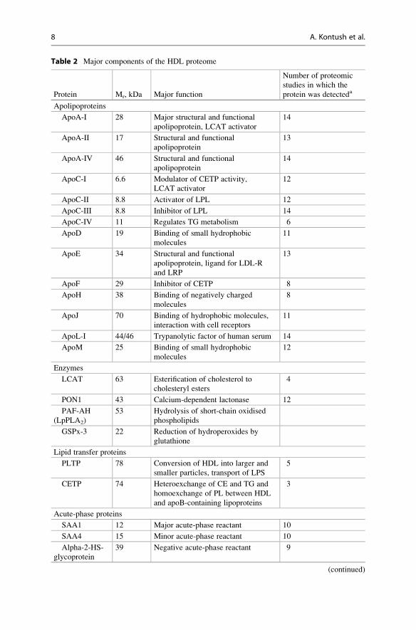
Table 2 Major components of the HDL proteome
Protein Mr, kDa Major function
Number of proteomic
studies in which the
protein was detecteda
Apolipoproteins
ApoA-I 28 Major structural and functional
apolipoprotein, LCAT activator
14
ApoA-II 17 Structural and functional
apolipoprotein
13
ApoA-IV 46 Structural and functional
apolipoprotein
14
ApoC-I 6.6 Modulator of CETP activity,
LCAT activator
12
ApoC-II 8.8 Activator of LPL 12
ApoC-III 8.8 Inhibitor of LPL 14
ApoC-IV 11 Regulates TG metabolism 6
ApoD 19 Binding of small hydrophobic
molecules
11
ApoE 34 Structural and functional
apolipoprotein, ligand for LDL-R
and LRP
13
ApoF 29 Inhibitor of CETP 8
ApoH 38 Binding of negatively charged
molecules
8
ApoJ 70 Binding of hydrophobic molecules,
interaction with cell receptors
11
ApoL-I 44/46 Trypanolytic factor of human serum 14
ApoM 25 Binding of small hydrophobic
molecules
12
Enzymes
LCAT 63 Esterification of cholesterol to
cholesteryl esters
4
PON1 43 Calcium-dependent lactonase 12
PAF-AH
(LpPLA2)
53 Hydrolysis of short-chain oxidised
phospholipids
GSPx-3 22 Reduction of hydroperoxides by
glutathione
Lipid transfer proteins
PLTP 78 Conversion of HDL into larger and
smaller particles, transport of LPS
5
CETP 74 Heteroexchange of CE and TG and
homoexchange of PL between HDL
and apoB-containing lipoproteins
3
Acute-phase proteins
SAA1 12 Major acute-phase reactant 10
SAA4 15 Minor acute-phase reactant 10
Alpha-2-HS-
glycoprotein
39 Negative acute-phase reactant 9
(continued)
8 A. Kontush et al.
(e.g. extreme pH and ionic strength involved in immunoaffinity separation). Thus,
proteomics of apoA-I-containing fractions isolated from human plasma by a
non-denaturing approach of fast protein liquid chromatography (FPLC) reveal the
presence of up to 115 individual proteins per fraction, only up to 32 of which were
identified as HDL-associated proteins in ultracentrifugally isolated HDL (Collins
et al. 2010). Indeed, co-elution with HDL of plasma proteins of matching size is
inevitable in FPLC-based separation; the presence of a particular protein across a
range of HDL-containing fractions of different size isolated by FPLC on the basis of
their association with phospholipid would however suggest that such a protein is
indeed associated with HDL (Gordon et al. 2010). Remarkably, several of the most
abundant plasma proteins, including albumin, haptoglobin and alpha-2-macroglob-
ulin, are indeed present in all apoA-I-containing fractions isolated by FPLC (Col-
lins et al. 2010), suggesting their partial association with HDL by a non-specific,
low-affinity binding.
Table 2 (continued)
Protein Mr, kDa Major function
Number of proteomic
studies in which the
protein was detecteda
Fibrinogen
alpha chain
95 Precursor of fibrin, cofactor in
platelet aggregation
10
Complement components
C3 187 Complement activation 9
Proteinase inhibitors
Alpha-1-
antitrypsin
52 Inhibitor of serine proteinases 11
Hrp 39 Decoy substrate to prevent
proteolysis
10
Other proteins
Transthyretin
55 Thyroid hormone binding and
transport
12
Serotransferrin
75 Iron binding and transport 10
Vitamin
D-binding
protein
58 Vitamin D binding and transport 10
Alpha-1B-
glycoprotein
54 Unknown 9
Hemopexin 52 Heme binding and transport 8aOut of total of 14 proteomic studies according to Shah et al. (2013). Only proteins detected in
more than 50 % of the studies are listed, together with seven others previously known to be
associated with HDL (apoC-IV, apoH, LCAT, PAF-AH, GSPx-3, PLTP, CETP)
CE cholesteryl ester, CETP cholesteryl ester transfer protein, GSPx-3 glutathione selenoperoxidase
3,Hrp haptoglobin-related protein, LDL-R LDL receptor, LCAT lecithin/cholesterol acyltransferase,
LPL lipoprotein lipase, LpPLA2 lipoprotein-associated phospholipase A2, LRP LDL receptor-related
protein, PAF-AH platelet-activating factor acetyl hydrolase, PL phospholipid, PLTP phospholipid
transfer protein, PON1 paraoxonase 1, SAA serum amuloid A, TG triglyceride
Structure of HDL: Particle Subclasses and Molecular Components 9

ApolipoproteinsApolipoprotein A-I is the major structural and functional HDL protein which
accounts for approximately 70 % of total HDL protein. Almost all HDL particles
are believed to contain apoA-I (Asztalos and Schaefer 2003a, b; Schaefer
et al. 2010). Major functions of apoA-I involve interaction with cellular receptors,
activation of lecithin/cholesterol acyltransferase (LCAT) and endowing HDL with
multiple anti-atherogenic activities. Circulating apoA-I represents a typical amphi-
pathic protein that lacks glycosylation or disulfide linkages and contains eight
alpha-helical amphipathic domains of 22 amino acids and two repeats of
11 amino acids. As a consequence, apoA-I binds avidly to lipids and possesses
potent detergent-like properties. ApoA-I readily moves between lipoprotein
particles and is also found in chylomicrons and very low-density lipoprotein
(VLDL). As for many plasma apolipoproteins, the main sites for apoA-I synthesis
and secretion are the liver and small intestine.
ApoA-II is the second major HDL apolipoprotein which represents approxi-
mately 15–20 % of total HDL protein. About a half of HDL particles may contain
apoA-II (Duriez and Fruchart 1999). ApoA-II is more hydrophobic than apoA-I and
circulates as a homodimer composed of two identical polypeptide chains (Shimano
2009; Puppione et al. 2010) connected by a disulfide bridge at position 6 (Brewer
et al. 1972). ApoA-II equally forms heterodimers with other cysteine-containing
apolipoproteins (Hennessy et al. 1997) and is predominantly synthesised in the liver
but also in the intestine (Gordon et al. 1983).
ApoA-IV, an O-linked glycoprotein, is the most hydrophilic apolipoprotein
which readily exchanges between lipoproteins and also circulates in a free form.
ApoA-IV contains thirteen 22-amino acid tandem repeats, nine of which are highly
alpha-helical; many of these helices are amphipathic and may serve as lipid-binding
domains. In man, apoA-IV is synthesised in the intestine and is secreted into the
circulation with chylomicrons.
ApoCs form a family of small exchangeable apolipoproteins primarily
synthesised in the liver. ApoC-I is the smallest apolipoprotein which associates
with both HDL and VLDL and can readily exchange between them. ApoC-I carries
a strong positive charge and can thereby bind free fatty acids and modulate activities
of several proteins involved in HDL metabolism. Thus, apoC-I is involved in the
activation of LCAT and inhibition of hepatic lipase and cholesteryl ester transfer
protein (CETP). ApoC-II functions as an activator of several triacylglycerol lipases
and is associated with HDL and VLDL.ApoC-III is predominantly present in VLDL
with small amounts found in HDL. The protein inhibits lipoprotein lipase (LPL) and
hepatic lipase and decreases the uptake of chylomicrons by hepatic cells. ApoC-IV
induces hypertriglyceridemia when overexpressed in mice (Allan and Taylor 1996;
Kim et al. 2008). In normolipidemic plasma, greater than 80 % of the protein resides
in VLDL, with most of the remainder in HDL. The HDL content of apoC-IV is much
lower as compared to the other apoC proteins.
ApoD is a glycoprotein mainly associated with HDL (McConathy and
Alaupovic 1973). The protein is expressed in many tissues, including liver and
intestine. ApoD does not possess a typical apolipoprotein structure and belongs to
10 A. Kontush et al.

the lipocalin family which also includes retinol-binding protein, lactoglobulin and
uteroglobulin. Lipocalins are small lipid transfer proteins with a limited amino acid
sequence identity but with a common tertiary structure. Lipocalins share a structur-
ally conserved beta-barrel fold, which in many lipocalins bind hydrophobic ligands.
As a result, apoD transports small hydrophobic ligands, with a high affinity for
arachidonic acid (Rassart et al. 2000). In plasma, apoD forms disulfide-linked
homodimers and heterodimers with apoA-II.
ApoE is a key structural and functional glycoprotein component of HDL despite
its much lower content in HDL particles as compared to apoA-I (Utermann 1975).
The major fraction of circulating apoE is carried by triglyceride-containing
lipoproteins where it serves as a ligand for apoB/apoE receptors and ensures
lipoprotein binding to cell-surface glycosaminoglycans. Similar to apoA-I and
apoA-II, apoE contains eight amphipathic alpha-helical repeats and displays
detergent-like properties towards phospholipids (Lund-Katz and Phillips 2010).
ApoE is synthesised in multiple tissues and cell types, including liver, endocrine
tissues, central nervous system and macrophages.
ApoF is a sialoglycoprotein present in human HDL and low-density lipoprotein
(LDL) (Olofsson et al. 1978), also known as lipid transfer inhibitor protein (LTIP)
as a consequence of its ability to inhibit CETP. ApoF is synthesised in the liver and
heavily glycosylated with both O- and N-linked sugar groups. Such glycosylation
renders the protein highly acidic and results in a molecular mass some 40 % greater
than predicted (Lagor et al. 2009).
ApoH, also known as beta-2-glycoprotein 1, is a multifunctional N- and
O-glycosylated protein. ApoH binds to various kinds of negatively charged
molecules, primarily to cardiolipin, and may prevent activation of the intrinsic
blood coagulation cascade by binding to phospholipids on the surface of damaged
cells. Such binding properties are ensured by a positively charged domain. ApoH
regulates platelet aggregation and is expressed by the liver.
ApoJ (also called clusterin and complement-associated protein SP-40,40) is an
antiparallel disulfide-linked heterodimeric glycoprotein. Human apoJ consists of
two subunits designated alpha (34–36 kDa) and beta (36–39 kDa) which share
limited homology (de Silva et al. 1990a, b) and are linked by five disulfide bonds.
The distinct structure of apoJ allows binding of both a wide spectrum of hydropho-
bic molecules on the one hand and of specific cell-surface receptors on the other.
ApoL-I is a key component of the trypanolytic factor of human serum associated
with HDL (Duchateau et al. 1997). ApoL-I possesses a glycosylation site and shares
structural and functional similarities with intracellular apoptosis-regulating
proteins of the Bcl-2 family. ApoL-I displays high affinity for phosphatidic acid
and cardiolipin (Zhaorigetu et al. 2008) and is expressed in pancreas, lung, prostate,
liver, placenta and spleen.
ApoM is an apolipoprotein found mainly in HDL (Axler et al. 2007) which
possesses an eight stranded antiparallel beta-barrel lipocalin fold and a hydrophobic
pocket that ensures binding of small hydrophobic molecules, primarily sphingo-
sine-1-phosphate (S1P) (Ahnstrom et al. 2007; Christoffersen et al. 2011). ApoM
reveals 19 % homology with apoD, another apolipoprotein member of the lipocalin
Structure of HDL: Particle Subclasses and Molecular Components 11
family (Sevvana et al. 2009), and is synthesised in the liver and kidney. The binding
of apoM to lipoproteins is assured by its hydrophobic N-terminal signal peptide
which is retained on secreted apoM, a phenomenon atypical for plasma
apolipoproteins (Axler et al. 2008; Christoffersen et al. 2008; Dahlback and Nielsen
2009).
ApoO, a minor HDL component expressed in several human tissues (Lamant
et al. 2006), is present in HDL, LDL and VLDL, belongs to the proteoglycan family
and contains chondroitin sulphate chains, a unique feature distinguishing it from
other apolipoproteins. The physiological function of apoO remains unknown
(Nijstad et al. 2011).
Minor apolipoprotein components isolated within the density range of HDL are
also exemplified by apoB and apo(a), which reflect the presence of lipoprotein
(a) and result from overlap in the hydrated densities of large, light HDL2 and
lipoprotein (a) (Davidson et al. 2009).
EnzymesLCAT catalyses the esterification of cholesterol to cholesteryl esters in plasma
lipoproteins, primarily in HDL but also in apoB-containing particles. Approxi-
mately 75 % of plasma LCAT activity is associated with HDL. In plasma, LCAT
is closely associated with apoD, which frequently co-purify (Holmquist 2002). The
LCAT gene is primarily expressed in the liver and, to a lesser extent, in the brain
and testes. The LCAT protein is heavily N-glycosylated. The tertiary structure of
LCAT is maintained by two disulfide bridges and involves an active site covered by
a lid (Rousset et al. 2009). LCAT contains two free cysteine residues at positions
31 and 184.
Human paraoxonases (PON) are calcium-dependent lactonases PON1, PON2
and PON3 (Goswami et al. 2009). In the circulation, PON1 is almost exclusively
associated with HDL; such association is mediated by HDL surface phospholipids
and requires the hydrophobic leader sequence retained in the secreted PON1.
Human PON1 is largely synthesised in the liver but also in the kidney and colon
(Mackness et al. 2010). Hydrolysis of homocysteine thiolactone has been proposed
to represent a major physiologic function of PON1 (Jakubowski 2000). The name
“PON” however reflects the ability of PON1 to hydrolyse the organophosphate
substrate paraoxone together with other organophosphate substrates and aromatic
carboxylic acid esters. Catalytic activities of the enzyme involve reversible binding
to the substrate as the first step of hydrolytic cleavage. PON1 is structurally
organised as a six-bladed beta-propeller, with each blade consisting of four beta-
sheets (Harel et al. 2004). Two calcium atoms needed for the stabilisation of the
structure and the catalytic activity of the enzyme are located in the central tunnel of
the enzyme. Three helices, located at the top of the propeller, are involved in the
anchoring of PON1 to HDL. The enzyme is N-glycosylated and may contain a
disulfide bond. PON2, another member of the PON family, is an intracellular
enzyme not detectable in serum despite its expression in many tissues, including
the brain, liver, kidney and testis. The enzyme hydrolyses organophosphate
substrates and aromatic carboxylic acid esters. PON3 possesses properties which
12 A. Kontush et al.

are similar to those of PON1, such as requirement for calcium, N-glycosylation,
secretion in the circulation with retained signal peptide and association with HDL.
PON3 displays potent lactonase, limited arylesterase and no PON activities and is
predominantly expressed in the liver.
Platelet-activating factor acetyl hydrolase (PAF-AH) equally termed
lipoprotein-associated phospholipase A2 (LpPLA2) is a calcium-independent,
N-glycosylated enzyme, which degrades PAF by hydrolysing the sn-2 ester bond
to yield biologically inactive lyso-PAF (Mallat et al. 2010). The enzyme cleaves
phospholipid substrates with a short residue at the sn-2 position and can therefore
hydrolyse proinflammatory oxidised short-chain phospholipids; however, it is inac-
tive against long-chain non-oxidised phospholipids. PAF-AH is synthesised
throughout the brain, white adipose tissue and placenta. Macrophages represent
the most important source of the circulating enzyme (McIntyre et al. 2009). Plasma
PAF-AH circulates in association with LDL and HDL particles, with the majority of
the enzyme bound to small, dense LDL and to lipoprotein (a) (Tselepis et al. 1995).
The crystal structure of PAF-AH reveals a typical lipase alpha/beta-hydrolase fold
and a catalytic triad (Samanta and Bahnson 2008). The active site is close to the
lipoprotein surface and at the same time accessible to the aqueous phase. Two
clusters of hydrophobic residues build a lipid-binding domain ensuring association
with lipoproteins.
Plasma glutathione selenoperoxidase 3 (GSPx-3), also called glutathione per-
oxidase 3, is distinct from two other members of the GSPx family termed GSPx-1
and GSPx-2 which represent erythrocyte and liver cytosolic enzymes. All GSPx
enzymes protect biomolecules from oxidative damage by catalysing the reduction
of hydrogen peroxide, lipid peroxides and organic hydroperoxide, in a reaction
involving glutathione. Human GSPx-3 is a homotetrameric protein containing
selenium as a selenocysteine residue at position 73. Human GSPx-3 is synthesised
in the liver, kidney, heart, lung, breast and placenta. In plasma, GSPx-3 is exclu-
sively associated with HDL (Chen et al. 2000).
Lipid Transfer ProteinsPhospholipid transfer protein (PLTP) belongs to the bactericidal permeability-
increasing protein (BPI)/lipopolysaccharide (LPS)-binding protein (LBP)/Plunc
superfamily of proteins. PLTP is synthesised in the placenta, pancreas, lung,
kidney, heart, liver, skeletal muscle and brain. In the circulation, PLTP is primarily
associated with HDL and converts it into larger and smaller particles. PLTP also
plays a role in extracellular phospholipid transport and can bind LPS. The protein
contains multiple glycosylation sites and is stabilised by a disulfide bond. PLTP is a
positive acute-phase reactant with a potential role in the innate immune system.
CETP equally belongs to the BPI/LBP/Plunc superfamily and contains multiple
N-glycosylation sites. It is primarily expressed by the liver and adipose tissue. In
the circulation, CETP shuttles between HDL and apoB-containing lipoproteins and
facilitates the bidirectional transfer of cholesteryl esters and triglycerides between
them. The structure of CETP includes a hydrophobic tunnel filled with two
cholesteryl ester molecules and plugged by an amphiphilic phosphatidylcholine
Structure of HDL: Particle Subclasses and Molecular Components 13
(PC) molecule at each end (Qiu et al. 2007). Such interactions additionally endow
CETP with PC transfer activity. CETP may also undergo conformational changes to
accommodate lipoprotein particles of different sizes (Qiu et al. 2007).
Acute-Phase Response ProteinsPositive acute-phase response proteins, whose plasma concentrations are markedly
elevated by acute inflammation, form a large family of HDL-associated proteins
(Vaisar et al. 2007; Heinecke 2009). Under normal conditions, the content of such
proteins in HDL is however much lower as compared to apolipoproteins. On the
other hand, plasma levels of several HDL apolipoproteins, such as apoA-I and
apoA-IV, are reduced during the acute-phase response (Navab et al. 2004); such
proteins can therefore be considered as negative acute-phase response proteins.
Serum amyloid A (SAA) proteins, major acute-phase reactants, are secreted
during the acute phase of the inflammatory response. In humans, three SAA
isoforms, SAA1, SAA2 and SAA4, are produced predominantly by the liver.
Hepatic expression of SAA1 and SAA2 in the liver is induced during the acute-
phase reaction, resulting in increase in their circulating levels by as much as 1,000-
fold from basal concentrations of about 1–5 mg/l (Khovidhunkit et al. 2004). By
contrast, SAA4 is expressed constitutively in the liver and is therefore termed
constitutive SAA. SAA1, the major member of this family, is predominantly carried
by HDL in human, rabbit and murine plasma (Hoffman and Benditt 1982; Marhaug
et al. 1982; Cabana et al. 1996). In the circulation, SAA1 does not exist in a free
form and associates with non-HDL lipoproteins in the absence of HDL (Cabana
et al. 2004).
LBP is an acute-phase glycoprotein capable of binding the lipid A moiety of
LPS of Gram-negative bacteria and facilitating LPS diffusion (Wurfel et al. 1994).
LBP/LPS complexes appear to interact with the CD14 receptor to enhance cellular
responses to LPS. LBP also binds phospholipids, thereby acting as a lipid exchange
protein (Yu et al. 1997), and belongs to the same BPI/LBP/Plunc protein superfam-
ily as PLTP and CETP.
Fibrinogen is a common acute-phase protein and a cofactor in platelet aggrega-
tion synthesised by the liver, which is converted by thrombin into fibrin during
blood coagulation. Fibrinogen is a disulfide-linked heterohexamer which contains
two sets of three non-identical chains (alpha, beta and gamma).
Alpha-1-acid glycoprotein 2, equally termed orosomucoid-2, belongs to the
calycin protein superfamily which also includes lipocalins and fatty acid-binding
proteins. The protein appears to modulate activity of the immune system during the
acute-phase reaction. In plasma, the protein is N-glycosylated and stabilised by
disulfide bonds.
Alpha-2-HS-glycoprotein (fetuin-A) promotes endocytosis, possesses opsonic
properties and influences the mineral phase of bone. The protein shows affinity for
calcium and barium ions and contains two chains, A and B, which are held together
by a single disulfide bond. Alpha-2-HS-glycoprotein is synthesised in the liver and
secreted into plasma.
14 A. Kontush et al.

Complement ComponentsSeveral complement components associate with HDL. Complement component
3 (C3) plays a central role in the activation of the complement system through both
classical and alternative activation pathways. C3 exists in a form of two chains, beta
and alpha, linked by a disulfide bond. C4 is a key component involved in the
activation of the classical pathway of the complement system, which circulates as a
disulfide-linked trimer of alpha, beta and gamma chains. C4b-binding protein
controls the classical pathway of complement activation and binds as a cofactor to
C3b/C4b inactivator, which then hydrolyses complement fragment C4b. C9 is a
pore-forming subunit of the membrane attack complex that provides an essential
contribution to the innate and adaptive immune response by forming pores in the
plasma membrane of target cells.
Vitronectin is another HDL-associated protein involved in complement regula-
tion. The protein belongs to cell-to-substrate adhesion molecules present in serum
and tissues, which interact with glycosaminoglycans and proteoglycans and can be
recognised by members of the integrin family. In complement regulation,
vitronectin serves as an inhibitor of the membrane-damaging effect of the terminal
cytolytic pathway. Vitronectin is largely expressed in the liver but also in visceral
tissue and adrenals. The presence of vitronectin in HDL raises the possibility that
certain HDL components can be derived from non-cellular sources or cells distinct
from those that synthesise apoA-I in the liver and intestine (Heinecke 2009).
Proteinase InhibitorsA family of proteins in HDL contains serine proteinase inhibitor domains (Vaisar
et al. 2007). Serine protease inhibitors (serpins) are important regulators of
biological pathways involved in inflammation, coagulation, angiogenesis and
matrix degradation. HDL-associated serpins are exemplified by alpha-1-
antitrypsin which in the circulation is exclusively present in HDL (Karlsson
et al. 2005; Ortiz-Munoz et al. 2009). Alpha-2-antiplasmin, a serpin that inhibits
plasmin and trypsin and inactivates chymotrypsin, is another key proteinase inhibi-
tor which circulates in part associated with HDL. HDL also carries inter-alpha-
trypsin inhibitor heavy chain H4 and bikunin, two components of inter-alpha-
trypsin inhibitors consisting of three of four heavy chains selected from the groups
1 to 4 and one light chain selected from the alpha-1-microglobulin/bikunin precur-
sor or Kunitz-type protease inhibitor 2 groups. The full complex inhibits trypsin,
plasmin and lysosomal granulocytic elastase. Inter-alpha-trypsin inhibitor heavy
chain H4 is produced in the liver and is cleaved by kallikrein to yield 100 and
35 kDa fragments in plasma, and the resulting 100 kDa fragment is further
converted to a 70 kDa fragment. Bikunin is a light chain of the inter-alpha-trypsin
inhibitor, which is produced via proteolytic cleavage of the alpha-1-microglobulin/
bikunin precursor protein together with alpha-1-microglobulin and trypstatin.
Other proteolysis-related proteins which are detected on HDL include
haptoglobin-related protein (Hrp), kininogen-1, prothrombin, angiotensinogen
and procollagen C-proteinase enhancer-2 (PCPE2). Hrp contains a crippled cata-
lytic triad residue that may allow it to act as a decoy substrate to prevent proteolysis.
Structure of HDL: Particle Subclasses and Molecular Components 15

Kininogen-1 (alpha-2-thiol proteinase inhibitor) plays an important role in blood
coagulation and inhibits the thrombin- and plasmin-induced aggregation of
thrombocytes. Prothrombin is a precursor of thrombin, a key serine protease of
the coagulation pathway. Angiotensinogen is a an alpha-2-globulin that is pro-
duced constitutively mainly by the liver and represents a substrate for renin whose
action forms angiotensin I. PCPE2 binds to the C-terminal propeptide of type I or II
procollagens and enhances the cleavage of the propeptide by bone morphogenetic
protein 1 (BMP-1, also termed procollagen C-proteinase).
Other Protein ComponentsHDL equally transports distinct proteins displaying highly specialised functions.
The metabolic purpose of such association is unclear; it might prolong the residence
time of a protein or represent a mechanism for protein conservation in the circula-
tion. For example, plasma retinol-binding protein, which delivers retinol from the
liver stores to peripheral tissues, co-isolates with HDL3 (Vaisar et al. 2007). In
plasma, the complex of retinol-binding protein and retinol interacts with
transthyretin, thereby preventing loss of retinol-binding protein by filtration
through the kidney glomeruli. As a corollary, transthyretin, a homotetrameric
thyroid hormone-binding protein, is equally present on HDL (Hortin et al. 2006;
Vaisar et al. 2007; Davidson et al. 2009). Serotransferrin, an iron-transport
glycoprotein largely produced in the liver, is also in part associated with HDL.
Hemopexin, an iron-binding protein that binds heme and transports it to the liver
for breakdown and iron recovery, equally co-isolates with HDL3 (Vaisar
et al. 2007).
HDL also carries proteins involved in the regulation of various biological
functions, such as Wnt signalling molecules, which participate in cell-to-cell
signalling (Neumann et al. 2009), and progranulin, a precursor of granulins
which play a role in inflammation, wound repair and tissue remodelling.
In addition, HDL transports lysosomal proteins, such as prenylcysteine oxidase,
which is involved in the degradation of prenylated proteins (Vaisar et al. 2007).
Other minor abundance proteins reported to be associated with HDL are albumin,
alpha-1B-glycoprotein, alpha-amylase, vitamin D-binding protein and platelet
basic protein (Vaisar et al. 2007; Davidson et al. 2009).
In addition to proteins, HDL carries a large number of small peptides in the mass
range from 1 to 5 kDa (Hortin et al. 2006). These peptides are present in HDL at low
concentrations of about 1 % of total HDL protein, with some representing
fragments of larger proteins, such as apoB, fibrinogen and transthyretin (Hortin
et al. 2006). The association of small peptides with HDL as a vehicle may represent
a pathway for peptide delivery or scavenging, in order to slow renal clearance and
proteolysis (Hortin et al. 2006).
Heterogeneity in HDL ProteinsHDL proteins are non-uniformly distributed across HDL subpopulations. Indeed,
proteomic analysis of five HDL subpopulations isolated from normolipidemic
subjects by isopycnic density gradient ultracentrifugation identifies five distinct
16 A. Kontush et al.
patterns of distribution of individual protein components across the HDL density
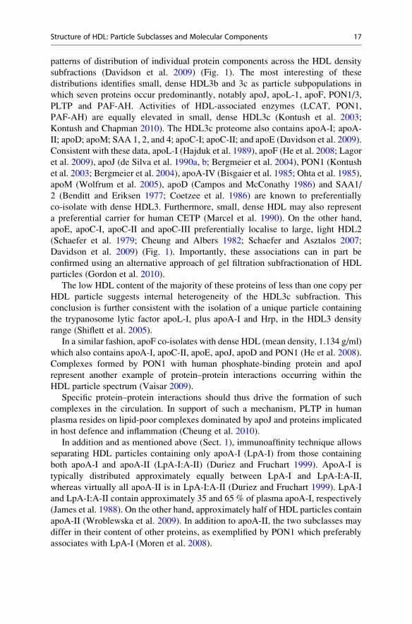
subfractions (Davidson et al. 2009) (Fig. 1). The most interesting of these
distributions identifies small, dense HDL3b and 3c as particle subpopulations in
which seven proteins occur predominantly, notably apoJ, apoL-1, apoF, PON1/3,
PLTP and PAF-AH. Activities of HDL-associated enzymes (LCAT, PON1,
PAF-AH) are equally elevated in small, dense HDL3c (Kontush et al. 2003;
Kontush and Chapman 2010). The HDL3c proteome also contains apoA-I; apoA-
II; apoD; apoM; SAA 1, 2, and 4; apoC-I; apoC-II; and apoE (Davidson et al. 2009).
Consistent with these data, apoL-I (Hajduk et al. 1989), apoF (He et al. 2008; Lagor
et al. 2009), apoJ (de Silva et al. 1990a, b; Bergmeier et al. 2004), PON1 (Kontush
et al. 2003; Bergmeier et al. 2004), apoA-IV (Bisgaier et al. 1985; Ohta et al. 1985),
apoM (Wolfrum et al. 2005), apoD (Campos and McConathy 1986) and SAA1/
2 (Benditt and Eriksen 1977; Coetzee et al. 1986) are known to preferentially
co-isolate with dense HDL3. Furthermore, small, dense HDL may also represent
a preferential carrier for human CETP (Marcel et al. 1990). On the other hand,
apoE, apoC-I, apoC-II and apoC-III preferentially localise to large, light HDL2
(Schaefer et al. 1979; Cheung and Albers 1982; Schaefer and Asztalos 2007;
Davidson et al. 2009) (Fig. 1). Importantly, these associations can in part be
confirmed using an alternative approach of gel filtration subfractionation of HDL
particles (Gordon et al. 2010).
The low HDL content of the majority of these proteins of less than one copy per
HDL particle suggests internal heterogeneity of the HDL3c subfraction. This
conclusion is further consistent with the isolation of a unique particle containing
the trypanosome lytic factor apoL-I, plus apoA-I and Hrp, in the HDL3 density
range (Shiflett et al. 2005).
In a similar fashion, apoF co-isolates with dense HDL (mean density, 1.134 g/ml)
which also contains apoA-I, apoC-II, apoE, apoJ, apoD and PON1 (He et al. 2008).
Complexes formed by PON1 with human phosphate-binding protein and apoJ
represent another example of protein–protein interactions occurring within the
HDL particle spectrum (Vaisar 2009).
Specific protein–protein interactions should thus drive the formation of such
complexes in the circulation. In support of such a mechanism, PLTP in human
plasma resides on lipid-poor complexes dominated by apoJ and proteins implicated
in host defence and inflammation (Cheung et al. 2010).
In addition and as mentioned above (Sect. 1), immunoaffinity technique allows
separating HDL particles containing only apoA-I (LpA-I) from those containing
both apoA-I and apoA-II (LpA-I:A-II) (Duriez and Fruchart 1999). ApoA-I is
typically distributed approximately equally between LpA-I and LpA-I:A-II,
whereas virtually all apoA-II is in LpA-I:A-II (Duriez and Fruchart 1999). LpA-I
and LpA-I:A-II contain approximately 35 and 65 % of plasma apoA-I, respectively
(James et al. 1988). On the other hand, approximately half of HDL particles contain
apoA-II (Wroblewska et al. 2009). In addition to apoA-II, the two subclasses may
differ in their content of other proteins, as exemplified by PON1 which preferably
associates with LpA-I (Moren et al. 2008).
Structure of HDL: Particle Subclasses and Molecular Components 17
01234
1020
ApoL-I Pon1PLTP ApoJ Pon 3 ApoF PAF-AH
05
10152080
120
ApoBapo(a)
Aver
age
Num
ber o
f Pep
tides
Iden
tifie
d
0
3
6
9
12
apoDapoMSAA 1+2
05
10152025
apoEapoC-II
06
12456075
ApoA-IapoA-II SAA4apoC-I
2b 2a 3a 3b 3c
Class A
Class B
Class C
Class D
Class E
Exclusively dense HDL3
Preferentially dense HDL3
No preferential distribution
Preferentially light HDL2
Exclusively light HDL2
Fig. 1 Abundance pattern of proteins across healthy normolipidemic HDL subpopulations. Class
A: exclusively present in small, dense HDL3b and 3c. Class B: enriched in small, dense HDL3b
and 3c. Class C: equally abundant across HDL subpopulations. Class D: enriched in large, light
HDL2b and 2a. Class E: exclusively present in large, light HDL2b and 2a [modified from
(Davidson et al. 2009)]
18 A. Kontush et al.
LpA-I particles can be further subfractionated according to size. The number of
apoA-I molecules in such subpopulations is increased from two to three to four with
an increase in the particle size (Gauthamadasa et al. 2010). On the other hand, the
entire population of LpA-I:A-II demonstrates the presence of only two apoA-I
molecules per particle, while the number of apoA-II molecules varies from one
dimeric apoA-II to two and then to three. Upon compositional analyses of individ-
ual subpopulations, LpA-I:A-II exhibits comparable proportions for major lipid
classes across subfractions, while LpA-I components show significant variability
(Gauthamadasa et al. 2010).
Another important subpopulation of HDL particles is formed by apoE-
containing HDL. The presence of apoE facilitates expansion of the lipid core as a
result of the accumulation of cholesteryl ester, with formation of large, lipid-rich
HDL; these particles represent an excellent ligand for the LDL receptor (Hatters
et al. 2006).
The diversity of molecules which bind to HDL suggests that the lipoprotein can
serve as a versatile adsorptive surface for proteins and peptides to form complexes
playing roles not only in lipid metabolism but equally in acute-phase response,
innate immune response, complement activation, plaque stability and proteolysis
inhibition (Heinecke 2009). As the abundance of the most of HDL-associated
proteins is below 1 mol/mol HDL (i.e. less than 1 copy per HDL particle), it
remains however unclear as to how they are distributed among minor HDL
subpopulations of potentially distinct origin and function. Specific proteins may
therefore be confined to distinct HDL subpopulations of distinct origin and func-
tion, which are differentially distributed across the HDL particle spectrum
(Davidson et al. 2009). The HDL fraction as a whole therefore appears to represent
“a collection of individualised species with distinct functionalities that happen to
have similar physicochemical properties” (Shah et al. 2013) and are primarily
defined by specific protein–protein interactions facilitated by the presence of
phospholipid, rather than “a transient ensemble of randomly exchanging proteins”
(Gordon et al. 2013; Shah et al. 2013).
2.1.2 Protein Isoforms, Translational and PosttranslationalModifications
In line with the evolvement of proteomics, a large number of proteins have been
identified in HDL as described in the previous section (Vaisar et al. 2007). In
addition, most proteins are expressed as different isoforms due to co- and posttrans-
lational modifications (Karlsson et al. 2005; Candiano et al. 2008). This makes the
proteome of HDL both complex and dynamic, which most likely result in various
HDL particles with different protein composition in respect to the environment.
Common posttranslational modifications (PTMs) such as glycosylations, truncations
and phosphorylations change the charge and/or the size of the protein, which can be
utilised for separation of the isoforms, andmodern mass spectrometric techniques can
be used to detect, characterise and nowadays also measure even small mass
differences in proteins. However, although isoforms of major HDL proteins have
been known for decades (Zannis et al. 1980; Hussain and Zannis 1990), surprisingly
Structure of HDL: Particle Subclasses and Molecular Components 19
little is still known on how these variations of the HDL proteome affect the function-
ality. The following is a comprehensive review of isoforms patterns described in
common human HDL apolipoproteins, apoA-I, apoA-II, apoC-III and SAA, and their
possible functional relevance.
ApoA-I (pI 5.3/28 kDa, accession no. P02647) is normally found as different
charge isoforms; besides the major isoform (70–75 % of total apoA-I) and the
slightly more basic pre-apoA-I (5–10 % of total apoA-I), also two more acidic
isoforms are generally detected by isoelectric focusing (IEF) and 2D gel electro-
phoresis (2-DE) (Contiero et al. 1997; Karlsson et al. 2005). The nature of these
acidic isoforms is still unclear. An early report suggested deamidation of Gln or Asn
residues, resulting in a +1 charge shift, which could be formed during the analytical
procedure (Ghiselli et al. 1985). At the same time, a few reports indicated the
importance of acidic apoA-I in vivo; increased levels of acidic apoA-I, while
decreased levels of the major form, were found in LDL from obese subjects,
especially in women (Karlsson et al. 2009). Also, higher degree of deamidated
apoA-I has been shown in relation to diabetes (Jaleel et al. 2010) and acidic apoA-I
may be more vulnerable to methionine oxidation (Fernandez-Irigoyen et al. 2005).
In 2-DE HDL protein patterns also 30–35 kDa variants of apoA-I are usually
detected (Karlsson et al. 2005). Although mass spectrometry (MS) analysis was
in agreement with O-glycosylation at two potential sites (Thr78 or Thr92), this has
not been confirmed by others, and it is generally regarded that apoA-I is not
N-linked or O-linked glycosylated. In contrast, non-enzymatically glycation of
apoA-I has been found in association to diabetes and believed to affect apoA-I
functions, such as LCAT activation (Fievet et al. 1995; Nobecourt et al. 2007; Park
et al. 2010).
Another potentially important PTM of apoA-I is truncation. During atheroscle-
rotic inflammation, apoA-I might be N-terminally and C-terminally truncated by
released proteases. Specific cleavage sites at Tyr42, Phe57, Tyr216 and Phe253 for
chymase have been identified that in reconstituted HDL reduces its ability to
promote cholesterol efflux (Lee et al. 2003; Usami et al. 2013). Low amounts of
C-terminally truncated apoA-I can be measured in normal serum (Usami
et al. 2011) and fragmented apoA-I is a feature in plasma from children with
nephrotic syndrome, a condition linked to higher risk of atherosclerosis (Santucci
et al. 2011). Truncation has also been implicated in apoA-I dimerisation as studied
in apoA-I Milano (R197C) and apoA-I Paris (R175C) (Calabresi et al. 2001; Favari
et al. 2007; Gursky et al. 2013). Notably, apoA-I with an apparent molecular mass
of 50 kDa, consistent with dimeric apoA-I, has been found in patients with
myocardial infarction (Majek et al. 2011) but also appears to be present in HDL
from healthy individuals (Karlsson et al. 2005). Finally, oxidatively modified
apoA-I have been extensively studied during the recent years by the help of MS
techniques, as described in detail in several reviews elsewhere (e.g. Nicholls and
Hazen 2009; Shao 2012). It has been proposed that myeloperoxidase-mediated
inflammation results in oxidation of apoA-I. Specific sites have been identified
for methionine oxidation, for nitrated/chlorinated tyrosines and for lysines modified
by reactive carbonyls. Importantly, the modifications have been coupled to
20 A. Kontush et al.
functional impairment in apoA-I activity such as ABCA1-mediated cholesterol
efflux and are linked to cardiovascular disease.
ApoA-II (pI 5.0/8.7 kDa, accession no. P02652) is mostly found as two isoforms
in HDL that differ slightly according to pI and molecular mass, probably due to
O-linked glycosylation/sialylation (Karlsson et al. 2005; Halim et al. 2013). Similar
to apoA-I, the protein is produced as a more basic pro-form (Hussain and Zannis
1990). In contrast, apoA-II appears to be quickly processed to the mature form, as
the pro-form is not found in the circulation. In addition to glycosylation, phosphor-
ylation at Ser68, C-terminal truncated variants (des-Gln and des-Thr-Gln) and a
cysteinylated variant has been detected in the circulation (Jin and Manabe 2005;
Nelsestuen et al. 2008; Zhou et al. 2009). ApoA-II also forms a homodimer at
Cys29 that is abundant in plasma (Gillard et al. 2005; Jin and Manabe 2005).
Overall, more than ten different variants of apoA-II are present in humans, but
the physiological relevance of this heterogeneity is unclear. However, sialylated
apoA-II appear to be selectively associated to HDL3 (Remaley et al. 1993; Karlsson
et al. 2005), and elevated levels of modified apoA-II isoforms have been linked to
premature delivery in pregnant women (Flood-Nichols et al. 2013).
ApoC-III (pI 4.7/8.8 kDa, accession no. P02656) is generally found as three
charge isoforms depending on O-linked glycosylation (GalGalNAc) at Thr94 with
or without sialylation; disialylated apoC-III2, monosialylated apoCIII1 and
non-sialylated apoC-III0 (Karlsson et al. 2005; Bruneel et al. 2008). An early report
showed that glycosylation is not necessary for apoC-III secretion and does not
affect its relative affinity to different lipoprotein particles (Roghani and Zannis
1988), and, as judged by gel electrophoresis and MS analysis of HDL and plasma,
the non-sialylated variant is least abundant, usually less than 5 % of total apoC-III
in normal individuals (Wopereis et al. 2003; Bruneel et al. 2008; Mazur et al. 2010;
Holleboom et al. 2011). In addition to glycosylation, apoC-III can also be
C-terminal truncated (des-Ala and des-Ala-Ala), which further increases the num-
ber of isoforms (Bondarenko et al. 1999; Jin and Manabe 2005; Nicolardi
et al. 2013a). Interestingly, novel results strongly suggest that glycosylation of
apoC-III is an important event in the regulation of lipid metabolism (Holleboom
et al. 2011; Baenziger 2012). Thus, apoC-III is exclusively glycosylated by GalNAc
transferase 2 (GALNT2) (Holleboom et al. 2011; Schjoldager et al. 2012), and
heterozygotes with a loss-of-function mutation in GALNT2 present with an altered
apoC-III isoform pattern with more of the non-sialylated variant and less of the
monosialylated variant, while the total apoC-III plasma concentration was about the
same as compared to wild-type controls (Holleboom et al. 2011). This is then linked
to reduced inhibition of lipoprotein lipase and improved triglyceride clearance. In
line, the production rate of apoC-III1 and -III2 is more strongly correlated with
plasma triglyceride levels than apoC-III0 (Mauger et al. 2006), increased apoC-III1/
apoC-III0 ratio has been found in diabetic subjects (Jian et al. 2013), and HDL3
from subjects with low HDL-C is characterised by higher levels of monosialylated
apoC-III than subjects with high HDL-C (Mazur et al. 2010). The evaluation of
apoC-III isoforms is complicated by the fact that apoC-III0 can be separated into a
non-glycosylated form and glycosylated but non-sialylated forms (Bruneel
Structure of HDL: Particle Subclasses and Molecular Components 21

et al. 2008; Holleboom et al. 2011; Nicolardi et al. 2013a). Moreover, a recent MS
study of 96 serum samples showed that 30 % of the individuals displayed an apoC-
III pattern with additional glycosylated variants, characterised by fucosylation
(Nicolardi et al. 2013b). The relevance of these glycosylated non-sialylated variants
of apoC-III, as of the C-terminal truncated forms, is yet unclear, but may explain
somewhat contradictory results showing higher relative levels of non- and less-
sialylated apoC-III in obese subjects than in lean subjects (Harvey et al. 2009;
Karlsson et al. 2009), although obesity is generally associated with high triglyceride
levels.
SAA exists in a form of SAA1 (pI 5.9/11.7 kDa, accession no. P0DJI8) and
SAA2 (pI 8.3/11.6 kDa, accession no. P0DJI9). The two proteins display about
93 % sequence homology, and depending on natural variation in alleles, SAA1 is
separated into five isoforms, SAA1.1 to SAA1.5, and SAA2 is separated into two
isoforms SAA2.1 and SAA2.2 (often also denoted as alpha, beta, etc.), with one to
three amino acid difference between the isoforms. In addition, both N-terminal and
C-terminal truncated variants of SAA1/SAA2 are detected in serum (Ducret
et al. 1996; Kiernan et al. 2003; de Seny et al. 2008). By using a combined 2-DE/
MS and SELDI-TOF approach, eight isoforms were identified in HDL after LPS
infusion in healthy individuals; besides native SAA1.1 and SAA2.1, N-terminal
truncations (des-R, -RS and -RSFF) of each variant were also found (Levels
et al. 2011). Interestingly, a subgroup, based on HDL protein profile, characterised
by elevated antioxidative PON1 activity showed a delayed response of SAA to LPS
in particular for the most truncated (des-RSFF) variants. Otherwise, very little is
known about differential physiological relevance of the SAA isoforms. However,
SAA2.1 but not SAA1.1 has been shown to promote cholesterol efflux from
macrophages (Kisilevsky and Tam 2003). Today, contradictory results make it
unclear whether the increased level of SAA in inflammation, believed to replace
apoA-I in HDL, actually is a mechanism in atherosclerosis or merely is a marker for
inflammation (de Beer et al. 2010; Chiba et al. 2011; Kisilevsky and Manley 2012).
Future differential quantitative MS analysis of the highly homologous SAA
isoforms such as described by Sung et al. (2012) may resolve this controversy.
Sensitive MS techniques have revealed a large number of proteins associated to
HDL. Most of them are also expressed as different isoforms depending on transla-
tional and posttranslational modifications. This leads to a need to develop
MS-based methods for specific and reliable quantification of protein isoforms.
With appropriate standards, measurements can be performed with low coefficient
of variation and with a specificity superior to, e.g. immunoassays. Consequently,
such applications in the field of HDL are being presented by using, e.g. multiple
reaction monitoring and top-down proteomics with high-resolution MS (Mazur
et al. 2010; Sung et al. 2012). Another interesting and fairly simple approach is to
use ratio determinations, e.g. modified/native protein expression (Nelsestuen
et al. 2008). As these measures are concentration-independent, they bear a potential
to reduce individual variations. Furthermore, such MS approaches are not only
useful for PTMs but also of value to understand the impact of protein variations
caused by genetic polymorphism. For example, a recent study of heterozygotes
22 A. Kontush et al.

with an apoA-I mutation (K131Del) showed that, in contrast to what could be
expected, the mutant protein was more abundantly expressed in HDL than the
native protein (Ljunggren et al. 2013). Herein four HDL proteins that are all
expressed as different isoforms have been discussed: two (apoA-I and apoC-III)
in which PTMs have been shown to be important for lipid metabolism and two
(apoA-II and SAA) in which the role of PTMs is still unclear. In light of the vital
importance of carboxylations and truncations in the processes of haemostasis,
which also involves other PTMs such as phosphorylation, hydroxylation, glycosyl-
ation and sulphation, it appears highly unlikely that the diversity in the “HDLome”
would not be relevant for lipid metabolism and cardiovascular disease. Therefore,
characterisation of PTMs is probably one of the most challenging but also one of the
most important tasks in order to understand the complex function of HDL.
2.2 Lipidome
The real power of lipidomic technologies involving mass spectrometry results from
their ability to provide quantitative data on individual molecular species of lipids
and on low-abundance lipid molecules. The pioneering study of Wiesner and
colleagues published in 2009 (Wiesner et al. 2009) provided reference values for
the lipidome of HDL isolated from healthy normolipidemic controls by FPLC. In an
attempt to further characterise HDL composition and address its inherent heteroge-
neity, we recently reported the phospho- and sphingolipidome of five major HDL
subpopulations isolated from healthy normolipidemic subjects (Camont
et al. 2013).
2.2.1 PhospholipidsPhosphatidylcholine is the principal plasma phospholipid that accounts for 32–
35 mol % of total lipids in HDL (Wiesner et al. 2009) (Table 3). PC is a structural
lipid, consistent with its even distribution across HDL subpopulations (Fig. 2).
Major molecular species of PC are represented by the 16:0/18:2, 18:0/18;2 and
16:0/20:4 species (Lhomme et al. 2012). As compared to other lipoproteins, HDL is
enriched in PC containing polyunsaturated fatty acid moieties (Wiesner et al. 2009).
LysoPC is an important phospholipid subclass in HDL (1.4–8.1 mol % of total
lipids; Table 3). It is derived from the regulated degradation of PC by
phospholipases, including LCAT, consistent with the preferential association of
the latter with HDL particles (Kontush et al. 2007). More specifically, LCAT was
reported earlier to associate mainly with small, dense HDL particles, which are also
enriched in lysoPC by approximately twofold as compared to large, light HDL
(Camont et al. 2013) (Fig. 2). LysoPC is also produced by the hydrolytic action of
Lp-PLA2 on oxidised PC or by secreted PLA2 under pro-atherogenic conditions,
such as oxidative stress and inflammation, and constitutes therefore a potential
biomarker of inflammation. Major molecular species of HDL lysoPC contain
saturated fatty acid moieties of predominantly 16 and 18 carbon atoms, reflecting
LCAT preference for 16 and 18 carbon atom long PCs (Lhomme et al. 2012). As
Structure of HDL: Particle Subclasses and Molecular Components 23
considerable amounts of serum lysoPC are also associated with albumin (Wiesner
et al. 2009), HDL contamination by the both compounds is typical for FPLC
isolation. However, in HDL isolated by isopycnic density gradient ultracentrifuga-
tion, lysoPC content in HDL is two- to tenfold lower (Camont et al. 2013; Stahlman
et al. 2013).
Phosphatidylethanolamine (PE) is moderately abundant in HDL (0.7–
0.9 mol % of total lipids; Table 3), and its content tends to increase with increasing
HDL hydrated density (Wiesner et al. 2009; Camont et al. 2013) (Fig. 2). PE
principal molecular species are represented by the 36:2 and 38:4 fatty acid residues
in HDL (Kontush et al. 2007).
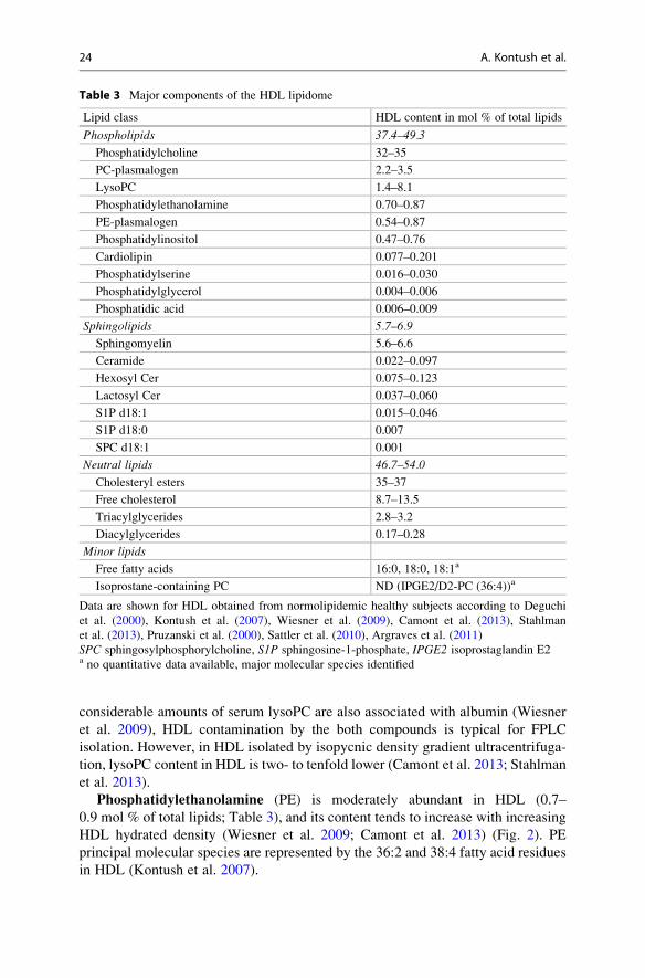
Table 3 Major components of the HDL lipidome
Lipid class HDL content in mol % of total lipids
Phospholipids 37.4–49.3
Phosphatidylcholine 32–35
PC-plasmalogen 2.2–3.5
LysoPC 1.4–8.1
Phosphatidylethanolamine 0.70–0.87
PE-plasmalogen 0.54–0.87
Phosphatidylinositol 0.47–0.76
Cardiolipin 0.077–0.201
Phosphatidylserine 0.016–0.030
Phosphatidylglycerol 0.004–0.006
Phosphatidic acid 0.006–0.009
Sphingolipids 5.7–6.9
Sphingomyelin 5.6–6.6
Ceramide 0.022–0.097
Hexosyl Cer 0.075–0.123
Lactosyl Cer 0.037–0.060
S1P d18:1 0.015–0.046
S1P d18:0 0.007
SPC d18:1 0.001
Neutral lipids 46.7–54.0
Cholesteryl esters 35–37
Free cholesterol 8.7–13.5
Triacylglycerides 2.8–3.2
Diacylglycerides 0.17–0.28
Minor lipids
Free fatty acids 16:0, 18:0, 18:1a
Isoprostane-containing PC ND (IPGE2/D2-PC (36:4))a
Data are shown for HDL obtained from normolipidemic healthy subjects according to Deguchi
et al. (2000), Kontush et al. (2007), Wiesner et al. (2009), Camont et al. (2013), Stahlman
et al. (2013), Pruzanski et al. (2000), Sattler et al. (2010), Argraves et al. (2011)
SPC sphingosylphosphorylcholine, S1P sphingosine-1-phosphate, IPGE2 isoprostaglandin E2a no quantitative data available, major molecular species identified
24 A. Kontush et al.

Plasmalogens contain a vinyl ether-linked fatty acid essential for their specific
antioxidative properties (Maeba and Ueta 2003). PC-plasmalogens are the most
abundant species in HDL (2.2–3.5 mol %) but represent less than 10 % of total PC
(Stahlman et al. 2013). On the contrary, PE-plasmalogens and PE are equally
abundant in HDL (0.6–0.9 mol %; Table 3). PC- and PE-plasmalogens contain
0.035
0.045
0.055
0.065
0.075
0.085
0.095Ceramide
0.0035
0.0055
LysoPCPEPI
0.4
0.8
1.2
1.6
Mol
% o
f tot
al li
pids
2b 2a 3a 3b 3c
Class B
Class C
Class D
Class E
0.000
0.010
PSPAS1P
0.02
0.08
0.14
0.20Class A
PG
4.55.56.57.5
Free cholesterolSphingomyelin
11
16
21048
CEPCTAG
1222324252
Highly enriched in dense HDL3
Enriched in dense HDL3
Equally abundant
Enriched in light HDL2
Highly enriched in light HDL2
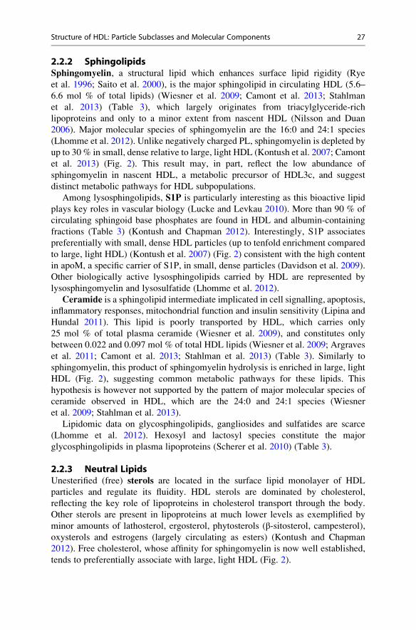
Fig. 2 Abundance pattern of lipids across healthy normolipidemic HDL subpopulations. Class A:
highly enriched in small, dense HDL3b and 3c (>1.5-fold relative to HDL2b). Class B: enriched in
small, dense HDL3b and 3c (1.2–1.5-fold relative to HDL2b). Class C: equally abundant across
HDL subpopulations (<1.2-fold variations between HDL2b and HDL3b+ 3c). Class D: enriched
in large, light HDL2b (1.2–1.5-fold relative to HDL3b + 3c). Class E: highly enriched in large,
light HDL2b (>1.5-fold relative to HDL3b + 3c). S1P, sphingosine-1-phosphate
Structure of HDL: Particle Subclasses and Molecular Components 25
mainly polyunsaturated species: 38:5 and 36:2 in PC-plasmalogens (Stahlman
et al. 2013) and 18:0/20:4 and 16:0/20:4 in PE-plasmalogens (Wiesner et al. 2009).
Phosphatidylinositol (PI), phosphatidylserine (PS), phosphatidylglycerol
(PG), phosphatidic acid (PA) and cardiolipin are negatively charged
phospholipids present in HDL (Table 3) which may significantly impact on its net
surface charge (Rosenson et al. 2011; Lhomme et al. 2012). The content of these
lipids can thereby modulate lipoprotein interactions with lipases, membrane proteins,
extracellular matrix and other protein components; indeed, such interactions are
largely charge-dependent.
PI, similarly to PE, is moderately abundant in HDL (0.5–0.8 mol %; Table 3) and
tends to be enriched in small, dense HDL (Fig. 2). Major molecular species of PI in
HDL include the 18:0/20:3 and 18:0/20:4 species (Lee et al. 2010).
PS is a minor negatively charged phospholipid component of HDL (0.016–
0.030 mol %; Table 3). This phospholipid was very recently reported to be highly
enriched (34-fold) in the small, dense HDL3c subpopulation relative to large, light
HDL2 (Fig. 2) (Camont et al. 2013) as well as in small discoid preβ HDL and small
nascent HDL formed by ABCA1 (up to 2.5 mol % of total lipids) (see Kontush and
Chapman 2012 for review). Interestingly, small, dense HDL also displayed potent
biological activities which correlated positively with PS content in HDL (Camont
et al. 2013). This lipid could therefore, in part, account for enhanced functionality
of HDL3c.
PA, a second messenger, is both a common metabolic precursor and an enzy-
matic product of phospholipid metabolism. This negatively charged lipid is present
in very low abundance in HDL (0.006–0.009 mol %; Table 3) but, similarly to PS, is
enriched in small, dense HDL (by more than threefold) (Camont et al. 2013)
(Table 3). This observation might reflect preferential association of PA with
apoL-I which is equally enriched in small, dense HDL (Kontush and Chapman
2012).
PG is a metabolic precursor of cardiolipin present in HDL in very low amounts
(0.004–0.006 mol %; Table 3). PG tends to be enriched in small, dense particles
(Camont et al. 2013) (Fig. 2).
Cardiolipin is a minor anionic phospholipid present in trace amounts in HDL
(0.08–0.2 mol %; Table 3). This lipid with potent anticoagulant properties may
contribute to the effects of lipoproteins on coagulation and platelet aggregation
(Deguchi et al. 2000).
Together, these data indicate that although negatively charged lipids represent
minor HDL constituents (0.8 mol % of total lipids), they are highly enriched in
small, dense HDL, consistent with the elevated surface electronegativity of this
subpopulation (Rosenson et al. 2011).
Isoprostanes are well established as biomarkers of oxidative stress and are
predominantly associated with HDL (see Kontush and Chapman 2012 for review).
Major molecular species of isoprostane-containing PCs include 5,6-epoxy-
isoprostaglandine A2-PC (EIPGA2-PC) 36:3, 5,6 EIPGE2-PC 36:4, IPGE2/D2-
PC 36:4, IPGF-PC 36:4, IPGE2/D2-PC 38:4 and IPGF-PC 38:4 (Pruzanski et al. 2000)
(Table 3).
26 A. Kontush et al.
2.2.2 SphingolipidsSphingomyelin, a structural lipid which enhances surface lipid rigidity (Rye
et al. 1996; Saito et al. 2000), is the major sphingolipid in circulating HDL (5.6–
6.6 mol % of total lipids) (Wiesner et al. 2009; Camont et al. 2013; Stahlman
et al. 2013) (Table 3), which largely originates from triacylglyceride-rich
lipoproteins and only to a minor extent from nascent HDL (Nilsson and Duan
2006). Major molecular species of sphingomyelin are the 16:0 and 24:1 species
(Lhomme et al. 2012). Unlike negatively charged PL, sphingomyelin is depleted by
up to 30 % in small, dense relative to large, light HDL (Kontush et al. 2007; Camont
et al. 2013) (Fig. 2). This result may, in part, reflect the low abundance of
sphingomyelin in nascent HDL, a metabolic precursor of HDL3c, and suggest
distinct metabolic pathways for HDL subpopulations.
Among lysosphingolipids, S1P is particularly interesting as this bioactive lipid
plays key roles in vascular biology (Lucke and Levkau 2010). More than 90 % of
circulating sphingoid base phosphates are found in HDL and albumin-containing
fractions (Table 3) (Kontush and Chapman 2012). Interestingly, S1P associates
preferentially with small, dense HDL particles (up to tenfold enrichment compared
to large, light HDL) (Kontush et al. 2007) (Fig. 2) consistent with the high content
in apoM, a specific carrier of S1P, in small, dense particles (Davidson et al. 2009).
Other biologically active lysosphingolipids carried by HDL are represented by
lysosphingomyelin and lysosulfatide (Lhomme et al. 2012).
Ceramide is a sphingolipid intermediate implicated in cell signalling, apoptosis,
inflammatory responses, mitochondrial function and insulin sensitivity (Lipina and
Hundal 2011). This lipid is poorly transported by HDL, which carries only
25 mol % of total plasma ceramide (Wiesner et al. 2009), and constitutes only
between 0.022 and 0.097 mol % of total HDL lipids (Wiesner et al. 2009; Argraves
et al. 2011; Camont et al. 2013; Stahlman et al. 2013) (Table 3). Similarly to
sphingomyelin, this product of sphingomyelin hydrolysis is enriched in large, light
HDL (Fig. 2), suggesting common metabolic pathways for these lipids. This
hypothesis is however not supported by the pattern of major molecular species of
ceramide observed in HDL, which are the 24:0 and 24:1 species (Wiesner
et al. 2009; Stahlman et al. 2013).
Lipidomic data on glycosphingolipids, gangliosides and sulfatides are scarce
(Lhomme et al. 2012). Hexosyl and lactosyl species constitute the major
glycosphingolipids in plasma lipoproteins (Scherer et al. 2010) (Table 3).
2.2.3 Neutral LipidsUnesterified (free) sterols are located in the surface lipid monolayer of HDL
particles and regulate its fluidity. HDL sterols are dominated by cholesterol,
reflecting the key role of lipoproteins in cholesterol transport through the body.
Other sterols are present in lipoproteins at much lower levels as exemplified by
minor amounts of lathosterol, ergosterol, phytosterols (β-sitosterol, campesterol),
oxysterols and estrogens (largely circulating as esters) (Kontush and Chapman
2012). Free cholesterol, whose affinity for sphingomyelin is now well established,
tends to preferentially associate with large, light HDL (Fig. 2).
Structure of HDL: Particle Subclasses and Molecular Components 27

Cholesteryl esters (CE) are largely (up to 80 %) formed in plasma HDL (Fig. 2),
as a result of transesterification of PL and cholesterol catalysed by LCAT. These
highly hydrophobic lipids form the lipid core of HDL and contribute up to 36 mol %
of total HDL lipid (Wiesner et al. 2009; Camont et al. 2013; Stahlman et al. 2013)
(Table 3). Most of HDL CE is accounted for by cholesteryl linoleate (Lhomme
et al. 2012). A pioneering work on CE molecular species distribution across HDL
subpopulations using gas chromatography showed very similar profiles between
HDL2 and HDL3 particles (Vieu et al. 1996).
HDL-associated triacylglycerides (TAG) are dominated by species containing
oleic, palmitic and linoleic acid moieties (Lhomme et al. 2012) and represent
around 3 mol % of total HDL lipids (Vieu et al. 1996; Kontush et al. 2007; Wiesner
et al. 2009; Camont et al. 2013; Stahlman et al. 2013) (Table 3). Similarly to CE,
TAG species profile is conserved between HDL2 and HDL3 (Vieu et al. 1996).
Minor bioactive lipids present in HDL include diacylglycerides (DAG),
monoacylglycerides (MAG) and free fatty acids (Lhomme et al. 2012). In the
study of Vieu et al., sn-1,2 and sn-1,3 isomers of DAG species were identified in
HDL (Vieu et al. 1996). More recently, detailed characterisation of DAG molecular
species in HDL revealed 16:0/18:1 and 18:1/18:1 as major species, estimating their
content at 0.2 mol % of total lipids (Stahlman et al. 2013) (Table 3).
HDL composition in free fatty acids shows a predominance of palmitic, stearic
and oleic acid-containing species (Lhomme et al. 2012).
Together, available lipidomic studies have already provided detailed
characterisation of HDL isolated using two complementary techniques, FPLC
(Wiesner et al. 2009) and ultracentrifugation (Kontush et al. 2007; Camont
et al. 2013). The latter studies demonstrated preferential association of negatively
charged lipids (PS, PA) and S1P with small, dense HDL together with preferential
association of sphingomyelin and ceramide with large, light particles, consistent
with distinct metabolic origins and potent biological activities of small, dense HDL
(Camont et al. 2013). These data illustrate the power of lipidomics to provide
crucial information on the metabolism and function of lipoproteins relevant for
the development of cardiovascular disease, which can in turn deliver novel
biomarkers of cardiovascular risk.
3 The Structure of HDL
3.1 Introduction/Brief History
When it was becoming clear that plasma levels of HDL were inversely correlated
with cardiovascular disease, numerous laboratories were already doing pioneering
work on the structure of human plasma HDL (Edelstein et al. 1972; Jonas 1972;
Laggner et al. 1973; Atkinson et al. 1974; Schonfeld et al. 1976; Tardieu
et al. 1976). Knowing little about the nature of the stabilising proteins, Scanu and
colleagues used detailed compositional studies to build models of human HDL3
28 A. Kontush et al.
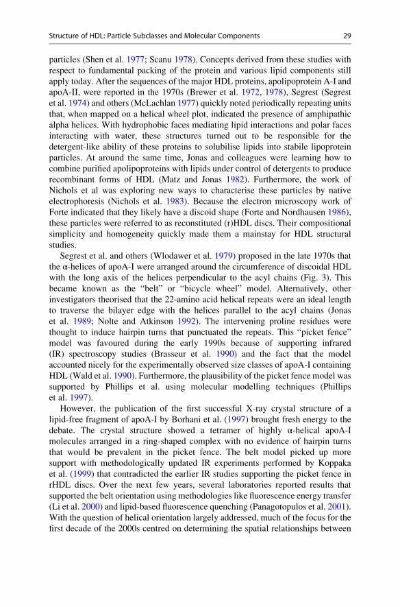
particles (Shen et al. 1977; Scanu 1978). Concepts derived from these studies with
respect to fundamental packing of the protein and various lipid components still
apply today. After the sequences of the major HDL proteins, apolipoprotein A-I and
apoA-II, were reported in the 1970s (Brewer et al. 1972, 1978), Segrest (Segrest
et al. 1974) and others (McLachlan 1977) quickly noted periodically repeating units
that, when mapped on a helical wheel plot, indicated the presence of amphipathic
alpha helices. With hydrophobic faces mediating lipid interactions and polar faces
interacting with water, these structures turned out to be responsible for the
detergent-like ability of these proteins to solubilise lipids into stabile lipoprotein
particles. At around the same time, Jonas and colleagues were learning how to
combine purified apolipoproteins with lipids under control of detergents to produce
recombinant forms of HDL (Matz and Jonas 1982). Furthermore, the work of
Nichols et al was exploring new ways to characterise these particles by native
electrophoresis (Nichols et al. 1983). Because the electron microscopy work of
Forte indicated that they likely have a discoid shape (Forte and Nordhausen 1986),
these particles were referred to as reconstituted (r)HDL discs. Their compositional
simplicity and homogeneity quickly made them a mainstay for HDL structural
studies.
Segrest et al. and others (Wlodawer et al. 1979) proposed in the late 1970s that
the α-helices of apoA-I were arranged around the circumference of discoidal HDL
with the long axis of the helices perpendicular to the acyl chains (Fig. 3). This
became known as the “belt” or “bicycle wheel” model. Alternatively, other
investigators theorised that the 22-amino acid helical repeats were an ideal length
to traverse the bilayer edge with the helices parallel to the acyl chains (Jonas
et al. 1989; Nolte and Atkinson 1992). The intervening proline residues were
thought to induce hairpin turns that punctuated the repeats. This “picket fence”
model was favoured during the early 1990s because of supporting infrared
(IR) spectroscopy studies (Brasseur et al. 1990) and the fact that the model
accounted nicely for the experimentally observed size classes of apoA-I containing
HDL (Wald et al. 1990). Furthermore, the plausibility of the picket fence model was
supported by Phillips et al. using molecular modelling techniques (Phillips
et al. 1997).
However, the publication of the first successful X-ray crystal structure of a
lipid-free fragment of apoA-I by Borhani et al. (1997) brought fresh energy to the
debate. The crystal structure showed a tetramer of highly α-helical apoA-I
molecules arranged in a ring-shaped complex with no evidence of hairpin turns
that would be prevalent in the picket fence. The belt model picked up more
support with methodologically updated IR experiments performed by Koppaka
et al. (1999) that contradicted the earlier IR studies supporting the picket fence in
rHDL discs. Over the next few years, several laboratories reported results that
supported the belt orientation using methodologies like fluorescence energy transfer
(Li et al. 2000) and lipid-based fluorescence quenching (Panagotopulos et al. 2001).
With the question of helical orientation largely addressed, much of the focus for the
first decade of the 2000s centred on determining the spatial relationships between
Structure of HDL: Particle Subclasses and Molecular Components 29
apoA-I molecules on these discs. This effort was aided by the detailed “double belt”
computer model put forth in the classic paper by Segrest (Segrest et al. 1999).
The preceding paragraphs quickly summarise a tremendous amount of work
between 1970 and 2000 aimed at understanding the structure of HDL and its
recombinant forms, and by no means does it do justice to all contributions. For
more complete discussions of this period of HDL structural research, the reader is
referred to the classic review series by Brouillette (Brouillette and Anantharamaiah
1995; Brouillette et al. 2001). In addition, due to space limitations, this section will
not focus on the structure of lipid-free apolipoproteins nor their proposed
mechanisms for lipid binding and particle generation. The reader is directed to
the recent review by Phillips (Phillips 2013) for an excellent discussion of these
issues. For the purposes of this section, we will focus on work done over the last
14 years that has built upon the concept of the double belt model in both
reconstituted and “real” circulating plasma HDL.
Fig. 3 The double belt model for a rHDL discoidal particle and its various refinements. The
classic LL 5/5 double belt model for apoA-I as proposed by Segrest (Segrest et al. 2000) is shown
in panel (a). Each amphipathic helical domain is shown in a different colour with helix 5 in greenat the front. This model did not include the N-terminal 43 a.a. The “belt and buckle” model (b)
does not form a continuous circle around the particle but has its N-terminal 43 a.a. folding back
across the belts (40). The “looped belt” (c) features a localised separation between residues
133 and 146 (helix 5) (41). The “solar flares” model (d) encapsulates the lipid with an antiparallel
double belt but has disorganised domains that resemble solar flares erupting between residues
165 and 180. The double superhelix model (e) maintains similar intermolecular contact among the
molecules of apoA-I as the double belt, but the lipid is stabilised as an elongated micelle and no
intramolecular contact is made between the N- and C-termini of apoA-I
30 A. Kontush et al.
3.2 HDL in the Test Tube
3.2.1 Discoid HDLDiscoid HDLs probably exist in human plasma but are extremely short-lived due to
the fact that they are excellent substrates for LCAT (Jonas 2002). HDL discs have
been detected in sequestered compartments like peripheral lymph and interstitial
fluid which likely have lower LCAT activity (Sloop et al. 1983). As stated above,
discs can be reconstituted in vitro, and these particles exhibit many of the traits of
native HDL such as LCAT activation (Jonas and McHugh 1984), lipid transfer
(Davidson et al. 1995) and receptor binding (Liadaki et al. 2000).
The most well-studied rHDL discs consist of two apoA-I molecules, are ~96 A in
diameter and ~47 A in thickness and contain 150–200 phospholipid molecules
(Jonas 1986; Jonas et al. 1989). This “benchmark” particle was used as a basis for
the original double belt molecular model (Segrest et al. 1999). In this model, two
ring-shaped apoA-I molecules encapsulate a lipid membrane leaflet in an antiparal-
lel orientation (Segrest 1977; Segrest et al. 1999; Brouillette et al. 2001; Klon
et al. 2002a) in which helix 5 of both apoA-I molecules directly oppose each other
(Fig. 3a). In this configuration, there are two possible interfaces between the
molecules depending on how they are stacked: left to left (LL) and right to right
(RR). Computer analysis indicated that an LL interface in which helix 5 (more
specifically, glycine 129) of both molecules are directly opposed produced the
highest weighted score of potential salt bridges between the apoA-I molecules
(Segrest et al. 1999). This is called the LL5/5 (G129j) registry and, interestingly,
is the same orientation found in the Borhani crystal structure (Borhani et al. 1997).
This orientation was experimentally validated in three studies that utilised chemical
cross-linking to derive the molecular orientation of the two apoA-I molecules in
these particles. Two from our laboratory were highly consistent with the 5/5
orientation across most of the molecule, although certain cross-links were also
consistent with a second 5/2 registry (Davidson and Hilliard 2003; Silva
et al. 2005). Cross-linking studies by Bhat et al. (2005) were also consistent with
the general 5/5 belt model for most of the molecule, but these investigators
proposed that the N- and C-terminal 40–50 residues doubled back on the molecule
(Fig. 3b). This refinement was called the “belt and buckle”. Electron paramagnetic
resonance studies performed by Martin and colleagues (Martin et al. 2006) on 96 A
rHDL particles were also consistent with a 5/5 antiparallel double belt orientation
over most of the particle circumference. However, they observed that the spin
coupling signatures of residues 134–145 (within helix 5) were consistent with a
looping region that causes a localised opening between the parallel belts. This
“looped belt” was proposed to be a potential site by which LCAT could gain access
to the cholesterol and phospholipid acyl chains that are otherwise buried in the lipid
bilayer (Fig. 3c). Using hydrogen/deuterium exchange (HDX) experiments, Wu and
colleagues proposed an alternative refinement of the double belt model which
included the full-length apoA-I (Wu et al. 2007). This “solar flares” model again
featured a 5/5 double belt for most of the particle, but the N-termini were modelled
as globular nodules (Fig. 3d). The HDX profile of the region between 165 and
Structure of HDL: Particle Subclasses and Molecular Components 31
180 suggested that this region loops off the particle edge and could be a site for
LCAT interaction. This argument was bolstered by results showing that added
LCAT could alter the exchange patterns of these residues and that mutations in
this region affected LCAT activity. The conclusions of this study were called into
some question by a more recent HDX study performed by Chetty et al. on similar
particles that failed to support the existence of the looped solar flares (Chetty
et al. 2013). They showed that almost all of apoA-I was helical in the discs with
the exception of the first 6 and last 7 residues. Interestingly, the region between
residues 125 and 158 exhibited bimodal exchange kinetics, suggesting that this
region flits between a helical and less ordered structure. This is consistent with the
looped belt idea (Martin et al. 2006), but the putative solar flares region
(Wu et al. 2007) was not found to be particularly dynamic in this study. The authors
suggested that this region is responsible for absorbing changes in particle size by
unfolding under conditions when lipid surface area is limiting. This is consistent
with previous studies from our laboratory which used lipid-based fluorescence
quenching agents to show that this same region changes its exposure to lipid
when the particle diameter is reduced (Maiorano et al. 2004).
The recent publication of a high-resolution (2.2 A) crystal structure of a lipid-
free truncation mutant of apoA-I (Δ185–243) also strongly supports the double beltmodel (Mei and Atkinson 2011). Although lipid-free, the dimeric apoA-I formed a
curved structure derived from long, kinked alpha helices. Interestingly, much of the
structure resembled the 5/5 double belt, even down to a similar degree of curvature.
The model also suggested increased temperature factors in helix 5 hinting that there
could be a separation of the helices at this site, consistent with the looped belt idea.
This advance should prove to be a valuable guide for future modelling of both the
lipid-free and lipid-bound forms of apoA-I.
The models discussed above conform to the generally accepted idea that these
rHDLs are discoid in shape. However, Hazen and colleagues proposed a model that
departed from this idea (Wu et al. 2009). Based on small-angle neutron scattering
(SANS), they proposed that antiparallel apoA-I molecules form a double superhe-
lix (DSH) that encapsulates a cigar-shaped micelle of phospholipids (Fig. 3e). Thus,
the particles resemble an ellipsoid rather than a disc. Despite the different shape, the
overall molecular interactions along the interface of the two apoA-I molecules are
the same as the 5/5 double belt model. However, unlike in the planar disc, the N-
and C-termini are not predicted interact in the DSH. The viability of this model
remains under debate as molecular dynamics studies suggest that this model rapidly
collapses to a disc-shaped structure (Jones et al. 2010). Even simulations done by
the group that proposed the model seem to indicate that the elongated structure
collapses to an oblate spheroid, though they argue it remains micellar (Gogonea
et al. 2010). Going forward, this model must be squared with observations of disc-
like shapes by electron microscopy (Zhang et al. 2011), EPR data (Jones
et al. 2010), and the quantised changes in particle size that can be readily
demonstrated by reconstituting different lipid to protein ratios (Wald et al. 1990).
In addition to experimental work, significant progress has been made using
molecular dynamics to simulate the structure of rHDL discs. The earliest
32 A. Kontush et al.

simulations by Klon et al. utilised apoA-I lacking the N-terminal 43 amino acids
(Klon et al. 2002b). The short 1 ns simulations were performed using the 5/5 double
belt model, encapsulating simulated phospholipids in a standard ~96 A particle as a
starting point. The simulations provided four major conclusions. First, the overall
structure remained intact over the course of the simulation, arguing the physical
plausibility of the structure. Second, specific salt bridges were found to be impor-
tant with regard to the molecular registry of the two apoA-I molecules. Third, the
punctuating proline residues were found to induce peri-helical “kinks” that also
played a role in molecular registry. Finally, the motions of the molecule suggested
that apoA-I may have the potential to change its molecular registry. This concept
was first proposed in the fluorescence resonance energy transfer experiments of Li
et al. (2002), and the concept was later supported by certain chemical cross-links
(Silva et al. 2005). This idea of molecular registry shifts on the HDL particle surface
raises fascinating implications with respect to alternative interactions with the wide
array of HDL docking proteins.
Further simulations in which lipids were added to or subtracted from the system
showed that apoA-I can twist into a saddle-shaped structure in response to lipid
removal (Gu et al. 2010). These results were supported by independent studies
(Miyazaki et al. 2010) and in subsequent temperature jump studies using full-length
apoA-I (Jones et al. 2011). Segrest and colleagues concluded that apoA-I maintains
minimal surface bilayers and responds to changes in surface lipid content by
winding or unwinding of this twisted saddle shape. It is important to emphasise,
however, that these twisted models of dimeric apoA-I maintain similar intermolec-
ular contacts as the planar double belt model and thus are consistent with the
fluorescence, EPR and cross-linking data described above.
3.2.2 Spherical rHDLAlthough the majority of HDL particles in human plasma are spherical, these
particles are extremely understudied from a structural point of view. Spherical
HDL particles contain a neutral lipid core composed of cholesteryl ester and
triglyceride and thus lack a particle “edge” to constrain the apolipoproteins as in
the discs. The helical domains have the potential to spread across the phospholipid
surface penetrating past the phosphate group to interact with the acyl chains.
Segrest et al. (1994) argued early on that if apoA-I exists in a belt-like model in
discoidal particles, the fundamental interactions of apoA-I helices with the phos-
pholipid acyl chains should not change significantly in spheres. Brasseur (Brasseur
et al. 1990) and Borhani (Borhani et al. 1997) have also argued the similarity in
apoA-I structure between the two shapes. These arguments were supported by
circular dichroism and fluorescence studies showing that apoA-I secondary struc-
ture content and the exposure of Trp residues do not undergo gross changes when a
reconstituted discoidal particle converts to a sphere in the presence of LCAT (Jonas
et al. 1990). In addition, careful NMR studies suggested that the changes in
conformation of apoA-I between reconstituted discs and spheres are primarily
limited to the apoA-I N-terminus (Sparks et al. 1992).
Structure of HDL: Particle Subclasses and Molecular Components 33

Using the cross-linking approach, we studied the structure of reconstituted
apoA-I-containing spherical particles generated by incubating rHDL discs with
LCAT under controlled conditions (Silva et al. 2008). These spheres contained a
largely cholesteryl ester core and were of roughly similar diameter to the “bench-
mark” discs described above. Cross-linking experiments showed that these spheres
contained 3 molecules of apoA-I, vs. only two in the discs. Quite surprisingly, the
cross-linking pattern derived in the spheres was almost identical to that of the discs.
Our solution to the problem of how the exact same molecular interactions occur
between two apoA-Is in a disc vs. three in a sphere was the “trefoil model” (Fig. 4).
In this scheme, kinks were added in helix 5 and 10 of apoA-I. Two antiparallel belts
as per the double belt model are then bent 120� on these kinks and a third molecule,
bent the same way, was inserted. This formed a three-dimensional cage-like
structure in which the helical domains in each of the three apoA-I molecules
made exactly the same salt bridge interactions postulated for the original double
belt. This cage can be envisioned to encapsulate a neutral lipid ester core and
support the surface polar lipids in the intervening open spaces, or lines, between the
molecules.
Studying the exact same particles using SANS, Wu and colleagues came to a
different conclusion (Wu et al. 2011). The low-resolution molecular envelope of the
protein component was consistent with three different architectures: (1) two apoA-I
molecules in a double belt with the third as a separate hairpin, called the helical
dimer with hairpin (HdHp), (2) three separate hairpins (3Hp), or (3) an integrated
trimer (iT) which is similar to the trefoil arrangement. Based on chemical cross-
linking, the authors argued that the data was most consistent with the HdHp model.
Despite the differences in the HdHp and the trefoil arrangements from a conceptual
point of view, the models are quite similar from a molecular perspective in that the
interactions in both models are predicted to be similar. All contacts in the trefoil are
intermolecular where the interactions within the hairpined apoA-I are intramolecu-
lar, but the same sequences interact in all molecules. Thus, the epitopes on the HDL
particles should be similar in both models and therefore might be expected to
Fig. 4 The development of the trefoil model for a spherical particle from the double belt. The
classic LL 5/5 double belt for apoA-I is shown in panel (a) coloured as in Fig. 3. (b) Kinks are
introduced within helix 5 and helix 10 and the molecules are bent 60� out of the plane of the disc. Athird molecule of apoA-I, bent the same way, can now be introduced. (c) The trefoil model
demonstrates similar intermolecular contacts between all three apoA-I molecules
34 A. Kontush et al.

interact similarly with HDL docking proteins. It should also be noted that an
alternative spherical model that is related to the trefoil organisation was proposed
by Gursky (Gursky 2013) that invokes different hinge domains as extrapolated
from the crystal structure of the lipid-free apoA-I fragment (Mei and Atkinson
2011). Distinction between these models awaits the development of strategies that
allow for the higher resolution determination of cross-link span on particles
containing more than two molecules of apoA-I.
Catte et al. made initial explorations into the structure of reconstituted spherical
particles using a combination of atomistic and course-grained molecular dynamics
simulations (Catte et al. 2008). The simulated particle consisted of a saddle-shaped
rHDL particle with two molecules of apoA-I in which surface phospholipids were
replaced in silico with cholesteryl ester (CE) molecules. During the simulations, the
CE is sequestered into a core that was well covered by the surface protein and
phospholipid, forming a prolate ellipsoid shape. The overall helicity and salt bridge
orientation between the resident apoA-I molecules remained similar to the disc
models, though there was an increase in overall salt bridge number. Despite an
uncertain relationship with experimentally derived spherical particles, these
simulations are consistent with the idea that apoA-I structures in discs and spheres
are generally similar. The same group later simulated particles that more closely
resembled those appearing in plasma (Segrest et al. 2013). Their data suggested that
spheres created under artificial conditions in the test tube may represent kinetically
trapped species with artificially high ratios of surface lipids with respect to the
scaffold proteins. However, again, the resident apoA-I particles exhibited overall
similar structures.
3.3 “Real” HDL Particles
Reconstituted HDL discs and spheres have been a critical tool for understanding the
fundamental arrangements of apoA-I on the HDL particle surface. The homogene-
ity of these preparations allowed the use of traditional spectrophotometric
approaches such as circular dichroism, NMR, EPR, etc. However, the ultimate
goal has always been to understand HDL in its natural plasma form. As outlined in
other sections of this chapter, human plasma HDL as isolated by density gradient
ultracentrifugation is heterogeneous in almost every property: protein composition,
lipid composition, particle size, particle shape and particle charge. This heteroge-
neity precludes the use of most traditional structural techniques. For example,
circular dichroism studies of a mixed system in which apoA-I is present either in
several different conformations or with other apolipoproteins will produce averaged
data that will likely not be applicable to any of the particles present in the sample.
Even attempts to simplify these particles, such as by immunoisolating only those
particles that contain apoA-I but not apoA-II (LpA-I), still result in preparations
Structure of HDL: Particle Subclasses and Molecular Components 35
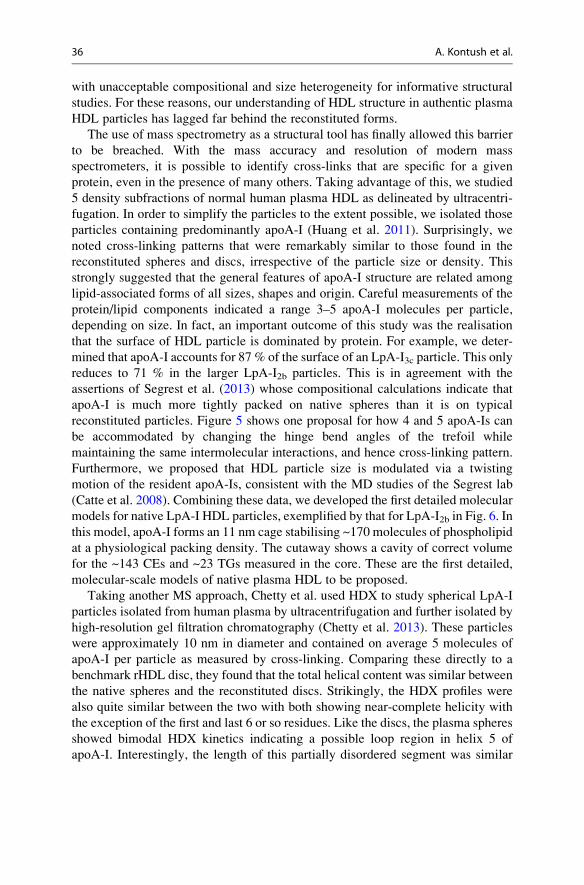
with unacceptable compositional and size heterogeneity for informative structural
studies. For these reasons, our understanding of HDL structure in authentic plasma
HDL particles has lagged far behind the reconstituted forms.
The use of mass spectrometry as a structural tool has finally allowed this barrier
to be breached. With the mass accuracy and resolution of modern mass
spectrometers, it is possible to identify cross-links that are specific for a given
protein, even in the presence of many others. Taking advantage of this, we studied
5 density subfractions of normal human plasma HDL as delineated by ultracentri-
fugation. In order to simplify the particles to the extent possible, we isolated those
particles containing predominantly apoA-I (Huang et al. 2011). Surprisingly, we
noted cross-linking patterns that were remarkably similar to those found in the
reconstituted spheres and discs, irrespective of the particle size or density. This
strongly suggested that the general features of apoA-I structure are related among
lipid-associated forms of all sizes, shapes and origin. Careful measurements of the
protein/lipid components indicated a range 3–5 apoA-I molecules per particle,
depending on size. In fact, an important outcome of this study was the realisation
that the surface of HDL particle is dominated by protein. For example, we deter-
mined that apoA-I accounts for 87 % of the surface of an LpA-I3c particle. This only
reduces to 71 % in the larger LpA-I2b particles. This is in agreement with the
assertions of Segrest et al. (2013) whose compositional calculations indicate that
apoA-I is much more tightly packed on native spheres than it is on typical
reconstituted particles. Figure 5 shows one proposal for how 4 and 5 apoA-Is can
be accommodated by changing the hinge bend angles of the trefoil while
maintaining the same intermolecular interactions, and hence cross-linking pattern.
Furthermore, we proposed that HDL particle size is modulated via a twisting
motion of the resident apoA-Is, consistent with the MD studies of the Segrest lab
(Catte et al. 2008). Combining these data, we developed the first detailed molecular
models for native LpA-I HDL particles, exemplified by that for LpA-I2b in Fig. 6. In
this model, apoA-I forms an 11 nm cage stabilising ~170 molecules of phospholipid
at a physiological packing density. The cutaway shows a cavity of correct volume
for the ~143 CEs and ~23 TGs measured in the core. These are the first detailed,
molecular-scale models of native plasma HDL to be proposed.
Taking another MS approach, Chetty et al. used HDX to study spherical LpA-I
particles isolated from human plasma by ultracentrifugation and further isolated by
high-resolution gel filtration chromatography (Chetty et al. 2013). These particles
were approximately 10 nm in diameter and contained on average 5 molecules of
apoA-I per particle as measured by cross-linking. Comparing these directly to a
benchmark rHDL disc, they found that the total helical content was similar between
the native spheres and the reconstituted discs. Strikingly, the HDX profiles were
also quite similar between the two with both showing near-complete helicity with
the exception of the first and last 6 or so residues. Like the discs, the plasma spheres
showed bimodal HDX kinetics indicating a possible loop region in helix 5 of
apoA-I. Interestingly, the length of this partially disordered segment was similar
36 A. Kontush et al.
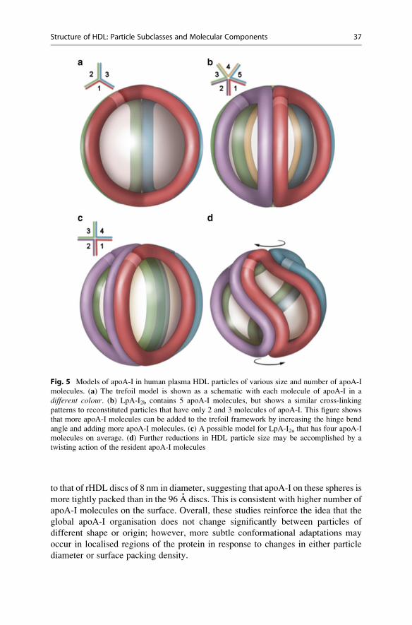
to that of rHDL discs of 8 nm in diameter, suggesting that apoA-I on these spheres is
more tightly packed than in the 96 A discs. This is consistent with higher number of
apoA-I molecules on the surface. Overall, these studies reinforce the idea that the
global apoA-I organisation does not change significantly between particles of
different shape or origin; however, more subtle conformational adaptations may
occur in localised regions of the protein in response to changes in either particle
diameter or surface packing density.
Fig. 5 Models of apoA-I in human plasma HDL particles of various size and number of apoA-I
molecules. (a) The trefoil model is shown as a schematic with each molecule of apoA-I in a
different colour. (b) LpA-I2b contains 5 apoA-I molecules, but shows a similar cross-linking
patterns to reconstituted particles that have only 2 and 3 molecules of apoA-I. This figure shows
that more apoA-I molecules can be added to the trefoil framework by increasing the hinge bend
angle and adding more apoA-I molecules. (c) A possible model for LpA-I2a that has four apoA-I
molecules on average. (d) Further reductions in HDL particle size may be accomplished by a
twisting action of the resident apoA-I molecules
Structure of HDL: Particle Subclasses and Molecular Components 37
Fig. 6 Molecular model of a human plasma LpA-I2b particle. (a) Five molecules of apoA-I are
shown in an organisation shown schematically in Fig. 5b. Phospholipid headgroups (tan) are
present in the intervening spaces (lunes) between the apoA-I belts (purple). (b) A cross section of the
same particle showing the helical belts floating atop the acyl chains of the phospholipids (brown).Note the large internal cavity that is of sufficient volume to contain the number of triglycerides and
cholesteryl ester molecules that have been measured in these particles (Huang et al. 2011). (Colour
figure online)
38 A. Kontush et al.
Conclusions and Perspectives
HDL, a lipoprotein class historically and classically defined and isolated on the
basis of its hydrated density alone, is a highly heterogeneous particle family
consisting of multiple subclasses differing in hydrated density, size, shape and
lipid and protein composition. HDL-associated proteins have been considered
until now to predominate in determining the particle structure and biological
function of all HDL particles. Indeed, HDL is distinguished from other lipopro-
tein classes by its cargo of a large number (>80) of different proteins whose
abundance may vary by more than 1,000-fold and which fall into several major
subgroups, including apolipoproteins, diverse enzymes, lipid transfer proteins,
acute-phase response proteins, complement components and proteinase
inhibitors; additional functions, such as host defence and inflammation, cannot
be excluded. Many of the individual proteins are present as distinct isoforms due
to co- and posttranslational modification, thereby adding a further dimension to
the complex and dynamic HDL structure.
It would be erroneous however to consider that the proteome(s) of HDL
particles solely determines their respective functions, particularly as cutting
edge lipidomic technologies have recently revealed significant—and indeed
determinant—roles of specific HDL lipid components in particle function. In
this regard, negatively charged phospholipids, and notably molecular species of
phosphatidylserines, are intimately associated with the cellular cholesterol
efflux, antioxidative, antiapoptotic and anti-inflammatory actions of small
dense HDL3 particles and equally modulate platelet activity (Camont
et al. 2013). In addition, earlier studies have suggested that both core neutral
lipids in HDL (cholesteryl esters and triacylglycerols) and surface lipids can
impact surface fluidity and, in turn, antioxidative and other functions (Zerrad-
Saadi et al. 2009; Camont et al. 2013).
Increasingly then, a new concept is emerging, one in which specific protein
and lipid components interact closely to form individual HDL particle species,
such as overall compositional and structural features translating into expression
of highly defined biological functions (Camont et al. 2011; Shah et al. 2013).
Clearly we are at the beginning of a new era in our understanding of HDL
structure and function, with the promise of exciting mechanistic insights into
function, which, in turn, will translate into new therapeutic strategies to harness
the anti-atherosclerotic potential of HDL.
The central thesis that specific protein–protein interactions may drive the
formation of lipoprotein complexes in the HDL particle spectrum is equally
emergent. Indeed, the diversity of molecules which bind to HDL suggests that
the lipoprotein can serve as a versatile adsorptive surface for proteins to form
complexes playing roles not only in lipid metabolism but equally in the acute-
phase response, innate immune response, complement activation, proteolysis
inhibition and plaque stability.
More comprehensive understanding of HDL structure is critical to future
progress in HDL biology. A molecular understanding of HDL-bound apoA-I
will allow further insight into its interactions with key plasma remodelling
Structure of HDL: Particle Subclasses and Molecular Components 39

factors, such as LCAT, PLTP or CETP, and in addition with cell-surface proteins
that mediate HDL assembly and clearance, such as ABCA1 and scavenger
receptor B-I. A firm handle on these interactions would contribute to therapeutic
manipulation that could enhance the protective features of HDL with respect to
cardiovascular disease, or perhaps other pathologies (Gordon et al. 2011).
Because HDL and its recombinant analogues have been resistant to traditional
high-resolution structural approaches such as NMR or X-ray crystallography,
investigators have been forced to apply a series of lower resolution techniques to
the problem. As described above, these approaches have led to a general
understanding of apoA-I structure in HDL that can be summarised by the
following statements: (1) apoA-I pairs with itself in a double belt-like arrange-
ment in almost all of its lipidated states; (2) apoA-I interacts via intermolecular
salt bridges that commonly stabilise the registry in the 5/5 orientation, though
there may be a possibility of dynamic shifts to other orientations; and (3) despite
a similar general organisation among different types of HDL particles, specific
regions within the scaffold can undergo conformational changes depending on
particle size or perhaps composition—possible sites include the N- and
C-termini and central region in helix 5 or between residues 165 and 180.
These conformationally dynamic sites are attractive possibilities for the interac-
tion of lipid-modifying docking proteins such as LCAT and CETP.
Now that these conformationally dynamic sites within apoA-I have been
proposed, it will be important to specifically define their importance for key
HDL remodelling reactions. This will require well-designed site-directed muta-
genesis experiments in reconstituted particles and antibody blocking
experiments in native plasma particles. Furthermore, cross-linking studies hold
much promise for directly mapping the sites of interaction for plasma
remodelling factors on HDL-bound apoA-I.
Finally, yet another key unanswered question relates to the unknown effects
of other HDL structural proteins on apoA-I structure. ApoA-II is commonly
found with apoA-I on many HDL particles and would be expected to have a
significant impact not only on properties such as protein packing on the particle
surface but also on the conformation of apoA-I in a functionally significant way.
For example, does apoA-II perform distinct structural and protein docking roles
vs. apoA-I? How does each of these proteins determine the components of the
bound lipidome in individual particle species? Resolution of these and other
questions will continue to require the use of both reconstituted and native,
authentic HDL particles combined with the appropriate application of multiple
structural techniques (including continued attempts at X-ray crystallography).
It is poignant to consider that in 2014 we still lack critically important insights
into HDL structure as it relates to the intravascular metabolism and
atheroprotective function of these pseudomicellar, quasi-spherical particles.
Had sufficient effort been invested in solving these unknowns heretofore, then
perhaps our costly forays—and failures—into HDL-targeted therapeutics over
the past 15 years could at least partially have been avoided.
40 A. Kontush et al.

Acknowledgements This article results from individual contributions of all the co-authors. The
highly complex world of HDL subclasses is presented by Laura Calabresi. The multifaceted
composition of the HDL proteome is discussed by Anatol Kontush. Protein isoforms, including
translational and posttranslational modifications, are reviewed by Mats Lindahl. Rapidly growing
knowledge of the HDL lipidome is discussed by Marie Lhomme. W. Sean Davidson presents a
brief history of structural studies of HDL, critically reviews existing models of HDL structure, and
together with M. John Chapman draws conclusions and discusses future perspectives.
Open Access This chapter is distributed under the terms of the Creative Commons Attribution
Noncommercial License, which permits any noncommercial use, distribution, and reproduction in
any medium, provided the original author(s) and source are credited.
References
Ahnstrom J et al (2007) Hydrophobic ligand binding properties of the human lipocalin apolipo-
protein M. J Lipid Res 48(8):1754–1762
Allan CM, Taylor JM (1996) Expression of a novel human apolipoprotein (apoC-IV) causes
hypertriglyceridemia in transgenic mice. J Lipid Res 37(7):1510–1518
Argraves KM et al (2011) S1P, dihydro-S1P and C24:1-ceramide levels in the HDL-containing
fraction of serum inversely correlate with occurrence of ischemic heart disease. Lipids Health
Dis 10:70
Asztalos BF, Schaefer EJ (2003a) High-density lipoprotein subpopulations in pathologic
conditions. Am J Cardiol 91(7A):12E–17E
Asztalos BF, Schaefer EJ (2003b) HDL in atherosclerosis: actor or bystander? Atheroscler Suppl 4
(1):21–29
Asztalos BF et al (2007) Role of LCAT in HDL remodeling: investigation of LCAT deficiency
states. J Lipid Res 48(3):592–599
Atkinson D et al (1974) The structure of a high density lipoprotein (HDL3) from porcine plasma.
Proc R Soc Lond B Biol Sci 186(083):165–180
Axler O et al (2007) An ELISA for apolipoprotein M reveals a strong correlation to total
cholesterol in human plasma. J Lipid Res 48(8):1772–1780
Axler O et al (2008) Apolipoprotein M associates to lipoproteins through its retained signal
peptide. FEBS Lett 582(5):826–828
Baenziger JU (2012) Moving the O-glycoproteome from form to function. Proc Natl Acad Sci
USA 109(25):9672–9673
Benditt EP, Eriksen N (1977) Amyloid protein SAA is associated with high density lipoprotein
from human serum. Proc Natl Acad Sci USA 74(9):4025–4028
Bergmeier C et al (2004) Distribution spectrum of paraoxonase activity in HDL fractions. Clin
Chem 50(12):2309–2315
Bhat S et al (2005) Intermolecular contact between globular N-terminal fold and C-terminal
domain of ApoA-I stabilizes its lipid-bound conformation: studies employing chemical
cross-linking and mass spectrometry. J Biol Chem 280(38):33015–33025
Bisgaier CL et al (1985) Distribution of apolipoprotein A-IV in human plasma. J Lipid Res 26
(1):11–25
Bondarenko PV et al (1999) Mass spectral study of polymorphism of the apolipoproteins of very
low density lipoprotein. J Lipid Res 40(3):543–555
Borhani DW et al (1997) Crystal structure of truncated human apolipoprotein A-I suggests a lipid-
bound conformation. Proc Natl Acad Sci USA 94(23):12291–12296
Brasseur R et al (1990) Mode of assembly of amphipathic helical segments in model high-density
lipoproteins. Biochim Biophys Acta 1043(3):245–252
Structure of HDL: Particle Subclasses and Molecular Components 41
Brewer HB Jr et al (1972) Amino acid sequence of human apoLp-Gln-II (apoA-II), an apolipo-
protein isolated from the high-density lipoprotein complex. Proc Natl Acad Sci USA 69
(5):1304–1308
Brewer HB Jr et al (1978) The amino acid sequence of human APOA-I, an apolipoprotein isolated
from high density lipoproteins. Biochem Biophys Res Commun 80(3):623–630
Brouillette CG, Anantharamaiah GM (1995) Structural models of human apolipoprotein A-I.
Biochim Biophys Acta 1256(2):103–129
Brouillette CG et al (2001) Structural models of human apolipoprotein A-I: a critical analysis and
review. Biochim Biophys Acta 1531(1–2):4–46
Bruneel A et al (2008) Two dimensional gel electrophoresis of apolipoprotein C-III and MALDI-
TOF MS are complementary techniques for the study of combined defects in N- and mucin
type O-glycan biosynthesis. Proteomics Clin Appl 2:1670–1674
Cabana VG et al (1996) HDL content and composition in acute phase response in three species:
triglyceride enrichment of HDL a factor in its decrease. J Lipid Res 37(12):2662–2674
Cabana VG et al (2004) Influence of apoA-I and apoE on the formation of serum amyloid
A-containing lipoproteins in vivo and in vitro. J Lipid Res 45(2):317–325
Calabresi L et al (2001) Limited proteolysis of a disulfide-linked apoA-I dimer in reconstituted
HDL. J Lipid Res 42(6):935–942
Camont L et al (2011) Biological activities of HDL subpopulations and their relevance to
cardiovascular disease. Trends Mol Med 17(10):594–603
Camont L et al (2013) Small, dense high-density lipoprotein-3 particles are enriched in negatively
charged phospholipids: relevance to cellular cholesterol efflux, antioxidative, antithrombotic,
anti-inflammatory, and antiapoptotic functionalities. Arterioscler Thromb Vasc Biol 33
(12):2715–2723
Campos E, McConathy WJ (1986) Distribution of lipids and apolipoproteins in human plasma by
vertical spin ultracentrifugation. Arch Biochem Biophys 249(2):455–463
Candiano G et al (2008) High-resolution 2-DE for resolving proteins, protein adducts and
complexes in plasma. Electrophoresis 29(3):682–694
Catte A et al (2008) Structure of spheroidal HDL particles revealed by combined atomistic and
coarse-grained simulations. Biophys J 94(6):2306–2319
Chapman MJ et al (1981) A density gradient ultracentrifugal procedure for the isolation of the
major lipoprotein classes from human serum. J Lipid Res 22(2):339–358
Chen N et al (2000) Physiologic concentrations of homocysteine inhibit the human plasma GSH
peroxidase that reduces organic hydroperoxides. J Lab Clin Med 136(1):58–65
Chetty PS et al (2013) Comparison of apoA-I helical structure and stability in discoidal and
spherical HDL particles by HX and mass spectrometry. J Lipid Res 54(6):1589–1597
Cheung MC, Albers JJ (1982) Distribution of high density lipoprotein particles with different
apoprotein composition: particles with A-I and A-II and particles with A-I but no A-II. J Lipid
Res 23(5):747–753
Cheung MC et al (2010) Phospholipid transfer protein in human plasma associates with proteins
linked to immunity and inflammation. Biochemistry 49(34):7314–7322
Chiba T et al (2011) Serum amyloid A facilitates the binding of high-density lipoprotein from mice
injected with lipopolysaccharide to vascular proteoglycans. Arterioscler Thromb Vasc Biol 31
(6):1326–1332
Christoffersen C et al (2008) The signal peptide anchors apolipoprotein M in plasma lipoproteins
and prevents rapid clearance of apolipoprotein M from plasma. J Biol Chem 283(27):18765–
18772
Christoffersen C et al (2011) Endothelium-protective sphingosine-1-phosphate provided by
HDL-associated apolipoprotein M. Proc Natl Acad Sci USA 108(23):9613–9618
Coetzee GA et al (1986) Serum amyloid A-containing human high density lipoprotein 3. Density,
size, and apolipoprotein composition. J Biol Chem 261(21):9644–9651
42 A. Kontush et al.
Collins LA et al (2010) Integrated approach for the comprehensive characterization of lipoproteins
from human plasma using FPLC and nano-HPLC-tandem mass spectrometry. Physiol Geno-
mics 40(3):208–215
Contiero E et al (1997) Apolipoprotein AI isoforms in serum determined by isoelectric focusing
and immunoblotting. Electrophoresis 18(1):122–126
Dahlback B, Nielsen LB (2009) Apolipoprotein M affecting lipid metabolism or just catching a
ride with lipoproteins in the circulation? Cell Mol Life Sci 66(4):559–564
Davidson WS, Hilliard GM (2003) The spatial organization of apolipoprotein A-I on the edge of
discoidal high density lipoprotein particles: a mass specrometry study. J Biol Chem 278
(29):27199–27207
DavidsonWS et al (1995) Effects of acceptor particle size on the efflux of cellular free cholesterol.
J Biol Chem 270(29):17106–17113
Davidson WS et al (2009) Proteomic analysis of defined HDL subpopulations reveals particle-
specific protein clusters: relevance to antioxidative function. Arterioscler Thromb Vasc Biol 29
(6):870–876
Davidsson P et al (2010) Proteomics of apolipoproteins and associated proteins from plasma high-
density lipoproteins. Arterioscler Thromb Vasc Biol 30(2):156–163
de Beer MC et al (2010) Impact of serum amyloid A on high density lipoprotein composition and
levels. J Lipid Res 51(11):3117–3125
De Lalla OF, Gofman JW (1954) Ultracentrifugal analysis of serum lipoproteins. Methods
Biochem Anal 1:459–478
de Seny D et al (2008) Monomeric calgranulins measured by SELDI-TOF mass spectrometry and
calprotectin measured by ELISA as biomarkers in arthritis. Clin Chem 54(6):1066–1075
de Silva HV et al (1990a) Purification and characterization of apolipoprotein J. J Biol Chem 265
(24):14292–14297
de Silva HV et al (1990b) A 70-kDa apolipoprotein designated ApoJ is a marker for subclasses of
human plasma high density lipoproteins. J Biol Chem 265(22):13240–13247
Deguchi H et al (2000) Cardiolipin is a normal component of human plasma lipoproteins. Proc
Natl Acad Sci USA 97(4):1743–1748
Duchateau PN et al (1997) Apolipoprotein L, a new human high density lipoprotein apolipoprotein
expressed by the pancreas. Identification, cloning, characterization, and plasma distribution of
apolipoprotein L. J Biol Chem 272(41):25576–25582
Ducret A et al (1996) Characterization of human serum amyloid A protein isoforms separated by
two-dimensional electrophoresis by liquid chromatography/electrospray ionization tandem
mass spectrometry. Electrophoresis 17(5):866–876
Duriez P, Fruchart JC (1999) High-density lipoprotein subclasses and apolipoprotein A-I. Clin
Chim Acta 286(1–2):97–114
Edelstein C et al (1972) On the subunit structure of the protein of human serum high density
lipoprotein. I. A study of its major polypeptide component (Sephadex, fraction 3). J Biol Chem
247(18):5842–5849
Favari E et al (2004) Depletion of pre-beta-high density lipoprotein by human chymase impairs
ATP-binding cassette transporter A1- but not scavenger receptor class B type I-mediated lipid
efflux to high density lipoprotein. J Biol Chem 279(11):9930–9936
Favari E et al (2007) A unique protease-sensitive high density lipoprotein particle containing the
apolipoprotein A-I Milano dimer effectively promotes ATP-binding Cassette A1-mediated cell
cholesterol efflux. J Biol Chem 282(8):5125–5132
Fernandez-Irigoyen J et al (2005) Oxidation of specific methionine and tryptophan residues of
apolipoprotein A-I in hepatocarcinogenesis. Proteomics 5(18):4964–4972
Fievet C et al (1995) Non-enzymatic glycosylation of apolipoprotein A-I and its functional
consequences. Diabete Metab 21(2):95–98
Flood-Nichols SK et al (2013) Longitudinal analysis of maternal plasma apolipoproteins in
pregnancy: a targeted proteomics approach. Mol Cell Proteomics 12(1):55–64
Structure of HDL: Particle Subclasses and Molecular Components 43
Forte TM, Nordhausen RW (1986) Electron microscopy of negatively stained lipoproteins.
Methods Enzymol 128:442–457
Franceschini G et al (1985) Effects of storage on the distribution of high density lipoprotein
subfractions in human sera. J Lipid Res 26(11):1368–1373
Franceschini G et al (2007) Effects of fenofibrate and simvastatin on HDL-related biomarkers in
low-HDL patients. Atherosclerosis 195(2):385–391
Gauthamadasa K et al (2010) Speciated human high-density lipoprotein protein proximity profiles.
Biochemistry 49(50):10656–10665
Ghiselli G et al (1985) Origin of apolipoprotein A-I polymorphism in plasma. J Biol Chem 260
(29):15662–15668
Gidez LI et al (1982) Separation and quantitation of subclasses of human plasma high density
lipoproteins by a simple precipitation procedure. J Lipid Res 23(8):1206–1223
Gillard BK et al (2005) Plasma factors required for human apolipoprotein A-II dimerization.
Biochemistry 44(2):471–479
Gogonea V et al (2010) Congruency between biophysical data from multiple platforms and
molecular dynamics simulation of the double-super helix model of nascent high-density
lipoprotein. Biochemistry 49(34):7323–7343
Gordon JI et al (1983) Biosynthesis of human preproapolipoprotein A-II. J Biol Chem 258
(22):14054–14059
Gordon SM et al (2010) Proteomic characterization of human plasma high density lipoprotein
fractionated by gel filtration chromatography. J Proteome Res 9(10):5239–5249
Gordon SM et al (2011) High density lipoprotein: it’s not just about lipid transport anymore.
Trends Endocrinol Metab 22(1):9–15
Gordon SM et al (2013) Multi-dimensional co-separation analysis reveals protein-protein
interactions defining plasma lipoprotein subspecies. Mol Cell Proteomics 12(11):3123–3134
Goswami B et al (2009) Paraoxonase: a multifaceted biomolecule. Clin Chim Acta 410(1–2):1–12
Gu F et al (2010) Structures of discoidal high density lipoproteins: a combined computational-
experimental approach. J Biol Chem 285(7):4652–4665
Gursky O (2013) Crystal structure of Delta(185–243)ApoA-I suggests a mechanistic framework
for the protein adaptation to the changing lipid load in good cholesterol: from flatland to
sphereland via double belt, belt buckle, double hairpin and trefoil/tetrafoil. J Mol Biol 425
(1):1–16
Gursky O et al (2013) Structural basis for distinct functions of the naturally occurring Cys mutants
of human apolipoprotein A-I. J Lipid Res 54(12):3244–3257
Hajduk SL et al (1989) Lysis of Trypanosoma brucei by a toxic subspecies of human high density
lipoprotein. J Biol Chem 264(9):5210–5217
Halim A et al (2013) LC-MS/MS characterization of O-glycosylation sites and glycan structures of
human cerebrospinal fluid glycoproteins. J Proteome Res 12(2):573–584
Harel M et al (2004) Structure and evolution of the serum paraoxonase family of detoxifying and
anti-atherosclerotic enzymes. Nat Struct Mol Biol 11(5):412–419
Harvey SB et al (2009) O-glycoside biomarker of apolipoprotein C3: responsiveness to obesity,
bariatric surgery, and therapy with metformin, to chronic or severe liver disease and to
mortality in severe sepsis and graft vs host disease. J Proteome Res 8(2):603–612
Hatters DM et al (2006) Apolipoprotein E structure: insights into function. Trends Biochem Sci 31
(8):445–454
He Y et al (2008) Control of cholesteryl ester transfer protein activity by sequestration of lipid
transfer inhibitor protein in an inactive complex. J Lipid Res 49(7):1529–1537
Heinecke JW (2009) The HDL proteome: a marker - and perhaps mediator - of coronary artery
disease. J Lipid Res Supp 50:S167–S171
Hennessy LK et al (1997) Isolation of subpopulations of high density lipoproteins: three particle
species containing apoE and two species devoid of apoE that have affinity for heparin. J Lipid
Res 38(9):1859–1868
44 A. Kontush et al.

Hoffman JS, Benditt EP (1982) Changes in high density lipoprotein content following endotoxin
administration in the mouse. Formation of serum amyloid protein-rich subfractions. J Biol
Chem 257(17):10510–10517
Holleboom AG et al (2011) Heterozygosity for a loss-of-function mutation in GALNT2 improves
plasma triglyceride clearance in man. Cell Metab 14(6):811–818
Holmquist L (2002) Selective extraction of lecithin:cholesterol acyltransferase (EC 2.3.1.43) from
human plasma. J Biochem Biophys Methods 52(1):63–68
Hoofnagle AN, Heinecke JW (2009) Lipoproteomics: using mass spectrometry-based proteomics
to explore the assembly, structure, and function of lipoproteins. J Lipid Res 50(10):1967–1975
Hortin GL et al (2006) Diverse range of small peptides associated with high-density lipoprotein.
Biochem Biophys Res Commun 340(3):909–915
Huang R et al (2011) Apolipoprotein A-I structural organization in high-density lipoproteins
isolated from human plasma. Nat Struct Mol Biol 18(4):416–422
Hussain MM, Zannis VI (1990) Intracellular modification of human apolipoprotein AII (apoAII)
and sites of apoAII mRNA synthesis: comparison of apoAII with apoCII and apoCIII
isoproteins. Biochemistry 29(1):209–217
Jakubowski H (2000) Calcium-dependent human serum homocysteine thiolactone hydrolase. A
protective mechanism against protein N-homocysteinylation. J Biol Chem 275(6):3957–3962
Jaleel A et al (2010) Identification of de novo synthesized and relatively older proteins: accelerated
oxidative damage to de novo synthesized apolipoprotein A-1 in type 1 diabetes. Diabetes 59
(10):2366–2374
James RW et al (1988) Protein heterogeneity of lipoprotein particles containing apolipoprotein A-I
without apolipoprotein A-II and apolipoprotein A-I with apolipoprotein A-II isolated from
human plasma. J Lipid Res 29(12):1557–1571
Jian W et al (2013) Relative quantitation of glycoisoforms of intact apolipoprotein C3 in human
plasma by liquid chromatography-high-resolution mass spectrometry. Anal Chem 85(5):2867–
2874
Jin Y, Manabe T (2005) Direct targeting of human plasma for matrix-assisted laser desorption/
ionization and analysis of plasma proteins by time of flight-mass spectrometry. Electrophoresis
26(14):2823–2834
Jonas A (1972) Studies on the structure of bovine serum high density lipoprotein using covalently
bound fluorescent probes. J Biol Chem 247(23):7773–7778
Jonas A (1986) Reconstitution of high-density lipoproteins. Methods Enzymol 128:553–582
Jonas A (2002) Lipoprotein structure. In: Vance DE, Vance JE (eds) Biochemistry of lipids,
lipoproteins and membranes. Elsevier, Amsterdam, pp 483–504
Jonas A, McHugh HT (1984) Reaction of lecithin: cholesterol acyltransferase with micellar
substrates. Effect of particle sizes. Biochim Biophys Acta 794(3):361–372
Jonas A et al (1989) Defined apolipoprotein A-I conformations in reconstituted high density
lipoprotein discs. J Biol Chem 264(9):4818–4824
Jonas A et al (1990) Apolipoprotein A-I structure and lipid properties in homogeneous,
reconstituted spherical and discoidal high density lipoproteins. J Biol Chem 265(36):22123–
22129
Jones MK et al (2010) Assessment of the validity of the double superhelix model for reconstituted
high density lipoproteins: a combined computational-experimental approach. J Biol Chem 285
(52):41161–41171
Jones MK et al (2011) “Sticky” and “promiscuous”, the Yin and Yang of apolipoprotein A-I
termini in discoidal high-density lipoproteins: a combined computational-experimental
approach. Biochemistry 50(12):2249–2263
Karlsson H et al (2005) Lipoproteomics II: mapping of proteins in high-density lipoprotein using
two-dimensional gel electrophoresis and mass spectrometry. Proteomics 5(5):1431–1445
Karlsson H et al (2009) Protein profiling of low-density lipoprotein from obese subjects. Proteo-
mics Clin Appl 3(6):663–671
Structure of HDL: Particle Subclasses and Molecular Components 45

Khovidhunkit W et al (2004) Effects of infection and inflammation on lipid and lipoprotein
metabolism: mechanisms and consequences to the host. J Lipid Res 45(7):1169–1196
Kiernan UA et al (2003) Detection of novel truncated forms of human serum amyloid A protein in
human plasma. FEBS Lett 537(1–3):166–170
Kim E et al (2008) Expression of apolipoprotein C-IV is regulated by Ku antigen/peroxisome
proliferator-activated receptor gamma complex and correlates with liver steatosis. J Hepatol 49
(5):787–798
Kisilevsky R, Manley PN (2012) Acute-phase serum amyloid A: perspectives on its physiological
and pathological roles. Amyloid 19(1):5–14
Kisilevsky R, Tam SP (2003) Macrophage cholesterol efflux and the active domains of serum
amyloid A 2.1. J Lipid Res 44(12):2257–2269
Klon AE et al (2002a) Comparative models for human apolipoprotein A-I bound to lipid in
discoidal high-density lipoprotein particles. Biochemistry 41(36):10895–10905
Klon AE et al (2002b) Molecular dynamics simulations on discoidal HDL particles suggest a
mechanism for rotation in the apo A-I belt model. J Mol Biol 324(4):703–721
Kontush A, Chapman MJ (2010) Antiatherogenic function of HDL particle subpopulations: focus
on antioxidative activities. Curr Opin Lipidol 21(4):312–318
Kontush A, Chapman MJ (2012) High-density lipoproteins: structure, metabolism, function and
therapeutics. Wiley, New York
Kontush A et al (2003) Small, dense HDL particles exert potent protection of atherogenic LDL
against oxidative stress. Arterioscler Thromb Vasc Biol 23(10):1881–1888
Kontush A et al (2007) Preferential sphingosine-1-phosphate enrichment and sphingomyelin
depletion are key features of small dense HDL3 particles: relevance to antiapoptotic and
antioxidative activities. Arterioscler Thromb Vasc Biol 27(8):1843–1849
Koppaka V et al (1999) The structure of human lipoprotein A-I. Evidence for the “belt” model. J
Biol Chem 274(21):14541–14544
Kulkarni KR et al (1997) Quantification of HDL2 and HDL3 cholesterol by the vertical auto
profile-II (VAP-II) methodology. J Lipid Res 38(11):2353–2364
Laggner P et al (1973) Studies on the structure of lipoprotein A of human high density lipoprotein
HDL3: the spherically averaged electron density distribution. FEBS Lett 33(1):77–80
LagorWR et al (2009) Overexpression of apolipoprotein F reduces HDL cholesterol levels in vivo.
Arterioscler Thromb Vasc Biol 29(1):40–46
Lamant M et al (2006) ApoO, a novel apolipoprotein, is an original glycoprotein up-regulated by
diabetes in human heart. J Biol Chem 281(47):36289–36302
Lee M et al (2003) Apolipoprotein composition and particle size affect HDL degradation by
chymase: effect on cellular cholesterol efflux. J Lipid Res 44(3):539–546
Lee JY et al (2010) Profiling of phospholipids in lipoproteins by multiplexed hollow fiber flow
field-flow fractionation and nanoflow liquid chromatography-tandem mass spectrometry. J
Chromatogr A 1217(10):1660–1666
Levels JH et al (2011) High-density lipoprotein proteome dynamics in human endotoxemia.
Proteome Sci 9(1):34
Lhomme M et al (2012) Lipidomics in lipoprotein biology. In: Ekroos K (ed) Lipidomics:
technologies and applications. Wiley, Weinheim, pp 197–231
Li H et al (2000) Structural determination of lipid-bound ApoA-I using fluorescence resonance
energy transfer. J Biol Chem 275(47):37048–37054
Li HH et al (2002) ApoA-I structure on discs and spheres. Variable helix registry and conforma-
tional states. J Biol Chem 277(42):39093–39101
Liadaki KN et al (2000) Binding of high density lipoprotein (HDL) and discoidal reconstituted
HDL to the HDL receptor scavenger receptor class B type I. Effect of lipid association and
APOA-I mutations on receptor binding. J Biol Chem 275(28):21262–21271
Lipina C, Hundal HS (2011) Sphingolipids: agents provocateurs in the pathogenesis of insulin
resistance. Diabetologia 54(7):1596–1607
46 A. Kontush et al.

Ljunggren S et al (2013) APOA-I mutations, L202P and K131DEL, IN HDL from heterozygotes
with low HDL-cholesterol. Proteomics Clin Appl 8(3–4):241–250
Lucke S, Levkau B (2010) Endothelial functions of sphingosine-1-phosphate. Cell Physiol
Biochem 26(1):87–96
Lund-Katz S, Phillips MC (2010) High density lipoprotein structure-function and role in reverse
cholesterol transport. Subcell Biochem 51:183–227
Mackness B et al (2010) Human tissue distribution of paraoxonases 1 and 2 mRNA. IUBMB Life
62(6):480–482
Maeba R, Ueta N (2003) Ethanolamine plasmalogen and cholesterol reduce the total membrane
oxidizability measured by the oxygen uptake method. Biochem Biophys Res Commun 302
(2):265–270
Maiorano JN et al (2004) Identification and structural ramifications of a hinge domain in apolipo-
protein A-I discoidal high-density lipoproteins of different size. Biochemistry 43(37):11717–
11726
Majek P et al (2011) Plasma proteome changes in cardiovascular disease patients: novel isoforms
of apolipoprotein A1. J Transl Med 9:84
Mallat Z et al (2010) Lipoprotein-associated and secreted phospholipases A in cardiovascular
disease: roles as biological effectors and biomarkers. Circulation 122(21):2183–2200
Marcel YL et al (1990) Distribution and concentration of cholesteryl ester transfer protein in
plasma of normolipemic subjects. J Clin Invest 85(1):10–17
Marhaug G et al (1982) Characterization of amyloid related protein SAA complexed with serum
lipoproteins (apoSAA). Clin Exp Immunol 50(2):382–389
Martin DD et al (2006) Apolipoprotein A-I assumes a “looped belt” conformation on reconstituted
high density lipoprotein. J Biol Chem 281(29):20418–20426
Matz CE, Jonas A (1982) Micellar complexes of human apolipoprotein A-I with phosphatidyl-
cholines and cholesterol prepared from cholate-lipid dispersions. J Biol Chem 257(8):4535–
4540
Mauger JF et al (2006) Apolipoprotein C-III isoforms: kinetics and relative implication in lipid
metabolism. J Lipid Res 47(6):1212–1218
Mazur MT et al (2010) Quantitative analysis of intact apolipoproteins in human HDL by top-down
differential mass spectrometry. Proc Natl Acad Sci USA 107(17):7728–7733
McConathy WJ, Alaupovic P (1973) Isolation and partial characterization of apolipoprotein D: a
new protein moiety of the human plasma lipoprotein system. FEBS Lett 37(2):178–182
McIntyre TM et al (2009) The emerging roles of PAF acetylhydrolase. J Lipid Res Supp 50:S255–
S259
McLachlan AD (1977) Repeated helical pattern in apolipoprotein-A-I. Nature 267(5610):465–466
Mei X, Atkinson D (2011) Crystal structure of C-terminal truncated apolipoprotein A-I reveals the
assembly of high density lipoprotein (HDL) by dimerization. J Biol Chem 286(44):38570–
38582
Miida T et al (2003) Analytical performance of a sandwich enzyme immunoassay for pre beta
1-HDL in stabilized plasma. J Lipid Res 44(3):645–650
Miyazaki M et al (2010) Static and dynamic characterization of nanodiscs with apolipoprotein A-I
and its model peptide. J Phys Chem B 114(38):12376–12382
Mora S (2009) Advanced lipoprotein testing and subfractionation are not (yet) ready for routine
clinical use. Circulation 119(17):2396–2404
Moren X et al (2008) HDL subfraction distribution of paraoxonase-1 and its relevance to enzyme
activity and resistance to oxidative stress. J Lipid Res 49(6):1246–1253
Navab M et al (2004) The oxidation hypothesis of atherogenesis: the role of oxidized
phospholipids and HDL. J Lipid Res 45(6):993–1007
Nelsestuen GL et al (2008) Top-down proteomic analysis by MALDI-TOF profiling:
concentration-independent biomarkers. Proteomics Clin Appl 2(2):158–166
Neumann S et al (2009) Mammalian Wnt3a is released on lipoprotein particles. Traffic 10(3):334–
343
Structure of HDL: Particle Subclasses and Molecular Components 47

Nicholls SJ, Hazen SL (2009) Myeloperoxidase, modified lipoproteins, and atherogenesis. J Lipid
Res Supp 50:S346–S351
Nichols AV et al (1983) Characterization of discoidal complexes of phosphatidylcholine, apoli-
poprotein A-I and cholesterol by gradient gel electrophoresis. Biochim Biophys Acta 750
(2):353–364
Nichols AV et al (1986) Nondenaturing polyacrylamide gradient gel electrophoresis. Methods
Enzymol 128:417–431
Nicolardi S et al (2013a) Mapping O-glycosylation of apolipoprotein C-III in MALDI-FT-ICR
protein profiles. Proteomics 13(6):992–1001
Nicolardi S et al (2013b) Identification of new apolipoprotein-CIII glycoforms with ultrahigh
resolution MALDI-FTICR mass spectrometry of human sera. J Proteome Res 12(5):2260–
2268
Nijstad N et al (2011) Overexpression of apolipoprotein O does not impact on plasma HDL levels
or functionality in human apolipoprotein A-I transgenic mice. Biochim Biophys Acta 1811
(4):294–299
Nilsson A, Duan RD (2006) Absorption and lipoprotein transport of sphingomyelin. J Lipid Res 47
(1):154–171
Nobecourt E et al (2007) The impact of glycation on apolipoprotein A-I structure and its ability to
activate lecithin:cholesterol acyltransferase. Diabetologia 50(3):643–653
Nolte RT, Atkinson D (1992) Conformational analysis of apolipoprotein A-I and E-3 based on
primary sequence and circular dichroism. Biophys J 63(5):1221–1239
Ohta T et al (1985) Studies on the in vivo and in vitro distribution of apolipoprotein A-IV in human
plasma and lymph. J Clin Invest 76(3):1252–1260
Olofsson SO et al (1978) Isolation and partial characterization of a new acidic apolipoprotein
(apolipoprotein F) from high density lipoproteins of human plasma. Biochemistry 17(6):1032–
1036
Ortiz-Munoz G et al (2009) HDL antielastase activity prevents smooth muscle cell anoikis, a
potential new antiatherogenic property. FASEB J 23(9):3129–3139
Otvos JD et al (1992) Development of a proton nuclear magnetic resonance spectroscopic method
for determining plasma lipoprotein concentrations and subspecies distributions from a single,
rapid measurement. Clin Chem 38(9):1632–1638
Panagotopulos SE et al (2001) Apolipoprotein A-I adopts a belt-like orientation in reconstituted
high density lipoproteins. J Biol Chem 276(46):42965–42970
Park KH et al (2010) Fructated apolipoprotein A-I showed severe structural modification and loss
of beneficial functions in lipid-free and lipid-bound state with acceleration of atherosclerosis
and senescence. Biochem Biophys Res Commun 392(3):295–300
Phillips MC (2013) New insights into the determination of HDL structure by apolipoproteins:
thematic review series: high density lipoprotein structure, function, and metabolism. J Lipid
Res 54(8):2034–2048
Phillips JC et al (1997) Predicting the structure of apolipoprotein A-I in reconstituted high-density
lipoprotein disks. Biophys J 73(5):2337–2346
Pruzanski W et al (2000) Comparative analysis of lipid composition of normal and acute-phase
high density lipoproteins. J Lipid Res 41(7):1035–1047
Puppione DL et al (2010) Mass spectral analyses of the two major apolipoproteins of great ape
high density lipoproteins. Comp Biochem Physiol Part D Genomics Proteomics 4(4):305–309
Qiu X et al (2007) Crystal structure of cholesteryl ester transfer protein reveals a long tunnel and
four bound lipid molecules. Nat Struct Mol Biol 14(2):106–113
Rassart E et al (2000) Apolipoprotein D. Biochim Biophys Acta 1482(1–2):185–198
Remaley AT et al (1993) O-linked glycosylation modifies the association of apolipoprotein A-II to
high density lipoproteins. J Biol Chem 268(9):6785–6790
Roghani A, Zannis VI (1988) Mutagenesis of the glycosylation site of human ApoCIII. O-linked
glycosylation is not required for ApoCIII secretion and lipid binding. J Biol Chem 263
(34):17925–17932
48 A. Kontush et al.
Rosenson RS et al (2011) HDL measures, particle heterogeneity, proposed nomenclature, and
relation to atherosclerotic cardiovascular events. Clin Chem 57(3):392–410
Rousset X et al (2009) Lecithin: cholesterol acyltransferase–from biochemistry to role in cardio-
vascular disease. Curr Opin Endocrinol Diabetes Obes 16(2):163–171
Rye KA et al (1996) The influence of sphingomyelin on the structure and function of reconstituted
high density lipoproteins. J Biol Chem 271(8):4243–4250
Saito H et al (2000) Inhibition of lipoprotein lipase activity by sphingomyelin: role of membrane
surface structure. Biochim Biophys Acta 1486(2–3):312–320
Samanta U, Bahnson BJ (2008) Crystal structure of human plasma platelet-activating factor
acetylhydrolase: structural implication to lipoprotein binding and catalysis. J Biol Chem 283
(46):31617–31624
Santucci L et al (2011) Protein-protein interaction heterogeneity of plasma apolipoprotein A1 in
nephrotic syndrome. Mol Biosyst 7(3):659–666
Sattler KJ et al (2010) Sphingosine 1-phosphate levels in plasma and HDL are altered in coronary
artery disease. Basic Res Cardiol 105(6):821–832
Scanu AM (1978) Ultrastructure of serum high density lipoproteins: facts and models. Lipids 13
(12):920–925
Schaefer EJ, Asztalos BF (2007) Increasing high-density lipoprotein cholesterol, inhibition of
cholesteryl ester transfer protein, and heart disease risk reduction. Am J Cardiol 100(11A):
S25–S31
Schaefer EJ et al (1979) The composition and metabolism of high density lipoprotein subfractions.
Lipids 14(5):511–522
Schaefer EJ et al (2010) Marked HDL deficiency and premature coronary heart disease. Curr Opin
Lipidol 21(4):289–297
Scherer M et al (2010) Sphingolipid profiling of human plasma and FPLC-separated lipoprotein
fractions by hydrophilic interaction chromatography tandem mass spectrometry. Biochim
Biophys Acta 1811(2):68–75
Schjoldager KT et al (2012) Probing isoform-specific functions of polypeptide GalNAc-
transferases using zinc finger nuclease glycoengineered SimpleCells. Proc Natl Acad Sci
USA 109(25):9893–9898
Schonfeld G et al (1976) Structure of high density lipoprotein. The immunologic reactivities of the
COOH- and NH2-terminal regions of apolipoprotein A-I. J Biol Chem 251(13):3921–3926
Segrest JP (1977) Amphipathic helixes and plasma lipoproteins: thermodynamic and geometric
considerations. Chem Phys Lipids 18(1):7–22
Segrest JP et al (1974) A molecular theory of lipid-protein interactions in the plasma lipoproteins.
FEBS Lett 38(3):247–258
Segrest JP et al (1994) The amphipathic alpha helix: a multifunctional structural motif in plasma
apolipoproteins. Adv Protein Chem 45:303–369
Segrest JP et al (1999) A detailed molecular belt model for apolipoprotein A-I in discoidal high
density lipoprotein. J Biol Chem 274(45):31755–31758
Segrest JP et al (2000) Detailed molecular model of apolipoprotein A-I on the surface of high-
density lipoproteins and its functional implications. Trends Cardiovasc Med 10(6):246–252
Segrest JP et al (2013) MD simulations suggest important surface differences between
reconstituted and circulating spherical HDL. J Lipid Res 54(10):2718–2732
Sevvana M et al (2009) Serendipitous fatty acid binding reveals the structural determinants for
ligand recognition in apolipoprotein M. J Mol Biol 393(4):920–936
Shah AS et al (2013) Proteomic diversity of high density lipoproteins: our emerging understanding
of its importance in lipid transport and beyond. J Lipid Res 54(10):2575–2585
Shao B (2012) Site-specific oxidation of apolipoprotein A-I impairs cholesterol export by ABCA1,
a key cardioprotective function of HDL. Biochim Biophys Acta 1821(3):490–501
Shen BW et al (1977) Structure of human serum lipoproteins inferred from compositional analysis.
Proc Natl Acad Sci USA 74(3):837–841
Shiflett AM et al (2005) Human high density lipoproteins are platforms for the assembly of multi-
component innate immune complexes. J Biol Chem 280(38):32578–32585
Structure of HDL: Particle Subclasses and Molecular Components 49
Shimano H (2009) ApoAII controversy still in rabbit? Arterioscler Thromb Vasc Biol 29
(12):1984–1985
Silva RA et al (2005) A mass spectrometric determination of the conformation of dimeric
apolipoprotein A-I in discoidal high density lipoproteins. Biochemistry 44(24):8600–8607
Silva RA et al (2008) Structure of apolipoprotein A-I in spherical high density lipoproteins of
different sizes. Proc Natl Acad Sci USA 105(34):12176–12181
Sloop CH et al (1983) Characterization of dog peripheral lymph lipoproteins: the presence of a
disc-shaped “nascent” high density lipoprotein. J Lipid Res 24(11):1429–1440
Sparks DL et al (1992) The conformation of apolipoprotein A-I in discoidal and spherical
recombinant high density lipoprotein particles. 13C NMR studies of lysine ionization behavior.
J Biol Chem 267(36):25830–25838
Stahlman M et al (2013) Dyslipidemia, but not hyperglycemia and insulin resistance, is associated
with marked alterations in the HDL lipidome in type 2 diabetic subjects in the DIWA cohort:
impact on small HDL particles. Biochim Biophys Acta 1831(11):1609–1617
Sung HJ et al (2012) Large-scale isotype-specific quantification of Serum amyloid A 1/2 by
multiple reaction monitoring in crude sera. J Proteomics 75(7):2170–2180
Tardieu A et al (1976) Structure of human serum lipoproteins in solution. II. Small-angle x-ray
scattering study of HDL and LDL. J Mol Biol 101(2):129–153
Tselepis AD et al (1995) PAF-degrading acetylhydrolase is preferentially associated with dense
LDL and VHDL-1 in human plasma. Catalytic characteristics and relation to the monocyte-
derived enzyme. Arterioscler Thromb Vasc Biol 15(10):1764–1773
Usami Y et al (2011) Detection of chymase-digested C-terminally truncated apolipoprotein A-I in
normal human serum. J Immunol Methods 369(1–2):51–58
Usami Y et al (2013) Identification of sites in apolipoprotein A-I susceptible to chymase and
carboxypeptidase A digestion. Biosci Rep 33(1):49–56
Utermann G (1975) Isolation and partial characterization of an arginine-rich apolipoprotein from
human plasma very-low-density lipoproteins: apolipoprotein E. Hoppe Seylers Z Physiol
Chem 356(7):1113–1121
Vaisar T (2009) Thematic review series: proteomics. Proteomic analysis of lipid-protein
complexes. J Lipid Res 50(5):781–786
Vaisar T et al (2007) Shotgun proteomics implicates protease inhibition and complement activa-
tion in the antiinflammatory properties of HDL. J Clin Invest 117(3):746–756
Vieu C et al (1996) Identification and quantification of diacylglycerols in HDL and accessibility to
lipase. J Lipid Res 37(5):1153–1161
Wald JH et al (1990) Structure of apolipoprotein A-I in three homogeneous, reconstituted high
density lipoprotein particles. J Biol Chem 265(32):20037–20043
Wiesner P et al (2009) Lipid profiling of FPLC-separated lipoprotein fractions by electrospray
ionization tandem mass spectrometry. J Lipid Res 50(3):574–585
Wlodawer A et al (1979) High-density lipoprotein recombinants: evidence for a bicycle tire
micelle structure obtained by neutron scattering and electron microscopy. FEBS Lett 104
(2):231–235
Wolfrum C et al (2005) Apolipoprotein M is required for prebeta-HDL formation and cholesterol
efflux to HDL and protects against atherosclerosis. Nat Med 11(4):418–422
Wopereis S et al (2003) Apolipoprotein C-III isofocusing in the diagnosis of genetic defects in
O-glycan biosynthesis. Clin Chem 49(11):1839–1845
Wroblewska M et al (2009) Phospholipids mediated conversion of HDLs generates specific apoA-
II pre-beta mobility particles. J Lipid Res 50(4):667–675
Wu Z et al (2007) The refined structure of nascent HDL reveals a key functional domain for
particle maturation and dysfunction. Nat Struct Mol Biol 14(9):861–868
Wu Z et al (2009) Double superhelix model of high density lipoprotein. J Biol Chem 284(52):
36605–36619
Wu Z et al (2011) The low resolution structure of ApoA1 in spherical high density lipoprotein
revealed by small angle neutron scattering. J Biol Chem 286(14):12495–12508
50 A. Kontush et al.

Wurfel MM et al (1994) Lipopolysaccharide (LPS)-binding protein is carried on lipoproteins and
acts as a cofactor in the neutralization of LPS. J Exp Med 180(3):1025–1035
Yu B et al (1997) Lipopolysaccharide binding protein and soluble CD14 catalyze exchange of
phospholipids. J Clin Invest 99(2):315–324
Zannis VI et al (1980) Isoproteins of human apolipoprotein A-I demonstrated in plasma and
intestinal organ culture. J Biol Chem 255(18):8612–8617
Zerrad-Saadi A et al (2009) HDL3-mediated inactivation of LDL-associated phospholipid
hydroperoxides is determined by the redox status of apolipoprotein A-I and HDL particle
surface lipid rigidity: relevance to inflammation and atherogenesis. Arterioscler Thromb Vasc
Biol 29(12):2169–2175
Zhang L et al (2011) Morphology and structure of lipoproteins revealed by an optimized negative-
staining protocol of electron microscopy. J Lipid Res 52(1):175–184
Zhaorigetu S et al (2008) ApoL1, a BH3-only lipid-binding protein, induces autophagic cell death.
Autophagy 4(8):1079–1082
Zhou W et al (2009) An initial characterization of the serum phosphoproteome. J Proteome Res
8(12):5523–5531
Structure of HDL: Particle Subclasses and Molecular Components 51