structure-function properties of hemp seed proteins and
TRANSCRIPT
Structure-function properties of hemp
seed proteins and protein-derived
acetylcholinesterase-inhibitory
peptides
by
Sunday Abiodun Malomo
A Thesis submitted to the
Faculty of Graduate Studies of
The University of Manitoba
in partial fulfillment of the requirements of
the degree of
DOCTOR OF PHILOSOPHY
Department of Human Nutritional Sciences
University of Manitoba
Winnipeg
Copyright ©2015 by Sunday Abiodun Malomo
ii
Abstract
Hemp seed proteins (HSP) were investigated for physicochemical and functional properties in
model food systems. In addition, the HSP were enzymatically digested and the released peptides
investigated as potential therapeutic agents. Membrane isolated HSP (mHPC) were the most
soluble with >60% solubility at pH 3-9 when compared to a maximum of 27% for isoelectric pH-
precipitated proteins (iHPI). However, iHPI formed emulsions with smaller oil droplet sizes (<1
µm) while mHPI formed bigger oil droplets. The iHPI was subjected to enzymatic hydrolysis
using different concentrations (1-4%) of six proteases (pepsin, pancreatin, flavourzyme,
thermoase, papain and alcalase) to produce various HSP hydrolysates (HPHs). HPHs had strong
in vitro inhibitions of angiotensin converting enzyme (ACE) and renin activities, the two main
enzyme systems involved in hypertension. Oral administration of the HPHs to spontaneously
hypertensive rats led to fast and persistent reductions in systolic blood pressure. The HPHs also
inhibited in vitro activities of acetylcholinesterase (AChE), a serine hydrolase whose excessive
activities lead to inadequate level of the cholinergic neurotransmitter, acetylcholine (ACh).
Inadequate ACh level in the brain has been linked to neurodegenerative diseases such as
dementia and Alzheimer’s disease (AD); therefore, AChE inhibition is a therapeutic target. The
1% pepsin HPH was the most active with up to 54% AChE inhibition at 10 µg/mL peptide
concentration. The 1% pepsin HPH (dominated by <1 kDa) was subjected to reverse-phase
HPLC peptide purification coupled with tandem mass spectrometry, which led to identification
of several peptide sequences. Some of the peptides inhibited activities of both animal and human
AChE forms with LYV being the most potent against human AChE (IC50 = 7 µg/ml). Thus the
LYV peptide may serve as a useful template for the development of future potent AChE-
inhibitory peptidomimetics. In conclusion, several novel AChE-inhibitory peptides were

iii
discovered and their amino acid sequences elucidated for the first time. Results from this work
identified HSP products that could serve as functional ingredients in the food industry. The work
also produced and confirmed the in vitro AChE-inhibitory activities of several new peptide
sequences that may serve as therapeutic agents for AD management.

iv
Acknowledgements
My able advisor and supervisor, Dr Rotimi Aluko, really did his worth in my life to mould me
with integrity in both academics and characters. I always thank God for giving me such a
hardworking and intelligent man like him. Thank you Sir. My Advisory committee members - Dr
Susan Arntfield, Dr Michel Aliani and Dr Vanu Ramprasath - were really appreciated for their
mentorship, good planning and always ready for the accomplishment of this research work. The
following grant and scholarship awarding bodies were appreciated too – UMGF, MGS, ARDI
and NSERC for their financial supports towards my research.
Members of staff of Department of Human Nutritional Sciences and Members of Dr Aluko’s lab
group are wonderful sets of people. I really know your worth in my life.
Before his departure from this world, he tried his best as a father; LATE (MR.) S. Malomo, GOD
rest his soul (Amen). My sweet mother; MRS. G.E. Malomo, you always give me the motherly
advice, prayer and needs. May God bless you abundantly to reap the seeds of motherhood.
“Efforts, they say, when cannot be returned, it must be appreciated.” I say a big thank you to all
my friends, Pastors and mentors who had strengthened me with their supports, advices, finances
and prayers. My appreciation specially goes to people who suffered but endured my absence in
their lives during the course of this study- Adedoyin, Michael and Peter. Thanks for your
enduring spirit.
“He, who started the work in my life, will surely finish it.” I therefore give praises, honour,
adoration and glory to my creator, beginning, end, benefactor and my hope. He is my “I AM
THAT I AM.” The ever trusting and ever supporting GOD!
Malomo, S.A.
July, 2015

v
Dedication
To the glory of Awesome and Ever-merciful GOD!
To those that put in all their efforts and supports to seeing me reaching higher heights in life.
vi
FOREWORD
This report is written using the manuscript format. It is composed of six manuscripts (studies)
after the general introduction and literature review chapters. The manuscripts are all prepared
according to the appropriate journals’ specifications and guidelines. For instance, study 1 is
written according to styles of Journal of Food Science; study 2 in Food Hydrocolloids journal
format; study 3 in Innovative Food Science and Emerging Technologies journal format; study 4
in Nutrients journal format; study 5 in Journal of the American Oil Chemists’ Society format and
study 6 in Journal of Functional Foods format. Manuscripts 1 and 2 have been published while
manuscripts 3, 4, 5 and 6 are being prepared for submission into the above listed appropriate
journals. All the studies (manuscripts) are concisely linked together by transition statements at
the end of each study for proper flow of study designs and objectives accomplishment. The list
of references cited for the general introduction and literature review parts is done according to
the Food Hydrocolloids journal format. The last chapter of this report therefore, provides a
general summary and conclusion of the study, limitations involved and future directions of the
study.
vii
Table of Contents
Pages
Title Page i
Abstract ii
Acknowledgements iv
Dedication v
Foreword vi
Table of Contents vii
List of Tables xvi
List of Figures xvii
List of copyright materials used in the preparation of thesis xxii
List of Abbreviations xxiii
Chapter One
1. General Introduction 1
1.1 Hypotheses of the study 4
1.2 Objectives of the study 5
1.3 Justification of the study 6
1.4 Significance of the study 7
Chapter Two
2. Literature Review 8
2.1 Why hemp? 8
2.2 The hemp seed protein 9
2.3. The isolation methods employed for the production of protein isolates 9
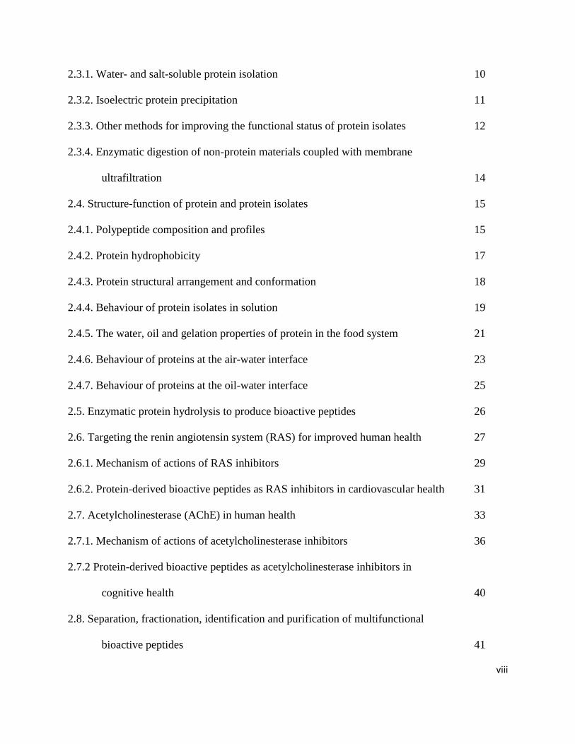
viii
2.3.1. Water- and salt-soluble protein isolation 10
2.3.2. Isoelectric protein precipitation 11
2.3.3. Other methods for improving the functional status of protein isolates 12
2.3.4. Enzymatic digestion of non-protein materials coupled with membrane
ultrafiltration 14
2.4. Structure-function of protein and protein isolates 15
2.4.1. Polypeptide composition and profiles 15
2.4.2. Protein hydrophobicity 17
2.4.3. Protein structural arrangement and conformation 18
2.4.4. Behaviour of protein isolates in solution 19
2.4.5. The water, oil and gelation properties of protein in the food system 21
2.4.6. Behaviour of proteins at the air-water interface 23
2.4.7. Behaviour of proteins at the oil-water interface 25
2.5. Enzymatic protein hydrolysis to produce bioactive peptides 26
2.6. Targeting the renin angiotensin system (RAS) for improved human health 27
2.6.1. Mechanism of actions of RAS inhibitors 29
2.6.2. Protein-derived bioactive peptides as RAS inhibitors in cardiovascular health 31
2.7. Acetylcholinesterase (AChE) in human health 33
2.7.1. Mechanism of actions of acetylcholinesterase inhibitors 36
2.7.2 Protein-derived bioactive peptides as acetylcholinesterase inhibitors in
cognitive health 40
2.8. Separation, fractionation, identification and purification of multifunctional
bioactive peptides 41
ix
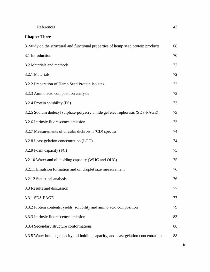
References 43
Chapter Three
3. Study on the structural and functional properties of hemp seed protein products 68
3.1 Introduction 70
3.2 Materials and methods 72
3.2.1 Materials 72
3.2.2 Preparation of Hemp Seed Protein Isolates 72
3.2.3 Amino acid composition analysis 72
3.2.4 Protein solubility (PS) 73
3.2.5 Sodium dodecyl sulphate-polyacrylamide gel electrophoresis (SDS-PAGE) 73
3.2.6 Intrinsic fluorescence emission 73
3.2.7 Measurements of circular dichroism (CD) spectra 74
3.2.8 Least gelation concentration (LGC) 74
3.2.9 Foam capacity (FC) 75
3.2.10 Water and oil holding capacity (WHC and OHC) 75
3.2.11 Emulsion formation and oil droplet size measurement 76
3.2.12 Statistical analysis 76
3.3 Results and discussion 77
3.3.1 SDS-PAGE 77
3.3.2 Protein contents, yields, solubility and amino acid composition 79
3.3.3 Intrinsic fluorescence emission 83
3.3.4 Secondary structure conformations 86
3.3.5 Water holding capacity, oil holding capacity, and least gelation concentration 88
x
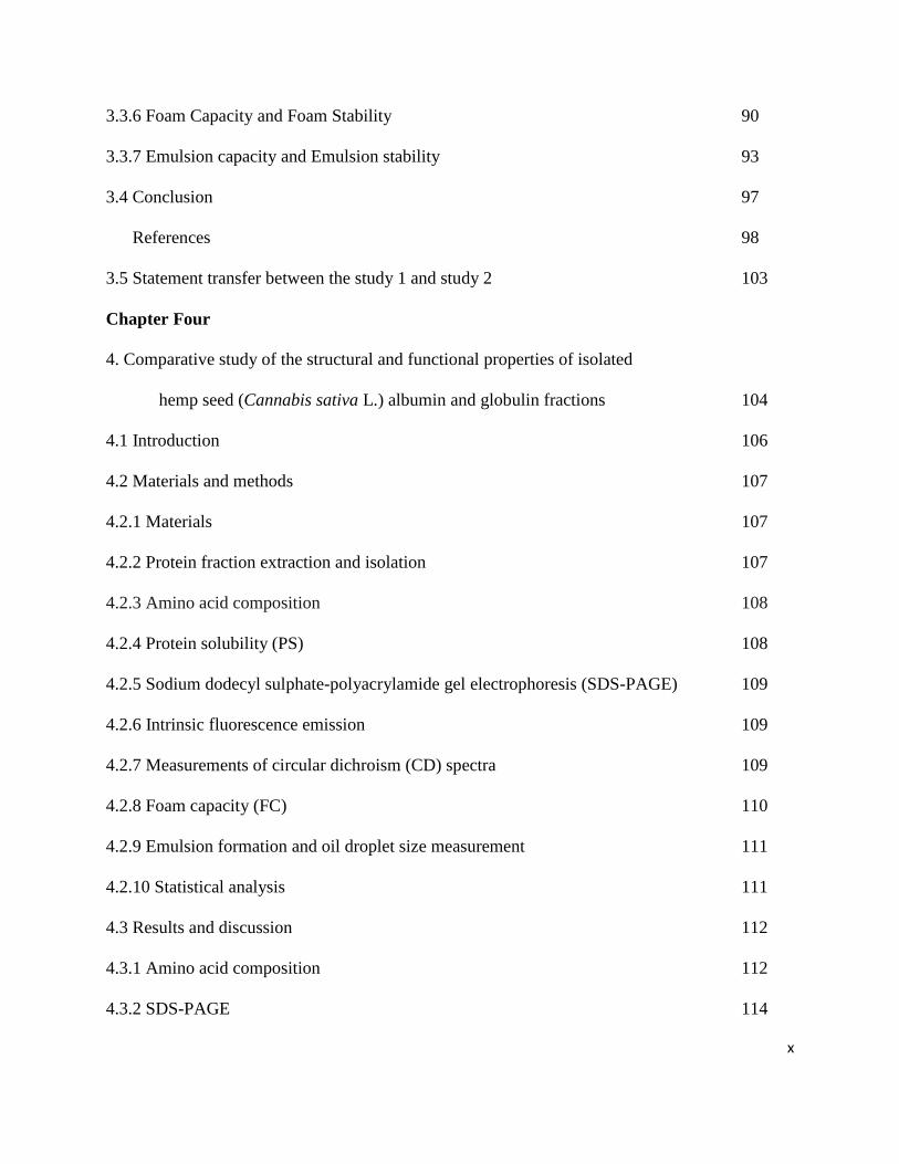
3.3.6 Foam Capacity and Foam Stability 90
3.3.7 Emulsion capacity and Emulsion stability 93
3.4 Conclusion 97
References 98
3.5 Statement transfer between the study 1 and study 2 103
Chapter Four
4. Comparative study of the structural and functional properties of isolated
hemp seed (Cannabis sativa L.) albumin and globulin fractions 104
4.1 Introduction 106
4.2 Materials and methods 107
4.2.1 Materials 107
4.2.2 Protein fraction extraction and isolation 107
4.2.3 Amino acid composition 108
4.2.4 Protein solubility (PS) 108
4.2.5 Sodium dodecyl sulphate-polyacrylamide gel electrophoresis (SDS-PAGE) 109
4.2.6 Intrinsic fluorescence emission 109
4.2.7 Measurements of circular dichroism (CD) spectra 109
4.2.8 Foam capacity (FC) 110
4.2.9 Emulsion formation and oil droplet size measurement 111
4.2.10 Statistical analysis 111
4.3 Results and discussion 112
4.3.1 Amino acid composition 112
4.3.2 SDS-PAGE 114
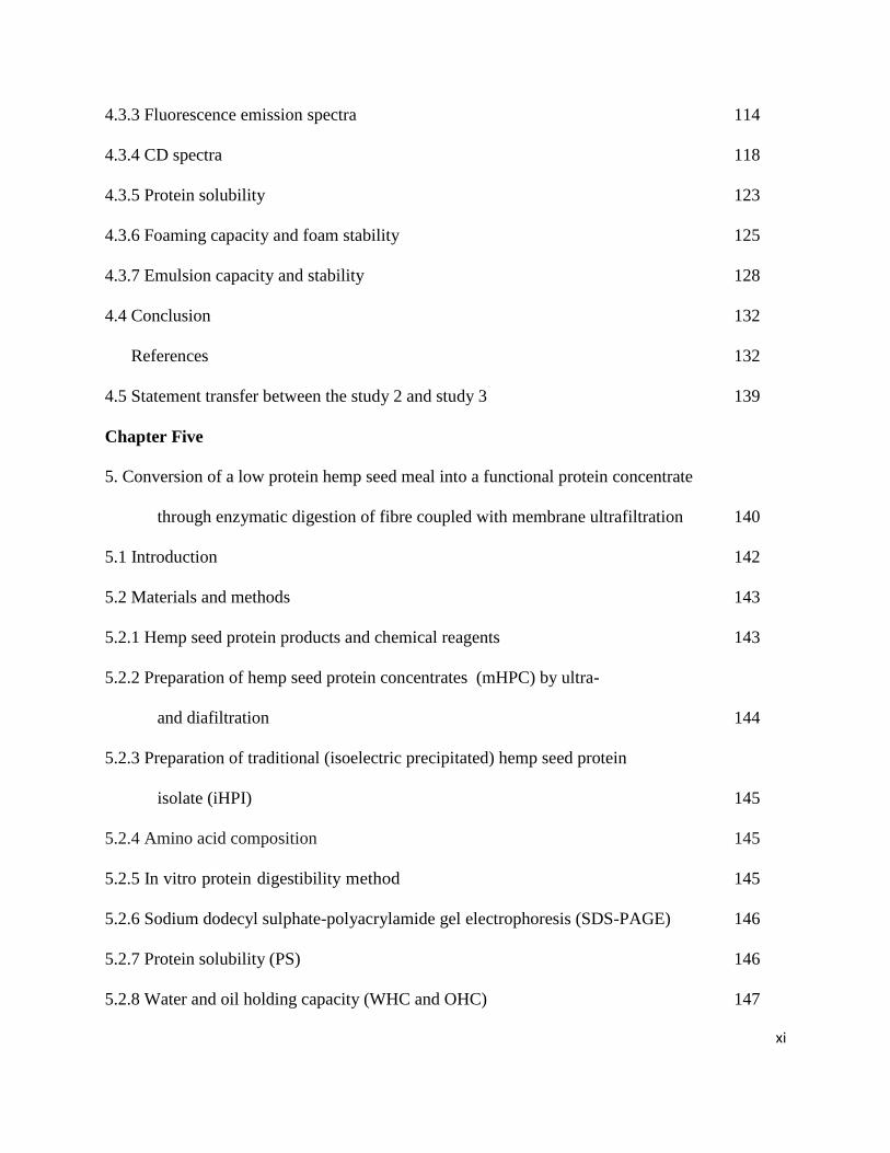
xi
4.3.3 Fluorescence emission spectra 114
4.3.4 CD spectra 118
4.3.5 Protein solubility 123
4.3.6 Foaming capacity and foam stability 125
4.3.7 Emulsion capacity and stability 128
4.4 Conclusion 132
References 132
4.5 Statement transfer between the study 2 and study 3 139
Chapter Five
5. Conversion of a low protein hemp seed meal into a functional protein concentrate
through enzymatic digestion of fibre coupled with membrane ultrafiltration 140
5.1 Introduction 142
5.2 Materials and methods 143
5.2.1 Hemp seed protein products and chemical reagents 143
5.2.2 Preparation of hemp seed protein concentrates (mHPC) by ultra-
and diafiltration 144
5.2.3 Preparation of traditional (isoelectric precipitated) hemp seed protein
isolate (iHPI) 145
5.2.4 Amino acid composition 145
5.2.5 In vitro protein digestibility method 145
5.2.6 Sodium dodecyl sulphate-polyacrylamide gel electrophoresis (SDS-PAGE) 146
5.2.7 Protein solubility (PS) 146
5.2.8 Water and oil holding capacity (WHC and OHC) 147
xii
5.2.9 Foam capacity (FC) 147
5.2.10 Emulsion formation and oil droplet size measurement 147
5.2.11 Statistical analysis 148
5.3 Results and discussion 148
5.3.1 Amino acid composition 148
5.3.2 SDS-PAGE 150
5.3.3 Protein contents and Protein solubility (PS) 152
5.3.4 In vitro Protein digestibility (IPD) 155
5.3.5 Water and oil holding capacity (WHC and OHC) 157
5.3.6 Foaming capacity and foam stability 159
5.3.7 Emulsion capacity and stability 162
5.4 Conclusion 166
References 167
5.5 Statement transfer between the study 3 and study 4 173
Chapter Six
6. Study on the structural and antihypertensive properties of enzymatic
hemp seed protein hydrolysates 174
6.1 Introduction 176
6.2 Materials and methods 177
6.2.1. Hemp seed products and chemical reagents 177
6.2.2. Preparation of hemp seed protein isolates (HPI) 178
6.2.3. Preparation of enzymatic hemp seed protein hydrolysates (HPHs) 178
6.2.4. Determination of degree of hydrolysis 179
xiii
6.2.5. Amino acid composition analysis 179
6.2.6. Analysis of molecular weight distribution 179
6.2.7. Intrinsic fluorescence 180
6.2.8. ACE inhibition assay 180
6.2.9. Renin inhibition assay 181
6.2.10. BP-lowering effect of peptides in Spontaneously Hypertensive Rats (SHRs) 182
6.2.11. Statistical analysis 183
6.3 Results 183
6.3.1. Amino acid composition of HPI and HPHs 183
6.3.2. DH and size exclusion chromatography analysis of HPHs 183
6.3.3. Intrinsic fluorescence properties 187
6.3.4. In vitro inhibition of ACE and renin activities of HPHs 187
6.3.5. In vivo reduction of blood pressure by HPHs 191
6.4 Discussion 193
6.5 Conclusion 198
References 199
6.6 Statement transfer between the study 4 and study 5 206
Chapter Seven
7. Study on the in vitro acetylcholinesterase-inhibitory properties of enzymatic
hemp seed protein hydrolysates 207
7.1 Introduction 209
7.2 Materials and methods 211
7.2.1. Hemp seed products and chemical reagents 211
xiv
7.2.2. Preparation of hemp seed protein isolate (HPI) 212
7.2.3. Preparation of enzymatic hemp seed protein hydrolysates (HPHs) 212
7.2.4. Amino acid composition analysis 213
7.2.5. Determination of degree of hydrolysis (DH) 213
7.2.6. Analysis of molecular weight distribution 213
7.2.7. Mass spectrometry analysis of protein hydrolysates 214
7.2.8. Acetylcholinesterase (AChE) inhibition assays 214
7.2.9. Statistical analysis 215
7.3 Results and discussion 215
7.3.1. Degree of hydrolysis (DH) and AChE inhibition 215
7.3.2. Analysis of molecular weight distribution 218
7.3.3. Amino acid composition and peptide sequences 220
7.4 Conclusion 226
References 227
7.5 Statement transfer between the study 5 and study 6 232
Chapter Eight
8. Study on the purification, amino acid sequences and potency of hemp seed
protein-derived acetylcholinesterase-inhibitory peptides 233
8.1 Introduction 235
8.2 Materials and methods 236
8.2.1 Hemp seed products and chemical reagents 236
8.2.2 Preparation of enzymatic hemp seed protein hydrolysates (HPHs) 237
8.2.3 Reverse-phase (RP)-HPLC separation of HPH 237
xv
8.2.4 LC/MS/MS identification of the purified peptides 238
8.2.5 Acetylcholinesterase (AChE) inhibition assays 239
8.2.6 Statistical analysis 239
8.3 Results and discussion 240
8.3.1 RP-HPLC fractions from 1% pepsin-produced HPH 240
8.3.2 Inhibition of AChE by different RP-HPLC fractions 240
8.3.3 Identification of AChE inhibitory peptides from hemp seed protein hydrolysates 243
8.3.4 Inhibition of AChE by different hemp protein-derived bioactive peptides 249
8.4 Conclusion 252
References 253
Chapter Nine
9. General Summary and Conclusion of the study 257
Chapter Ten
10. Limitations of the study 262
Chapter Eleven
11. Future directions of the study 263
xvi
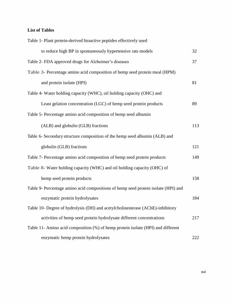
List of Tables
Table 1- Plant protein-derived bioactive peptides effectively used
to reduce high BP in spontaneously hypertensive rats models 32
Table 2- FDA approved drugs for Alzheimer’s diseases 37
Table 3- Percentage amino acid composition of hemp seed protein meal (HPM)
and protein isolate (HPI) 81
Table 4- Water holding capacity (WHC), oil holding capacity (OHC) and
Least gelation concentration (LGC) of hemp seed protein products 89
Table 5- Percentage amino acid composition of hemp seed albumin
(ALB) and globulin (GLB) fractions 113
Table 6- Secondary structure composition of the hemp seed albumin (ALB) and
globulin (GLB) fractions 121
Table 7- Percentage amino acid composition of hemp seed protein products 149
Table 8- Water holding capacity (WHC) and oil holding capacity (OHC) of
hemp seed protein products 158
Table 9- Percentage amino acid compositions of hemp seed protein isolate (HPI) and
enzymatic protein hydrolysates 184
Table 10- Degree of hydrolysis (DH) and acetylcholinesterase (AChE)-inhibitory
activities of hemp seed protein hydrolysate different concentrations 217
Table 11- Amino acid composition (%) of hemp protein isolate (HPI) and different
enzymatic hemp protein hydrolysates 222
xvii
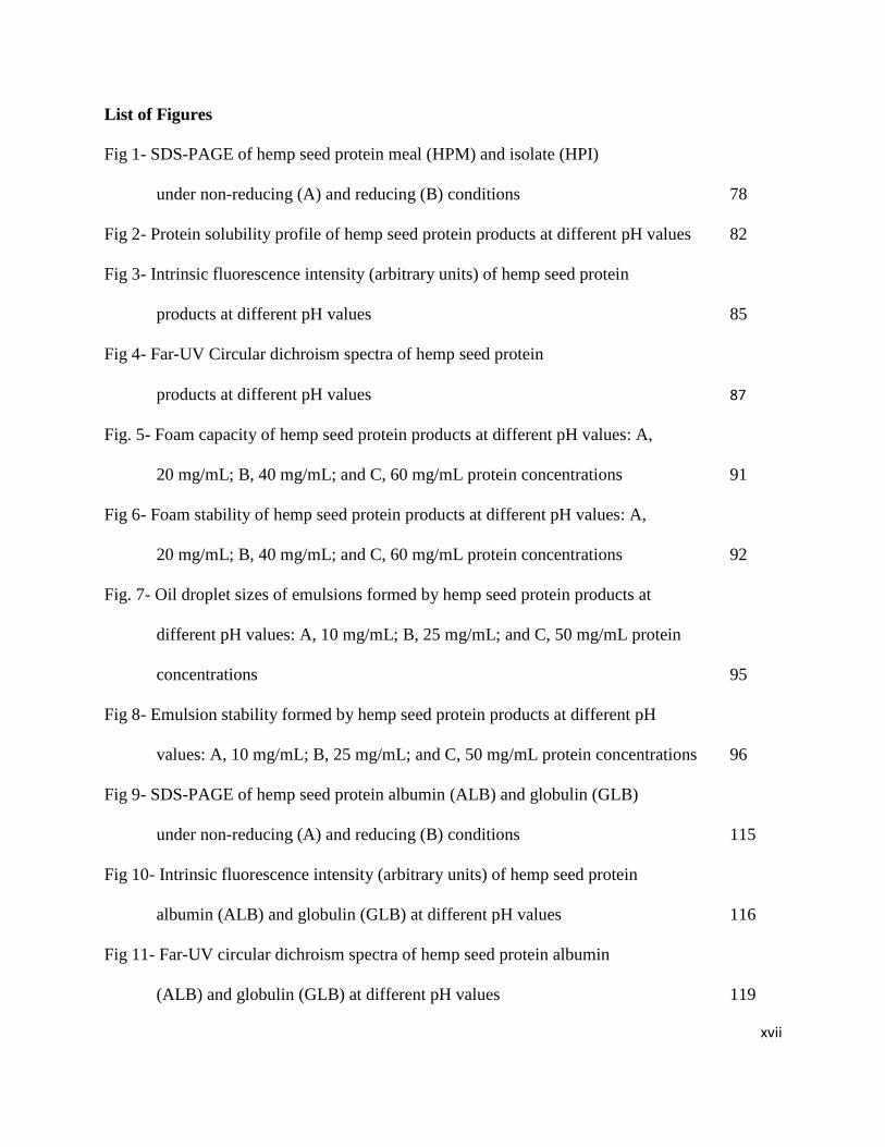
List of Figures
Fig 1- SDS-PAGE of hemp seed protein meal (HPM) and isolate (HPI)
under non-reducing (A) and reducing (B) conditions 78
Fig 2- Protein solubility profile of hemp seed protein products at different pH values 82
Fig 3- Intrinsic fluorescence intensity (arbitrary units) of hemp seed protein
products at different pH values 85
Fig 4- Far-UV Circular dichroism spectra of hemp seed protein
products at different pH values 87
Fig. 5- Foam capacity of hemp seed protein products at different pH values: A,
20 mg/mL; B, 40 mg/mL; and C, 60 mg/mL protein concentrations 91
Fig 6- Foam stability of hemp seed protein products at different pH values: A,
20 mg/mL; B, 40 mg/mL; and C, 60 mg/mL protein concentrations 92
Fig. 7- Oil droplet sizes of emulsions formed by hemp seed protein products at
different pH values: A, 10 mg/mL; B, 25 mg/mL; and C, 50 mg/mL protein
concentrations 95
Fig 8- Emulsion stability formed by hemp seed protein products at different pH
values: A, 10 mg/mL; B, 25 mg/mL; and C, 50 mg/mL protein concentrations 96
Fig 9- SDS-PAGE of hemp seed protein albumin (ALB) and globulin (GLB)
under non-reducing (A) and reducing (B) conditions 115
Fig 10- Intrinsic fluorescence intensity (arbitrary units) of hemp seed protein
albumin (ALB) and globulin (GLB) at different pH values 116
Fig 11- Far-UV circular dichroism spectra of hemp seed protein albumin
(ALB) and globulin (GLB) at different pH values 119

xviii
Fig 12- Near-UV circular dichroism spectra of hemp seed protein albumin
(ALB) and globulin (GLB) at different pH values 122
Fig 13- Protein solubility profile of hemp seed protein albumin and globulin
at different pH values 124
Fig 14- Foam capacity of hemp seed albumin and globulin in water and at different pH
values: A, 20 mg/mL; B, 40 mg/mL; and C, 60 mg/mL protein concentrations 126
Fig 15- Foam stability of hemp seed albumin and globulin in water and at different pH
values: A, 20 mg/mL; B, 40 mg/mL; and C, 60 mg/mL protein concentrations 127
Fig 16- Oil droplet sizes of emulsions formed by hemp seed albumin and globulin in
water and at different pH values: A, 10 mg/mL; B, 25 mg/mL; and C,
50 mg/mL protein concentrations 129
Fig 17- Emulsion stability formed by hemp seed albumin and globulin in water and at
different pH values: A, 10 mg/mL; B, 25 mg/mL; and C,
50 mg/mL protein concentrations 130
Fig 18- Polypeptide composition of hemp seed protein products under non-reducing
(A) and reducing (B) sodium dodecyl sulfate polyacrylamide gel electrophoresis
conditions: cHPC, commercial hemp seed protein concentrate; iHPI, isoelectric pH
precipitated hemp seed protein isolate; mHPC, membrane ultrafiltration hemp seed
concentrate; HPM, hemp seed protein meal. 151
Fig 19- Protein solubility profile of hemp seed protein products at different pH
values: cHPC, commercial hemp seed protein concentrate; iHPI,
isoelectric pH-precipitated protein isolate; mHPC, membrane
ultrafiltration protein concentrate; HPM, hemp seed protein meal. 153
xix
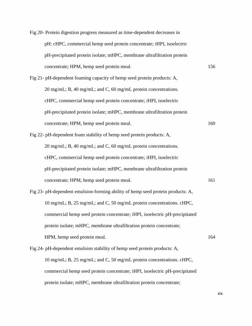
Fig 20- Protein digestion progress measured as time-dependent decreases in
pH: cHPC, commercial hemp seed protein concentrate; iHPI, isoelectric
pH-precipitated protein isolate; mHPC, membrane ultrafiltration protein
concentrate; HPM, hemp seed protein meal. 156
Fig 21- pH-dependent foaming capacity of hemp seed protein products: A,
20 mg/mL; B, 40 mg/mL; and C, 60 mg/mL protein concentrations.
cHPC, commercial hemp seed protein concentrate; iHPI, isoelectric
pH-precipitated protein isolate; mHPC, membrane ultrafiltration protein
concentrate; HPM, hemp seed protein meal. 160
Fig 22- pH-dependent foam stability of hemp seed protein products: A,
20 mg/mL; B, 40 mg/mL; and C, 60 mg/mL protein concentrations.
cHPC, commercial hemp seed protein concentrate; iHPI, isoelectric
pH-precipitated protein isolate; mHPC, membrane ultrafiltration protein
concentrate; HPM, hemp seed protein meal. 161
Fig 23- pH-dependent emulsion-forming ability of hemp seed protein products: A,
10 mg/mL; B, 25 mg/mL; and C, 50 mg/mL protein concentrations. cHPC,
commercial hemp seed protein concentrate; iHPI, isoelectric pH-precipitated
protein isolate; mHPC, membrane ultrafiltration protein concentrate;
HPM, hemp seed protein meal. 164
Fig 24- pH-dependent emulsion stability of hemp seed protein products: A,
10 mg/mL; B, 25 mg/mL; and C, 50 mg/mL protein concentrations. cHPC,
commercial hemp seed protein concentrate; iHPI, isoelectric pH-precipitated
protein isolate; mHPC, membrane ultrafiltration protein concentrate;
xx
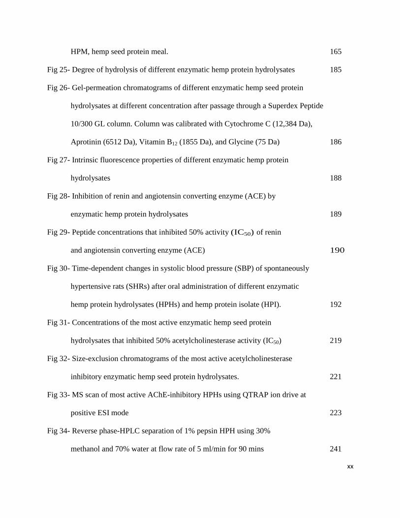
HPM, hemp seed protein meal. 165
Fig 25- Degree of hydrolysis of different enzymatic hemp protein hydrolysates 185
Fig 26- Gel-permeation chromatograms of different enzymatic hemp seed protein
hydrolysates at different concentration after passage through a Superdex Peptide
10/300 GL column. Column was calibrated with Cytochrome C (12,384 Da),
Aprotinin (6512 Da), Vitamin B12 (1855 Da), and Glycine (75 Da) 186
Fig 27- Intrinsic fluorescence properties of different enzymatic hemp protein
hydrolysates 188
Fig 28- Inhibition of renin and angiotensin converting enzyme (ACE) by
enzymatic hemp protein hydrolysates 189
Fig 29- Peptide concentrations that inhibited 50% activity (IC50) of renin
and angiotensin converting enzyme (ACE) 190
Fig 30- Time-dependent changes in systolic blood pressure (SBP) of spontaneously
hypertensive rats (SHRs) after oral administration of different enzymatic
hemp protein hydrolysates (HPHs) and hemp protein isolate (HPI). 192
Fig 31- Concentrations of the most active enzymatic hemp seed protein
hydrolysates that inhibited 50% acetylcholinesterase activity (IC50) 219
Fig 32- Size-exclusion chromatograms of the most active acetylcholinesterase
inhibitory enzymatic hemp seed protein hydrolysates. 221
Fig 33- MS scan of most active AChE-inhibitory HPHs using QTRAP ion drive at
positive ESI mode 223
Fig 34- Reverse phase-HPLC separation of 1% pepsin HPH using 30%
methanol and 70% water at flow rate of 5 ml/min for 90 mins 241
xxi
Fig 35- AChE inhibition assay (%) of RP-HPLC separation of 1% pepsin HPH 242
Fig 36- MS scan of RP-HPLC separated fraction F7 using QTRAP ion drive at
positive ESI mode 244
Fig 37- LC/MS/MS of RP-HPLC separated fraction F7 at different m/z using
QTRAP ion drive at positive ESI mode 245
Fig 38- Inhibition of AChE (from Eel) by different enzymatic hemp protein-derived
peptides at a 50% level of inhibition (IC50) 250
Fig 39- Inhibition of AChE (from Eel) by different enzymatic hemp protein-derived
peptides at a 50% level of inhibition (IC50) 251
xxii
LIST OF COPYRIGHT MATERIALS USED IN THE PREPARATION OF THE THESIS
1. Malomo, S. A., He, R., & Aluko, R. E. (2014). Structural and functional properties of
hemp seed protein products. Journal of Food Science, 79 (8), C1512-1521. Copyright ©
2014, with permission from John Wiley & Sons.
2. Malomo, S. A., & Aluko, R. E. (2015). A comparative study of the structural and
functional properties of isolated hemp seed (Cannabis sativa L.) albumin and globulin
fractions. Food Hydrocolloids, 43, 743-752. Copyright © 2015, with permission from
Elsevier.
xxiii
List of Abbreviations
ACE - Angiotensin converting enzyme
ACh – Acetylcholine
AChE – Acetylcholinesterase
ALB – Albumin
BP - Blood pressure
CD – Circular dichroism
DPPH - 1,1- diphenyl-2-picrylhydrazyl
FI - fluorescence intensity
GLB – Globulin
HPLC – High Performance Liquid Chromatography
HPI - Hemp protein isolate
HPH - Hemp protein hydrolysate
IPD – In vitro Protein Digestibility
LGC – Least gelation concentration
ME - Mercaptoethanol
OHC – Oil holding capacity
PS - Protein solubility
SDS - Sodium dodecyl sulfate
So - Surface hydrophobicity
THC - Tetrahydrocannibinol
WHC - Water holding capacity
1
CHAPTER ONE
1. General Introduction
The structure and function of proteins have always been critical indicators that determine the
various physiological processes and functions occurring in any biological system (Barac et al.,
2014). Scientific investigations into the dynamics of proteins centred on structure and function
determinations in order to give a better understanding of their potential uses in foods and
biological systems (MuneMune et al., 2014; Tan et al., 2014). For instance, protein activities’
research works have brought extensive insight into various biological processes such as protein
folding, enzyme functions and target identification of ligand binding to the receptor. Other
processes include interaction of proteins with molecules such as the protein itself, lipids (oils),
and water. All these processes aid the use of proteins as basic and bioactive ingredients in food
and human health applications (Rasool et al., 2013).
As of today, many simple and easily-adopted methods available for protein isolation include,
but are not limited to salt extraction, traditional isoelectric precipitation, and enzyme pre-
digestion coupled with membrane filtration to remove non-protein materials. The analytical
methods involved in protein dynamics studies related to food and human health applications are
directed towards investigating physicochemical and functional properties in relation to
environmental factors (pH, heat and concentration) (Barac et al., 2014). Apart from native
proteins, research works have also been geared towards determining bioactivity potentials of low
molecular weight polypeptide chains (peptides) of proteins in the management of several human
diseases (Udenigwe & Mohan, 2014).
Several physiological imbalances and malfunctioning of body homeostasis have been the
causative background of aggravated human chronic disease conditions such as Alzheimer’s
2
disease (AD) and cardiovascular diseases (CVD) like hypertension (Health Canada, 2014).
Various reports (Kumar & Chowdhury, 2014; Iannello et al., 2014; Saravanaraman et al., 2014;
Ghribia et al., 2014) have shown the acetylcholinesterase (AChE) enzyme as an important target
in research aimed at developing therapeutic tools against a wide range of human metabolic
disorders. This is because AChE hydrolyzes the neurotransmitter acetylcholine (ACh) at
cholinergic synapses, with a higher catalytic efficiency than other known enzymes. The
dysfunction of cholinergic neurotransmission in the brain, the formation and growth of brain
amyloid lesions, including the senility-promoting plaques have been widely reported as
contributing factors to a Alzheimer’s disease (AD) pathogenesis (Willcox et al., 2014). As of
today the current available therapy for AD consists of the administration of AChE inhibitors due
to their clinical efficacy in prolonging the half-life of ACh (Kumar & Chowdhury, 2014).
Recently, there is awareness in the food industry and in preventive health care activities
with respect to the development of natural nutraceuticals and functional foods ingredients from
plant materials due to their high biodegradability rate, which results in little or no toxicity
(residual effects or negative side effects). An example of such plant materials is the industrial
hemp (Cannabis sativa L.), which was traditionally grown for its high fibre content. However,
the production of fibre from hemp leaves behind the hemp seed, which is a rich source of high
quality oil (30%) and proteins (25%) (Radocaj et al., 2014). Therefore, recent efforts at hemp
commercialization have actually focused on the seed because the high quality oil and proteins
can serve as ingredients to formulate functional foods and nutraceuticals. This has led to a
substantial growth in the hemp seed processing industry and an increase in the economic value of
the crop.
3
In Canada, hemp seed processing primarily involves cold-pressing to expel the oil, which
leaves behind a high protein (30-50%) residue. The increased utilization of hemp seed for edible
oil production has led to abundant amounts of protein-rich meal, which has been converted into
various forms of powdered protein products. These protein products are sold as ingredients for
the formulation of foods such as plant-based protein shakes, hemp milk (vanilla or chocolate
flavoured) and energy bars (House et al., 2010). Current commercial hemp seed protein (HSP)
products are mainly in the form of protein concentrates that have less than 70% protein content
but with high fibre levels (Lu et al., 2010). The high fibre and phytate contents of these HSP
products reduce protein functionality and limit their use in the manufacture of novel food
products (Teh et al., 2014). Reports have shown HSP to contain 65% globulin (edestin) and 33%
albumin; edestin is composed of six identical subunits with the acidic subunit (AS) linked by one
disulphide bond to a basic subunit (BS) (Teh et al., 2014; Teh et al., 2013; Park et al., 2012). In
the plant kingdom, these two high-quality storage HSP have been reported to be readily
digestible, and are rich in all essential amino acids with exceptionally high arginine and glutamic
acid contents (Park et al., 2012).
HSP can be efficiently obtained from defatted hemp seed meal (HSM) by alkali
solubilization followed by acid precipitation (Girgih et al., 2014a). The poor food functionality
of current hemp seed protein products is due primarily to two reasons, first is the high fibre and
phytate content in the concentrates and second is the poor solubility of the protein isolate, which
is due to the use of harsh alkali extraction, acid-induced protein precipitation and cross-linking of
proteins by phytate molecules (Teh et al., 2014). Therefore, in order to produce novel protein
isolates with improved food functionalities, a method that involved fibre and phytate digestion
followed by removal of non-protein materials through membrane ultrafiltration was used. The
4
membrane ultrafiltration obviates the need for the use of undesirable harsh protein precipitation
protocols while ensuring high protein yield with minimal denaturation and a potential for strong
functional performance in food formulations. This fibre and phytate digestion method had been
previously used to obtain high protein, yield and quality soy (Wang et al., 2014), flaxseed
(Udenigwe et al., 2009) and rice bran (Wang et al., 1999) protein isolates. However, pre-
digestion with carbohydrases and phytases plus coupling with membrane ultrafiltration has not
been reported for hemp seed protein isolate production.
The functional properties of the enzyme pre-digested and membrane-produced protein
isolates that was determined in the present study include protein solubility (important in the
formulation of clear beverages), emulsification (important for the manufacture of oil-in-water
emulsions like salad dressings and soups), foaming (important in formulation of desserts such as
meringues), and gelation (critical for formulation of semi-solid to solid foods). In addition, the
work also studied the relationships between physicochemical properties (polypeptide
composition, hydrophobicity, secondary & tertiary structures) and functional properties in order
to gain fundamental understanding of the structural factors of the proteins that enhance their
functional properties. The obtained protein isolate was then subjected to in vitro enzymatic
digestion using different proteases to yield AChE-inhibitory and antihypertensive peptides. The
active protein hydrolysates were then separated and purified by column chromatography in order
to identify peptide amino acid sequences.
1.1. Hypotheses
In order to obtain basic knowledge existing between the structure and function of a protein in
the biological system, the following hypotheses were proposed:
5
1) Enzyme and ultrafiltration membrane-assisted removal of non-protein materials will lead
to the production of a hemp seed protein isolate with higher protein content than the
isoelectric-precipitated equivalent.
2) The membrane protein isolate will have higher contents of native structural
conformations and produce higher level of food functionality when compared to protein
materials produced by isoelectric precipitation.
3) Optimized in vitro enzymatic hydrolysis of hemp seed protein isolate will lead to the
release of multifunctional peptides that possess acetylcholinesterase inhibitory and
antihypertensive activities.
1.2. Objectives
This study aimed to address some unanswered questions related to the effects of structure-
functions properties of hemp seed protein products in food and human health applications in
vitro and in vivo through the following main objectives:
1) Compare the physicochemical and functional properties of hemp seed albumin and
globulin protein fractions,
2) Optimize cellulase and phytase degradation of hemp seed fibre and phytate, respectively,
into low molecular weight products that can be removed by membrane ultrafiltration to
produce HSP isolate with high protein content and food functionality,
3) Determine the in vitro digestibility, physicochemical and functional properties of
laboratory-prepared HSP isolates in comparison with commercial hemp seed protein
products,
6
4) Modify the HSP isolate using various enzymes (alcalase, thermoase, trypsin, papain,
chymotrypsin, pepsin) to produce acetylcholinesterase-inhibitory and antihypertensive
peptides, and
5) Purify and determine the amino acid sequence of acetylcholinesterase-inhibitory peptides
present in active protein hydrolysates followed by a study of their structure-function
properties.
1.3. Justification
Bioactive peptides with low serine-histidine ratio have the potentials to be used as potent
agents for fast and effective managing of cognitive malfunction in the body system. Natural
peptide products do not have negative side effects associated with synthetic drugs and the hemp
seed peptides could serve as alternative anti-AChE agents. Moreover, peptides are more effective
nutrition agents when compared to free amino acids. This is because unlike free serine and
histidine molecules, low serine or histidine containing peptides have the advantage of being able
to penetrate cells directly without the need for transporters. The use of this type of novel peptides
is anchored on the fact that AChE is a serine hydrolase with specificity for serine and histidine;
so the low ratio of these amino acids would make it unavailable for the enzyme or totally block
the active site of action of the enzyme. Therefore, optimized feeding of low serine/histidine
containing peptides could provide relief from Alzheimer’s disease without the use of drugs. In
addition to cognitive decline, hypertension is also a recognized health problem in the elederly
population. Thus, the development of multifunctional peptides that can act simultaneously
against cognitive decline and hypertension could provide innovative means of managing chronic
diseases. More importantly such multifunctional peptides could reduce health care costs
7
associated with therapeutic treatment since one agent (as against the current practice of multiple
agents) will be effective against more than one disease condition.
Conversion of protein constituents into clinical nutrition tools as well as functional and
nutraceutical peptides are excellent ways of increasing the economic value of hemp seed.
Moreover, different studies have revealed a strong market demand for bioactive peptides and
thus, this research work would provide hemp seed peptides that satisfy part of this demand.
1.4. Significance
The significance of this study is based on the growing economic importance of hemp
seed protein in Manitoba and Canada at large. For instance, there are currently two hemp seed
processing companies (Hemp Oil Canada, St. Agathe; and Manitoba Harvest, Winnipeg) in
Manitoba that produce various products including protein concentrates. Thus, this study is
expected to increase value-added utilization of the products from these companies which, could
lead to improved economic power of individuals (farmers, processors, suppliers and traders),
provinces (Manitoba and environs) and Canada as a whole. Also critical to the choice is the fact
that HSPs contain the highest level (~11% when compared to <6% for most other food proteins)
of arginine (a precursor of the vasodilatory nitric oxide) among foods and as such, it could be
used as a rich source of cardiovascular health-promoting amino acid. Finally, the outcome of this
work may provide hemp seed peptides for use as ingredients to formulate therapeutic foods for
AD prevention and treatment. Peptides could provide these health benefits without the negative
side effects such as failure of cholinergic synaptic transmission, deterioration of neuromuscular
junctions, flaccid muscle paralysis, and central nervous system seizures that are associated with
use of AD drugs.
8
CHAPTER TWO
2. Literature Review
2.1. Why hemp?
Industrial hemp (Cannabis sativa L.) is one of dozens of plant species that represent at
least 22 genera and contain <0.1% Δ-9-tetrahydrocannabinol (THC), a known phytochemical
drug component in most Cannabis species. This particular species has been disruptively selected
for bast (phloem) fibre in the stem, multipurpose oil in the achenes, and an intoxicating resin
secreted by epidermal glands (Small et al., 2003). It is industrially grown primary for utilization
in fibre production; however, the seeds recovered as by-products are then further processed for
high quality oil and protein-rich meal (Radocaj et al., 2014). Despite the prohibition on its
cultivation in many countries due to the presence of THC, this important crop still serves as an
excellent source of food, fibre, dietary oil and medicine (Lu et al., 2010).
In Canada, industrial hemp has gained ground as an important agricultural commodity
and now supports the operations of the two Manitoba hemp seed processing companies (The
Hemp Oil Canada, St. Agathe; and Manitoba Harvest, Winnipeg). Besides its edible and high
quality oil, the fibre-rich hemp plant had been widely used for paper and clothing productions in
Canada (Lu et al., 2010). The most important hemp products in the health food market are whole
hemp seed, hulled hemp seed, hemp seed oil and hemp protein meal. These hemp seed food
products contain very low THC levels that cannot produce intoxication when ingested (Karimi &
Hayatghaibi, 2005). In addition to the high quality oil, the legumin protein fraction (edestin) is
specifically found in the hemp seed alone but the albumin fraction is very similar to those found
in high-proteinous egg white, hence their higher protein quality when compared to other plant
albumins (Callaway et al., 2002). Besides, unlike soybean proteins, the HSP is reportedly devoid
9
of the trypsin inhibitors (the albumin-like compounds that can reduce protein digestion) and
flatulence-causing oligosaccharides (Karimi & Hayatghaibi, 2005).
2.2. The hemp seed protein
The major storage protein fractions in hemp seed are 2S albumin and 12S globulin. The
12S globulin – (edestin) is a legumin with a similar tertiary structure to 7S vicilins. These two
storage protein fractions are secretory proteins synthesized with a signal peptide, which ensure
their folded conformations within the lumen of the endoplasmic reticulum. They are also
characterized by heterogeneity in molecular weights and charges (Callaway et al., 2002). The
albumin fraction has a relatively high solubility in water whereas the globulin fraction is water-
insoluble but can be solubilized mostly in dilute salt solution at pH values above or below the
isoelectric points. The higher number of hydrophobic associations between these storage proteins
helps to stabilize their interactions with other food system compounds (Haard, 1985). The HSP
has been proven to contain all the nutritionally-essential amino acids, especially the highest level
of arginine among plant proteins, consisting of up to 11% of protein weight when compared to
<6% for most other food proteins (Yin et al., 2008).
The HSP has been utilized to produce bioactive compounds with desirable health
benefits. Such beneficial effects include as an antioxidant (Girgih et al., 2014a), oxidative
apoptosis protective agent (Chakrabarti et al., 2014), antihypertensive (Girgih et al., 2014b),
hydrogen peroxide-induced apoptosis protective agent (Lu et al., 2010) and industrial basic
functional ingredients (Teh et al., 2014; Teh et al., 2013; Yin et al., 2009; Yin et al., 2008).
2.3. Isolation methods employed for the production of plant protein isolates
The preparation methods (isolation/extraction) for most plant protein isolates are believed
to contribute to the physicochemical, digestibility, functional and nutritional properties of
10
different protein isolate fractions. The protein extraction methods and conditions, downstream
processing of extracted proteins (purification and drying), pH, temperature and ionic strength of
the food system, have been the important factors that affect functional properties in food systems
(Vioque et al., 2000). These intrinsic (structure and size) and environmental (protein
separation/production method, pH, ionic strength) factors can vary or determine the dynamic
utilization of plant proteins in various food industries. For instance, high oil and water binding
proteins could find desirable applications in the food processing industries dealings in meats,
sausages, breads, and cakes. Proteins with very high emulsifying and foaming capacity would
find useful applications in salad dressing, sausages, bologna, soups, confectionery, frozen
desserts and cakes (He et al., 2014b; Vioque et al., 2000). The different isolation methods that
have been used to produce plant protein isolates are highlighted below.
2.3.1. Water- and salt-soluble protein isolation
The salt extraction protein isolation method is based on the ionic strength of the solution
to dissolve and fractionate the two major storage proteins found in hemp seed - the 12S globulin
and the 2S albumin (Park et al., 2012). The high glycoprotein (up to 45%) present in albumin
fractions enhances protein-water interactions and thus aids its significant solubility in water. In
contrast, the low glycoprotein (3.9%) present in globulin fraction might as well contribute to its
water insolubility (Mundi & Aluko, 2012). The dissociation of subunits in these storage proteins
might be affected by appropriate conditions like solvent, pH and the ionic strength (Haard,
1985). The salt extraction method has been previously used to isolate proteins from plant sources
like canola and flaxseed (Karaca et al., 2011), buckwheat (Choi et al., 2007), kidney bean
(Mundi & Aluko, 2012), Ginkgo biloba bean (Deng et al., 2011) and African locust bean (Lawal
et al., 2005).
11
Dialysis of the salt extract against water followed by centrifugation produces a globulin-
rich precipitate and an albumin-rich supernatant (Mundi & Aluko, 2012). Generally, the
homologous storage globular HSP (edestin) has a molar mass of 300 kDa and its quaternary
structure is composed of six subunits (Park et al., 2012). Albumin, which has a low molecular
mass of 12–14 kDa is the other protein fraction of interest in most oilseeds. This typical rigid 2S
protein fraction (albumin) is well stabilized by inter-chain and intra-chain disulfide bridges with
40–45% helix structure (Park et al., 2012). Since most native proteins have poor functional
properties, different studies have shown that structural modification may be used to increase
protein functionality of albumin and globulin (Tang et al., 2011; Bora, 2002). For example,
chemical modification such as acylation was used to influence the surface functionality of
rapeseed albumin and globulin fractions (Gerbanowski et al., 1999).
2.3.2. Isoelectric protein precipitation
The most common and simple method of protein isolation involves the use of alkaline
solubilisation followed by acid precipitation at the isoelectric point (pI), for instance between pH
4.0 and 6.0 for seed proteins. At pH values above the pI (alkaline environment), the protein will
be solubilized with or without heat application while at lower pH values (acidic environment)
that coincide with the pI, the solubilized protein becomes insoluble, is precipitated out and
recovered as a precipitate after centrifugation. In some cases, the protein isolates generated from
this method may have high protein contents; however, protein recovery is incomplete, which
might be due to lack of adequate solubilisation or loss in the supernatant during centrifugation of
the precipitated proteins (Chavan et al., 2001). Moreover, all the proteins may not have the same
pI, hence selective precipitation at a single pH value results in low protein recovery.
12
Another limitation of this method, besides the poor functional properties, is that the
isoelectric-produced protein isolates are composed of higher amounts of ash generated during the
acid-base neutralization procedure (Chavan et al., 2001; Tang et al., 2011; Yin et al., 2008,
2009).
2.3.3. Other methods for improving the functional status of protein isolates
In order to improve the status of the plant protein isolates as functional ingredients,
several scientific methods have been put into use, which had invariably positively or negatively
affected their functional and physicochemical properties. For example, succinylation of lentil
seed globulin isolate significantly changed interfacial adsorption kinetics leading to improved
foam capacity and emulsion stability (Bora, 2002). High pressure, high heat and combination of
both methods were used to improve the physicochemical and functional properties of peanut, soy
and rapeseed protein isolates, respectively (He et al., 2014b; Li et al., 2011a; He et al., 2014a).
Higher solubility pea, chickpea and lentil protein isolates were obtained from ultrafiltration-
based protein isolation (Boye et al., 2010).
The probable positive or negative effects resulted from types of isolation method to
obtain plant protein isolates on its functional properties are described below. For instance, peanut
roasting during production of peanut protein isolate was found to reduce all the functional
properties whereas a reverse effect was reported when a fermentation method was used (Yu et
al., 2007). Significantly higher differences in the functional properties, particularly emulsifying
capacity, foaming capacity and oil binding, were observed for peanut protein isolates obtained by
spray drying and vacuum oven drying (Yu et al., 2007). Other methods of improving the
structure-function properties of plant protein isolates have involved the use of glycation on
kidney bean vicilin (Tang et al., 2011); micellization on amaranth protein isolates (Cordero-de-
13
los-Santos et al., 2005) and limited enzyme hydrolysis on lentil protein isolate (Avramenko et al.,
2013). The limited enzymatic hydrolysis method by trypsin was used to modify the functional
properties of hemp protein isolates (Yin et al., 2008). The enzymatic hydrolysis improved only
the protein solubility but led to decreased water and oil holding capacities as well as reduced
foaming and emulsion properties. The improved protein solubility resulted from the
transformation of insoluble protein components to soluble protein aggregates due to exposure of
ionizable amino and carboxyl groups of the protein during the limited enzymatic protein
hydrolysis. Meanwhile, the higher soluble aggregates in the hydrolysates were found to inhibit
surface viscoelastic membrane formation, caused oil droplets coalescence and led to decreased
emulsion capacity of the hydrolysates (Gbogouri et al., 2004). The shorter-chain lengths and low
molecular weight sizes of the limited enzymatic hydrolysis products (hydrolysates) might
account for the decreased foaming potentials (Yin et al., 2008).
The succinylation and acetylation (or acylation) methods were effectively employed to
improve or modify the structure-function properties of hemp seed protein isolates (Yin et al.,
2009). The succinylation and acetylation methods gradually increased the proteins’ solubility,
emulsion capacity (but decreased stability) and produced more compact structural conformation
of the proteins. The limited hydrolysis of rapeseed protein isolates led to improved functional
properties such as increased emulsification (Vioque et al., 2000). The limited proteolysis study
showed a positive relationship between the degree of hydrolysis and functional properties of a
protein. The hydrolysis process may have led to protein unfolding and enhanced exposure of
hydrophobic amino acid residues, which increased protein interactions with the oil and hence
better emulsification ability. Although, the limited enzymatic hydrolysis method of protein
isolation helped to expose the hydrophobic amino acid residues which are buried in the protein
14
core (mostly inaccessible), the resultant hydrolyzed proteins could be bitter due to presence of
low-molecular weight hydrophobic amino acid-rich peptides (Gong et al., 2015).
Hence, the limitations in most of these modification methods suggest the need to develop
more efficient protein isolation protocols such as those that involve enzymatic pre-digestion of
carbohydrates and phytates, which is then coupled to membrane ultrafiltration. Thus, the
enzymatic digestion and membrane-based removal of non-protein compounds might be a good
opportunity to obtain protein isolates with improved structure-function properties.
2.3.4. Enzymatic digestion of non-protein materials coupled with membrane ultrafiltration
Some plant proteins, especially hemp seeds have higher proportions of carbohydrates
(mainly fibres, and soluble sugars) and phytate (antinutrient), which limit the yield and
functional properties of protein isolates. These fibrous materials and phytate could be subjected
to digestion or degradation by several types of carbohydrases like cellulase, hemicellulase,
xylanase, amyloglucosidase and phytase. The reason for using a combination of carbohydrases
rather than single entity is due to the differences in the sites (bonds) of their cleavage actions. For
instance, cellulase breaks down the β(1→4)-linkage bond of the glucose structure (cellulose)
while hemicellulase and xylanase will hydrolyze the β(1→4)-linkage bond of the xylose
(hemicellulose and xylans) structure (Haard, 1985). Therefore, combined use of these
carbohydrases enhances the possibility that most of the fibres will be hydrolyzed and can be
separated from proteins. Use of the pre-digestion by these carbohydrases and phytase enzymes
followed by the removal of the digested products by the membrane ultrafiltration is novel and
has not been widely reported for plant protein isolate production.
Although, some recent works have reported the successful application of this novel
method to produce high quality protein isolates, there is no report for HSP isolate production.

15
For instance, Wang et al. (2014) reported a protein isolate with higher protein contents (91-93%)
and better in vitro digestibility when phytase was used to pre-process soybean flour. In the same
vein, the works of Udenigwe et al. (2009) have shown that pre-digestion of the fibre in defatted
flaxseed meal (known for its higher glucan and gum contents) with cellulase resulted in a
flaxseed protein isolate having 23% protein content higher than the protein isolated from
untreated flaxseed meal. Wang et al. (1999) produced a high protein content (92%), yield (75%)
and better functional (foam capacity) protein isolates from the combination of xylanase and
phytase pre-digestion of rice bran. Another study reported the use of microfiltration membranes
processes without enzyme pre-digestion to obtain soy protein isolate of improved protein yield,
solubility, foaming and emulsifying properties (Chove et al., 2007).
2.4. Structure-function of protein and protein isolates
The utilization of proteins as foods or food ingredients is dependent on its structural and
functional properties. These properties (the important determinants of quality protein) can be
modified and improved by any processing to enhance its utilization in the food and human health
applications (He et al., 2014a). Hence, the various structural and functional characteristics of
plant proteins that are essential for modification by appropriate processing (treatments) methods
are discussed below.
2.4.1. Polypeptide composition and profiles
The protein polypeptide composition and profiles can be analyzed in the laboratory using
the gel electrophoresis method. Polyacrylamide gel electrophoresis (PAGE) is used for
separating proteins ranging in size from 5 to 2,000 kDa due to the uniform pore size provided by
the polyacrylamide gel (Schägger, 2006). The protein samples are usually analyzed by sodium
dodecyl sulfate PAGE (SDS-PAGE) and by native-PAGE. The polypeptide profiles of the non-
16
denatured protein in their natural forms are determined by native-PAGE while the denatured
forms are analyzed by SDS-PAGE (Niepmann & Zheng, 2006). Gel electrophoresis is a common
method used to study mostly the size or length of the protein molecules. The main principle
behind gel electrophoresis is that proteins travel through the gel towards the positive pole and
away from the negative pole of the electrophoresis machine (Arndt et al., 2012). In the native-
PAGE analysis, the protein charges and mobility are dependent on the primary amino acid
sequence of the protein and the attained pH during the electrophoresis procedures (Arndt et al.,
2012).
Prior to the SDS-PAGE analysis, the proteins undergo denaturation in the presence of
a detergent such as sodium dodecyl sulfate (SDS) that coats the proteins to make them negatively
charged (Berg et al., 2002). Once denatured, the proteins form long rod-like structures instead of
their spiral complex tertiary shapes and move across the gel strictly based on molecular size
because they have the same SDS:protein ratio and therefore, have similar net charge (Niepmann
& Zheng, 2006). The charge to mass ratio of the protein is very important in this type of analysis;
therefore, the resulting denatured proteins have an overall negative charge while the relative
protein size is dependent on the amount of bound SDS (Berg et al., 2002). Since the SDS binds
to the same amount of proteins, the polypeptides will have similar negative charge density and
differ only in mass (or size).
Comparing the molecular sizes of the protein bands formed on the resultant gel, the
smaller the molecular size of the protein compounds, the further it would travel towards the
anode due to faster passage through the gel. Thus, the larger molecular size polypeptides would
move slowly towards the anode and can be separated from the faster moving smaller
polypeptides. Typical polyacrylamide gel density (degree of cross-linking) used for proteins is 4-

17
25% either as homogenous or density gradient type. In order to fully resolve very small proteins
sizes and peptides, homogenous high density (usually >20%) should be used (Schägger, 2006;
Berg et al., 2002). Besides molecular size and polypeptide composition determination, PAGE is
also used to determine protein purity after isolation and extraction.
2.4.2. Protein hydrophobicity
Research works that investigate extensive protein conformational studies involves
determination of intrinsic and surface hydrophobicity, which are mainly based on the
fluorescence properties of tyrosine, phenylalanine and tryptophan (Szabo, 2000). It is noteworthy
that most intrinsic fluorescence studies involve determination of tyrosine and tryptophan
emissions due to their high molar absorptivity, although tryptophan has always been the most
dominant (Schmid, 1989). Fluorescence spectroscopy has been used in various protein research
works to study structural hydrophobicity due to its high sensitivity level, rapid data acquisition,
wide dynamic range and convenient instrumentation (Szabo, 2000). Cheftel et al. (1985) reported
that hydrophobic interactions of native protein structures are solution-dependent. This is because
hydrophobic side chain amino acid residues that are buried in the protein interior need to be
exposed to the surface in order to measure hydrophobicity. Therefore, the solution properties
(pH, salt, temperature) will determine position of the aromatic amino acids and dictate the
magnitude of measured hydrophobicity. The distribution of polar and hydrophobic groups of
amino acid side chains has been suggested to determine protein solubility since these two groups
always have opposite interactions in solution (He et al., 2013).
Surface hydrophobicity, which is measured by ANS-binding experiments, can be coupled
with modifications that produce conformational changes and alter surface functional groups in
the protein (Matsudomi et al., 1985). Such modifications, which could be by deamidation-

18
induced and acid-induced denaturations were found to increase protein surface hydrophobicity.
For example, a previous study reported great improvement (good adsorption kinetics in the
interface and decreased molecular size) on the surface behaviours of soybean glycinin after mild
acidic treatments (Wagner & Gueguen, 1995).
2.4.3. Protein structural arrangement and conformation
Protein structural conformations are supported by the carbon-bonded sulfhydryl,
thioethers and carbonyls functional groups, which help to govern all the functional and biological
activities (Marsh & Teichmann, 2013). These activities are dependent on the primary structure
(amino acid sequence), secondary structure (α-helices and β-sheets stabilized largely by
hydrogen bonds), tertiary structure (3-D organization of secondary structures via disulphide
bonds) and quaternary structure (assembly of multiple folded or coiled protein subunits) of the
protein (Forman-Kay & Mittag, 2013). Therefore, any alteration in the secondary, tertiary and
quaternary structures of the protein could lead to denaturation, a resultant effect that takes place
when hydrogen, ionic or hydrophobic bonds are disrupted due to changes in temperature, pH,
interfacial area and/or presence of organic compounds (Nooren & Thornton, 2003). Proteins are
linear polymeric compounds composed of well-defined amino acid sequences that become
folded in required specific conformations for functional and biological activities (Rodger &
Ismail, 2000). The various intramolecular hydrogen bonding arrangements and relative
orientations within the environment defines a protein’s planar and rigid, but rotational, structure.
The common features associated with chiral secondary (α-helix, β-sheet, β-turns and
unordered) and tertiary protein structures have been studied using circular dichroism (CD)
techniques. In protein chemistry, CD has been used extensively to give useful information about
the protein structure (structural conformation), the stability of the designed protein fragments, as
19
well as the extent and rate of structural changes that occurred within the protein structure (Kelly
& Price, 2000). During extraction, isolation, digestion and characterisation, the structural
integrity of proteins can become altered; therefore CD is an extremely useful technique for
assessing protein conformational changes or alterations (Rodger & Ismail, 2000). CD technology
is based on the differential absorption of left and right circularly polarised radiation by
chromophores of the protein samples when placed in either intrinsic chirality or chiral
environments. This would give an empirical gauge of the structural arrangement and
conformation of the protein (Rankin et al., 1998).
The peptide bond absorption in the protein chromophores gives rise to the CD signals at
the far ultraviolet (UV) region of 240-180 nm to represent the contents of regular secondary
structural features (Cheftel et al., 1985). The other spectrum in the near UV region at 320-250
nm reflects the aromatic amino acid side chain environments to give information about the
tertiary structure of the protein (Kelly & Price, 2000).
2.4.4. Behaviour of protein isolates in solution
The behaviour of plant proteins in solution has been used as an arbitrary means of
classifying storage proteins into different fractions. Protein molecule solubilization is a
simultaneous process that involves wetting, swelling, solvation, and dissolution within the
solution. The critical determinant of protein functionality in food processing and applications has
always been solubility in an aqueous solution (Karaca et al., 2011; Chove et al., 2007). Protein
solubility results from the intermolecular repulsion caused by modification (e.g. succinylation)
process of the protein’ charges and thereby ionize the interior non-polar groups of a protein
(Bora, 2002). The modification disrupts native protein structure through polypeptide chain
unfolding and subsequently exposure of buried functional groups. Several factors have been
20
documented to affect protein solubility, among which are, ionic strength, pH, food matrix
medium and temperature. Another critical factor is the isoelectric point of the protein which is
the pH at which the protein molecule carries no net electrical charge (Tsumura et al., 2005). The
protein molecule derives its overall charges from the different positive, negative, neutral or polar
amino acids that make up its complex structure. Thus, the protein will possess a net positive and
negative charge at a pH below and above its isoelectric points, respectively. The amphoteric
nature (containing both acidic and basic functional groups) of the protein, causes precipitation at
the pH corresponding to the isoelectric point where solubility is minimum (Tsumura et al.,
2005).
Additionally, the variety or species of plant protein sources may also have great
importance in determining solubility and hence, quality functional properties of the final isolates
or any other products from the protein (Barac et al., 2014). While reports have been made on use
of SDS-PAGE to profile protein molecular weights, the isoelectric focusing (IEF) has been a tool
or technique used to relate the functional properties (solubility) of a protein to the charge carried
by individual polypeptide chains (Chove et al., 2007). Apart from the effect of isoelectric point
in determining the protein solubility, the environment pH is also an important determinant
(Achouri et al., 2005). Protein solubility is one of the major functional properties that have been
known to be pH-dependent and this had been observed and reported for different protein sources
and fractions. For example, the minimum solubility of some proteins have been reported to be at
pH 4.5 for glycated soy 11S globulin (Achouri et al., 2005), and pH 4.8 for kidney bean globulin
(Mundi & Aluko. 2012).
21
2.4.5. The water, oil and gelation properties of protein in the food system
The indices of protein interactions with water and oil as well as protein’ gelation
attributes in the food systems have been termed as water-holding capacity, oil-holding capacity
and least gelation concentration of the protein. The water holding capacity is a quantitative
indication of the amount of retained water (i.e. entrapped water) within a protein matrix under
certain defined conditions (Chou & Morr, 1979). For example, functional food protein
ingredients are customarily produced in a dehydrated form with the understanding that once
present in the food system, the polypeptides will absorb and hold water for proper processing of
such food products (Chavan et al., 2001). This is because the functional properties (reflected in
the foaming and emulsifying capacity) of any protein isolate are closely related to the moisture
content and water activity of the dried isolates. The protein isolates interact with water in the
food system at different levels such as structural, monolayer, un-freezable, hydrophobic,
imbibition/capillary and hydrodynamic hydration water (Chou & Morr, 1979).
The water holding capacity of protein isolates is also affected by the protein molecular
conformation changes brought about by formation of new polar-polar or hydrophilic-
hydrophobic interaction pairs of the protein (Adebowale et al., 2011). The poor water holding
capacity of some proteins could possibly result from their low hydrophilic properties as
determined by the proportion of hydrophilic to hydrophobic amino acids (Nosenko et al., 2014).
For example, rapeseed proteins (Nosenko et al., 2014) have abundant levels of hydrophobic
amino acids and therefore, possessed lower water holding capacities than the soy proteins. Water
holding capacity is also affected by the protein isolate extraction method as shown by the higher
capacity obtained for bambara groundnut, soybean (Adebowale et al., 2011) and chickpea
(Paredes-Lopez et al., 1991) protein isolates prepared by micellization when compared to
22
isoelectric precipitated isolates. The differences could be due to greater exposure of polar groups
and ability to form hydrogen bonds with water in the micellar protein structures, whereas the
isoelectric precipitation method caused the protein aggregation to produce a structure that have
limited interactions with water (Stone et al., 2014).
On the other hand, oil holding capacity is related to emulsifying ability, another
functional property that deals with hydrophobicity. The method of extraction, protein species and
sources determine the oil holding capacity of proteins, which is also dependent on protein
surface properties. For example, the oil holding capacity of spring rapeseed protein isolate was
~30% higher than that of winter rapeseed protein isolate (Nosenko et al., 2014). More so, the salt
extraction and micellization methods produced pea protein isolates with higher oil holding
capacity than the isoelectric precipitated proteins (Paredes-Lopez et al., 1991). Besides, another
study reported higher oil holding capacity for micellized Bambara nut protein isolates than the
isoelectric pH-precipitated ones (Adebowale et al., 2011). While the protein with higher amounts
of hydrophilic groups near the surface will hold onto more water, the protein with higher
amounts of hydrophobic groups near the surface holds more oil. The increased non-exposure of
protein hydrophobic amino acids to the surface when present in the aqueous solution also
influences oil holding capacity. The oil holding capacity has influence on the use of proteins as
potential functional ingredients in foods such as high-fat bakery products, doughnuts and
emulsion-type foods (Liu et al., 2013).
Protein gelation results from aggregation through limited protein swelling and as a result
of the formation of disulfide bonds. Gelation also arises from heating that increased protein-
protein interactions thereby causing a tighter gel structure (Chou & Morr, 1979). The protein
network is needed to form a gel; therefore, a proper balance between the attractive and repulsive

23
forces on the respective polypeptide chains has to be present. The dependency of gel formation
on protein concentration is depicted when a higher protein concentration was used to form a rigid
gel structure from denatured globular proteins. The attractive forces produced an insoluble
protein precipitate while the rich disulfide cross-linkages led to an irreversible gel formation. The
interaction between protein and water plays an important role in gel formation once the protein
molecules are arranged in the appropriate (unfolded) molecular conformation (Okezie & Bello,
1988). This interaction properly activates the protein molecules into an unfolded conformation,
which enhances protein-protein interactions that lead to three-dimensional gel network formation
(Okezie & Bello, 1988).
2.4.6. Behaviour of proteins at the air-water interface
Protein-rich meals from industrial oil extraction processes have been utilized as valuable
raw materials in the production of highly-functional ingredients for health application and novel
food formulations (Hojilla-Evangelista et al., 2013). Therefore, the behaviour of proteins at
interfaces, for instance at the air-water interface has been a special interest for every food
processor due to the much valued foam formation (Hojila-Evangelista et al., 2014). Foams
essentially constitute the group of dispersed air bubbles in continuous aqueous, semi-aqueous or
solid phases of a food system (Ptaszek, 2013; Balerin et al., 2007). It is noteworthy that the
presence of air determines density of the phase structure, the mechanical properties and
conditions of the continuous phase while increasing spreadability, homogeneity appearance and
distribution uniformity of the foam (Ptaszek et al., 2015; Żmudziński et al., 2014).
Good foam is characterized by protein molecular flexibility, structural disorder and
metastability; thus once formed, it is thermodynamically stable over a period of time (Ptaszek et
al., 2015; Żmudziński, et al., 2014). Foam disintegration or collapse can be halted by modifying
24
the interface through surface tension reduction and essentially opening the protein molecule to
activate its hydrophilic and hydrophobic groups. The presence of long polysaccharide chains
(sugars) might influence the foaming mechanism of the protein molecules (for example,
albumin) thereby leading to increase in foam formation and behavioural hydrocolloids as
previously observed (Mundi & Aluko, 2012). The foaming capacity of any protein is the ability,
under certain conditions (i.e. concentration, pH, temperature) to form a foam, while foam
stability indicates how well such protein can retain the foam volume over a desirable period of
time (Barac et al., 2010). The foaming properties of protein isolates were reported to be
influenced by protein concentration, pH, high pressure, thermal treatment, foam formation
procedure, nature and behavior at interfaces (denaturation, protein–protein interactions) as well
as their interactions with other food ingredients (Stone et al., 2014). At high pH values (alkaline),
very low foam stability is obtained as a result of increased net charge-induced weak protein-
protein interactions, which reduce the ability of the protein to form strong interfacial membranes
at the air-water interface (Tan et al., 2011). The methods of extraction and drying (spray, freeze
and drum drying) also have influence on the foaming properties of the protein isolates.
The differences in the foaming capacity and foam stability of plant protein isolates as
affected by the extraction method have been fully documented by several authors (Hojila-
Evangelista et al., 2014; Aluko et al., 2005; Pedroche et al., 2004). For example, salt solution-
extracted and spray-dried pea protein isolates produced higher foams than the isoelectric
precipitated proteins (Stone et al., 2014). However, foam stability is influenced by various
factors like the protein adsorption at the water-air interface, the surface rheological properties,
diffusion of the air out and into foam cells, size distributions of the cells, liquid surface tension,
external pressure and temperature (Hojila-Evangelista et al., 2014). Several plant protein isolates
25
have been studied in the past for their foaming property, which is one of the desirable protein
functional properties in food processing industries such as milk and dairies, breweries and
confectioneries (Hojilla-Evangelista et al., 2013; Mundi & Aluko, 2012; Barac et al., 2010;
Aluko et al., 2005; Pedroche et al., 2004).
2.4.7. Behaviour of proteins at the oil-water interface
Normally, interactions between water and oils (lipids) are not possible in food systems;
therefore, the ability of proteins to act as the intermediary agent that facilitates mixing of the two
surfaces (water and oil) is defined as the emulsifying capacity (Vioque et al., 2000). This is very
important in the food processing industry and human health applications because the capacity of
the hydrophobic residues of the protein to interact with oil while the hydrophilic parts interact
with water would help to form stable emulsions and enhanced formed-oil-water-phases of most
formulated foods. The surface properties of proteins, which include amino acid composition and
structural conformation, have extensively aided applications in food processing industries for
coatings, films and emulsions.
Besides their structures, the functional properties of proteins are dependent on their
interactions with water and lipids (Barac et al., 2012). Various detailed and comprehensive
investigations have been done on the emulsification properties of the major storage hemp seed
proteins (12S globulin and 2S albumin). For example, the high level of disulfide bridge-linked
polypeptide chains found in the hemp seed globulin protein fractions may contribute to the
formation of stable emulsions.The suitability of any protein isolate as an emulsifier is dependent
on the rate at which the polypeptides diffuse into the interface and their structural conformation
as influenced by interfacial tension (surface denaturation). This is achieved through a relative
low molecular weight, balanced amino acid composition (charged, polar and non-polar residues),
26
solubility, well-developed surface hydrophobicity, and a relatively stable conformation (Barac et
al., 2012).
A positive correlation between the surface hydrophobicity, surface tension, and
emulsifying activity index of the protein has been reported (Wagner & Gueguen, 1999;
Matsudomi et al., 1985). For example, the emulsion forming property of proteins is dependent on
a substantial decrease in the interfacial tension as a result of protein adsorption at the oil-water
interface (Lqari et al., 2002). The energy barriers (electrostatic, structural and mechanical) from
the interfacial layer that oppose destabilization processes determine the emulsion-forming ability
of protein (Lqari et al., 2002).
2.5. Enzymatic protein hydrolysis to produce bioactive peptides
The concept of bioactive foods was actualized to provide natural foods (functional
foods), food ingredients or dietary supplements (nutraceuticals) that demonstrate specific health
or medical benefits towards the prevention, management and treatment of diseases beyond basic
nutritional functions (Sharma & Singh, 2010). Broadly, it is believed that complex food
constituents (like plant proteins) can be hydrolyzed by natural gastrointestinal tract or microbial
enzymes and their digestion products (bioactive peptides) used as treatment or preventive agents
against various human metabolic disorders (Sharma & Singh, 2010). The in vitro protein
digestibility of plant proteins determines their level of hydrolysis (digestion) by proteinases to
produce these bioactive peptides (Marambe et al., 2013). Thus, environmental conditions like
pH, temperature, time and concentration are the major factors that contribute to the successful in
vitro protein digestion (Moyano et al., 2014).
Another factor is the specificity of the digestive proteases as well as that of the targeted
enzymes that are responsible for pathogenesis of the chronic diseases. This specificity (whether
27
open or folded conformation) determines their activity and interactions with the substrates (plant
proteins) and bioactive peptides (in the case of disease-targeted enzymes) in a competitive or
non-competitive manner. Therefore, enzymatic protein digestion involves breaking down the
long polypeptide chains into shorter chains that could fit into the specific active site of the
disease-related metabolic enzymes. The peptides may also bind to non-active sites on the target
disease-causing enzymes through hydrophobic or electrostatic interactions. This is the basis for
using bioactive peptides to inactivate disease-related metabolic enzymes during management of
chronic diseases (Udenigwe & Mohan, 2014).
Although, the clinical (human intervention) trial issues still remain unclear for some
products, different scientific studies have confirmed the production of bioactive peptides from
plant proteins through in vitro digestion processes. The multi-functionality and potential health
benefits of protein-derived bioactive peptides in the management of human diseases include anti-
inflammatory, cardioprotective, radioprotective, antihyperglycemic, antitumor, antigenotoxic,
antidiabetic, prevention of atherosclerosis and dyslipidemia treatment (Ferreira et al., 2010). The
bioactivity of food plant protein-derived peptides is dependent on the degree of hydrolysis (Onuh
et al., 2013), protein solubility (He at al., 2013), protein content (Mundi & Aluko, 2014),
hydrophobicity (He at al., 2013), structural conformation (He at al., 2013) and amino acid
arrangement (Girgih et al., 2014b).
2.6. Targeting the renin angiotensin system (RAS) for improved human health
The dual effects of excessive angiotensin I-converting enzyme (ACE) and renin activities
in preventing adequate vasodilation coupled with elevated vasoconstriction (a hypertension-
causing factor) have been the major causes of high blood pressure (BP) in human cardiovascular
health. Inhibition of ACE and renin activities has been the main mechanism responsible for the
28
health benefits of several antihypertensive agents, especially synthesized drugs (Ibrahim, 2006).
The common pharmaceutical drugs such as captopril, lisinopril, enalapril, ramparil, and zestril
have been used for partial or total control of hypertension. However, the use of some of these
drugs is associated with some negative side effects such as erectile dysfunction, dry cough,
angioedemas and diarrhea (Hernandez-Ledesma et al., 2011). Another shortcoming of these
drugs is that they function mainly as ACE inhibitors, which causes elevated plasma renin
concentration through increased renal secretion (Pihlanto & Mäkinen, 2013).
Meanwhile, in order to regulate RAS more efficiently, direct renin-inhibition could
potentially prevent the occurrence of elevated BP because renin is the first enzyme in the RAS
pathway and it converts angiotensinogen to angiotensin-I, the latter being an ACE substrate
(Udenigwe & Aluko, 2012). Inhibition of renin reaction would provide a more effective
blockade of the RAS and concurrently lead to normal physiological blood flow conditions. The
search for effective antihypertensive agents has triggered research in the areas of plant protein-
derived bioactive peptides, specifically peptides that have dual inhibitory activities against renin
and ACE (Lopez-Barrios et al., 2014; Udenigwe & Aluko, 2012). Single or dual-acting bioactive
peptides with higher hydrophobicity and different amino acid residues have been produced,
purified by RP-HPLC and found to possess strong RAS regulating ability (Fitzgerald et al., 2014;
Mundi & Aluko, 2014; Yu et al., 2013; Siow & Gan, 2013; Harnedy & FitzGerald, 2013;
Barbana & Boye, 2011; Chel-Guerrero et al., 2012; Barbana & Boye, 2010; Udenigwe et al.,
2009; Li et al., 2005).
Besides functioning as in vitro RAS regulators, some of these purified plant protein-
derived bioactive peptides have also shown antihypertensive effects during animal and human
intervention trials. For example, the trypsin-produced tripeptide (HQG) and hexapeptide

29
(IVGRPR) caused a decrease in systolic BP of spontaneously hypertensive rats (Li et al., 2004).
Likewise, the thermoase-produced dipeptides (VY, IY, FY and IW) had very strong BP-lowering
effects in SHRs (Sato et al., 2008). More so, Li et al. (2011b) reported the efficacy of pea protein
hydrolysates in reducing elevated BP in humans when given at a dose of 3g/day in order to
substantiate the efficacy of plant protein-derived peptides in human chronic diseases
intervention.
2.6.1. Mechanism of actions of RAS inhibitors
The mechanisms of action of ACE and renin in the RAS pathway have been fully
reviewed and reported (Udenigwe & Aluko, 2012). The main mechanism involves the first and
rate-limiting step activation of the catalytic enzyme, renin to convert angiotensinogen into
angiotensin-I. Once formed, the angiotensin-I is then converted to angiotensin–II by ACE, which
cleaves a dipeptide residue from angiotensin-I C-terminus. The resultant angiotensin-II has been
described as a powerful vasoconstrictor that binds to tissue receptors, which causes blood vessel
contractions. In some instances, ACE degrades bradykinin, a vasodilatory peptide that is
involved in nitric oxide production. Although, this sequence of renin- and ACE-catalyzed
reactions constitute the normal physiological BP-regulating events in the human body, the
excessive production of angiotensin-II leads to severe blood vessel contractions (without
adequate relaxation) and is a causative factor for hypertension (Lopez-Barrios et al., 2014).
Therefore, inhibition of ACE and renin activities can lead to RAS down-regulation, which
contributes to BP reduction and is a major means by which antihypertensive agents exert their
health benefits.
Previous works have shown that some peptides inhibit ACE or renin activity through
mixed inhibition mode of action, which means either binding to both the enzyme active catalytic
30
site or non-active site residues (Aluko et al., 2015; Girgih et al., 2014b; Li et al., 2004; Sato et
al., 2002). For instance, when the bioactive peptides (inhibitor) bind to the targeted enzymes
(ACE and renin) at the active site, the catalytic site is altered and no substrate can bind to it but
the inhibitor can be displaced at high substrate concentrations. This is known as competitive
inhibition process in which the peptides have a close structural resemblance (amino acid
sequence, hydrophobicity, or hydrophilicity) to the enzyme substrate (Sato et al., 2002). In this
case, it competes with the substrate molecules for the active site of the targeted enzyme and the
peptides remain bound to the enzyme and exclude substrate molecules from the active site of the
enzymes while it remains attached (Sharma, 2012; Sharma & Singh, 2010).
In contrast, the binding to the non-active site residues of the targeted enzymes (otherwise
known as non-competitive inhibition) involves peptides that have no structural resemblance to
the enzyme’s substrates, which then binds to enzyme at a region other than the active site. The
enzyme-peptide interaction leads to a change in the structural conformation of the
enzyme protein such that the active site configuration is modified to prevent adequate fit (or
entry) of substrates. This process renders a proportion of the targeted enzyme molecules inactive
and as a result, the effective targeted enzyme activity is decreased (Sharma, 2012).
Moreover, the peptides can bind to the enzyme-substrate complex and change the
structural configuration of the substrates in such a way that it will not be able to bind to the
active sites of the targeted enzymes. This process is known as uncompetitive inhibition, in which
the peptides (inhibitors) is neither binding to the active sites nor non-active residues of the
targeted enzymes (ACE and renin) but rather bind to the enzyme-substrate complex to limit
catalysis rate (Udenigwe & Aluko, 2012). The following plant protein-derived bioactive peptides
viz: Ser-Val-Tyr-Thr (Girgih et al., 2014b); Leu-Trp; Ile-Tyr (Sato et al., 2002) and Ile-Trp; Phe-
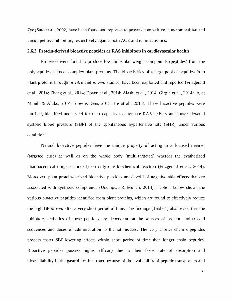
31
Tyr (Sato et al., 2002) have been found and reported to possess competitive, non-competitive and
uncompetitive inhibition, respectively against both ACE and renin activities.
2.6.2. Protein-derived bioactive peptides as RAS inhibitors in cardiovascular health
Proteases were found to produce low molecular weight compounds (peptides) from the
polypeptide chains of complex plant proteins. The bioactivities of a large pool of peptides from
plant proteins through in vitro and in vivo studies, have been exploited and reported (Fitzgerald
et al., 2014; Zhang et al., 2014; Doyen et al., 2014; Alashi et al., 2014; Girgih et al., 2014a, b, c;
Mundi & Aluko, 2014; Siow & Gan, 2013; He at al., 2013). These bioactive peptides were
purified, identified and tested for their capacity to attenuate RAS activity and lower elevated
systolic blood pressure (SBP) of the spontaneous hypertensive rats (SHR) under various
conditions.
Natural bioactive peptides have the unique property of acting in a focused manner
(targeted cure) as well as on the whole body (multi-targeted) whereas the synthesized
pharmaceutical drugs act mostly on only one biochemical reaction (Fitzgerald et al., 2014).
Moreover, plant protein-derived bioactive peptides are devoid of negative side effects that are
associated with synthetic compounds (Udenigwe & Mohan, 2014). Table 1 below shows the
various bioactive peptides identified from plant proteins, which are found to effectively reduce
the high BP in vivo after a very short period of time. The findings (Table 1) also reveal that the
inhibitory activities of these peptides are dependent on the sources of protein, amino acid
sequences and doses of administration to the rat models. The very shorter chain dipeptides
possess faster SBP-lowering effects within short period of time than longer chain peptides.
Bioactive peptides possess higher efficacy due to their faster rate of absorption and
bioavailability in the gastrointestinal tract because of the availability of peptide transporters and
32
Table 1- Plant protein-derived bioactive peptides effectively used to reduce high BP in spontaneously hypertensive rats models
Plant protein sources Identified peptide sequences
Dose/ Administration
route
Response/ Inference
(ΔSBP mmHg)
References
Sweet potato tuber ITP, IIP, GQY 500 mg/kg hydrolysate; Oral -30 after 8 h Ishiguro et al., 2012
Rapeseed protein VW, IY 7.5 mg/kg; Oral -9.8 to -10.8 after 2 h Marczak et al., 2003
Rice protein TQVY 50 mg/kg; Oral -40 after 6 h Li et al., 2007
Corn gluten meal
protein
AT 30 mg/kg; Oral -9.5 after 2 h Yang et al., 2007
Flaxseed protein RW, SVR 200 mg/kg; Oral -17.9 after 2 h Udenigwe et al., 2012
Mung bean protein KDYRL, VTPALR 600 mg/kg hydrolysate; Oral -30.8 after 6 h Li et al., 2006
Wheat bran LQP, IQP, VY 10 mg/ml; Oral -45 after 2 h Nogata et al., 2011
Hemp seed protein WVYY 30 mg/kg; Oral -34 after 2 h Girgih et al., 2014b
Rapeseed protein GHS 30 mg/kg; Oral -17.29 after 6 h He et al., 2013
Pea seed protein LTFPG, IFENLQN 30 mg/kg; Oral
-37 after 2 h and 4 h,
respectively
Aluko et al., 2015

33
ability to diffuse into cells based on chain hydrophobicity.
The use of plant protein-derived bioactive peptides in human clinical trials is still under
serious investigation and critical consideration in order to avoid any unethical application of
these bioactive compounds. Although, a past study has substantiated the efficacy of plant
protein-derived peptides in human chronic diseases intervention to reduce elevated-BP when the
human volunteers were given a 3 g/day-dose of pea protein hydrolysate (Li et al., 2011). Yet,
various concerns are still being generated to ascertain the purity and safety of these plant protein-
derived bioactive peptides in human clinical trials; therefore, these concerns need to be looked
into before the peptides can be adopted for human therapeutic uses. The low molecular weight
peptides are absorbed faster than the unhydrolyzed proteins that require digestion in the
gastrointestinal tract. Thus the biological effects of unhydrolyzed proteins are invariably delayed
when compared to peptides as therapeutic tools for chronic disease management (He et al., 2013;
Alashi et al., 2014).
2.7. Acetylcholinesterase (AChE) in human health
The cholinesterase is the generic name given to the enzymes; Acetylcholinesterase
(AChE, E.C.3.1.1.7) and butyrylcholinesterase (BChE, E.C.3.1.1.8), which are found as part of
the human nervous/neurological systems (Kozurkova et al., 2011). While AChE hydrolyses
acetylcholine (ACh) that is mainly associated with nerves and muscles and typically found on the
synapsies, BChE, a plasma cholinesterase or pseudocholinesterase, hydrolyses butyrylcholine
(BCh). Although not naturally found in the body, BCh is a known synthetic compound used to
distinguish between AChE and BChE (Chen et al., 2015; Pohanka, 2013).
BChE is a non-specific cholinesterase enzyme that binds nerve agent in the bloodstream
before it can exert effects in the nervous system (Chen et al., 2015; Brus et al., 2014). BChE is

34
synthesized by the liver and found in large concentrations in serum (Delfino et al., 2009). The
assay of plasma-BChE activity can be used as a pathological liver function test (Pohanka, 2013).
The major differences between these cholinesterases are their respective preferences for
substrates and degradation half-life. The substrates and half-life of AChE are ACh and 2 days
while that of BChE are BCh and approximately 8–16 hours (Huang et al., 2007; Wenthold et al.,
1974).
Besides its role as an effective modulator of the central and peripheral nervous systems,
the ubiquitous process-regulatory nature of AChE such as in cell growth, locomotion or
apoptosis has given it a priority over BChE during recent investigations (Tumiatti et al., 2010;
Camps et al., 2010). Therefore, the emphasis in this thesis work is on AChE activities and its
inhibition by hemp seed protein-derived peptides. AChE (a serine hydrolase) has long been
known to play a very crucial physiological role in maintaining the major neurotransmitter (ACh)
in the body by rapidly clearing free ACh from the synapse (Kumar & Chowdhury, 2014). ACh is
released by presynaptic cholinergic terminals and is the main neurotransmitter involved in
cholinergic neurotransmission through activation of nicotinic and/or muscarinic receptors to
modify postsynaptic cell functions. Normally, AChE rapidly and efficiently degrades ACh into
acetate and choline, thereby terminating its signaling action (Kolář et al., 2010). AChE is a
membrane-bound glycoprotein, which is present in cells in the form of a monomer or an
oligomer; however, this is dependent on the type of cells and the enzyme variant (Kolář et al.,
2010).
During neurotransmission, ACh is released from the ACh receptor when its concentration
in the synaptic cleft is very low, so that a cholinergic neuron can receive another impulse. The
release of ACh can lead to accumulation of ACh in the synaptic cleft and then results in impeded
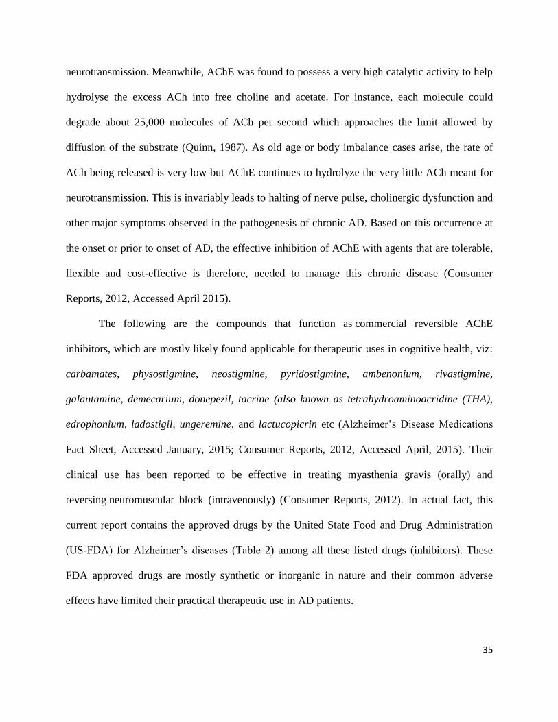
35
neurotransmission. Meanwhile, AChE was found to possess a very high catalytic activity to help
hydrolyse the excess ACh into free choline and acetate. For instance, each molecule could
degrade about 25,000 molecules of ACh per second which approaches the limit allowed by
diffusion of the substrate (Quinn, 1987). As old age or body imbalance cases arise, the rate of
ACh being released is very low but AChE continues to hydrolyze the very little ACh meant for
neurotransmission. This is invariably leads to halting of nerve pulse, cholinergic dysfunction and
other major symptoms observed in the pathogenesis of chronic AD. Based on this occurrence at
the onset or prior to onset of AD, the effective inhibition of AChE with agents that are tolerable,
flexible and cost-effective is therefore, needed to manage this chronic disease (Consumer
Reports, 2012, Accessed April 2015).
The following are the compounds that function as commercial reversible AChE
inhibitors, which are mostly likely found applicable for therapeutic uses in cognitive health, viz:
carbamates, physostigmine, neostigmine, pyridostigmine, ambenonium, rivastigmine,
galantamine, demecarium, donepezil, tacrine (also known as tetrahydroaminoacridine (THA),
edrophonium, ladostigil, ungeremine, and lactucopicrin etc (Alzheimer’s Disease Medications
Fact Sheet, Accessed January, 2015; Consumer Reports, 2012, Accessed April, 2015). Their
clinical use has been reported to be effective in treating myasthenia gravis (orally) and
reversing neuromuscular block (intravenously) (Consumer Reports, 2012). In actual fact, this
current report contains the approved drugs by the United State Food and Drug Administration
(US-FDA) for Alzheimer’s diseases (Table 2) among all these listed drugs (inhibitors). These
FDA approved drugs are mostly synthetic or inorganic in nature and their common adverse
effects have limited their practical therapeutic use in AD patients.
36
Based on these negative side effects and recent health awareness of the natural and
organic materials, the use of plant protein-derived bioactive peptides have now developed and if
successful, may be commercialized for use as drug alternatives for human cognitive health
promotion. AChE action also causes decreases in choline acetyltransferase (ChAT) level, which
is the rate-limiting enzyme during ACh synthesis (Lane et al., 2006). Apart from the typical role
of AChE in the termination of cholinergic neurotransmission, AChE has also been observed in
non-neuronal tissues (erythrocytes, megakaryocytes etc.) but without significant catalytic
functions (Sperling et al., 2008). The expression of AChE under neurodegenerative disease
conditions has been reported to undergo changes at different levels during proliferation, neurite
growth, apoptosis and differentiation (Sperling et al., 2008).
Equation 1 summarizes the catalytic action of the AChE on the neurotransmitter ACh as
follows:
2.7.1. Mechanism of actions of acetylcholinesterase inhibitors
Currently, the precise mechanisms of potent AChE inhibitors have not been fully elucidated,
although the ability of the plant protein-derived bioactive peptides to exhibit mixed inhibition
mode of action (as described under the RAS inhibition section), can serve as a potential
mechanism of action for AChE inhibitors. For instance, the bioactive peptides (inhibitor) can
competitively bind to the targeted enzyme (AChE) at the catalytic site to alter its
Acetylcholine Acetate Choline
(Eqn 1)

37
Table 2- FDA approved drugs for Alzheimer’s diseases*
Drug
(Brand)
Class and Indication Mechanism of Action Common Adverse Effects
Donepezil
(Aricept)
Cholinesterase inhibitor prescribed to
treat symptoms of mild-to-moderate and
moderate-to-severe AD
Prevents the breakdown of ACh in the
brain.
Nausea, vomiting, diarrhea
Galantamine
(Razadyne)
Cholinesterase inhibitor prescribed to
treat symptoms of mild-to-moderate AD
Prevents the breakdown of ACh and
stimulates nicotinic receptors to release
more ACh in the brain.
Nausea, vomiting, diarrhea,
loss of appetite, weight loss
Memantine
(Namenda)
NMDA antagonist prescribed to treat
symptoms of moderate-to-severe AD
Blocks the toxic effects associated with
excess glutamate and regulates
glutamate activation
Dizziness, headache,
constipation, confusion
Rivastigmine
(Exelon)
Cholinesterase inhibitor prescribed to
treat symptoms of mild-to-moderate AD.
Prevents the breakdown of ACh and
BCh in the brain.
Nausea, vomiting, diarrhea,
loss of appetite, weight loss,
muscle weakness
*AD - Alzheimer’s disease; NMDA – N-methyl-D-aspartate. Table adapted from Alzheimer’s Disease Medications Fact Sheet (2015) (National Institute on
Aging) with open access permission.
38
active site (Ser-His-Glu) and hence, hinder the attack/bind on the substrate (ACh). There could
be non-competitive and uncompetitive inhibition cases, when the bioactive peptides (having no
amino acid sequence to bind to the active sites of the AChE) attached to the targeted enzyme at
the non-active residues. In both cases, there exists a change in the structural conformation of the
targeted enzyme molecule and substrate, respectively thereby decreasing or terminating AChE
activity (Saravanaraman et al., 2014; Singh et al., 2013).
However, there is a previous report that AChE can terminate cholinergic nerve
transmission by inactivating ACh (Katzung, 2001). This enzyme, which is found both on the
post-synaptic membrane of cholinergic synapses, and in other tissues e.g. red blood cells, binds
to and hydrolyzes ACh to acetate and choline (as illustrated in equation 1 above). Therefore, the
use of AChE inhibitors prevent excessive hydrolysis and increases ACh concentration at the
synaptic cleft, which enhances proper nerve impulse functioning. Brain AChE has been the
major therapeutic target of AChE inhibitors as a treatment strategy for AD. The dysfunction of
cholinergic neurotransmission in the brain, the formation and growth of brain amyloid lesions
and the senile plaques, are widely believed to be contributing factors to a crucial event in the
pathogenesis of AD (Kumar & Chowdhury, 2014).
AD, a leading cause of dementia in developed countries, has been reported as a
progressive, degenerative disease characterized by memory loss, language deterioration, poor
judgment and impaired visuospatial skills, among others (Darvesh et al., 2003). Besides
cognitive improvement, AChE inhibitors with low cytotoxicity could be widely used as
multifunctional agents for neuroprotective effects, such as antioxidants to suppress the increased
oxidative stress resulting from free radical damage to cellular functions (Fernández-Bachiller et
al., 2010). Although interactions of AChE inhibitors with other apoptotic molecules during
39
apoptosis are not completely clear, their actions have been suggested to improve AD therapy. For
instance, recent studies have reported the beneficial apoptotic capacity of AChE inhibitors (Ye et
al., 2010; Toiber et al., 2008). This action of AChE helps to potentially down regulate p53 and
Bcl-2 family protein expressions during ischemia/reperfusion-induced apoptosis and also blocks
the overexpression of the cell death-induction synaptic variant, N-AChE-S.
Recently, available therapies that have been selectively designed for human health in the
form of commercial AChE-inhibitory drugs include Donepezil, Galantamine, Rivastigmine,
among others. The essence of administering these AChE-inhibitory drugs is as a result of their
clinical efficacy in prolonging the half-life of ACh in the cognitive system (Darvesh et al., 2003).
Meanwhile, the dietary plant polyphenols, a broad class of naturally occurring and potent
compounds (also regarded as antioxidants), have been found to provide neuroprotection against
AD (Wang et al., 2006; Ramassamy, 2006; Dai et al., 2006; Arendash et al., 2006; Rezai-Zadeh
et al., 2005; Yang et al., 2005; Lindsay et al., 2002; Lim et al., 2001). For instance, plant
polyphenols such as quercetin, catechin, curcumin and resveratrol have shown decreased
prevalence rate of AD when consumed from their dietary sources such as red wine, curry, green
tea, coffee, fruit and vegetable juices among others (Dai et al., 2006; Ringman et al., 2005;
Lindsay et al., 2002; Chandra et al., 2001; Orgogozo et al., 1997).
The areas of ethnopharmaceutical research in AD management indicate promising
potential of Berberis vulgaris as source of new compounds for the AChE inhibition when its
polyphenolic-hexane fraction showed active inhibition (64%; IC50 68 μg/ml) activities (Kolar et
al., 2010). The neurotrophic effects of plant polyphenols (IC50 16-73 μg/ml) to improve
cognition in AD patients was demonstrated and reported when several plant species used
traditionally exhibited a high in vitro AChE inhibition (Vinutha et al., 2007). It is noteworthy

40
that human clinical studies reported the polyphenolic extract, EGb 761 from Ginkgo biloba, to
produce neuroprotective and cognition-enhancing effects by maintaining the neurotransmitter,
ACh in the cognitively-affected adult patients (Birks & Grimley, 2009; Gold et al., 2002). For
instance, the polyphenolic extract provided modest cognitive benefits in patients with mild-to-
severe AD when 120 mg/day of the polyphenolic extract was orally administered (Le Bars et al.,
2002; 2000; 1997). Besides, the mild-to-moderate AD patients reportedly performed
significantly better (cognition, behaviour, daily living activities) than the placebo-treated group
after consumption of the polyphenolic extracts (Napryeyenko & Borzenko, 2007).
2.7.2 Protein-derived bioactive peptides as acetylcholinesterase inhibitors in cognitive
health
The major limitation to the utilization of the synthetic drugs that have been described
earlier lies with the fact that they are all characterized by common negative side effects like
nausea, vomiting, muscle cramps, decreased heart rate (bradycardia), decreased appetite and
weight loss as well as increased gastric acid production (Ghribia et al., 2014). However, recent
approaches employed to address this critical problem of negative side effects of these synthetic
drugs are based on the use of plants products and their secondary metabolites (Ghribia et al.,
2014; Iannello et al., 2014; Kumar & Chowdhury, 2014; Zare-Zardini et al., 2013). The
emerging innovation and nutritional knowledge in food and food products to improve the human
health result from the concept of functional food (i.e. the relationship between food and health).
Hence, the conceptual fact about functional foods in consumer’s health is presumed to help in
overcoming all these negative side effects associated with synthetic drugs in cognitive health
(Peressini & Sensidoni, 2009).
41
However, there has been scanty report on the use of plant protein hydrolysates and
peptides as AChE inhibitors. Naturally occurring bioactive peptides (hydrolysates) from plant
proteins are attractive alternatives because they are not known to exhibit negative side effects
like synthetic drugs (Zhang, Mu, & Sun, 2014; Barbana & Boye, 2010). As previously indicated,
peptide-dependent AChE inhibition can be achieved either through competitive (active site
blockage) or non-competitive (attachment to non-active site) actions (Saravanaraman et al.,
2014; Singh et al., 2013).
2.8. Separation, fractionation, identification and purification of multifunctional bioactive
peptides
The most widely used technique for multifunctional bioactive peptides separation,
fractionation, identification and purification is the high or ultra-performance liquid
chromatography (HPLC and UPLC respectively). This process can also be employed for the
structural characterization and amino acid sequencing of these bioactive peptides by coupling the
HPLC with tandem mass spectrometry (Singh et al., 2014). The amino acid sequencing
identification is done by consulting updated and accurate peptide banks of significant MS/MS
data using search engines such as MASCOT, SEQUEST, X!TANDEM and/or OMSSA (Craig &
Beavis, 2004, Geer et al., 2004).
The accurate peptide database is updated with lists of precursor proteins in their active
forms as well as their known post translational modifications. Such databases used in peptide
sequences identification include, but are not limited to, UniProtKB, BioPep, PepBank, EROP-
Moscow, BioPD, PeptideDB, Peptidome and APD (Panchaud et al., 2012). The bioactive
peptides from different plant proteins have been compiled and annotated in various peptide
databases, which provide information on their activities (Panchaud et al., 2012).
42
The de novo sequencing method can also be an important tool to derive the potential
sequences of the bioactive peptides with high level of confidence for proteins whose primary
structures are not yet available (Seidler et al., 2010; Picotti et al., 2008). This method involves
the use of reliable computer-based interpretation of a product ion spectrum from potential
sequences or sequence tags of a particular parent ion. For instance, excellent commercial
software such as PEAKS Studio, with detailed modules for dynamic procedures like de novo
search, database search, PTM search and quantification, has been a handy tool for peptide de
novo sequence identification (Seidler et al., 2010). The utilization of tandem MS system for de
novo peptide sequencing through the purity of the peptide ions being selected for fragmentation,
is influenced mainly by the performances of both the MS/MS and the connected liquid
chromatography system (Schlosser & Lehmann, 2002).
Prior to the use of HPLC-techniques, another separation or fractionation method,
ultrafiltration membrane system (UMS), could be used to separate or fractionate the peptides
based on their molecular weight. For example, the UMS-screening separation of soy protein
hydrolysate produced the potential anticancer, antioxidative, antiviral and immuno-modulatory
bioactive hydrolysates (Roblet et al., 2012) for human health applications. Some of the
equipments attached to HPLC for further peptide identification or purification are: electrospray
ionization (ESI), matrix-assisted laser desorption ionization-time of flight (MALDI-TOF)-mass
spectrometry (specially designed for peptide profiles generation) and tandem mass spectrometry
detection (LC–MS/MS) (specially designed for peptide sequencing and identification) (Singh et
al., 2014).
Various multifunctional bioactive peptides have been separated, fractionated, identified
and purified for application in the management and improvement of several diseases that affect
43
human health. It is noteworthy that RAS-inhibitory bioactive peptides from different plant
proteins, have been isolated, purified and identified for hypertension management and general
human cardiovascular health (Udenigwe & Mohan, 2014). For instance, a strong (IC50 6.25
µg/ml) ACE-inhibitory tripeptide (Gly-Pro-Pro) was purified and identified from buckwheat
protein hydrolysates using protein sequencing and electrospray-LC–MS system (Ma et al., 2006).
Likewise, a low molecular weight (0.4 kDa) antioxidant tetrapeptide (Ala-Asp-Ala-Phe) was
obtained (using RP-HPLC-ESI-MS) from walnut protein hydrolysates (Chen et al., 2012).
Moreover, a strong (IC50 14 µg/ml) adipogenesis-inhibitory tripeptide (Ile-Gln-Asn) was purified
and identified from black soybean protein hydrolysates (Kim et al., 2007). The bioactive
tetrapeptide (Trp-Val-Tyr-Tyr) and pentapeptide (Pro-Ser-Leu-Pro-Ala) were both identified by
LC-MS/MS from hemp seed protein hydrolysates and shown to exert maximum systolic blood
pressure reduction of -34 and -40 mmHg, respectively, after oral administration to SHRs (Girgih
et al., 2014).
Despite the abundance of information on the purification and identification of RAS-
inhibitory and antihypertensive bioactive peptides from plant proteins, there exists little or no
information on AChE-inhibitory peptides from plant proteins. Therefore, additional efforts are
needed to fractionate, purify and identify plant protein-derived AChE-inhibitory peptides, which
could be used for future management of AD and other human cognitive diseases.
References
Abugoch, L. E., Romero, N., Tapia, C. A., Silva, J., & Rivera, M. (2008). Study of some
physicochemical and functional properties of quinoa (Chenopodium quinoa Willd)
protein isolates. Journal of Agricultural and Food Chemistry, 56, 4745-4750.

44
Adebowale, Y. A., Schwarzenbolz, U., & Henle, T. (2011). Protein isolates from Bambara
groundnut (Voandzeia Subterranean L.): Chemical characterization and functional
properties. International Journal of Food Properties, 14, 758–775.
Adebowale, Y. A., Adeyemi, I. A., Oshodi, A. A., & Niranjana, K. (2007). Isolation,
fractionation and characterisation of proteins from Mucuna bean. Food Chemistry, 104,
287–299.
Achouri, A., Boye, J., Yaylayan, V., & Yeboah, F. (2005). Functional properties of glycated soy
11S Glycinin. Journal of Food Science, 70(4), 270–274.
Alashi, A. M., Blanchard, C. L., Mailer, R. J., Agboola, S. O., Mawson, A. J., He, R., Malomo,
S. A., Girgih, A. T., & Aluko, R. E. (2014). Blood pressure lowering effects of Australian
canola protein hydrolysates in spontaneously hypertensive rats. Food Research
International, 55, 281-287.
Aluko, R. E., Girgih, A. T., He, R., Malomo, S. A., Li, H., Offengenden, M., & Wu, J. (2015).
Structural and functional characterization of yellow field pea seed (Pisum sativum L.)
protein-derived antihypertensive peptides. Food Research International, 1-7.
http://dx.doi.org/10.1016/j.foodres.2015.03.029
Aluko, R. E., McIntosh, T., & Katepa-Mupondwa, F. (2005). Comparative study of the
polypeptide profiles and functional properties of Sinapis alba and Brassica juncea seed
meals and protein concentrates. Journal of the Science of Food and Agriculture, 85,
1931–1937.
Alzheimer’s Disease Medications Fact Sheet (2015). National Institute on Aging.
www.nia.nih.gov/alzheimers/publication/alzheimers-disease-medications-fact-sheet.
Accessed January 9, 2015.
45
Arendash, G. W., Schleif, W., Rezai-Zadeh, K., Jackson, E. K., Zacharia, L. C., Cracchiolo, J.
R., Shippy, D., & Tan, J. (2006). Caffeine protects Alzheimer’s mice against cognitive
impairment and reduces brain beta-amyloid production. Neuroscience. 142(4), 941-952.
Arndt, C., Koristka, S., Bartsch, H., & Bachmann, M. (2012). Native polyacrylamide gels. In B.
T. Kurien & R. H. Scofield (eds.), Protein Electrophoresis: Methods and Protocols,
Methods in Molecular Biology, 869, 49-53.
Avramenko, N. A., Low, N. H., & Nickerson, M. T. (2013). The effects of limited enzymatic
hydrolysis on the physicochemical and emulsifying properties of a lentil protein isolate.
Food Research International, 51, 162-169.
Balerin, C., Aymard, P., Ducept, F., Vaslin, S., & Cuvelier, G. (2007). Effect of formulation and
processing factors on the properties of liquid food foams. Journal of Food Engineering,
78, 802-809.
Barac, M. B., Pesic, M. B., Stanojevic, S. P., Kostic, A. Z., & Bivolarevic, V. (2014).
Comparative study of the functional properties of three legume seed isolates: adzuki, pea
and soy bean. Journal of Food Science & Technology, DOI 10.1007/s13197-014-1298-6,
1-9.
Barac, M., Cabrilo, S., Stanojevic, S., Pesic, M., Pavlicevic, M., Zlatkovic, B., & Jankovic, M.
(2012). Functional properties of protein hydrolysates from pea (Pisum sativum, L) seeds.
International Journal of Food Science and Technology, 47, 1457–1467.
Barac, M., Cabrilo, S., Pesic, M., Stanojevic, S., Zilic, S., Macej, O., et al. (2010). Profile and
functional properties of seed proteins from six pea (Pisum sativum) genotypes.
International Journal of Molecular Sciences, 11, 4973–4990.
46
Barbana, C., & Boye, J. I. (2011). Angiotensin I-converting enzyme inhibitory properties of
lentil protein hydrolysates: determination of the kinetics of inhibition. Food Chemistry,
127(1), 94–101.
Barbana, C., & Boye, J. I. (2010). Angiotensin I-converting enzyme inhibitory activity of
chickpea and pea protein hydrolysates. Food Research International, 43(6), 1642–1649.
Berg, J. M., Tymoczko, J. L., & Stryer, L. (2002). Biochemistry (5th ed.). WH Freeman, New
York. ISBN 0-7167-4955-6.
Birks, J., & Grimley, E. V. (2009). Ginkgo biloba for cognitive impairment and dementia.
Cochrane Database of Systematic Reviews, 1, DOI: 10.1002/14651858.CD003120.pub3.
Bora, P. S. (2002). Functional properties of native and succinylated lentil (Lens culinaris)
globulins. Food Chemistry, 77, 171-176.
Boye, J. I., Aksay, S., Roufik, S., Ribéreau, S., Mondor, M., Farnworth, E., & Rajamohamed, S.
H. (2010). Comparison of the functional properties of pea, chickpea and lentil protein
concentrates processed using ultrafiltration and isoelectric precipitation techniques. Food
Research International, 43, 537–546.
Brus, B., Košak, U., Turk, S., Pišlar, A., Coquelle, N., Kos, J., Stojan, J., Colletier, J., & Gobec,
S. (2014). Discovery, biological evaluation, and crystal structure of a novel nanomolar
selective butyrylcholinesterase inhibitor. Journal of Medicinal Chemistry, 57 (19), 8167-
8179.
Callaway, J. C., Summers, L. K., Bradshaw, H., & Frayn, K. N. (2002). Changes in LDL particle
composition after the consumption of meals containing different amounts and types of
fat. American Journal of Clinical Nutrition, 76, 345-350.
47
Camps, P., Formosa, X., Galdeano, C., Gómez, T., Munoz-Torrero, D., Ramírez, L., Viayna, E.,
Gómez, E., Isambert, N., Lavilla, R., Badia, A., Clos, M. V., Bartoliny, M., Mancini, F.,
Andrisano, V., Bidon-Chanal, A., Huertas, Ó., Dafni, T., & Luque, F. J. (2010). Tacrine
based dual binding site acetylcholinesterase inhibitors as potential disease-modifying
anti-Alzheimer drug candidates. Chem. Biol. Interact., 187, 411-415.
Chakrabarti, S., Jahandideh, F., & Wu, J. (2014). Food-derived bioactive peptides on
inflammation and oxidative stress. BioMed Research International, 1-11.
http://dx.doi.org/10.1155/2014/608979
Chandra, V., Pandav, R., Dodge, H. H., Johnston, J. M., Belle, S. H., Dekosky, S. T., & Ganguli,
M. (2001). Incidence of Alzheimer’s disease in a rural community in India: the Indo-US
study. Neurology, 57(6), 985-989.
Chavan, U. D., McKenzie, D. B., & Shahidi, F. (2001). Functional properties of protein isolates
from beach pea (Lathyrus maritimus L.). Food Chemistry, 74: 177-187.
Cheftel, J. C., Cuq, J. L., & Lorient, D. (1985). Amino acids, peptides, and proteins. In: O. R.
Fennema (Ed), Food Chemistry (2nd
Edition). Marcel Dekker Inc, New York, USA. P
245-369. ISBN 0-8247-7271-7.
Chel-Guerrero, L., Dom´ınguez-Maga˜na, M., Mart´ınez-Ayala, A., D´avila-Ortiz, G., &
Betancur-Ancona, D. (2012). Lima bean (Phaseolus lunatus) protein hydrolysates with
ACE-I inhibitory activity. Food & Nutrition Sciences, 3(4), 511–521.
Chen, A., Yang, H., Sun, Y., Niu, J., & Liu, S. (2012). Purification and identification of
antioxidant peptides from walnut (Juglans regia L.) protein hydrolysates. Peptides, 38,
344-349.
48
Chen, V. P., Gao, Y., Geng, L., Parks, R. J., Pang, Y. P., & Brimijoin, S. (2015). Plasma
butyrylcholinesterase regulates ghrelin to control aggression. Proceedings of the National
Academy of Sciences of the United States of America, 112(7), 2251–2256.
Choi, S. M., & Ma, C. Y. (2007). Structural characterization of globulin from common
buckwheat (Fagopyrum esculentum Moench) using circular dichroism and Raman
spectroscopy. Food Chemistry, 102, 150-160.
Chou, D. H., & Morr, C. V. (1979). Protein-water interactions and functional properties. Journal
of American Oil Chemists’ Society, 56, 53-62.
Chove, B. E., Grandison, A. S., & Lewis, M. J. (2007). Some functional properties of
fractionated soy protein isolates obtained by microfiltration. Food Hydrocolloid, 21,
1379–1388.
Consumer Reports (2012). Drug Effectiveness Review Project, Evaluating prescription drugs
used to treat: Alzheimer’s disease comparing effectiveness, safety and price. Best Buy
Drugs. Accessed April 2015.
Cordero-de-los-Santos, J., Osuna-Castro, A., Borodanenko, A., & Paredes-Lo´pez, O. (2005).
Physicochemical and functional characterization of Amaranth (Amaranthus
hypochondriacus) protein isolates obtained by isoelectric precipitation and micellisation.
Food Sci. Technol. Int., 11, 269–280.
Craig, R., & Beavis, R. C. (2004). TANDEM: matching proteins with tandem mass spectra.
Bioinformatics, 20, 1466–1467.
Dai, Q., Borenstein, A. R., Wu, Y., Jackson, J. C., & Larson, E. B. (2006). Fruit and vegetable
juices and Alzheimer’s disease: the Kame Project. American Journal of Medicine, 119(9),
751-759.
49
Darvesh, S., Walsh, R., Kumar, R., Caines, A., Roberts, S., Magee, D., Rockwood, K., & Martin,
E. (2003) Inhibition of human cholinesterases by drugs used to treat Alzheimer Disease.
Alzheimer Disease and Associated Disorders, 17(2), 117–126.
Delfino, E. T., Ribeiro, T. S., & Figueroa-Villar, J. D. (2009). Organophosphorus compounds as
chemical warfare agents: a review. J. Braz. Chem. Soc., 20, 407-428.
Deng, Q. C., Wang, L., Wei, F., Xie, B. J., Huang, F. H., Huang, W., Shi, J., Huang, Q. D., Tian,
B. Q., & Xue, S. (2011). Functional properties of protein isolates, globulin and albumin
extracted from Ginkgo biloba seeds. Food Chemistry, 124, 1458-1465.
Doyen, A., Udenigwe, C.C., Mitchell, P.L., Marette, A., & Aluko, R.E. (2014). Anti-diabetic and
antihypertensive activities of two flaxseed protein hydrolysate fractions revealed
following their simultaneous separation by electrodialysis with ultrafiltration membranes.
Food Chemistry, 145, 66–76.
Fernández-Bachiller, M. I., Pérez, C., González-Muňoz, G. C., Conde, S., López, M. G.,
Villaroya, M., García, A. G., & Rodríquez-Franco, M. I. (2010). Novel tacrine-8
hydroxyquinoline hybrids as multifunctional agents for the treatment of Alzheimer’s
disease, with neuroprotective, cholinergic, antioxidant, and copper-complexing
properties. J. Med. Chem., 53, 4927-4937.
Ferreira, D., Marais, J. P. J., Coleman, C. M., & Slade, D. (2010). Proanthocyanidins: Chemistry
and Biology. In: M. Lew, & L. Hung-Wen (Eds.), Comprehensive natural products II.
Elsevier, Oxford, New York, USA. p 605-661.
Fitzgerald, C., Aluko, R.E., Hossain, M., Rai, D.K., & Hayes, M. (2014). Potential of a renin
inhibitory peptide from the red seaweed (Palmaria palmate) as a functional food
ingredient following confirmation and characterization of a hypotensive Effect in
50
spontaneously hypertensive rats. Journal of Agricultural and Food Chemistry, 62, 8352-
8356.
Forman-Kay, J. D., & Mittag, T. (2013). From sequence and forces to structure, function, and
evolution of intrinsically disordered proteins. Structure, 21(9), 1492–1499.
Gbogouri, G. B., Linder, M., Fanni, J., & Parmentier, M. (2004). Influence of hydrolysis degree
on the functional properties of salmon by-products hydrolysis. Journal of Food Science,
69, C615–C622.
Geer, L. Y., Markey, S. P., Kowalak, J. A., Wagner, L., Xu, M., Maynard, D. M., Yang, X., Shi,
W., & Bryant, S. H. (2004). Open mass spectrometry search algorithm. Journal of
Proteome Research, 3, 958–964.
Gerbanowski, A., Malabat, C., Rabiller, C., & Gue´guen, J. (1999). Grafting of aliphatic and
aromatic probes on rapeseed 2S and 12S proteins: influence on their structural and
physicochemical properties. Journal of Agricultural and Food Chemistry, 47,
5218−5226.
Ghribia, L., Ghouilaa, H., Omrib, A., Besbesb, M., & Janneta, H. B. (2014). Antioxidant and
anti-acetylcholinesterase activities of extracts and secondary metabolites from Acacia
cyanophylla. Asian Pacific Journal of Tropical Biomedicine, 4(Suppl 1), S417-S423.
Gibbs, B. F., Zougman, A., Masse, R., & Mulligan, C. (2004). Production and characterization
bioactive peptides from soy hydrolysate and soy-fermented food. Food Research
International, 37, 123-131.
Girgih, A.T., He, R., Alashi, A.M., Malomo, S.A., & Aluko, R.E. (2014a). Preventive and
treatment effects of hemp seed (Cannabis sativa L.) meal protein hydrolysate against
51
blood pressure in spontaneously hypertensive rats. European Journal of Nutrition, 53 (5),
1237-1246.
Girgih, A. T., He, R., Malomo, S. A., & Aluko, R. E. (2014b). Structural and functional
characterization of hemp seed (Cannabis sativa L.) protein-derived antioxidant and
antihypertensive peptides. Journal of Functional Foods, 6, 384-394.
Girgih, A.T., He, R., & Aluko, R.E. (2014c). Kinetics and molecular docking studies of the
inhibitions of angiotensin converting enzyme and renin activities by hemp seed
(Cannabis sativa L.) peptides. Journal of Agricultural and Food Chemistry, 62,
4135−4144.
Gold, P. E., Cahill, L., & Wenk, G. L. (2002). Ginkgo biloba: a cognitive enhancer?
Psychological Science in the Public Interest, 3(1), 2-11.
Gong, M., Mohan, A., Gibson, A., & Udenigwe, C.C. (2015). Mechanisms of plastein formation,
and prospective food and nutraceutical applications of the peptide aggregates
Biotechnology Reports, 5, 63–69.
Grisaru, D., Sternfeld, M., Eldor, A., Glick, D., & Soreq, H. (1999). Structural roles of
acetylcholinesterase variants in biology and pathology, Eur. J. Biochem., 264, 672-686.
Haard, F. N. (1985). Characteristics of edible plant tissues. In: O. R. Fennema (Ed), Food
Chemistry (2nd
Edition). Marcel Dekker Inc, New York, USA. p 857-911.
ISBN 0-8247-7271-7.
Harnedy, P. A., & FitzGerald, R. J. (2013). In vitro assessment of cardioprotective, anti-diabetic
and antioxidant potential of Palmaria palmata protein hydrolysates. Journal of Applied
Phycology, 25, 1793–1803.
52
Hasnat, M. A., Pervin, M., & Lim, B. O. (2013). Acetylcholinesterase inhibition and in vitro and
in vivo antioxidant activities of Ganoderma lucidum grown on germinated brown rice.
Molecules, 18, 6663-6678.
He, R., He, H., Chao, D., Ju, X., & Aluko, R. (2014a). Effects of high pressure and heat
treatments on physicochemical and gelation properties of rapeseed protein isolate. Food
Bioprocess Technology, 7, 1344–1353.
He, X. H., Liu, H. Z., Liu, L., Zhao, G. L., Wang, Q., & Chen, Q. L. (2014b). Effects of high
pressure on the physicochemical and functional properties of peanut protein isolates.
Food Hydrocolloids, 36, 123-129.
He, R., Malomo, S. A., Girgih, A. T., Ju, X., & Aluko, R. E. (2013). Glycinyl-histdinyl-serine
(GHS), a novel rapeseed protein-derived peptide has blood pressure-lowering effect in
spontaneously hypertensive rats. Journal of Agricultural and Food Chemistry, 61, 8396-
8402
Herna´ndez-Ledesma, B., Del Mar Contreras, & M., Recio, I. (2011). Antihypertensive peptides:
production, bioavailability and incorporation into foods. Advance Colloid Interface
Science, 165, 23–35.
Hojila-Evangelista, M. P., Selling, G. W., Berhow, M. A., & Evangelista, R. L. (2014).
Preparation, composition and functional properties of pennycress (Thlaspi arvense L.)
seed protein isolates. Industrial Crops and Products, 55, 173-179.
Hojilla-Evangelista, M. P., Evangelista, R. L., Isbell, T. A., & Selling, G. W. (2013). Effects of
cold-pressing and seed cooking on functional properties of protein in pennycress (Thlaspi
arvense L.) seed and press cakes. Industrial Crops and Products, 45, 223–229.

53
House, J. D., Neufeld, J., & Leson, G. (2010). Evaluating the quality of protein from hemp seed
(Cannabis sativa L.) products through the use of the protein digestibility-corrected amino
acid score method. Journal of Agricultural and Food Chemistry, 58, 11801-11807.
Huang, Y. J., Huang, Y., Baldassarre, H., Wang, B., Lazaris, A., Leduc, M., Bilodeau, A. S.,
Bellemare, A., Côté, M., Herskovits, P., Touati, M., Turcotte, C., Valeanu, L., Lemée, N.,
Wilgus, H., Bégin, I., Bhatia, B., Rao, K., Neveu, N., Brochu, E., Pierson, J., Hockley, D.
K., Cerasoli, D. M., Lenz, D. E., Karatzas, C. N., & Langermann, S. (2007).
Recombinant human butyrylcholinesterase from milk of transgenic animals to protect
against organophosphate poisoning. Proceedings of the National Academy of Sciences of
the United States of America, 104(34), 13603–13608.
Iannello, C., Pigni, N. B., Antognoni, F., Poli, F., Maxia, A., de Andrade, J. P., & Bastida, J.
(2014). A potent acetylcholinesterase inhibitor from Pancratium illyricum L. Fitoterapia,
92, 163–167.
Ibrahim, M. M. (2006). RAS inhibition in hypertension. Journal of Human Hypertension, 20,
101–108.
Ishiguro, K., Sameshima, Y., Kume, T., Ikeda, K., Matsumoto, J., & Yoshimoto, M. (2012).
Hypotensive effect of a sweet potato protein digest in spontaneously hypertensive rats
and purification of angiotensin I-converting enzyme inhibitory peptides. Food Chemistry,
131, 774-779.
Karaca, A. C., Low, N., & Nickerson, M. (2011). Emulsifying properties of canola and flaxseed
protein isolates produced by isoelectric precipitation and salt extraction. Food Research
International, 44, 2991-2998.

54
Katzung, B. G. (2001). Introduction to autonomic pharmacology. In: Sedative-hypnotic drugs.
In: Basic and clinical pharmacology, 8th edition. San Francisco, CA: The McGraw Hill
Companies, Inc.75-91.
Kelly, S. M., & Price, N. C. (2000). The use of circular dichroism in the investigation of protein
structure and function. Current Protein and Peptide Science, 1(4), 349-384
Kim, H. J., Bae, I. Y., Ahn, C. W., Lee, S., & Lee, H. G. (2007). Purification and identification of
adipogenesis inhibitory peptide from black soybean protein hydrolysate. Peptides, 28,
2098–2103.
Kolář, D., Wimmerová, L., & Kádek, R. (2010). Acetylcholinesterase and Butyrylcholinesterase
inhibitory activities of Berberis vulgaris. Phytopharmacology, 1(1) 7-11.
Kozurkova, M., Hamulakova, S., Gazova, Z., Paulikova, H., & Kristian, P. (2011). Neuroactive
multifunctional tacrine congeners with cholinesterase, anti-amyloid aggregation and
neuroprotective properties. Pharmaceuticals, 4, 382-418.
Kumar, S., & Chowdhury, S. (2014). Kinetics of acetylcholinesterase inhibition by an aqueous
extract of Cuminum cyminum seeds. International Journal of Applied Science and
Biotechnology, 2(1), 64-68.
Lane, R. M., Potkin, S. G., & Enz, A. (2006). Targeting acetylcholinesterase and
butyrylcholinesterase in dementia. International Journal of Neuropsychopharmacology,
9, 101-124.
Lawal, O. S., Adebowale, K. O., Ogunsanwo, B. M., Sosanwo, O. A., & Bankole, S. A. (2005).
On the functional properties of globulin and albumin protein fractions and flours of
African locust bean (Parkia biglobossa). Food Chemistry, 92, 681-691.
55
Le Bars, P. L., Velasco, F. M., Ferguson, J. M., Dessain, E. C., Kieser, M., & Hoerr, R. (2002).
Influence of the severity of cognitive impairment on the effect of the Ginkgo biloba
extract EGb 761 in Alzheimer’s disease. Neuropsychobiology, 45(1), 19-26.
Le Bars, P. L., Kieser, M., & Itil, K. Z. (2000). A 26-week analysis of a double-blind, placebo
controlled trial of the ginkgo biloba extract EGb 761 in dementia. Demential and
Geriatric Cognitive Disorders, 11(4), 230-237.
Le Bars, P. L., Katz, M. M., Berman, N., Itil, T. M., Freedman, A. M., & Schatzberg, A. F.
(1997). A placebo-controlled, double-blind, randomized trial of an extract of Ginkgo
biloba for dementia. North American EGb Study Group. Journal of the American
Medical Association, 278(16), 1327-1332.
Li, F., Kong, X., Zhang, C., & Hua, Y. (2011a). Effect of heat treatment on the properties of soy
protein-stabilised emulsions. International Journal of Food Science and Technology, 46,
1554–1560.
Li, H., Prairie, N., Udenigwe, C. C., Adebiyi, A. P., Tappia, P. S., Aukema, H. M., Jones, P. J.
H., & Aluko, R. E. (2011b). Blood pressure lowering effect of a pea protein hydrolysate
in hypertensive rats and humans. Journal of Agricultural and Food Chemistry, 59, 9854
9860.
Li, G. H., Qu, M. R., Wan, J. Z., & You, J. M. (2007). Antihypertensive effect of rice protein
hydrolysate with in vitro angiotensin I converting enzyme inhibitory activity in
spontaneously hypertensive rats. Asia Pacific Journal of Clinical Nutrition, 16(Suppl. 1),
275–280.
56
Li, G., Shi, Y., Liu, H., & Le, G. (2006). Antihypertensive effect of alcalase generated mung
bean protein hydrolysates in spontaneously hypertensive rats. European Food research
and Technology, 222: 733–736.
Li, G. H., Le, G. W., Liu, H., & Shi, Y. H. (2005). Mung-bean protein hydrolysates obtained
with alcalase exhibit angiotensin I-converting enzyme inhibitory activity. Food Science
& Technology International, 11, 281-287.
Li, G. H., Le, G. W., Shi, Y. H. & Shresthe, S. (2004). Angiotensin I-converting enzyme
inhibitory peptides derived from food proteins and their physiological and
pharmacological effects. Nutrition Research, 24, 469-486.
Lim, G. P., Chu, T., Yang, F., Beech, W., Frautschy, S. A., & Cole, G. M. (2001). The curry
spice curcumin reduces oxidative damage and amyloid pathology in an Alzheimer
transgenic mouse. Journal of Neuroscience, 21(21), 8370-8377.
Lindsay, J., Laurin, D., Verreault, R., Hebert, R., Helliwell, B., Hill, G. B., & McDowell, I.
(2002). Risk factors for Alzheimer’s disease: a prospective analysis from the Canadian
Study of Health and Aging. American Journal of Epidemiology, 156(5), 445-453.
L´opez-Barrios, L., Guti´errez-Uribe, J. A., & Serna-Sald´ıvar, S. O. (2014). Bioactive peptides
and hydrolysates from pulses and their potential use as functional ingredients. Journal of
Food Science, 79(3), R273-R283.
Lopez, S., Bastida, J., Viladomat, F., & Codina, C. (2002). Acetylcholinesterase inhibitory
activity of some Amaryllidaceae alkaloids and Narcissus extracts. Life Sciences, 71,
2521–2529.
Lu, R. R., Qian, P., Sun, Z., Zhou, X. H., Chen, T. P., He, J. F., Zhang, H., & Wu, J. P. (2010).
Hempseed protein derived antioxidative peptides: Purification, identification and
57
protection from hydrogen peroxide-induced apoptosis in PC12 cells. Food Chemistry,
123(4), 1210-1218.
Lqari, H., Vioque, J., Pedroche, J., & Millan, F. (2002). Lupinus angustifolius protein isolates:
chemical composition, functional properties and protein characterization. Food
Chemistry, 76, 349-356.
Ma, M. S., Bae, I. Y., Lee, H. G., Yang, C. B. (2006). Purification and identification of
angiotensin I-converting enzyme inhibitory peptide from buckwheat (Fagopyrum
esculentum Moench). Food Chemistry, 96, 36–42.
Marambe, H. K., Shand, P. J., & Wanasundara, J. P. D. (2013). In vitro digestibility of flaxseed
(Linum usitatissimum L.) protein: effect of seed mucilage, oil and thermal processing.
International Journal of Food Science and Technology, 48, 628–635.
Marczak, E., Usui, H., Fujita, H., Yang, Y., Yokoo, M., Lipkowski, A., & Yoshikawa, M.
(2003). New antihypertensive peptides isolated from rapeseed. Peptides, 24, 791-798
Marsh, J. A., & Teichmann, S. A. (2013). Parallel dynamics and evolution: protein
conformational fluctuations and assembly reflect evolutionary changes in sequence and
structure. Bioessays, 36, 209–218.
Matsudomi, N., Sasaki, T., Kato, A., & Kobayashi, K. (1985). Conformational changes and
functional properties of acid-modified soy protein. Journal of Agricultural and Food
Science, 49, 1251-1256.
Moyano, F. J., Saenz de Rodriganez, M. A., Dıaz, M., & Tacon, A. G. J. (2014). Application of
in vitro digestibility methods in aquaculture: constraints and perspectives. Reviews in
Aquaculture, 6, 1–20.
58
Mundi, S, & Aluko, R.E. (2014). Inhibitory properties of kidney bean protein hydrolysate and its
membrane fractions against renin, angiotensin converting enzyme, and free radicals.
Austin Journal of Nutrition and Food Sciences, 2(1), 1-11
Mundi, S., & Aluko, R. E. (2012). Physicochemical and functional properties of kidney bean
albumin and globulin protein fractions. Food Research International, 48, 299– 306.
MuneMune, M. A., Minka, S. R., & Mbome, I. L. (2014). Optimising functional properties
during preparation of cowpea protein concentrate. Food Chemistry, 154, 32–37.
Napryeyenko, O., & Borzenko, I. (2007). Ginkgo biloba special extract in dementia with
neuropsychiatric features. A randomised, placebo-controlled, double-blind clinical trial.
Arzneimittelforschung, 57(1), 4-11.
Niepmann, M., & Zheng, J. (2006). Discontinuous native protein gel electrophoresis.
Electrophoresis, 27, 3949–3951.
Nogata, Y., Nagamine, T., & Sekiya, K. (2011). Antihypertensive effect of angiotensin I
converting enzyme inhibitory peptides derived from wheat bran in spontaneously
hypertensive rats. Journal of Japanese Society for Food Science and Technology, 58, 67-
70.
Nooren, I. M. A., & Thornton, J. M. (2003). Structural characterisation and functional
significance of transient protein-protein interactions. Journal of Molecular Biology,
325(5), 991–1018.
Nosenko, T., Kot, T., & Kichshenko, V. (2014). Rape seeds as a source of feed and food
proteins. Polish Journal of Food and Nutrition Sciences, 64(2), 109–114.
Okezie, B. O., & Bello, A. B. (1988). Physicochemical and functional properties of winged bean
flour and isolate compared with soy isolate. Journal of Food Science, 53, 450-454.
59
Onuh, J. O., Girgih, A. T., Aluko, R. E., & Aliani, M. (2013). Inhibitions of renin and
angiotensin converting enzyme activities by enzymatic chicken skin protein hydrolysates.
Food Research International, 53, 260–267.
Orgogozo, J. M., Dartigues, J. F., Lafont, S., Letenneur, L., Commenges, D., Salamon, R.,
Renaud, S., & Breteler, M. B. (1997). Wine consumption and dementia in the elderly: a
prospective community study in the Bordeaux area. Neurological Review (Paris), 153(3),
185-192.
Panchaud, A., Affolter, M., & Kussmann, M. (2012). Mass spectrometry for nutritional
peptidomics: How to analyze food bioactives and their health effects. Journal of
Proteomics, 75, 3546-3559.
Paredeslopez, O., Ordoricafalomir, C., & Olivaresvazquez, M. R. (1991). Chickpea protein
isolates - physicochemical, functional and nutritional characterization. Journal of Food
Science, 56, 726-729.
Park, S. K., Seo, J. B., & Lee, M. Y. (2012). Proteomic profiling of hempseed proteins from
Cheungsam. Biochimica et Biophysica Acta-Proteins and Proteomics, 1824, 374-382.
Pedroche, J., Yust, M. M., Lqari, H., Giron-Calle, J,. Alaiz, M,. Vioque, J., & Millan, F. (2004).
Brassica carinata protein isolates: chemical composition, protein characterization and
improvement of functional properties by protein hydrolysis. Food Chemistry, 88, 337–
346.
Peressini, D., & Sensidoni, A. (2009). Effect of soluble dietary fibre addition on rheological and
bread-making properties of wheat doughs. Journal of Cereal Science, 49(2), 190-201.
60
Picotti, P., Lam, H., Campbell, D., Deutsch, E. W., Mirzaei, H., Ranish, J., Domon, B., &
Aebersold, R. (2008). A database of mass spectrometric assays for the yeast proteome.
Nature Methods, 5(11), 913-914.
Pihlanto, A., & Mäkinen, S. (2013). Antihypertensive properties of plant protein derived
peptides. In B. Hernandez-Ledesma, C. Hsieh (Eds.), Bioactive Food Peptides in Health
and Disease. INTECH Publishers, Rijeka, Croatia. p 145-182. ISBN 978-953-51-0964-8.
Pohanka, M. (2013). Butyrylcholinesterase as a biochemical marker. Bratislava Medical
Journal, 114,(12), 726–734.
Pownall, T. L., Udenigwe, C. C., & Aluko, R. E. (2010). Amino acid composition and
antioxidant properties of pea seed (Pisum sativum L.) enzymatic protein hydrolysate
fractions. Journal of Agricultural and Food Chemistry, 58, 4712–4718.
Ptaszek, P., Kabzinski, M., Kruk, J., Kaczmarczyk, K., Żmudziński, D., Liszka-Skoczylas, M.,
Mickowska, B., Lukasiewicz, M., & Banas, J. (2015). The effect of pectins and xanthan
gum on physicochemical properties of egg white protein foams. Journal of Food
Engineering, 144, 129-137.
Ptaszek, P. (2013). The non-linear rheological properties of fresh wet foams based on egg white
proteins and selected hydrocolloids. Food Research International, 54(1), 478–486.
Quinn, D. M. (1987). Acetylcholinesterase: enzyme structure, reaction dynamics, and virtual
transition states. Chemical Review, 87(5), 955–979.
Radocaj, O., Dimi´c, E., & Tsao, R. (2014). Effects of hemp (Cannabis sativa L.) seed oil press-
cake and decaffeinated green tea leaves (Camellia sinensis) on functional characteristics
of gluten-free crackers. Journal of Food Science, 79, C318-325.
61
Ramassamy C. (2006). Emerging role of polyphenolic compounds in the treatment of
neurodegenerative diseases: a review of their intracellular targets. European Journal of
Pharmacology, 545(1), 51-64.
Rankin, S. E., Watts, A., & Pinheiro, T. J. T. (1998). Electrostatic and hydrophobic
contributions to the folding mechanism of apocytochrome C driven by the interaction
with lipid. Biochemistry, 37, 12588-12595
Rasool, M., Malik, A., Manan, A., Qazi, A. M., & Qazi, M. H. (2013). The recent methodologies
of protein structural determination and dynamic developments. In G. M. Ashraf, & I. A.
Sheikh (Eds.), Advances in Protein Chemistry (pp. 4-18). USA: OMICS Group eBooks.
Rezai-Zadeh, K., Shytle, D., Sun, N., Mori, T., Hou, H., Jeanniton, D., Ehrhart, J., Townsend,
K., Zeng, J., Morgan, D., Hardy, J., Town, T., & Tan, J. (2005). Green tea
epigallocatechin-3-gallate (EGCG) modulates amyloid precursor protein cleavage and
reduces cerebral amyloidosis in Alzheimer transgenic mice. Journal of Neuroscience,
25(38), 8807-8814.
Ringman, J. M., Frautschy, S. A., Cole, G. M., Masterman, D. L., & Cummings, J. L. (2005). A
potential role of the curry spice curcumin in Alzheimer’s disease. Current Alzheimer
Research, 2(2), 131-136.
Roblet, C., Amiot, J., Lavigne, C., Marette, A., Lessard, M., Jean, J., Ramassamy, C., Moresoli,
C., & Bazinet, L. (2012). Screening of in vitro bioactivities of a soy protein hydrolysate
separated by hollow fiber and spiral-wound ultrafiltration membranes. Food Research
International, 46, 237–249.
Rodger, A., & Ismail, M. A. (2000). Introduction to circular dichroism. In: M. G. Gore (Ed),
Spectrophotometry & Spectrofluorimetry. Oxford, New York, USA. p 99-139.
62
Saravanaraman, P., Chinnadurai, R. K., & Boopathy, R. (2014). A new role for the non-
pathogenic non-synonymous single-nucleotide polymorphisms of acetylcholinesterase in
the t reatment of Alzheimer’s disease: A computational study. Journal of
computational biology, 21(8), 632-647.
Sato, M., Hosokawa, T., Yamaguchi, T., Nakano, T., Muramoto, K., Kahara, T., Funayama, K.,
Kobayashi, A., & Nakano, T. (2002). Angiotensin I-converting enzyme inhibitory
peptides derived from wakame (Undaria pinnatifida) and their antihypertensive effect in
spontaneously hypertensive rats. Journal of Agricultural and Food Chemistry, 50(21),
6245-6252.
Schägger, H. (2006). "Tricine–SDS-PAGE". Nature protocols 1(1), 16–22.
Schlosser, A., & Lehmann, W. D. (2002). Patchwork peptide sequencing: extraction of sequence
information from accurate mass data of peptide tandem mass spectra recorded at high
resolution. Proteomics, 2, 524–533
Seidler, J., Zinn, N., Boehm, M. E., & Lehmann, W. D. (2010). De novo sequencing of peptides
by MS/MS. Proteomics, 10, 634–649.
Sharma, R. (2012). Enzyme inhibition: mechanisms and scope, In R. Sharma (Ed.), Enzyme
Inhibition and Bioapplications. INTECH Publishers, Rijeka, Croatia. p 1-36. ISBN 978
953-51-0964-8.
Sharma, R., & Singh, R. B. (2010). Bioactive Foods and Nutraceutical Supplementation Criteria
in Cardiovascular Protection. The Open Nutraceuticals Journal, 3, 141-153.
Singh, B. P., Vij, S., & Hatib, S. (2014). Functional significance of bioactive peptides derived
from soybean. Peptides, 54, 171–179.
63
Singh, M., Kaur, M., Kukreja, H., Chugh, R., Silakari, O., & Singh, D. (2013).
Acetylcholinesterase inhibitors as Alzheimer therapy: From nerve toxins to
neuroprotection. European Journal of Medicinal Chemistry, 70, 165-188.
Siow, H., & Gan, C. (2013). Extraction of antioxidative and antihypertensive bioactive peptides
from Parkia speciosa seeds. Food Chemistry, 141, 3435–3442.
Small, E., Pocock, T., & Cavers, P. B. (2003). The biology of Canadian weeds. 119. Cannabis
sativa L. Canadian Journal of Plant Science, 83, 217–237.
Sperling, L. E., Steinert, G., Boutter, J., Landgraf, D., Hescheler, J., Pollet, D., & Layer, P. G.
(2008). Characterisation of cholinesterase expression during murine embryonic stem cell
differentiation. Chem. Biol. Interact., 175, 156-160.
Stone, A. K., Karalash, A., Tyler, R. T., Warkentin, T. D., & Nickerson, M. T. (2014).
Functional attributes of pea protein isolates prepared using different extraction methods
and cultivars. Food Research International, 1-8.
http://dx.doi.org/10.1016/j.foodres.2014.11.017
Szabo, A. G. (2000). Fluorescence principles and measurement. In: M. G. Gore (Ed),
Spectrophotometry & Spectrofluorimetry. Oxford, New York, USA. p 33-67.
Tan, E., Ngoh, Y., & Gan, C. (2014). A comparative study of physicochemical characteristics
and functionalities of pinto bean protein isolate (PBPI) against the soybean protein isolate
(SPI) after the extraction optimisation. Food Chemistry, 152, 447–455.
Tan, S. H., Mailer, R. J., Blanchard, C. L., & Agboola, S. O. (2011). Canola proteins for human
consumption: extraction profile, and functional properties. Journal of Food Science, 76,
R16–R28.
64
Tang, C. H., Sun, X., & Foegeding, E. A. (2011). Modulation of physicochemical and
conformational properties of kidney bean vicilin (Phaseolin) by glycation with glucose:
implications for structure-function relationships of legume vicilins. Journal of
Agricultural and Food Chemistry, 59, 10114-10123.
Teh, S. S., Bekhit, A. E. D., Carne, A. & Birch, J. (2014). Effect of the defatting process, acid
and alkali extraction on the physicochemical and functional properties of hemp, flax and
canola seed cake protein isolates. Food Measure, 8, 92-104.
Teh, S. S., & Birch, J. (2013). Physicochemical and quality characteristics of cold-pressed hemp,
flax and canola seed oils. Journal of Food Composition and Analysis, 30, 26-31.
Toiber, D., Berson, A., Greenberg, D., Melamed-Book, N., Diamant, S., & Soreq, H. (2008). N
acetylcholinesterase-induced apoptosis in Alzheimer's disease. PLoS One, 3, e3108
Tsumura, K., Saito, T., Tsuge, K., Ashida, H., Kugimiya, W., & Inouye, K. (2005). Functional
properties of soy protein hydrolysates obtained by selective proteolysis. Lebensmittel-
Wissenschaft und- Technologie (LWT), 38(3), 255–261.
Tumiatti, V., Minarini, A., Bolognesi, M. L., Milelli, A., Rosini, M., & Melchiorre, C. (2010).
Tacrine derivatives and Alzheimer's disease. Current Medicinal Chemistry, 17, 1825-
1838.
Udenigwe, C. C., & Mohan, A. (2014). Mechanisms of food protein-derived antihypertensive
peptides other than ACE inhibition. Journal of Functional Foods, 8C, 45–52.
Udenigwe, C. C., & Aluko, R. E. (2012). Food protein-derived bioactive peptides: production,
processing and potential health benefits. Journal of Food Science, 71, R11–R24.
Udenigwe, C. C., Adebiyi, A. P., Doyen, A., Li, H., Bazinet, L., & Aluko, R. E. (2012). Low
molecular weight flaxseed protein-derived arginine-containing peptides reduced blood
65
pressure of spontaneously hypertensive rats faster than amino acid form of arginine and
native flaxseed protein. Food Chemistry, 132, 468-475.
Udenigwe, C. C., Lin, Y. S., Hou, W. C., & Aluko, R. E. (2009). Kinetics of the inhibition of
renin and angiotensin I-converting enzyme by flaxseed protein hydrolysate fractions.
Journal of Functional Foods, 1(2), 199-207.
Vinutha, B., Prashanth, D., Salma, K., Sreeja, S. L., Pratiti, D., Padmaja, R., Radhika, S., Amit,
A., Venkateshwarlu, K., & Deepak, M. (2007). Screening of selected Indian medicinal
plants for acetylcholinesterase inhibitory activity. Journal of Ethnopharmacology, 109,
359-363.
Vioque, J., Sánchez-Vioque, R., Clemente, A., Pedroche, J., & Millán, F. (2000). Partially
hydrolyzed rapeseed protein isolates with improved functional properties. Journal of the
American Oil Chemists’ Society, 77(4), 447-450.
Wagner, J. R., & Gueguen, J. (1999). Surface functional properties of native, acid-treated, and
reduced soy glycinin. 2. Emulsifying properties. Journal of Agricultural and Food
Chemistry, 47, 2181-2187.
Wagner, J. R., & Gue´guen, J. (1995). Effects of dissociation, deamidation, and reducing
treatment on structural and surface active properties of soy glycinin. Journal of
Agricultural and Food Chemistry, 43, 1993-2000.
Wang, H., Chen, Y., Hua, Y., Kong, X., & Zhang, C. (2014). Effects of phytase-assisted
processing method on physicochemical and functional properties of soy protein isolate.
Journal of Agricultural and Food Chemistry, 62, 10989−10997.
Wang, J., Ho, L., Zhao, Z., Seror, I., Humala, N., Dickstein, D. L., Thiyagarajan, M., Percival, S.
S., Talcott, S. L., & Pasinetti, G. M. (2006). Moderate consumption of Cabernet
66
Sauvignon attenuates Abeta neuropathology in a mouse model of Alzheimer’s disease.
Journal of Federation of American Societies for Experimental Biology, 20(13), 2313-
2320.
Wenthold, R. J., Mahler, H. R., & Moore, W. J. (1974). The half-life of acetylcholinesterase in
mature rat brain. Journal of Neurochemistry, 22(6), 941-943.
Willcox, D. C., Scapagnini, G., & Willcox, B. J. (2014). Healthy aging diets other than the
Mediterranean: A focus on the Okinawan diet. Mechanisms of Ageing and Development,
136-137, 148-162.
Yang, Y., Guanjun, T. A. O., Ping, L. I. U., & Liu, J. I. A. (2007). Peptide with angiotensin I
converting enzyme inhibitory activity from hydrolyzed corn gluten meal. Journal of
Agricultural and Food Chemistry, 55, 7891–7895.
Yang, F., Lim, G. P., Begum, A. N., Ubeda, O. J., Simmons, M. R., Ambegaokar, S. S., Chen, P.
P., Kayed, R., Glabe, C. G., Frautschy, S. A., & Cole, G. M. (2005). Curcumin inhibits
formation of amyloid beta oligomers and fibrils, binds plaques, and reduces amyloid in
vivo. Journal of Biological Chemistry, 280(7), 5892-5901.
Ye, W., Gong, X., Xie, J., Wu, J., Zhang, X., Ouyang, Q., Zhao, X., Shi, Y., & Zhang, X. (2010).
AChE deficiency or inhibition decreases apoptosis and p53 expression and protects renal
function after ischemia/reperfusion. Apoptosis, 15, 474-487.
Yin, S. W., Tang, C. H., Wen, Q. B., & Yang, X. Q. (2009). Functional and structural properties
and in vitro digestibility of acylated hemp (Cannabis sativa L.) protein isolates.
International Journal of Food Science and Technology, 44, 2653–2661.
67
Yin, S. W., Tang, C. H., Cao, J. S., Hu, E. K., Wen, Q. B., & Yang, X. Q. (2008). Effects of
limited enzymatic hydrolysis with trypsin on functional properties of hemp (Cannabis
sativa L.) protein isolate. Food Chemistry, 106, 1004–1013.
Yu, W., Gao, J., Xue, Z., Kou, X., Wang, Y., & Zhai, L. (2013). Radical-scavenging activity,
ACE-inhibiting capability and identification of rapeseed albumin hydrolysate. Food
Science and Human Wellness, 2, 93–98.
Yu, J., Ahmedna, M., & Goktepe, I. (2007). Peanut protein concentrates: production and
functional properties as affected by processing. Food Chemistry, 103, 121–129.
Zare-Zardini, H., Tolueinia, B., Hashemi, A., Ebrahimi, L., & Fesahat, F. (2013). Antioxidant
and cholinesterase inhibitory activity of a new peptide from Ziziphus jujuba fruits.
American Journal of Alzheimer's Disease and other dementials, 00(0), 1-8. DOI:
10.1177/1533317513500839.
Zhang, M., Mu, T., & Sun, M. (2014). Purification and identification of antioxidant peptides
from sweet potato protein hydrolysates by alcalase. Journal of Functional Foods, 7, 191–
200.
Żmudziński, D., Ptaszek, P., Kruk, J., Kaczmarczyk, K., Rożnowski, W., Berski, W., Ptaszek,
A., & Grzesik, M. (2014). The role of hydrocolloids in mechanical properties of fresh
foams based on egg white proteins. Journal of Food Engineering, 121, 128-134.
68
CHAPTER THREE
3. Study on the structural and functional properties of hemp seed protein products
Sunday A. Malomo1, Rong He
1,2 and Rotimi E. Aluko
1*
1Department of Human Nutritional Sciences, University of Manitoba, Winnipeg, Manitoba,
Canada R3T 2N2
2College of Food Science and Engineering, Nanjing University of Finance and Economics,
Nanjing 210046, People’s Republic of China
*Corresponding author: Dr. Rotimi E. Aluko; E-Mail: [email protected]; Tel.: +1-204-
474-9555; Fax: +1-204-474-7593
Word count: 7,100
Short version: Structure-function of hemp seed proteins
Reprinted from Structural and functional properties of hemp seed protein products. Journal of
Food Science, 79(8), C1512-1521.
Copyright © 2014, with permission from John Wiley & Sons.
69
ABSTRACT: The effects of pH and protein concentration on some structural and functional
properties of hemp seed protein isolate (HPI, 84.15% protein content) and defatted hemp seed
protein meal (HPM, 44.32% protein content) were determined. The HPI had minimum protein
solubility (PS) at pH 4.0, which increased as pH was decreased or increased. In contrast, the
HPM had minimum PS at pH 3.0, which increased at higher pH values. Gel electrophoresis
showed that some of the high molecular weight proteins (>45 kDa) present in HPM were not
well extracted by the alkali and were absent or present in low ratio in the HPI polypeptide
profile. The amino acid composition showed that the isolation process increased the Arg/Lys
ratio of HPI (5.52%) when compared to HPM (3.35%). Intrinsic fluorescence and circular
dichroism data indicate that the HPI proteins had a well-defined structure at pH 3.0, which was
lost as pH value increased. The differences in structural conformation of HPI at different pH
values were reflected as better foaming capacity at pH 3.0 when compared to pH 5.0, 7.0 and
9.0. At 10 and 25 mg/mL protein concentrations, emulsions formed by the HPM had smaller oil
droplet sizes (higher quality), when compared to the HPI-formed emulsions. In contrast at 50
mg/mL protein concentration, the HPI-formed emulsions had smaller oil droplet sizes (except at
pH 3.0). We conclude that the functional properties of hemp seed protein products are dependent
on structural conformations as well as protein concentration and pH.
Keywords: circular dichroism, emulsion, hemp seed, intrinsic fluorescence, protein functionality
Practical Application
Hemp seed is a popular oil seed crop grown in Canada and globally for its oil and protein
products. Hemp seed proteins (HSP), which account for 25% of its composition, have been
widely studied in detail, but their structural and functional properties at varying processing
70
conditions are not well understood. This work provides information on the structural
conformation and functional performance in model systems of HSP under varying pH conditions
that cover the food processing range. The data presented could enhance manipulation of
environmental pH conditions for the use of HSP as novel food ingredients.
3.1 Introduction
The structure–function relationships of proteins are known to be dependent on environmental
factors such as pH, ionic strength, and temperature, which are commonly encountered in food
systems at various stages including food preparation, processing, storage and consumption.
Therefore, these environmental factors can be manipulated in order to enhance the functional and
nutritional properties of foods or for the development of novel food products. Due to their lower
cost in comparison to animal proteins, plant proteins have been well studied as a means of
producing cheap food ingredients. For example, some previous works have studied the
physicochemical and functional properties of legume flours, protein isolates and their 7S
globulins in order to determine their potential functionality in foods (Tan and others 2014; Mune
Mune and others 2014; Du and others 2014). Besides functional properties, plant proteins have
also been studied for their physicochemical characteristics such as amino acid composition,
hydrophobicity, and polypeptide composition (Mundi and Aluko 2012; 2013; Tan and others
2014). Hemp (Cannabis sativa L.) originated from Central Asia (Girgih and others 2011) and has
been commonly used to produce fibres (Yin and others 2008) for ropes and fabrics. However,
some authors (Callaway and others 2005; Svennerstedt and Svensson 2006) have reported hemp
seeds as useful materials for animal feed production due to its high levels of edible oil. In
Canada, various commercial hemp seed-derived products are available such as snacks, defatted
71
meal, protein shakes, hemp milk (vanilla or chocolate flavoured), energy bars and oils (House
and others 2010; Radocaj and others 2014). The composition of hemp seed protein (HSP) as an
excellent natural source of highly digestible amino acids when compared to other protein sources
such as borage meal, canola meal and heated canola meal has been documented (Wang and
others 2008). The by-product obtained after hemp seed processing into edible oil is called hemp
seed protein meal (HPM), which has been reported to be nutritionally similar to other protein
sources (Gibb and others 2005). HPM also contains all the essential amino acids in nutritionally
sufficient amounts for infants or children as recommended by FAO/WHO (Tang and others
2006).
HPM can be further refined through alkali extraction followed by isoelectric protein
precipitation to obtain hemp seed protein isolate (HPI), which also serves as a good source of
essential amino acids for human nutrition (Tang and others 2006). While the potential of HSP as
a good source of nutritious protein has been shown, there is limited information on potential use
as ingredients to formulate high quality foods. In order to enhance value-added utilization of
HSP in foods, knowledge of structural properties under various environmental conditions is
needed. This is because the structural conformation of proteins is known to be a determinant of
their behaviour in food systems; therefore, availability of such information could enhance
manufacture of high quality HSP-containing food products through manipulation of various
environmental factors. To the best of our knowledge, there is scanty information on the structural
and functional properties of HSP, especially under varying pH conditions. Therefore, the aim of
this work was to investigate the physiochemical and functional properties of HSP contained in
HPI in comparison with HPM. The results obtained could enhance the value of HPI as a food

72
ingredient since current commercial HSP products are mainly in the form of protein concentrates
that have less than 70% protein content.
3.2 Materials and Methods
3.2.1 Materials
Defatted HPM was purchased from Manitoba Harvest Fresh Hemp Foods Ltd (Winnipeg, MB,
Canada) and stored at -20 oC until used for protein extraction. Other analytical grade chemicals
and reagents were procured from Fisher Scientific (Oakville, ON, Canada).
3.2.2 Preparation of Hemp Seed Protein Isolates
HPI was produced from HPM according to the method described by Tang and others (2006) with
slight modifications. HPM was dispersed in deionized water (1:20, w/v) and the dispersion was
adjusted to pH 10.0 using 2 M NaOH to solubilize the proteins while stirring at 37 oC for 2 h;
this was followed by centrifugation (7000 x g, 60 min at 4 oC). The precipitate was discarded and
the supernatant filtered with cheese-cloth, adjusted to pH 5.0 with 2 M HCl to precipitate the
proteins and thereafter centrifuged (7000 x g, 60 min at 4 oC). The resultant precipitate was re-
dispersed in deionized water, adjusted to pH 7.0 with 2 M NaOH and freeze-dried to obtain the
HPI. Protein concentrations of the HPI and HPM were determined using the modified Lowry
method (Markwell and others 1978).
3.2.3 Amino acid composition analysis
The amino acid profiles of the hemp seed protein fractions were determined using the HPLC
Pico-Tag system according to the method previously described after samples were digested with
6 M HCl for 24 h (Bidlingmeyer and others 1984). The cysteine and methionine contents were
73
determined after performic acid oxidation (Gehrke and others 1985) and the tryptophan content
was determined after alkaline hydrolysis (Landry and Delhaye 1992).
3.2.4 Protein solubility (PS)
PS of HPM and HPI was determined according to the method described by Adebiyi and Aluko
(2011) with slight modifications. Briefly, 10 mg of sample (protein weight basis) was dispersed
in 1 mL of 0.1 M phosphate buffer solutions (at pH 3.0-9.0) to obtain a 0.1% (w/v) concentration
and the resulting mixture was vortexed for 2 min and centrifuged at 10,000 x g for 20 min.
Protein content of the supernatant was determined using the modified Lowry method (Markwell
and others 1978). Total protein content was determined by dissolving the protein samples in 0.1
M NaOH solution. Protein solubility (PS) was expressed as percentage ratio of supernatant
protein content to the total protein content.
3.2.5 Sodium dodecyl sulphate-polyacrylamide gel electrophoresis (SDS-PAGE)
The freeze-dried HPI and meal were subjected to SDS-PAGE (reducing and non-reducing)
according to the method of Aluko and McIntosh (2004) with minor modifications. The protein
samples were each dispersed (10 mg/mL) in Tris/HCl buffer, pH 8.0 containing 10% (w/v) SDS
only (non-reducing buffer) or SDS + 10% (v/v) β-mercaptoethanol (β-ME), followed by heating
at 95 oC for 10 min, cooled and centrifuged (10000xg, 15 min). After centrifugation, 1 µl of
supernatant was loaded onto 8-25% gradient gels and electrophoresis was performed with
Phastsystem Separation and Development units according to the manufacturer’s instructions (GE
Healthcare, Montréal, PQ, Canada). A mixture of protein standards (14.4-116 kDa) was used as
the molecular weight marker and the gels were stained with Coomassie brilliant blue.
3.2.6 Intrinsic fluorescence emission
74
The method as described by Li and Aluko (2006) was used to record intrinsic fluorescence
spectra on a JASCO FP-6300 spectrofluorimeter (JASCO, Tokyo, Japan) at 25 oC using a 1 cm
path length cuvette. Protein stock solution was prepared by dispersing 10 mg in 1 mL of 0.1 M
sodium phosphate buffer, followed by centrifugation and determination of protein content of the
supernatant. The supernatant was then diluted to 0.002% protein content (w/v) and fluorescence
spectra recorded at excitation wavelengths of 275 (tyrosine and tryptophan) with emission
recorded from 280 to 500 nm. Emissions of the buffer blanks were subtracted from those of the
respective samples to obtain fluorescence spectra of the sample.
3.2.7 Measurements of circular dichroism (CD) spectra
CD spectra of samples were measured at 25 oC in a J-810 spectropolarimeter (JASCO, Tokyo,
Japan) using the spectral range of 190-240 nm (far-UV) for secondary structure determinations
according to the method described by Omoni and Aluko (2006) with minor modifications.
Protein stock solutions were prepared as described above (intrinsic fluorescence) using 10 mM
phosphate buffer that contained appropriate NaCl concentration followed by centrifugation at
10000xg for 30 min; the supernatant was then diluted to the concentration required for CD
structural analysis. Secondary structure was determined using a cuvette with pathlength of 0.05
cm containing 2 mg/ml protein solution. All the CD spectra were obtained as the average of three
consecutive scans with automatic subtraction of respective buffer spectra. Since the samples
contain protein mixtures, the obtained spectra reflect the sum of individual conformation of the
various polypeptide components.
3.2.8 Least gelation concentration (LGC)
LGC was determined according to the method of Adebiyi and Aluko (2011) by suspending the
samples in water at different concentrations (2-20%, w/v, protein weight basis). The mixture was
75
vortexed, placed in a water bath at 95 oC for 1 h, cooled rapidly under tap water and left in the
refrigerator (4 oC) for 14 h. The sample concentration at which the gel did not slip when the tube
was inverted was taken as the LGC.
3.2.9 Foam capacity (FC)
FC was determined according to the method described by Adebiyi and Aluko (2011) using
slurries that were prepared as 20, 40, or 60 mg/mL (protein weight basis) sample dispersions in
50 mL graduated centrifuge tubes containing 0.1 M phosphate buffer, pH 3.0, 5.0, 7.0, and 9.0.
Sample slurry was homogenized at 20,000xg for 1 min using a 20 mm foaming shaft on the
polytron PT 3100 homogenizer (Kinematica AG, Lucerne, Switzerland). The capacity of the
continuous phase to include air (FC) was determined as follows (Eqn 2) using the mean of three
measurements;
Foam Capacity (FC) = Vol. after homogenization – Vol. before homogenization x 100
Vol. before homogenization (Eqn 2)
The ability to retain air for a certain period of time (foam stability, FS) was calculated by
measuring the foam volume after storage at room temperature for 30 min and expressed as
percentage of the original foam volume.
3.2.10 Water and oil holding capacity (WHC and OHC)
The WHC and OHC were determined using the method of Adebiyi and Aluko (2011) with slight
modifications. Protein sample (1g) was dispersed in 10 mL distilled water (or pure canola oil) in
a 15 mL pre-weighed centrifuge tube. The dispersions were vortexed for 1 min, allowed to stand
for 30 min and then centrifuged at 7, 000 x g for 25 min at room temperature. The supernatant
was decanted, excess water (or oil) in the upper phase drained for 15 min and tube containing the
76
protein residue was weighed again to determine amount of water or oil retained per gram of
sample.
3.2.11 Emulsion formation and oil droplet size measurement
Oil-in-water emulsion was prepared according to the method described by Adebiyi and Aluko
(2011) with slight modifications. Protein slurries of 10, 25, or 50 mg/mL concentrations were
each separately prepared in 0.1 M phosphate buffer pH 3.0, 5.0, 7.0, or 9.0 followed by addition
of 1 mL of pure canola oil. The oil/water mixture was homogenized at 20,000 rpm for 2 min,
using the 20 mm non-foaming shaft on a Polytron PT 3100 homogenizer. The oil droplet size
(d3,2) of the emulsions was determined in a Mastersizer 2000 (Malvern Instruments Ltd.,
Malvern, U.K.) with distilled water as dispersant. Under constant shearing, emulsion sample
taken from the emulsified layers of the samples was added to about 100 mL of water contained
in the small volume wet sample dispersion unit (Hydro 2000S) attached to the instrument until
the required level of obscuration is attained. The instrument was set to automatically measure the
oil droplet size of each emulsion in triplicate and each sample was prepared in triplicate; the
results were used as indicators of emulsifying capacity (EC). Emulsions were kept at room
temperature for 30 min without agitation and the oil droplet size distribution and mean particle
diameter were measured again to assess emulsion stability (ES) as in Eqn 3.
Emulsion Stability (ES) = Oil droplet size at 0 min (d3,2) x 100
Oil droplet size after 30 min (d3,2) (Eqn 3)
3.2.12 Statistical analysis
Triplicate replications were used to obtain mean values and standard deviations. Statistical
analysis was performed with SAS (Statistical Analysis Software 9.1) using one-way ANOVA.
77
Duncan's multiple-range test was carried out to compare the mean values for samples with
significant differences taken at p<0.05.
3.3 Results and Discussions
3.3.1 SDS-PAGE
The SDS-PAGE profiles of HPM and HPI in the absence of β-ME as presented in Fig. 1A and B
showed the presence of three (23, 34 & 47 kDa) and two (23 & 34 kDa) main polypeptide bands,
respectively. Both HPM and HPI also contained a minor band with <14 kDa in size; the intensity
of this band increased after treatment of HPM (but not HPI) with β-ME. This minor protein is
likely to be the albumins as previously suggested by Tang and others (2006). The HPM was
characterized by highly stained but diffused bands that were >66 kDa in size, suggesting reduced
solubility in the electrophoresis buffer. Addition of β-ME resulted in the 34 kDa band splitting
into 2 closely migrating bands (34 & 36 kDa), suggesting the presence of disulfide bonds in the
native polypeptide chain. The presence of 20 and 33 kDa protein bands (identified as
components of the main edestin protein fraction) in hemp seed protein products, results that are
similar to data presented in the current study have also been previously reported (Tang and
others 2006; Wang and others 2008). Intensity of the 47 kDa band was reduced in the presence
of β-ME while a heavier polypeptide was detected at ~64 kDa, which probably came from a
higher molecular weight disulfide-bonded protein. Previous reports (Tang and others 2006;
Wang and others 2008) have also showed the presence of a similar 48 kDa polypeptide in hemp
seed proteins. Thus, under reducing conditions, the HPM had three major and two minor
polypeptide bands while the HPI had two major and one minor band. The results suggest that the
HPI did not contain many disulfide bonds since there was no substantial increase in the number
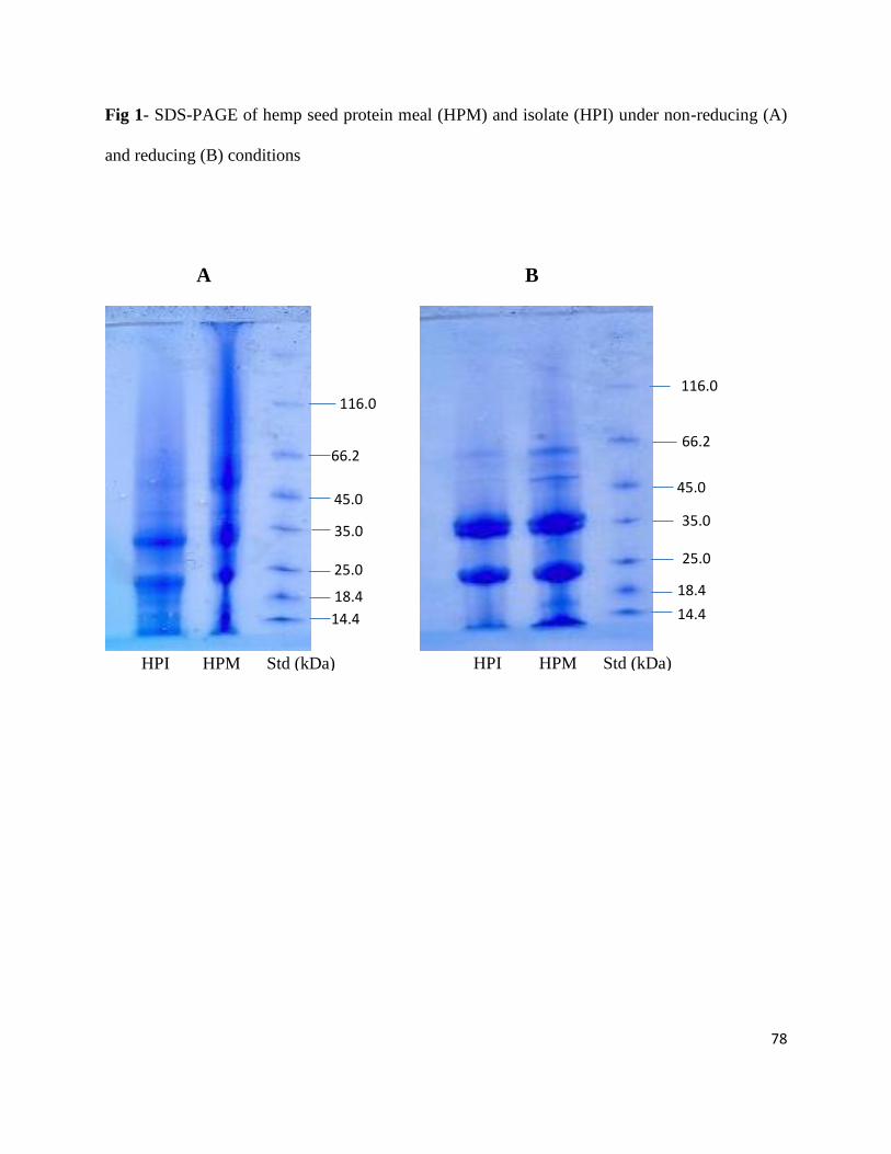
78
Fig 1- SDS-PAGE of hemp seed protein meal (HPM) and isolate (HPI) under non-reducing (A)
and reducing (B) conditions
116.0
66.2
45.0
35.0
25.0
18.4
14.4 14.4
18.4
25.0
35.0
45.0
66.2
116.0
HPI HPM Std (kDa) HPI HPM Std (kDa)
A B
79
of polypeptide bands when mercaptoethanol was present in the buffer. The proteins found in
both HPI and HPM electrophoretic profiles were in the same molecular weight range with 7S
vicilin (41 kDa) and phytohemagglutinins (16 to 30 kDa) reported for pinto bean protein isolates
(Tan and others 2014).
3.3.2 Protein contents, yields, solubility and amino acid composition
The protein content obtained for HPI (84.15%) is significantly (P<0.05) higher than the
corresponding value (44.32%) for HPM. The protein content for HPI in the present study is
higher than those previously reported by Mwasaru and others (1999) for pigeon pea isoelectric-
precipitated isolates (82.4%), pigeon pea micellization-precipitated isolates (82.8%) and cowpea
micellization-precipitated isolates (83.2%) but lower than the cowpea isoelectric-precipitated
isolates (89.5%) and the value (90.5%) that was reported by Wang and others (2008) for HPI.
The differences in protein content may be due to differences in extraction materials as well as the
method of protein precipitation. The protein content obtained for HPM in the present study is in
the 31.0-53.3% range reported by House and others (2010) and higher than the previously
reported value of 30.7% by Silversides and Lefrancois (2005). Meanwhile, Tang and others
(2006) reported a higher protein value (50%) for HPM, which is similar to the values claimed by
some commercial producers of HPM (House and others 2010). The yield obtained for the
isolates based on protein weight and gross weight was 37.90% and 16.80% respectively.
The HPI was fairly insoluble across the pH 3-9 range with a 25% PS at pH 3.0 and minimal
values (0.5-1%) at pH 4.0-5.0, which represents the isoelectric point range for hemp seed
proteins (Fig. 2). PS gradually increased as pH was raised from 5 to 9 as shown in Fig. 2. The
results are similar to the report of Tang and others (2006) who showed increased PS at pH values
>pI. The low PS of hemp seed proteins is believed to be due to edestin (major protein)
80
aggregation at pH below 7.0 and their dissociation at higher pH values (Friedman and Brandon
2001). There is linear proportionality between PS of HPM and pH with minimum PS at pH 3.0
(Fig. 2), and then increased to pH 9.0. Overall, with the exception of pH 3.0, HPM had higher PS
values than HPI at all the pH values used in this work. The higher PS of HPM suggests the
proteins may be in a more native state than the polypeptides present in HPI. Thus, the results
indicate that the isoelectric precipitation method used during protein isolation may have
contributed to increased aggregation and hence reduced PS of the HPI when compared to HPM.
However, the presence of higher level of non-protein materials in HPM could have also modified
protein structure (e.g. protein-sugar interactions) to enhance observed PS. Generally, the PS
values obtained in this work are lower than those reported by Tang and others (2006), which
probably reflects the seed processing history. The higher PS obtained by Tang and others (2006)
reflects the gentle seed processing history (supercritical fluid extraction at <40 oC), which would
have reduced excessive protein denaturation. In contrast, the protein products used in this work
were obtained from seeds that have undergone high level of force (cold press) to extract the oil;
hence it is more likely that the polypeptides have undergone severe protein-protein interactions,
which may be responsible for the observed low PS.
The amino acid composition data (Table 3) showed that the isolation process did not
cause any major changes in the content of most amino acids. However, there was a slight
increase in the Arg content of HPI (15.06%) when compared to its HPM (13.20%). Moreover,
the Arg/Lys ratio was higher for HPI (5.52) when compared to the HPM (3.35). A higher ratio of
Arg/Lys in the diet has been shown to produce hypocholesterolemic effects and could be of
benefit to improving cardiovascular health (Giroux and others 1999). In this work, the results
showed that the HPI may have better positive effects on the cardiovascular system when
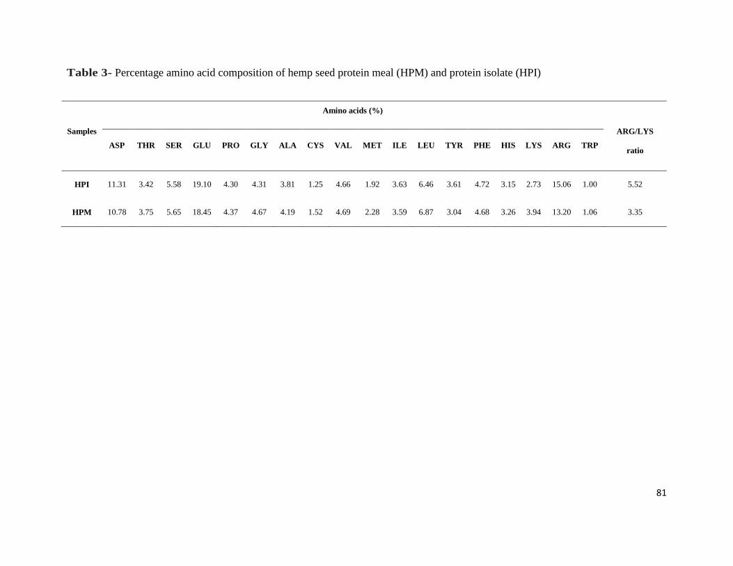
81
Table 3- Percentage amino acid composition of hemp seed protein meal (HPM) and protein isolate (HPI)
Samples
Amino acids (%)
ARG/LYS
ratio ASP THR SER GLU PRO GLY ALA CYS VAL MET ILE LEU TYR PHE HIS LYS ARG TRP
HPI 11.31 3.42 5.58 19.10 4.30 4.31 3.81 1.25 4.66 1.92 3.63 6.46 3.61 4.72 3.15 2.73 15.06 1.00 5.52
HPM 10.78 3.75 5.65 18.45 4.37 4.67 4.19 1.52 4.69 2.28 3.59 6.87 3.04 4.68 3.26 3.94 13.20 1.06 3.35
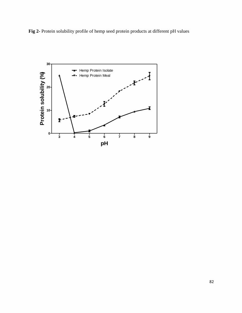
82
Fig 2- Protein solubility profile of hemp seed protein products at different pH values
pH
Pro
tein
so
lub
ilit
y (
%)
3 4 5 6 7 8 90
10
20
30
Hemp Protein Isolate
Hemp Protein Meal
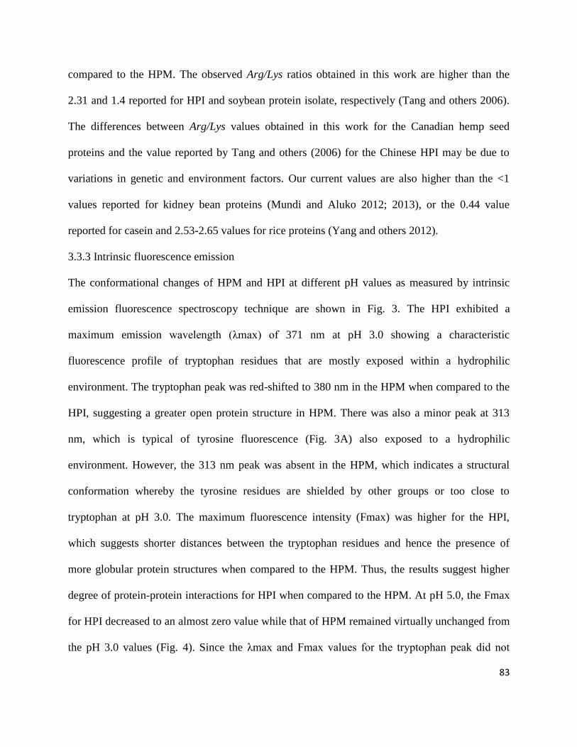
83
compared to the HPM. The observed Arg/Lys ratios obtained in this work are higher than the
2.31 and 1.4 reported for HPI and soybean protein isolate, respectively (Tang and others 2006).
The differences between Arg/Lys values obtained in this work for the Canadian hemp seed
proteins and the value reported by Tang and others (2006) for the Chinese HPI may be due to
variations in genetic and environment factors. Our current values are also higher than the <1
values reported for kidney bean proteins (Mundi and Aluko 2012; 2013), or the 0.44 value
reported for casein and 2.53-2.65 values for rice proteins (Yang and others 2012).
3.3.3 Intrinsic fluorescence emission
The conformational changes of HPM and HPI at different pH values as measured by intrinsic
emission fluorescence spectroscopy technique are shown in Fig. 3. The HPI exhibited a
maximum emission wavelength (λmax) of 371 nm at pH 3.0 showing a characteristic
fluorescence profile of tryptophan residues that are mostly exposed within a hydrophilic
environment. The tryptophan peak was red-shifted to 380 nm in the HPM when compared to the
HPI, suggesting a greater open protein structure in HPM. There was also a minor peak at 313
nm, which is typical of tyrosine fluorescence (Fig. 3A) also exposed to a hydrophilic
environment. However, the 313 nm peak was absent in the HPM, which indicates a structural
conformation whereby the tyrosine residues are shielded by other groups or too close to
tryptophan at pH 3.0. The maximum fluorescence intensity (Fmax) was higher for the HPI,
which suggests shorter distances between the tryptophan residues and hence the presence of
more globular protein structures when compared to the HPM. Thus, the results suggest higher
degree of protein-protein interactions for HPI when compared to the HPM. At pH 5.0, the Fmax
for HPI decreased to an almost zero value while that of HPM remained virtually unchanged from
the pH 3.0 values (Fig. 4). Since the λmax and Fmax values for the tryptophan peak did not
84
change for HPM from pH 3.0 to 5.0, it means there was no change in the location or environment
of the tryptophan residues as a result of the reduced acidity. However, the tyrosine fluorescence
at 313 nm was observed also for HPM at pH 5.0 (Fig. 3B), which is an indication of
translocation of these residues into a more conserved protein structure and within a hydrophobic
environment, when compared to their location at pH 3.0. The Fmax for HPI tyrosine residues at
pH 5.0 was about 300% higher than the value obtained at pH 3.0, which suggests increased
interactions between the residues. Thus it seems that at pH 5.0, the HPI tryptophan residues
became highly shielded from light while the tyrosine residues became more exposed to light
source when compared to the structural conformation at pH 3.0.
At pH 7.0 and 9.0, there were slight increases in the Fmax values obtained for both
tyrosine and tryptophan peaks, which indicate structural rearrangements that enhanced tyrosine-
tyrosine and tryptophan-tryptophan interactions. It is possible that at pH 7.0 and 9.0 where the
environment is more hydrophilic, the aromatic groups are moved away from the surface and
towards the more hydrophobic core, which led to enhanced emission intensity. This is supported
by the fact that there were slight blue shifts in λmax, especially for the HPI at pH 7.0 (376 nm)
and 9.0 (368 nm). For the HPM, λmax values of 378 and 375 nm were obtained at pH 7.0 and
9.0, respectively (Fig. 3). The observed blue shifts in λmax at high pH values in the present study
in similar to a previous report on quinoa protein isolates (Abugoch and others 2008). The red
shift in λmax values and reduced Fmax observed from pH 3.0 to 5.0 for HPI reflect increased
exposure of the aromatic residues to the hydrophilic environment and is in agreement with a past
report (Naseem and others 2004) on the relationship between isoelectric point and
conformational changes in proteins.
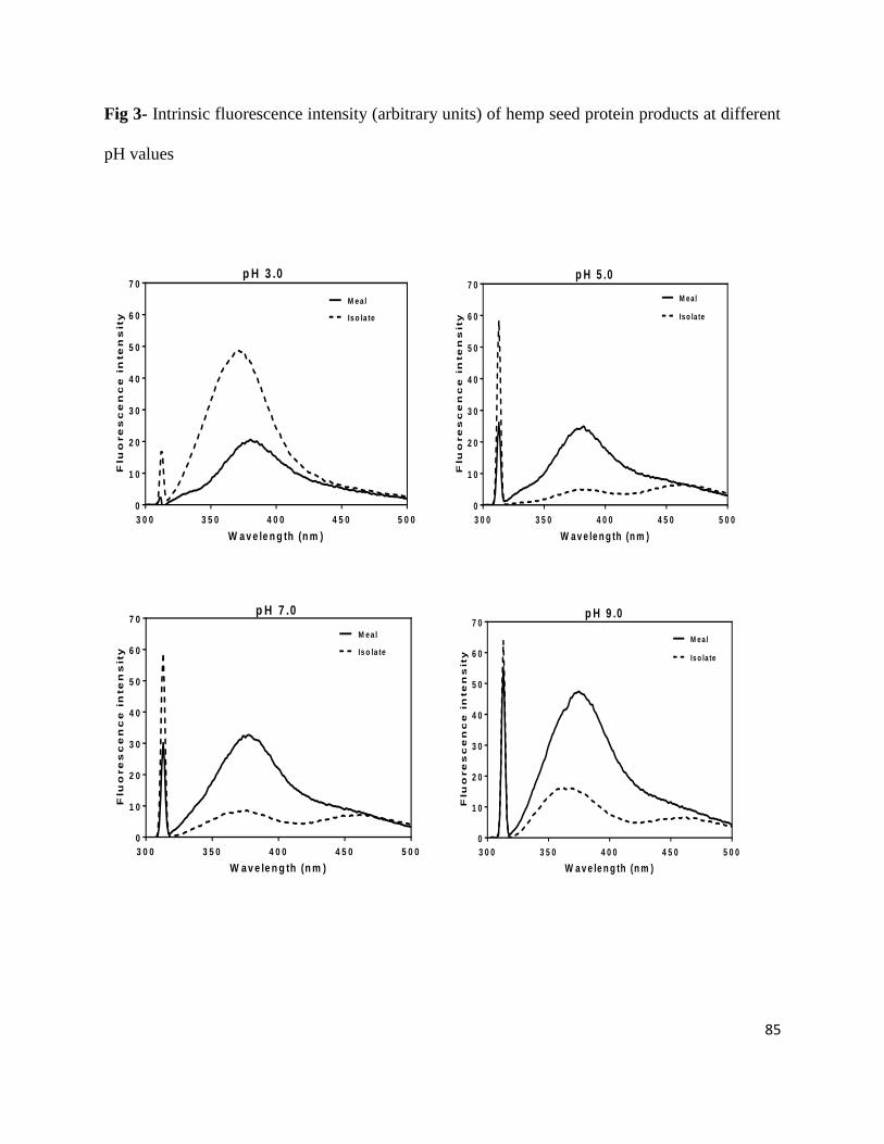
85
Fig 3- Intrinsic fluorescence intensity (arbitrary units) of hemp seed protein products at different
pH values
3 0 0 3 5 0 4 0 0 4 5 0 5 0 0
0
1 0
2 0
3 0
4 0
5 0
6 0
7 0
Is o la te
M ea l
W a v e le n g th (n m )
Flu
ore
sc
en
ce
in
te
ns
ity
p H 3 .0
3 0 0 3 5 0 4 0 0 4 5 0 5 0 0
0
1 0
2 0
3 0
4 0
5 0
6 0
7 0
Is o la te
M ea l
W a v e le n g th (n m )
Flu
ore
sc
en
ce
in
te
ns
ity
p H 5 .0
3 0 0 3 5 0 4 0 0 4 5 0 5 0 0
0
1 0
2 0
3 0
4 0
5 0
6 0
7 0
Is o la te
M ea l
W a v e le n g th (n m )
Flu
ore
sc
en
ce
in
te
ns
ity
p H 7 .0
3 0 0 3 5 0 4 0 0 4 5 0 5 0 0
0
1 0
2 0
3 0
4 0
5 0
6 0
7 0
Is o la te
M ea l
W a v e le n g th (n m )
Flu
ore
sc
en
ce
in
te
ns
ity
p H 9 .0

86
3.3.4 Secondary structure conformations
The effect of pH on CD spectra obtained for secondary structures of HPM and HPI is shown in
Fig. 4. Due to reduced protein solubility, CD determination was not possible at pH 5.0 since
required level of soluble proteins could not be obtained. At pH 3.0, HPI showed a strong
secondary structure dominated mostly by the α-helix conformation as evident in the intense
ellipticity between 200 and 220 nm. The HPM had similar ellipticity pattern but was less intense
(less α-helix structure) than that of the HPI. However, the ellipticity at 195 nm was more intense
for the HPM, which indicates presence of more β-sheet structure when compared to the HPI. The
presence of a higher level of β-sheet structure in HPM indicates a more open conformation when
compared to HPI. The far-UV CD results are consistent with the fluorescence intensity data that
showed reduced Fmax and higher λmax (indication of high degree of exposure of aromatic
groups to the hydrophilic environment) exhibited at pH 3.0. When the environment was changed
to pH 7.0, the HPM had a very high increase in ellipticity values between 200-220 nm, indicating
presence of more α-helix structure than at pH 3.0 (Fig. 4). In contrast, the HPI had less α-helix
structure as indicated by the substantial decreases in ellipticity values between 200-220 nm,
indicating a more loose protein conformation than present at pH 3.0. The far-UV CD data at pH
7.0 also support the fluorescence emission data (Fig. 3) that showed a lower Fmax value and a
red shift in λmax (compared to pH 3.0), which are indications of increased interactions with the
hydrophilic environment. The CD ellipticity patterns obtained for HPI and HPM at pH 9.0 were
similar to those obtained at pH 7.0, though with reduced intensity, which suggests that increased
charge at alkaline pH 9.0 may have produced greater protein-protein electrostatic repulsions and
hence a more disorganized structure when compared to pH 7.0.
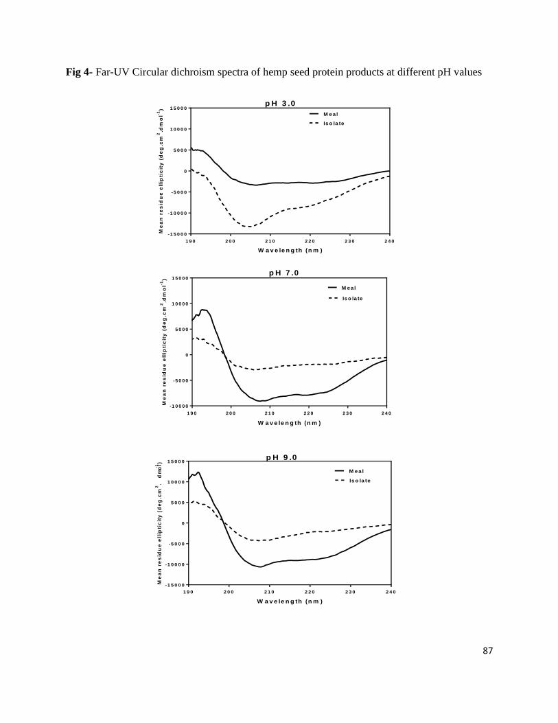
87
Fig 4- Far-UV Circular dichroism spectra of hemp seed protein products at different pH values
W a v e le n g th (n m )
Me
an
re
sid
ue
ell
ipti
cit
y (
de
g.c
m2
.dm
ol-1
)
1 9 0 2 0 0 2 1 0 2 2 0 2 3 0 2 4 0
-1 5 0 0 0
-1 0 0 0 0
-5 0 0 0
0
5 0 0 0
1 0 0 0 0
1 5 0 0 0
M ea l
Is o la te
p H 3 .0
W a v e le n g th (n m )
Me
an
re
sid
ue
ell
ipti
cit
y (
de
g.c
m2
.dm
ol-1
)
1 9 0 2 0 0 2 1 0 2 2 0 2 3 0 2 4 0
-1 0 0 0 0
-5 0 0 0
0
5 0 0 0
1 0 0 0 0
1 5 0 0 0
M ea l
Is o la te
p H 7 .0
W a v e le n g th (n m )
Me
an
re
sid
ue
ell
ipti
cit
y (
de
g.c
m2
.d
mo
l-1
)
1 9 0 2 0 0 2 1 0 2 2 0 2 3 0 2 4 0
-1 5 0 0 0
-1 0 0 0 0
-5 0 0 0
0
5 0 0 0
1 0 0 0 0
1 5 0 0 0
M ea l
Is o la te
p H 9 .0
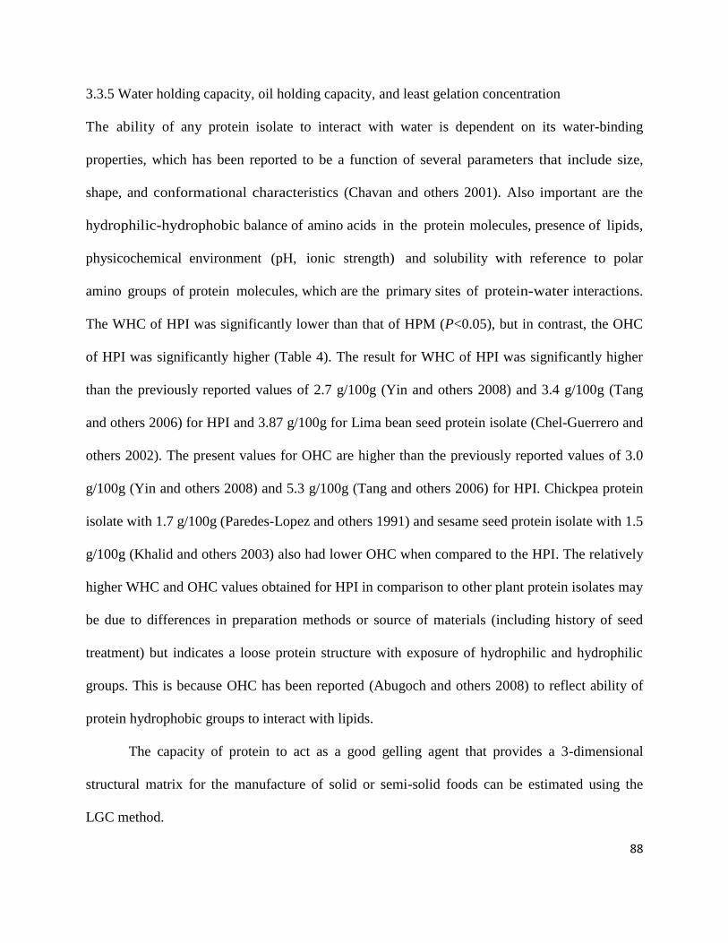
88
3.3.5 Water holding capacity, oil holding capacity, and least gelation concentration
The ability of any protein isolate to interact with water is dependent on its water-binding
properties, which has been reported to be a function of several parameters that include size,
shape, and conformational characteristics (Chavan and others 2001). Also important are the
hydrophilic-hydrophobic balance of amino acids in the protein molecules, presence of lipids,
physicochemical environment (pH, ionic strength) and solubility with reference to polar
amino groups of protein molecules, which are the primary sites of protein-water interactions.
The WHC of HPI was significantly lower than that of HPM (P<0.05), but in contrast, the OHC
of HPI was significantly higher (Table 4). The result for WHC of HPI was significantly higher
than the previously reported values of 2.7 g/100g (Yin and others 2008) and 3.4 g/100g (Tang
and others 2006) for HPI and 3.87 g/100g for Lima bean seed protein isolate (Chel-Guerrero and
others 2002). The present values for OHC are higher than the previously reported values of 3.0
g/100g (Yin and others 2008) and 5.3 g/100g (Tang and others 2006) for HPI. Chickpea protein
isolate with 1.7 g/100g (Paredes-Lopez and others 1991) and sesame seed protein isolate with 1.5
g/100g (Khalid and others 2003) also had lower OHC when compared to the HPI. The relatively
higher WHC and OHC values obtained for HPI in comparison to other plant protein isolates may
be due to differences in preparation methods or source of materials (including history of seed
treatment) but indicates a loose protein structure with exposure of hydrophilic and hydrophilic
groups. This is because OHC has been reported (Abugoch and others 2008) to reflect ability of
protein hydrophobic groups to interact with lipids.
The capacity of protein to act as a good gelling agent that provides a 3-dimensional
structural matrix for the manufacture of solid or semi-solid foods can be estimated using the
LGC method.
89
Table 4- Water holding capacity (WHC), oil holding capacity (OHC) and least gelation
concentration (LGC) of hemp seed protein products
Samples WHC (g/g) OHC (g/g) LGC (%)
Meal 12.32±0.03a 12.54±0.08
b 12.00±0.00
a
Isolate 12.01±0.08b 13.70±0.29
a 22.00±0.00
b
Mean values±standard deviation of replicate analysis; means followed by different superscript in
each column indicates significant differences at P<0.05.
90
The gelation capacity of HPM (12%) in this study was significantly better (P<0.05) when
compared to HPI (22%) but is similar to the value reported for lupin protein concentrate (Lqari
and others 2002). The LGC value for HPM is also better than the previously reported value of
14% for soybean protein isolate (Okezie and Bello 1988). The poor gel-forming ability of HPI
may be due to a high level of protein aggregation during isoelectric protein precipitation, which
reduced the flexibility required for network formation. In contrast, the HPM did not undergo
isoelectric protein precipitation and probably contained less number of aggregated proteins,
hence the higher LGC value.
3.3.6 Foam Capacity and Foam Stability
Besides possessing flexible protein surfactant molecules, a good foaming protein must be rapidly
adsorbed at the air–water interface and rearranged to form a cohesive viscoelastic film via
intermolecular interactions (Yin and others 2008; Aluko and Yada 1997). At 20 mg/ml protein
concentration, the FC of HPM and HPI was similar across various pH values with the exception
of pH 3.0 where the HPI had significantly (P<0.05) higher value (Fig. 5). Protein solubility and
hydrophobicity/hydrophilicity ratio of samples can also influence some functional properties
(Damodaran 1996). Therefore, the high FC at pH 3.0 for the HPI may be due to its superior
solubility property when compared to other pH values. The results are also consistent with the
higher intrinsic fluorescence intensity and better secondary structure conformation obtained for
HPI at pH 3.0. The fluorescence intensity data indicates greater exposure of aromatic groups,
which could have enhanced protein-protein interactions to form stronger interfacial membranes
and hence higher FC at pH 3.0. The results also showed that the FC of the HPM was consistent
with the solubility properties across the pH range studied. Increase in protein concentration from
20 to 60 mg/ml led to reductions in FC at pH 3.0 whereas there were increases at pH 5.0 to 9.0.
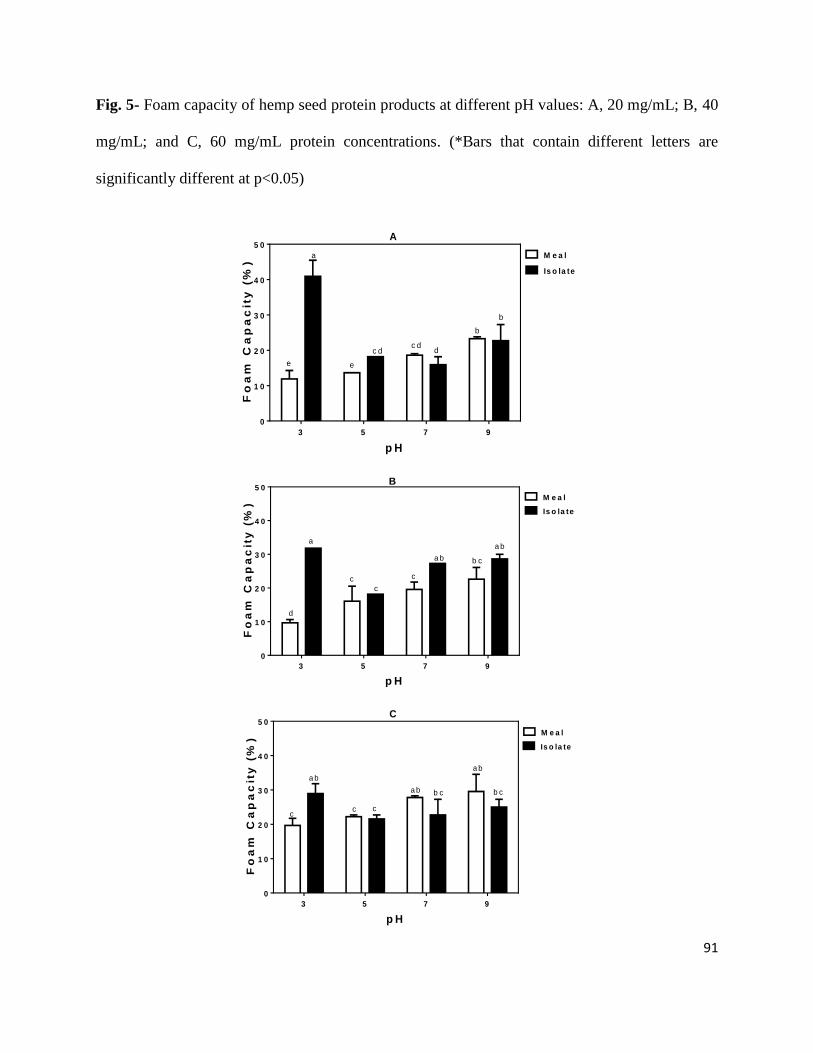
91
Fig. 5- Foam capacity of hemp seed protein products at different pH values: A, 20 mg/mL; B, 40
mg/mL; and C, 60 mg/mL protein concentrations. (*Bars that contain different letters are
significantly different at p<0.05)
p H
Fo
am
Ca
pa
cit
y (
%)
3 5 7 9
0
1 0
2 0
3 0
4 0
5 0
M e a l
Is o la te
c d
a
dc d
b
e e
b
A
p H
Fo
am
Ca
pa
cit
y (
%)
3 5 7 9
0
1 0
2 0
3 0
4 0
5 0
M e a l
Is o la te
d
c
c
c
aa b
a b b c
B
p H
Fo
am
Ca
pa
cit
y (
%)
3 5 7 9
0
1 0
2 0
3 0
4 0
5 0
M e a l
Is o la te
a b
a b
c
b c
c c
a b b c
C
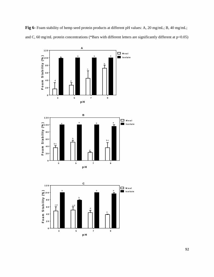
92
Fig 6- Foam stability of hemp seed protein products at different pH values: A, 20 mg/mL; B, 40 mg/mL;
and C, 60 mg/mL protein concentrations (*Bars with different letters are significantly different at p<0.05)
p H
Fo
am
Sta
bil
ity
(%
)
3 5 7 9
0
2 0
4 0
6 0
8 0
1 0 0
1 2 0
M e a l
Is o la teaa
d d
a a
b
c
A
p H
Fo
am
Sta
bil
ity
(%
)
3 5 7 9
0
2 0
4 0
6 0
8 0
1 0 0
1 2 0
M e a l
Is o la te
b c
aa
d
a
b c
a
b
B
p H
Fo
am
Sta
bil
ity
(%
)
3 5 7 9
0
2 0
4 0
6 0
8 0
1 0 0
1 2 0
M e a l
Is o la tea
e
a
d
a
c d c d
b
C
93
The results suggest that at pH 3.0, a dilute protein concentration provided greater protein
flexibility and hence higher FC; increased concentration probably led to a crowded solution that
reduced protein flexibility. In contrast, at pH 7.0 and 9.0 there was an increased charge density
which could have minimized the crowding effect and hence FC increased with increased protein
concentration. Only slight changes in FC were observed when protein concentration was
increased at pH 5.0 (Fig. 5). The foams formed by HPI were general more stable at all the
different pH values and protein concentrations with no significant differences (P>0.05) between
them (Fig. 6). All the HPI foams had significantly higher FS values when compared to the HPM-
formed foams. The results agree with a previous work, which showed that samples with higher
protein contents formed more stable foams than samples with lower protein contents (Aluko and
others 2009). This is because proteins are able to form viscoelastic interfacial membranes
through protein-protein interactions and enhance resistance of air bubbles to destabilization.
3.3.7 Emulsion capacity and Emulsion stability
The EC recorded as surface diameter (µm) of the HPM and HPI emulsion oil droplets (d3,2) at
different pH and protein concentrations are shown in Figure 7. At a low protein concentration of
10 mg/ml, the HPM formed significantly (P<0.05) better emulsions (lower d3,2 values) at pH 3.0
and 5.0 when compared to pH 7.0 and 9.0 or all the HPI emulsions. The higher HPM emulsion
forming ability may be due to the presence of a higher level of non-protein materials such as
polysaccharides that could enhance emulsification, especially at low protein concentrations. In
contrast, the HPI has less non-protein materials and coupled with the rigid molecules from the
isoelectric protein precipitation produced emulsions with slightly higher d3,2 values at 10 and 25
mg/ml protein concentrations. However at a higher protein concentration of 50 mg/ml, the HPI
produced emulsions with significantly lower d3,2 values at all the tested pH value with the
94
exception of pH 3.0. Thus, the results suggest that higher protein concentration may be able to
compensate for the poor emulsification property of a protein. The exception at pH 3.0 can be
attributed to the increased solubility (Fig. 2) and hence higher net protein charge coupled with
protein crowding, which may have limited the interfacial properties as a result of increased
protein-protein repulsions. In contrast, the decreased PS at pH 7.0 and 9.0 indicates weaker
intensity of the protein-protein repulsion forces and hence higher ability to form stronger
interfacial protein membranes, which led to better emulsion formation than at pH 3.0 where the
forces are stronger. The values presented for the 50 mg/ml protein concentration is in contrast to
a previous report (Tang and others 2006) that showed lack of a relationship between EC and PS.
This is because at pH 3.0 where higher PS value was recorded for HPI in this work, there was a
significantly (P<0.05) lower emulsification ability (higher d3,2). However, the higher emulsifying
ability (lower d3,2) of HPI at a high protein concentration of 50 mg/ml (pH 5.0, 7.0 and 9.0) is
consistent with a previous report (Aluko and others 2009) that showed enhanced emulsion ability
when sample protein concentration was increased. ES increases when there is strong protein-
protein interactions at the oil-water interface of the protein samples, which prevents oil droplet
coalescence. At 10 mg/ml protein concentration, all the emulsions were very stable (~100% ES)
with the exception of HPI emulsion at pH 5.0, which had only 50% stability (Fig. 8). As protein
concentration was increased to 25 and 50 mg/ml, all the emulsions had similar ES values.
Therefore, the overall data seem to indicate that the HPM and HPI formed emulsions that were
stabilized by formation of strong interfacial membranes that reduced the rate of oil droplet
coalescence.
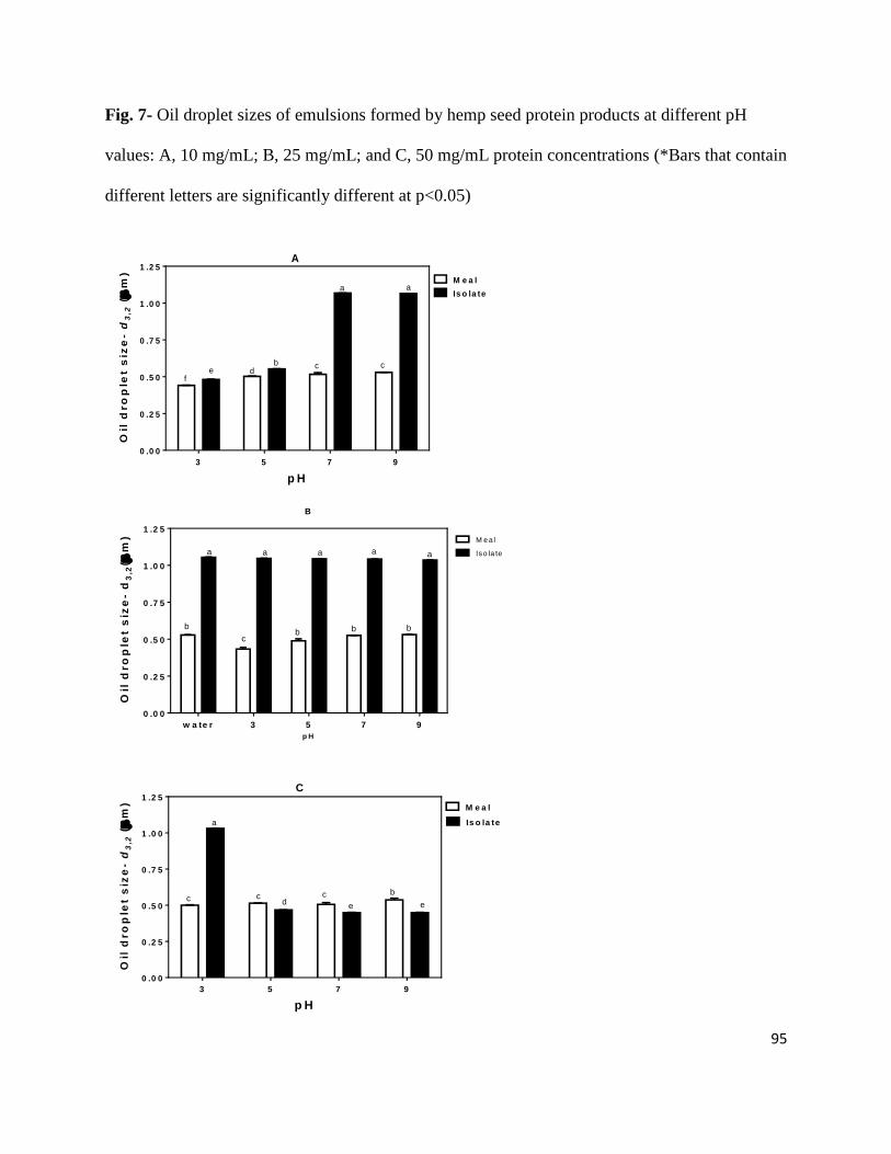
95
Fig. 7- Oil droplet sizes of emulsions formed by hemp seed protein products at different pH
values: A, 10 mg/mL; B, 25 mg/mL; and C, 50 mg/mL protein concentrations (*Bars that contain
different letters are significantly different at p<0.05)
p H
Oil
dro
ple
t s
ize
-d
3,2
(
m)
3 5 7 9
0 .0 0
0 .2 5
0 .5 0
0 .7 5
1 .0 0
1 .2 5
M e a l
Is o la te
ef
cd
b
a
c
a
A
B
p H
Oil
dro
ple
t s
ize
- d
3,2
(m
)
w a te r 3 5 7 9
0 .0 0
0 .2 5
0 .5 0
0 .7 5
1 .0 0
1 .2 5
M eal
Iso la te
b
a
c
a
b
a
b
a
b
a
p H
Oil
dro
ple
t s
ize
-d
3,2
(
m)
3 5 7 9
0 .0 0
0 .2 5
0 .5 0
0 .7 5
1 .0 0
1 .2 5
M e a l
Is o la te
bcc
c
a
de e
C
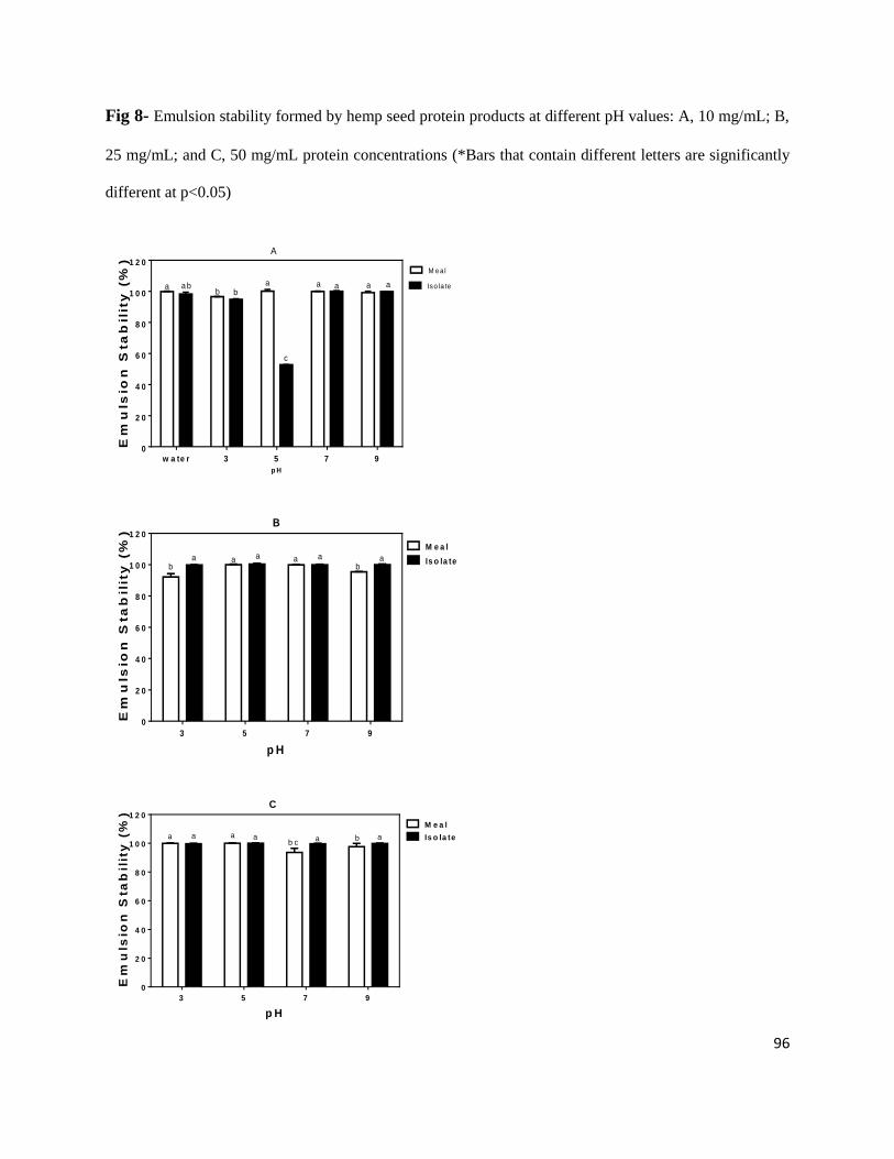
96
Fig 8- Emulsion stability formed by hemp seed protein products at different pH values: A, 10 mg/mL; B,
25 mg/mL; and C, 50 mg/mL protein concentrations (*Bars that contain different letters are significantly
different at p<0.05)
p H
Em
uls
ion
Sta
bil
ity
(%
)
w a te r 3 5 7 90
2 0
4 0
6 0
8 0
1 0 0
1 2 0M eal
Iso la teaaaaabb
a a b
c
A
p H
Em
uls
ion
Sta
bil
ity
(%
)
3 5 7 9
0
2 0
4 0
6 0
8 0
1 0 0
1 2 0
M e a l
Is o la teaa aaaab b
B
p H
Em
uls
ion
Sta
bil
ity
(%
)
3 5 7 9
0
2 0
4 0
6 0
8 0
1 0 0
1 2 0
M e a l
Is o la teabab c
aaaa
C
97
3.4 Conclusion
The findings from this study have shown that, besides the nutritional role of hemp seed
protein, there is the potential for use as a basic functional ingredient in the food industry. The
results showed that manipulation of pH could be used to increase FC, which is dependent on PS.
The oil droplet sizes of some of the emulsions were very small (< 0.5 µm) and comparable to
values previously reported for milk and soybean protein emulsifiers. The high ES is an indication
of the potential use of the hemp seed proteins to form food emulsions that can maintain stable oil
droplet size during short-term storage. The results obtained at pH 3.0 for HPI suggest that the
mechanisms responsible for foam formation are probably different from those involved in
emulsion formation. Intrinsic fluorescence and CD data revealed that proteins in HPM and HPI
exist in different conformational states when evaluated at various pH values. The results suggest
that the effect of pH on protein functionality is dependent of protein concentration. The results
confirm that protein functionality is highly dependent on structural conformation, which can be
modulated through changes in pH of the environment. Moreso, since hemp proteins are not listed
as priority allergens which require labelling, therefore, both HSP products could serve as
alternative protein ingredients to the priority allergens (e.g. soybean and pea proteins) that
require labelling.
Acknowledgement
This work was funded by the Natural Sciences and Engineering Research Council of Canada
(NSERC) Discovery grant and a Manitoba Agri-Food Research and Development Initiative
(ARDI) grant to Dr. R.E. Aluko. S.A. Malomo is a recipient of University of Manitoba Graduate
98
Fellowship (UMGF) and Manitoba Graduate Scholarship (MGS) Awards for doctoral studies.
The authors hereby declare that there is no conflict of interest.
Author contributions
R.E. Aluko designed the experiments, assisted with data interpretation and edited final draft of
the manuscript; S.A. Malomo performed the laboratory experiments, data analysis, data
interpretation and produced the draft manuscript; R. He assisted in performing data analysis and
interpretation.
References
Abugoch LE, Romero N, Tapia CA, Silva J, Rivera M. 2008. Study of some physicochemical
and functional properties of quinoa (Chenopodium quinoa Willd) protein isolates. J Agric
Food Chem 56: 4745-4750.
Adebiyi AP, Aluko RE. 2011. Functional properties of protein fractions obtained from
commercial yellow field pea (Pisum sativum L.) seed protein isolate. Food Chem 128:
902-908.
Aluko RE, McIntosh T. 2004. Electrophoretic and functional properties of mustard seed meals
and protein concentrates. J Amer Oil Chem Soc 81: 679-683.
Aluko RE, Mofolasayo OA, Watts BA. 2009. Emulsifying and foaming properties of commercial
yellow pea (Pisum sativum L.) seed flours. J Agric Food Chem 57: 9793-9800.
Aluko RE, Yada RY. 1997. Some physicochemical and functional properties of cowpea (Vigna
unguiculata) isoelectric protein isolate as a function of pH and salt concentration. Int J
Food Sci Nutr 48: 31-39.
99
Bidlingmeyer BA, Cohen SA, Tarvin TL. 1984. Rapid analysis of amino acids using pre-column
derivatization. J Chrom 336: 93-104.
Callaway J, Schwab U, Harvima I, Halonen P, Mykkänen P. Järvinen T. 2005. Efficacy of
dietary hemp seed oil in patients with atopic dermatitis. J Derm Treatm 16: 87-94.
Chavan UD, McKenzie DB, Shahidi F. 2001. Functional properties of protein isolates from
beach pea (Lathyrus maritimus L.). Food Chem 74: 177-187.
Chel-Guerrero L, Perez-Flores V, Betancur-Ancona D, Davila-Ortiz G. 2002. Functional
properties of flours and protein isolates from Phaseolus lunatus and Canavalia ensiformis
seeds. J Agric Food Chem 50: 584-591.
Damodaran S. 1996. Amino acids, peptides, and proteins. In O. R. Fennema (Ed.), Food
Chemistry (3rd ed., pp. 322–424). New York: Marcel Dekker, Inc.
Du SK, Jiang HX, Yu XZ, Jane JL. 2014. Physicochemical and functional properties of whole
legume flour. LWT Food Sci Technol 55: 308-313.
Friedman M, Brandon DL. 2001. Nutritional and health benefits of soy proteins. J Agric Food
Chem 49: 1069-1086.
Gehrke CW, Wall LL, Absheer JS, Kaiser FE, Zumwalt RW. 1985. Sample preparation for
chromatography of amino-acids: acid hydrolysis of proteins. J Assoc Official Anal Chem,
68: 811-821.
Gibb DJ, Shah MA, Mir RS, McAllister TA. 2005. Effect of full-fat hemp seed on performance
and tissue fatty acids of feedlot cattle. Can J Animal Sci 85: 223-230.
Girgih AT, Udenigwe CC, Aluko RE. 2011. In vitro antioxidant properties of hemp seed
(Cannabis sativa L.) protein hydrolysate fractions. J Am Oil Chem Soc 88:381-389.
100
Giroux I, Kurowska EM, Freeman DJ, Carroll KK. 1999. Addition of arginine but not glycine to
lysine plus methionine–enriched diets modulates serum cholesterol and liver
phospholipids in rabbits. J Nutr 129: 1807-1813.
House JD, Neufeld J, Leson G. 2010. Evaluating the quality of protein from hemp seed
(Cannabis sativa L.) products through the use of the protein digestibility-corrected amino
acid score method. J Agric Food Chem 58: 11801-11807.
Khalid, E. K., Babiker, E. E., EL Tinay, A. H. (2003). Solubility and functional properties of
sesame seed proteins as influenced by pH and/or salt concentration. Food Chem 82: 361-
366.
Landry J, Delhaye S. 1992. Simplified procedure for the determination of tryptophan of foods
and feedstuffs from barytic hydrolysis. J Agric Food Chem 40: 776-779.
Li H, Aluko RE. 2006. Structural modulation of calmodulin and calmodulin-dependent protein
kinase II by pea protein hydrolysates. Int J Food Sci Nutr 57: 178-189.
Lqari H, Vioque J, Pedroche J, Millan F. 2002. Lupinus angustifolius protein isolates: chemical
composition, functional properties and protein characterization. Food Chem 76: 349-356.
Markwell MAK, Haas SM, Bieber LL, Tolbert NE. 1978. Modification of lowry procedure to
simplify protein determination in membrane and lipoprotein samples. Anal Biochem 87:
206-210.
Mundi S, Aluko RE. 2012. Physicochemical and functional properties of kidney bean albumin
and globulin protein fractions. Food Res Int 48: 299-306.
Mundi S, Aluko RE. 2013. Effects of NaCl and pH on the structural conformations of kidney
bean vicilin. Food Chem 139: 624-630.
101
Mune Mune MA, Minka SR, Mbome IL. 2014. Optimising functional properties during
preparation of cowpea protein concentrate. Food Chem 154: 32-37
Mwasaru MA, Muhammad K, Bakar J, Man YBC. 1999. Effects of isolation technique and
conditions on the extractability, physicochemical and functional properties of pigeonpea
(Cajanus cajan) and cowpea (Vigna unguiculata) protein isolates. I. Physicochemical
properties. Food Chem 67: 435-443.
Naseem F, Ahmad B, Ashaf MT, Khan RH. 2004. Molten globule-like folding intermediate of
asialofetuin of acidic pH. Biochim Biophy Acta 1699: 191-199.
Okezie BO, Bello AB. 1988. Physicochemical and functional properties of winged bean flour
and isolate compared with soy isolate. J Food Sci 53: 450-454.
Omoni AO, Aluko RE. 2006. Effect of cationic flaxseed protein hydrolysate fractions on the in
vitro structure and activity of calmodulin-dependent endothelial nitric oxide synthase.
Mol Nutri Food Res 50: 958-966.
Paredeslopez O, Ordoricafalomir C, Olivaresvazquez MR. 1991. Chickpea protein isolates -
physicochemical, functional and nutritional characterization. J. Food Sci 56: 726-729.
Radocaj O, Dimi´c E, Tsao R. 2014. Effects of hemp (Cannabis sativa L.) seed oil press-cake
and decaffeinated green tea leaves (Camellia sinensis) on functional characteristics of
gluten-free crackers. J Food Sci 79: C318-325.
Silversides FG, Lefrancois MR. 2005. The effect of feeding hemp seed meal to laying hens. Brit
Poultry Sci 46: 231-235.
Svennerstedt B, Svensson G. 2006. Hemp (Cannabis sativa L.) trials in southern Sweden 1991-
2001. J Ind Hemp 11: 17-25.
102
Tan E, Ngoh Y, Gan C. 2014. A comparative study of physicochemical characteristics and
functionalities of pinto bean protein isolate (PBPI) against the soybean protein isolate
(SPI) after the extraction optimisation. Food Chem 152: 447–455
Tang CH, Ten Z, Wang XS, Yang XQ. 2006. Physicochemical and functional properties of hemp
(Cannabis sativa L.) protein isolate. J Agric Food Chem 54: 8945-8950.
Wang XS, Tang CH, Yang XQ, Gao WR. 2008. Characterization, amino acid composition and in
vitro digestibility of hemp (Cannabis sativa L.) proteins. Food Chem 107: 11-18.
Yang L, Chen JH, Zhang H, Qiu W, Liu QH, Peng X, Li YN, Yang HK. 2012. Alkali treatment
affects in vitro digestibility and bile acid binding activity of rice protein due to varying its
ratio of arginine to lysine. Food Chem 132: 925–930.
Yin SW, Tang CH, Cao JS, Hu EK, Wen QB, Yang XQ. 2008. Effects of limited enzymatic
hydrolysis with trypsin on the functional properties of hemp (Cannabis sativa L.) protein
isolate. Food Chem 106: 1004-1013.

103
3.5 Statement transfer between the study 1 and study 2
The isoelectric precipitation method produces protein isolates that consist of different fractions
through the alkaline solubilisation and acid precipitation. This isoelectric precipitation method of
protein isolation leads to limited functionality because of poor structural conformations and
partial protein denaturation. The limited functionality arises from protein rigidity and
hydrophobic interactions that cause protein aggregation within the harsh acid/alkaline
environments used for during extraction. Besides, some of the non-protein seed components
(starch, soluble sugars, fibre, and phytate) are also extracted and could thus affects the solubility
(and other functionalities) of the resultant protein isolates. Therefore, the next chapter focuses on
the use of conventional dialysis method to separate pure and distinct protein fractions (albumin
and globulin) from the hemp seed defatted flour. The two protein fractions also have different
solubility profiles, hence the conformational properties are expected to differ from each and
when compared with the protein isolates. This is because the albumin fraction is highly soluble
in water while the pure globulin fraction is only soluble in salt solution. In contrast the protein
isolates used in the preceding chapter are soluble mainly in dilute alkaline solutions.
104
CHAPTER FOUR
4. Comparative study of the structural and functional properties of isolated hemp seed
(Cannabis sativa L.) albumin and globulin fractions
Sunday A. Malomo, Rotimi E. Aluko*
The Department of Human Nutritional Sciences, University of Manitoba, Winnipeg, Manitoba,
Canada R3T 2N2
*Corresponding author: Dr. Rotimi E. Aluko; E-Mail: [email protected]; Tel.: +1-204-
474-9555; Fax: +1-204-474-7593.
Reprinted from A Comparative Study of the Structural and Functional Properties of Isolated
Hemp Seed (Cannabis sativa L.) Albumin and Globulin fractions. Food Hydrocolloids, 43, 743-
752.
Copyright © 2015, with permission from Elsevier
105
ABSTRACT
The aim of this work was to determine the structural and functional characteristics of two major
hemp seed proteins, the water-soluble albumin (ALB) and the salt-soluble globulin (GLB). 0.5 M
NaCl extract of hemp seed protein flour was dialyzed against water to obtain the two fractions:
ALB in the water phase and GLB in the insoluble precipitate. The protein fractions were
subjected to structural (gel electrophoresis, intrinsic fluorescence and circular dichroism) and
functional (protein solubility, foaming, and emulsion) tests. Amino acid composition data
showed the presence of higher contents of aromatic and hydrophobic residues in GLB. Gel
electrophoresis indicated that the ALB has less disulfide bonds and hence a more open (flexible)
structure; this was confirmed by the intrinsic fluorescence and circular dichroism data showing
greater exposure of tyrosine residues when compared to GLB. ALB had significantly (p<0.05)
higher protein solubility and foaming capacity than the GLB at all the pH or sample
concentration values but emulsion forming ability was similar for both protein fractions.
Differences in emulsion stability were observed mostly at 10 mg/mL sample concentration; these
differences were minimized at 25 mg/mL and eliminated at 50 mg/mL. We conclude that the
ALB fraction will serve as excellent ingredient for food foam formulation while the GLB may be
slightly more useful than the ALB in the formulation of food emulsions.
Keywords: hemp seed; albumin, globulin, functional properties; intrinsic fluorescence, circular
dichroism
106
4.1. Introduction
The amount and structure of proteins present in a food system have been reported to play
an important role in making such ingredients useful for novel food formulations (Mundi &
Aluko, 2013; Yin et al., 2011). Barac, Pesic, Stanojevic, Kostic and Bivolarevic (2014) studied
and reported the influence of the functional properties of a food or its components (in addition to
its nutritional properties) in enhancing utilization for novel food product development. This
functionality is achieved through the protein’s activity (in free or structure changing state) as
observed during interactions with other food components (Tang, Sun, & Foegeding, 2011). An
example is the tendency of hydrophobic portions of a protein to interact with lipids and lipid
soluble compounds (Karaca, Low, & Nickerson, 2011). Critical efforts have been aimed at
effective utilization of inexpensive plant proteins as food hydrocolloids for nutritional and
functional purposes under different pH conditions. Proteins from plants like canola (Tan, Mailer,
Blanchard, & Agboola, 2014), peanut (He, Liu, et al., 2014), kidney bean (Mundi & Aluko,
2012, 2013), lentils (Avramenko, Low, & Nickerson, 2013), pinto bean (Tan, Ngoh, & Gan,
2014), adzuki, pea and soy bean (Barac et al., 2014), amaranth (Shevkani, Singh, Rana, & Kaur,
2014) and cowpea (Oduro-yeboah, Onwulata, Tortoe, & Thomas-Gahring, 2014) have been
examined for their structural and functional properties in model food systems.
Food-grade hemp seed is a new but widely cultivated plant of industrial importance and
requires detailed functional characterization of component proteins. This is very important
because availability of non-drug varieties of hemp seed possessing low δ-9-tetrahydrocannabinol
(THC) contents (Lu et al., 2010) has increased industrial utilization for food product
manufacture. The legal use of the seeds from low-THC plants as consumables food for humans
has been reported in Canada and other North American countries due to its considerable amounts
107
of over 30% oil and about 25% easily digestible proteins (Teh & Birch, 2013). The uniqueness of
hemp seed protein (HSP) has been reported (Park, Seo, & Lee, 2012) to contain 65% globulin
called edestin and 33% albumin; edestin is composed of six identical subunits having an acidic
subunit (AS) and a basic subunit (BS) linked by one disulfide bond. Previous works have
reported the functional (Tang, Ten, Wang, & Yang, 2006; Teh, Bekhit, Carne, & Birch, 2013;
Yin et al., 2008) or bioactive (Girgih, He, Malomo, & Aluko, 2014; Girgih, Udenigwe, & Aluko,
2011; Wang, Tang, Chen, & Yang, 2009) properties of HSP isolates and hydrolysates but there is
scanty information on the structural and functional properties of the seed globulin and albumin
fractions. Therefore, this study was aimed at determining the structural and functional properties
of albumin (ALB) and globulin (GLB) fractions of HSP. Specifically, pH-dependent changes in
protein solubility, foaming and emulsifying properties were studied in relative to protein
conformational changes. The information from this work may provide opportunities for
industrial application of HSP as a useful food ingredient and a suitable alternative source of
functional proteins to traditional ingredients.
4.2. Materials and Methods
4.2.1. Materials
Defatted hemp seed protein meal (HPM) containing ~40% (w/w) protein content, the by-
product obtained from commercial oil extraction was purchased from Manitoba Harvest Fresh
Hemp Foods Ltd (Winnipeg, MB, Canada) and stored at -20 oC until used for protein extraction.
Spectra/Por dialysis membrane with 6-8 kDa molecular weight cut-off (MWCO), gel
electrophoresis protein markers as well as other analytical grade reagents were purchased from
Fisher Scientific (Oakville, ON, Canada).
4.2.2. Protein fraction extraction and isolation

108
The method of Aluko (2004), with slight modifications was employed in the extraction
and fractionation of HPM protein into ALB and GLB. The HPM was extracted (1:10 w/v) with
0.5 M NaCl solution for 1 h at 24+2 oC under continuous stirring followed by centrifugation
(7000 g, 60 min at 4 oC) and the supernatant clarified with Whatman No 1 filter while the
precipitate was discarded. The supernatant was dialyzed against water at 4 oC for 5 days using
the 6-8 kDa MWCO dialysis tubing (with dialysis water changed 3 times daily). The content of
the dialysis bag was then centrifuged (7000 g, 60 min at 4 oC) and the supernatant collected as
the albumin protein fraction. The precipitate was washed with 100 ml of distilled water,
centrifuged again under similar conditions as above and the precipitate collected as the globulin
protein fraction. The two protein fractions were individually freeze-dried followed by storage at -
20 oC until required for subsequent analyses. Protein concentration of the fractions was
determined using the modified Lowry method (Markwell, Haas, Bieber, & Tolbert, 1978).
4.2.3. Amino acid composition
The amino acid profiles of ALB and GLB were determined using the HPLC Pico-Tag
system according to the method previously described after samples were digested with 6 M HCl
for 24 h (Bidlingmeyer, Cohen, & Tarvin, 1984). The cysteine and methionine contents were
determined after performic acid oxidation (Gehrke, Wall, Absheer, Kaiser, & Zumwalt, 1985)
while the tryptophan content was determined after alkaline hydrolysis (Landry & Delhaye,
1992).
4.2.4. Protein solubility (PS)
PS of ALB and GLB was determined according to the method described by Adebiyi and
Aluko (2011) with slight modifications. Briefly, 10 mg of sample was dispersed in 1 ml of 0.1 M
phosphate buffer solutions (pH 3.0-9.0) and the resulting mixture was vortexed for 2 min and
109
centrifuged at 10000 g for 20 min. Protein content of the supernatant was determined using the
modified Lowry method (Markwell et al., 1978). Total protein content was determined by
dissolving the protein samples in 0.1 M NaOH solution. PS was expressed as percentage ratio of
supernatant protein content to the total protein content.
4.2.5. Sodium dodecyl sulphate-polyacrylamide gel electrophoresis (SDS-PAGE)
The freeze-dried ALB and GLB were subjected to SDS-PAGE according to the method
of Aluko and McIntosh (2004) with minor modifications. The protein samples were each
dispersed (10 mg/mL) in Tris/HCl buffer, pH 8.0 containing 10% (w/v) SDS only (non-reducing
buffer) or SDS + 10% (v/v) β-mercaptoethanol (β-ME) as the reducing buffer followed by
heating at 95 oC for 10 min, cooled and centrifuged (10000 g, 15 min). After centrifugation, 1 µl
of supernatant was loaded onto 8-25% gradient gels and electrophoresis was performed with
Phastsystem Separation and Development units according to the manufacturer’s instructions (GE
Healthcare, Montréal, PQ, Canada). A mixture of protein standards (14.4-116 kDa) was used as
the molecular weight marker and the gels were stained with Coomassie brilliant blue.
4.2.6. Intrinsic fluorescence emission
The method described by Li and Aluko (2006) was used to record intrinsic fluorescence
spectra on the JASCO FP-6300 spectrofluorimeter (JASCO, Tokyo, Japan) at 25 oC using a 1 cm
path length cuvette. Protein stock solution (10 mg/ml) was diluted with 0.1 M sodium phosphate
buffer to 0.002% (w/v) and fluorescence spectra recorded at excitation wavelengths of 275 nm
(tyrosine and tryptophan) with emission recorded from 280 to 450 nm. Emissions of the buffer
blanks were subtracted from those of the respective samples to obtain fluorescence spectra of the
sample.
4.2.7. Measurements of circular dichroism (CD) spectra

110
CD spectra of samples was measured at 25 oC in a J-810 spectropolarimeter (JASCO,
Tokyo, Japan) using the spectral range of 190-240 nm (far-UV) for secondary structure
determinations and 250-320 nm (near-UV) for tertiary structure according to the method
described by Omoni & Aluko (2006). Protein stock solutions were diluted to required
concentration in 10 mM phosphate and the secondary structure determined using a cuvette with
pathlength of 0.05 cm containing 2 mg/ml protein solution while the tertiary structure was
measured in a 0.1 cm cuvette containing 4 mg/ml protein concentration. All the CD spectra were
obtained as the average of three consecutive scans with automatic subtraction of the buffer
spectra.
4.2.8. Foam capacity (FC)
FC was determined according to the method described by Adebiyi and Aluko (2011)
using slurry that were prepared as 20, 40, or 60 mg/mL (protein weight basis) sample dispersions
in 50 mL graduated centrifuge tubes containing 0.1 M phosphate buffer, pH 3.0, 5.0, 7.0, and
9.0. Sample slurry was homogenized at 20,000 rpm for 1 min using a 20 mm foaming shaft on
the polytron PT 3100 homogenizer (Kinematica AG, Lucerne, Switzerland). The capacity of the
continuous phase to include air (FC) was determined as follows (Eqn 4) using the mean of three
measurements;
Foam Capacity (FC) = Vol. after homogenization – Vol. before homogenization x 100
Vol. before homogenization (Eqn 4)
The ability to retain air for a certain period of time (foam stability, FS) was calculated by
measuring the foam volume after storage at room temperature for 30 min and expressed as
percentage of the original foam volume.
111
4.2.9. Emulsion formation and oil droplet size measurement
Oil-in-water emulsion was prepared according to the method described by Adebiyi and
Aluko (2011), with slight modifications. Protein slurries of 10, 25, or 50 mg/mL concentrations
were prepared using 5 mL of 0.1 M phosphate buffer pH 3.0, 5.0, 7.0, or 9.0 followed by
addition of 1 mL of pure canola oil. The oil/aqueous mixture was homogenized at 20,000 rpm for
2 min, using the 20 mm non-foaming shaft on a Polytron PT 3100 homogenizer. The oil droplet
size (d3,2) of the emulsions was determined in a Mastersizer 2000 (Malvern Instruments Ltd.,
Malvern, U.K.) with distilled water as dispersant. Under constant shearing, emulsion sample
taken from the emulsified layers of the samples was added to about 100 mL of water contained
in the small volume wet sample dispersion unit (Hydro 2000S) attached to the instrument until
the required level of obscuration is attained. The instrument was set to automatically measure the
oil droplet size of each emulsion in triplicate and each sample was prepared in triplicate.
Emulsion formed was kept at room temperature for 30 min without agitation and the particle size
distribution and mean particle diameter was measured again to assess emulsion stability (ES),
which was calculated as the percentage ratio of oil droplet size at time zero to oil droplet size
measured at 30 min (Eqn 5).
Emulsion Stability (ES) = Particle size at 0 min (d3,2) x 100
Particle size at 30 min (d3,2) (Eqn 5)
4.2.10 Statistical analysis
Triplicate replications were used to obtain mean values and standard deviations.
Statistical analysis was performed with SAS (Statistical Analysis Software 9.1) using one-way
112
ANOVA. Duncan's multiple-range test was carried out to compare the mean values for samples
with significant differences taken at p<0.05.
4.3. Results and discussion
4.3.1. Amino acid composition
The amino acid composition data show that in general the amino acid pattern of the ALB was
comparable with that of GLB with only some slight differences (Table 5). For example, the GLB
has a higher content of sulfur-containing amino acids, especially methionine when compared to
ALB. Legume seed proteins are generally known to be poor in sulfur-containing amino acids but
our results show that the GLB fraction could be a better source of this amino acid if used
separately. The GLB fraction was also higher in hydrophobic and aromatic amino acids, which
could contribute to increased protein-protein interactions between the polypeptide chains. The
content of branched chain amino acids (BCAA) was higher in the GLB (11.84%) and suggests a
nutritionally superior protein in comparison to the ALB (8.97). This is because BCAAs are
recognized as important fuels for muscle metabolism and could reduce the muscle wasting
symptom associated with liver disease. Arginine to lysine ratio was higher in GLB (4.37) when
compared to ALB (1.74), which also adds to the nutritional superiority of the GLB fraction. This
is due to the presumed cardiovascular disease risk-lowering effect of high arginine to lysine ratio
(Giroux, Kurowska, Freeman, & Carroll, 1999). The cold extraction method employed in the
industry to defat the oil seeds has a great influence on the properties of the isolated fractions by
preventing the degradation or destruction of these heat labile molecules present in the oil seeds.
The protein concentration of the recovered isolated fractions were higher than that of the starting
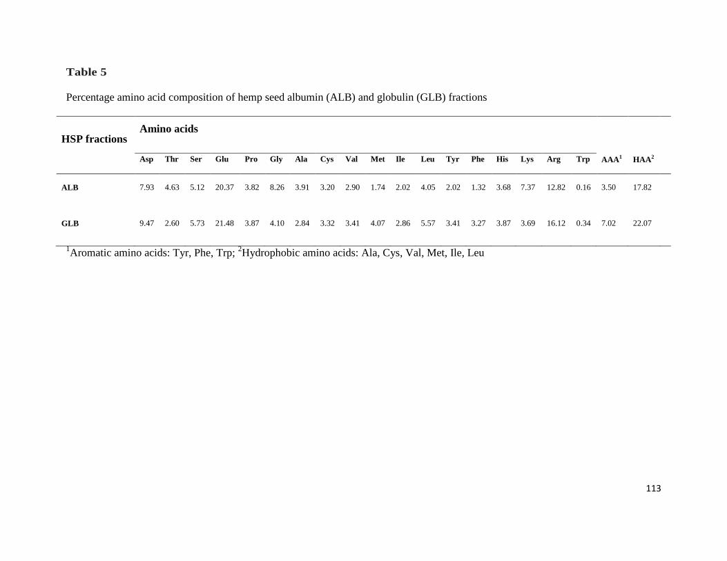
113
Table 5
Percentage amino acid composition of hemp seed albumin (ALB) and globulin (GLB) fractions
HSP fractions Amino acids
AAA1
HAA2
Asp Thr Ser Glu Pro Gly Ala Cys Val Met Ile Leu Tyr Phe His Lys Arg Trp
ALB 7.93 4.63 5.12 20.37 3.82 8.26 3.91 3.20 2.90 1.74 2.02 4.05 2.02 1.32 3.68 7.37 12.82 0.16 3.50 17.82
GLB 9.47 2.60 5.73 21.48 3.87 4.10 2.84 3.32 3.41 4.07 2.86 5.57 3.41 3.27 3.87 3.69 16.12 0.34 7.02 22.07
1Aromatic amino acids: Tyr, Phe, Trp;
2Hydrophobic amino acids: Ala, Cys, Val, Met, Ile, Leu
114
material (HPM, 40%), for instance, the protein concentration of ALB is 80% and that of GLB IS
100%. In contrast to their protein concentration, the protein yield of ALB (6.33%) is relatively
higher than that of GLB (6.07%). The low protein yields are a reflection of the harsh seed
processing condition (mechanical press) to extract edible oil. The applied strong mechanical
force could have caused severe protein-protein interactions and hence low solubility during
extraction.
4.3.2. SDS-PAGE
The non-reducing SDS-PAGE profiles (Fig. 9A) showed that ALB and GLB had seven
and three major polypeptides, respectively. For the ALB, the estimated molecular weights ranged
from 10 to 42 kDa while those for the globulin were estimated to be between 6 and 35 kDa.
Addition of β-ME did not have any substantial effect on the ALB polypeptide profile whereas
three intense bands with estimated sizes of 21, 34 and 45 kDa were present in the GLB (Fig. 9B).
The results suggest that the GLB protein had more disulfide bridges that link polypeptides of
different molecular sizes while the ALB had less. The presence of more disulfide bridges in the
GLB protein may also contribute to the poor SDS solubility and hence reduced polypeptide band
intensity as evident in Fig. 9A. By breaking the disulfide bonds, the β-ME converts the less
soluble proteins into smaller size polypeptides with increased solubility in the SDS buffer and
hence the higher band intensity obtained for GLB in Fig 9B. However, overall, the
electrophoresis separations indicate similar sizes (10-50 kDa) of polypeptides present in both
protein fractions.
4.3.3. Fluorescence emission spectra
Fig10 shows that the ALB protein exhibited a typical tyrosine-tyrosine interaction with a
major peak at a maximum emission wavelength (λ max) of 314 nm showing a characteristic
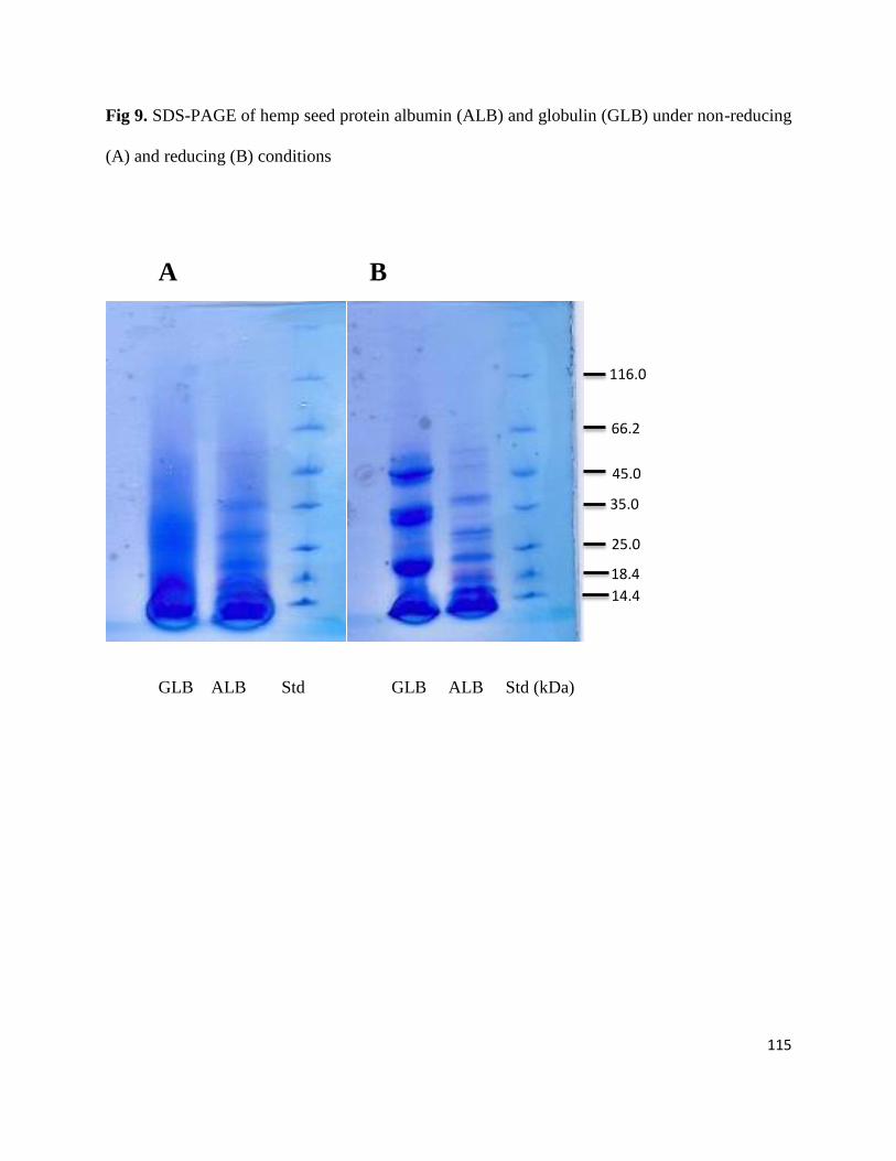
115
Fig 9. SDS-PAGE of hemp seed protein albumin (ALB) and globulin (GLB) under non-reducing
(A) and reducing (B) conditions
A B
GLB ALB Std GLB ALB Std (kDa)
14.4
25.0
18.4
35.0
45.0
66.2
116.0
0
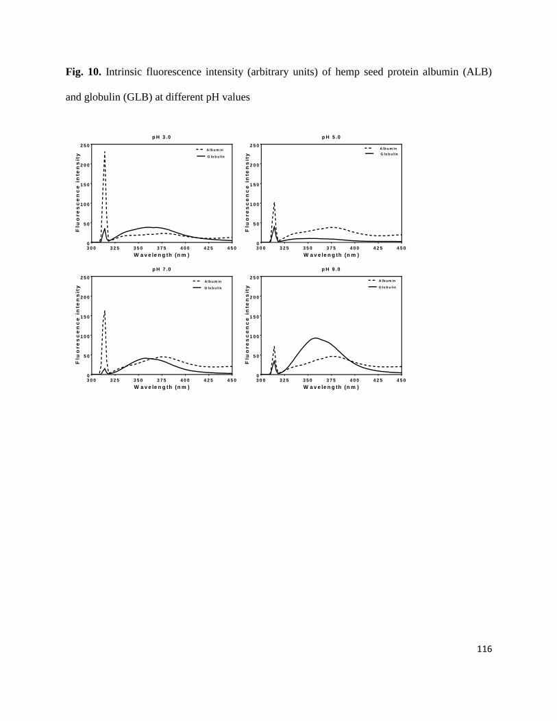
116
Fig. 10. Intrinsic fluorescence intensity (arbitrary units) of hemp seed protein albumin (ALB)
and globulin (GLB) at different pH values
p H 3 .0
W a v e le n g th (n m )
Flu
ore
sc
en
ce
in
te
ns
ity
3 0 0 3 2 5 3 5 0 3 7 5 4 0 0 4 2 5 4 5 00
5 0
1 0 0
1 5 0
2 0 0
2 5 0A lb u m in
G lo b u lin
p H 5 .0
W a v e le n g th (n m )
Flu
ore
sc
en
ce
in
te
ns
ity
3 0 0 3 2 5 3 5 0 3 7 5 4 0 0 4 2 5 4 5 00
5 0
1 0 0
1 5 0
2 0 0
2 5 0 A lb u m in
G lo b u lin
p H 7 .0
W a v e le n g th (n m )
Flu
ore
sc
en
ce
in
te
ns
ity
3 0 0 3 2 5 3 5 0 3 7 5 4 0 0 4 2 5 4 5 00
5 0
1 0 0
1 5 0
2 0 0
2 5 0 A lb u m in
G lo b u lin
p H 9 .0
W a v e le n g th (n m )
Flu
ore
sc
en
ce
in
te
ns
ity
3 0 0 3 2 5 3 5 0 3 7 5 4 0 0 4 2 5 4 5 00
5 0
1 0 0
1 5 0
2 0 0
2 5 0A lb u m in
G lo b u lin
117
fluorescence profile of tyrosine residues that are more and strongly exposed within a
hydrophobic environment (Yin et al., 2011). In contrast, the tyrosine residues in GLB were either
highly exposed in a hydrophilic environment or there was increased tyrosine-tryptophan energy
transfer as a result of intense protein-protein interactions. The latter effect seems more likely
judging from Table 5 that showed higher contents of aromatic and hydrophobic amino acids in
GLB, which could promote greater protein-protein interactions when compared to the ALB with
less amounts of these amino acids. The λmax of tyrosine residues obtained from this work (314
nm) is comparable to the 318 nm value reported by Yin et al. (2011) but higher than the 303 nm
reported by Mundi and Aluko (2013) for kidney bean globulins. In general, the tyrosine
environment in GLB did not respond to pH changes when compared to the ALB where
fluorescence intensity (FI) was less at pH 5.0 and 9.0. At pH 3.0 and 7.0, tryptophan
fluorescence (350-360 nm) indicates the residues were located in a polar environment, which
probably contributed to quenching and hence low and broad FI maximum. At pH 5.0, which is
near the isoelectric point of hemp seed proteins, tryptophan fluorescence was almost zero. This
indicates an almost complete shielding of the residues from excitation light; this result is
consistent with the fact that proteins experience intense protein-protein aggregation near the
isoelectric point. However, as the pH shifts away from the isoelectric point, the introduction of
net charges reduces protein-protein interactions, which contributes to increased exposure of
tryptophan residues and hence the observed higher FI especially at pH 9.0. The effect of
increased net pH is more apparent in the GLB at pH 9.0 where the tryptophan FI indicates a
more conserved structure within a hydrophobic environment, hence the blue shift in the λmax
when compared to the ALB protein. This means that the increased charge at pH 9.0 led to a
rearrangement in the GLB protein whereby the tryptophan residues were moved away from the
118
hydrophilic surface into a more hydrophobic interior and the higher FI is consistent with a higher
content of this residue (Table 5) when compared to the ALB. Thus the FI data indicates that the
GLB protein conformation was more globular in nature at pH 9.0 than at lower pH values.
However, the reduced tyrosine FI could also reflect greater closer proximity to tryptophan, which
increased tyrosine-tryptophan energy transfer. The values obtained from this work support the
report of Yin et al. (2011), which showed that the λmax of tryptophan residues experienced
significant shifts as buffer acidity reduced from pH 3.0 to 9.0; this confirms that the micro-
environment of tryptophan residues can be manipulated by environmental conditions. Overall,
the λmax value obtained for GLB (355 nm) is similar to the previously reported values of 354
and 360 nm for α-lactalbumin (Stanciuc, Aprodu, Rapeanu, & Bahrim, 2013; Stanciuc, Rapeanu,
Bahrim, & Aprodu, 2012) and kidney bean vicilin (Mundi & Aluko, 2013), respectively.
4.3.4. CD spectra
In this work, CD determination was not possible at pH 5.0 due to reduced protein
solubility which made required level of soluble proteins difficult to obtain. Far-UV spectra
showed that at pH 3.0 and 7.0, the ALB had more secondary structure conformation than GLB;
however, the conformation at pH 9.0 was similar for both proteins (Fig. 11). The secondary
structure was dominated mostly by the α-helix conformation as evident by the double minimum
ellipticity observed around 200-210 nm and 222 nm. At pH 3.0 and 7.0, the ALB showed
stronger ellipticity, suggesting the presence of a higher degree of α-helix conformation than the
GLB. At pH 9.0, there was an increased ellipticity for the GLB when compared to the pH 3.0 and
7.0 spectra, which suggests increased structural compactness (Kelly, Jess, & Price, 2005). The
pH 9.0 GLB CD spectra is supported by the intrinsic fluorescence data, which showed increased
FI at pH 9.0 and suggests translocation of the tryptophan residues away from the hydrophilic

119
Fig. 11. Far-UV circular dichroism spectra of hemp seed protein albumin (ALB) and globulin
(GLB) at different pH values
p H 3 .0
W a v e le n g th (n m )
Me
an
re
sid
ue
ell
iptic
ity
(d
eg
.cm
2.d
mo
l-1)
1 9 0 2 0 0 2 1 0 2 2 0 2 3 0 2 4 0
-8 0 0 0 0
-6 0 0 0 0
-4 0 0 0 0
-2 0 0 0 0
0
2 0 0 0 0
4 0 0 0 0A lb u m in
G lo b u lin
p H 7 .0
W a v e le n g th (n m )
Me
an
re
sid
ue
ell
iptic
ity
(d
eg
.cm
2.d
mo
l-1)
1 9 0 2 0 0 2 1 0 2 2 0 2 3 0 2 4 0
-8 0 0 0 0
-6 0 0 0 0
-4 0 0 0 0
-2 0 0 0 0
0
2 0 0 0 0
4 0 0 0 0A lb u m in
G lo b u lin
p H 9 .0
W a v e le n g th (n m )
Me
an
re
sid
ue
ell
iptic
ity
(d
eg
.cm
2.d
mo
l-1)
1 9 0 2 0 0 2 1 0 2 2 0 2 3 0 2 4 0
-8 0 0 0 0
-6 0 0 0 0
-4 0 0 0 0
-2 0 0 0 0
0
2 0 0 0 0
4 0 0 0 0A lb u m in
G lo b u lin
120
exterior into the hydrophobic interior. The results differ from the work of Mundi and Aluko
(2013) who reported that kidney bean globulins showed no defined secondary structure at pH
3.0. Our results also differ from the report of Yin et al. (2011) who showed that kidney bean
phaseolin had ellipticity minimum mostly at 216-218 nm, which indicates a predominance of β-
sheet structure. However, the present results are consistent with previous reports (Choi & Ma,
2007; Mundi & Aluko, 2013; Yin et al., 2011) that protein secondary structure conformation can
be altered as a result of pH-dependent variations in electrostatic interactions. Besides, data from
Table 6 confirmed the higher degree of α-helix conformation in ALB than the GLB at pH 3.0-
9.0. In contrast, the β-sheet contents in GLB were higher than that of ALB. Decrease in α-helix
contents and an increase of β-sheet structure contents at different pH, were also observed
between the heat-treated rapeseed protein isolates (He, He, Chao, Ju, & Aluko et al., 2014). This
supports the fact that processing conditions like pH and heat can cause alteration in the protein
secondary structure conformation. The unordered protein structure contents present in ALB were
higher than that of GLB (Table 6) and this might have an effect on their PS. For instance, the
higher contents of unordered protein in ALB might suggests a decreased structural compactness
in ALB which encourages its higher PS, while lower contents of unordered protein in GLB
causes its increased structural compactness, which could have contributed to the observed lower
PS as shown in Fig. 13.
The near-UV CD spectra show that the shape and magnitude of both protein fractions
were substantially affected by varying the pH conditions (Fig. 12). For instance, at pH 3.0, the
ALB had ellipticity minimum at 276 nm, which is consistent with tyrosine residues within a
hydrophobic environment; in contrast the GLB showed almost no ellipticity, which indicates
residues in a hydrophilic environment. The results are consistent with intrinsic fluorescence data
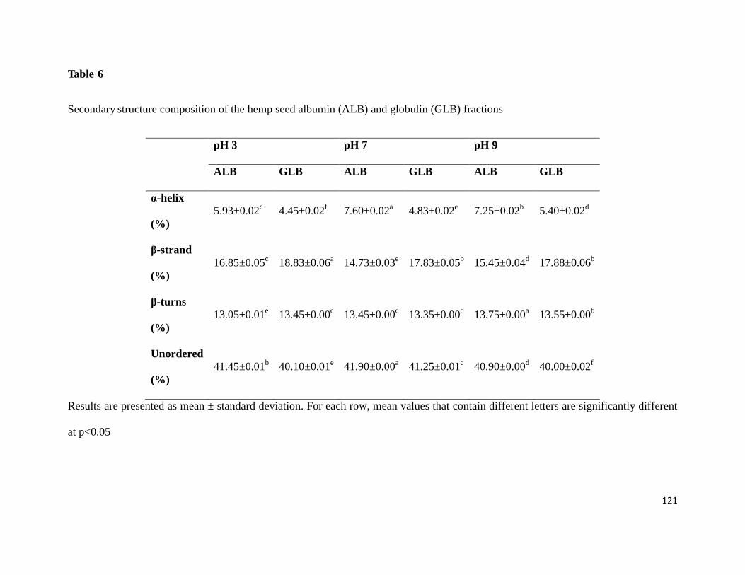
121
Table 6
Secondary structure composition of the hemp seed albumin (ALB) and globulin (GLB) fractions
pH 3 pH 7 pH 9
ALB GLB ALB GLB ALB GLB
α-helix
(%)
5.93±0.02c 4.45±0.02
f 7.60±0.02
a 4.83±0.02
e 7.25±0.02
b 5.40±0.02
d
β-strand
(%)
16.85±0.05c 18.83±0.06
a 14.73±0.03
e 17.83±0.05
b 15.45±0.04
d 17.88±0.06
b
β-turns
(%)
13.05±0.01e 13.45±0.00
c 13.45±0.00
c 13.35±0.00
d 13.75±0.00
a 13.55±0.00
b
Unordered
(%)
41.45±0.01b 40.10±0.01
e 41.90±0.00
a 41.25±0.01
c 40.90±0.00
d 40.00±0.02
f
Results are presented as mean ± standard deviation. For each row, mean values that contain different letters are significantly different
at p<0.05

122
Fig. 12. Near-UV circular dichroism spectra of hemp seed protein albumin (ALB) and globulin
(GLB) at different pH values
p H 3 .0
W a v e le n g th (n m )
Me
an
re
sid
ue
ell
ipti
cit
y
(de
g.c
m2
.dm
ol-1
)
2 5 0 2 6 0 2 7 0 2 8 0 2 9 0 3 0 0 3 1 0 3 2 0
-8 0 0
-7 0 0
-6 0 0
-5 0 0
-4 0 0
-3 0 0
-2 0 0
-1 0 0
0
1 0 0
2 0 0
3 0 0
A lb u m in
G lo b u lin
p H 7 .0
W a v e le n g th (n m )
Me
an
re
sid
ue
ell
ipti
cit
y
(de
g.c
m2
.dm
ol-1
)
2 5 0 2 6 0 2 7 0 2 8 0 2 9 0 3 0 0 3 1 0 3 2 0
-8 0 0
-7 0 0
-6 0 0
-5 0 0
-4 0 0
-3 0 0
-2 0 0
-1 0 0
0
1 0 0
2 0 0
3 0 0A lb u m in
G lo b u lin
p H 9 .0
W a v e le n g th (n m )
Me
an
re
sid
ue
ell
ipti
cit
y
(de
g.c
m2
.dm
ol-1
)
2 5 0 2 6 0 2 7 0 2 8 0 2 9 0 3 0 0 3 1 0 3 2 0
-8 0 0
-7 0 0
-6 0 0
-5 0 0
-4 0 0
-3 0 0
-2 0 0
-1 0 0
0
1 0 0
2 0 0
3 0 0A lb u m in
G lo b u lin
123
(Fig. 10) that showed tyrosine fluorescence was dominant in the ALB but almost absent in the
GLB. At pH 7.0, there was an increased GLB and ALB ellipticity when compared to pH 3.0,
which may be due to transfer of the aromatic amino acid residues into the more hydrophobic
interior as the exterior becomes more hydrophilic, which also conforms with the intrinsic
fluorescence data. This is reflected in an even greater ellipiticity values at pH 9.0 for both
proteins and a more prominent tyrosine peak for GLB, which are also consistent with the
intrinsic fluorescence data showing increased tryptophan emission. The results are supported by
the report of Kelly et al. (2005) who indicated that the actual shape and magnitude of the near-
UV CD spectrum of a protein is dependent on various factors like the number of each type of
aromatic amino acid present, their mobility, the nature of their environment (H-bonding, polar
groups and polarizability) and their spatial disposition in the protein.
4.3.5. Protein solubility
The effective utilization of proteins in various novel food applications is highly
dependent on solubility (Karaca et al., 2011). Protein solubility occurs as a result of
intermolecular repulsion caused by electrostatic interactions which ionizes the interior non-polar
groups of a protein, disrupts its native structure through polypeptide chain unfolding and
subsequently leads to exposure of buried functional groups (Bora, 2002). While ALB had
minimum PS at pH 3.0, the minimum PS for GLB was observed at pH 5.0 (Fig. 13). The PS
pattern for GLB is consistent with previous observations showing that legume globulins have
minimum solubility at pH values near the isoelectric point; solubility then increases as pH values
move away from the isoelectric point (Mundi & Aluko, 2012; Sai-Ut, Ketnawa, & Rawdkuen,
2009; Ragab, Babiker, & Eltinay, 2004; Yin et al., 2010). This solubility pattern is due to
increased protein-protein interactions at the isoelectric point, but which gradually reduces in
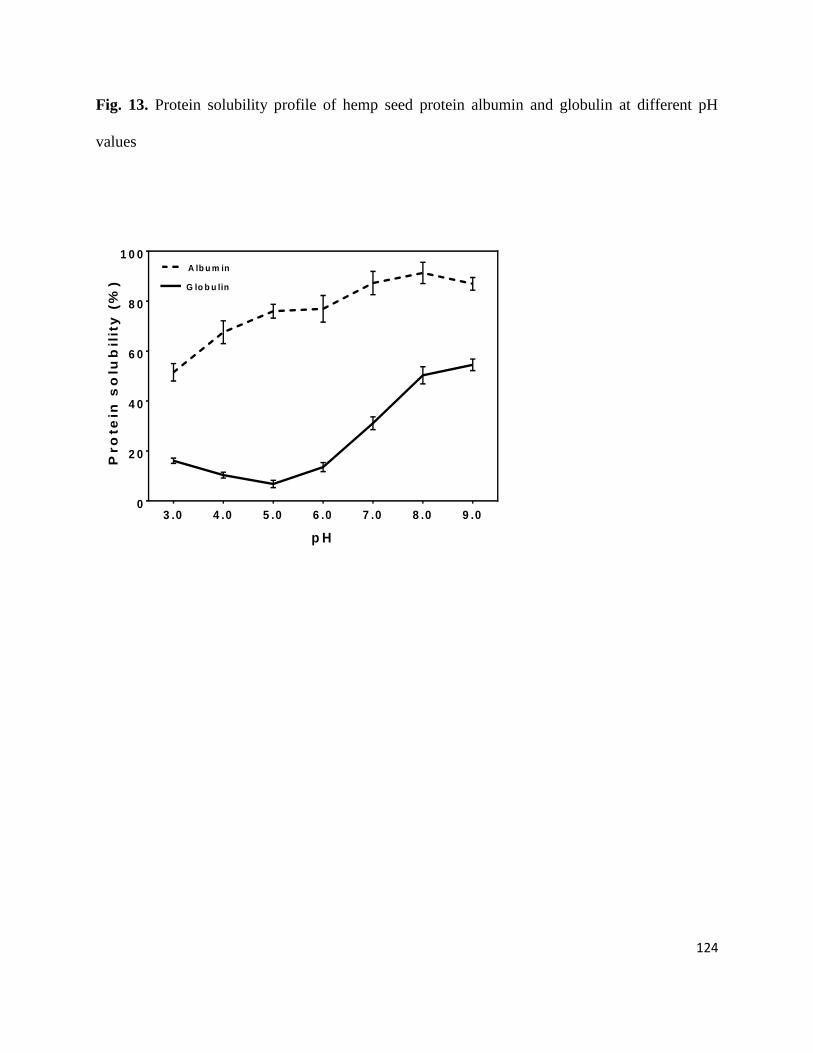
124
Fig. 13. Protein solubility profile of hemp seed protein albumin and globulin at different pH
values
p H
Pro
te
in s
olu
bil
ity
(%
)
3 .0 4 .0 5 .0 6 .0 7 .0 8 .0 9 .00
2 0
4 0
6 0
8 0
1 0 0A lb u m in
G lo b u lin
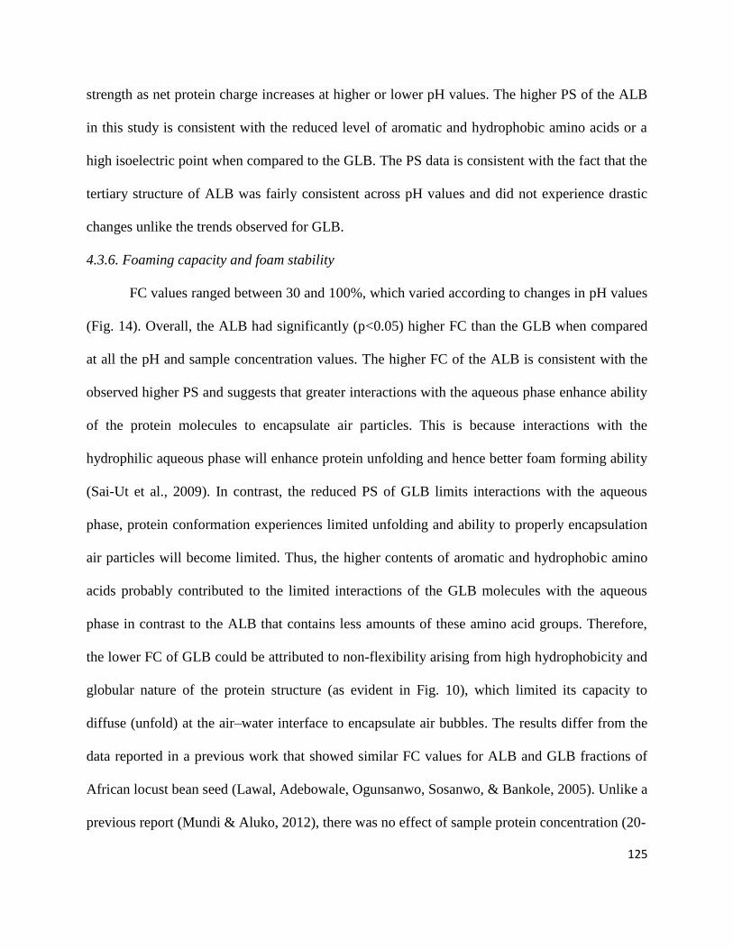
125
strength as net protein charge increases at higher or lower pH values. The higher PS of the ALB
in this study is consistent with the reduced level of aromatic and hydrophobic amino acids or a
high isoelectric point when compared to the GLB. The PS data is consistent with the fact that the
tertiary structure of ALB was fairly consistent across pH values and did not experience drastic
changes unlike the trends observed for GLB.
4.3.6. Foaming capacity and foam stability
FC values ranged between 30 and 100%, which varied according to changes in pH values
(Fig. 14). Overall, the ALB had significantly (p<0.05) higher FC than the GLB when compared
at all the pH and sample concentration values. The higher FC of the ALB is consistent with the
observed higher PS and suggests that greater interactions with the aqueous phase enhance ability
of the protein molecules to encapsulate air particles. This is because interactions with the
hydrophilic aqueous phase will enhance protein unfolding and hence better foam forming ability
(Sai-Ut et al., 2009). In contrast, the reduced PS of GLB limits interactions with the aqueous
phase, protein conformation experiences limited unfolding and ability to properly encapsulation
air particles will become limited. Thus, the higher contents of aromatic and hydrophobic amino
acids probably contributed to the limited interactions of the GLB molecules with the aqueous
phase in contrast to the ALB that contains less amounts of these amino acid groups. Therefore,
the lower FC of GLB could be attributed to non-flexibility arising from high hydrophobicity and
globular nature of the protein structure (as evident in Fig. 10), which limited its capacity to
diffuse (unfold) at the air–water interface to encapsulate air bubbles. The results differ from the
data reported in a previous work that showed similar FC values for ALB and GLB fractions of
African locust bean seed (Lawal, Adebowale, Ogunsanwo, Sosanwo, & Bankole, 2005). Unlike a
previous report (Mundi & Aluko, 2012), there was no effect of sample protein concentration (20-

126
Fig. 14. Foam capacity of hemp seed albumin and globulin in water and at different pH values:
A, 20 mg/mL; B, 40 mg/mL; and C, 60 mg/mL protein concentrations (*Bars that contain
different letters are significantly different at p<0.05)
p H
Fo
am
Ca
pa
cit
y (
%)
3 .0 5 .0 7 .0 9 .00
2 0
4 0
6 0
8 0
1 0 0A lb u m in
G lo b u lin
d
b a
e
c
d e d e
b
A
p H
Fo
am
Ca
pa
cit
y (
%)
3 .0 5 .0 7 .0 9 .00
2 0
4 0
6 0
8 0
1 0 0 A lb u m in
G lo b u lin
c
c d
a a ab
c dc
B
p H
Fo
am
Ca
pa
cit
y (
%)
3 .0 5 .0 7 .0 9 .00
2 0
4 0
6 0
8 0
1 0 0 A lb u m in
G lo b u lin
aa
c
a
e
b
de
C
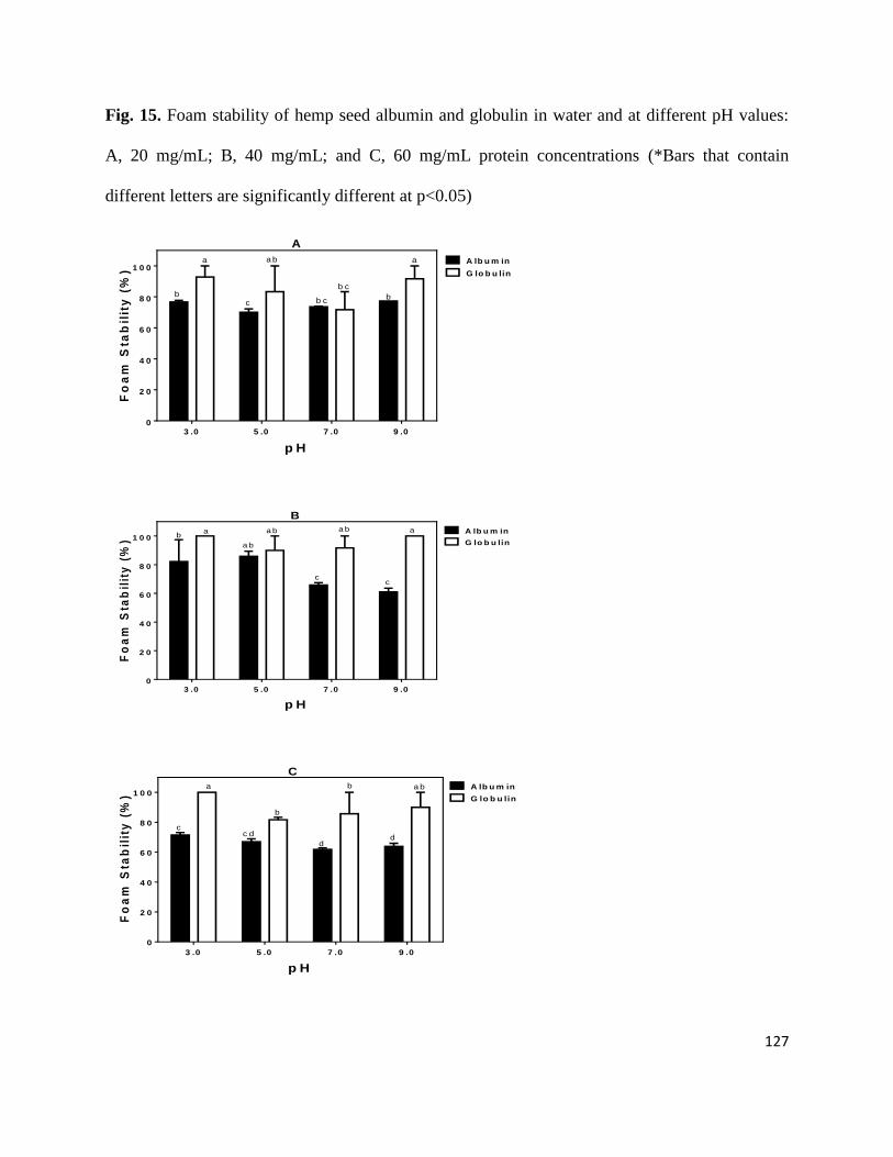
127
Fig. 15. Foam stability of hemp seed albumin and globulin in water and at different pH values:
A, 20 mg/mL; B, 40 mg/mL; and C, 60 mg/mL protein concentrations (*Bars that contain
different letters are significantly different at p<0.05)
p H
Fo
am
Sta
bil
ity
(%
)
3 .0 5 .0 7 .0 9 .00
2 0
4 0
6 0
8 0
1 0 0A lb u m in
G lo b u lin
a
bc
a b
b c
b c
a
b
A
p H
Fo
am
Sta
bil
ity
(%
)
3 .0 5 .0 7 .0 9 .00
2 0
4 0
6 0
8 0
1 0 0A lb u m in
G lo b u lina b
c
aa b
c
a bb
a
B
p H
Fo
am
Sta
bil
ity
(%
)
3 .0 5 .0 7 .0 9 .0
0
2 0
4 0
6 0
8 0
1 0 0A lb u m in
G lo b u lin
a b
d
b
d
c d
a
c
b
C
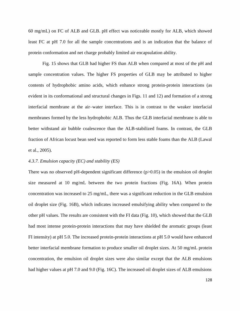
128
60 mg/mL) on FC of ALB and GLB. pH effect was noticeable mostly for ALB, which showed
least FC at pH 7.0 for all the sample concentrations and is an indication that the balance of
protein conformation and net charge probably limited air encapsulation ability.
Fig. 15 shows that GLB had higher FS than ALB when compared at most of the pH and
sample concentration values. The higher FS properties of GLB may be attributed to higher
contents of hydrophobic amino acids, which enhance strong protein-protein interactions (as
evident in its conformational and structural changes in Figs. 11 and 12) and formation of a strong
interfacial membrane at the air–water interface. This is in contrast to the weaker interfacial
membranes formed by the less hydrophobic ALB. Thus the GLB interfacial membrane is able to
better withstand air bubble coalescence than the ALB-stabilized foams. In contrast, the GLB
fraction of African locust bean seed was reported to form less stable foams than the ALB (Lawal
et al., 2005).
4.3.7. Emulsion capacity (EC) and stability (ES)
There was no observed pH-dependent significant difference (p>0.05) in the emulsion oil droplet
size measured at 10 mg/mL between the two protein fractions (Fig. 16A). When protein
concentration was increased to 25 mg/mL, there was a significant reduction in the GLB emulsion
oil droplet size (Fig. 16B), which indicates increased emulsifying ability when compared to the
other pH values. The results are consistent with the FI data (Fig. 10), which showed that the GLB
had most intense protein-protein interactions that may have shielded the aromatic groups (least
FI intensity) at pH 5.0. The increased protein-protein interactions at pH 5.0 would have enhanced
better interfacial membrane formation to produce smaller oil droplet sizes. At 50 mg/mL protein
concentration, the emulsion oil droplet sizes were also similar except that the ALB emulsions
had higher values at pH 7.0 and 9.0 (Fig. 16C). The increased oil droplet sizes of ALB emulsions

129
Fig. 16. Oil droplet sizes of emulsions formed by hemp seed albumin and globulin in water and at
different pH values: A, 10 mg/mL; B, 25 mg/mL; and C, 50 mg/mL protein concentrations (*Bars that
contain different letters are significantly different at p<0.05)
p H
Oil
dro
ple
t s
ize
(d
3,2
) -
M
3 .0 5 .0 7 .0 9 .00 .0
0 .2
0 .4
0 .6
0 .8
1 .0
A lb u m in
G lo b u lin
aaa a
b
aa a
B
p H
Oil
dro
ple
t s
ize
(d
3,2
) -
M
3 .0 5 .0 7 .0 9 .00 .0
0 .2
0 .4
0 .6
0 .8
1 .0
A lb u m in
G lo b u lin
bb
a
bb cb c
a
b
C
p H
Oil
dro
ple
t s
ize
(d
3,2
) -
M
3 .0 5 .0 7 .0 9 .00 .0
0 .2
0 .4
0 .6
0 .8
1 .0
A lb u m in
G lo b u lin
A
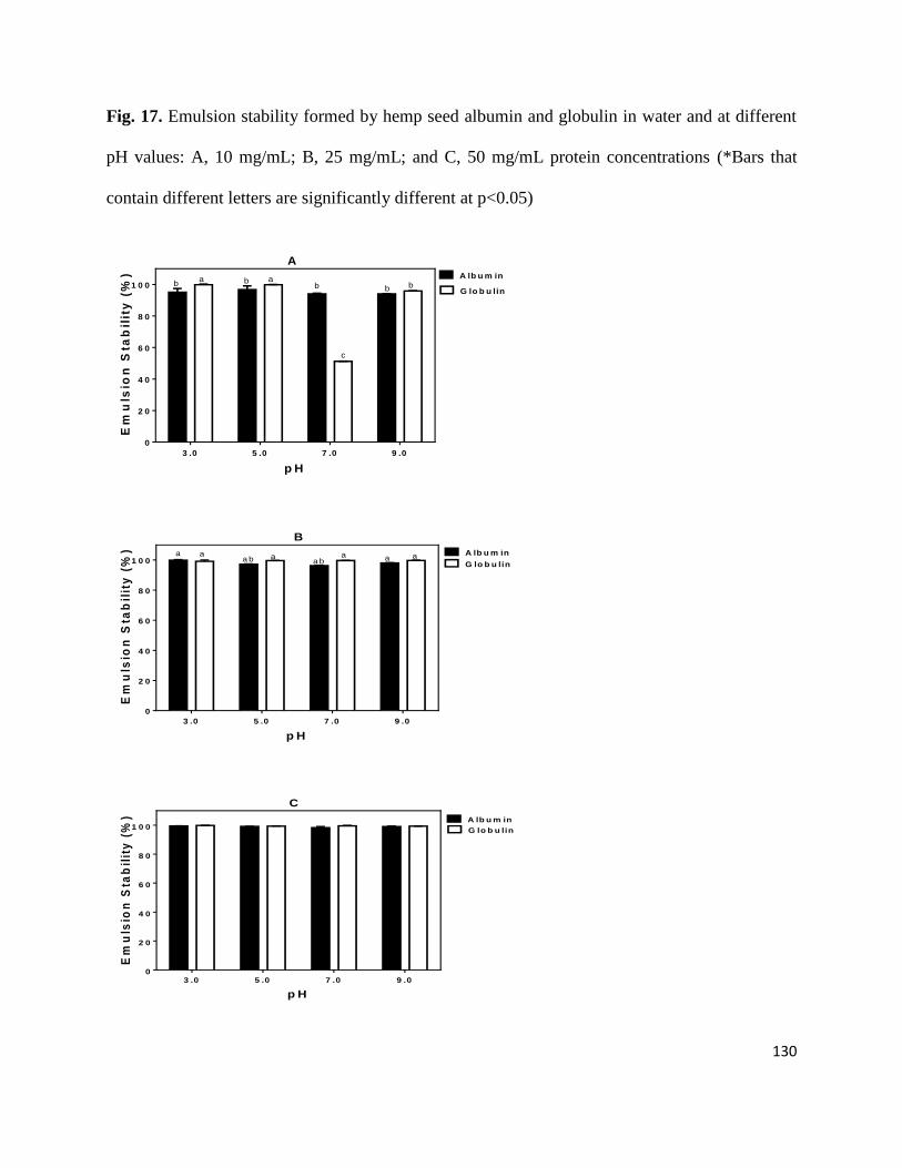
130
Fig. 17. Emulsion stability formed by hemp seed albumin and globulin in water and at different
pH values: A, 10 mg/mL; B, 25 mg/mL; and C, 50 mg/mL protein concentrations (*Bars that
contain different letters are significantly different at p<0.05)
p H
Em
uls
ion
Sta
bil
ity
(%
)
3 .0 5 .0 7 .0 9 .0
0
2 0
4 0
6 0
8 0
1 0 0
A lb u m in
G lo b u linbb
c
baba
b
A
p H
Em
uls
ion
Sta
bil
ity
(%
)
3 .0 5 .0 7 .0 9 .0
0
2 0
4 0
6 0
8 0
1 0 0A lb u m in
G lo b u linaa ba b
a a a a a
B
p H
Em
uls
ion
Sta
bil
ity
(%
)
3 .0 5 .0 7 .0 9 .00
2 0
4 0
6 0
8 0
1 0 0A lb u m in
G lo b u lin
C
131
may be attributed to higher net charge effects, which would have reduced ability of the protein
molecules to form strong interfacial membranes (due to increased protein-protein repulsions) at
neutral and alkaline pH values. The increased protein concentration would have also contributed
to the reduced emulsifying ability of ALB at pH 7.0 and 9.0 since there will be more charged
protein molecules in solution when compared to lower protein concentrations. Overall, both
protein fractions formed emulsions with small particle sizes similar to those reported for dairy
whey protein isolate (Taherian, Britten, Sabik, & Fustier, 2011) and indicate better emulsifiers
when compared to other plant proteins (Aluko, Mofolasayo, & Watts, 2009; Li, Kong, Zhang, &
Hua, 2011).
The results presented in Fig 17 showed that differences in ES was mostly dependent on
protein concentration; there were less differences as sample concentration increased from 10 to
50 mg/mL. At 10 mg/mL, the GLB emulsions were the most stable at pH 3.0 and 5.0 but there
were no differences between the ALB emulsions (Fig. 17A). The low ES of the GLB emulsion at
pH 7.0 indicates formation of very weak interfacial membrane, which was less resistant
coalescence when compared to the other pH values. At 25 and 50 mg/mL, the GLB emulsions
had slightly higher stability but there were no significant differences. Therefore, it is possible
that the effects of differences in molecular structure were minimized by the higher protein
contents used to form the emulsions. The increased protein concentrations (25 and 50 mg/mL)
would have contributed to formation of thicker interfacial membranes and hence higher ES when
compared to the 10 mg/mL emulsions. The results are in contrast to previous reports (Deng et al.,
2011; Chavan, McKenzie, & Shahidi, 2001) that demonstrated reduced ES at low pH values due
to increased interactions between the emulsified droplets as a result of decreased net charge on
the proteins.
132
4.4. Conclusion
The differences in functionalities of hemp seed ALB and GLB protein fractions may be
attributed to variations in amino acid composition and pH-dependent changes in structural
conformation. Polypeptide composition indicates the GLB has more disulfide bonded proteins,
which produced a rigid structure with reduced exposure of aromatic amino acids when compared
to the ALB. The higher level of hydrophobic amino acids and the rigid conformational structure
in GLB contributed to decreased PS and FC, which confirms the dependence of these functional
properties flexibility and ability of proteins to interact with the aqueous phase. In contrast,
emulsion-forming ability was not dependent on PS, which suggests that different mechanisms
are involved in foam and emulsion formations. Both protein fractions are potentially good
ingredients for the manufacture of food emulsions but only the ALB may find role in the
manufacture of food foams.
Acknowledgement
This work was funded by the Natural Sciences and Engineering Research Council of Canada
(NSERC) Discovery grant and a Manitoba Agri-Food Research and Development Initiative
(ARDI) grant to Dr. R.E. Aluko. S.A. Malomo is a recipient of University of Manitoba Graduate
Fellowship (UMGF) and Manitoba Graduate Scholarship (MGS) Awards for doctoral studies.
References
Adebiyi, A. P., & Aluko, R. E. (2011). Functional properties of protein fractions obtained from
commercial yellow field pea (Pisum sativum L.) seed protein isolate. Food Chemistry,
128, 902-908.
133
Aluko, R. E. (2004). The extraction and purification of proteins: An introduction. In R. Y. Yada
(Ed.), Proteins in food processing (pp. 323–346). Woodhead Publishing Limited and
CRC Press LLC.
Aluko, R. E., Mofolasayo, O. A., & Watts, B. A. (2009). Emulsifying and foaming properties of
commercial yellow pea (Pisum sativum L.) seed flours. Journal of Agricultural and Food
Chemistry, 57, 9793-9800.
Aluko, R. E., & McIntosh, T. (2004). Electrophoretic and functional properties of mustard seed
meals and protein concentrates. Journal of the American Oil Chemists Society, 81, 679-
683.
Avramenko, N. A., Low, N. H., & Nickerson, M. T. (2013). The effects of limited enzymatic
hydrolysis on the physicochemical and emulsifying properties of a lentil protein isolate.
Food Research International, 51, 162-169.
Barac, M. B., Pesic, M. B., Stanojevic, S. P., Kostic, A. Z., & Bivolarevic, V. (2014).
Comparative study of the functional properties of three legume seed isolates: adzuki, pea
and soy bean. Journal of Food Science & Technology, DOI 10.1007/s13197-014-1298-6,
1-9.
Bidlingmeyer, B. A., Cohen, S. A., & Tarvin, T. L. (1984). Rapid analysis of amino acids using
pre-column derivatization. Journal of Chromatography B: Biomedical Sciences and
Applications 336, 93-104.
Bora, P. S. (2002). Functional properties of native and succinylated lentil (Lens culinaris)
globulins. Food Chemistry, 77, 171-176.
Chavan, U. D., McKenzie, D. B., & Shahidi, F. (2001). Functional properties of protein isolates
from beach pea (Lathyrus maritimus L.). Food Chemistry, 74, 177-187.
134
Choi, S. M., & Ma, C. Y. (2007). Structural characterization of globulin from common
buckwheat (Fagopyrum esculentum Moench) using circular dichroism and Raman
spectroscopy. Food Chemistry, 102, 150-160.
Deng, Q. C., Wang, L., Wei, F., Xie, B. J., Huang, F. H., Huang, W., Shi, J., Huang, Q. D., Tian,
B. Q., & Xue, S. (2011). Functional properties of protein isolates, globulin and albumin
extracted from Ginkgo biloba seeds. Food Chemistry, 124, 1458-1465.
Gehrke, C. W., Wall, L. L., Absheer, J. S., Kaiser, F. E., & Zumwalt, R. W. (1985). Sample
preparation for chromatography of amino-acids: acid hydrolysis of proteins. Journal of
Association of Official Analytical Chemists, 68, 811-821.
Girgih, A. T., He, R., Malomo, S. A., & Aluko, R. E. (2014). Structural and functional
characterization of hemp seed (Cannabis sativa L.) protein-derived antioxidant and
antihypertensive peptides. Journal of Functional Foods, 6, 384-394.
Girgih, A. T., Udenigwe, C. C., & Aluko, R. E. (2011). In vitro antioxidant properties of hemp
seed (Cannabis sativa L.) protein hydrolysate fractions. Journal of the American Oil
Chemists Society, 88, 381-389.
Giroux, I., Kurowska, E. M., Freeman, D. J., & Carroll, K. K. (1999). Addition of arginine but
not glycine to lysine plus methionine–enriched diets modulates serum cholesterol and
liver phospholipids in rabbits. Journal of Nutrition, 129, 1807-1813.
He, R., He, H., Chao, D., Ju, X., & Aluko, R. (2014). Effects of high ressure and heat treatments
on physicochemical and gelation properties of rapeseed protein isolate. Food Bioprocess
Technology, 7, 1344–1353.
135
He, X. H., Liu, H. Z., Liu, L., Zhao, G. L., Wang, Q., & Chen, Q. L. (2014). Effects of high
pressure on the physicochemical and functional properties of peanut protein isolates.
Food Hydrocolloids, 36, 123-129.
Karaca, A. C., Low, N., & Nickerson, M. (2011). Emulsifying properties of canola and flaxseed
protein isolates produced by isoelectric precipitation and salt extraction. Food Research
International, 44, 2991-2998.
Kelly, S. M., Jess, T. J., & Price, N. C. (2005). How to study proteins by circular dichroism.
Biochimica et Biophysica Acta-Proteins and Proteomics, 1751, 119-139.
Landry, J., & Delhaye, S. (1992). Simplified procedure for the determination of tryptophan of
foods and feedstuffs from barytic hydrolysis. Journal of Agricultural and Food
Chemistry, 40, 776-779.
Lawal, O. S., Adebowale, K. O., Ogunsanwo, B. M., Sosanwo, O. A., & Bankole, S. A. (2005).
On the functional properties of globulin and albumin protein fractions and flours of
African locust bean (Parkia biglobossa). Food Chemistry, 92, 681-691.
Li, H., & Aluko, R. E. (2006). Structural modulation of calmodulin and calmodulin-dependent
protein kinase II by pea protein hydrolysates. International Journal of Food Sciences and
Nutrition, 57, 178-189.
Li, F., Kong, X., Zhang, C., & Hua, Y. (2011). Effect of heat treatment on the properties of soy
protein-stabilised emulsions. International Journal of Food Science and Technology, 46,
1554–1560.
Lu, R. R., Qian, P., Sun, Z., Zhou, X. H., Chen, T. P., He, J. F., Zhang, H., & Wu, J. (2010).
Hempseed protein derived antioxidative peptides: Purification, identification and
136
protection from hydrogen peroxide-induced apoptosis in PC12 cells. Food Chemistry,
123, 1210-1218.
Markwell, M. A. K., Haas, S. M., Bieber, L. L., & Tolbert, N. E. (1978). Modification of lowry
procedure to simplify protein determination in membrane and lipoprotein samples.
Analytical Biochemistry, 87, 206-210.
Mundi, S., & Aluko, R. E., (2013). Effects of NaCl and pH on the structural conformations of
kidney bean vicilin. Food Chemistry, 139, 624-630.
Mundi, S., & Aluko, R. E. (2012). Physicochemical and functional properties of kidney bean
albumin and globulin protein fractions. Food Research International, 48, 299– 306.
Oduro-yeboah, C., Onwulata, C., Tortoe, C. & Thomas-Gahring, A. (2014). Functional
properties of plantain, cowpea flours and oat fiber in extruded products. Journal of Food
Processing and Preservation, 38, 347–355.
Omoni, A. O., & Aluko, R. E. (2006). Effect of cationic flaxseed protein hydrolysate fractions on
the in vitro structure and activity of calmodulin-dependent endothelial nitric oxide
synthase. Molecular Nutrition and Food Research, 50, 958-966.
Park, S. K., Seo, J. B., & Lee, M. Y. (2012). Proteomic profiling of hempseed proteins from
Cheungsam. Biochimica et Biophysica Acta-Proteins and Proteomics, 1824, 374-382.
Ragab, D. D.M., Babiker, E. E., & Eltinay, A. H. (2004). Fractionation, solubility and functional
properties of cowpea (Vigna unguiculata) proteins as affected by pH and/or salt
concentration. Food Chemistry, 84, 207–212.
Sai-Ut, S., Ketnawa, P., & Rawdkuen, S. (2009). Biochemical and functional properties of
proteins from red kidney, navy and adzuki beans. Asian Journal of Food and Agro-
Industry, 2, 493–504.
137
Shevkani, K., Singh, N., Rana, J. C., & Kaur, A. (2014). Relationship between physicochemical
and functional properties of amaranth (Amaranthus hypochondriacus) protein isolates.
International Journal of Food Science and Technology, 49, 541-550.
Stanciuc, N., Aprodu, I., Rapeanu, G., & Bahrim, G. (2013). pH- and heat-induced structural
changes of bovine alpha-lactalbumin in response to oleic acid binding. European Food
Research and Technology, 236, 257-266.
Stanciuc, N., Rapeanu, G., Bahrim, G., & Aprodu, I. (2012). pH and heat-induced structural
changes of bovine apo alpha-lactalbumin. Food Chemistry, 131, 956-963.
Taherian, A. R., Britten, M., Sabik, H., & Fustier, P. (2011). Ability of whey protein isolate
and/or fish gelatin to inhibit physical separation and lipid oxidation in fish oil-in-water
beverage emulsion. Food Hydrocolloids, 25, 868-878.
Tan, S. H., Mailer, R. J., Blanchard, C. L. & Agboola, S. O. (2014). Emulsifying properties of
proteins extracted from Australian canola meal. LWT - Food Science and Technology, 57,
376-382.
Tan, E., Ngoh, Y. & Gan, C. (2014). A comparative study of physicochemical characteristics and
functionalities of pinto bean protein isolate (PBPI) against the soybean protein isolate
(SPI) after the extraction optimization. Food Chemistry, 152, 447–455.
Tang, C-H., Sun, X., & Foegeding, E. A. (2011). Modulation of physicochemical and
conformational properties of kidney bean vicilin (Phaseolin) by glycation with glucose:
implications for structure-function relationships of legume vicilins. Journal of
Agricultural and Food Chemistry, 59, 10114-10123.
138
Tang, C-H., Ten, Z., Wang, X-S., & Yang, X-Q. (2006). Physicochemical and functional
properties of hemp (Cannabis sativa L.) protein isolate. Journal of Agricultural and Food
Chemistry, 54, 8945-8950.
Teh, S. S., & Birch, J. (2013). Physicochemical and quality characteristics of cold-pressed hemp,
flax and canola seed oils. Journal of Food Composition and Analysis, 30, 26-31.
Teh, S. S., Bekhit, A. E. D., Carne, A. & Birch, J. (2013). Effect of the defatting process, acid
and alkali extraction on the physicochemical and functional properties of hemp, flax and
canola seed cake protein isolates. Food Measure, DOI 10.1007/s11694-013-9168-x, 1-13.
Wang, X-S., Tang, C-H., Chen, L., Yang, X-Q. (2009). Characterization and antioxidant
properties of hemp protein hydrolysate obtained with neutrase. Food Technology and
Biotechnology, 47, 428–434.
Yin, S-W., Huang, K-L., Tang, C-H., Yang, X-Q., Wen, Q-B., & Qi, J-R. (2011). Surface charge
and conformational properties of phaseolin, the major globulin in red kidney bean
(Phaseolus vulgaris L): effect of pH. International Journal of Food Science and
Technology, 46, 1628-1635.
Yin, S-W., Tang, C-H., Cao, J-S., Hu, E-K., Wen, Q-B., Yang, X-Q. (2008). Effects of limited
enzymatic hydrolysis with trypsin on functional properties of hemp (Cannabis sativa L.)
protein isolate. Food Chemistry, 106, 1004–1013.
Yin, S-W., Tang, C-H.,Wen, Q. B., & Yang, X-Q. (2010). Functional and conformational
properties of phaseolin (Phaseolus vulgris L.) and kidney bean protein isolate: A
comparative study. Journal of the Science of Food and Agriculture, 90, 599–607.
139
4.5 Statement transfer between the study 2 and study 3
The conventional dialysis process used in the preceding chapter allowed the study of pure protein
fractions but production yield coupled with the isolation method (dialysis) may not be
compatible with practical commercialization. Therefore, the next chapter deals with conversion
of a waste by-product from hemp seed processing industry into protein isolates using processes
that could be readily scaled-up for commercialization. The product will have high solubility
properties and better functionalities when compared with protein isoelectric pH-precipitated
protein isolates and isolated protein fractions. The starting raw material is a non-functional hemp
seed meal residue, which is characterized by low protein content, high phytate and high fibre.
The hemp seed meal residue is normally dumped in landfills or used as a source of fertilizer on
farms. Therefore, a method was developed to remove some of the fibre and phytate components
of the meal residue in order to leave behind a product with higher protein content and improved
functionality in foods. This involved the use of an enzyme (carbohydrase and phytase) pre-
digestion method to breakdown the fibres (sugars) and phytate into low molecular weight (<10
kDa) digests, which are then removed through membrane filtration process. The modified protein
is expected to have entirely different functionality (even when added in small amounts to food
products) when compared with parent protein (meal) and isolates from the other methods of
isolation. It is believed that isolate from this enzyme pre-digestion cum membrane filtration
method can be used in novel food processing and add value to the hemp processing industry.
140
CHAPTER FIVE
5. Conversion of a low protein hemp seed meal into a functional protein concentrate
through enzymatic digestion of fibre coupled with membrane ultrafiltration
Sunday A. Malomo and Rotimi E. Aluko*
The Department of Human Nutritional Sciences, University of Manitoba, Winnipeg, Manitoba,
Canada R3T 2N2
*Corresponding author: Dr. Rotimi E. Aluko; E-Mail: [email protected]; Tel.: +1-204-
474-9555; Fax: +1-204-474-7593.
141
Abstract
Industrial hemp seed protein meal (HPM) containing ~37% protein content was digested with
carbohydrases and a phytase. The digest was then passed through a 10 kDa ultrafiltration
membrane system and the retentate freeze-dried as the membrane hemp seed protein concentrate
(mHPC), which had 74% protein content. Functional properties of the mHPC was compared with
a commercial hemp seed protein concentrate (cHPC, 70% protein) and a traditional protein
isolate produced by isoelectric pH precipitation (iHPI, 84% protein). Four main polypeptides
(14, 24, 34, and 47 kDa) were most apparent under non-reducing gel electrophoresis, but the
mHPC has less intensity of the 24 and 34 kDa bands. There was reduced intensity of the 47 kDa
band in the presence of mercaptoethanol, which indicates the protein contains disulfide bonds.
The mHPC had the highest contents of essential amino acids as well as branched-chain amino
acids among the hemp seed products. Protein digestibility (89%) was significantly (P<0.05)
higher than the ~85% obtained for the HPM, iHPI, and cHPC. The mHPC also had significantly
(P<0.05) higher protein solubility than other hemp products in the pH 3-9 range with a minimum
of 76% at pH 4.0. In contrast the HPM and iHPI had <25% maximum protein solubility with
minimum of 7.5% and 0.35% solubility, respectively. Foaming capacity was highest for mHPC
(55-98%) when compared to 10-70% for the other protein products. However, emulsions formed
by mHPC and cHPC had bigger oil droplet sizes (4.5-15.5 µm) when compared with <1 µm for
HPM and iHPI emulsions. The excellent and improved functional properties for mHPC suggest
that the mHPC could be used for the nutritional fortification of low protein foods and drinks (for
example fruit juices).
Keywords: hemp seed; protein solubility and digestibility; protein isolate; membrane
ultrafiltration; foaming; emulsion
142
5.1. Introduction
The increased utilization of hemp plant for various industrial purposes has led to
abundant amounts of protein-rich seed residue, which can be converted into various forms of
powdered protein products. In Canada, hemp seed processing primarily involves the cold-
pressing method to expel oil and leave behind a high protein (30-50%) residue/meal (Malomo et
al., 2014). The hemp seed protein (HSP) products have been shown as promising raw materials
for the production of peptides with potential human health applications (Girgih et al., 2014a,
2014b). However, the hemp seed protein materials are poorly utilised in applications with added
value. For instance, the current commercial HSP products are mainly in the form of protein
concentrates that have less than 70% protein content and contain high levels of fibre and phytate
(Lu et al., 2010). The high fibre and phytate contents of these HSP products have been reported
to cause protein functionality reduction, which limits their use as new food ingredients (Yin et
al., 2009).
Moreover, current HSP isolates are generally prepared by alkaline extraction, which is
then followed by isoelectric precipitation and is the most commonly used traditional procedure
for plant protein isolate production (Malomo et al., 2014). The harsh conditions used in the
traditional procedure have negative effects on protein functionalities, especially protein solubility
and foaming properties (Liu et al., 2013). The poor food functionality of current HSP products
can also be attributed to the high phytate content, which can cause protein cross-linking and
reduce protein solubility. Therefore, in order to produce novel protein isolates with improved
food functionalities, a method that involves fibre and phytate digestion followed by removal of
non-protein materials by membrane filtration was developed. The use of membrane ultrafiltration
obviates the need for harsh protein precipitation protocols while ensuring high yield of proteins
143
with minimal conformational denaturation. Recent studies have confirmed the successful
application of this novel method of protein isolate production with high quality properties. For
instance, Wang et al. (2014) reported a soy protein isolate with higher protein contents (91-93%)
and better in vitro digestibility from phytase-assisted processing method. A better functional
(foam capacity) protein isolates with high protein content (92%) and yield (75%) was produced
from rice bran using a combination of xylanase and phytase pre-digestion process (Wang et al.,
1999). Another study (Udenigwe et al., 2009) had shown that pre-digestion of the fibre with
cellulase resulted in up to 23% higher protein content of flaxseed protein isolate. However, pre-
digestion with carbohydrases and phytases coupled to membrane ultrafiltration is novel and has
not been previously reported for HSP isolate production.
During industrial hemp seed processing, the defatted meal is usually milled and passed
through several sieves to obtain high-value protein products. The milling and sieving process
leaves behind a high-fibre waste product that is normally dumped in farms as a nitrogen source
or simply discarded in landfills. Therefore, this study aimed to convert this high-fibre but low
protein industrial side-stream of hemp seed processing into a high protein product through
enzymatic digestion of the main non-protein materials (fibre and phytate) followed by removal
of the low molecular weight digests by membrane ultrafiltration. The membrane protein isolate
was then compared with the traditional protein isolate from isoelectric pH precipitation and a
commercial hemp seed protein concentrate to determine protein digestibility and potential
functionality in food systems.
5.2. Materials and Methods
5.2.1. Hemp seed protein products and chemical reagents
144
Hemp seed protein meal (HPM) a 37% protein content (~9% residual oil) product of the hemp
seed oil processing industry was kindly provided by Hemp Oil Canada (St. Agathe, Manitoba,
Canada). To produce HPM, the hemp seeds are mechanically pressed to remove the highly
valued oil. The defatted hemp seed cake is then milled in a classifier milling system to the
desired particle size and sifted through various screens to remove seed coat and other coarse
materials. Hemp Pro70, a commercial hemp seed protein concentrate (cHPC) produced through
proprietary methods was purchased from Manitoba Harvest Hemp Foods (Winnipeg, MB,
Canada). All enzymes were purchased from Sigma-Aldrich (St. Louis, MO, USA). Other
analytical-grade reagents were obtained from Fisher Scientific (Oakville, ON, Canada).
5.2.2. Preparation of hemp seed protein concentrates (mHPC) by ultra- and diafiltration
mHPC was produced from HPM according to the method previously described (Udenigwe et al.,
2009) with slight modifications. Approx. 5% (w/v) slurry of HMR was adjusted to the following
conditions: 4.5% cellulase (Aspergillus niger; E.C. 3.2.1.4; ~0.8 U/mg) (w/w), 0.75%
hemicellulase (Aspergillus niger; 647-004-00-4; 1.5 U/mg solid) (w/w), 0.75% xylanase
(Thermomyces lanuginosus; ≥2500 U/g) (w/w), and 0.5% phytase (wheat; 0.01-0.04 U/mg solid)
(w/w) at 37 oC and pH 5.0. The digestion was allowed to proceed for 4 h, cooled to room
temperature and then subjected to membrane ultrafiltration/diafiltration processing using a 10
kDa molecular weight cut-off (MWCO) membrane. The digested fibre and phytate fragments
were removed in the permeate while the target proteins remained in the retentate. Three rounds
of ultrafiltration/diafiltration were used in order to ensure that most of the digested non-protein
materials were removed, after which the retentate was freeze-dried as the mHPC. Protein
concentration of the mHPC was determined using the modified Lowry method (Markwell et al.,
1978).
145
5.2.3. Preparation of traditional (isoelectric precipitated) hemp seed protein isolate (iHPI)
iHPI was produced from HPM according to the previously described method of Malomo et al.
(2014) with slight modifications. HPM was dispersed in deionized water (1:20, w/v) and the
dispersion adjusted to pH 8.0 using 2 M NaOH to solubilize the proteins while stirring at 37 oC
for 2 h; this was followed by centrifugation (7000 x g, 60 min at 4 oC). The precipitate was
discarded and the supernatant filtered with cheese-cloth; adjusted to pH 5.0 with 2 M HCl to
precipitate the proteins and thereafter centrifuged (7000 x g, 60 min at 4 oC). The resultant
precipitate was re-dispersed in deionized water, adjusted to pH 7.0 with 2 M NaOH and freeze-
dried to obtain the iHPI. Protein concentration of the iHPI was determined using the modified
Lowry method (Markwell et al., 1978).
5.2.4. Amino acid composition
The amino acid profiles of HSP products were determined using the HPLC Pico-Tag system
according to the method previously described after samples were digested with 6 M HCl for 24 h
(Bidlingmeyer et al., 1984). The cysteine and methionine contents were determined after
performic acid oxidation (Gehrke et al., 1985) and the tryptophan content was determined after
alkaline hydrolysis (Landry & Delhaye 1992).
5.2.5. In vitro protein digestibility method
The in vitro protein digestibility of the HSP products was carried out according to a previously
described method (Hsu et al., 1977) with slight modifications using an enzyme system consisting
of trypsin (bovine pancreas; E.C. 3.4.21.4; 1000 BAEE U/mg) and chymotrypsin (bovine
pancreas; E.C. 3.4.21.1). A 10 mL aliquot of aqueous protein suspension (6.25 mg protein/mL)
in double distilled water was adjusted to pH 8.0 with 0.1 M NaOH while stirring at 37 °C. The
enzyme solution (containing 1.6 mg trypsin and 3.1 mg chymotrypsin/mL) was maintained in an
146
ice bath and 1 mL of the solution was then added to the protein suspension. The pH drop was
recorded automatically over a 10 min period using a Metrohm 842 Titrando system
(Mississauga, ON, Canada). The % protein digestibility of each protein sample was calculated
using the regression equation predicted by Hsu et al. (1977) as follows:
% Protein digestibility (Y) = 210.46 – 18.10Xf
Where Xf is the final pH value of each sample after a 10 min digestion. (Eqn 6)
5.2.6. Sodium dodecyl sulphate-polyacrylamide gel electrophoresis (SDS-PAGE)
The HSP products were subjected to SDS-PAGE (reducing and non-reducing) according to the
method of Aluko & McIntosh (2004) with some modifications. The protein samples were each
dispersed (10 mg/mL) in Tris/HCl buffer, pH 8.0 containing 10% (w/v) SDS only (non-reducing
buffer) or SDS + 10% (v/v) β-mercaptoethanol (reducing buffer), followed by heating at 95 oC
for 10 min, cooled and centrifuged (10000 x g, 15 min). After centrifugation, 1 µL of supernatant
was loaded onto 8-25% gradient gels and electrophoresis performed with Phastsystem Separation
and Development units according to the manufacturer’s instructions (GE Healthcare, Montréal,
PQ, Canada). A mixture of protein standards (14.4-116 kDa) was used as the molecular weight
marker and the gels were stained with Coomassie brilliant blue.
5.2.7. Protein solubility (PS)
PS of HSP products was determined according to the method described by Malomo et al. (2014)
with slight modifications. Briefly, 10 mg of sample was dispersed in 1 mL of 0.1 M phosphate
buffer solutions (pH 3.0-9.0) and the resulting mixture was vortexed for 2 min and centrifuged at
10,000 x g for 20 min. Protein content of the supernatant was determined using the modified
Lowry method (Markwell et al., 1978). Total protein content was determined by dissolving the
147
protein samples in 0.1 M NaOH solution. PS was expressed as percentage ratio of supernatant
protein content to the total protein content.
5.2.8. Water and oil holding capacity (WHC and OHC)
The WHC and OHC were determined using the method of Malomo et al. (2014) with slight
modifications. Protein sample (1 g) was dispersed in 10 mL of distilled water (or pure canola oil)
in a 15 mL pre-weighed centrifuge tube. The dispersions were vortexed for 1 min, allowed to
stand for 30 min and then centrifuged at 7, 000 x g for 25 min at room temperature. The
supernatant was decanted, excess water (or oil) in the upper phase drained for 15 min and tube
containing the protein residue was weighed again to determine amount of water or oil retained
per gram of sample.
5.2.9. Foaming capacity (FC)
FC was determined according to the method of Malomo et al. (2014) using slurries that were
prepared as 20, 40, or 60 mg/mL (protein weight basis) sample dispersions in 50 mL graduated
centrifuge tubes containing 0.1 M sodium phosphate buffer, pH 3.0, 5.0, 7.0, and 9.0. Sample
slurry was homogenized at 20,000 x g for 1 min using a 20 mm foaming shaft on the polytron PT
3100 homogenizer (Kinematica AG, Lucerne, Switzerland). The foam volume was recorded as
the FC. The ability to retain air for a certain period of time (foam stability, FS) was calculated by
measuring the foam volume after storage at room temperature for 30 min and expressed as
percentage of the original foam volume.
5.2.10. Emulsion formation and oil droplet size measurement
Oil-in-water emulsions were prepared as previously described (Malomo et al., 2014) but with
slight modifications. Slurries of 10, 25 and 50 mg protein/mL concentrations were prepared in
0.1 M sodium phosphate buffer at pH 3.0, 5.0, 7.0, or 9.0 and 1 mL of pure canola oil added. The
148
oil/water mixture was homogenized at 20,000 rpm for 2 min with a 20 mm non-foaming shaft on
the Polytron PT 3100 homogenizer (Kinematica AG, Lucerne, Switzerland). The emulsion oil
droplet size (d3,2) was determined using a Mastersizer 2000 (Malvern Instruments Ltd., Malvern,
U.K.) with distilled water as dispersant. Under constant shearing, an emulsion sample was taken
from the emulsified layer and added to about 100 mL of water contained in the small volume wet
sample dispersion unit (Hydro 2000S) until the required level of obscuration was attained. The
instrument setting automatically measured the oil droplet size of each emulsion in triplicate
while each sample was prepared in triplicate. Emulsions formed were kept at room temperature
for 30 min without agitation and the particle size distributions and mean particle diameters were
measured again to assess emulsion stability (ES), which was calculated as the percentage ratio of
oil droplet size at time zero to oil droplet size measured at 30 min.
Emulsion Stability (ES) = Particle size at 0 min (d3,2) x 100
Particle size at 30 min (d3,2) (Eqn 7)
5.2.11. Statistical analysis
Triplicate replications were used to obtain mean values and standard deviations. Statistical
analysis was performed with SAS (Statistical Analysis Software 9.1) using one-way ANOVA.
Duncan's multiple-range test was carried out to compare the mean values for samples with
significant differences taken at p<0.05.
5.3. Results and discussions
5.3.1. Amino acid composition
There seemed to be no notable differences in the amino acid composition data of all the HSP
products before and after protein isolation process, with the exception that the mHPI had slight
increases in valine content but decreased arginine (Table 7). However, the essential amino acid
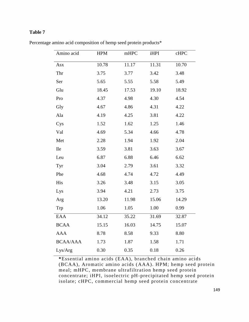
149
Table 7
Percentage amino acid composition of hemp seed protein products*
Amino acid HPM mHPC iHPI cHPC
Asx 10.78 11.17 11.31 10.70
Thr 3.75 3.77 3.42 3.48
Ser 5.65 5.55 5.58 5.49
Glu 18.45 17.53 19.10 18.92
Pro 4.37 4.98 4.30 4.54
Gly 4.67 4.86 4.31 4.22
Ala 4.19 4.25 3.81 4.22
Cys 1.52 1.62 1.25 1.46
Val 4.69 5.34 4.66 4.78
Met 2.28 1.94 1.92 2.04
Ile 3.59 3.81 3.63 3.67
Leu 6.87 6.88 6.46 6.62
Tyr 3.04 2.79 3.61 3.32
Phe 4.68 4.74 4.72 4.49
His 3.26 3.48 3.15 3.05
Lys 3.94 4.21 2.73 3.75
Arg 13.20 11.98 15.06 14.29
Trp 1.06 1.05 1.00 0.99
EAA 34.12 35.22 31.69 32.87
BCAA 15.15 16.03 14.75 15.07
AAA 8.78 8.58 9.33 8.80
BCAA/AAA 1.73 1.87 1.58 1.71
Lys/Arg 0.30 0.35 0.18 0.26
*Essent ial amino acids (EAA), branched chain amino acids
(BCAA), Aromatic amino acids (AAA). HPM; hemp seed protein
meal; mHPC, membrane ult raf i l t rat ion hemp seed protein
concentrate; iHPI, isoelectric pH-precipitated hemp seed protein
isolate; cHPC, commercial hemp seed protein concentrate
150
(EAA) and branched-chain amino acid (BCAA) contents of mHPI (35.22% and 16.03%,
respectively) were higher than those of the other products. The BCAA values obtained in this
study are higher than the 12.40-13.00% reported for flaxseed protein products but lower than
17.00% of soy protein (Oomah, 2001). The higher BCAA/AAA ratio in mHPC could contribute
to better muscle metabolism and maintenance of protein homeostasis (Herman et al., 2010; Sun
et al., 2011). However, the BCAA/AAA ratios obtained in this work are lower than the values
reported for Brassica carinata protein hydrolysates (Pedroche et al., 2006) as well as flaxseed
high-molecular weight 12S protein and soy protein (Oomah, 2001).But the high Lys/Arg ratio
reduces potential contributions to improved cardiovascular health. This is because arginine is a
precursor for the vasodilatory nitric oxide; therefore, higher arginine levels are desirable for a
healthy cardiovascular system. On the other hand, the Lys/Arg ratios obtained in this work
(mHPC, 0.35) are lower than the 0.37-1.00 and 0.88 reported for flaxseed protein products and
soy proteins (Oomah, 2001), respectively. The results suggest that the hemp seed proteins may
have better impact than flaxseed protein fractions and soy protein in improving cardiovascular
health.
5.3.2. SDS-PAGE
The non-reducing SDS-PAGE profiles revealed that the 47 kDa band contains the main
polypeptide in the HSP products (Fig. 18A). Other polypeptides with lower band intensity are
apparent in the 14, 24, and 34 kDa bands. However, the 24 and 35 kDa bands seem to be absent
in the mHPC. The similarity in the polypeptide composition of cHPC, iHPI and HPM indicates
that the processing methods used for the protein products extracted similar proteins from HMR.
In contrast the mHPC had a slightly different polypeptide composition, which suggests that the
membrane ultrafiltration protocol led to retention of higher ratios of the 14 and 47 kDa

151
Fig. 18. Polypeptide composition of hemp seed protein products under non-reducing (A) and
reducing (B) sodium dodecyl sulfate polyacrylamide gel electrophoresis conditions: cHPC,
commercial hemp seed protein concentrate; iHPI, isoelectric pH-precipitated hemp seed protein
isolate; mHPC, membrane ultrafiltration hemp seed concentrate; HPM, hemp protein meal.
cHPC iHPI mHPC HPM Std (kDa) cHPC iHPI mHPC HPM Std (kDa)
116.0
25.018.4
14.4
66.2
45.0
35.0
14.4
18.425.0
35.045.0
66.2
116.0
152
polypeptides and hence less intensity of the 24 and 34 kDa proteins. The results are consistent
with our previous report which showed the presence of these main polypeptides in other types of
HSP products under non-reducing SDS-PAGE (Malomo et al., 2014). However, unlike the
present work where the 47 kDa was the main polypeptide, our previous work showed that the 24
and 34 kDa polypeptides were the major proteins in a previously reported protein isolate
(Malomo et al., 2014). The difference could be because the protein isolate used by Malomo et
al. (2014) was prepared from a different defatted meal (50% protein content) and not the seed
waste product used for the present work. However, under the reducing conditions (presence of
mercaptoethanol), the 14 and 47 kDa bands were dissociated into lower molecular weight
polypeptides, which suggest the presence of disulfide bonds (Fig. 18B). Thus the mHPC lane
showed more visible polypeptide bands under reducing SDS-PAGE than non-reducing. For the
cHPC, iHPI and HPM, there were increased band intensities for the 24 and 34 kDa polypeptides
under reducing conditions, which may have been due to dissociation of disulfide bonds in the
higher molecular weight proteins. The results are also consistent with reports of Tang et al.
(2006) and Wang et al. (2008) who showed presence of a 48 kDa protein in hemp seed protein
isolates.
5.3.3. Protein contents and Protein solubility (PS)
The mHPC (74% protein, wet weight basis) and iHPI (84% protein, wet weight basis) were
successfully obtained from the starting material, HPM (~37% protein) in this study after
employing the enzyme pre-digestion coupled with membrane ultrafiltration and isoelectric
precipitation methods, respectively. The results indicate that the enzyme cocktail used was able
to degrade most of the non-protein materials in the original waste product to produce a 200%
protein enriched product when compared with HPM. The mHPC also had slightly higher protein
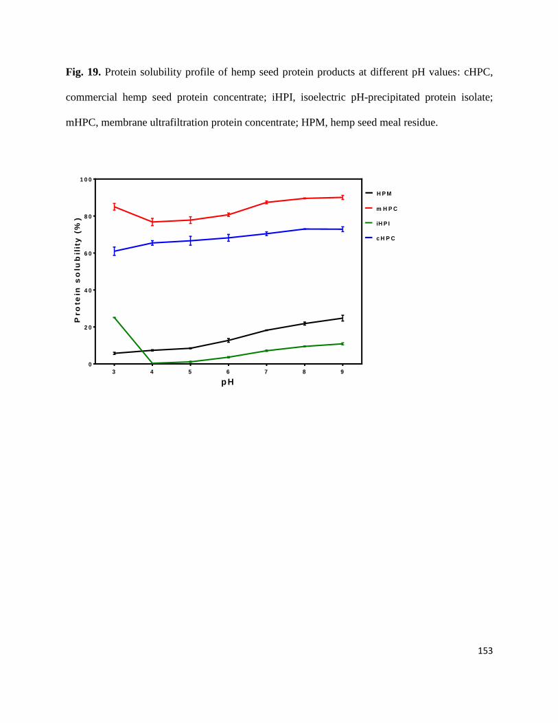
153
Fig. 19. Protein solubility profile of hemp seed protein products at different pH values: cHPC,
commercial hemp seed protein concentrate; iHPI, isoelectric pH-precipitated protein isolate;
mHPC, membrane ultrafiltration protein concentrate; HPM, hemp seed meal residue.
p H
Pro
tein
so
lub
ilit
y (
%)
3 4 5 6 7 8 9
0
2 0
4 0
6 0
8 0
1 0 0
H P M
m H P C
iH P I
c H P C

154
content (74%) than the 70% for commercial product (cHPC). Teh et al. (2014) obtained hemp
(alkali soluble, 95%; acid soluble, 91% protein) and canola (alkali soluble, 93% acid soluble,
94% protein) protein isolates from their starting materials, hemp (33% protein) and canola (37%
protein) seed cakes, respectively. These findings indicate that the type and sources of raw
materials as well as methods used for protein product preparation are the major determinants of
protein contents of the resultant protein isolates.
The PS of any protein at various pH levels has been a useful indicator in determining
potential incorporation and performance in the food systems (Mohamed et al., 2009; Adebowale
et al., 2007). Fig. 19 shows that the mHPC and cHPC had >60% PS across the pH 3-9 range but
the former had higher values. In contrast the iHPI and HPM had very poor PS (<25% at pH 3-9);
the iHPI was virtually insoluble at pH 4-5, which is the isoelectric point range for these proteins.
However, the poor solubility of iHPI could have been caused by intense protein-protein
interactions that led to aggregation and precipitation during the isoelectric pH protein isolation
protocol (Adebowale et al., 2007). As expected HPM proteins were also very insoluble because
crosslinking by phytate may have reduced ability to properly interact with water. Similarly, the
HPM contained high levels of insoluble fibre that reduced protein-water interactions. In contrast,
the removal of some of the fibre and phytate provided beneficial effects in terms of protein-water
interactions.
The high PS of the mHPC could have also been due to the absence of protein aggregation
that is common to iHPI. Thus the mHPC would have experienced less protein denaturation (such
as increased exposure of hydrophobic groups), which ensures better water interactions by the
native polypeptide chains. The high PS of the cHPC suggests that the proprietary method used
for protein isolation did not involve isoelectric pH-induced precipitation. The PS reported for
155
mHPI (>75%) in this study is in the range of those reported for thermally-improved adzuki (28-
79%), soy (49-82%) and pea (40-82%) protein isolates in the same pH (3-9) range (Barac et al.,
2015). The high PS observed for mHPC (>75%) in the pH 3-5 range indicates potential use in
the formulation of acidic foods such as protein-enriched beverages (Adebowale et al., 2007).
5.3.4. In vitro Protein digestibility (IPD)
The trypsin-chymotrypsin combination has been previously used and reported to simulate
gastrointestinal enzymatic processes that occurred in the normal human digestion of food
proteins (Pihlanto & Markinen, 2013). During protein digestion, peptide bond hydrolysis releases
H+, which causes pH to decrease; faster decreases represent higher rates of digestion and can be
used as an index of protein digestibility. Fig. 20 shows that the pH of mHPC decreased rapidly
from 8.0 to 7.15 about 25 sec after addition of trypsin-chymotrypsin cocktail; in contrast the
iHPI and cHPC digests were each at pH 7.45 and 7.43, respectively. After the first 25 sec, the
mHPC digest experienced slow but continuous pH decreases until 475 sec and were then stable
till 600 sec for a final pH 6.7 value. Meanwhile, other products experienced pH stability at 225
sec and final pH 6.91-6.93 values. The faster rate of mHPC digestion is consistent with easier
protease accessibility to peptide bonds because of the reduced levels of non-protein materials
(Adebowale et al., 2007). Based on Fig. 20, the calculated IPD of mHPC was significantly
(p<0.05) higher than similar values for iHPI and cHPC (Table 8). The higher IPD of mHPC can
be attributed to the pre-digestion reduction in polysaccharides and phytate. This is because fibres
have been reported to have negative effect on IPD as a result of non-specific interactions
between proteins and polysaccharides constituents of foods (Marambe et al., 2013).
The IPD obtained for mHPC in this study is higher than the 68, 71, 85, and 84% reported
for flaxseed (Marambe et al., 2013), pinto bean, soybean (Tan et al., 2014) protein isolates and
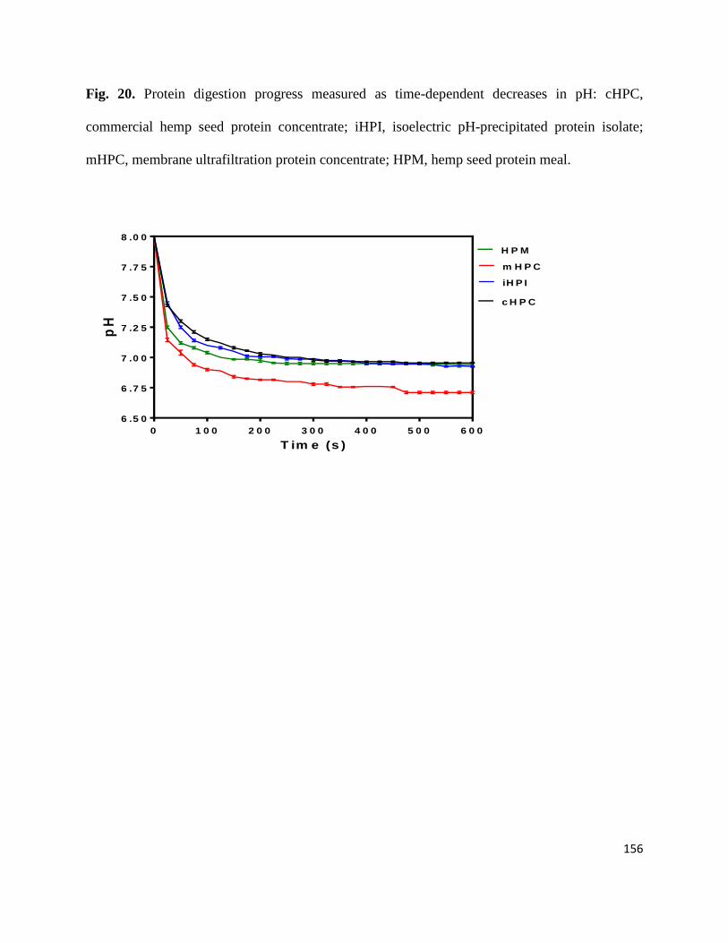
156
Fig. 20. Protein digestion progress measured as time-dependent decreases in pH: cHPC,
commercial hemp seed protein concentrate; iHPI, isoelectric pH-precipitated protein isolate;
mHPC, membrane ultrafiltration protein concentrate; HPM, hemp seed protein meal.
T im e (s )
pH
0 1 0 0 2 0 0 3 0 0 4 0 0 5 0 0 6 0 0
6 .5 0
6 .7 5
7 .0 0
7 .2 5
7 .5 0
7 .7 5
8 .0 0
H P M
m H P C
iH P I
c H P C
157
soy protein concentrate (Mohamed et al., 2009), respectively. However, the mHPC has lower
IPD when compared to the 91-95% reported for Mucuna bean protein isolates (Adebowale et al.,
2007) and 96% for chickpea protein isolate (Sanchez-Vioque et al., 1999).
5.3.5. Water and oil holding capacity (WHC and OHC)
The importance of WHC of any protein is centered on the hydrophilic-hydrophobic
balance of its amino acids. The polar amino groups, which are the primary sites for protein-
water interactions, contributed greatly to this hydrophilic-hydrophobic balance (Malomo et al.,
2014; Chavan et al., 2001; Chou & Morr, 1979). Likewise, the OHC has been interestingly
known to reflect the interaction ability of protein hydrophobic groups with lipids (Abugoch et al.,
2008; Liu et al., 2013). The presence of lipids, physicochemical environment (pH, ionic
strength) and solubility are some of the factors that could contribute to the WHC and OHC of
any protein molecules in the food system (Malomo et al., 2014). Table 8 indicates that mHPC
has a significantly (p<0.05) higher WHC (13.18 g/g) when compared to iHPI (12.01 g/g) and
cHPC (12.05 g/g). WHC is considered to be an important factor in maintaining flavour and
texture of foods (Liu et al., 2013; Mohamed et al., 2009); thus, mHPC seem to be of higher
quality as a food ingredient in this aspect. The present WHC results are higher than the 8.7, 8.2,
8.5, 7.8, 3.99 and 3.55 g/g reported for alkali-soluble hemp, acid-soluble hemp, alkali-soluble
canola, acid-soluble canola (Teh et al., 2014), defatted wheat germ (Liu et al., 2013) and
commercial soy (Zhu et al., 2010) protein isolates, respectively. Results of the WHC test seem to
suggest a positive association with the PS data, which may be due to the fact that both tests
depend on protein-water interactions. mHPC had a similar OHC as iHPI but the values are
significantly (p<0.05) higher than those obtained for cHPI and HPM.

158
Table 8
Water holding capacity (WHC) and oil holding capacity (OHC) of hemp seed protein products*
Samples WHC (g/g) OHC (g/g) % Protein
digestibility
HPM 12.32±0.03b 12.54±0.08
b 84.85±0.51
b
mHPC 13.18±0.06a 13.76±0.19
a 89.00±0.26
a
iHPI 12.01±0.08c 13.70±0.29
a 85.12±0.13
b
cHPC 12.05±0.04c 12.58±0.06
b 84.58±0.13
b
* cHPC, commercial hemp seed protein concentrate; iHPI,
Isoelectric pH-precipitated protein isolate; mHPC, membrane
ultrafiltration protein concentrate; HPM, hemp seed protein meal
Results are presented as mean ± standard deviation. For each column, mean values that contain
different letters are significantly different at p<0.05
159
The higher OHC of mHPC and iHPI is probably a reflection of their higher protein
contents, since interactions with the lipid phase is enhanced more by the protein molecules and
less by the insoluble polysaccharides. The results indicate a high potential for mHPC usage as a
functional ingredient in high-fat foods, such as bakery products and emulsion-type foods. The
OHC values obtained in this study are higher than the 8.0, 7.5, 7.5, 7.0, 2.98, 2.43, 1.80 and 1.43
g/g reported for alkali-soluble hemp, acid-soluble hemp, alkali-soluble canola, acid-soluble
canola (Teh et al., 2014), defatted wheat germ (Liu et al., 2013), commercial soy (Zhu et al.,
2010), pinto bean (Tan et al., 2014) protein isolates and optimized cowpea concentrates (Mune
Mune et al., 2014), respectively. The enzymatic pre-digestion and removal of some of the non-
protein components during mHPC preparation may have contributed to a loose protein structure
which enhanced interactions of its hydrophilic and hydrophilic groups with the environment.
5.3.6. Foaming capacity (FC) and foam stability (FS)
Foaming properties are important aspects of protein utilization in foods but are highly
influenced by environmental factors. Factors such as protein concentration, PS and
hydrophobicity/hydrophilicity ratio can also influence foaming properties (Malomo et al., 2014;
Chel-Guerrero et al., 2002; Damodaran, 1996). At all pH values and protein concentrations, the
mHPI formed significantly (p<0.05) higher foam volumes (FC) than iHPI and cHPC (Fig 21).
The higher FC of mHPC indicates a more flexible structure that reflects minimal level of protein
aggregation and is also consistent with the observed high solubility. In general, the cHPC had
significantly (P<0.05) higher FC than the iHPI and HPM, especially at pH 7.0 and 9.0, which
may also be due to the higher solubility of cHPC. This is because increased PS enhances protein-
water interactions, which can unfold protein structure for better air encapsulation to give higher
FC as previously demonstrated for other hemp seed proteins (Malomo & Aluko, 2015).
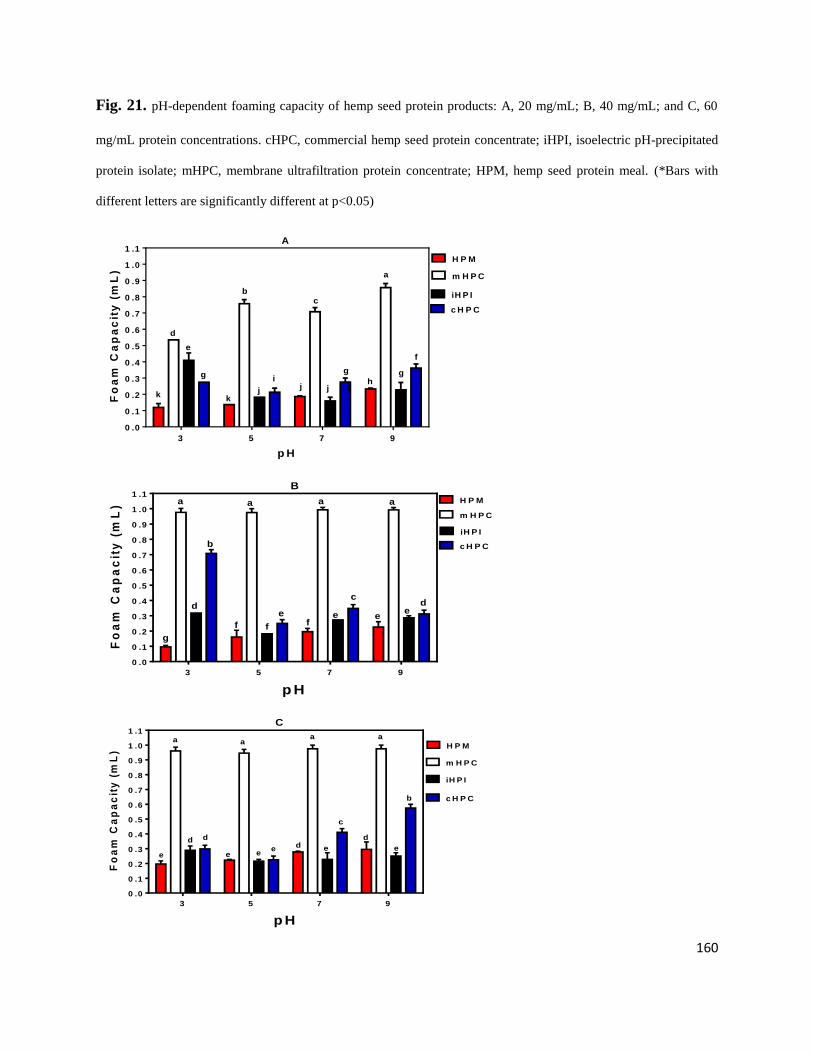
160
Fig. 21. pH-dependent foaming capacity of hemp seed protein products: A, 20 mg/mL; B, 40 mg/mL; and C, 60
mg/mL protein concentrations. cHPC, commercial hemp seed protein concentrate; iHPI, isoelectric pH-precipitated
protein isolate; mHPC, membrane ultrafiltration protein concentrate; HPM, hemp seed protein meal. (*Bars with
different letters are significantly different at p<0.05)
p H
Fo
am
Ca
pa
cit
y (
mL
)
3 5 7 9
0 .0
0 .1
0 .2
0 .3
0 .4
0 .5
0 .6
0 .7
0 .8
0 .9
1 .0
1 .1
H P M
m H P C
gh
g
b
ji
d
c
a
iH P I
c H P C
ef
g
jj
kj
k
A
p H
Fo
am
Ca
pa
cit
y (
mL
)
3 5 7 9
0 .0
0 .1
0 .2
0 .3
0 .4
0 .5
0 .6
0 .7
0 .8
0 .9
1 .0
1 .1H P M
m H P C
iH P I
c H P C
B
aa aa
e
d
e
cd
e
fff
b
g
e
p H
Fo
am
Ca
pa
cit
y (
mL
)
3 5 7 9
0 .0
0 .1
0 .2
0 .3
0 .4
0 .5
0 .6
0 .7
0 .8
0 .9
1 .0
1 .1
H P M
m H P C
iH P I
c H P C
C
dd
ee
b
aaaa
e
c
ee
de
d
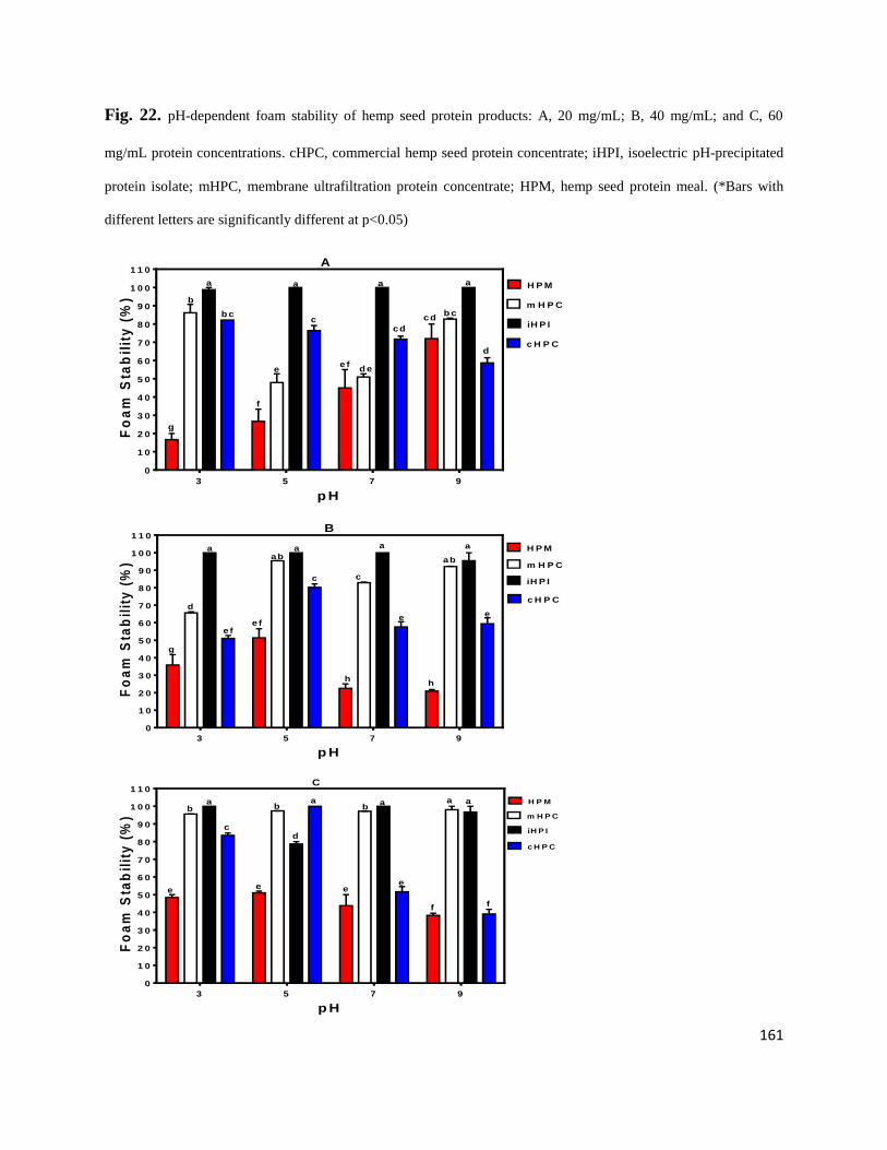
161
Fig. 22. pH-dependent foam stability of hemp seed protein products: A, 20 mg/mL; B, 40 mg/mL; and C, 60
mg/mL protein concentrations. cHPC, commercial hemp seed protein concentrate; iHPI, isoelectric pH-precipitated
protein isolate; mHPC, membrane ultrafiltration protein concentrate; HPM, hemp seed protein meal. (*Bars with
different letters are significantly different at p<0.05)
p H
Fo
am
Sta
bil
ity
(%
)
3 5 7 9
0
1 0
2 0
3 0
4 0
5 0
6 0
7 0
8 0
9 0
1 0 0
1 1 0
H P M
m H P C
a
a
b
c
a a
b cb c
iH P I
c H P C
A
cd
cd
e fe d e
d
g
f
a
p H
Fo
am
Sta
bil
ity
(%
)
3 5 7 9
0
1 0
2 0
3 0
4 0
5 0
6 0
7 0
8 0
9 0
1 0 0
1 1 0
H P M
m H P C
c
aa
c
aa
abab
iH P I
c H P C
B
e fe
de
e f
h
g
h
p H
Fo
am
Sta
bil
ity
(%
)
3 5 7 9
0
1 0
2 0
3 0
4 0
5 0
6 0
7 0
8 0
9 0
1 0 0
1 1 0
H P M
m H P C
a
g
iH P I
c H P C
C
d
a ab
aab b
cd
ee e e
f f
162
In contrast, the lower PS of iHPI and HPM reduces protein-water interactions, which is
unfavourable for foam formation, hence the lower FC values. FC was slightly increased when
protein concentration increased from 20 to 40 mg/mL but this was noticeable mostly for the
mHPI. The higher FC of mHPC at 40 mg/mL suggests that the additional protein molecules were
used to form more foam particles. Increasing the concentration from 40 to 60 mg/mL did not
have any substantial effect on FC, except at pH 9.0 where the cHPC showed a higher value. The
results agree with a previous report indicating increased protein concentration could favour
higher FC (Mundi & Aluko, 2012).
At all the protein concentrations, FS of iHPI were significantly (p<0.05) higher than the
other foams samples (Fig. 22). The higher FS of iHPI may be because of formation of thick or
strong interfacial membranes, which are favoured by the aggregated nature of this protein isolate.
Formation of such a cohesive protein membrane will reduce foam drainage and assist in
maintaining a stable foam product. FS increased as mHPC concentration was increased to 60
mg/mL, which could be due to formation of stronger interfacial membranes as more protein
molecules become available to participate in membrane formation. FS values of cHPC and HPM
were lower than those of iHPI and mHPC at all the pH values and protein concentrations. The
results suggest that the lower protein contents in cHPC and HPM may have limited formation of
strong interfacial membranes and hence foam drainage is more than those of iHPI and mHPC.
This is consistent with a previous work which showed that pea flour products with higher protein
contents tended to form more stable foams than those with lower protein (Aluko et al., 2009).
5.3.7. Emulsion capacity (EC) and stability (ES)
The emulsifying activity of any protein lies in its potential to interact with and stabilize
an oil-water mixture to prevent phase separation. Therefore, exposure of hydrophobic amino acid
163
residues would enhance formation of good emulsions because of the propensity to interact with
the hydrophobic lipid phase. Good emulsifiers will form emulsions with smaller oil droplet sizes
(larger surface area) than poor emulsifiers. Fig. 23 shows that the HPM and iHPI formed
excellent emulsions with oil droplet sizes that are <1 µm. In contrast the mHPC and cHPC
emulsion-forming abilities were much poorer with oil droplet sizes in the 6-15 µm range. Thus,
the iHPI interacted better with the oil phase probably as a result of protein denaturation that
exposed the hydrophobic amino acids. The excellent emulsion-forming ability of the HPM was
unexpected but could be due to the high level of insoluble polysaccharides that have better
interactions with oil rather than water. The results are consistent with the PS data, which
suggests the presence of more hydrophilic surfaces in mHPC and cHPC but higher levels of
hydrophobic surfaces in iHPI and HPM. For mHPC and cHPC, the oil droplet size decreased as
protein concentration or pH was increased. The results suggest better formation of interfacial
membranes with availability of higher number of protein molecules. The better emulsion
formation at alkaline pH values suggests increased ability of the proteins to unfold, which is a
prerequisite for oil encapsulation. Protein molecules will acquire more negative charges at higher
pH values, which would enhance protein unfolding and enable better formation of emulsified oil
droplets.
The oil droplet sizes obtained in this work are lower (i.e. better emulsions) than the 18-30
µm reported for kidney bean proteins (Mundi & Aluko, 2012) and the 18-85 µm reported for
canola and soy proteins (Tan et al., 2014) at the same pH (3-9) range. The oil droplet sizes for
HPM and iHPI are very similar to values reported for a different hemp seed meal and protein
isolate (Malomo et al., 2014) as well as the albumin and globulin fractions (Malomo & Aluko,
2015). However, the emulsions formed by mHPC and cHPC have bigger oil droplet sizes
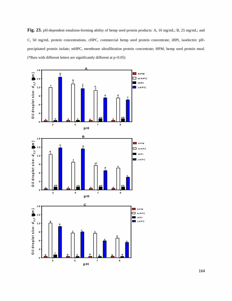
164
Fig. 23. pH-dependent emulsion-forming ability of hemp seed protein products: A, 10 mg/mL; B, 25 mg/mL; and
C, 50 mg/mL protein concentrations. cHPC, commercial hemp seed protein concentrate; iHPI, isoelectric pH-
precipitated protein isolate; mHPC, membrane ultrafiltration protein concentrate; HPM, hemp seed protein meal.
(*Bars with different letters are significantly different at p<0.05)
p H
Oil
dro
ple
t s
ize
-d
3,2
(
m)
3 5 7 9
0
3
6
9
1 2
1 5
1 8
H P M
m H P C
iH P I
c H P C
a aab
dc
a
d
aaab
ef
g
a
f
h
A
p H
Oil
dro
ple
t s
ize
-d
3,2
(
m)
3 5 7 9
0
3
6
9
1 2
1 5
1 8
H P M
m H P C
iH P I
c H P C
aab
c
e
aab
e f
d
a
h
f
h
g
aba
ab
B
p H
Oil
dro
ple
t s
ize
-d
3,2
(
m)
3 5 7 9
0
3
6
9
1 2
1 5
1 8
H P M
m H P C
iH P I
c H P Ca
C
ccc
b
d
ee
fgg g g g g g
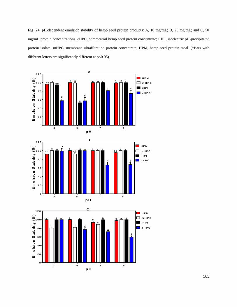
165
Fig. 24. pH-dependent emulsion stability of hemp seed protein products: A, 10 mg/mL; B, 25 mg/mL; and C, 50
mg/mL protein concentrations. cHPC, commercial hemp seed protein concentrate; iHPI, isoelectric pH-precipitated
protein isolate; mHPC, membrane ultrafiltration protein concentrate; HPM, hemp seed protein meal. (*Bars with
different letters are significantly different at p<0.05)
p H
Em
uls
ion
Sta
bil
ity
(%
)
3 5 7 9
0
2 0
4 0
6 0
8 0
1 0 0
1 2 0
H P M
m H P C
iH P I
c H P C
ab
c
aaa
c
aaa
d
e
aa
d
aba
A
p H
Em
uls
ion
Sta
bil
ity
(%
)
3 5 7 9
0
2 0
4 0
6 0
8 0
1 0 0
1 2 0
H P M
m H P C
iH P I
c H P C
abaa
ab
c
aaaaaab
a
aa a
c
B
p H
Em
uls
ion
Sta
bil
ity
(%
)
3 5 7 9
0
2 0
4 0
6 0
8 0
1 0 0
1 2 0H P M
m H P C
iH P I
c H P C
a
C
aaaaa a a ab
c
dd d
e
f

166
(i.e. poorer emulsions) than those of the previously reported hemp seed protein products
(Malomo et al., 2014) and protein fractions (Malomo & Aluko, 2015). These differences suggest
that the type of raw material used for protein product preparation is an important determinant of
protein functionality. Contrary to the oil droplet size results, the data presented in Fig 24 show
that most of the emulsions were stable with values >50%. The mHPI emulsions were particularly
stable at 10 and 25 mg/mL protein concentrations, which suggest formation of strong interfacial
membranes to reduce oil droplet coalescence. The decreased ES of mHPC at 50 mg/mL could be
due to protein crowding, which limits the level of protein-protein interactions required to form
strong interfacial membranes. The 100% ES presented in this study is higher than the 50% value
reported by Teh et al. (2014) for alkali- and acid-soluble hemp protein isolates. However, a
similar 100% ES has been reported for alkali- and acid-soluble canola protein isolates (Teh et al.,
2014). The high ES produced by mHPC may be due to its molecular flexibility, which enhances
formation of strong interfacial membranes that resist oil droplet coalescence. This is because
formation of a stable emulsion is dependent on the ability of proteins to adequately balance their
molecular size, charge, surface hydrophobicity and molecular flexibility (Wang et al., 2014; Liu
et al., 2013; Avramenko et al., 2013; Zhang et al., 2009).
5.4. Conclusions
The potential uses of protein isolates are largely dependent on their physicochemical and
functional properties with great influence from protein digestibility. A low protein industrial
hemp seed meal (37% protein content) was successfully converted into a functional protein
concentrate (mHPC) with 74% protein content after digestion and removal of some of the non-
protein materials. The high protein solubility over the pH 3-9 range, excellent foaming properties
and high protein digestibility most probably indicate that most of the mHPC polypeptides were
167
present mostly in their native forms with little or no denaturation. The mHPC solubility in the pH
range is highly desirable for protein fortification of acidic foods and beverages, such as fruit
juices. The high level of essential amino acids and branched-chain amino acids suggests that the
mHPC could be used for the fortification of products with low protein content. However, the
native conformation of mHPC did not favour formation of good emulsions, which indicates lack
of exposure of hydrophobic groups and hence poor interactions with the oil phase. In contrast,
the original low protein waste material (HPM) and the isoelectric-precipitated protein isolate
(iHPI) had excellent emulsifying ability probably due to the presence of polysaccharides and
denatured protein molecules with exposed hydrophobic groups, respectively.
Acknowledgement
This work was funded through the Natural Sciences and Engineering Research Council of
Canada (NSERC) Discovery grant and a Manitoba Agri-Food Research and Development
Initiative (ARDI) grant to Dr. R.E. Aluko. S.A. Malomo is a recipient of University of Manitoba
Graduate Fellowship (UMGF) and Manitoba Graduate Scholarship (MGS) Awards for doctoral
studies.
References
Abugoch, L.E., Romero, N., Tapia, C.A., Silva, J., Rivera, M., 2008. Study of some
physicochemical and functional properties of quinoa (Chenopodium quinoa Willd)
protein isolates. J. Agric. Food Chem. 56, 4745-4750.
Adebowale, Y.A., Adeyemi, I.A., Oshodi, A.A., Niranjana, K., 2007. Isolation, fractionation and
characterisation of proteins from Mucuna bean. Food Chem. 104, 287–299.

168
Aluko, R.E., Mofolasayo, O.A., Watts, B.A., 2009. Emulsifying and foaming properties of
commercial yellow pea (Pisum sativum L.) seed flours. J. Agric. Food Chem. 57, 9793
9800.
Aluko, R.E., McIntosh, T., 2004. Electrophoretic and functional properties of mustard seed
meals and protein concentrates. J. American Oil Chem. Soc. 81 (7), 679-683.
Avramenko, N.A., Low, N.H., Nickerson, M.T., 2013. The effects of limited enzymatic
hydrolysis on the physicochemical and emulsifying properties of a lentil protein isolate.
Food Res. Int. 51, 162-169.
Barac, M.B., Pesic, M.B., Stanojevic, S.P., Kostic, A.Z., Bivolarevic, V., 2015. Comparative
study of the functional properties of three legume seed isolates: adzuki, pea and soy bean.
J. Food Sci. Technol. 52 (5), 2779–2787.
Bidlingmeyer, B.A., Cohen, S.A., Tarvin, T.L., 1984. Rapid analysis of amino acids using pre
column derivatization. J. Chrom. B: Biomed. Sci. Appl. 336 (1), 93-104.
Chavan, U. D., McKenzie, D. B., & Shahidi, F. (2001). Functional properties of protein isolates
frombeach pea (Lathyrus maritimus L.). Food Chemistry, 74: 177-187.
Chel-Guerrero, L., Perez-Flores, V., Betancur-Ancona, D., Davila-Ortiz, G. (2002). Functional
properties of flours and protein isolates from Phaseolus lunatus and Canavalia ensiformis
seeds. Journal of Agricultural and Food Chemistry, 50: 584-591.
Chou, D. H., & Morr, C. V. (1979). Protein-water interactions and functional properties. Journal
of American Oil Chemists’ Society, 56, 53-62.
Damodaran, S. (1996). Amino acids, peptides, and proteins. In O. R. Fennema (Ed.), Food
Chemistry (3rd ed., pp. 322–424). New York: Marcel Dekker, Inc.
169
Gehrke, C.W., Wall, L.L., Absheer, J.S., Kaiser, F.E., Zumwalt, R.W., 1985. Sample preparation
for chromatography of amino-acids: acid hydrolysis of proteins. J. Assoc. Official Anal.
Chem. 68, 811-821.
Girgih, A.T., He, R., Alashi, A.M., Malomo, S.A., Aluko, R.E., 2014a. Preventive and treatment
effects of hemp seed (Cannabis sativa L.) meal protein hydrolysate against blood
pressure in spontaneously hypertensive rats. Eur. J. Nutr. 53 (5), 1237-1246.
Girgih, A.T., Alashi, A., He, R., Malomo, S.A., Raj, P., Netticadan, T., Aluko, R.E., 2014b. A
novel hemp seed meal protein hydrolysate reduces oxidative stress factors in
spontaneously hypertensive rats. Nutri. 6, 5652-5666.
Herman, M.A., She, P., Peroni, O.D., Lynch, C.J., Kahn, B.B., 2010. Adipose tissue branched
chain amino acid (BCAA) metabolism modulates circulating BCAA levels. J. Biol.
Chem. 285, 11348- 11356.
Hsu, H.W., Vavak, D.L., Satterlee, D.L., Mill, G.A., 1977. A multienzyme technique for
estimating protein digestibility. J. Food Sci. 42 (5), 1269-1273.
Landry, J., Delhaye, S., 1992. Simplified procedure for the determination of tryptophan of foods
and feedstuffs from barytic hydrolysis. J. Agric. Food Chem. 40, 776-779.
Liu, F., Chen, Z., Wang, L., Wang, R., 2013. Effects of protein solubilisation and precipitation
pH values on the functional properties of defatted wheat germ protein isolates. Int. J.
Food Sci. Technol. 48, 1490–1497.
Lu, R.R., Qian, P., Sun, Z., Zhou, X.H., Chen, T.P., He, J.F., Zhang, H., Wu, J. P., 2010.
Hempseed protein derived antioxidative peptides: Purification, identification and
protection from hydrogen peroxide-induced apoptosis in PC12 cells. Food Chem. 123 (4),
1210-1218.
170
Malomo, S.A., Aluko, R. E., 2015. A comparative study of the structural and functional
properties of isolated hemp seed (Cannabis sativa L.) albumin and globulin fractions.
Food Hydrocol. 43, 743-752.
Malomo, S.A., He, R., Aluko, R.E., 2014. Structural and functional properties of hemp seed
protein products. J. Food Sci. 79 (8), C1512-1521.
Marambe, H.K., Shand, P.J., Wanasundara, J.P.D., 2013. In vitro digestibility of flaxseed (Linum
usitatissimum L.) protein: effect of seed mucilage, oil and thermal processing. Int. J.
Food Sci. Technol. 48, 628–635.
Markwell, M.A.K., Haas, S.M., Bieber, L.L., Tolbert, N.E., 1978. Modification of Lowry
procedure to simplify protein determination in membrane and lipoprotein samples. Anal.
Biochem. 87 (1), 206-210.
Mohamed, T.K., Zhu, K., Issoufou, A., Fatmata, T., Zhou, H., 2009. Functionality, in vitro
digestibility and physicochemical properties of two varieties of defatted Foxtail millet
protein concentrates. Int. J. Mol. Sci. 10, 5224-5238.
Mundi, S., Aluko, R.E., 2012. Physicochemical and functional properties of kidney bean albumin
and globulin protein fractions. Food Res. Int. 48, 299-306.
Mune Mune, M.A., Minka, S.R., Mbome, I.L., 2014. Optimising functional properties during
preparation of cowpea protein concentrate. Food Chem. 154, 32-37
Oomah, B.D., 2001. Flaxseed as a functional food source. J. Sci. Food Agric., 81, 889-894.
Pedroche, J., Yust, M.D.M., Lqari, H., Megiäas, C., N-Calle, J.G., Alaiz, M., Vioque, J.,
MillaäN, F., 2006. Production of Brassica carinata protein hydrolysates with a high
fischer’s ratio using immobilized proteases. J. Agric. Food Chem. 54, 7621-7627.
171
Pihlanto, A., Mäkinen, S., 2013. Antihypertensive properties of plant protein derived peptides.
In: Hernandez-Ledesma, B., Hsieh, C. (Eds.), Bioactive Food Peptides in Health and
Disease. INTECH Publishers, Rijeka, Croatia. p 145-182. ISBN 978-953-51-0964-8.
Sa'nchez-Vioque, R., Clemente, A., Vioque, J., Bautista, J., Milla'n, F., 1999. Protein isolates
from chickpea (Cicer arietinum L.): chemical composition, functional properties
and protein characterization. Food Chem. 64, 237-243.
Sun, H., Lu, G., Ren, S., Chen, J., Wang, Y., 2011. Catabolism of Branched-Chain Amino Acids
in Heart Failure: Insights from Genetic Models. Ped. Cardio. 32, 305-310.
Tan, E., Ngoh, Y., Gan, C., 2014. A comparative study of physicochemical characteristics and
functionalities of pinto bean protein isolate (PBPI) against the soybean protein isolate
(SPI) after the extraction optimisation. Food Chem. 152, 447–455.
Tan, S.H., Mailer, R.J., Blanchard, C.L. Agboola, S.O., 2014. Emulsifying properties of proteins
extracted from Australian canola meal. LWT - Food Sci. Technol. 57, 376-382.
Tang, C.H, Ten, Z., Wang, X.S., Yang, X.Q., 2006. Physicochemical and functional properties of
hemp (Cannabis sativa L.) protein isolate. J. Agric. Food Chem. 54, 8945–8950.
Teh, S.S., Bekhit, A.E.D., Carne, A. Birch, J., 2013. Effect of the defatting process, acid and
alkali extraction on the physicochemical and functional properties of hemp, flax and
canola seed cake protein isolates. Food Meas. 8, 92–104.
Udenigwe, C.C., Lin, Y.S., Hou, W.C., Aluko, R.E., 2009. Kinetics of the inhibition of renin and
angiotensin I-converting enzyme by flaxseed protein hydrolysate fractions. J. Funct.
Foods, 1 (2), 199-207.
172
Wang, H., Chen, Y., Hua, Y., Kong, X., Zhang, C., 2014. Effects of phytase-assisted processing
method on physicochemical and functional properties of soy protein isolate. J. Agric.
Food Chem. 62, 10989−10997.
Wang, X.S., Tang, C.H., Yang, X.Q., Gao, W.R., 2008. Characterization, amino acid
composition and in vitro digestibility of hemp (Cannabis sativa L.) proteins. Food Chem.
107, 11–18.
Wang, M., Hettiarachchy, N.S., Qi, M., Burks, W., Siebenmorgen, T., 1999. Preparation and
functional properties of rice bran protein isolates. J. Agric. Food Chem. 47, 411−416.
Yin, S.W., Tang, C.H., Wen, Q.B., Yang, X.Q., 2009. Functional and structural properties and in
vitro digestibility of acylated hemp (Cannabis sativa L.) protein isolates. Int. J. Food Sci.
Technol. 44, 2653–2661.
Zhang, T., Jiang, B., Mu, W.M., Wang, Z., 2009. Emulsifying properties of chickpea protein
isolates: influence of pH and NaCl. Food Hydrocol. 23, 146–152.
Zhu, K.X., Sun, X.H., Chen, Z.C., Peng, W., Qian, H.F., & Zhou, H.M., 2010. Comparison of
functional properties and secondary structures of defatted wheat germ proteins separated
by reverse micelles and alkaline extraction and isoelectric precipitation. Food Chem. 123,
1163–1169.
173
5.5 Statement transfer between the study 3 and study 4
The isolated hemp seed protein products in the first 3 chapters are made of complex structures
(amino acid composition and three dimensional structures) with different subunits linked
together by the disulphide bonds. Thus, polypeptide chains were the principal structural elements
present in the protein molecules. In order to further exploit value-added utilization, the complex
proteins need to be broken down through enzymatic digestions to produce small-size peptides
that could be used as bioactive peptides. Globally, human health is being shaped by rapid
environmental changes (age, stress, disease, urbanization, globalized unhealthy lifestyles) that
cause oxidative stress and homeostatic imbalance in the body. Ultimately, these negative changes
lead to the development of chronic diseases, one of which is hypertension. High blood pressure
is a recognized risk factor for the development of various cardiovascular diseases, which are
currently managed with synthetic compounds (drugs). However, the use of several
antihypertensive drugs is associated with negative side effects that reduce compliance with
physician’s prescribed dosage. This has led to increased consumer demand for natural
antihypertensive agents that will have reduced or nil negative side effects. This chapter deals
with the enzymatic modifications of hemp seed proteins using selected proteases to cleave the
specific peptide bonds to produce distinct peptide fractions that could be used as
antihypertensive agents. The hemp seed bioactive peptides work as modulators of the renin-
angiotensin system, which is the main controller of mammalian blood pressure.
174
CHAPTER SIX
6. Study on the structural and antihypertensive properties of enzymatic hemp seed protein
hydrolysates
Sunday A. Malomo; John O. Onuh; Abraham T. Girgih and Rotimi E. Aluko*
The Department of Human Nutritional Sciences, University of Manitoba, Winnipeg, Manitoba,
Canada R3T 2N2
*Corresponding author: Dr. Rotimi E. Aluko; E-Mail: [email protected]; Tel.: +1-204-
474-9555; Fax: +1-204-474-7593.
175
Abstract
The aim of this work was to produce antihypertensive protein hydrolysates through different
enzymatic hydrolysis (2% pepsin, 4% pepsin, 1% alcalase, 2% alcalase, 2% papain, and 2%
pepsin+pancreatin) of hemp seed proteins (HSP). The hemp seed protein hydrolysates (HPHs)
were tested for in vitro inhibitions of renin and angiotensin converting enzyme (ACE), two of the
enzymes that regulate human blood pressure. The HPHs were then administered orally (200
mg/kg body weight) to spontaneously hypertensive rats and systolic blood pressure (SBP)-
lowering effects measured over a 24 h period. Size exclusion chromatography showed mainly a
300-9560 Da peptide size range for the HPHs while amino acid composition data had the 2%
pepsin HPH with the lowest cysteine content. Fluorescence spectroscopy revealed higher
fluorescence intensities for the peptides when compared to the unhydrolyzed hemp seed protein.
Overall, the 1% alcalase HPH was the most effective (p<0.05) SBP-reducing agent (-32.5 + 0.7
mmHg after 4 h) while the pepsin HPHs produced longer-lasting effects (-23.0 + 1.4 mmHg after
24 h). We conclude that an optimized combination of the fast-acting HPH (1% alcalase) with the
longer-lasting HPHs (2% and 4% pepsin) could provide daily effective SBP reductions.
Keywords: hemp seed; protein hydrolysates; renin; angiotensin converting enzyme; degree of
hydrolysis; fluorescence intensity; systolic blood pressure; spontaneously hypertensive rats
176
6.1 Introduction
Increased rate of high blood pressure (BP) at a certain age in life has led to critical
hypertensive conditions in most nations and is responsible for ~45-51% of total global deaths
[1]. BP is said to be under normal and adequate control when the systolic and diastolic values are
≤140 and ≤90 mm Hg, respectively [1]. Occurrence of elevated BP is a known risk factor for the
development of several cardiovascular diseases like, coronary heart disease, heart failure, stroke,
peripheral arterial disease, and renal failure [2]. Antihypertensive drug therapy (a key aspect of
hypertension management), which is currently being employed to screen, treat and control high
BP in order to reduce the incidence of hypertension, has posed a significant healthcare burden on
several nations [3]. For instance, in order to control this public health burden, the USA incurred a
direct and indirect cost of about US$93.5 billion in 2010 for hypertension awareness, treatment
and control [3]. Likewise, an increasingly significant health, policy and financing challenges
have been recorded in some countries, which inevitably create unsustainable pharmaceutical
costs for government [4].
The renin–angiotensin aldosterone system (RAAS) is the primary physiological pathway that
has been described for BP regulation and management [5]. To control BP, renin is synthesized in
the kidneys and then released into the blood circulatory system where it cleaves the N-terminal
region of angiotensinogen to release a decapeptide, angiotensin (AT)-I [6]. AT-I further
circulates in the blood until its C-terminal dipeptide residue is cleaved by angiotensin I-
converting enzyme (ACE) to form an octapeptide AT-II (a potent vasoconstrictor). The
pharmaceutical industry has historically exploited ACE inhibition to produce commercial
antihypertensive drugs like captopril, enalapril, and lisinopril [7]. Meanwhile, a comparative
reduction in elevated BP that could lead to treatment for hypertension other than ACE inhibition
177
has been suggested for research [8]. Such alternative antihypertensive compounds target renin
and bioactive peptides from plant proteins have been shown to have renin- and ACE-inhibitory
properties [9-15]. The need to reduce negative side effects (nausea, vomiting, dry cough) of
antihypertensive therapy has spurred research into alternative natural sources of effective
compounds such as food protein-derived peptides. Thus there are various reports that have
shown bioactive peptides as potential antihypertensive agents [9-14,16-24].
Hemp seed protein (HSP), which is obtained from the industrial production of edible oil, has
been shown to be a suitable raw material for antihypertensive peptide production [12,14].
However, the previous works have focused solely on the use of simulated gastrointestinal
digestion as a tool to produce blood pressure-reducing peptides from hemp seed proteins.
Therefore, there is the need to expand hemp seed antihypertensive research scope by examining
the potential use of other commercially available enzymes as suitable proteolytic agents. This is
because past studies have demonstrated the possibility of generating antihypertensive peptides
with varying potencies simply by using different enzymes and enzyme:substrate ratios
[9,17,22,25,26]. This work aimed to determine the structural properties and BP-lowering effects
of different hemp seed protein hydrolysates (HPHs) produced using four enzymes and different
enzyme:substrate ratios. Specifically, the HPHs were tested for in vitro inhibitions of renin and
ACE activities, which were then related to observed BP-lowering effects after oral
administration to spontaneously hypertensive rats.
6.2. Materials and methods
6.2.1. Hemp seed products and chemical reagents
Hemp seed protein meal (HPM, 37% protein content) produced as a by-product of the
hemp seed oil processing industry was a gift from Hemp Oil Canada (St. Agathe, Manitoba,
178
Canada). Human recombinant Renin Inhibitor Screening Assay Kit was purchased from Cayman
Chemicals (Ann Arbor, MI, USA) while other enzymes such as pepsin (porcine gastric mucosa;
E.C. 3.4.23.1; ≥250 U/mg), pancreatin (porcine pancreas; 647-014-00-9), papain (papaya latex;
E.C. 3.4.22.2), alcalase (Bacillus licheniformis; E.C. 3.4.21.62), and ACE (rabbit lung; E.C.
3.4.15.1; ≥250 U/mg) were purchased from Sigma-Aldrich (St. Louis, MO, USA). Other
analytical-grade reagents were obtained from Fisher Scientific (Oakville, ON, Canada).
6.2.2. Preparation of hemp seed protein isolates (HPI)
HPI was produced from HPM according to the method previously described [12] with
slight modifications. HPM was dispersed in a glass beaker that contained deionized water (1:20,
w/v) and the dispersion was adjusted to pH 8.0 using 2 M NaOH to solubilize the proteins while
stirring at 37oC for 2 h; this was followed by centrifugation (7000 x g, 60 min at 4
oC). The
precipitate was discarded and the supernatant filtered with cheese-cloth, adjusted to pH 5.0 with
2 M HCl to precipitate the proteins and thereafter centrifuged (7000 x g, 60 min at 4oC). The
resultant precipitate was re-dispersed in deionized water, adjusted to pH 7.0 with 2 M NaOH and
freeze-dried to obtain the HPI. Protein concentration of the HPI was determined using the
modified Lowry method [27].
6.2.3. Preparation of enzymatic hemp seed protein hydrolysates (HPHs)
Hydrolysis of the HPI was conducted using each of the following enzyme and reaction
conditions as previously reported [9] with slight modifications: alcalase (50oC, pH 8.0, 4 h);
papain (65oC, pH 6, 4 h); and PP (pepsin at 37
oC, pH 2.0, 2 h followed by pancreatin at 37
oC, pH
7.5, 4 h). HPI (5%, w/v, protein weight basis) was suspended in deionized water in a glass beaker
equipped with a stirrer, heated to the appropriate temperature and adjusted to the appropriate pH
value prior to addition of the proteolytic enzymes. Proteases were added to the HPI slurry based
179
on the optimized enzyme to substrate ratio (E/S) viz: 1:100 (alcalase), 2:100 (pepsin, alcalase,
papain, pepsin+pancreatin (PP), and 4:100 (pepsin), based on HPI protein content. During
digestion, the required pH was maintained constant by addition of NaOH after which the
enzymes were inactivated by adjusting to pH 4.0 with 2 M HCl followed by immersing the
reaction vessel in boiling water bath for 10 min. The undigested proteins were precipitated by
centrifugation (8000 x g, 60 min at 4oC) after cooling and the supernatant (contains target
peptides) was freeze dried as the HPH, which was then stored at -20oC until needed for further
analysis. The protein contents of the freeze dried HPH was determined using the modified Lowry
method [27].
6.2.4. Determination of degree of hydrolysis
The percent degree of hydrolysis (DH) of HPHs was determined according to the
trinitrobenzene sulfonic acid method as previously described [28]. Briefly, HPI was digested
under vacuum with 6 M HCl for 24 h and the digest was used to determine total amino groups as
L-leucine equivalent. The DH was calculated as percentage ratio of the leucine equivalent of
HPHs to that of HPI.
6.2.5. Amino acid composition analysis
The amino acid profiles of HPI and HPHs were determined using the HPLC S4300
Amino Acid Analyzer, (Sykam Mfd Co., Eresing, Bavaria, Germany) according to the method
previously described [29] after samples were digested with 6 M HCl for 24 h. The cysteine and
methionine contents were determined after performic acid oxidation [30] and the tryptophan
content was determined after alkaline hydrolysis [31].
6.2.6. Analysis of molecular weight distribution

180
Molecular weight (MW) distribution of HPH peptides was determined by size exclusion
chromatography (SEC) as previously described [9] with slight modifications, using an AKTA
FPLC system (GE Healthcare, Montreal, PQ) equipped with a Superdex Peptide 10/300 GL
column (10 x 300 mm), and UV detector (λ= 214 nm). An aliquot (100 µl) of the sample (5
mg/mL in 50 mM phosphate buffer, pH 7.0 containing 0.15 M NaCl) was loaded onto the
column and elution was performed at room temperature using the phosphate buffer at a flow rate
of 0.5 mL/min. The column was calibrated with Cytochrome C (12,384 Da), Aprotinin (6512
Da), Vitamin B12 (1855 Da), and Glycine (75 Da) as the standard proteins. Peptide sizes of the
samples were estimated from a plot of log MW versus elution volume of the standard proteins.
6.2.7. Intrinsic fluorescence
Fluorescence intensity spectra of protein and peptide samples were obtained using a
previously described method [32] on a JASCO FP-6300 spectrofluorimeter (JASCO, Tokyo,
Japan) at 25 °C in a 1-cm path length cuvette. The sample stock solution was prepared as 10
mg/mL in 0.1 M sodium phosphate buffer, pH 7.0; this was followed by centrifugation and
determination of protein content of the supernatant. After diluting the supernatant to 0.002%
protein content (w/v), the fluorescence spectra was recorded at 280 nm excitation wavelengths
and 300 to 500 nm emission. The final fluorescence emission spectrums of each sample were
obtained after subtraction of the buffer emission spectrum.
6.2.8. ACE inhibition assay
The ability of HPHs to inhibit in vitro ACE activity was measured according to a
spectrophotometric method using synthetic N-[3-(2-Furyl)acryloyl]-L-phenylalanyl-glycyl-
glycine (FAPGG) as the substrate (Sigma-Aldrich, St. Louis, MO, USA) [33]. Briefly, 1 mL of
0.5 mM FAPGG (dissolved in 50mM Tris-HCl buffer containing 0.3 M NaCl, pH 7.5) was
181
mixed with 20 µL of ACE (20 mU final reaction activity) and 200 µL sample dissolved in same
buffer. The rate of decrease in absorbance at 345 nm was recorded for 2 min at room temperature
using Varian Cary 50-UV/Visible spectrophotometer (Varian Inc., Belrose, NSW, Australia).
The buffer was used instead of sample solutions in the blank experiment. The concentration of
sample that inhibited ACE activity by 50% (IC50) was calculated from a non-linear regression
plot of percentage ACE activity versus sample concentrations. ACE activity was expressed as the
rate of reaction (ΔA/min) and inhibitory activity was calculated as:
ACE inhibition (%) = [1 - ΔA.min
-1(sample)] x 100
ΔA.min
-1(blank) (Eqn 8)
where ΔA.min
-1(sample) and ΔA
.min
-1(blank) represent ACE activity in the presence and absence of
the HPHs, respectively.
6.2.9. Renin inhibition assay
In vitro inhibition of human recombinant renin activity by HPHs was conducted using the
Renin Inhibitor Screening Assay Kit (Cayman Chemical Co., Ann Arbor, MI, USA) according to
the method previously described [9]. Prior to the assay, renin buffer was diluted in 50 mM Tris–
HCl, pH 8.0, containing 100 mM NaCl. The renin protein solution was diluted 20 times with the
assay buffer before use and pre-warmed to 37oC prior to initiating the reaction in a fluorometric
microplate reader (Spectra MAX Gemini, Molecular Devices, Sunnyvale, CA, USA) maintained
at 37oC. Before the reaction, (i) 20 µL substrate, 160 µL assay buffer, and 10 µL Milli-Q water
were added to the background wells; (ii) 20 µL substrate, 150 µL assay buffer, and 10 µL Milli-
Q water were added to the blank wells; and (iii) 20 µL substrate, 150 µL assay buffer, and 10 µL
sample were added to the inhibitor wells. The reaction was initiated by adding 10 µL renin to the
blank and sample wells. The microplate was shaken for 10 sec to mix, incubated at 37oC for 15
182
min, and the fluorescence intensity (FI) was recorded using excitation and emission wavelengths
of 340 and 490 nm, respectively. The concentration of sample that inhibited renin activity by
50% (IC50) was calculated from a non-linear regression plot of percentage renin activity versus
peptide concentrations. The percentage renin inhibition was calculated as follows:
Renin inhibition (%) = [1 – FI sample)] x 100
FI (control) (Eqn 9)
6.2.10. BP-lowering effect of peptides in Spontaneously Hypertensive Rats (SHRs)
Animal experiments were carried out following the Canadian Council on Animal Care
Ethics guidelines with a protocol approved by the University of Manitoba Animal Protocol and
Management Review Committee. The 30-week old male SHRs (Charles River Laboratories,
Montreal, QC, Canada) with 340–380 g body weight (bw) were kept in the Animal Housing
Facility at the Richardson Centre for Functional Foods and Nutraceuticals, under a 12 hr day
and night cycle at 22 ± 2oC and were fed regular diet and tap water. The rats were divided into
three groups with 4 rats per group: HPH (test sample), captopril (positive control) and
phosphate buffered saline (PBS, pH 7.4) as the negative control. HPHs (each at 200 mg/kg bw)
and captopril (10 mg/kg bw) were dissolved in 1 mL PBS and administered to the SHRs by oral
gavage followed by measurement of systolic blood pressure (SBP) at 2, 4, 6, 8, and 24 h using
the tail-cuff method in slightly anesthetized rats as previously described [12]. Prior to sample
administration, the baseline (time-zero) SBP was determined. The change in SBP (ΔSBP,
mmHg) was determined by subtracting the baseline data from the data obtained at different time
points. Oral gavage was used to ensure all rats received the same amount of the HPHs while the
200 mg/kg rat bw translates to 32.43 mg/kg bw (~2 g/day for 60 kg) in an adult human being
[34].
183
6.2.11. Statistical analysis
Triplicate replications were used to obtain mean values and standard deviations.
Statistical analysis was performed with SAS (Statistical Analysis Software 9.1) using one-way
ANOVA. Duncan's multiple-range test was carried out to compare the mean values for samples
with significant differences taken at p<0.05.
6.3. Results
6.3.1. Amino acid composition of HPI and HPHs
Table 9 shows that the protein isolate and hydrolyzed samples had very similar amino
acid composition, which is consistent with the hydrolyzed samples being derived from same
protein starting material. However, the 2% pepsin hydrolysate had very low cysteine content
(0.12%) in comparison to the 1.2-1.43% for the other samples. The 2% papain HPH tended to
have the highest proline content.
6.3.2. DH and size exclusion chromatography analysis of HPHs
Extent of hydrolysis can be estimated from the DH values, which can be used an
indication of peptide chain length; higher and lower values indicate mean shorter and longer
lengths, respectively. As expected, the PP digest had significantly (p < 0.05) higher DH (28.16 +
0.34%) than the other enzyme digests (Figure 25), probably due to the exo- and endo-proteinase
activities of the enzyme preparations. Similar to our results, a flavourzyme protein hydrolysate
was shown to exhibit the highest DH, which was attributed to the presence of endo- and exo-
proteinase activities in this enzyme [35]. The peptide sizes were estimated by SEC and the
results showed distribution into four major peaks with approx. MW ranging from 300 to 9560 Da
(Figure 26). These peptide peaks were most noticeable for the 2% pepsin HPH; increasing the
enzyme concentration to 4% led to reduced intensities of the bigger peptide peaks (2775 and

184
Table 9: Percentage amino acid compositions of hemp seed protein isolate (HPI) and enzymatic
protein hydrolysates
Amino acid HPI 2% pepsin 4% pepsin 1% alcalase 2% alcalase 2% papain 2% pepsin+ pancreatin
Asx 11.31 11.36 11.37 11.51 11.55 11.18 10.74
Thr 3.42 2.92 2.85 2.89 2.78 2.93 2.82
Ser 5.58 5.98 5.95 6.10 6.09 6.00 5.84
Glu 19.10 19.36 19.28 19.63 19.64 19.56 19.06
Pro 4.30 4.94 4.74 4.83 4.75 5.23 5.17
Gly 4.31 3.82 3.84 3.73 3.70 3.63 3.56
Ala 3.81 4.79 4.76 4.53 4.60 4.75 4.48
Cys 1.25 0.12 1.34 1.20 1.22 1.31 1.43
Val 4.66 4.60 4.33 4.25 4.20 4.36 4.71
Met 1.92 2.26 2.18 2.28 2.24 2.34 2.21
Ile 3.63 3.17 3.04 2.95 2.90 3.03 3.33
Leu 6.46 6.64 6.48 6.38 6.39 6.35 6.60
Tyr 3.61 3.49 3.39 3.39 3.38 3.28 3.62
Phe 4.72 4.65 4.52 4.45 4.48 4.44 4.80
His 3.15 3.09 3.13 3.12 3.13 3.02 3.00
Lys 2.73 3.19 3.32 2.96 3.03 3.35 3.33
Arg 15.06 14.63 14.47 14.77 14.80 14.24 14.21
Trp 1.00 1.01 1.01 1.05 1.11 1.00 1.08
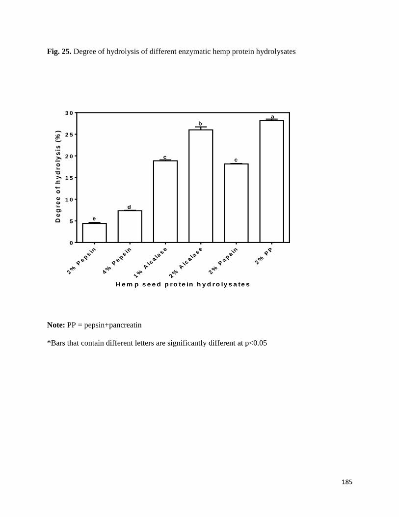
185
Fig. 25. Degree of hydrolysis of different enzymatic hemp protein hydrolysates
Note: PP = pepsin+pancreatin
*Bars that contain different letters are significantly different at p<0.05
H e m p s e e d p ro te in h y d ro ly s a te s
De
gre
e o
f h
yd
roly
sis
(%
)
2%
Pep
sin
4%
Pep
sin
1%
Alc
ala
se
2%
Alc
ala
se
2%
Pap
ain
2%
PP
0
5
1 0
1 5
2 0
2 5
3 0
b
e
d
cc
a
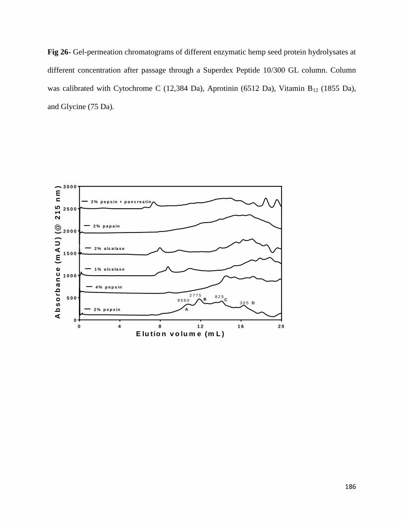
186
Fig 26- Gel-permeation chromatograms of different enzymatic hemp seed protein hydrolysates at
different concentration after passage through a Superdex Peptide 10/300 GL column. Column
was calibrated with Cytochrome C (12,384 Da), Aprotinin (6512 Da), Vitamin B12 (1855 Da),
and Glycine (75 Da).
E lu tio n v o lu m e (m L )
Ab
so
rb
an
ce
(m
AU
) (@
21
5 n
m)
0 4 8 1 2 1 6 2 0
0
5 0 0
1 0 0 0
1 5 0 0
2 0 0 0
2 5 0 0
3 0 0 0
2 % p e p s in
4 % p e p s in
1 % a lc a la s e
2 % a lc a la s e
2 % p a p a in
2 % p e p s in + p a n c r e a tin
2 7 7 5
9 5 6 0 B C8 2 5
A
3 0 5 D
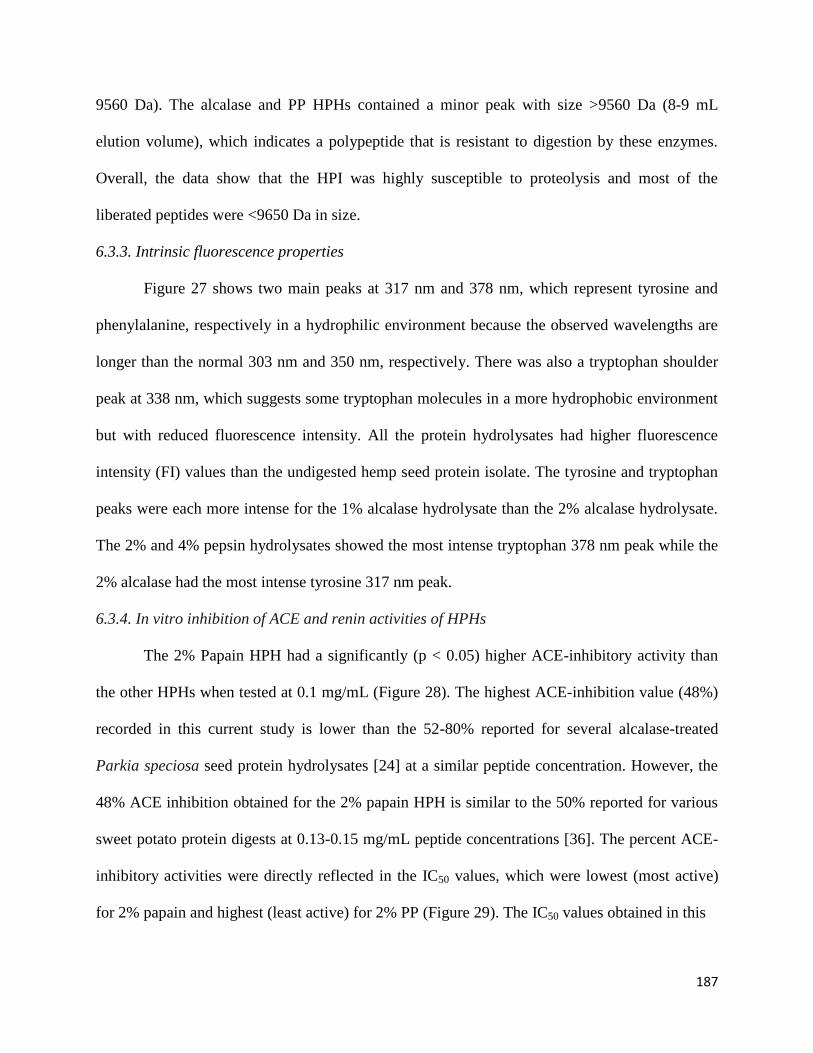
187
9560 Da). The alcalase and PP HPHs contained a minor peak with size >9560 Da (8-9 mL
elution volume), which indicates a polypeptide that is resistant to digestion by these enzymes.
Overall, the data show that the HPI was highly susceptible to proteolysis and most of the
liberated peptides were <9650 Da in size.
6.3.3. Intrinsic fluorescence properties
Figure 27 shows two main peaks at 317 nm and 378 nm, which represent tyrosine and
phenylalanine, respectively in a hydrophilic environment because the observed wavelengths are
longer than the normal 303 nm and 350 nm, respectively. There was also a tryptophan shoulder
peak at 338 nm, which suggests some tryptophan molecules in a more hydrophobic environment
but with reduced fluorescence intensity. All the protein hydrolysates had higher fluorescence
intensity (FI) values than the undigested hemp seed protein isolate. The tyrosine and tryptophan
peaks were each more intense for the 1% alcalase hydrolysate than the 2% alcalase hydrolysate.
The 2% and 4% pepsin hydrolysates showed the most intense tryptophan 378 nm peak while the
2% alcalase had the most intense tyrosine 317 nm peak.
6.3.4. In vitro inhibition of ACE and renin activities of HPHs
The 2% Papain HPH had a significantly (p < 0.05) higher ACE-inhibitory activity than
the other HPHs when tested at 0.1 mg/mL (Figure 28). The highest ACE-inhibition value (48%)
recorded in this current study is lower than the 52-80% reported for several alcalase-treated
Parkia speciosa seed protein hydrolysates [24] at a similar peptide concentration. However, the
48% ACE inhibition obtained for the 2% papain HPH is similar to the 50% reported for various
sweet potato protein digests at 0.13-0.15 mg/mL peptide concentrations [36]. The percent ACE-
inhibitory activities were directly reflected in the IC50 values, which were lowest (most active)
for 2% papain and highest (least active) for 2% PP (Figure 29). The IC50 values obtained in this
188
Fig 27. Intrinsic fluorescence properties of different enzymatic hemp protein hydrolysates
W a v e le n g th (n m )
Flu
ore
sc
en
ce
in
ten
sit
y
3 0 0 3 2 0 3 4 0 3 6 0 3 8 0 4 0 0 4 2 0 4 4 0 4 6 0 4 8 0 5 0 0
0
2 0
4 0
6 0
8 0
1 0 0
1 2 0
1 4 0
2 % P e p s in
4 % P e p s in
1 % A lc a la s e
2 % A lc a la s e
2 % P a p a in
2 % P e p s in + P a n c r e a t in
H e m p s e e d p r o te in is o la te

189
Fig 28- Inhibition of renin and angiotensin converting enzyme (ACE) by enzymatic hemp
protein hydrolysates
Note: PP = pepsin+pancreatin
*Bars that contain different letters are significantly different at p<0.05
H e m p s e e d p ro te in h y d ro ly s a te s
Pe
rce
nt
inh
ibit
ion
2%
Pep
sin
4%
Pep
sin
1%
Alc
ala
se
2%
Alc
ala
se
2%
Pap
ain
2%
PP
0
1 0
2 0
3 0
4 0
5 0
6 0
7 0
8 0
A C E
R e n in
a a
ab
a
b
c
C
abb
DD D
B
A
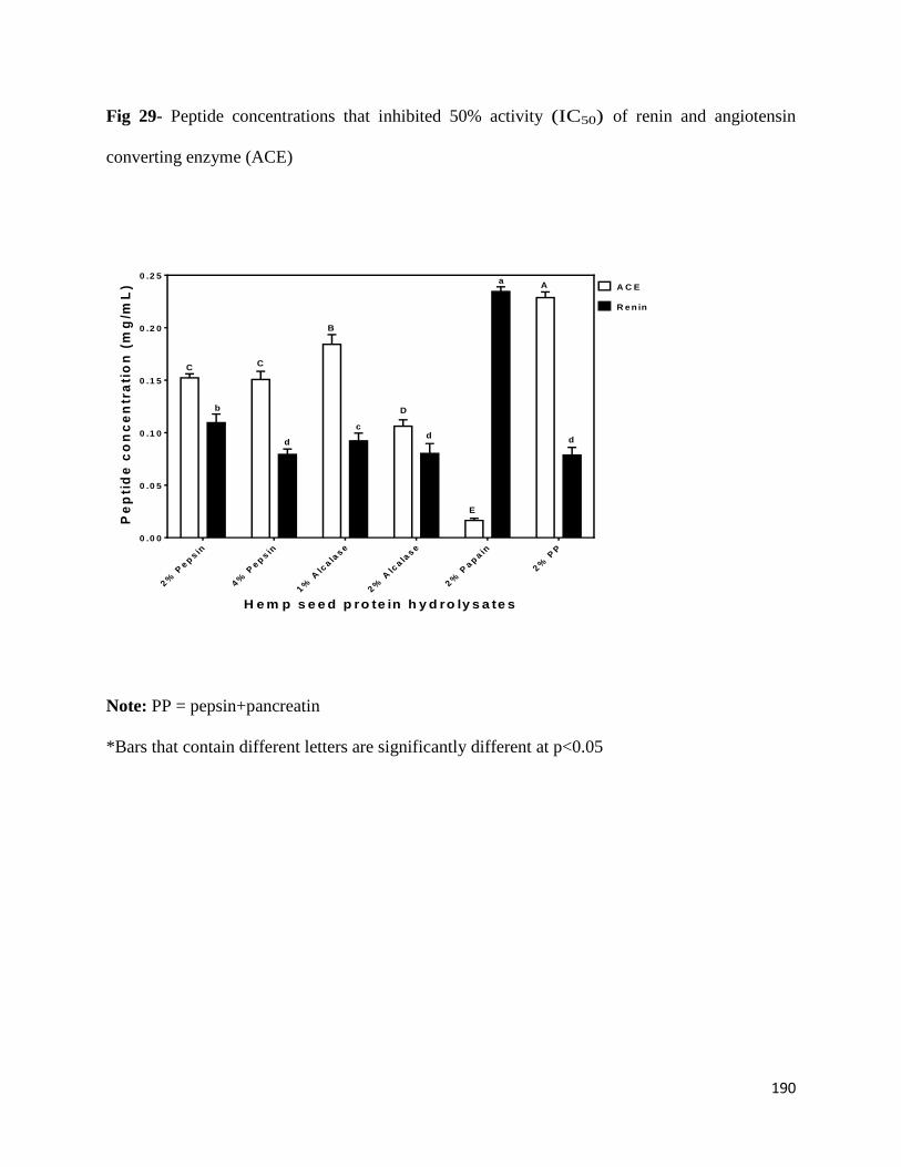
190
Fig 29- Peptide concentrations that inhibited 50% activity (IC50) of renin and angiotensin
converting enzyme (ACE)
Note: PP = pepsin+pancreatin
*Bars that contain different letters are significantly different at p<0.05
H e m p s e e d p ro te in h y d ro ly s a te s
Pe
pti
de
co
nc
en
tra
tio
n (
mg
/mL
)
2%
Pep
sin
4%
Pep
sin
1%
Alc
ala
se
2%
Alc
ala
se
2%
Pap
ain
2%
PP
0 .0 0
0 .0 5
0 .1 0
0 .1 5
0 .2 0
0 .2 5
A C E
R e n in
a
a
b
c
d dd
A
C
B
D
C
E
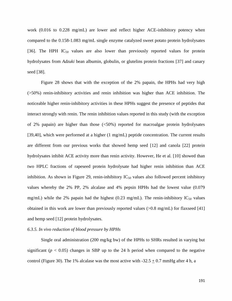
191
work (0.016 to 0.228 mg/mL) are lower and reflect higher ACE-inhibitory potency when
compared to the 0.158-1.083 mg/mL single enzyme catalyzed sweet potato protein hydrolysates
[36]. The HPH IC50 values are also lower than previously reported values for protein
hydrolysates from Adzuki bean albumin, globulin, or glutelins protein fractions [37] and canary
seed [38].
Figure 28 shows that with the exception of the 2% papain, the HPHs had very high
(>50%) renin-inhibitory activities and renin inhibition was higher than ACE inhibition. The
noticeable higher renin-inhibitory activities in these HPHs suggest the presence of peptides that
interact strongly with renin. The renin inhibition values reported in this study (with the exception
of 2% papain) are higher than those (<50%) reported for macroalgae protein hydrolysates
[39,40], which were performed at a higher (1 mg/mL) peptide concentration. The current results
are different from our previous works that showed hemp seed [12] and canola [22] protein
hydrolysates inhibit ACE activity more than renin activity. However, He et al. [10] showed than
two HPLC fractions of rapeseed protein hydrolysate had higher renin inhibition than ACE
inhibition. As shown in Figure 29, renin-inhibitory IC50 values also followed percent inhibitory
values whereby the 2% PP, 2% alcalase and 4% pepsin HPHs had the lowest value (0.079
mg/mL) while the 2% papain had the highest (0.23 mg/mL). The renin-inhibitory IC50 values
obtained in this work are lower than previously reported values (>0.8 mg/mL) for flaxseed [41]
and hemp seed [12] protein hydrolysates.
6.3.5. In vivo reduction of blood pressure by HPHs
Single oral administration (200 mg/kg bw) of the HPHs to SHRs resulted in varying but
significant (p < 0.05) changes in SBP up to the 24 h period when compared to the negative
control (Figure 30). The 1% alcalase was the most active with -32.5 + 0.7 mmHg after 4 h, a
192
Fig 30- Time-dependent changes in systolic blood pressure (SBP) of spontaneously hypertensive
rats (SHRs) after oral administration of different enzymatic hemp protein hydrolysates (HPHs)
and hemp protein isolate (HPI). (*Bars that contain different letters are significantly different at
p<0.05)
2 4 6 8 2 4-4 0
-3 5
-3 0
-2 5
-2 0
-1 5
-1 0
-5
0
5
1 0
S a lin e C a p to p r i l 4 % P e p s in
2 % A lc a la s e 2 % P a p a in 2 % P e p s in + P a n c re a tin H e m p s e e d p r o te in is o la te
T im e a fte r o ra l a d m in is tra tio n (h )
Ch
an
ge
s i
n S
BP
(m
mH
g)
ef
c
oo
nn
bb c
i
o pp
ppp
l
a a
de d
f ffff
e ffg
gg
gg ggg
g gh
hii i
ij
k
l
m
o
2 % P e p s in 1 % A lc a la s e
193
result that is similar to that of the lower dose (10 mg/kg bw) drug (captopril). The 2% pepsin had
significantly (p < 0.05) lower reductions after 2-6 h than the 4% pepsin, which is similar to the
trend for the alcalase HPHs. However, both the 2% and 4% pepsin HPHs produced the longest-
lasting effects with -23.0 + 1.4 mmHg after 24 h of oral administration. The 1% and 2% alcalase
HPHs had least persistence effects with 24 h values of -10.0 + 1.4 mmHg and -7.0+ 1.4 mmHg,
respectively. Interestingly, the SBP-reducing effects did not have a strict direct relationship with
observed in vitro ACE and renin inhibitions. However, the 2% papain with the highest ACE-
inhibitory activity still produced highly significant (p < 0.05) SBP reductions, especially after 4 h
(-30.5 + 0.7 mmHg). But the 2% alcalase which had low ACE and renin inhibition IC50 values
also produced less SBP reductions when compared to other hydrolysates with higher in vitro
inhibitory values.
6.4. Discussion
Apart from elucidating the arrangement of amino acids on a peptide chain, knowledge of
the levels of amino acids present may also provide information on structural basis for observed
hydrolysate inhibitory activities. Only the cysteine level tended to differ among the hydrolysates
but the reason for the low cysteine level in 2% pepsin HPH is unclear. However, we can infer
that at the 2% pepsin concentration, native protein structural restrictions may have limited
proteolysis in areas that contained rigid disulfide bonds. At a higher 4% pepsin concentration,
this restriction was probably overcome with availability of more enzyme molecules, hence the
cysteine content increased to a comparable level with other samples. The significantly higher DH
value for the PP HPH may be attributed to the endo- and exo-peptidase activities of pancreatin,
which enhances protein digestion through hydrolysis of more peptide bonds when compared to
an enzyme with only endopeptidase activity. Moreover, predigestion with pepsin could have
194
enhanced subsequent digestion by pancreatin and may have contributed to the high DH.
Gonzalez-Garcia et al. [35] have also shown that Flavourzyme, an enzyme with similar endo-
and exo-peptidase activities as pancreatin also produced a hydrolysate with highest DH. In
contrast, the 2% pepsin digest had significantly lowest DH value of 4.40 + 0.22%, which may be
due to the specificity of the enzyme for mostly peptide bonds formed by aromatic amino acids.
Alcalase and papain are non-specific endopeptidases, hence the high DH values when compared
to pepsin. DH was higher when enzyme concentration increased, which is consistent with
availability of more enzyme molecules for protein digestion.
The peptide sizes were estimated by SEC and the results showed distribution into four
major peaks with approx. MW ranging from 300 to 9560 Da. These peptide peaks were most
noticeable for the 2% pepsin HPH; increasing the enzyme concentration to 4% led to reduced
intensities of the bigger peptide peaks (2775 and 9560 Da). The results support a higher
proteolysis level at 4% pepsin, which is consistent with the higher DH when compared to the 2%
pepsin. The alcalase and PP HPHs contained a minor peak with size >9560 Da (8-9 mL elution
volume), which indicates a polypeptide that is probably resistant to digestion by these enzymes.
The presence of minor peaks with size <305 Da (most likely free amino acids) were most intense
in the PP, which could have been due to the presence of exoproteases in pancreatin. Overall, the
data show that the HPI was highly susceptible to proteolysis and most of the liberated peptides
were <9650 Da in size. The interrelationship between the MWs of hydrolysates and their
bioactivities in human health application is very crucial in functional foods and nutraceutical
formulations. Thus the presence of such low molecular weight peptides enhances the absorption
potential and ability of the peptides to have bioactive effects during in vivo tests.
195
Variations in fluorescence intensity (FI) reflect structural conformation of proteins and
peptides. Tyrosine and tryptophan are the main chromophores with emission maxima at 303 and
350 nm, respectively [42]. However, fluorescence intensity decreases and wavelength of
maximum emission increases (red shift) upon exposure to aqueous environment because of
water-dependent chromophore quenching. In contrast, fluorescence intensity increases and
wavelength of maximum emission decreases (blue shift) when the chromophores are moved into
a hydrophobic environment. Figure 3 shows two main peaks at 317 nm and 378 nm, which
represent tyrosine and phenylalanine, respectively in a hydrophilic environment. There was also
a tryptophan shoulder peak at 338 nm, which suggests some tryptophan molecules in a more
hydrophobic environment. Thus the tryptophan residues were in two different
microenvironments, probably consisting of folded (338 nm) and open linear (378 nm)
conformations. The HPI FI was very low, which indicates a denatured protein structure with
most of the chromophores exposed to the hydrophilic environment. The higher FI of the peptides
indicates close proximity between the tryptophan molecules and between the tyrosine molecules,
which indicates some level of peptide-peptide interactions. These interactions could have been
facilitated by the shorter and more flexible peptide conformations. The FI data confirms the
structural differences between peptides, which are of short-chain amino acid sequence and the
proteins with long amino acid sequences.
The better ACE-inhibitory activity of the 2% papain HPH may be due to the slightly
higher proline content, when compared to that of the other HPHs. This is because proline has
been suggested as a potency-enhancing factor for ACE-inhibitory peptides based on the fact that
the first two ACE-inhibitory peptides (VPP and IPP) isolated from fermented milk both
contained proline [43]. However, proline content alone was not directly related to ACE-
196
inhibitory activity of all the HPHs, which suggests that other peptide structural factors (e.g.,
proline position, presence of aromatic amino acids, or absence of disulfide bonds) may be
responsible for the observed results. For example, the 2% pepsin HPH has very low cysteine
level while the 2% PP HPH had slightly higher tyrosine level, both of which may have
contributed to the poor ACE-inhibitory activities. The ACE-inhibitory levels are lower than
some of the previously reported values for enzymatic food protein hydrolysates such as Parkia
speciosa seed protein hydrolysates [24]. These differences may be due to the use of different
protein materials for enzyme hydrolysis, since variations in the primary structure will lead to the
liberation of peptides with distinct amino acid composition and sequences.
Unlike ACE, only very few reports [10,11,14,16,38,44] are available for peptide-
dependent renin inhibition. A higher renin-inhibition seems to be related to phenylalanine
content since this amino acid was highest in the 2% pepsin, 4% pepsin and 2% PP HPHs.
However, other structural features (e.g., amino acid arrangement and type) are likely to be
involved in determining renin-inhibitory properties of the HPHs. A previous report has suggested
the importance of branched-chain amino acids and aromatic amino acids in potentiating renin-
inhibitory properties of food protein-derived peptides [45]. Generally, renin inhibitory activity of
the hydrolysates was higher than ACE-inhibitory activity, which is different from other reports
that showed a reverse trend [9,12,22,46]. Overall, these results confirm previously published
literature data that indicate peptide-dependent ACE-inhibitory potency are more likely to be
different from that renin inhibition. Since catalytic mechanisms differ between ACE and renin,
molecules that have strong inhibition against one enzyme may not necessary have the same
inhibition level against the other enzyme.
197
Single oral administration of the HPHs to SHRs resulted in varying but significant
(P<0.05) changes in SBP up to the 24 h period when compared to the negative control (Figure
30). The 1% alcalase was the most active with -32.5 + 0.7 mmHg after 4 h, a result that is similar
to that of the lower dose drug (captopril). The 2% pepsin had significantly (P<0.05) lower
reductions than the 4% pepsin after 2-6 h, which is similar to the trend for the alcalase HPHs and
indicate higher enzyme concentrations may not necessarily produce more effective
antihypertensive peptides. The results are similar to the work of Alashi et al. [22] who also
showed that alcalase and pepsin canola protein digests produced the most SBP reductions in
SHR also at 200 mg/kg bw dose. He et al. [9] also showed that an alcalase digest of rapeseed was
the most effective SBP-reducing agent among several enzymes used. The results suggest that the
lower renin-inhibition potency of the 2% papain HPH may have been compensated for by the
higher ACE-inhibitory activity, hence strong reductions in SBP. The lack of direct correlation
between in vitro and in vivo activities of the HPHs is consistent with previous reports [9,22]. The
maximum SBP reductions obtained in this work are similar to the maximum value obtained for a
sweet potato protein hydrolysate, which was administered at a higher 500 mg/kg bw dose [36].
Pistachio [17] and almond [19] protein hydrolysates at 1000 and 800 mg/kg bw doses,
respectively also produced lower SBP-reducing effects than the HPHs used in this work. In
contrast to the HPHs, the unhydrolyzed HPI was less active (SBP values of -1 to -3 mmHg),
which suggests that enzymatic predigestion converted the inactive proteins into bioactive
peptides. Even though predigestion with pepsin alone or in combination with pancreatin
produced SBP-reducing HPHs, the lack of effect after ingestion of HPI suggests that other in
vivo factors (buffering capacity and duration of hydrolysis) may have prevented effective
digestion and liberation of antihypertensive peptides in the SHR gastrointestinal tract. Similar
198
works with pea [23] and rapeseed [9] have also shown better and faster SBP-reducing effects of
predigested proteins when compared to undigested proteins.
6.5 Conclusion
The present study confirmed the dual in vitro ACE and renin inhibition capacity of
enzymatic HSP digests. There was no relationship between DH and ACE inhibitory activity of
the protein hydrolysates though renin inhibition tended to be inversely related to DH, which
suggests longer hemp seed peptide chains may be more potent in vitro renin inhibitors than
shorter chains. The DH and in vitro ACE or renin inhibition had no relationship with actual SBP-
reducing effects in SHR, which suggests peptide amino acid sequence or synergistic effects
between different peptides may be the determining factors for the observed antihypertensive
potency. The significantly (P<0.05) longer-lasting SBP-reducing effects of the pepsin digests,
which also had the least DH (longer peptide chains) is an indication of reduced rate of absorption
coupled with higher resistance to enzymatic clearance after absorption from the gastrointestinal
tract. The alcalase digest had the fastest and most SBP-reduction and may be combined with the
papain digest (also highly potent with long-lasting effects) to produce consistent SBP-lowering
effect on a 24 h basis. However, a combination of pepsin and alcalase digests may also produce
desirable 24 h SBP-reducing effects. Further studies are needed to determine the amino acid
sequence of the most active HPH peptides (pepsin, alcalase, and papain) in order to perform
structure-function studies. Overall, the results provide critical information on HPHs that may be
used as active ingredients to formulate antihypertensive functional foods and nutraceuticals.
Acknowledgement
This work was funded through the Natural Sciences and Engineering Research Council of
Canada (NSERC) Discovery grant to Dr. R.E. Aluko. SA Malomo is a recipient of University of
199
Manitoba Graduate Fellowship (UMGF) and Manitoba Graduate Scholarship (MGS) Awards for
doctoral studies.
Author Contributions
Sunday Malomo performed all the in vitro tests and data analysis; he also wrote the first
draft of this manuscript. Sunday Malomo, John Onuh and Abraham Girgih conducted the rat
feeding and blood pressure measurements. Dr. Rotimi Aluko is the grant holder for the project;
he produced the experimental concept and edited the final manuscript draft.
References
1. WHO. World Health Organization. (2013). A global brief on hypertension; silent killer,
global public health crisis. Geneva, USA. WHO/DCO/WHD/2013.2. 40pp. Internet:
http://www.who.int/cardiovascular_diseases/publications/global_brief_hypertension/en/
(accessed July 04, 2015).
2. Guessous, I.; Bochud, M.; Theler, J-M.; Gaspoz, J-M.; Pechère-Bertschi, A. 1999–2009
Trends in prevalence, unawareness, treatment and control of hypertension in Geneva,
Switzerland. PLoS One 2012, 7, e39877.
3. Cai, B.; McAdam-Marx, C. The determinants of antihypertensive use and expenditure in
patients with hypertension in the USA. J. Pharm. Health Serv. Res. 2014, 5, 11–18.
4. Anderson, I.; Ivatts, S.; Somanathan, A.; Rolfe, B. The challenges of health financing in five
South Pacific countries. Asian-Pacific Econ. Lit. 2014, 28, 29–46.
5. Daien, V.; Duny, Y.; Ribstein, J.; Du, C.G.; Mimran, A.; Villain, M.; Daures, J.P.; Fesler, P.
Treatment of hypertension with renin-angiotensin system inhibitors and renal dysfunction: a
systematic review and meta-analysis. Am. J. Hypertens. 2012, 25, 126-132.
200
6. Aluko, R.E. Antihypertensive peptides from food proteins. Annu. Rev. Food Sci. Technol.
2015, 6, 235-262.
7. Pihlanto, A.; Mäkinen, S. Antihypertensive properties of plant protein derived peptides. In
Bioactive Food Peptides in Health and Disease; B. Hernandez-Ledesma, C. Hsieh, Eds.;
INTECH Publishers, Rijeka, Croatia, 2013; pp. 145-182.
8. Norris, R.; FitzGerald, R. Antihypertensive peptides from food proteins. In Bioactive Food
Peptides in Health and Disease; B. Hernandez-Ledesma, C. Hsieh, Eds.; INTECH
Publishers, Rijeka, Croatia, 2013; pp. 45–72..
9. He, R.; Alashi, A.; Malomo, S.A.; Girgih, A.T.; Chao, D.; Ju, X.; Aluko, R.E.
Antihypertensive and free radical scavenging properties of enzymatic rapeseed protein
hydrolysates. Food Chem. 2013, 141, 153-159.
10. He, R.; Malomo, S.A.; Alashi, A.; Girgih, A.T.; Ju, X.; Aluko, R.E. Purification and
hypotensive activity of rapeseed protein-derived renin and angiotensin converting enzyme
inhibitory peptides. J. Funct. Foods 2013, 5, 781–789.
11. He, R.; Malomo, S.A.; Girgih, A.T.; Ju, X.; Aluko, R.E. Glycinyl-histidinyl-serine (GHS), a
novel rapeseed protein-derived peptide has blood pressure-lowering effect in spontaneously
hypertensive rats. J. Agric. Food Chem. 2013, 61, 8396–8402.
12. Girgih, A.T.; Udenigwe, C.C.; Li, H.; Adebiyi, A.P.; Aluko, R.E. Kinetics of enzyme
inhibition and antihypertensive effects of hemp seed (Cannabis sativa L.) protein
hydrolysate. J. Am. Oil Chem. Soc. 2011, 88, 1767–1774.
13. Girgih, A.T.; He, R.; Alashi, A.M.; Malomo, S.A.; Aluko, R.E. Preventive and treatment
effects of hemp seed (Cannabis sativa L.) meal protein hydrolysate against blood pressure
in spontaneously hypertensive rats. Eur. J. Nutr. 2014, 53, 1237-1246.
201
14. Girgih, A.T.; He, R.; Malomo, S.A.; Aluko, R.E. Structural and functional characterization
of hemp seed (Cannabis sativa L.) protein-derived antioxidant and antihypertensive
peptides. J. Funct. Foods, 2014, 6, 384-394.
15. Mundi, S.; Aluko, R.E. Inhibitory properties of kidney bean protein hydrolysate and its
membrane fractions against renin, angiotensin converting enzyme, and free radicals. Austin
J. Nutr. Food Sci. 2014, 2, 1-11.
16. Fitzgerald, C.; Aluko, R.E.; Hossain, M.; Rai, D.K.; Hayes, M. Potential of a renin
inhibitory peptide from the red seaweed (Palmaria palmata) as a functional food ingredient
following confirmation and characterization of a hypotensive effect in spontaneously
hypertensive rats. J. Agric. Food Chem. 2014, 62, 8352−8356.
17. Li, P.; Jia, J.; Fang, M.; Zhang, L.; Guo, M.; Xie, J.; Xia, Y.; Zhou, L.; Wei, D. In vitro and
in vivo ACE inhibitory of pistachio hydrolysates and in silico mechanism of identified
peptide binding with ACE. Process Biochem. 2014, 49, 898–904.
18. Zhang, C.; Cao, W.; Hong, P.; Ji, H.; Qin, X.; He, J. Angiotensin I-converting enzyme
inhibitory activity of Acetes chinensis peptic hydrolysate and its antihypertensive effect in
spontaneously hypertensive rats. Int. J. Food Sci. Technol. 2009, 44, 2042–2048.
19. Wang, C.; Tian, J.; Wang, Q. ACE inhibitory and antihypertensive properties of apricot
almond meal hydrolysate. Eur. Food Res. Technol. 2011, 232, 549–556.
20. Huang, W.H.; Sun, J.; He, H.; Dong, H.W.; Li, J.T. Antihypertensive effect of corn
peptides, produced by a continuous production in enzymatic membrane reactor, in
spontaneously hypertensive rats. Food Chem. 2011, 128, 968–973.
21. Doyen, A.; Udenigwe, C.C.; Mitchell, P.L.; Marette, A.; Aluko, R.E. Anti-diabetic and
antihypertensive activities of two flaxseed protein hydrolysate fractions revealed following
202
their simultaneous separation by electrodialysis with ultrafiltration membranes. Food Chem.
2014, 145, 66–76.
22. Alashi, A.M.; Blanchard, C.L.; Mailer, R.J.; Agboola, S.O.; Mawson, A.J.; He, R.; Malomo,
S.A.; Girgih, A.T.; Aluko, R.E. Blood pressure lowering effects of Australian canola protein
hydrolysates in spontaneously hypertensive rats. Food Res. Int. 2014, 55, 281-287.
23. Li, H.; Prairie, N.; Udenigwe, C.C.; Adebiyi, A.P.; Tappia, P.S.; Aukema, H.M.; Jones,
P.J.H.; Aluko, R.E. Blood pressure lowering effect of a pea protein hydrolysate in
hypertensive rats and humans. J. Agric. Food Chem. 2011, 59, 9854-9860.
24. Siow, H.; Gan, C. Extraction of antioxidative and antihypertensive bioactive peptides from
Parkia speciosa seeds. Food Chem. 2013, 141, 3435–3442.
25. Marczak, E.; Usui, H.; Fujita, H.; Yang, Y.; Yokoo, M.; Lipkowski, A.; Yoshikawa, M.
New antihypertensive peptides isolated from rapeseed. Peptides, 2003, 24, 791-798.
26. Ambigaipalan, P., Al-Khalifa, A.S., Shahidi, F. Antioxidant and angiotensin I converting
enzyme inhibitory activities of date seed protein hydrolysates prepared using Alcalase,
Flavourzyme and Thermolysin. J. Funct. Foods, 2015, doi: 10.1016/j.jff.2015.01.021
27. Markwell, M.A.K.; Haas, S.M.; Bieber, L.L.; Tolbert, N.E. Modification of Lowry
procedure to simplify protein determination in membrane and lipoprotein samples. Anal.
Biochem. 1978, 87, 206-210.
28. Adler-Nissen, J. Determination of the degree of hydrolysis of food protein
hydrolysates by trinitrobenzenesulfonic acid. J. Agric. Food Chem. 1979, 27,
1256-1262.
29. Bidlingmeyer, B.A.; Cohen, S.A.; Tarvin, T.L. Rapid analysis of amino acids using pre-
column derivatization. J. Chrom. B 1984, 336, 93-104.
203
30. Gehrke, C.W.; Wall, L.L.; Absheer, J.S.; Kaiser, F.E.; Zumwalt, R.W. Sample preparation
for chromatography of amino-acids: acid hydrolysis of proteins. J. Assoc. Official Anal.
Chem. 1985, 68, 811-821.
31. Landry, J.; Delhaye, S. Simplified procedure for the determination of tryptophan of foods
and feedstuffs from barytic hydrolysis. J. Agric. Food Chem. 1992, 40, 776-779.
32. Li, H.; Aluko, R.E. Structural modulation of calmodulin and calmodulin-dependent protein
kinase II by pea protein hydrolysates. Int. J. Food Sci. Nutr. 2006, 57, 178-189.
33. Holmquist, B.; Bunning, P.; Riordan, J.F. A continuous spectrophotometric assay for
angiotensin converting enzyme. Anal. Biochem. 1979, 95, 540-548.
34. Reagan-Shaw, S.; Nihal, M.; Ahmad, N. Dose translation from animal to human studies
revisited. FASEB J. 2007, 22, 659-661.
35. Gonzalez-Garcia, E.; Marina, M.L.; Garcia, M.C. Plum (Prunus domestica L.) by-product as
a new and cheap source of bioactive peptides: Extraction method and peptides
characterization. J. Funct. Foods, 2014, 11, 428-437.
36. Ishiguro, K.; Sameshima, Y.; Kume, T.; Ikeda, K.; Matsumoto, J.; Yoshimoto, M.
Hypotensive effect of a sweet potato protein digests in spontaneously hypertensive rats and
purification of angiotensin I-converting enzyme inhibitory peptides. Food Chem. 2012, 131,
774-779.
37. Durak, A.; Baraniak, B.; Jakubczyk, A.; Swieca, M. Biologically active peptides obtained
by enzymatic hydrolysis of Adzuki bean seeds. Food Chem. 2013, 141, 2177-2183.
38. Estrada-Salas, P.A.; Montero-Moran, G.M.; Martínez-Cuevas, P.P.; Gonzalez, C.; Barba de
la Rosa, A.P. Characterization of antidiabetic and antihypertensive properties of canary seed
(Phalaris canariensis L.) peptides. J. Agric. Food Chem. 2014, 62, 427-433.
204
39. Harnedy, P.A.; FitzGerald, R.J. In vitro assessment of cardioprotective, anti-diabetic and
antioxidant potential of Palmaria palmata protein hydrolysates. J. Appl. Phycol. 2013, 25,
1793–1803.
40. Fitzgerald, C.; Mora-Soler, L.; Gallagher, E.; O’Connor, P.; Prieto, J.; Soler-Vila, A.;
Hayes, M. Isolation and characterization of bioactive pro-peptides with in vitro renin
inhibitory activities from the macroalga Palmaria palmata. J. Agric. Food Chem. 2012, 60,
7421–7427.
41. Udenigwe, C.C.; Lin, Y.S.; Hou, W.C.; Aluko, R.E. Kinetics of the inhibition of renin and
angiotensin I-converting enzyme by flaxseed protein hydrolysate fractions. J. Funct. Foods,
2009, 1, 199-207.
42. Schmid, F.X. Spectra methods of characterizing protein conformation and conformational
changes. In Protein structure: A practical Approach; T.E. Creighton, Ed.; Springer
Publishing Co., NY, USA, 1989; pp. 251–285.
43. Nakamura, Y.; Yamamoto, N.; Sakai, K.; Takano, T. Antihypertensive effect of sour milk
and peptides isolated from it that are inhibitors of angiotensin converting enzyme. J. Dairy
Sci. 1995, 78, 1253–1257.
44. Li, H.; Aluko, R.E. Identification and inhibitory properties of multifunctional peptides from
pea protein hydrolysate. J. Agric. Food Chem. 2010, 58, 11471-11476.
45. Aluko, R.E. Structure and function of plant protein-derived antihypertensive peptides. Curr.
Op. Food Sci. 2015, 4, 44–50.
46. Onuh, J.O.; Girgih, A.T.; Malomo, S.A.; Aluko, R.E.; Aliani, M. Kinetics of in vitro renin
and angiotensin converting enzyme inhibition by chicken skin protein hydrolysates and their

205
blood pressure lowering effects in spontaneously hypertensive rats. J. Funct. Foods 2015,
14, 133-143.
206
6.6 Statement transfer between the study 4 and study 5
In recent years, research findings have made progress toward the development of naturally
occurring bioactive multifunctional peptides (or protein hydrolysates). The previous chapter
reported the effective lowering of blood pressure by hemp seed protein hydrolysates. In this
chapter, enzymatic protein hydrolysis was also used to produce a different set of protein fractions
that can modulate activities of one of the agents involved in the nervous system.
Acetylcholinesterase (AChE) is a main factor involved in neurological disorders and is therefore,
a target for therapeutic agents against dementia and Alzheimer’s disease. Since the AChE
substrate has a structural resemblance to peptides, enzymatic digestion of hemp seed proteins
could produce very active peptide inhibitory agents. Therefore, in this chapter, optimized
enzymatic hydrolysis of hemp seed proteins was carried out at different enzyme:substrate
concentrations in order to generate a wide range of peptides with varied structural properties.
The hemp seed protein hydrolysates were then tested for in vitro AChE-inhibitory properties as
potential agents for human cognitive health improvement. The most active peptide fractions were
examined for peptide sequences to determine type of amino acids that are important for potent
AChE-inhibition.
207
CHAPTER SEVEN
7. Study on the in vitro acetylcholinesterase-inhibitory properties of enzymatic hemp seed
protein hydrolysates
Sunday A. Malomo and Rotimi E. Aluko*
The Department of Human Nutritional Sciences, University of Manitoba, Winnipeg, Manitoba,
Canada R3T 2N2
*Corresponding author: Dr. Rotimi E. Aluko; E-Mail: [email protected]; Tel.: +1-204-
474-9555; Fax: +1-204-474-7593.
208
Abstract
The aim of this work was to produce acetylcholinesterase (AChE)-inhibitory peptides through
enzymatic hydrolysis of hemp seed proteins. Hydrolysis was performed using six different
proteases (pepsin, papain, thermoase, flavourzyme, alcalase and pepsin+pancreatin) at different
concentrations (1-4%). Degree of hydrolysis (DH) was directly related to amount of protease
used but had no relationship with AChE-inhibitory activity. Amino acid composition results
showed that the hemp seed protein hydrolysates (HPHs) had high levels of negatively charged
amino acids (39.62-40.18%) as well as arginine. The 1% pepsin HPH was the most active AChE
inhibitor with ~6 µg/mL IC50 value when compared to 8-11.6 µg/mL for the other HPHs. The
1% pepsin HPH was dominated by high cysteine-containing peptides though negatively charged
and aromatic amino acids were also present. We conclude that formation of disulfide bonds
between peptides and AChE polypeptides may constitute an important use as potential inhibitory
mechanism. The HPHs could be an important source of agents for maintaining a healthy nervous
system.
Key words: hemp seed; protein hydrolysate; peptides; acetylcholinesterase
209
7.1. Introduction
The serine hydrolase, acetylcholinesterase (AChE), has long been known to play a very
crucial physiological role in maintaining the major neurotransmitter, acetylcholine (ACh) in the
body by rapidly clearing free ACh from the synapse (Kumar & Chowdhury, 2014). During
neurotransmission, ACh is synthesized from choline and acetyl-CoA by the enzyme choline
acetyltransferase. ACh is then released from the nerve into the synaptic cleft where it binds to
receptors on the post-synaptic membrane thereby relaying the signal from the nerve to the brain
(Singh et al., 2013). AChE converts excess ACh into the inactive metabolites, choline and
acetate, thus promoting normal brain and muscles functions by preventing over-stimulation of
the nervous system. The liberated choline is taken up again by the pre-synaptic nerve and
combined with acetyl-CoA to synthetize ACh through the action of choline acetyltransferase
(Singh et al., 2013). Thus normal functioning of AChE also ensures proper recycling of choline
and acetyl-CoA such that optimal level of ACh is maintained within the nervous system. As
aging and dietary changes set in, less ACh is synthesized due to non-regeneration of used
neurons and shortage of choline. Meanwhile, AChE continues to hydrolyze the little amounts of
ACh; net result is low levels of ACh. This leads to reduced functioning of nerve impulse and
inadequate signal transmission to the brain, thus eventually contributing to memory impairment
such as in Alzheimer’s disease (AD) and dementia (Willcox et al., 2014).
The increasing occurrence of AD has been implicated as a contributing factor to higher
global morbidity/ mortality rate (US Burden of Disease Collaborators, 2013). This is because AD
is a progressive, degenerative disease which is characterized by memory loss, language
deterioration, poor judgment and impaired visuospatial skill. The cholinergic neurotransmission
dysfunction in the brain, formation and growth of brain amyloid lesions, and senile plaques, are
210
widely believed and reported as crucial events in the pathogenesis of AD (Alzheimer’s
Association, 2013). The various socio-economic factors leading to AD have been suggested to
include diet, population aging and lifestyle. For instance, the aging process affects the diet of
individuals by partially or totally changing their diet type as well as the quality and quantity of
consumed foods (Willcox et al., 2014).
AChE inhibition is an important research in the wide range of associated health
implications in humans because of the ability to hydrolyze ACh at cholinergic synapses with
high catalytic efficiency than other known enzymes (Saravanaraman et al., 2014; Zare-Zardini
et al., 2013). AChE inhibitors are therefore, employed to reduce the rate at which ACh is broken
down, thereby increasing brain ACh concentration and combating the loss caused by the death of
cholinergic neurons (Hasnat et al., 2013). The approach involves the use of compounds that
possess structural similarity to ACh such that they can fit into the enzyme active site, thereby
reducing catalysis rate (Saravanaraman et al., 2014; Singh et al., 2013). As of today the current
available therapy for AD consists of the administration of synthetic AChE inhibitors due to their
clinical efficacy in prolonging the half-life of ACh (Kumar and Chowdhury, 2014). Such
synthetic drugs (like tacrine, rivastigmine, galantamine, memantine and donepezil) are
characterized by common negative side effects like nausea, vomiting, muscle cramps, decreased
heart rate (bradycardia), decreased appetite and weight as well as increased gastric acid
production (Ghribia et al., 2014; Iannello et al., 2014; Saravanaraman et al., 2014).
However, many approaches have been recently employed to address this critical problem
of negative side effects, some of which includes the uses of natural polyphenolic plant products
and their secondary metabolites (Borowiec et al., 2014; Ghribia et al., 2014; Iannello et al., 2014;
Kumar & Chowdhury, 2014). Zare-Zardini et al. (2013) recently isolated a 31-mer peptide
211
(Snakin-Z) from Ziziphus jujuba fruits, which inhibited in vitro AChE activity. However, to the
best of our knowledge, there is scanty information on the potential use of naturally occurring
bioactive peptides (hydrolysates) from plant proteins as AChE inhibitors. Food protein
hydrolysates contain peptides that consist of peptide bonds, which have structural similarity to
the ester bond in ACh. Moreover, several peptides contain positively charged nitrogen atoms just
like found in ACh. Therefore, theoretically, small-size peptides could simulate the structural
conformation of ACh and enable peptide interaction with the AChE active site. Since peptides
are not AChE substrates, such binding to the active site could exclude ACh, slow down enzyme
catalysis and potentially enhance physiological levels of ACh during aging.
Food protein-derived hydrolysates have reduced risk of imparting negative side effects in
consumers and therefore, could serve as suitable alternatives or complementary treatment to AD
drugs. Previous works have demonstrated several in vitro (Girgih et al., 2011a, 2013) and in vivo
(Girgih et al., 2011b, 2014a,b) antioxidant and antihypertensive effects of hemp seed protein
hydrolysates (HPH). A previous work also showed the oxidative apoptosis protective effect of
hemp seed peptides (Lu et al., 2010) but so far there has been no report of AChE inhibition by
food protein hydrolysates. Therefore, the aim of this study was to determine, for the first time,
the in vitro AChE-inhibitory properties of several HPH generated from the action of different
enzymes during proteolysis of hemp seed plant proteins. The amino acid composition, peptide
size distribution and potential peptide sequences of the most active HPHs were determined.
7.2. Materials and methods
7.2.1. Hemp seed products and chemical reagents
Hemp seed protein meal (HPM) (37% protein content) produced as a by-product of the hemp
seed oil processing industry was a gift from Hemp Oil Canada (St. Agathe, Manitoba, Canada).
212
All enzymes- pepsin (porcine gastric mucosa; E.C. 3.4.23.1; ≥250 U/mg), papain (papaya latex;
E.C. 3.4.22.2), alcalase (Bacillus licheniformis; E.C. 3.4.21.62), flavourzyme (Aspergillus
oryzae; ≥500 U/g), pancreatin (porcine pancreas; 647-014-00-9) and Electrophorus electricus
(electric eel; 1256 U/mg protein) acetylcholinesterase Type VI-S were purchased from Sigma-
Aldrich (St. Louis, MO, USA). Thermoase (Aspergillus sp.; Protease A "Amano" 2 SD) was a
gift from Amano Enzyme Inc (Nishiki, Naka-ku, Nagoya Japan). Dithio-bis-nitrobenzoic acid
(DTNB) and acetylthiocholine were also purchased from Sigma-Aldrich. Other analytical-grade
reagents were obtained from Fisher Scientific (Oakville, ON, Canada).
7.2.2. Preparation of hemp seed protein isolate (HPI)
HPI was produced from HPP according to the method of Girgih et al. (2011a) with slight
modifications. HPP was dispersed in deionized water (1:20, w/v) and the dispersion was adjusted
to pH 8.0 using 2 M NaOH to solubilize the proteins while stirring at 37 oC for 2 h; this was
followed by centrifugation (7000g, 1 h, 4 oC). The precipitate was discarded and the supernatant
filtered with cheese-cloth, adjusted to pH 5.0 with 2 M HCl to precipitate the proteins and then
centrifuged (7000g, 1 h, 4 oC). The resultant precipitate was re-dispersed in deionized water,
adjusted to pH 7.0 with 2 M NaOH and freeze-dried to obtain the HPI. Protein concentration of
the HPI was determined using the modified Lowry method (Markwell et al., 1978).
7.2.3. Preparation of enzymatic hemp seed protein hydrolysates (HPHs)
Hydrolysis of the HPI was conducted using each of the following enzymes and reaction
conditions: alcalase (50 oC, pH 8.0, 4 h); papain (65
oC, pH 6.0, 4 h), pepsin (37
oC, pH 2.0, 2 h),
thermoase (50 oC, pH 8.0, 4 h), flavourzyme (50
oC, pH 8.0, 4 h) and pepsin+pancreatin (first, 37
oC, pH 2.0, 2 h; then at the same 37
oC, pH 7.5, 4 h). HPI (5%, w/v, protein basis) was suspended
in deionized water in a reaction vessel equipped with a stirrer, heated to the appropriate
213
temperature and adjusted to the appropriate pH value prior to addition of the proteolytic
enzymes. Each protease was added to the HPI slurry at five different enzymes to substrate
protein ratios (E/S): 0.5:100, 1:100, 2:100, 3:100, and 4:100. During digestion, the reaction
mixture pH was maintained constant by addition of 1 M NaOH. After digestion, the enzymes
were inactivated by immersing reaction vessel in hot water bath at 95 oC for 10 min and allowed
to cool down. The undigested proteins were precipitated by centrifugation (8000 g, 60 min at 4
oC) while the supernatant containing target peptides was freeze dried as the HPH and stored at -
20 oC until needed for further analysis. The protein contents of the freeze dried HPHs were
determined using the modified Lowry method (Markwell et al., 1978).
7.2.4. Amino acid composition analysis
The amino acid profiles of HPI and HPHs were determined using the HPLC S4300 Amino Acid
Analyzer, (Sykam Mfd Co., Eresing, Bavaria, Germany) according to the method previously
described (Bidlingmeyer et al., 1984) after samples were digested with 6 M HCl for 24 hr. The
cysteine and methionine contents were determined after performic acid oxidation (Gehrke et al.,
1985) and the tryptophan content was determined after alkaline hydrolysis (Landry & Delhaye,
1992).
7.2.5. Determination of degree of hydrolysis (DH)
The percent DH of each HPH was determined according to the trinitrobenzene sulfonic acid
(TNBS) method as previously described (Adler-Nissen, 1979). Briefly, HPI was digested under
vacuum with 6 M HCl for 24 hr and the digest was used to determine total amino groups as L-
leucine equivalent. The DH was calculated as percentage ratio of the leucine equivalent of HPHs
to that of HPI.
7.2.6. Analysis of molecular weight distribution
214
Molecular weight (MW) distribution of HPHs was determined as previously described (He at al.,
2013) using an AKTA FPLC system (GE Healthcare, Montreal, PQ) equipped with a Superdex
Peptide 10/300 GL column (10 x 300 mm) and UV detector (λ= 214 nm). The column was
calibrated with Cytochrome C (12,384 Da), Aprotinin (6512 Da), Vitamin B12 (1855 Da), and
Glycine (75 Da) as the standard proteins. Peptide sizes of the samples were estimated from a plot
of log MW versus elution volume of the standard proteins.
7.2.7. Mass spectrometry analysis of protein hydrolysates
The mass spectrometry analysis of the protein hydrolysates is done according to the previously
described method (He et al., 2013) with slight modifications. Briefly, a 10 ng/ µL aliquout of the
active impure HPHs, being dissolved in deionized water (containing 0.1% formic acid as solvent
A) was subjected to mass spectrometry (MS) scan analysis. The elution and MS scan was
monitored with the AB SCIEX QTRAP® 6500 MS System (AB SCIEX Pte. Ltd., Foster City,
California, USA) coupled with electrospray ionization source using the following parameters:
Ion source, Turbo Spray IonDrive; Curtain Gas, 30.0; IonSpray Voltage, 3.5 kV; Temperature,
150 °C; Ion Source Gas, 30; Declustering Potential, 100; Enterance Potential, 10; Flow rate, 30
µL/min for 3 min in the positive ion mode.
7.2.8. Acetylcholinesterase (AChE) inhibition assays
AChE inhibition assay was performed as previously described (Ellman et al., 1961) with slight
modifications. Briefly, the reaction was carried out at room temperature using 0.1 M sodium
phosphate buffer (pH 7.5) and the final assay mixture was 200 μL in a 96-well microplate
(Costar, Corning, NY, USA). Aliquots of 20 µL HPHs were added to the microplate wells that
contained 130 µL of the buffer. DTNB (20 µL) and acetylthiocholine (10 µL) were added to
each well. Then, 20 µL of AChE (0.5 U/mL final assay concentration) was added to initiate the

215
reaction and the mixture incubated for 15 min at room temperature. The absorbance and the
reaction kinetics were read for 2 min at wavelength of 412 nm using a Synergy H4 microplate
reader (Biotek Instruments Inc., Winooski, Vermont, USA). In the uninhibited wells, 20 µL of
the buffer was used to replace HPH. In the background wells, 20 µL of the buffer was used to
replace AChE while 40 µL of buffer was used to replace both AChE and HPH in the blank wells.
The concentration of sample that inhibited 50% AChE activity (IC50) was calculated from a non-
linear regression plot of percentage AChE activity versus HPH concentration.
7.2.9. Statistical analysis
Triplicate determinations were used to obtain mean values and standard deviations. Statistical
analysis was performed with SAS (Statistical Analysis Software 9.1) using one-way ANOVA.
Duncan's multiple-range test was carried out to compare the mean values for samples and
significant differences taken at p < 0.05.
7.3 Results and Discussion
7.3.1. Degree of hydrolysis (DH) and AChE inhibition
Table 10 shows that the DH was significantly (p < 0.05) affected by the type and level of
proteases, which is consistent with the fact that these proteases have different specificities and
will produce peptides at different rates. The flavourzyme-produced HPHs have the highest (p <
0.05) DH values, which can be attributed to the dual endoprotease and exopeptidase activity
possessed by the enzyme. In contrast, thermoase and pepsin hydrolysates had the least DH
values, which suggest that several peptide bonds within the hemp seed proteins were not
accessible or were resistant to the catalytic activities of these two enzymes. As expected, higher
DH values were obtained as the amount of protease used was increased, which indicates
216
increased proteolysis. Protein content of the HPHs ranged from ~81-91%; therefore, AChE
assays were performed on protein weight basis.
The data on inhibitory properties of the HPHs show that there was no direct relationship
between level of enzyme used and the ability of the protein hydrolysate to reduce AChE activity
(Table 10). Unlike the DH data, there was no relationship between amount of enzyme used and
the AChE-inhibitory activity of the resultant HPH. Thus, the results suggest that there is no
direct relationship between peptide size (inversely related to DH) and AChE-inhibitory activity
of the HPHs. It is more likely that AChE-inhibitory activity of the HPHs was determined mostly
by the type and sequence of amino acids on the peptide chain. The results show that several of
the HPHs have higher AChE-inhibitory activity than the 31 amino acid residue peptide (Snakin-
Z) isolated from Ziziphus jujuba fruits (Zare-Zardini et al., 2013). At 10 µg/mL, the AChE
activity inhibition by Snakin-Z was <20% when compared to several of the HPHs with >20%
inhibition at same peptide concentration.
The higher AChE-inhibitory effects of some of the protein hydrolysates may be due to
synergistic effects of several peptides when compared to the single Snakin-Z peptide. The
highest (p < 0.05) AChE inhibition of 54% was by the 1% pepsin hydrolysate, which suggests
the presence of peptides that bind strongly to AChE protein. The AChE-inhibitory activities of
the HPHs which was up to 54% for the 1% pepsin hydrolysate is similar to the maximum value
of 57% that was reported by Hasnat et al. (2013) for a Ganoderma lucidum aqueous
polyphenolic extract at 2.00 mg/mL. Danis et al. (2014) also reported about 56% AChE inhibition
by a methanolic extract of Rhus coriaria leaves but at 1.2 mg/mL. While it may not be totally
correct to compare polyphenolic activity with that of a protein hydrolysate, it should be noted
that the HPHs were tested at 120-200 times less than the natural plant extract concentrations.
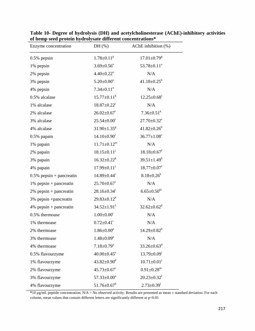
217
Table 10- Degree of hydrolysis (DH) and acetylcholinesterase (AChE)-inhibitory activities
of hemp seed protein hydrolysate different concentrations*
Enzyme concentration DH (%) AChE inhibition (%)
0.5% pepsin 1.78±0.11p 17.01±0.79
g
1% pepsin 3.69±0.56o 53.78±0.11
a
2% pepsin 4.40±0.22o N/A
3% pepsin 5.20±0.00o 41.18±0.25
b
4% pepsin 7.34±0.11n N/A
0.5% alcalase 15.77±0.11k 12.25±0.68
i
1% alcalase 18.87±0.22j N/A
2% alcalase 26.02±0.67i 7.36±0.51
k
3% alcalase 25.54±0.00i 27.70±0.32
e
4% alcalase 31.90±1.35g 41.82±0.26
b
0.5% papain 14.10±0.90l 36.77±1.08
c
1% papain 11.71±0.12m N/A
2% papain 18.15±0.11j 18.18±0.67
f
3% papain 16.32±0.22k 39.51±1.49
b
4% papain 17.99±0.11j 18.77±0.07
f
0.5% pepsin + pancreatin 14.89±0.44l 8.18±0.26
k
1% pepsin + pancreatin 25.70±0.67i N/A
2% pepsin + pancreatin 28.16±0.34i 6.65±0.50
kl
3% pepsin +pancreatin 29.83±0.12h N/A
4% pepsin + pancreatin 34.52±1.91f 32.62±0.62
d
0.5% thermoase 1.00±0.00r N/A
1% thermoase 0.72±0.41r N/A
2% thermoase 1.86±0.00p 14.29±0.82
h
3% thermoase 1.48±0.09p N/A
4% thermoase 7.18±0.79n 33.26±0.63
d
0.5% flavourzyme 40.00±0.45e 13.79±0.09
i
1% flavourzyme 43.82±0.90d 10.71±0.01
j
2% flavourzyme 45.73±0.67c 0.91±0.28
m
3% flavourzyme 57.33±0.00a 20.23±0.32
f
4% flavourzyme 51.76±0.67b 2.73±0.39
l
*10 µg/mL peptide concentration; N/A = No observed activity. Results are presented as mean ± standard deviation. For each
column, mean values that contain different letters are significantly different at p<0.05
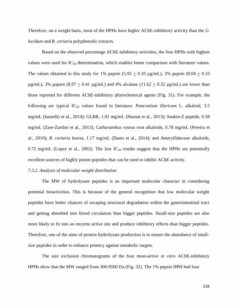
218
Therefore, on a weight basis, most of the HPHs have higher AChE-inhibitory activity than the G.
lucidum and R. coriaria polyphenolic extracts.
Based on the observed percentage AChE-inhibitory activities, the four HPHs with highest
values were used for IC50 determination, which enables better comparison with literature values.
The values obtained in this study for 1% pepsin (5.95 + 0.10 µg/mL), 3% pepsin (8.04 + 0.33
µg/mL), 3% papain (8.97 + 0.41 µg/mL) and 4% alcalase (11.62 + 0.32 µg/mL) are lower than
those reported for different AChE-inhibitory phytochemical agents (Fig. 31). For example, the
following are typical IC50 values found in literature: Pancratium illyricum L. alkaloid, 3.5
mg/mL (Iannello et al., 2014); GLBR, 1.01 mg/mL (Hasnat et al., 2013); Snakin-Z peptide, 0.58
mg/mL (Zare-Zardini et al., 2013); Catharanthus roseus root alkaloids, 0.78 mg/mL (Pereira et
al., 2010); R. coriaria leaves, 1.17 mg/mL (Danis et al., 2014); and Amaryllidaceae alkaloids,
0.72 mg/mL (Lopez et al., 2002). The low IC50 results suggest that the HPHs are potentially
excellent sources of highly potent peptides that can be used to inhibit AChE activity.
7.3.2. Analysis of molecular weight distribution
The MW of hydrolysate peptides is an important molecular character in considering
potential bioactivities. This is because of the general recognition that low molecular weight
peptides have better chances of escaping structural degradation within the gastrointestinal tract
and getting absorbed into blood circulation than bigger peptides. Small-size peptides are also
more likely to fit into an enzyme active site and produce inhibitory effects than bigger peptides.
Therefore, one of the aims of protein hydrolysate production is to ensure the abundance of small-
size peptides in order to enhance potency against metabolic targets.
The size exclusion chromatograms of the four most-active in vitro AChE-inhibitory
HPHs show that the MW ranged from 300-9500 Da (Fig. 32). The 1% pepsin HPH had four

219
Fig 31. Concentrations of the most active enzymatic hemp seed protein hydrolysates that
inhibited 50% acetylcholinesterase activity (IC50)
*Bars that contain different letters are significantly different at p<0.05
H e m p s e e d p ro te in h y d ro ly s a te s
Pe
ptid
e c
on
ce
ntra
tio
ns
(µ
g/m
L)
1 % p e p s in 3 % p e p s in 3 % p a p a in 4 % a lc a la s e0 .0
2 .0
4 .0
6 .0
8 .0
1 0 .0
1 2 .0 d
a
c
b

220
major peaks (A-D) but the intensity decreased when the concentration was increased to 3% level,
which is consistent with the increased DH (more extensive proteolysis) at the higher enzyme
level. The 4% alcalase hydrolysate showed lower levels of the 300-9500 Da peaks, which is
consistent with having the highest DH among the four protein hydrolysates. Thus, alcalase seems
to be more efficient than papain and pepsin in hydrolyzing the hemp seed proteins. However,
protein hydrolysate activity is also highly dependent on the type of peptides produced and not
just the hydrolysis efficiency. Moreso, the peaks produced by the proteases were different in
nature and this could have been the basis for their in vitro AChE inhibitory activities. This is
supported by the past study (He et al., 2013) which had understandably reported that the
structure (size) and activity of peptides is largely dependent on its method of production
(enzymes used). The results (<300 Da) obtained in this study for the alcalase HPH (even though
at 4% level) is in the range with those reported (Alashi et al., 2014) for canola protein
hydrolysates (6.47–0.027 kDa) and 2.6-0.14 kDa being reported (He et al., 2013) for rapeseed
protein hydrolysates.
It is noted in this study that alcalase enzyme produced a higher peak of smaller MW
HPHs probably due to its endoprotease activity and its broad specificity for protein digestion. It
could also be that HPI was more susceptible to alcalase-induced proteolysis as being observed
by past studies (Alashi et al., 2014; He et al., 2013) for canola and rapeseed protein hydrolysates
respectively.
7.3.3. Amino acid composition and peptide sequences
Table 11 shows that the HPHs are composed of mostly negatively charged amino acids
(NCAA), which was followed by hydrophobic amino acids (HAA), while content of aromatic
amino acids (AAA) was the least. The peripheral anionic site (PAS) is a unique surface area on
221
Fig 32. Size-exclusion chromatograms of the most active acetylcholinesterase-inhibitory
enzymatic hemp seed protein hydrolysates.
E lu t io n v o lu m e (m L )
Ab
so
rb
an
ce
at 2
15
nm
(m
AU
)
0 2 4 6 8 1 0 1 2 1 4 1 6 1 8 2 0 2 2 2 4 2 6
0
5 0 0
1 0 0 0
1 5 0 0
2 0 0 0
2 5 0 0
3 0 0 0
3 5 0 0
4 0 0 0
4 5 0 0
2 7 7 49 5 6 2
B C
8 2 3
A 3 0 3
D
1 % p e p s in
3 % p e p s in
3 % p a p a in
4 % a lc a la s e
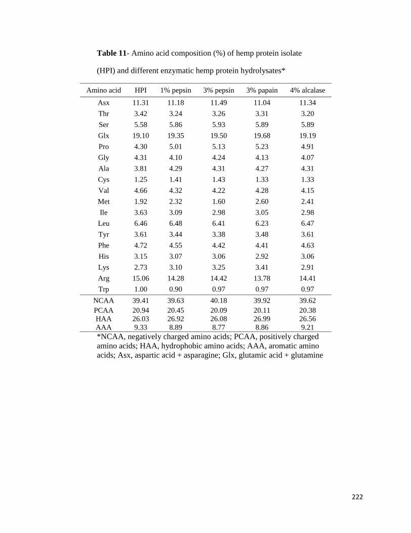
222
Table 11- Amino acid composition (%) of hemp protein isolate
(HPI) and different enzymatic hemp protein hydrolysates*
Amino acid HPI 1% pepsin 3% pepsin 3% papain 4% alcalase
Asx 11.31 11.18 11.49 11.04 11.34
Thr 3.42 3.24 3.26 3.31 3.20
Ser 5.58 5.86 5.93 5.89 5.89
Glx 19.10 19.35 19.50 19.68 19.19
Pro 4.30 5.01 5.13 5.23 4.91
Gly 4.31 4.10 4.24 4.13 4.07
Ala 3.81 4.29 4.31 4.27 4.31
Cys 1.25 1.41 1.43 1.33 1.33
Val 4.66 4.32 4.22 4.28 4.15
Met 1.92 2.32 1.60 2.60 2.41
Ile 3.63 3.09 2.98 3.05 2.98
Leu 6.46 6.48 6.41 6.23 6.47
Tyr 3.61 3.44 3.38 3.48 3.61
Phe 4.72 4.55 4.42 4.41 4.63
His 3.15 3.07 3.06 2.92 3.06
Lys 2.73 3.10 3.25 3.41 2.91
Arg 15.06 14.28 14.42 13.78 14.41
Trp 1.00 0.90 0.97 0.97 0.97
NCAA 39.41 39.63 40.18 39.92 39.62
PCAA
HAA
20.94
26.03
20.45
26.92
20.09
26.08
20.11
26.99
20.38
26.56
AAA 9.33 8.89 8.77 8.86 9.21
*NCAA, negatively charged amino acids; PCAA, positively charged
amino acids; HAA, hydrophobic amino acids; AAA, aromatic amino
acids; Asx, aspartic acid + asparagine; Glx, glutamic acid + glutamine
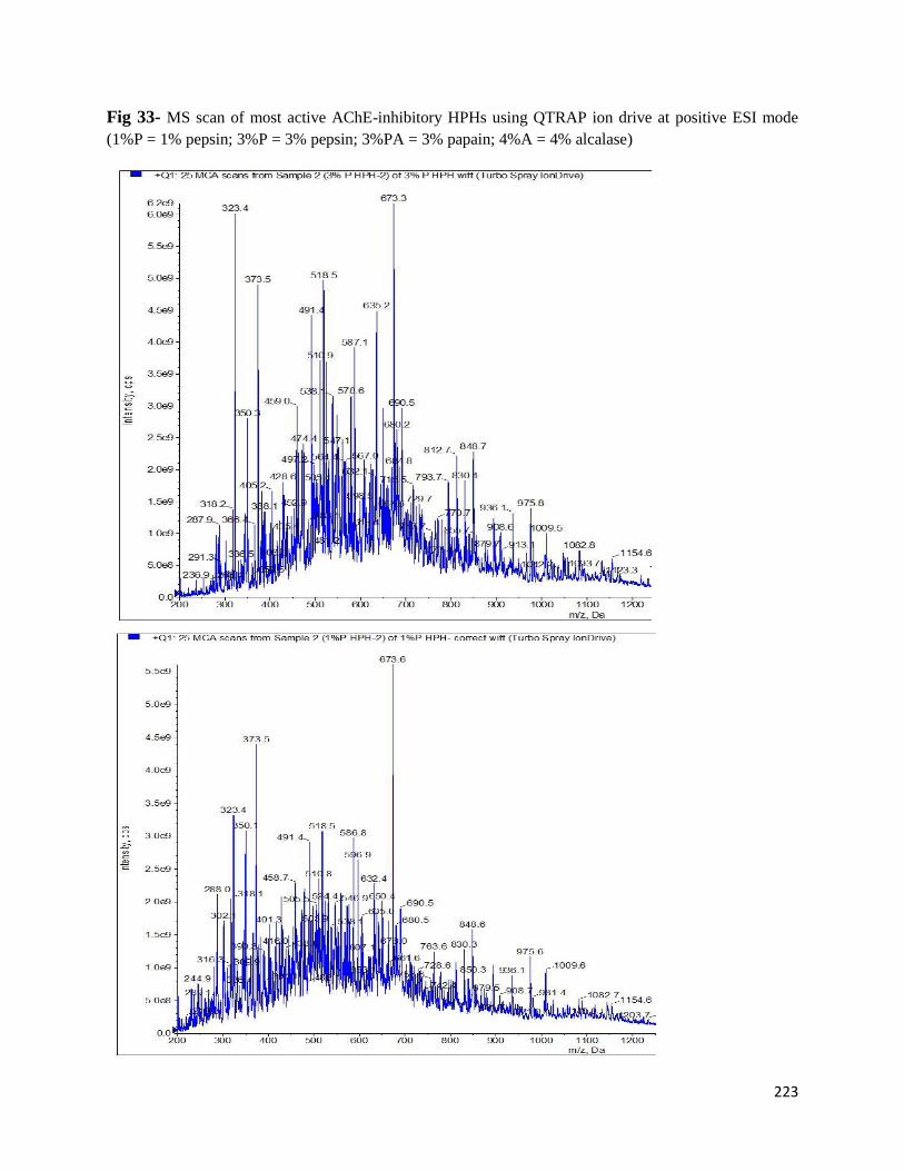
223
Fig 33- MS scan of most active AChE-inhibitory HPHs using QTRAP ion drive at positive ESI mode
(1%P = 1% pepsin; 3%P = 3% pepsin; 3%PA = 3% papain; 4%A = 4% alcalase)

224
225
the AChE protein that is believed to be an important binding site for inhibitors (Saravanaraman
et al., 2014). Within this binding site, PCAA of inhibitory peptides can form a stable complex
with the Trp286 indole ring of the AChE protein, which then prevents entry of substrates to the
enzyme active site. Therefore, it is possible that the high arginine contents of the HPHs may have
facilitated peptide-AChE interactions, which resulted in the observed inhibitions of enzyme
activity.
The most active AChE-inhibitory hydrolysates viz: 1% and 3% pepsin, 3% papain and
4% alcalase HPHs were injected to the mass analyzers of a QTRAP ion mass spectrometer (MS)
to isolate the targeted peptide precursor ion and several molecular species were obtained as
shown in Fig. 33a-d. From the result obtained, the MS scan for 1% and 3% pepsin HPHs are
very similar and almost contain the same peptide species of similar molecular masses. For
instance, the species with molecular mass of 673.6 Da is very more exposed in both 1% and 3%
pepsin HPHs (Fig. 33a-b). The similarity in the result might be from the fact that the same
enzyme (pepsin) is used but at different concentration; meanwhile, higher masses are more
exposed out in 3% HPH due to its higher enzyme concentration. Likewise, the MS scan obtained
for 3% papain and 4% alcalase HPHs are very similar with virtually the same molecular masses.
For instance, the species with molecular mass of 288.1 Da is predominantly observed in both
their MS scans (Fig. 33c-d). The reason might be connected to the fact that both papain and
alcalase have the same optimal condition of activity (pH, 6-8; temperature, 50-65 oC; time, 4 h).
Meanwhile, some of the species with higher molecular masses (for example, those with
masses ≥800 Da) observed in the MS scans of 1% and 3% pepsin HPHs are seemingly
diminished in the MS scans of 3% papain and 4% alcalase HPHs. For example, only ≥400 Da are
predominant in 3% papain and 4% alcalase HPHs unlike ≥600 Da that are observed in 1% and
226
3% pepsin HPHs. This might be connected to the differences in time involved for their
hydrolytic actions on HSP. For instance, both papain and alcalase use 4 h while pepsin uses 2 h
for optimum digestion. The results suggest that disulfide bond formation between peptides and
AChE polypeptide may be involved in reducing enzyme catalysis rate. Presumably, the disulfide
bond formation could distort AChE conformation such that the substrate does not fit properly
into the active site. However, other important amino acid residues include aromatic and
negatively charged, which suggest multiple mechanisms of action are involved in the peptide-
induced AChE inhibition.
7.4. Conclusion
This study, to our knowledge, is the first to report the in vitro AChE inhibition activities
of a food protein hydrolysate. The lack of relationship between DH and AChE-inhibitory of the
HPHs suggest peptide chain amino acid sequence was more important than peptide chain length
as an important structural feature for potency. The results showed the 1% pepsin digest as the
most active and the peptides are dominated by cysteine-containing amino acids, which indicate
involvement of disulfide bond formation during interactions with AChE polypeptides. The IC50
values are lower than values reported for polyphenols and suggest high AChE-inhibitory potency
of the peptides within the context of currently available inhibitory natural products. The
dominance of low molecular peptides suggests that the HPHs contain potentially bioavailable
components. Therefore, the HPHs may constitute important sources of natural AChE-inhibitory
agents and provide potential health benefits during metabolic disorders that involve the nervous
system. Meanwhile, further work will be performed to determine actual amino acid sequences of
peptides present in the most active HPH.
227
Acknowledgement
This work was funded through a Natural Science and Engineering Research Council of Canada
(NSERC) Discovery grant and a Manitoba Agri-Food Research and Development Initiative
(ARDI) grant to Dr. RE Aluko. SA Malomo is a recipient of University of Manitoba Graduate
Fellowship (UMGF) and Manitoba Graduate Scholarship (MGS) Awards for doctoral studies.
References
Adler-Nissen, J. (1979). Determination of the degree of hydrolysis of food protein hydrolysates
by trinitrobenzenesulfonic acid. Journal of Agricultural and Food Chemistry, 27,
1256-1262.
Alashi, A. M., Blanchard, C. L., Mailer, R. J., Agboola, S. O., Mawson, A. J., He, R., Malomo,
S. A., Girgih, A. T., & Aluko, R.E. (2014). Blood pressure lowering effects of Australian
canola protein hydrolysates in spontaneously hypertensive rats. Food Research
International, 55, 281-287.
Alzheimer’s Association (2013). Alzheimer’s disease facts and figures. Alzheimer’s & Dementia,
9, 2.
Bidlingmeyer, B. A., Cohen, S. A., & Tarvin, T. L. (1984). Rapid analysis of amino acids using
pre-column derivatization. Journal of Chromatography B: Biomedical Sciences and
Applications, 336(1), 93-104.
Borowiec, K., Szwajgier, D., Targońsk, Z., Demchuk, O. M., Cybulska, J., Czernecki, T., &
Malik, A. (2014). Cholinesterase inhibitors isolated from bilberry fruit. Journal of
Functional Foods, 11, 313-321.
228
Danis, O., Yuce-Dursun, B., Çimen, T., Demir, S., Salan, Ü., Yalçın, G., & Ogan, A. (2014).
Evaluation of antioxidant, radical-Scavenging and acetylcholinesterase inhibitory
activities of various culinary herbs cultivated in Southern Turkey. Journal of Food
Biochemistry, 38(6), 602-611.
Ellman, G.L., Courtney, K.D., Andres, V., & Featherstone, R.M. (1961). A new and rapid
colorimetric determination of Acetylcholinesterase activity. Biochemical
Pharmacology, 7, 88-95.
Gehrke, C. W., Wall, L. L., Absheer, J. S., Kaiser, F. E., & Zumwalt, R. W. (1985). Sample
preparation for chromatography of amino-acids: acid hydrolysis of proteins. Journal of
Association of Official Analytical Chemists, 68, 811-821.
Ghribia, L., Ghouilaa, H., Omrib, A., Besbesb, M., & Janneta, H. B. (2014). Antioxidant and
anti-acetylcholinesterase activities of extracts and secondary metabolites from Acacia
cyanophylla. Asian Pacific Journal of Tropical Biomedicine, 4(Suppl 1), S417-S423.
Girgih, A.T., He, R., Malomo, S.A., & Aluko, R.E. (2014a). Structural and functional
characterization of hemp seed (Cannabis sativa L.) protein-derived antioxidant and
antihypertensive peptides. Journal of Functional Foods, 6, 384-394.
Girgih, A. T., He, R., Malomo, S. A., Alashi, A. M., & Aluko, R. E. (2014b). Preventive and
treatment effects of hemp seed (Cannabis sativa L.) meal protein hydrolysate against
blood pressure in spontaneously hypertensive rats. European Journal of Nutrition,
53:1237–1246.
Girgih, A. T., Udenigwe, C. C., & Aluko, R. E. (2013). Reverse-phase HPLC separation of hemp
seed (Cannabis sativa L.) protein hydrolysate produced peptide fractions with enhanced
antioxidant capacity. Plant Foods for Human Nutrition, 68, 39–46.
229
Girgih, A. T., Udenigwe, C. C., & Aluko, R. E. (2011a). In vitro antioxidant properties of
hempseed (Cannabis sativa L.) protein hydrolysate fractions. Journal of the American
Oil Chemists’ Society, 88, 381-389.
Girgih, A. T., Udenigwe, C. C., Li, H., Adebiyi, A. P., & Aluko, R. E. (2011b). Kinetics of
enzyme inhibition and antihypertensive effects of hemp seed (Cannabis sativa L.) protein
hydrolysates. Journal of American Oil Chemists Society, 88, 1767–1774.
Hasnat, M. A., Pervin, M., & Lim, B. O. (2013). Acetylcholinesterase inhibition and in vitro and
in vivo antioxidant activities of Ganoderma lucidum grown on germinated brown rice.
Molecules, 18, 6663-6678.
He, R., Malomo, S. A., Alashi, A., Girgih, A. T., Ju, X., Aluko, R. E. (2013). Purification and
hypotensive activity of rapeseed protein-derived renin and angiotensin converting
enzyme inhibitory peptides. Journal of Functional Foods 5, 781-789.
Iannello, C., Pigni, N. B., Antognoni, F., Poli, F., Maxia, A., de Andrade, J. P., & Bastida, J.
(2014). A potent acetylcholinesterase inhibitor from Pancratium illyricum L. Fitoterapia,
92, 163–167.
Kumar, S., & Chowdhury, S. (2014). Kinetics of acetylcholinesterase inhibition by an aqueous
extract of Cuminum cyminum seeds. International Journal of Applied Science and
Biotechnology, 2(1), 64-68.
Landry, J., & Delhaye, S. (1992). Simplified procedure for the determination of tryptophan of
foods and feedstuffs from barytic hydrolysis. Journal of Agricultural and Food
Chemistry, 40, 776-779.
230
Lopez, S., Bastida, J., Viladomat, F., & Codina, C. (2002). Acetylcholinesterase inhibitory
activity of some Amaryllidaceae alkaloids and Narcissus extracts. Life Sciences, 71,
2521–2529.
Lu, R-R., Qian, P., Sun, Z., Zhou, X-H., Chen, T-P., He, J-F., Zhang, H., & Wu, J. (2010).
Hempseed protein derived antioxidative peptides: Purification, identification and
protection from hydrogen peroxide-induced apoptosis in PC12 cells. Food Chemistry,
123, 1210-1218.
Markwell, M.A.K., Haas, S.M., Bieber, L.L., & Tolbert, N.E. (1978). Modification of Lowry
procedure to simplify protein determination in membrane and lipoprotein samples.
Analytical Biochemistry, 87(1), 206-210.
Pereira, D. M., Ferreres, F., Oliveira, J. M. A., Gaspar, L., Faria, J., Valentao, P., Sottomayor, M.
& Andrade, P. B. (2010). Pharmacological effects of Catharanthus roseus root alkaloids
in acetylcholinesterase inhibition and cholinergic neurotransmission. Phytomedicine, 17,
646–652.
Saravanaraman, P., Chinnadurai, R. K., & Boopathy, R. (2014). A new role for the non-
pathogenic non-synonymous single-nucleotide polymorphisms of acetylcholinesterase in
the t reatment of Alzheimer’s disease: A computational study. Journal of
Computational Biology, 21(8), 632-647.
Singh, M., Kaur, M., Kukreja, H., Chugh, R., Silakari, O., & Singh, D. (2013).
Acetylcholinesterase inhibitors as Alzheimer therapy: From nerve toxins to
neuroprotection. European Journal of Medicinal Chemistry, 70, 165-188.
231
US Burden of Disease Collaborators. (2013). The State of US Health, 1990–2010: burden of
diseases, injuries, and risk factors. Journal of American Medical Association, http://
dx.doi.org/10.1001/jama.2013.13805.
Willcox, D. C., Scapagnini, G., & Willcox, B. J. (2014). Healthy aging diets other than the
Mediterranean: A focus on the Okinawan diet. Mechanisms of Ageing and Development,
1-15. doi.org/10.1016/j.mad.2014.01.002
Zare-Zardini, H., Tolueinia, B., Hashemi, A., Ebrahimi, L., & Fesahat, F. (2013). Antioxidant
and cholinesterase inhibitory activity of a new peptide from Ziziphus jujuba fruits.
American Journal of Alzheimer's Disease and other Dementias, 28, 702-709.
232
7.5 Statement transfer between the study 5 and study 6
The preceding evaluated several peptide fractions for AChE-inhibitory properties followed by
preliminary identification of probable peptide sequences. However, in order to perform structure-
functions, definitive identification of peptide sequences is required. Therefore, this chapter deals
with chromatographic separation and purification of peptides present within the most active
AChE-inhibitory hemp seed enzymatic digest identified from the preceding chapter. Peptide
purification was carried out using reverse-phase (RP) HPLC coupled with in vitro assay of
AChE-inhibitory properties of fractions. The most active RP-HPLC was rechromatographed to
obtain purer peptide fractions that were then subjected to liquid chromatography tandem mass
spectrometry (LC/MS/MS) to decipher amino acid sequences. De novo amino acid sequencing
was performed based on fragmentation pattern obtained from LC/MS/MS. Sequenced peptides
were then synthesized and tested for in vitro AChE-inhibitory properties in order to confirm
potency and as potential agents against neurological disorders.

233
CHAPTER EIGHT
8. Study on the purification, amino acid sequences and potency of hemp seed protein-
derived acetylcholinesterase-inhibitory peptides
Sunday A. Malomo and Rotimi E. Aluko*
The Department of Human Nutritional Sciences, University of Manitoba, Winnipeg, Manitoba,
Canada R3T 2N2
*Corresponding author: Dr. Rotimi E. Aluko; E-Mail: [email protected]; Tel.: +1-204-
474-9555; Fax: +1-204-474-7593.

234
Abstract
This work aimed at identification and testing of the in vitro potentials of protein-derived
acetylcholinesterase (AChE) inhibitory peptides present in 1% pepsin-produced hemp seed
protein hydrolysates (HPH). The reverse phase HPLC (RP-HPLC) was used to separate the HPH
into eight different fractions (F1-F8), which were analyzed for their in vitro AChE-inhibitory
properties. After preparative and analytical RP-HPLC separations, the most active fraction was
characterized to produce eleven purified peptides and the amino acid sequence determined by
tandem mass spectrometry (LC–MS/MS). The study revealed that the RP-HPLC fraction F7
exhibited the strongest (p<0.05) in vitro AChE-inhibitory properties (98%) while F3 had the
least (20%); however, no activity was recorded for F2, F5 and F6. The higher peptide purity of
F7 may have contributed to its higher AChE-inhibitory activity (98%; IC50, 5.80 µg/ml) when
compared to the unfractionated 1%-HPH (54%; IC50, 6.00 µg/ml), hence F7 was selected for
peptide characterization. The de-novo sequencing using the LC/MS/MS data led to identification
of peptide sequences that were mainly composed of branched-chain and hydrophobic amino
acids (V, I, L, Y and F). LYV (IC50, 6.80 µg/ml) was the most potent (p<0.05) against AChE
(from Eel) activity when compared to LSA (IC50, 21.90 µg/ml), VQFWG (IC50, 17.50 µg/ml)
and GGYRS (IC50, 11.60 µg/ml). LYV (IC50, 7.50 µg/ml) was also the most potent (p<0.05)
inhibitor of human AChE activity. We therefore, conclude that hydrophobic amino acids
contribute to the potency of HSP-derived inhibitory peptides, which were very active against
both Eel and human AChE.
Key words: hemp protein, acetylcholinesterase, acetylcholine, Alzheimer’s disease, hydrolysates,
branched chain amino acids, hydrophobicity
235
8.1. Introduction
The physiological functions of the serine hydrolase, acetylcholinesterase (AChE) is to
maintain homeostatic recycling of acetylcholine (ACh), the major neurotransmitter (Kumar &
Chowdhury, 2014; Singh et al., 2013). However, the aging process, dietary changes and other
body factors (such as stress, disease, sickness, etc) can affect this delicate homeostasis by
enhancing excessive AChE activity. High AChE activity levels can cause excessive ACh
degradation, which can disrupt neurotransmission and reduce normal signal transmission to the
brain. This abberant brain and neurotransmission conditions lead to Alzheimer’s disease (AD)
pathogenesis (Willcox et al., 2014) that contributes to increased global morbidity/mortality rate
(US Burden of Disease Collaborators, 2013). The high catalytic efficiency of the AChE and the
importance of its inhibition in AD management has been an important research focus in the
development of therapeutic agents (Saravanaraman et al., 2014; Zare-Zardini et al., 2013).
Several synthetic AChE-inhibitory drugs have been developed for use against
neurodegenerative diseases, including dementia and AD; however, these drugs are associated
with severe negative side effects (Kumar and Chowdhury, 2014; Ghribia et al., 2014; Iannello et
al., 2014). Therefore, recent efforts have focused on development of natural AChE-inhibitory
alternatives such as plant secondary metabolites, mainly polyphenolic compounds (Ghribia et al.,
2014; Iannello et al., 2014; Kumar & Chowdhury, 2014). Zare-Zardini et al. (2013) have also
reported isolation of a polypeptide that showed strong in vitro AChE-inhibitory activity. But
information on food protein-derived peptides as agents against neurodegenerative diseases
remains scanty.
Recent findings have shown that plant protein-derived peptides from various
enzymatically hydrolyzed food proteins could emerge as new sources of natural multifunctional
236
compounds without marked adverse effects (Zhang, Mu, & Sun, 2014). It is noteworthy that
several biomedical research works have revealed the efficacy of plant protein-derived peptides as
human health-improving agents, but there still exists scanty or no information on their uses in
AChE inhibition and AD management.
In a previous study, we found that enzymatic hemp seed protein (HSP) hydrolysates
(HPH) possessed significant in vitro AChE-inhibitory properties that are superior to various
polyphenolic extracts. However, there is paucity of information on the actual amino acid
sequence of the hemp seed protein-derived AChE-inhibitory peptides. Information on the amino
acid sequence of AChE-inhibitory peptides will enhance structure-function studies and could
lead to development of highly potent therapeutic peptides for AD or other neurodegenerative
disease management. Therefore, the main aim of this study was to separate, purify and identify
the amino acid sequence of in vitro AChE-inhibitory peptides from 1% pepsin-HPH. The peptide
sequences were then synthesized and used to confirm AChE-inhibitory properties.
8.2. Materials and methods
8.2.1 Hemp seed products and chemical reagents
Hemp seed protein meal (HPM) (37% protein content) produced as a by-product of the hemp
seed oil processing industry was a gift from Hemp Oil Canada (St. Agathe, Manitoba, Canada).
All enzymes- pepsin (porcine gastric mucosa; E.C. 3.4.23.1; ≥250 U/mg), papain (papaya latex;
E.C. 3.4.22.2), alcalase (Bacillus licheniformis; E.C. 3.4.21.62), Electrophorus electricus
(electric eel; 1256 U/mg protein) acetylcholinesterase Type VI-S and Human Recombinant
acetylcholinesterase (expressed in HEK 293 cells; ≥1000 U/mg protein) were purchased from
Sigma-Aldrich (St. Louis, MO, USA). Dithio-bis-nitrobenzoic acid (DTNB) and acethylcholine
237
iodide (AChSI) were also purchased from Sigma-Aldrich (St. Louis, MO, USA). Other
analytical-grade reagents were obtained from Fisher Scientific (Oakville, ON, Canada).
8.2.2 Preparation of enzymatic hemp seed protein hydrolysates (HPHs)
A 5% (w/v) hemp seed protein isolate (HPI) was obtained according to the previously
described method (Malomo et al., 2014) and subjected to hydrolysis using the following enzyme
and reaction conditions (pepsin; 37oC, pH 2.0, 2 hr). The pepsin was added to the HPI slurry at
an enzyme to substrate ratio (E/S) of 1:100, based on the HPI protein content. Digestion was
performed (pH maintained constant by addition of NaOH) after which the enzyme was
inactivated by immersing the reaction vessel in boiling water bath at 95oC for 10 min and
allowed to cool down. The undigested proteins were precipitated by centrifugation (8000 x g, 60
min at 4oC) and the supernatant containing target hydrolysates was freeze dried to make 1%
pepsin-HPH, which was stored at -20oC until needed for further analysis. The protein content of
the freeze dried HPH was determined using the modified Lowry method (Markwell et al., 1978).
8.2.3 Reverse-phase (RP)-HPLC separation of HPH
The separation of HPH using preparative RP-HPLC was performed on a Varian 940-LC
system as previously described (Girgih et al., 2013) with slight modifications. Briefly, freeze-
dried HPH was dissolved in double-distilled water (DDW) that contained 0.1% trifluoroacetic
acid (TFA) as solvent B at a concentration of 10 mg/ml; a 4 ml volume (filtered through the 0.2
μm membrane discs) was injected onto a 21×250 mm C12 preparative column (Phenomenex
Technologies Inc., Torrance, CA, USA). Fractions were eluted from the column at a flow rate of
5 ml/min using a linear gradient of 0-30% solvent A (methanol containing 0.1 % TFA) over 90
min. Elution of peptide fractions was monitored at 220 nm. Fractions were collected using an

238
automated fraction collector every 1 min and pooled into eight fractions according to elution
time as shown in Fig. 1.
The pooled fractions (F1-F8) were freeze-dried (after solvent evaporation under vacuum
using the rotary evaporator, maintained at a temperature range between 35 and 45°C) and stored
at −20°C until further use. The protein contents of the freeze-dried RP-HPLC fractions were
determined using the modified Lowry method (Markwell et al., 1978). Each fraction was tested
for AChE-inhibitory activity and the most active subjected to further LC/MS characterization.
8.2.4 LC/MS/MS identification of the purified peptides
The identification of the purified peptide was done according to the previously described
method (He et al., 2013b) with slight modifications. A 1 µg/µL aliquot of the most active peptide
fraction (F7) was loaded (10 µL) onto an Aeris peptide 2.6 um XB-C18 column (250 x 4.6 mm)
(Phenomenex Technologies Inc, Torrance, CA, USA) connected to a Shidmazu UPLC system
(Shidmazu Corporation, Nakagyo-ku, Kyoto, Japan) at a flow rate of 0.4 ml/min. The bound
peptides were then eluted over 30 min with a gradient elution of 0–30% acetonitrile (containing
0.1% formic acid as solvent B) at a flow rate of 0.35 µL/min. The LC/MS/MS analysis of the
peptide was monitored with an AB SCIEX QTRAP® 6500 LC/MS/MS System (AB SCIEX Pte.
Ltd., Foster City, California, USA) coupled with electrospray ionization source using the
following parameters: Ion source, Turbo Spray IonDrive; Curtain Gas, 30.0; IonSpray Voltage,
3.5 kV; Temperature, 150 °C; Ion Source Gas, 30; Declustering Potential, 100; Enterance
Potential, 10; Collision Energy, 50; Collision Energy Spread, 20; in the positive ion mode.
The amino acid sequences of the bioactive peptides were identified using PEAKS®
Complete Software for Proteomics (Bioinformatics Solutions Inc., Waterloo, ON, Canada). The
239
identified peptide sequences obtained were then synthesized (>95% purity) by GenWay Biotech
(GenWay Biotech Inc. San Diego, CA, USA).
8.2.5 Acetylcholinesterase (AChE) inhibition assays
AChE inhibition assay was performed as previously described (Ellman, Courtney,
Andres, & Featherstone, 1961) with slight modifications. Briefly, the reaction was carried out at
room temperature using 0.1 M sodium phosphate buffer (pH 7.5) and the final assay mixture was
200 μl in the 96-well microplates (Costar, Corning, NY). A 20 µl aliquot of peptides (39.06,
78.12 and 156.25 µg/ml) was added to 96-well microplate containing 130 µl of 0.1 M sodium
phosphate buffer (pH 7.5). This was followed by addition of 20 µl of DTNB and 10 µl of
acethylcholine iodide (AChSI). Then, 20 µl of Electrophorus electrical or human AChE (5 U/
ml) was added to initiate the reaction and the mixture was incubated for 15 min at room
temperature. The absorbance and the reaction kinetics were read for 2 min at wavelength of 412
nm using a microplate reader (Biotek Instruments Inc., Winooski, Vermont, USA). In the control
wells, 20 µl of 0.1 M sodium phosphate buffer (pH 7.5) was used to replace peptides. In the
background wells, 20 µl of 0.1 M sodium phosphate buffer (pH 7.5) was used to replace AChE
while 40 µl of 0.1 M sodium phosphate buffer (pH 7.5) was used to replace both AChE and
peptides in the blank wells. The concentration of sample that inhibited AChE activity by 50%
(IC50) was calculated from a non-linear regression plot of percentage AChE activity versus
peptide concentration.
8.2.6 Statistical analysis
Triplicate replications were used to obtain mean values and standard deviations. Statistical
analysis was performed with SAS (Statistical Analysis Software 9.1) using one-way ANOVA.
240
Duncan's multiple-range test was carried out to compare the mean values for samples with
significant differences taken at p<0.05.
8.3. Results and Discussion
8.3.1 RP-HPLC fractions from 1% pepsin-produced HPH
The reverse phase-HPLC purification of 1% pepsin HPH after 90 min separation
produced eight different fractions (F1-F8) of different hydrophobicity as presented in Fig. 34.
The RP-HPLC separation of protein-derived peptides is based on hydrophobicity as previously
described (Girgih et al., 2014; Girgih et al., 2013). Thus, while the weakly hydrophobic peptides
would elute in the earliest period of separation, the strongly hydrophobic peptides will elute at a
later time (Girgih et al., 2013). The results in Fig. 34 indicate that F1 and F8 are, respectively the
least and most hydrophobic fractions. Similar results (eight different fractions) were obtained
from the RP-HPLC separation and purification of a 4% pepsin + pancreatin antihypertensive
hemp seed protein hydrolysate (Girgih et al., 2013).
8.3.2 Inhibition of AChE by different RP-HPLC fractions
The eight RP-HPLC purified-fractions were screened for their in vitro AChE inhibition
activities and the results are presented in Fig. 35. Fraction F7 showed an overall significantly
(p<0.05) higher AChE-inhibitory effect when compared to the other fractions. For example, F7
and F3 had in vitro AChE-inhibitory values of 98% and 20%, respectively while no activities
were observed and recorded for fractions 2, 5 and 6. Interestingly, there seemed no significant
differences between fractions F1 and F4 (Eel AChE inhibition of 78%) as well as fraction F8 and
the unfractionated HPH (AChE inhibition of 54%; IC50, 6.00 µg/ml). Peptide purification has
been described (Zhang et al., 2014) as a means to enhance the level of active molecules with
potential increase in physiological impact. Therefore, the purified peptides are normally expected
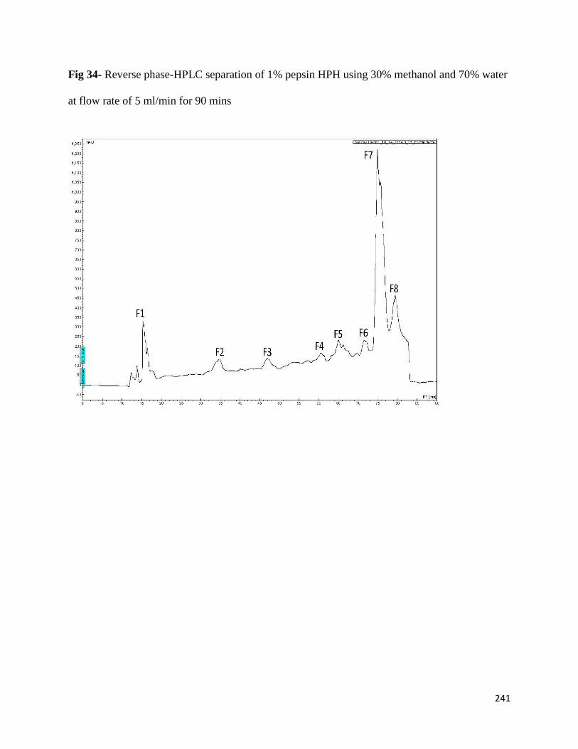
241
Fig 34- Reverse phase-HPLC separation of 1% pepsin HPH using 30% methanol and 70% water
at flow rate of 5 ml/min for 90 mins
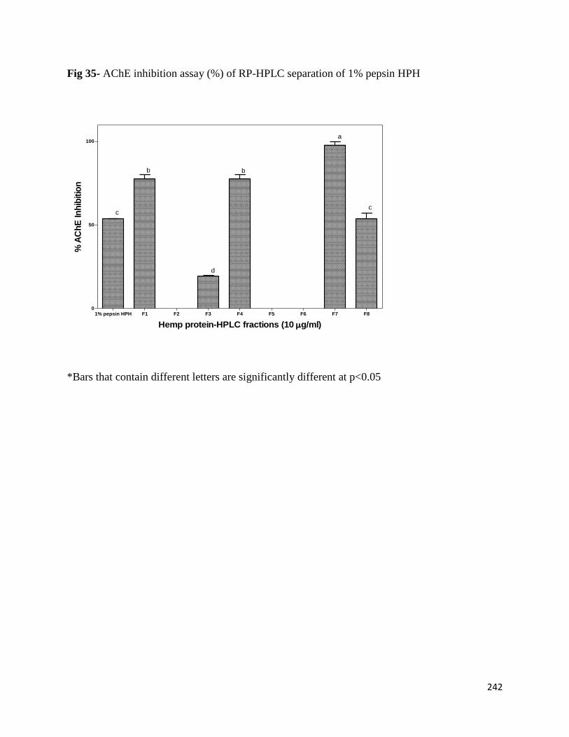
242
Fig 35- AChE inhibition assay (%) of RP-HPLC separation of 1% pepsin HPH
*Bars that contain different letters are significantly different at p<0.05
Hemp protein-HPLC fractions (10 g/ml)
% A
Ch
E In
hib
itio
n
1% pepsin HPH F1 F2 F3 F4 F5 F6 F7 F80
50
100
b
d
b
a
cc
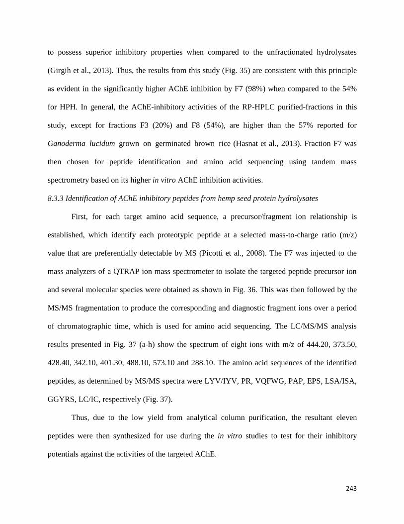
243
to possess superior inhibitory properties when compared to the unfractionated hydrolysates
(Girgih et al., 2013). Thus, the results from this study (Fig. 35) are consistent with this principle
as evident in the significantly higher AChE inhibition by F7 (98%) when compared to the 54%
for HPH. In general, the AChE-inhibitory activities of the RP-HPLC purified-fractions in this
study, except for fractions F3 (20%) and F8 (54%), are higher than the 57% reported for
Ganoderma lucidum grown on germinated brown rice (Hasnat et al., 2013). Fraction F7 was
then chosen for peptide identification and amino acid sequencing using tandem mass
spectrometry based on its higher in vitro AChE inhibition activities.
8.3.3 Identification of AChE inhibitory peptides from hemp seed protein hydrolysates
First, for each target amino acid sequence, a precursor/fragment ion relationship is
established, which identify each proteotypic peptide at a selected mass-to-charge ratio (m/z)
value that are preferentially detectable by MS (Picotti et al., 2008). The F7 was injected to the
mass analyzers of a QTRAP ion mass spectrometer to isolate the targeted peptide precursor ion
and several molecular species were obtained as shown in Fig. 36. This was then followed by the
MS/MS fragmentation to produce the corresponding and diagnostic fragment ions over a period
of chromatographic time, which is used for amino acid sequencing. The LC/MS/MS analysis
results presented in Fig. 37 (a-h) show the spectrum of eight ions with m/z of 444.20, 373.50,
428.40, 342.10, 401.30, 488.10, 573.10 and 288.10. The amino acid sequences of the identified
peptides, as determined by MS/MS spectra were LYV/IYV, PR, VQFWG, PAP, EPS, LSA/ISA,
GGYRS, LC/IC, respectively (Fig. 37).
Thus, due to the low yield from analytical column purification, the resultant eleven
peptides were then synthesized for use during the in vitro studies to test for their inhibitory
potentials against the activities of the targeted AChE.
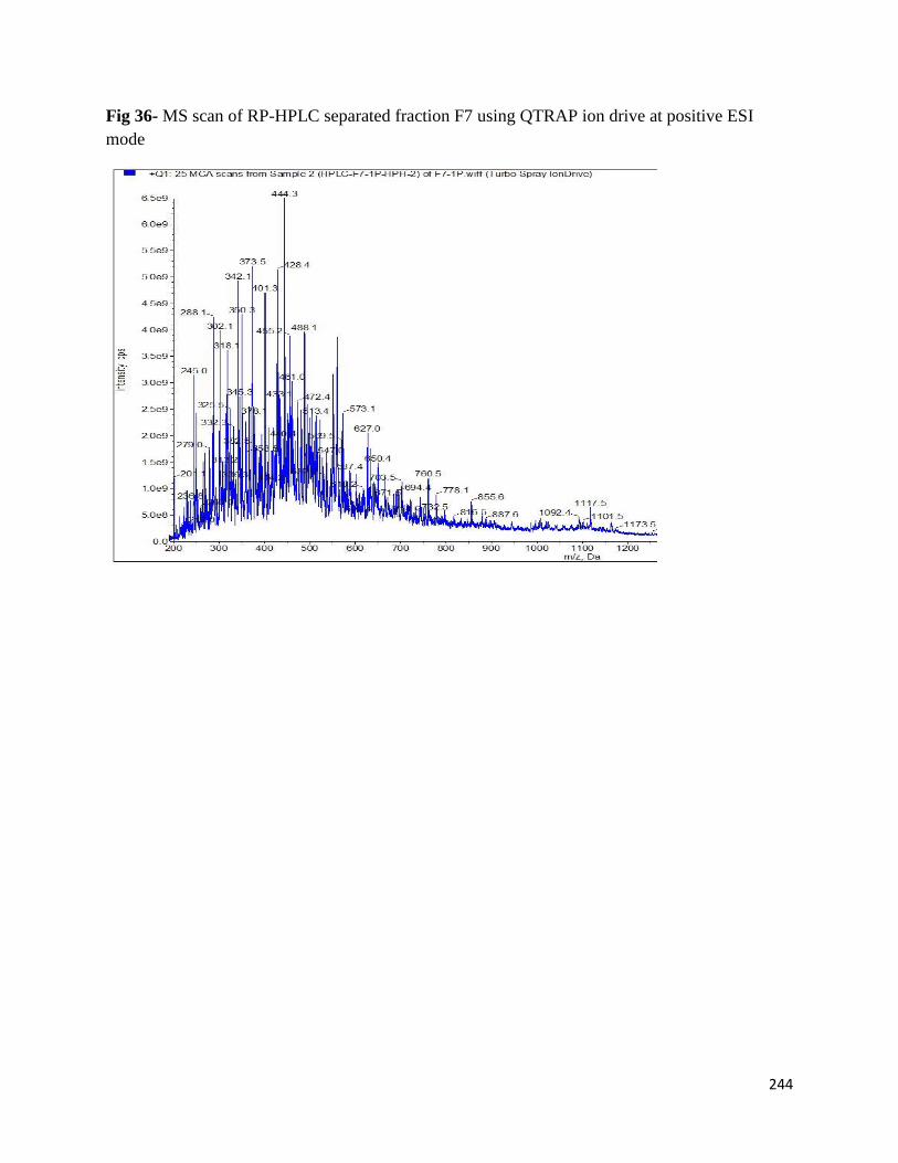
244
Fig 36- MS scan of RP-HPLC separated fraction F7 using QTRAP ion drive at positive ESI
mode

245
Fig 37- LC/MS/MS of RP-HPLC separated fraction F7 at different m/z using QTRAP ion drive
at positive ESI mode
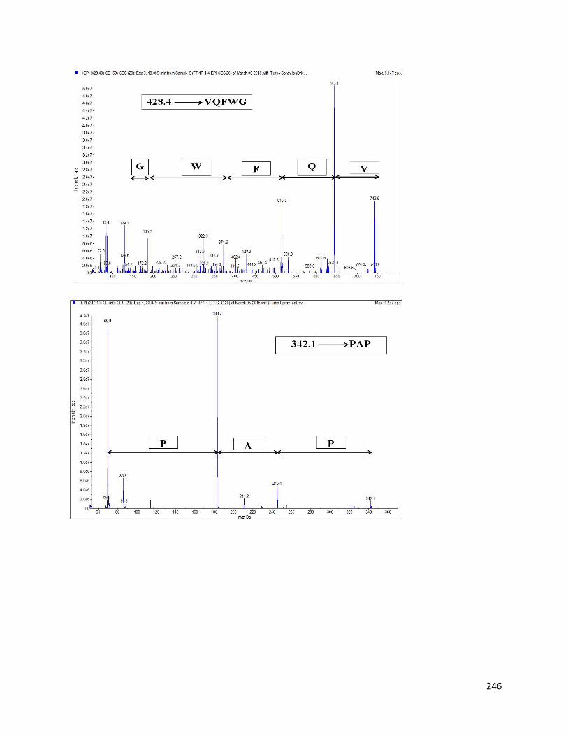
246
247
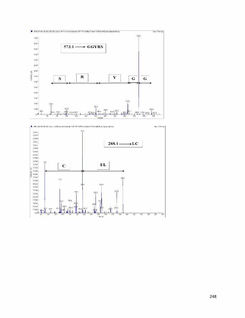
248
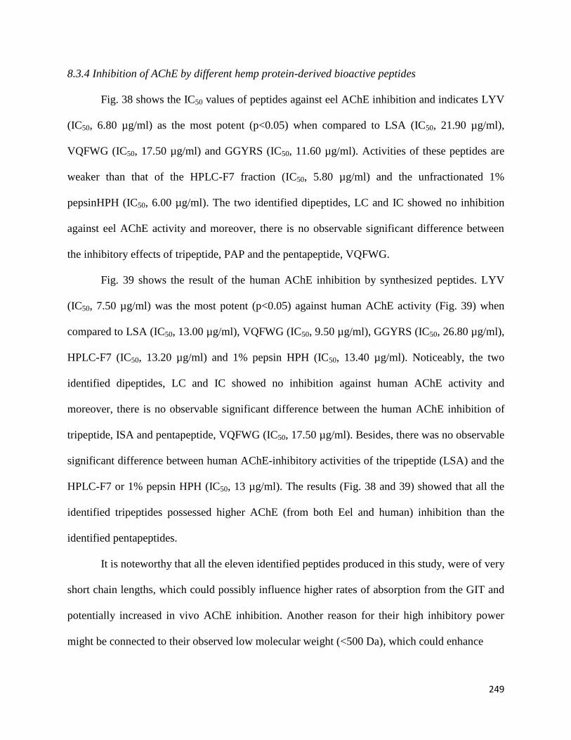
249
8.3.4 Inhibition of AChE by different hemp protein-derived bioactive peptides
Fig. 38 shows the IC50 values of peptides against eel AChE inhibition and indicates LYV
(IC50, 6.80 µg/ml) as the most potent (p<0.05) when compared to LSA (IC50, 21.90 µg/ml),
VQFWG (IC50, 17.50 µg/ml) and GGYRS (IC50, 11.60 µg/ml). Activities of these peptides are
weaker than that of the HPLC-F7 fraction (IC50, 5.80 µg/ml) and the unfractionated 1%
pepsinHPH (IC50, 6.00 µg/ml). The two identified dipeptides, LC and IC showed no inhibition
against eel AChE activity and moreover, there is no observable significant difference between
the inhibitory effects of tripeptide, PAP and the pentapeptide, VQFWG.
Fig. 39 shows the result of the human AChE inhibition by synthesized peptides. LYV
(IC50, 7.50 µg/ml) was the most potent (p<0.05) against human AChE activity (Fig. 39) when
compared to LSA (IC50, 13.00 µg/ml), VQFWG (IC50, 9.50 µg/ml), GGYRS (IC50, 26.80 µg/ml),
HPLC-F7 (IC50, 13.20 µg/ml) and 1% pepsin HPH (IC50, 13.40 µg/ml). Noticeably, the two
identified dipeptides, LC and IC showed no inhibition against human AChE activity and
moreover, there is no observable significant difference between the human AChE inhibition of
tripeptide, ISA and pentapeptide, VQFWG (IC50, 17.50 µg/ml). Besides, there was no observable
significant difference between human AChE-inhibitory activities of the tripeptide (LSA) and the
HPLC-F7 or 1% pepsin HPH (IC50, 13 µg/ml). The results (Fig. 38 and 39) showed that all the
identified tripeptides possessed higher AChE (from both Eel and human) inhibition than the
identified pentapeptides.
It is noteworthy that all the eleven identified peptides produced in this study, were of very
short chain lengths, which could possibly influence higher rates of absorption from the GIT and
potentially increased in vivo AChE inhibition. Another reason for their high inhibitory power
might be connected to their observed low molecular weight (<500 Da), which could enhance
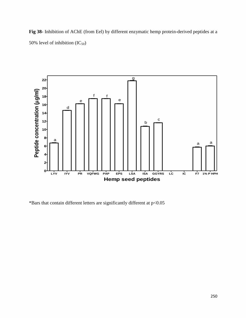
250
Fig 38- Inhibition of AChE (from Eel) by different enzymatic hemp protein-derived peptides at a
50% level of inhibition (IC50)
Hemp seed peptides
Pe
pti
de
co
nc
en
tra
tio
n (
g/m
l)
LYV IYV PR VQFWG PAP EPS LSA ISA GGYRS LC IC F7 1% P HPH0
2
4
6
8
10
12
14
16
18
20
22
aaa
c
g
b
f f
e
d
e
*Bars that contain different letters are significantly different at p<0.05
251
Fig 39- Inhibition of AChE (from human) by different enzymatic hemp protein-derived peptides
at a 50% level of inhibition (IC50)
Hemp seed peptides
Pe
pti
de
co
nc
en
tra
tio
n (
g/m
l)
LYV IYV PR VQFWG PAP EPS LSA ISA GGYRS LC IC F7 1% P HPH0
2
4
6
8
10
12
14
16
18
20
22
24
26
28
a
ee
g
b
e
f
d
bc
a
*Bars that contain different letters are significantly different at p<0.05
252
their ability to interact with the enzyme active site. A past study (although produced for
cardiovascular-related targeted enzymes) had reported the high inhibition and disease-
management power of low molecular weight bioactive peptides from pepsin-produced HPHs
(Girgih et al., 2014). About seven out of the eleven identified peptides were mainly composed of
branched-chain amino acids (L, V, I) that has been described as a group of amino acids found to
enhance the hydrophobic character of bioactive peptides (Aluko et al., 2015). The important role
of these branched-chain amino acids to enhance hydrophobicity and inhibitory potentials of
bioactive functional components has been also observed in our previous findings. The effect of
high hydrophobicity of bioactive peptides lies in their ability to influence strong interactions
between peptide and non-polar amino acid residues within the enzyme active site (Aluko et al.,
2015; Girgih et al., 2014; Sarmadi & Ismail, 2010).
8.4 Conclusion
The importance of separating and purifying the hemp seed protein-derived bioactive peptides
for cognitive human health application had been demonstrated in this study. The higher
hydrophobicity of fraction F7 was reflected in the hydrophobic character of the identified
peptides. Eleven peptides of very short chain lengths and low molecular weights <500 Da were
identified from F7, which were composed mainly of branched chain and hydrophobic amino
acids residues that contributed to their AChE-inhibitory potency. The HPLC-separated and
purified bioactive peptides possessed higher in vitro AChE (from both Eel and human) inhibitory
properties (IC50, 5.80-26.80 µg/ml). Therefore, for the first time in literature, this work reported
the amino acid sequences of food protein-derived peptides that inhibited both Eel and human
AChE activities.
253
Acknowledgement
This work was funded through the Natural Science and Engineering Research Council of Canada
(NSERC) Discovery grant and Manitoba Agri-Food Research and Development Initiative
(ARDI) grant to Dr. RE Aluko. SA Malomo is a recipient of University of Manitoba Graduate
Fellowship (UMGF) and Manitoba Graduate Scholarship (MGS) Awards for doctoral studies.
References
Aluko, R. E., Girgih, A. T., He, R., Malomo, S. A., Li, H., Offengenden, M., & Wu, J. (2015).
Structural and functional characterization of yellow field pea seed (Pisum sativum L.)
protein-derived antihypertensive peptides. Food Research International, 1-7.
http://dx.doi.org/10.1016/j.foodres.2015.03.029
Bigliardi, B., & Galati, F. (2013). Innovation trends in the food industry: the case of functional
foods. Trends in Food Science & Technology, 31, 118-129.
Ellman, G. L., Courtney, K. D., Andres, V., & Featherstone, R. M. (1961). A new and rapid
colorimetric determination of Acetylcholinesterase activity. Biochemical
Pharmacology, 7, 88-95.
Ghribia, L., Ghouilaa, H., Omrib, A., Besbesb, M., & Janneta, H. B. (2014). Antioxidant and
anti-acetylcholinesterase activities of extracts and secondary metabolites from Acacia
cyanophylla. Asian Pacific Journal of Tropical Biomedicine, 4(Suppl 1), S417-S423.
Girgih, A.T., He, R., Malomo, S.A., & Aluko, R.E. (2014). Structural and functional
characterization of hemp seed (Cannabis sativa L.) protein-derived antioxidant and
antihypertensive peptides. Journal of Functional Foods, 6, 384-394.
254
Girgih, A. T., Udenigwe, C. C., & Aluko, R. E. (2013). Reverse-phase HPLC separation of hemp
seed (Cannabis sativa L.) protein hydrolysate produced peptide fractions with enhanced
antioxidant capacity. Plant Foods for Human Nutrition, 68, 39–46.
Hasnat, M. A., Pervin, M., & Lim, B. O. (2013). Acetylcholinesterase inhibition and in vitro and
in vivo antioxidant activities of Ganoderma lucidum grown on germinated brown rice.
Molecules, 18, 6663-6678.
He, R., Malomo, S. A., Girgih, A. T., Ju, X., & Aluko, R. E. (2013a). Antihypertensive and free
radical scavenging properties of enzymatic rapeseed protein hydrolysates. Journal of
Agricultural and Food Chemistry, 61, 8396-8402.
He, R., Girgih, A. T., Malomo, S. A., Alashi, A. M. & Aluko, R.E. (2013b). Purification and
hypotensive activity of rapeseed protein-derived renin and angiotensin converting
enzyme inhibitory peptides. Journal of Functional Foods. 5, 781-789.
Iannello, C., Pigni, N. B., Antognoni, F., Poli, F., Maxia, A., de Andrade, J. P., & Bastida, J.
(2014). A potent acetylcholinesterase inhibitor from Pancratium illyricum L. Fitoterapia,
92, 163–167.
Kumar, S., & Chowdhury, S. (2014). Kinetics of acetylcholinesterase inhibition by an aqueous
extract of Cuminum cyminum seeds. International Journal of Applied Science and
Biotechnology, 2(1), 64-68.
Malomo, S. A., He, R., & Aluko, R. E. (2014). Structural and functional properties of hemp seed
protein products. Journal of Food Science, 79 (8), C1512-1521.
Markwell, M.A.K., Haas, S.M., Bieber, L.L., & Tolbert, N.E. (1978). Modification of Lowry
procedure to simplify protein determination in membrane and lipoprotein samples.
Analytical Biochemistry, 87(1), 206-210.
255
Picotti, P., Lam, H., Campbell, D., Deutsch, E., W., Mirzaei, H., Ranish, J., Domon, B., &
Aebersold, R. (2008). A database of mass spectrometric assays for the yeast proteome.
Nature Methods, 5(11), 913-914.
Saravanaraman, P., Chinnadurai, R. K., & Boopathy, R. (2014). A new role for the non-
pathogenic non-synonymous single-nucleotide polymorphisms of acetylcholinesterase in
the t reatment of Alzheimer’s disease: A computational study. Journal of
Computational Biology, 21(8), 632-647.
Sarmadi, B. H., & Ismail, A. (2010). Antioxidative peptides from food proteins: A review.
Peptides, 31, 1949–1956.
Seidler, J., Zinn, N., Boehm, M. E., & Lehmann, W. D. (2010). De novo sequencing of peptides
by MS/MS. Proteomics, 10, 634–649.
Singh, M., Kaur, M., Kukreja, H., Chugh, R., Silakari, O., & Singh, D. (2013).
Acetylcholinesterase inhibitors as Alzheimer therapy: From nerve toxins to
neuroprotection. European Journal of Medicinal Chemistry, 70, 165-188.
Sussman, J. L., Harel, M., & Silman, I. (1993). Three-dimensional structure of
acetylcholinesterase and of its complexes with anticholinesterase drugs. Chemico-
Biological Interractions, 87(1-3), 187-197.
Sussman, J. L., Harel, M., Frolow, F., Oefner, C., Goldman, A., Toker, L., & Silman, I. (1991).
Atomic structure of acetylcholinesterase from Torpedo californica: a prototypic
acetylcholine-binding protein. Science, 253(5022), 872-879.
US Burden of Disease Collaborators. (2013). The State of US Health, 1990–2010: burden of
diseases, injuries, and risk factors. Journal of American Medical Association, http://
dx.doi.org/10.1001/jama.2013.13805.

256
Willcox, D. C., Scapagnini, G., & Willcox, B. J. (2014). Healthy aging diets other than the
Mediterranean: A focus on the Okinawan diet. Mechanisms of Ageing and Development,
1-15. doi.org/10.1016/j.mad.2014.01.002
Zare-Zardini, H., Tolueinia, B., Hashemi, A., Ebrahimi, L., & Fesahat, F. (2013). Antioxidant
and cholinesterase inhibitory activity of a new peptide from Ziziphus jujuba fruits.
American Journal of Alzheimer's Disease and other dementials, 00(0), 1-8. DOI:
10.1177/1533317513500839
Zhang, M., Mu, T. H., & Sun, M. J. (2014). Purification and identification of antioxidant
peptides from sweet potato protein hydrolysates by alcalase. Journal of Functional
Foods, 7, 191-200.
257
CHAPTER NINE
9. General Summary and Conclusion of the study
The first study reveals a potential use of hemp seed protein products as basic functional
ingredients in the food industry besides their nutritional role. The findings show that
manipulation of pH could be used to increase foaming ability, which is dependent on protein
solubility. The oil droplet sizes of some of the emulsions produced by these proteins were very
small and comparable to the well-known and commonly used milk and soybean protein
emulsifiers. Although, their highly stable emulsions indicated potential use to form food
emulsions that could maintain stability during short-term storage, the findings showed a different
mechanism responsible for foam and emulsion formation. Intrinsic fluorescence and CD data
from this study revealed that hemp seed proteins existed in different conformational states within
different pH environments. The study suggested that the effect of pH on protein functionality is
dependent on protein concentration and also confirmed the high dependency of protein
functionality on structural conformation as being affected by modulation in pH changes.
The second study gives a concise description of how variations in amino acid
composition and pH-dependent changes in structural conformation could differentially affect the
functionalities of both hemp seed albumin and globulin protein fractions. The salt-extracted
globulin is made up of more disulfide-bonded proteins than the water-soluble albumin. This
produces a rigid structure with reduced exposure of aromatic amino acids within the globulins.
This study also reported the contribution of higher levels of hydrophobic amino acids and the
rigid conformational structure in globulin on its decreased protein solubility and foaming
capacity. This is a confirmation of the dependence of these functional properties on protein
flexibility and the ability of proteins to interact with the aqueous phase. In contrast, emulsion-
258
forming ability was reportedly not dependent on protein solubility, suggesting different
mechanisms are involved in foam and emulsion formations. However, both protein fractions
were observed to be potentially good ingredients for the manufacture of food emulsions with
only the albumin playing a superior role in the manufacture of food foams.
The third study proved the potential use of waste products from oil seed processing
industries, especially the hemp seed processing industry as well as the resultant effects of the
preparation methods of protein isolates on their physicochemical and functional properties. This
is because the potential uses of protein isolates are largely dependent on these properties with
great influence from protein digestibility. This study revealed the high improvement on the
physicochemical and functional properties of enzyme pre-digested and membrane produced-
isolates when compared to those from the isoelectric precipitation method. The combination of
carbohydrase and phytase treatments led to the production of a 74% protein product, which s
twice the 37% protein in the original hemp seed waste product. The significant improvements in
the functional properties (protein solubility, foam capacity and emulsion stability) and the
excellent in vitro protein digestibility reported for this improved protein isolates provides a big
advantage over utilization of the acid/alkaline-harsh conditions produced-isolates. The findings
also showed the presence of large numbers of disulfide bridge-linked polypeptide chains that is
well-known to keep the protein molecules intact against any processing environmental
conditions in the membrane produced-isolate.
The fourth study showed the potential of the hemp seed protein derived-peptides as dual
in vitro ACE and renin inhibitors. The work also demonstrated bioactive effects when these
physiologically functional hydrolysates were shown to significantly lower elevated-blood
pressure (BP) in vivo after oral administration to SHRs. The in vivo BP-lowering effects of the

259
peptides did not correspond directly to their in vitro renin-ACE inhibition activities, which
suggest other factors may have contributed to peptide-induced antihypertensive effects.
To the best of our knowledge, the fifth study is the first to report the in vitro AChE
inhibition activities of hemp seed protein derived-peptides obtained from enzymatic hydrolysis
of hemp seed proteins. The study showed higher improvement and increase in the protein content
and extraction yield of the hydrolysed protein products when compared with its non-hydrolysed
forms. The liberation of indigenous bioactive-encypted peptides from the primary structure of
plant proteins through in vitro enzyme-catalyzed proteolysis (hydrolysis) for disease
management is well justified in this study. The study further revealed the lack of relationships
between degree of hydrolysis and in vitro AChE-inhibition activities. However, the pronounced
in vitro AChE-inhibitory activities of the hydrolysates in this study was dependent on their high
hydrophobic and branched chain amino acid residues as well as the composition and presence of
very short chain peptides (e.g. di-, tri- and tetrapeptides).
The sixth study shows the importance of separating and purifying the plant protein-
derived AChE-inhibitory peptides. This was revealed from the study when the HPLC-separated
and purified bioactive peptides possessed higher in vitro AChE-inhibitory properties than the
crude and unfractionated hydrolysate. Apart from reporting the probable or suggested amino acid
sequences of the bioactive peptide that could be obtained from the most active AChE-inhibitory
HPHs, the study confirmed the presence of a very short chain peptide sequence (LYV) with
improved in vitro AChE-inhibitory properties. The very short chain peptide sequences were
composed mainly of branched-chain and hydrophobic-enriched amino acids. Moreover, LYV is
a novel peptide with an AChE-inhibitory IC50 value of 7 µg/ml that makes it a potential
therapeutic tool in human cognitive health applications. Finally, this work also presented other

260
novel peptide sequences with superior AChE-inhibitory values than previously reported natural
products and lays a solid foundation for further studies on the use of bioactive plant protein-
derived peptides as agents against neurodegenerative diseases.
Although the antihypertensive activity of 1% pepsin HPH is not shown in this report, but
its multifunctionality effects on renin-ACE-inhibition, great reduction of elevated-SBP in SHRs
(after 4 h) as well as effective inhibition against AChE activities were observed in this research
work. Hence, there is possibility that the identified-bioactive peptides obtained from 1% pepsin
HPH might probably inhibit both AChE and RAS enzymes (renin and ACE). This is because
most of the identified-bioactive peptides (e.g. PR, LYV, PAP and VQFWG) contain aromatic
(tyrosine, phenylalanine and tryptophan), hydrophobic (proline and alanine) and branched-chain
(valine and leucine) amino acid residues required for optimal and efficient AChE-inhibition and
antihypertensive acitivities. Thus, there seems no notable difference between the bioactive
peptides with cardiovascular (renin-ACE inhibition) and cognitive (AChE inhibition) health
benefits but their (peptides) metabolic pathway of inhibition of the disease-causative enzymes
might be different.
It is therefore, concluded that the improved physicochemical properties of the enzyme
pre-digested and membrane-produced protein isolate could make it a suitable functional
ingredient in the food processing industries. Potential food applications include formulation of
clear beverages, manufacture of oil-in-water emulsions (e.g. salad dressings and soups);
formulation of desserts (such as meringues) and meat and dairy products with desired fat/flavour
retention. The results also indicate potential use of the hemp seed protein-derived peptides as
beneficial multifunctional effects (for example, towards the human cardiovascular and cognitive
health applications). The protein hydrolysates may be used as a source of natural AChE-
261
inhibitory agent and with potential cognitive health benefits applications, especially in the
treatment of Alzheimer’s disease.
Lastly, this research work has established some of the relationships between
physicochemical properties (polypeptide composition, amino acid composition, amino acid
sequence), in vitro protein digestibility and functional properties of hemp seed proteins and its
derived peptides. For instance, the definitive peptide sequences identification exercise reveals
that most peptides with aromatic, hydrophobic and branched-chain amino acid residues might
possess the ability to improve both functional properties and human health benefits of the HSP.
This type of data is needed for fundamental understanding of the relationships between
structural properties of the proteins or peptides and their functions in food systems or human
health applications.
262
CHAPTER TEN
10. Limitations of the study
a. The enzyme pre-digestion and membrane ultrafiltration method of protein isolation was
not able to achieve the production of protein isolates with >90% protein content earlier
proposed in the course of this research work. The use of more effective carbohydrases
and phytases will be required to produce another 10-15% increase in protein content of
the membrane isolate.
b. Although, the work reported on the peptide identification and in vitro AChE-inhibitory
activities of the identified bioactive peptides from the 1% pepsin-produced hydrolysates,
similar peptide identification was not performed for the most active RAS-modulatory
hydrolysates.
c. Currently, there is no report on the human AChE-inhibitory bioactive peptides identified
from plant proteins to serve as reference point for our newly identified hemp seed AChE-
inhibitory peptides.
d. The AChE inhibition work involved only in vitro assays, but tests on animal disease
models of dementia or AD are required to confirm potential human health benefits. The
peptides will then need to be tested using human intervention trials before the product can
be made available to consumers.
263
CHAPTER ELEVEN
11. Future directions of the study
The data from in vitro studies alone cannot propagate the application of bioactive hemp seed
protein-derived-AChE-inhibitory peptides as therapeutic agents against cognitive health
disorders without in vivo and human clinical trial studies. Thus, in vivo animal and human
intervention trials are highly recommended before confirming peptides use in cognitive health. It
has been revealed from recent scientific findings that identification of amino acid sequences of
plant protein-derived bioactive peptides can pave the way for efficient utilization in developing
potent products for human health improvement. Therefore, further studies are required to isolate
and determine amino acid sequences of peptides present in the most active antihypertensive
HPHs reported in this study. Moreover, there still exist some unresolved cases around the
proposed utilization of hemp seed protein products as functional food ingredients in the food
industry and human health applications. For instance, the protein content of the enzyme pre-
digested coupled with membrane-produced hemp seed protein isolates need further improvement
to bring it up to the 85-90% range. Moreover, the exact mechanism of AChE-inhibition by the
hemp seed protein-derived peptides needs to be resolved using kinetics of enzyme inhibition
studies. These proposed studies would ensure industrial applications of the beneficial features of
this plant protein in food product development and human health. Emerging evidences have also
revealed the bioactive properties of some compounds obtained during interaction of plant protein
hydrolysates with some proteases (plastein aggregates). Therefore, it becomes very imperative to
consider emergence or production of such compounds from hemp seed protein hydrolysates and
study the beneficial values that these peptide aggregates might contribute to food and human
health applications.