structurally abnormalinsulin in diabetic...
TRANSCRIPT

Structurally Abnormal Insulin in a Diabetic PatientCharacterization of the Mutant Insulin A3 (Val Lou) Isolated from the Pancreas
H. Sakura, Y. Iwamoto, Y. Sakamoto, T. Kuzuya, and H. HirataDivision of Endocrinology and Metabolism, Jichi Medical School, Tochigi, Japan, and Department of Biochemistry,Jichi Medical School, Tochigi, Japan
Abstract
Wehave recently identified a diabetic patient with marked fastinghyperinsulinemia. Family study revealed that the abnormalitywas an autosomal dominant trait. High-performance liquidchromatography (HPLC) profile of the patient's serum insulinshowed that she had an abnormal insulin in addition to a normalinsulin. Wehave purified her insulin(s) from the specimen ofher pancreas, which was biopsied during an operation of cho-lelithiasis. Insulin was also immunologically purified from theserum of her portal vein. The reverse-phase HPLCanalysis re-vealed that the ratios of normal to abnormal insulin in the pan-creas, portal vein, and peripheral vein were 5:4, 4:5, and 1:7,respectively. Radioreceptor assay for insulin using guinea pigkidney membrane revealed that the binding activities of the nor-mal component insulin, the abnormal component insulin and herpancreatic insulin containing, both components were 100, 5, and50% of standard human insulin, respectively. The biological ac-tivities of the normal components the abnormal component andher pancreatic insulin to stimulate glucose oxidation in rat adi-pocytes were found to be 100, 8, and 60% of standard humaninsulin, respectively. Analysis of amino acid sequences of theabnormal insulin purified from her pancreas strongly suggestedthe substitution of leucine for valine at the third position of theA chain, A3 (Val -- Leu).
Introduction
Diabetes mellitus is a clinical syndrome induced by variouscauses, and the possibility had been considered for many years(1-5) that a structurally abnormal insulin might be one causeof diabetes. The first case with a mutant insulin was reportedby Tager et al. (6) and Given et al. (7) in 1979, 1980, and sincethat time, a few cases (8-12) have been described. The pointmutation sites of the insulin genes and resulting amino acidsubstitutions have been determined in three cases; e.g., insulinChicago B25 (Phe -. Leu) (13, 14), insulin Los Angeles B24(Phe -) Ser) (15), and insulin Wakayama A3 (Val -o Leu) (12,16). The abnormality is an autosomal dominant trait and the
Address reprint requests to Dr. Iwamoto, Division of Endocrinology andMetabolism, Jichi Medical School, Minamikawachi-machi, Tochigi-ken329-04, Japan. Dr. Sakura's present address is Third Department of In-ternal Medicine, University of Tokyo, 7-3-1 Hongo, Bunkyo-ku, Tokyo113, Japan.
Receivedfor publication 2 October 198S.
families with abnormal insulins are hyperinsulinemic, but notall are diabetic. The biologic activities of the abnormal insulinsare low, and hyperinsulinemia is thought to result from feedbackcompensatory mechanism and/or reduced degradation of theinsulins.
Werecently identified a new female patient with abnormalinsulin causing diabetes (17). During the investigations, she wasfound to have multiple gallstones. Since she had had a historyof severe right hypochondral pain after meals, and one of thesestones was found to be nearly dropped to the commonbile ductby intravenous cholecystography, a cholecystectomy was per-formed. At that time, as the informed consent of the patient andher family was obtained, a piece of pancreas (-0.6 g) from thepancreatic tail was biopsied and a venous blood was obtainedfrom the portal vein.
Wedescribe the characteristics of the patient's pancreaticinsulin and serum insulin.
Methods
Clinical characteristics of the patientThe clinical description of the patient can be found in detail elsewhere(17). Her clinical characteristics are summarized as follows; (a) Initialfasting plasma glucose was 244 mg/dl and serum insulin was 128 AU/ml; (b) C-peptide/insulin molar ratio was reduced to - 1.0. (c) disap-pearance rate of endogenous insulin was decreased; (d) the levels ofcounter-insulin hormones were normal; (e) antibodies to insulin andinsulin-receptor were absent; (f) '25I-insulin binding to the patient's redblood cells was normal; (g) sensitivity to exogenous insulin was nearlynormal; (h) her diabetes was mild and could be treated by oral hypogly-cemic drugs.
Family studiesExamination of the patient's family by an oral glucose tolerance testrevealed that four members (mother, sister, brother, and daughter) hadmarked fasting hyperinsulinemia and two (mother and sister) were overtlydiabetic. The abnormality thus was thought to be an autosomal dominanttrait (17) (Table I).
Insulin isolation procedure from the pancreas (18)The specimen of pancreas (0.6 g) from the patient was excised at thetime of the operation of cholecystectomy, and a 0.43-g specimen wasused for insulin isolation after the specimen for histological studies wasremoved. Before surgery, the purpose and procedures of the pancreasbiopsy were explained to the patient and her family in detail, and theconsent was obtained. As a control specimen, we used the pancreas fromthe other patient, who had advanced gastric cancer and had a total gas-trectomy plus partial pancreatectomy. First, the specimens were frozenwith dry ice and sliced into very thin pieces, which were dissolved in 1.3ml of 80% (vol/vol) ethanol and were adjusted to pH 3.0 with phosphatidicacid. The solution was mixed extensively for 60 min and the tissue was
extracted after centrifugation at 3,000 rpm for 15 min. The tissue residuewas reextracted with the same procedure. The combined extracts were
brought to pH 8.0 with ammonium hydroxide, mixed for 5 min, and
1666 H. Sakura, Y. Iwamoto, Y. Sakamoto, T. Kuzuya, and H. Hirata
J. Clin. Invest.© The American Society for Clinical Investigation, Inc.0021-9738/86/12/1666/07 $1.00Volume 78, December 1986, 1666-1672
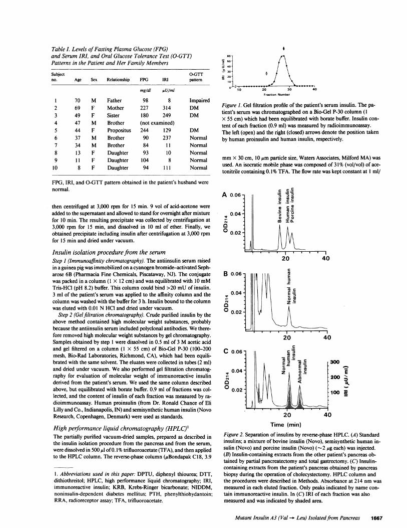
Table I. Levels of Fasting Plasma Glucose (FPG)and Serum IRI, and Oral Glucose Tolerance Test (O-GTT)Patterns in the Patient and Her Family Members
Subject O-GTTno. Age Sex Relationship FPG IRI pattern
mg/dt AU/mt
1 70 M Father 98 8 Impaired2 69 F Mother 227 314 DM3 49 F Sister 180 249 DM4 47 M Brother (not examined)5 44 F Propositus 244 129 DM6 37 M Brother 90 237 Normal7 34 M Brother 84 1 1 Normal8 1 3 F Daughter 93 10 Normal9 1 1 F Daughter 104 8 Normal
10 8 F Daughter 94 111 Normal
FPG, IRI, and O-GTT pattern obtained in the patient's husband werenormal.
then centrifuged at 3,000 rpm for 15 min. 9 vol of acid-acetone wereadded to the supernatant and allowed to stand for overnight after mixturefor 10 min. The resulting precipitate was collected by centrifugation at3,000 rpm for 15 min, and dissolved in 10 ml of ether. Finally, weobtained precipitate including insulin after centrifugation at 3,000 rpmfor 15 min and dried under vacuum.
Insulin isolation procedure from the serum
Step 1 (Immunoaffinity chromatography). The antiinsulin serum raisedin a guinea pig was immobilized on a cyanogen bromide-activated Seph-arose 6B (Pharmacia Fine Chemicals, Piscataway, NJ). The conjugatewas packed in a column (1 X 12 cm) and was equilibrated with 10 mMTris-HC1 (pH 8.2) buffer. This column could bind >20 mUof insulin.3 ml of the patient's serum was applied to the affinity column and thecolumn was washed with the buffer for 3 h. Insulin bound to the columnwas eluted with 0.01I N HC1 and dried under vacuum.
Step 2 (Gelfiltration chromatography). Crude purified insulin by theabove method contained high molecular weight substances, probablybecause the antiinsulin serum included polyclonal antibodies. Wethere-fore removed high molecular weight substances by gel chromatography.Samples obtained by step 1 were dissolved in 0.5 ml of 3 Macetic acidand gel filtered on a column (1 X 55 cm) of Bio-Gel P-30 (100-200mesh, Bio-Rad Laboratories, Richmond, CA), which had been equili-brated with the same solvent. The eluates were collected in tubes (2 ml)and dried under vacuum. Wealso performed gel filtration chromatog-raphy for evaluation of molecular weight of immunoreactive insulinderived from the patient's serum. Weused the same column describedabove, but equilibrated with borate buffer. 0.9 ml of fractions was col-lected, and the content of insulin of each fraction was measured by ra-dioimmunoassay. Human proinsulin (from Dr. Ronald Chance of EliLilly and Co., Indianapolis, IN) and semisynthetic human insulin (NovoResearch, Copenhagen, Denmark) were used as standards.
High performance liquid chromatography (HPLC)The partially purified vacuum-dried samples, prepared as described inthe insulin isolation procedure from the pancreas and from the serum,were dissolved in 500 ,ul of 0.1I% trifluoroacetate (TFA), and then appliedto the HPLCcolumn. The reverse-phase column (,gBondapak C18, 3.9
1. Abbreviations used in this paper: DPTU, diphenyl thiourea; DTT,dithiothreitol; HPLC, high performance liquid chromatography; IRI,immunoreactive insulin; KRB, Krebs-Ringer bicarbonate; NIDDM,noninsulin-dependent diabetes mellitus; PTH, phenylthiohydantoin;RRA, radioreceptor assay; TFA, trifluoroacetate.
I60-
-E50 -
:) 40 -
10-
10-l' 4020 30
Fraction Number
Figure 1. Gel filtration profile of the patient's serum insulin. The pa-tient's serum was chromatographed on a Bio-Gel P-30 column (1X 55 cm) which had been equilibrated with borate buffer. Insulin con-tent of each fraction (0.9 ml) was measured by radioimmunoassay.The left (open) and the right (closed) arrows denote the position takenby human proinsulin and human insulin, respectively.
mmX 30 cm, 10 Mmparticle size, Waters Associates, Milford MA) wasused. An isocratic mobile phase was composed of 3 1% (vol/vol) of ace-tonitrile containing 0.1I% TFA. The flow rate was kept constant at I ml/
A 0.06-
0.04-
00.02-
B 0.06 -
0.04 -
o 0.02-
C 0.06
le0.04-
00.02-
C ___C* 5 m3(a)C C.
co O-
20 40
C(U
40
f300
200 "100
2.0
Time (min)Figure 2. Separation of insulins by reverse-phase HPLC. (A) Standardinsulins; a mixture of bovine insulin (Novo), semisynthetic human in-sulin (Novo) and porcine insulin (Novo) (~-2 Mg each) was injected.(B) Insulin-containing extracts from the other patient's pancreas ob-tained by partial pancreatectomy and total gastrectomy. (C) Insulin-containing extracts from the patient's pancreas obtained by pancreasbiopsy during the operation of cholecystectomy. HPLCcolumn andthe procedures were described in Methods. Absorbance at 214 nm wasmeasured in each eluted fraction. Only peaks indicated by name con-tain immunoreactive insulin. In (C) IRI of each fraction was alsomeasured and was indicated by shaded area.
Mutant Insulin A3 (Val --o Leu) Isolated from Pancreas 1667
I-?? 0 0 9 0 316.0- - - - - - -I-- ( k..a,

min, and fractions (0.5 ml) were collected at 280C, and dried undervacuum. The residues were dissolved in 0.01 N HCI and borate buffer,and each fraction was measured by radioimmunoassay for insulin. Theapparatus used for HPLCconsisted of model 441 Waters liquid chro-matograph equipped with model 510 solvent delivery system (WatersAssociates), and Toyo Soda CP8000 chromato processor (Toyo Soda,Tokyo, Japan).
Radioreceptor assay (RRA)Suzuki et al. (19) reported that kidney of guinea pig has many insulinreceptors on the plasma membrane and can be used for radioreceptorassay for unextracted serum insulin. Weexamined the receptor bindingactivity of the patient's insulin according to their methods with a slightmodification (17).
The kidneys from five male guinea pigs (200-250 g) were removedunder nembutal anesthesia, rinsed with saline, trimmed of connectivetissue, divided into small pieces weighing - 11 mg, and homogenizedextensively in 100 ml of 0.3 Msucrose solution. Then the homogenatewas centrifuged at 10,000 g for 20 min and the resulting supernatantwas centrifuged at lO g for 90 min. The precipitate was suspended in 5ml of 25 mMTris-HCI buffer and homogenized strongly and kept frozenat -40'C. '25I-insulin binding to the insulin receptors on the kidneymembranes was examined in 400 gl of buffer (50 mMTris-HCI, 0.1%bovine serum albumin (BSA), 10 mMCaC12, 2 mMN-ethylmaleimidepH 7.6) containing kidney pellets (100 Mug of protein), '25I-insulin (50 ,l)and various concentration of either standard insulin or samples (50 M1).Samples included sera from the patient's peripheral and portal vein, serafrom her family members, partially purified pancreatic insulin preparedas described above, and normal or abnormal component insulin of pa-tient's pancreas purified by HPLC. Whensamples such as purified pan-creatic insulin were assayed, they were dissolved in insulin-free serumand then the incubation for RRAwas carried out. After an overnightincubation, 1 ml of the buffer was added and centrifuged at 3,000 rpmfor 20 min and the radioactivity of the precipitate was counted.
Biological activityWemeasured the biological activity of the patient's insulin by glucoseoxidation. Male Wistar rats (150 g, fed ad lib.) were killed by decapitation,and the epididynmal adipose tissues were removed. Isolated adipocyteswere prepared by the use of collagenase (Worthington Biochemical Corp.,Freehold, NJ) according to the method of Rodbell (20). Stimulation ofglucose oxidation was measured by means of incubating isolated ratadipocytes at 370C with [1-'4C]glucose in Krebs-Ringer bicarbonate
E-
C
a
a
ac0
C
EE
20
10 -
0-
20 -
10
0-
20 -
10 -
0-_
100 -
50 -
0-
(KRB) buffer containing various concentrations of either standard insulinor samples. Samples included the patient's pancreatic insulin preparedas described above, and normal or abnormal component insulin of pa-tient's pancreas purified by HPLC. They were dissolved in KRBbuffersupplemented with 1% BSA, and the incubation was carried out. After1 h of incubation the generated 14CO2 was collected and counted in aliquid scintillation counter as described previously by Kasuga et al. (21).
Determination of amino acid sequence of thepatient's insulinAmino acid sequences of both normal and abnormal component insulinsof the patient's pancreas purified by HPLCwere then determined usinga model 470A protein sequencer equipped with a model 120A phenyl-thiohydantoin (PTH) analyzer (both from Applied Biosystems, FosterCity, CA) and a model SP 4200 computing integrator (Spectra-PhysicsInc., Mountain View, CA). Since we analyzed purified insulins (normaland abnormal components) without dividing into A- and B-chains, wecould detect two N-terminal amino acids at one cycle.
ResultsGelfiltration chromatography. The patient's serum was gel-fil-tered on Bio-Gel P-30 to yield the profile shown in Fig. 1. Mostof insulin immunoreactivity was eluted at the position of insulin,indicating that the hyperinsulinemia of the patient is not due tohyperproinsulinemia.
HPLC. The patient's insulin purified from her pancreas,portal venous blood, and peripheral venous blood, and thosepurified from peripheral venous blood of her family memberswere applied to HPLCcolumn. The results are shown in Fig. 2and Fig. 3. The elution profiles of the insulins from the patientand family members demonstrated two peaks of immunoreactiveinsulins. One peak appeared at the position of normal humaninsulin. The insulin peak eluted later was thought to be a struc-turally abnormal insulin that was more hydrophobic than normalhuman insulin. The ratios of the immunoreactivity of normalinsulin to abnormal insulin were 5:4 in insulin from the patient'spancreas (Fig. 2 C), 4:5 in the patient's serum from the portalvein (Fig. 3 B), 1:7 in the patient's serum from the peripheralvein (Fig. 3 A), and 1:4-1:7 in her family member's serum fromthe peripheral vein (Fig. 3 C-F). The ratio of normal to abnormal
Figure 3. Separation of immuno-purified serum insulins by reverse-phase HPLC. (A) Insulin from pa-
C tient's peripheral vein. (B) Insulinfrom patient's portal vein. (C-F)Insulins from peripheral vein of
..________________________<family members with hyperinsulin-emia. HPLCcolumn and the pro-
D cedures were described in Meth-ods. Immunoreactive insulin was
measured in each eluted fraction.Closed and open vertical arrows
E indicate the position of normaland abnormal insulin, respectively.Note that the positions of the insu-
l Ailin peaks between A, B, and C-Fare slightly different. Since HPLC
F analyses of these two groups werecarried out at different times, the
jXk, difference may be caused by theslightly different experimental con-
1ZO30 40 50 ditions such as the composition of1 0 20 30 40 50mobile phase or temperature of the
Fraction Number column.
1668 H. Sakura, Y. Iwamoto, Y. Sakamoto, T. Kuzuya, and H. Hirata
Fraction Number

insulin from the patient's pancreas (5:4) determined by the ab-sorbance at 214 nmwas almost identical to that determined byimmunoreactive insulin, suggesting that there was no differencein the immunoreactivity between the normal and the abnormalinsulins (Fig. 2 C).
Radioreceptor assay. Weperformed RRAof the insulinsobtained from the pancreas and portal vein of the patient, fromthe peripheral vein of both the patient and her families. Further,normal and abnormal insulins separated by HPLC from herpancreatic insulin were also assayed by RRA. As shown in Fig.4, the binding activities of insulins, which contained abnormalinsulin, were all decreased. Compared with standard human in-sulin, the binding activities were 50% for the patient's pancreaticinsulin, 24% for the patient's serum insulin of the portal vein,12% for insulins from the peripheral vein of both the patientand her family members with abnormal insulinemia, and -5%
E
.0_
m
c
x
0.0
3
N
100 1000
Immunoreactive Insulin (,uU/ml)
for the abnormal insulin component purified by HPLC fromher pancreatic insulin. In contrast, displacement curves of theinsulin from the normal component purified by HPLC fromher pancreatic insulin and the serum of a family member withouthyperinsulinemia (subject 7 in Table I) were completely super-imposed on that of standard human insulin.
Biological activity. Fig. 5 shows the results of the ability ofthe patient's pancreatic insulin (mixture of normal and abnormalinsulin), purified insulins (normal component and abnormalcomponent) by HPLC, and standard human insulin to stimulateglucose oxidation in isolated rat adipocytes. The biological po-tency of the pancreatic insulin was reduced to 60%, and that ofthe abnormal component insulin was reduced to as little as 8%of the standard insulin, while the biological potency of the normalcomponent insulin of the patient was identical to that of thestandard insulin. However, maximally stimulated levels of glu-
10000
Cum
Ex
0
.0.0c
._
Immunoreactive Insulin (,uU/mi)
Figure 4. Ability of various insulinsto inhibit '25I-labeled porcine insulinbinding to guinea pig kidney mem-brane. (upper) Standard human insu-lin (-); the insulin isolated from pa-tient's pancreas (mixture of normaland abnormal insulins (s6); normal(o) and abnormal component insulin(o) purified by HPLCfrom patient'spancreas. Details of purification pro-cedure of each sample was describedin text. (lower) Standard human in-sulin (.); the insulin isolated fromsera of the patient (e), sera of familymembers with hyperinsulinemia(subject 2: ., subject 3: A, subject 6:v, subject 10: x), and sera of familymembers without hyperinsulinemia(subject 7) (v); and the immunopuri-fied insulin isolated from patient'sportal vein serum (A).
Mutant Insulin A3 (Val -- Leu) Isolatedfrom Pancreas 1669

10000
8000
6000f
* 4000
Normal I insulin
Standard humaninsulin
10 100
Immunoreactive Insulin (,u U/ml)
cose oxidation were observed in the presence of higher concen-trations of the abnormal insulin (>500 jU/ml).
Amino acid sequence. Weobtained -500 pmol of normaland 400 pmol of abnormal insulin by HPLCfrom 0.2 mg ofextract from the patient's pancreas. Weperformed amino acidsequencing both normal and abnormal insulin. As shown in Fig.6, we found that the third amino acids from N-terminal of theabnormal insulin consisted of leucine and asparagine, althoughthat position of the normal insulin were occupied with valineand asparagine. Since we could not find other differences betweennormal and abnormal insulin (Fig. 7), we concluded that theabnormal insulin of this patient contained a leucine for valinesubstitution at position 3 of the A-chain.
Discussion
Werecently found a patient whose insulin was a substitution ofleucine for valine at the third position of the A-chain. This phe-
Figure 5. Ability of various insulins to stimulate glu-cose oxidation in isolated rat adipocytes. Standard hu-man insulin (.); the insulin isolated from patient'spancreas (mixture of normal and abnormal insulins)(A); normal (o) and abnormal insulin (0) purified by
1000 HPLCfrom patient's pancreas. Details of purificationprocedure of each sample was described in text.
notype is identical to insulin Wakayama (9, 12, 16), but wecould not trace any relationship between two families. Two fam-ilies have lived far apart, in the western and eastern parts ofJapan. Since this substitution is not fatal and induces only (ma-turity-onset) noninsulin dependent diabetes mellitus (NIDDM),there is a possibility that this mutant insulin may have existedfor a long time and is widely scattered in Japan. Alternatively,this mutation may have occurred entirely independently in dif-ferent parts of Japan. However, we must consider that a differentgenotype can produce the same phenotype. The DNAsequencethat corresponds to valine is GTG, and if one point mutationinduces substitution of leucine for valine, two DNAsequences(e.g., TTGand CTG) can be candidates, and insulin Wakayamawas a former one (12, 16). DNAsequence of our family is nowunder investigation.
The abilities to bind to the insulin receptors and to stimulateglucose oxidation of the patient's abnormal insulin purified byHPLCfrom her pancreatic insulin were 5%and 8% that of nor-
B Abnormal Insulin: A3-13
L -Asn -
Val
Lou
* DTT: Dlthlothreltol** DPTU: Dlphenyl thiourea
Figure 6. HPLCprofiles of the PTHamino acids obtained by the Edmandegradation procedure (3rd cycle) ofnormal (A) and abnormal (B) insulin.
Asn These insulins were purified from thepatient's pancreas by the insulin isola-tion procedures and reverse-phaseHPLCas described in Methods. Thirdposition of the A and B chains of nor-
mal insulin consisted of asparagine(Asn) and valine (Val), and that of ab-normal insulin asparagine (Asn) andleucine (Leu), indicating the substitutionof leucine for valine at the A3 position.Peaks by DTT (W) and DPTU(**) were
detected as internal standards at eachcycle of HPLCanalysis of PTHaminoacids.
1670 H. Sakura, Y. Iwamoto, Y. Sakamoto, T. Kuzuya, and H. Hirata
A Normal Insulin: A3-B3
If,-
1

Sample \ cycle 1 2 3 4 5 6 7 8 9 10 11 12 13 14 15 16 17 18 19 20 21
Human A chain G I V E Q C C T S I C S L Y Q L E N Y C N
insulin B chain F V N Q H L C G S H L V E A L Y L V C G E
G I V E Q T I S LV Q L E N Y NNormal insulin S
F V N Q H L G H L V E A L Y L V G E
G I L E Q T I S LY Q L E N Y NAbnormal insulin S
F V N Q H L G H L V E A L Y L V G E
Sample \cycle 22 23 24 25 26 27 28 29 30
Human insulin R G F F Y T P K TB chain
Normal insulin R G F F Y T P K T
Abnormal insulin R G F F Y T P K T
Figure 7. Total amino acid sequences of normal and abnormal insulins purified from the patient's pancreas by the insulin isolation proceduresand reverse-phase HPLCas described in Methods. Each amino acid is expressed as a single letter. Since cys (C) can not be detected in this analy-sis, several cycles (cycle 6, 7, 11, 19, and 20) remained to be blank.
mal human insulin, respectively. The results indicate that thereis a good parallelism between binding activity and biologicalactivity of the abnormal insulin in the patient. Tager et al. (22)and Olefsky et al. (23) reported that the biological activity ofinsulin Chicago was much lower than its binding activity andsuggested that the abnormal insulin was an active antagonist ofinsulin action. Keefer et al. (24) and Kobayashi et al. (25), how-ever, found no antagonistic effect in those analogues. Further-more, Kobayashi et al. (26) clarified that the biological activityof insulin Los Angeles was proportional to its receptor bindingactivity. In concert with their results (24-26) the biological ac-tivity of the abnormal insulin of this patient was reduced toalmost the same extent as the binding activity.
Shoelson et al. (9) described that the variant insulin repre-sented 57% and the normal insulin 43% of the patient's storedpancreatic insulin in the case of insulin Chicago. Our resultsshow that the ratio of abnormal to normal insulin in the pancreasis 4:5, and support the idea that both normal and abnormalinsulins are secreted in almost equal proportions, indicating thecodominant expression of both alleles of the insulin gene. How-ever, we cannot insist that the expression of both alleles is exactlythe same, because it was known that the quantities of HbS het-erozygotes, which also have a point mutation of an allele likean abnormal insulin gene, are only 20-50% of all hemoglo-bins (27).
The reason why the ratio of an abnormal insulin to a normalinsulin in the peripheral vein is higher than that in the pancreasis probably due to a decreased metabolism of an abnormal in-sulin. The ratio of an abnormal insulin to a normal insulin inthe portal vein was found to be slightly lower than that in thepancreas as expected.
N-terminal and C-terminal of A-chain and C-terminal of B-chain (especially B23-26) are thought to constitute the receptorbinding site which is important for the action of insulin (28),and the amino acid sequences in these parts are conservedthrough evolution. Substitution of A3 amino acid is thereforesupposed to influence the conformation of the receptor bindingsite like B24 or B25 substitution, and to induce the decreasedbinding activity to the insulin receptor, which may result in
decreased bioactivity and decreased metabolism of the abnormalinsulin.
It is interesting to consider the reasons why patients withmutant insulin have diabetes. Family study shows that not allof those with a mutant insulin are diabetic. Noninsulin-depen-dent diabetes developes most frequently after middle age. Itseems that in this family diabetes develops with age. There is adiscussion that diabetes may develop only when ,8-cell secretionis no longer able to compensate the reduced bioactivity of itssecretory products (8), but in the present family, immunoreactiveinsulin (IRI) level of the propositus' mother who is overtly di-abetic is highest and that of the propositus' daughter whose GTTpattern is normal is lowest among the members with hyperin-sulinemia. There were no differences in the results of HPLCprofiles and RRAbetween those with and without diabetes. Wetherefore suppose that insulin insensitivity related to the post-receptor factors may contribute to the development of diabetesat least in part of this family. The analysis of development ofdiabetes by a mutant insulin will provide valuable informationsalso for the understanding of the pathogenesis of NIDDM ingeneral.
Acknowledgments
The authors thank Dr. Y. Ishii (Ohmiya Red Cross Hospital) for referringthe patient, Dr. K. Kasahara (Department of Surgery, Jichi MedicalSchool) for surgical operation, Dr. Kouga (Shimizu Pharmaceutical Co.)for isolation of insulin from the pancreas. Wealso thank Dr. M. Yoshida(Department of Biochemistry, Jichi Medical School), Dr. T. Ohta (De-partment of Biology, Jichi Medical School), Dr. Y. Oka (University ofTokyo) and Mr. Y. Sengoku (Japan Scientific Instrument Co., Ltd.) fortheir helpful technical advices.
This study was supported in part by a grant from the Ministry ofEducation, Science and Culture 60480276, Japan.
References1. Williams, R. H. 1965. Recent advances relative to diabetes mellitus.
Ann. Intern. Med. 63:512-529.2. Elliot, R. B., D. O'Brien, and C. C. Roy. 1965. An abnormal
insulin in juvenile diabetes mellitus. Diabetes. 14:780-787.3. Schwartz, I. L., and 0. Hechter. 1966. Insulin structure and func-
Mutant Insulin A3 (Val -- Leu) Isolatedfrom Pancreas 1671

tion. Reflections on the present state of the problem. Am. J. Med. 40:765-772.
4. Kimmel, J. R., and H. G. Pollock. 1967. Studies of human insulinfrom nondiabetic and diabetic pancreas. Diabetes. 16:687-694.
5. Brunfeldt, K., T. Deckert, and J. Thomsen. 1969. Human crys-talline insulin from non-diabetic and diabetic patients. Acta Endocrinol.60:543-549.
6. Tager, H., B. Given, D. Baldwin, M. Mako, J. Markese, A. Rub-enstein, J. Olefsky, M. Kobayashi, 0. Kolterman, and R. Poucher. 1979.A structurally abnormal insulin causing human diabetes. Nature (Lond.).281:122-125.
7. Given, B. D., M. E. Mako, H. S. Tager, D. Baldwin, J. Markese,A. H. Rubenstein, J. Olefsky, M. Kobayashi, 0. Kolterman, and R.Poucher. 1980. Diabetes due to secretion of an abnormal insulin. N.Engl. J. Med. 302:129-135.
8. Haneda, M., K. S. Polonsky, R. M. Bergenstal, J. B. Jaspan, S. E.Shoelson, P. M. Blix, S. J. Chan, S. C. M. Kwok, W. B. Wishner, A.Zeidler, J. M. Olefsky, G. Freidenberg, H. S. Tager, D. F. Steiner, andA. H. Rubenstein. 1984. Familial hyperinsulinemia due to a structurallyabnormal insulin. Definition of an emerging new clinical syndrome. N.Engl. J. Med. 310:1288-1294.
9. Shoelson, S., M. Haneda, P. Blix, A. Nanjo, T. Sanke, K. Inouye,D. Steiner, A. Rubenstein, and H. Tager. 1983. Three mutant insulinsin man. Nature (Lond.). 302:540-543.
10. Vinik, A. I., S. Seino, A. Funakoshi, J. Schwartz, M. Matsumoto,D. E. Schteingart, Z.-Z. Fu, and S.-T. Tsai. 1986. Familial hyperinsulin-emia associated with secretion of an abnormal insulin, and coexistenceof insulin resistance in the propositus. J. Clin. Endocrinol. Metab. 62:645-652.
11. Tager, H. S. 1984. Abnormal products of the human insulingene. Diabetes. 33:693-699.
12. Nanjo, K., T. Sanke, M. Miyano, K. Okai, R. Sowa, M. Kondo,S. Nishimura, K. Iwo, K. Miyamura, B. D. Given, S. J. Chan, H. S.Tager, D. F. Steiner, and A. H. Rubenstein. 1986. Diabetes due to se-cretion of a structurally abnormal insulin (insulin Wakayama). Clinicaland functional characteristics of [LeuA3] insulin. J. Clin. Invest. 77:514-519.
13. Kwok, S. C. M., S. J. Chan, A. H. Rubenstein, R. Poucher, andD. F. Steiner. 1981. Loss of a restriction endonuclease cleavage site inthe gene of a structurally abnormal human insulin. Biochem. Biophys.Res. Commun. 98:844-849.
14. Kwok, S. C. M., D. F. Steiner, A. H. Rubenstein, and H. S. Tager.1983. Identification of a point mutation in the human insulin gene givingrise to a structurally abnormal insulin (Insulin Chicago). Diabetes. 32:872-875.
15. Haneda, M., S. J. Chan, S. C. M. Kwok, A. H. Rubenstein, andD. F. Steiner. 1983. Studies on mutant human insulin genes: identificationand sequence analysis of a gene encoding [SerB24] insulin. Proc. Natl.Acad. Sci. USA. 80:6366-6370.
16. Sanke, T., M. Kondo, and K. Nanjo. 1985. Two families ofabnormal insulinemia. Folia Endocrinol. Jpn. 61 :257a. (Abstr.)
17. Iwamoto, Y., H. Sakura, Y. Ishii, R. Yamamoto, S. Kumakura,Y. Sakamoto, A. Matsuda, and T. Kuzuya. 1986. A new case of abnormalinsulinemia with diabetes: Reduced insulin values determined by radi-oreceptor assay. Diabetes. 35 (In press.)
18. Davoren, P. R. 1962. The isolation of insulin from a single catpancreas. Biochim. Biophys. Acta. 63:150-153.
19. Suzuki, K., N. Ohsawa, and K. Kosaka. 1976. Radioreceptorassay for insulin. J. Clin. Endocrinol. Metab. 42:399-402.
20. Rodbell, M. 1964. Metabolism of isolated fat cells. 1. Effects ofhormones on glucose metabolism and lipolysis. J. Biol. Chem. 239:375-380.
21. Kasuga, M., Y. Akanuma, Y. Iwamoto, and K. Kosaka. 1978.Insulin binding and glucose metabolism in adipocytes of streptozotocindiabetic rats. Am. J. Physiol. 235:E175-E182.
22. Tager, H., N. Thomas, R. Assoian, A. Rubenstein, M. Saekow,J. Olefsky, and E. T. Kaiser. 1980. Semisynthesis and biological activityof porcine [LeuB24] insulin and [LeuB2S] insulin. Proc. Nati. Acad. Sci.USA. 77:3181-3185.
23. Olefsky, J. M., M. Saekow, H. Tager, and A. H. Rubenstein.1980. Characterization of a mutant human insulin species. J. Biol. Chem.255:6098-6105.
24. Keefer, L. M., M.-A. Piron, P. DeMeyts, H.-G. Gattner, C. Dia-conescu, D. Saunders, and D. Brandenburg. 1981. Impaired negativecooperativity of the semisynthetic analogues human [LeuB24]- and[LeuB2J]-insulins. Biochem. Biophys. Res. Commun. 100: 1229-1236.
25. Kobayashi, M., S. Ohgaku, M. Iwasaki, H. Maegawa, Y. Shigeta,and K. Inouye. 1982. Characterization of [LeuB24]- and [LeuB2]-insulinanalogues. Receptor binding and biological activity. Biochem. J. 206:597-603.
26. Kobayashi, M., M. Haneda, H. Maegawa, N. Watanabe, Y. Tak-ada, Y. Shigeta, and K. Inouye. 1984. Receptor binding and biologicalactivity of [SerB241-insulin, an abnormal mutant insulin. Biochem. Bio-phys. Res. Commun. 119:49-57.
27. Huisman, T. H. J. 1979. Sickle cell anemia as a syndrome: Areview of diagnostic features. Am. J. Hematol. 6:173-184.
28. Pullen, R. A., D. G. Lindsay, S. P. Wood, I. J. Tickle, T. L.Blundell, A. Wollmer, G. Krail, D. Brandenburg, H. Zahn, J. Gliemann,and S. Gammeltoft. 1976. Receptor-binding region of insulin. Nature(Lond.). 259:36-38.
1672 H. Sakura, Y. Iwamoto, Y. Sakamoto, T. Kuzuya, and H. Hirata