structural studies of human gbe1 and relevance to apbd
TRANSCRIPT

Structural studies of human GBE1 and relevance to APBD
Wyatt YueStructural Genomics Consortium (SGC)
University of Oxford
APBDRF Meeting Dec 2013

Combining structural biology, protein biochemistry to understand inborn errors of metabolism
www.thesgc.org/wyatt [email protected]
• Public-private partnership
• High-throughput structural & chemical biology of human proteins
• Diverse protein families/biology areas
• Open access research model
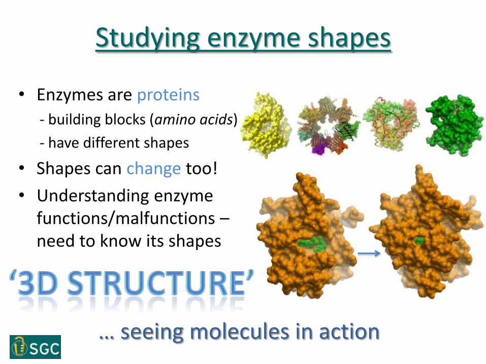
Studying enzyme shapes
• Enzymes are proteins
- building blocks (amino acids)
- have different shapes
• Shapes can change too!
• Understanding enzyme functions/malfunctions –need to know its shapes
… seeing molecules in action

‘Taking pictures’ of protein structures
1. make proteins of interest (expression, purification)
2. arrange them in order (crystallization)
3. take picture! (x-ray diffraction)
4. develop the film (modelling)
Our work so far …
APBDRF Meeting Dec 2013

Glycogen Synthesis
glycogenin
glycogenin
glycogenin
glycogensynthase
glycogensynthase
branchingenzyme
priming
elongation
branching
GSD type XV
GSD type 0
GSD type IV (liver)Adult Polyglucosan Body Disease
human GYG1 structure
human GBE1 structure
Cartoon impression of glycogen granule

1 702700
Multi-construct approach
E coli structure
6363
7070
7979
c004c005
c000c001c002c003
54
c101c102
c104c105c106c107
c103
c108
1116163838
54
c109c110c111c112
2nd roundSoluble in insect cells
L28P36
Y41R47
c011, c012c013, c014c015, c016c017, c018
3rd roundFine-tune
• Full length and a series of truncations• N- and C-termini nibbling• N-terminal His6 tagged fusion
1st roundInsoluble in E. coli
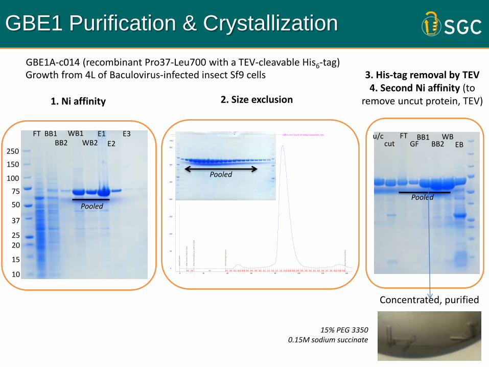
GBE1 Purification & Crystallization
150
100
75
50
37
2520
15
10
FT BB1 WB1 E1E2
E3BB2 WB2
Pooled
250
GBE1A c011 E1 s200 GF 300812:Sample1Title_UV GBE1A c011 E1 s200 GF 300812:Sample1Title_Fractions GBE1A c011 E1 s200 GF 300812:Sample1Title_Inject GBE1A c011 E1 s200 GF 300812:Sample1Title_Logbook
0
50
100
150
200
250
300
350
mAU
0 20 40 60 80 100 120 140 ml
Sa
mp
le 1
Na
me
Wa
sh
Fra
ctio
n C
olle
cto
r O
utle
t
Fin
ish
Wa
sh
ing
Fra
ctio
n C
olle
cto
r O
utle
t
Sta
rt C
olle
ctin
g F
ractio
ns
Sto
p C
olle
ctin
g F
ractio
ns
A2 A4 A6 A7 A9 A11 B12 B10 B8 B6 B4 B2 C1 C3 C5 C7 C9 C11 D12 D10 D8 D6 D4 D2 E1 E3 E5 E7 E9 E11 F12 F10 F8
Pooled
u/ccut
FTGF BB2
WBEB
BB1
Pooled
Growth from 4L of Baculovirus-infected insect Sf9 cells
1. Ni affinity 2. Size exclusion
3. His-tag removal by TEV4. Second Ni affinity (to
remove uncut protein, TEV)
Concentrated, purified
15% PEG 3350 0.15M sodium succinate
GBE1A-c014 (recombinant Pro37-Leu700 with a TEV-cleavable His6-tag)
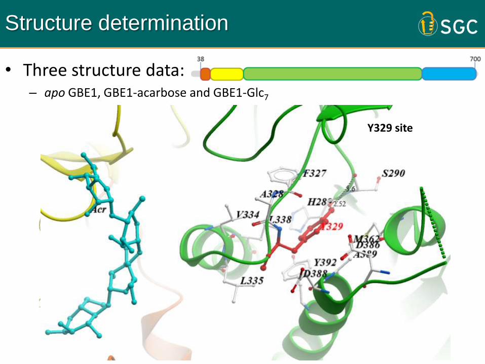
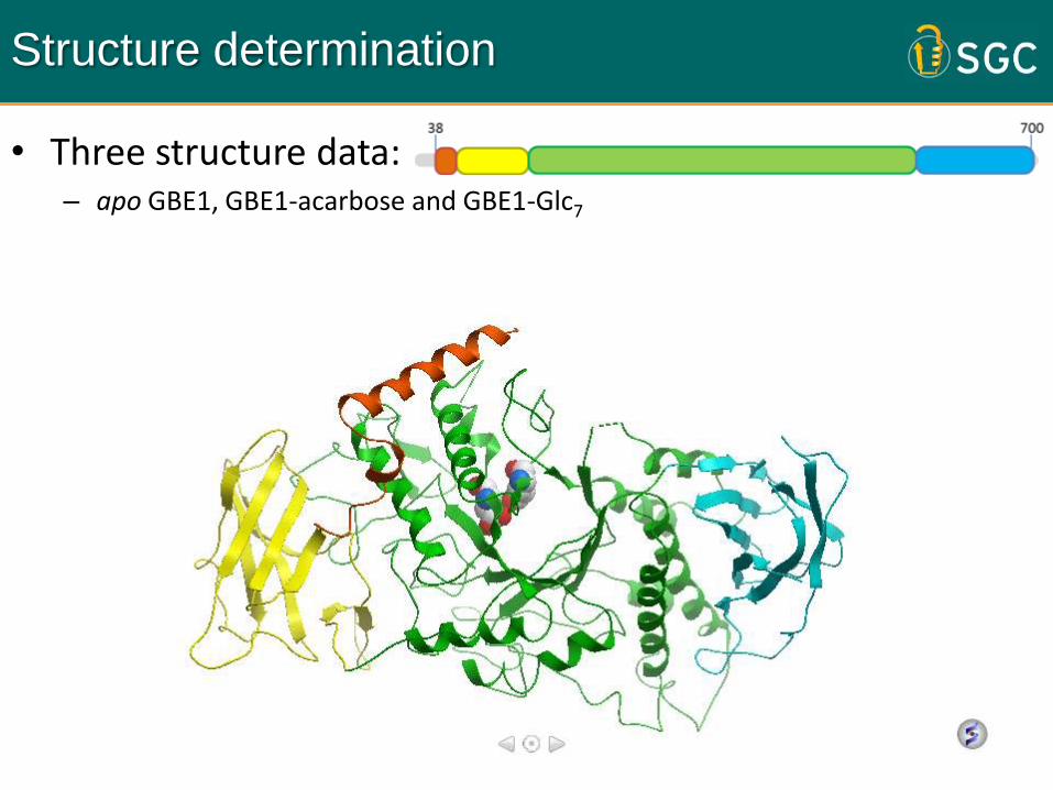
Structure determination
• Three structure data:– apo GBE1, GBE1-acarbose and GBE1-Glc7
Overall structureMapping of gbe1 mutationsComplex with sugar chains
Y329 site
1 702
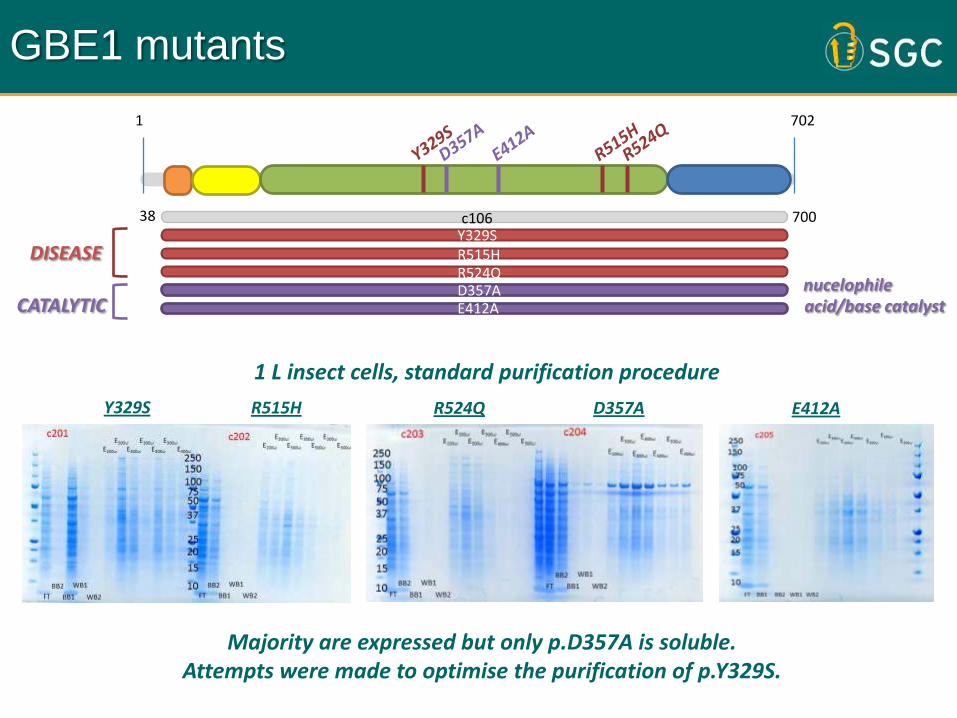
GBE1 mutants
c10638 700
DISEASE
CATALYTICnucelophileacid/base catalyst
Majority are expressed but only p.D357A is soluble.Attempts were made to optimise the purification of p.Y329S.
1 L insect cells, standard purification procedure
Y329S R515H R524Q D357A E412A
Y329SR515HR524QD357AE412A
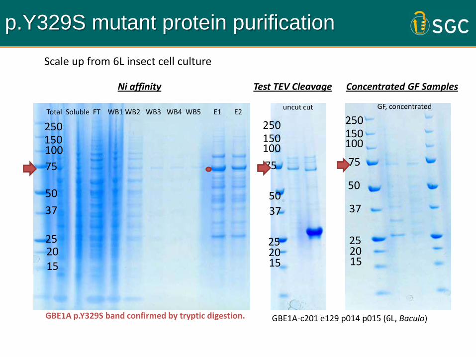
GBE1A-c201 e129 p014 p015 (6L, Baculo)
250150100
75
50
37
252015
uncut cut
Test TEV Cleavage Concentrated GF Samples
250150100
75
50
37
252015
250150100
75
50
37
252015
Total Soluble FT WB1 WB2 WB3 WB4 WB5 E1 E2GF, concentrated
Ni affinity
p.Y329S mutant protein purification
GBE1A p.Y329S band confirmed by tryptic digestion.
Scale up from 6L insect cell culture
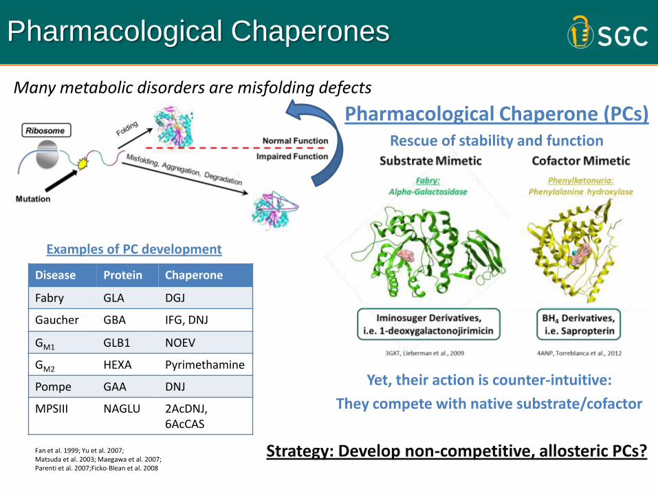
Many metabolic disorders are misfolding defects
Strategy: Develop non-competitive, allosteric PCs?
Pharmacological Chaperones
Yet, their action is counter-intuitive:
They compete with native substrate/cofactor
Disease Protein Chaperone
Fabry GLA DGJ
Gaucher GBA IFG, DNJ
GM1 GLB1 NOEV
GM2 HEXA Pyrimethamine
Pompe GAA DNJ
MPSIII NAGLU 2AcDNJ, 6AcCAS
Fan et al. 1999; Yu et al. 2007; Matsuda et al. 2003; Maegawa et al. 2007; Parenti et al. 2007;Ficko-Blean et al. 2008
Pharmacological Chaperone (PCs)Rescue of stability and function
Examples of PC development
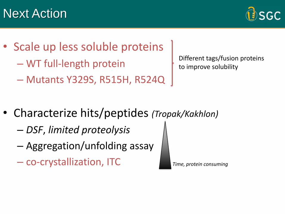
Next Action
• Scale up less soluble proteins
– WT full-length protein
– Mutants Y329S, R515H, R524Q
• Characterize hits/peptides (Tropak/Kakhlon)
– DSF, limited proteolysis
– Aggregation/unfolding assay
– co-crystallization, ITC
Different tags/fusion proteins to improve solubility
Time, protein consuming
Future: collaboration with APBD
APBDRF Meeting Dec 2013
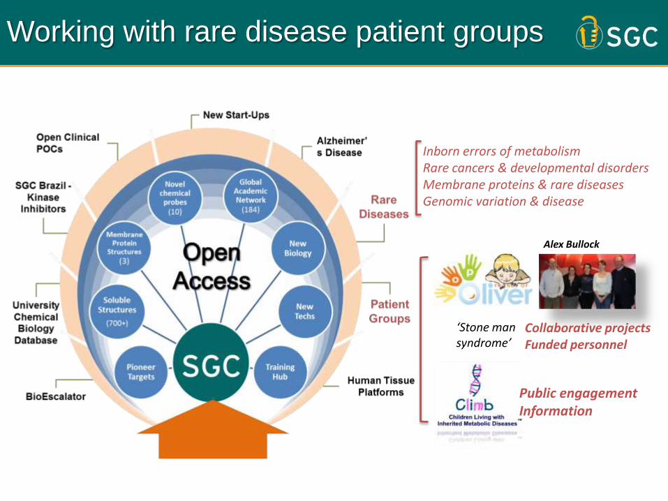
Working with rare disease patient groups
Collaborative projectsFunded personnel
Inborn errors of metabolism Rare cancers & developmental disordersMembrane proteins & rare diseasesGenomic variation & disease
‘Stone man syndrome’
Public engagementInformation
Alex Bullock

What can we do to help APBDRF?
xtal soaking
Crystals, 3D StructureWT/mutant protein
in silico Docking
Binding, BiochemistryDSF, ITC, BLI, co-xtal
dose response, affinity
hit finding
validation
Mode of Actionfolding, proteolysis, aggregation
Effects on activity, stability
characterization
HT Compound Screening (Michael Tropak)
in silico Ligand Design (peptides – Or Kakhlon)
Rescue in vivo?
patient fibroblast cellsAnimal model (e.g. mouse)
enzyme activity
Cellular Assays
Test Mutantsrescue stability?
Effects on activity
Clinical ‘know-how’
Goal: To develop compounds into a pharmacological chaperone treatment

Example of our In Vitro capabilities
Allosteric domain(AdoMet)
Catalytic domain(PLP, haem)
Pathfinder Awards for Orphan Diseases
human CBS structure
Developed In vitro assays to deconvolute binding modes
Recombinant proteinStructural Biology
Compound libraryCellular assay
Structurally diverse, drug-like hits
kp = 0.381 ± 0.037 min-1
Limited proteolysisIn silico docking Domain mappingFragment DSF
Aim: look for binders at different pockets/regions as chemical starting point
Cystathionine beta synthase (CBS) deficiency

ACKNOWLEDGEMENTSTHE MOBThomas McCorvieDipali PatelJolanta KopecStephanie OerumFiona FitzpatrickSean FroeseWasim Kiyani
CRYSTALLOGRAPHY
Frank von DelftTobias Krojer
BIOTECHNOLOGY
Claire DamerellPravin Mahajan
FUNDING PARTNERS
The Canadian Institutes for Health Research, the Canada Foundation for Innovation, Genome Canada, GlaxoSmithKline, Lilly Canada, the Novartis Research Foundation, Pfizer, Takeda, the Ontario Ministry of Economic Development and Innovation, and the Wellcome Trust.
[email protected]/wyatt
2013
2012

Structure determination
• Three structure data:– apo GBE1, GBE1-acarbose and GBE1-Glc7
Overall structureMapping of gbe1 mutationsComplex with sugar chains
Y329 site

Updates on DSF
APBDRF Meeting Dec 2013

Differential Scanning Fluorimetry (DSF), ‘Tm shift’ Niesen et al 2007 Nat Methods
WT
Thermal stability as a ligand binding assay
Destabilization due to mutation – left shift (- Tm)
Stabilization by native ligands –right shift (+ Tm)
Small molecule stabilization? –right shift towards WT
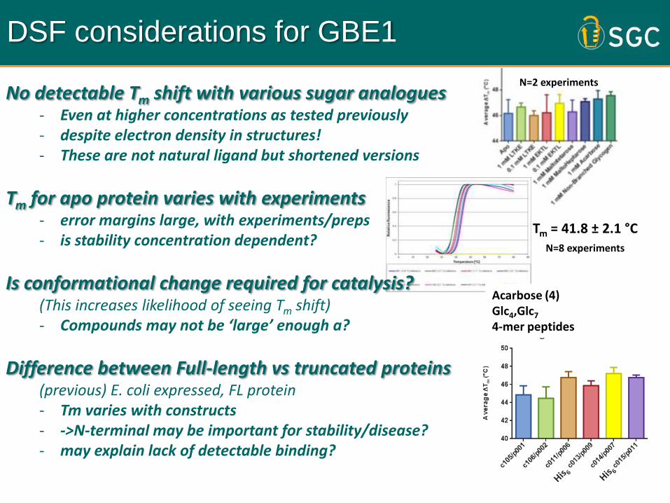
DSF considerations for GBE1
Tm = 41.8 ± 2.1 °CN=8 experiments
N=2 experimentsNo detectable Tm shift with various sugar analogues
- Even at higher concentrations as tested previously- despite electron density in structures! - These are not natural ligand but shortened versions
Tm for apo protein varies with experiments - error margins large, with experiments/preps- is stability concentration dependent?
Is conformational change required for catalysis?(This increases likelihood of seeing Tm shift) - Compounds may not be ‘large’ enough a?
Difference between Full-length vs truncated proteins(previous) E. coli expressed, FL protein - Tm varies with constructs- ->N-terminal may be important for stability/disease? - may explain lack of detectable binding?
Acarbose (4)Glc4,Glc7
4-mer peptides

1702
c105c106c011
700
c013c014
3838
283636
41c015
p001 = cut c105 usedp002 = cut c106 usedp003 = cut c106 usedp004 = uncut c106 ~200ul 16.5mg/mlp005 = cut c106 ~100ul 2.2mg/mlp006 = cut c011 ~120ul 6.9mg/ml*p007 = cut c014 ~530ul 16.1mg/ml
*p009 = uncut c013 ~550ul 20.2mg/mlp010 = cut c013 ~330ul 3.5mg/mlp011 = uncut c015 ~250ul 7.3mg/mlp012 = cut c015 ~80ul 7.3mg/mlp013 = uncut c106 ~50ul 10.7mg/ml
702700
702702
700702
02 Oct 2012, 15 Jan 2013
15 Sep 2013
GBE1 constructs purified

Y329 forms hydrogen bond between its side-chain and backbone carbonyl of His289.2.5 Å distance in WT increases to 8.5 Å in p.Y329S.
Virus re-amplified , extraction buffer optimised to sodium phosphate based,
Talon (cobalt) used instead of Ni-NTA (Nickel).
WT p.Y329S
Structural analysis of Y329 site
Structure determination
• Three structure data:– apo GBE1, GBE1-acarbose and GBE1-Glc7