structural rearrangements of sucrose phosphorylase from ... · beldman2, jette s. kastrup1 and...
TRANSCRIPT

Structural rearrangements of Sucrose Phosphorylase from Bifidobacterium adolescentis during sucrose conversion. Osman Mirza1, Lars K. Skov1*, Desiree Sprogøe1, Lambertus A. M. van den Broek2, Gerrit Beldman2, Jette S. Kastrup1 and Michael Gajhede1 From the 1 Biostructural Research, Department of Medicinal Chemistry, The Danish University of Pharmaceutical Sciences, Universitetsparken 2, DK-2100 Copenhagen, Denmark,2 Laboratory of Food Chemistry, Wageningen University, P.O. Box 8129, 6700 EV Wageningen, The Netherlands. Running title: Structural rearrangements of Sucrose Phosphorylase during sucrose conversion Address correspondence to: Michael Gajhede, Department of Medicinal Chemistry, The Danish University of Pharmaceutical Sciences, Universitetsparken 2, DK-2100 Denmark. Telephone: +45 35306407 FAX: +45 35306040 E-mail: [email protected] The reaction mechanism of sucrose phosphorylase from Bifidobacterium adolescentis (BiSP) has been studied by site directed mutagenesis and x-ray crystallography. An inactive mutant of BiSP (E232Q) has been co-crystallized with sucrose. The structure revealed a substrate binding mode comparable to that seen in other related sucrose-acting enzymes. Wild-type BiSP has also been crystallized in the presence of sucrose. In the dimeric structure a covalent glucosyl intermediate was formed in one molecule of the BiSP dimer and (after hydrolysis of the glucosyl intermediate) a β–D-glucose product complex was formed in the other molecule. While the overall structure of the glucosyl intermediate complex of BiSP is similar to that of the BiSP E232Q:sucrose complex, the glucose complex discloses major differences in loop conformations. Two loops (residues 336-344 and residues 132-137) in proximity of the active site are found to move up to 16 Å and 4 Å, respectively. Based on these findings, a reaction cycle has been suggested that takes the large movements in the active site entrance loops into account. *Present address: Novozymes A/S, Smørmosevej 9, DK-2880 Bagsværd, Denmark. Funding information: This work was supported by the Danish Natural Science
Research Council, the Danish Medical Research Council, Novozymes A/S and the Danish Synchrotron User Center (DANSYNC). Abbreviations used are: AS, amylosucrase; BiSP, sucrose phosphorylase from Bifidobacterium adolescentis; BiSP:glucose, structure of the wild-type BiSP mixed dimer; E232Q:sucrose, structure of BiSP mutant E232Q in complex with sucrose; GH, glycoside hydrolase; HPAEC, high performance anion exchange chromatography; MP, maltose phosphorylase; r.m.s, root mean square; SDS-PAGE, SDS polyacrylamide gel electrophoresis; vdW, van der Waals. INTRODUCTION
Sucrose phosphorylase (EC 2.4.1.7) is an enzyme that reversibly catalyzes the reaction: sucrose + orthophosphate = D-fructose + α-D-glucose 1-phosphate (Scheme 1). This reaction enables the production of the essential glucose moiety from sucrose. Sucrose phosphorylase from Bifidobacterium adolescentis (BiSP) has recently been sequenced, cloned and characterised (1), and the three-dimensional structure has also been determined by crystallographic methods (2). BiSP consists of 504 amino-acid residues resulting in a molecular weight of 56,189 g/mol.
http://www.jbc.org/cgi/doi/10.1074/jbc.M605611200The latest version is at JBC Papers in Press. Published on September 21, 2006 as Manuscript M605611200
Copyright 2006 by The American Society for Biochemistry and Molecular Biology, Inc.
by guest on January 3, 2020http://w
ww
.jbc.org/D
ownloaded from

2
Based on amino-acid sequence similarities, BiSP has been placed in the retaining glycoside hydrolase (GH) family 13 (3), also called the α-amylase family where, however, more complex functions than hydrolysis are not unprecedented. In this family other members show transglycosidase activity, e.g. cyclomaltodextrin glucanotransferase and amylosucrase (AS). Structurally, the GH family 13 is characterized by having a (β/α)8-barrel comprising the catalytic domain that is referred to as domain A. Apart from the catalytic domain, the enzymes of the family typically contain several other domains. These include the N-terminal domain (N), a domain formed by the usually long loop 2 in the (β/α)8-barrel (B) and the C-terminal domain (C). The structure of BiSP is composed of the four domains A, B, B’ and C (2); see Figure 1. Domain B contains two short antiparallel β-sheets and two short α-helices, whereas domain B’ is mainly a coil region, but contains one long and one short α-helix. The first 56 residues of the C-terminal domain form a single 5-stranded anti-parallel β-sheet with a topology described as 1,1,1,1 in algebraic notation, and this is unique amongst the GH family 13 domains.
Gel-filtration (1), dynamic light scattering and crystal packing analysis (2) have suggested that BiSP is a dimer. The majority of the interactions are confined to the two B domains, but interactions between the loop 8 regions of the two A domain barrels are also observed. This results in a large cavity in the dimer, which includes the entrance to the two active sites. In the GH family 13, dimers are also seen for the enzymes cyclomaltodextrinase (4), neopullulanase (5) and maltogenic amylase (6). In contrast to the BiSP dimer, the other dimers were primarily formed by the hydrolase N-terminal domains. To our knowledge, the BiSP structure represents the first assignment of a functional role of a B domain for dimerization in the entire GH family 13.
The proposed reaction mechanism of the GH family 13 is a double displacement reaction (7) involving a covalent glucosyl-enzyme intermediate (Scheme 1). For sucrose phosphorylase from Pseudomonas saccharophila, the existence of the intermediate was indicated already in 1947 by Doudoroff et al. (8) and this was experimentally confirmed by Voet and Abeles (9). The reaction is initiated by simultaneous protonation of the ether linkage oxygen atom by the proton donor (identified as Glu232 for BiSP) and a nucleophilic attack by Asp192 on the anomeric carbon of the glucosyl moiety. This leads to the covalently linked substrate-enzyme intermediate and the release of fructose. The intermediate can then react with phosphate (HPO4
-2 or H2PO4-), and
finally glucose 1-phosphate is released. Reactions with other nucleophiles such as water or saccharides are also possible. A detailed kinetic study of BiSP has not been performed yet, but sucrose phosphorylase from P. saccharophila has been thoroughly examined previously (10).
In this paper, we present the structures of wild-type BiSP reacted with sucrose and the BiSP E232Q mutant co-crystallized with sucrose. The structures represent the first intact complexes of a sucrose phosphorylase and provide significant new information on the catalytic mechanism. This study opens for a number of biochemical investigations that will further enhance our understanding of the basis for substrate specificity of enzymes involved in the starch and sucrose metabolism.
MATERIALS AND METHODS Chemicals and enzymes.
Chemicals and primers were obtained from Sigma unless stated otherwise. Recombinant sucrose phosphorylase from Bifidobacterium adolescentis was isolated and purified as described (1).
by guest on January 3, 2020http://w
ww
.jbc.org/D
ownloaded from

3
Construction of BiSP mutant. The QuickChange Site-Directed
Mutagenesis Kit (Stratagene) was used to make point mutations into the gene (AF543301) according to the instructions of the manufacturer. The following primers were used for the modification of E232Q (introduced mutation in italics and underlined); E232Qreverse (5’-GTAGTAGGAGTGCACTTGGATGAGGATTTCCAGACC-3’) and E232Qforward (5’-GGTCTGGAAATCCTCATCCAAGTGCACTCCTACTAC-3′). The wild-type sucrose phophorylase gene from B. adolescentis (1) was used as template. After mutagenesis, plasmid DNA from two blue colonies was isolated using the Qiagen plasmid purification kit. To verify if the desired mutation was introduced the DNA-nucleotide sequence of the plasmids were determined using a DYEnamic™ ET Terminator Cycle Sequencing Kit (Amersham) and an automated ABI PRISM 3100 Analyzer (Applied Biosystems). Isolation of mutant sucrose phosphorylase.
Cells from an Escherichia coli culture (1 l, LB medium, 50 mg l-1 ampicillin, 37oC) containing the mutated BiSP gene were grown overnight. The mutated sucrose phosphorylase present in the cells was isolated in the same way as described for the recombinant sucrose phosphorylase (1). Fractions containing high amounts of mutated BiSP were pooled. The presence of mutated BiSP was determined by SDS-PAGE using the Pharmacia PhastSystem according to the instructions of the supplier. Coomassie brilliant blue staining was used for detection of proteins on the PhastGel 10-15% gradient gels (Amersham). Protein concentration was determined by the method of Bradford (11) using bovine serum albumin as the standard.
Sucrose phosphorylase activity.
The sucrose phosphorylase activity was measured in 20 mM potassium phosphate buffer pH 6 containing 1 g l-1 sucrose and incubated for 1 h at 37oC. The
concentration of BiSP and mutated BiSP was 2.8 and 34.0. µg ml-1, respectively. The reaction was stopped by raising the temperature to 100oC for 10 min. After centrifugation for 10 min. at 10,000 g, the supernatant was analyzed by high performance anion exchange chromatography (HPAEC) as described (1). Crystallization and data collection of the BiSP E232Q mutant in complex with sucrose (E232Q:sucrose).
Crystals of the E232Q mutant were obtained similarly to the native BiSP crystals (2). Hanging drops were prepared by mixing 2.5 µl of protein solution (0.9 mg ml-1, 10 mM Tris/HCl pH 7.1) with 2.5 µl of precipitant solution (30% w/v polyethylene glycol 4000, 0.1 M bicine pH 8.6, and 50 mM Na-acetate) and equilibrated against 500 µl precipitant solution at 20°C. These crystals were soaked with a solution mimicking the crystallization conditions but supplemented with 20 mM sucrose for one hour. Small crystals could also be obtained by co-crystallization with sucrose which, however, were too small for data collection. The soaked crystals were mounted in a thin fibre loop and flash cooled in liquid nitrogen. Data collection was performed at beamline BW7B (λ = 0.843 Å) at the EMBL-Hamburg synchrotron radiation facility at 120 K, using a crystal to detector distance of 325 mm (300 mm scan mode of a MAR 345 image plate) and an oscillation range of 0.5°. The collected reflection data were processed with Mosflm (12) and Scala (13). The crystals belong to the orthorhombic space group P212121, with two molecules in the asymmetric unit. A summary of data collection statistics can be found in Table 1. Crystallization and data collection of wild-type BiSP reacted with sucrose (BiSP:glucose).
Crystals of wild-type BiSP reacted with sucrose were obtained by a combination of the hanging-drop vapor-diffusion technique and streak-seeding.
by guest on January 3, 2020http://w
ww
.jbc.org/D
ownloaded from

4
Hanging drops were prepared by mixing 2.5 µl of protein solution (0.9 mg ml-1, 10 mM Tris/HCl pH 7.1) with 2.5 µl of precipitant solution (30% w/v polyethylene glycol 4000, 0.1 M bicine pH 8.6, 50 mM Na-acetate and 5% w/v sucrose) and equilibrated against 500 µl precipitant solution at 20°C. After 3-4 days the drops were streak-seeded using wild-type microcrystals. Crystals grew to a maximum size of 50×50×50 µm within 14 days and were prior to data collection flash cooled in liquid nitrogen. Data collection was performed at beamline BW7B (λ = 0.841 Å) at the EMBL-Hamburg synchrotron radiation facility at 120 K, using a crystal to detector distance of 250 mm (240 mm scan mode of a MAR 345 image plate) and an oscillation range of 0.5°. The collected reflection data were processed with Denzo and Scalepack (14). The crystals belong to the orthorhombic space group P212121, with two molecules in the asymmetric unit. A summary of data collection statistics can be found in Table 1.
Structure determination and refinements.
In both structures, the resulting electron density maps were easily traced using the program Arp/Warp (15). The models were refined with CNS (16) with the mlf target function using a bulk solvent model and anisotropic B-factor correction. Refinement steps were accepted if they produced a lowering of Rfree. Water molecules were picked among spherical peaks of 1.2σ in the 2Fo-Fc maps, and were analyzed for hydrogen-bonding interactions with the protein or other water molecules. The B-values were refined for every atom, but restrained to the values of neighboring atoms. The data and refinement statistics are listed in Table 2.
The BiSP E232Q:sucrose structure.
Two BiSP molecules (A and B) resulting in a total of 1008 amino acid residues, two sucrose molecules and 999 water molecules were included in the final
model. In both chains, amino-acid residue Cys356 has been oxidized (probably by the synchrotron radiation) to a sulfone, and residue 232 has been modeled as Gln according to the introduced mutation.
The Ramachandran plot as calculated by the program PROCHECK (17) shows 88.4% of the residues in the most favorable regions, 11.1% in the additional allowed regions, 0.4% (Asp446 in both molecules, Asp447 in molecule A and Phe156 in molecule B) in the generously allowed regions and no residues in disallowed regions of the plot.
The wild-type BiSP:glucose structure.
Two BiSP molecules (A and B) resulting in a total of 1008 amino acid residues, two glucose molecules of which one is covalently attached to Asp192 of chain A and 1006 water molecules were included in the final model. The Ramachandran plot as calculated by the program PROCHECK (17) shows 90.9% of the residues in the most favorable regions, 8.7% in the additional allowed regions, 0.3% (Phe156 and Asp446 in molecule A and Asp486 in molecule B) in the generously allowed regions and one residue (Thr448 in molecule B) in a disallowed region of the plot. Preparation of figures.
All figures presented here have been created with PYMOL: DeLano, W.L. The PyMOL Molecular Graphics System (2002) DeLano Scientific, San Carlos, CA, USA. http://www.pymol.org PDB accession numbers.
The atomic coordinates and structure factors of E232Q:sucrose and BiSP:glucose have been deposited in the RCSB Protein Data Bank, Rutgers, Research Collaboration for Structural Bioinformatics, NJ, with the accession codes 2GDU and 2GDV, respectively.
by guest on January 3, 2020http://w
ww
.jbc.org/D
ownloaded from

5
RESULTS AND DISCUSSION Sucrose phosphorylases are
retaining enzymes, and the crystal structure of wild-type BiSP indicated that Glu232 and Asp192 are the catalytic residues acting as acid/base catalyst and nucleophile, respectively (2). To obtain detailed information on the binding site of the substrate, mutants can be created that capture sucrose in the active site. In this study, the BiSP gene was subjected to site-directed mutagenesis to replace Glu232 with Gln to inactivate the enzyme. The products formed by sucrose phosphorylase were measured by HPAEC. In comparison with wild-type BiSP the mutant BiSP showed no activity towards sucrose, not even when using more than a 10 times higher protein concentration (data not shown).
The BiSP substrate complex.
To obtain the sucrose complex, crystals of the inactive BiSP E232Q mutant were soaked in sucrose. Both monomers (A and B) have identical overall conformations (r.m.s. deviation of 0.3 Å on 504 Cα atoms; and the estimated coordinate error of the structure 0.3 Å) that are highly similar to the wild-type BiSP:Tris complex (r.m.s. deviation of 0.3 Å on 504 Cα atoms of molecules A) (2). The sucrose molecule is bound within the active site pocket (see Figure 2A) at approximately the same position as the Tris molecule in the previously determined structure (2). The interactions of BiSP E232Q and sucrose are shown in Figure 2B and Table 3. The binding of sucrose is very similar to that observed in the amylosucrase mutant E328Q:sucrose complex (see Figure 2C) (18). The hydroxyl groups of the glucosyl moieties (4C1 conformation) have conserved hydrogen-bonding partners. Also, the water molecule that mediates the contact between Asp50 (Asp144 in AS) and the glucosyl O4 is conserved. However, differences are observed in the binding of the fructosyl moieties. O3’ is within hydrogen-bonding distance of His234, whereas O3’ is interacting with a water molecule in AS. Furthermore, E232Q:sucrose has one
hydrogen bond to a water molecule and one to Asp342; instead a water-mediated hydrogen bond to O4’ is formed in AS E328Q:sucrose. The AS E328Q:sucrose O6’ interactions with Asp394 and Arg446 are in the BiSP E232Q:sucrose complex replaced by hydrogen bonds to Gln345 only. Unlike in AS E328Q and other related enzymes, the sucrose molecule in BiSP E232Q is packed tightly within the active site pocket.
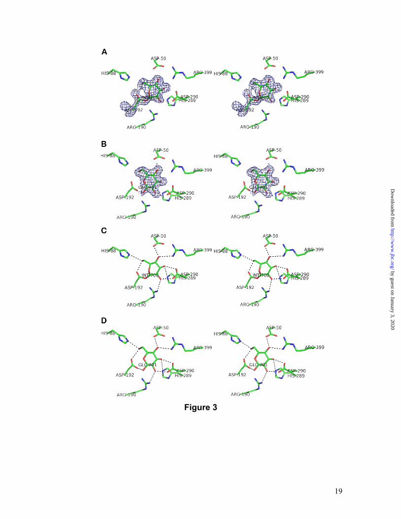
The BiSP:glucosyl intermediate complex.
Crystals of the wild-type BiSP complex were obtained by co-crystallization with sucrose. The asymmetric unit of the crystals comprises two molecules forming the same dimer interface as observed in the Tris complex. Here, however, the two molecules adopt different conformations. The conformation of molecule A is very similar to the E232Q:sucrose complex (molecule A; r.m.s. deviation of 0.3 Å on 504 Cα atoms), whereas the conformation of molecule B shows larger deviations (1.6 Å to molecules A of the Tris and E232Q complexes on all 504 Cα atoms). Both enzyme molecules contain glucose within the active site but only one glucose is covalently bound to the nucleophile, i.e. forming the catalytic covalent intermediate (molecule A; Figure 3A) The other glucose is non-covalently bound (molecule B; Figure 3B). The interactions of the glucosyl intermediate are shown in Figure 3C and Table 3. Since the anomeric carbon of sucrose changes configuration from α to β upon formation of the covalent intermediate, we observe the β-anomer bound as expected. To date, two covalent intermediate structures have been solved for enzymes of GH family 13, namely cyclodextrin glucosyltransferase (19) and AS (20). The most relevant structure for comparison with the BiSP covalent intermediate is that of AS, since AS and BiSP share sucrose as primary substrate while hydrolysis is only a minor side reaction for both enzymes. The glucosyl moiety in BiSP is in a slightly twisted 1,4B
by guest on January 3, 2020http://w
ww
.jbc.org/D
ownloaded from

6
conformation compared to 4C1 observed in AS. However, OD2, O2, O3 and O4 occupy nearly the same positions in the two structures. O6 is shifted by approximately 1 Å but is seen to form a hydrogen bond to a conserved His residue in both structures. The active site architecture of the covalent intermediate appears to be very similar to that of the sucrose bound form. The glucosyl moiety occupies the same position in the covalent intermediate as in the E232Q:sucrose complex, and all residues in the close vicinity of the glucosyl moieties are located at very similar positions; i.e. no structural rearrangements accompany the formation of a covalent intermediate in BiSP. The flipping of the glucosyl ring from 4C1 in sucrose to 1,4B in the intermediate does, however, have consequences in terms of binding. C1 moves by 1.2 Å towards Phe53 and this movement adds to the vdW interactions. A similar situation was observed upon formation of the covalent intermediate in AS where the glucosyl moiety was translated towards Tyr147 with a shift of 1.5 Å (20). This suggests that favorable vdW interactions contribute to stabilize the covalent intermediates in BiSP as well as in AS. The BiSP product complex after hydrolysis.
As the enzyme goes from the covalent intermediate bound state to the product bound state a number of changes to the active site topology occur. The glucosyl-binding site remains unaltered. Hence, the active site interactions with the glucose are very similar to those seen to the glucosyl part of sucrose in the E232Q:sucrose complex (Figure 3D). The non-covalently bound glucose (in molecule B) adopts the β-configuration with a 4C1 conformation. The most striking difference between the glucose product bound form (after hydrolysis of the covalently bound intermediate) and the substrate and intermediate forms are the conformations of the loops between residues 336-344 (loop A) and 130-140 (loop B) that differ up to 16 Å and 4 Å, respectively (Figure 4A).
Excluding these regions, the r.m.s. deviation is 0.3 Å (on 484 Cα atoms) compared to all other BiSP monomers. The rigid-body movement of loop B can be ascribed to major changes in backbone torsion angles of residues 133 and 137. Both loops A and B are in close proximity in the E232Q:sucrose and BiSP:glucosyl intermediate structures. Therefore, it is conceivable that the conformational changes observed in the wild-type BiSP:glucose structure are not independent. As a consequence of these differences in loop conformations, the architecture of the active site changes. Asp342 of loop A moves out of the binding site and becomes solvent exposed. Tyr344 replaces the position of Asp342, while Arg135 of loop B moves into the active site with the guanidino group replacing the side chain of Leu341. Due to the movement of Asp342 out and movement of Arg135 and Tyr344 into the active site, a net change of charge of +2 is seen. Towards an understanding of the reaction mechanism.
The present studies provide structural information on the substrate (sucrose) bound form of the enzyme, the glucosyl-enzyme intermediate and a product (glucose) bound form. So far, the structures of the apo enzyme and α-D-glucose 1-phosphate product form remain to be determined. The first step in the proposed reaction cycle (Scheme 2) is binding of sucrose to the active site of the enzyme. Upon formation of the covalent intermediate, the fructose group is cleaved off and is required to move away from the covalent intermediate atom C1 by approximately 3 Å. This is to some extent compensated by the 1.2 Å movement caused by the 4C1 to 1,4B conformational change but it still requires loop A to be pushed approximately 2 Å away from the active site residues. In the covalent intermediate structure, loop A and loop B adopt similar conformations to those observed in the sucrose complex; conformations that do not allow the exit of fructose because the entry
by guest on January 3, 2020http://w
ww
.jbc.org/D
ownloaded from

7
to the active site is too narrow. In the BiSP substrate and intermediate complexes, loop A is hydrogen bonded to loop B through the backbone atoms of Pro134 and Leu343. Upon hydrolysis of the enzyme-glucosyl intermediate and formation of the glucose product form, this interaction is lost due to large movements of loop A. The loop A now adopts an entirely different conformation, which brings Tyr344 into the fructose binding site. The large conformational change in loop A possibly causes loop B to move further in towards the active site. Phosphorolysis of the glucosyl intermediate is not observed in the wild-type BiSP structure as phosphate ions were not present during crystallization. In order to locate possible phosphate binding site(s) in BiSP soaking and co-crystallization experiments with phosphate, sulfate, vanadate and cacodylate were performed. Only co-crystallization of wild-type BiSP with cacodylate resulted in a complex structure to a resolution of 3 Å (data not shown). However, no cacodylate ions were observed within the active site. To speculate on the phosphorylase activity of BiSP, the position of the hydrolysis product glucose molecule can be used to dock the reaction product glucose 1-phosphate into the active site (Figure 4B). Arg135 and Tyr344 are positioned nearly optimal for the binding of a phosphate group. By extruding Arg135 into the active site, the BiSP enzyme ensures that phosphate is able to compete out water, i.e. prevent hydrolysis via strong electrostatic interaction. Hydrolysis is a significant reaction in other transferases utilizing a covalent intermediate. Importantly, Asp342 and Tyr344 of loop A are completely conserved and Arg135 of loop B is only substituted by lysine residues among the known sucrose phosphorylases, emphasizing the importance of these residues for sucrose phosphorylase activity. A question that remains to be answered is why a mixed dimer consisting of the intermediate form and the hydrolysis product form is found upon treatment of wild-type BiSP with sucrose. One possible explanation is that it is a consequence of
crystal packing. However, it has previously been indirectly observed using complementary methods that such mixed dimers also exist in solution (9). Interestingly, although structurally different from BiSP the crystal structure of maltose phosphorylase (21) (an enzyme that undertakes phosphorolysis of maltose into glucose and glucose 1-phosphate by inversion) also shows a mixed dimer when phosphate is present. In MP, the phosphate-binding loops rearrange in only one molecule to create a phosphate-binding site near the proposed maltose-binding site. As in BiSP, these phosphate-binding loops are also involved in forming the dimer interface in MP and it is possible that both enzymes work in a similar fashion, even though they do not belong to the same family of enzymes. Overall, essentially the same dimer as identified in the BiSP:Tris structure (2) is found in the two present structures. When analyzing the protein-protein contacts using the PISA server at EMBL-EBI it is found that for the E232Q:sucrose complex the size of the accessible surface area is 957 Å2. For the glucosyl intermediate the dimer interface is generally maintained, but due to the movements of the loops in the glucose product form of the enzyme the interactions are slightly different. The size has increased to 1052 Å2 and the number of hydrogen bonds has also increased. This asymmetry of the dimers may reflect a way that phosphate ions are brought to the active site. Analyses of the dimer interface in the BiSP:sucrose and BiSP:glucosyl intermediate reveal that the guanidino groups of Arg135 are pointing towards the interface and that the distance between them is ca. 10 Å. Slight movements of these side chains would make the guanidino groups from both monomers create a phosphate-binding site (Figure 4C). This is in agreement with an observation that cacodylate ions are capable of binding to this site (data not shown). It implicates that BiSP has only one surface phosphate-binding site from which phosphate ions are transported to the active site by Arg135.
by guest on January 3, 2020http://w
ww
.jbc.org/D
ownloaded from

8
Thus, BiSP might work in such a way that the two monomers are out of phase and require phosphate at different times; while one monomer is phosphorylating the covalent intermediate and releasing glucose
1-phosphate, the other monomer is binding sucrose and creating the covalent intermediate.
Acknowledgement We acknowledge EMBL-Hamburg and the staff at beamline BW7B for provision of synchrotron radiation facilities.
by guest on January 3, 2020http://w
ww
.jbc.org/D
ownloaded from

9
REFERENCES 1. van den Broek, L. A., van Boxtel, E. L., Kievit, R. P., Verhoef, R., Beldman, G., and
Voragen, A. G. (2004) Appl.Microbiol.Biotechnol. 65, 219-227
2. Sprogoe, D., van den Broek, L. A. M., Mirza, O., Kastrup, J. S., Voragen, A. G. J., Gajhede, M., and Skov, L. K. (2004) Biochemistry 43, 1156-1162
3. Henrissat, B. (1991) Biochem.J. 280, 309-316
4. Park, K. H., Kim, T. J., Cheong, T. K., Kim, J. W., Oh, B. H., and Svensson, B. (2000) Biochim.Biophys.Acta 1478, 165-185
5. Hondoh, H., Kuriki, T., and Matsuura, Y. (2003) Journal Of Molecular Biology 326, 177-188
6. Kim, J. S., Cha, S. S., Kim, H. J., Kim, T. J., Ha, N. C., Oh, S. T., Cho, H. S., Cho, M. J., Kim, M. J., Lee, H. S., Kim, J. W., Choi, K. Y., Park, K. H., and Oh, B. H. (1999) J.Biol.Chem. 274, 26279-26286
7. Koshland, D. E. (1953) Biol.Rev.Camb.Philos.Soc. 28, 416-436. 1953. 8. Doudoroff, M., Barker, H. A., and Hassid, W. Z. (1947) J.Biol.Chem. 168, 725-732
9. Voet, J. G. and Abeles, R. H. (1970) J.Biol.Chem. 245, 1020-1031
10. Silverstein, R., Voet, J., Reed, D., and Abeles, R. H. (1967) J.Biol.Chem. 242, 1338-1346
11. Bradford, M. M. (1976) Anal.Biochem. 72, 248-254
12. Leslie, A. G. W. Recent changes to the MOSFLM package for processing film and image plate data. Joint CCP4 and ESF-EACMB Newsletter on Protein Crystallography 26. 1992. Daresbury Laboratory.
13. Evans, P. (2006) Acta Cryst. D. 62, 72-82
14. Otwinowski, Z. and Minor, W. (1997) Method Enzymol. 276, 307-326
15. Perrakis, A., Morris, R., and Lamzin, V. S. (1999) Nature Struc.Biol. 6, 458-463
16. Brunger, A. T., Adams, P. D., Clore, G. M., DeLano, W. L., Gros, P., Grosse-Kunstleve, R. W., Jiang, J. S., Kuszewski, J., Nilges, M., Pannu, N. S., Read, R. J., Rice, L. M., Simonson, T., and Warren, G. L. (1998) Acta Cryst. D 54, 905-921
17. Laskowski, R. A., Macarthur, M. W., Moss, D. S., and Thornton, J. M. (1993) J.Appl.Cryst. 26, 283-291
18. Mirza, O., Skov, L. K., Remaud-Simeon, M., De Montalk, G. P., Albenne, C., Monsan, P., and Gajhede, M. (2001) Biochemistry 40, 9032-9039
by guest on January 3, 2020http://w
ww
.jbc.org/D
ownloaded from

10
19. Uitdehaag, J. C., Mosi, R., Kalk, K. H., van der Veen, B. A., Dijkhuizen, L., Withers, S. G., and Dijkstra, B. W. (1999) Nat.Struct.Biol. 6, 432-436
20. Jensen, M. H., Mirza, O., Albenne, C., Remaud-Simeon, M., Monsan, P., Gajhede, M., and Skov, L. K. (2004) Biochemistry 43, 3104-3110
21. Egloff, M. P., Uppenberg, J., Haalck, L., and van Tilbeurgh, H. (2001) Structure 9, 689-697
by guest on January 3, 2020http://w
ww
.jbc.org/D
ownloaded from

11
FIGURE LEGENDS Figure 1
Cartoon representation of the domain organization of the mixed dimer of BiSP. The glucosyl
intermediate (molecule A; left) and the hydrolysis product glucose (molecule B; right) are shown
as red spheres. The domain A, B, B’ and C are coloured green, yellow, blue and orange. The
labels N and C correspond to the N-terminus and C-terminus, respectively, while labels A and B
indicate the position of loops A and B.
Figure 2
Close-up view of the BiSP sucrose-binding site (in stereo). A, simulated annealing Fo-Fc omit
electron-density map contoured at 3σ, calculated with the sucrose atoms omitted. B, Potential
hydrogen bonds between sucrose and the enzyme of the E323Q:sucrose complex are displayed.
Potential hydrogen bonds are shown as stippled lines. C, superimposition of the sucrose binding
sites of BiSP (green) and AS (yellow). The figure was prepared by superimposing all sucrose
atoms from BiSP and AS.
Figure 3
Close-up view on the active site of the BiSP:glucose complex (in stereo). A and B, simulated
annealing Fo-Fc omit electron-density maps contoured at 3σ, calculated with the respective
ligand atoms omitted. In A, the glucosyl intermediate (molecule A) and in B, the non-covalently
bound β-D-glucose complex (molecule B). C and D, potential hydrogen bonds from ligand to
enzyme. In C, the glucosyl intermediate and in D, the non-covalently bound β-D-glucose
complex. Potential hydrogen bonds are shown as stippled lines.
by guest on January 3, 2020http://w
ww
.jbc.org/D
ownloaded from

12
Figure 4
Structural changes occurring during the enzyme reaction. A, close-up view of loops A and B of
the wild-type BiSP covalent intermediate (molecule A; cyan) superimposed on the glucose
product form (molecule B; yellow). The bound glucose of molecule B is shown for clarity. B,
close-up view of loops A and B and the non-covalently bound glucose molecule of the wild-type
BiSP:glucose complex. In yellow, a glucose 1-phosphate has been modelled based on the position
of the glucose with interactions to Arg135 and Tyr344. C, proposed inter-molecule phosphate-
binding site created by two Arg135 residues. The distances indicated by dashed lines are 3.9 Å.
The bound sucrose molecules are shown as red vdW spheres.
Scheme 1
Schematic representation of the reaction mechanism of sucrose phosphorylase. See
“Introduction” for further details.
Scheme 2
Structural cartoon of the reaction. Step A corresponds to the apo enzyme that binds the substrate
sucrose in step B. The covalent intermediate is formed in step C and fructose has left the active
site. In step D, the glucosyl intermediate reacts with either phosphate ions (phosphorolysis) or
water (hydrolysis). Phosphate/water is depicted as a gray sphere. Step E shows the product form
of the enzyme (hydrolysis – glucose; phosphorolysis – glucose 1-phosphate). The three structures
reported in the present study correspond to step B, C and E. Only the carboxylate group of
residues Glu232 and Asp192 are shown for clarity. The glucosyl and fructosyl groups are
represented by a hexagon and pentagon, respectively.
by guest on January 3, 2020http://w
ww
.jbc.org/D
ownloaded from

13
TABLE 1
Data collection statistics for the wild-type BiSP:glucose and E232Q:sucrose complexes Wild-type BiSP:glucosea E232Q:sucroseb Space group P212121 P212121 Unit cell, Å3 75.7 ×103.1 × 150.7 55.3 × 124.2 × 152.4 Resolution range, Å 20-2.0 (2.07-2.0)c 20-2.1 (2.21-2.1)c Total number of reflections measured
337,497 222,019
Number of unique reflections 80,388 61,222 Mosaicity, degrees 0.30 0.37 Completeness, % 99.6 (96.9)c 98.7 (99.7)c I/σI 21.5 (2.6)c 9.5 (3.1)c Rsym,d % 8.2 (43.3)c 13.8 (37.6)c a Data processed with Denzo/Scalepack (14). b Data processed with Mosflm/Scala (12)/(13). c Statistics for the highest resolution shell are given in parentheses. dRsym = ∑hkl(∑i(|Ihkl, i - <Ihkl>|)) / ∑hkl, i<Ihkl>, where Ihkl, I is the intensity of an individual measurement of the reflection with Miller indicies h, k, and l, and <Ihkl> is the mean intensity of that reflection.
by guest on January 3, 2020http://w
ww
.jbc.org/D
ownloaded from
14
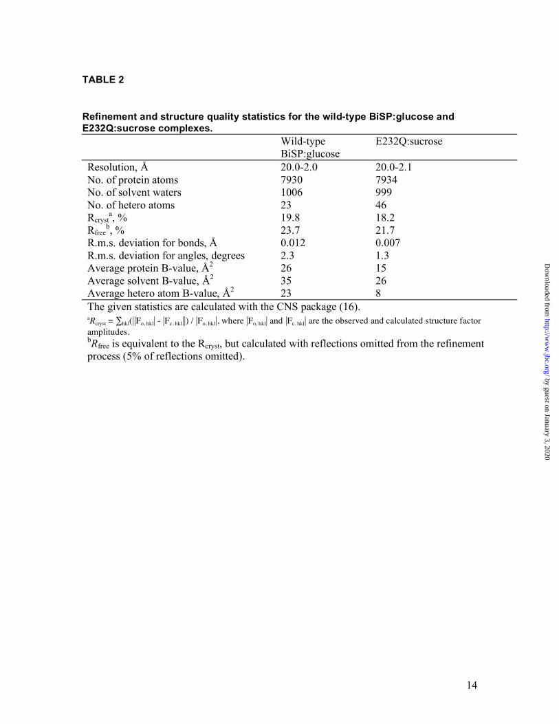
TABLE 2
Refinement and structure quality statistics for the wild-type BiSP:glucose and E232Q:sucrose complexes. Wild-type
BiSP:glucose E232Q:sucrose
Resolution, Å 20.0-2.0 20.0-2.1 No. of protein atoms 7930 7934 No. of solvent waters 1006 999 No. of hetero atoms 23 46 Rcryst
a, % 19.8 18.2 Rfree
b, % 23.7 21.7 R.m.s. deviation for bonds, Å 0.012 0.007 R.m.s. deviation for angles, degrees 2.3 1.3 Average protein B-value, Å2 26 15 Average solvent B-value, Å2 35 26 Average hetero atom B-value, Å2 23 8 The given statistics are calculated with the CNS package (16). aRcryst = ∑hkl(||Fo, hkl| - |Fc, hkl||) / |Fo, hkl|, where |Fo, hkl| and |Fc, hkl| are the observed and calculated structure factor amplitudes. bRfree is equivalent to the Rcryst, but calculated with reflections omitted from the refinement process (5% of reflections omitted).
by guest on January 3, 2020http://w
ww
.jbc.org/D
ownloaded from

15
Table 3 Potential hydrogen bond distances (up to 3.5 Å) of the two BiSP complexes.
Ligand Protein Interatomic Distance (Å)
E232Q:sucrose Wild-type BiSP:glucose
Mol A Glucosyl-intermediate
β–D-glucose
Glucosyl O2 ARG190 NH2
GLN/GLU232 NE2/OD2 HIS289 NE2 ASP290 OD2
3.0 3.3 3.1 2.6
3.0 2.9 3.1 2.7
2.9
2.9 2.7
O3 HIS289 NE2 ASP290 OD2
2.9 2.7
3.1 2.8
2.9 2.7
O4 ASP50 OD2 Arg399 NH1
2.8 2.6
3.0 2.7
2.8 2.7
O6 HIS88 NE2 ASP192 OD2
2.9 2.9
3.0 2.7
2.9 2.8
Fructosyl
O1’ GLN232 OE1 2.6 O3’ HIS234 NE2 3.5 O4’ ASP342 OD2 2.8 O5’ GLN345 OE1 2.8 by guest on January 3, 2020
http://ww
w.jbc.org/
Dow
nloaded from


Beldman, Jette S. Kastrup and Michael GajhedeOsman Mirza, Lars K. Skov, Desiree Sprogøe, Lambertus A.M. van den Brook, Gerrit
adolescentis during sucrose conversionStructural rearrangements of sucrose phosphorylase from bifidobacterium
published online September 21, 2006J. Biol. Chem.
10.1074/jbc.M605611200Access the most updated version of this article at doi:
Alerts:
When a correction for this article is posted•
When this article is cited•
to choose from all of JBC's e-mail alertsClick here
by guest on January 3, 2020http://w
ww
.jbc.org/D
ownloaded from