structural insight into how bacteria prevent …mbio.asm.org/content/6/6/e01867-15.full.pdf ·...
TRANSCRIPT
Structural Insight into How Bacteria Prevent Interference betweenMultiple Divergent Type IV Secretion Systems
Joseph J. Gillespie,a Isabelle Q. H. Phan,b,c Holger Scheib,d Sandhya Subramanian,b,c Thomas E. Edwards,b,e Stephanie S. Lehman,a
Hanna Piitulainen,f M. Sayeedur Rahman,a Kristen E. Rennoll-Bankert,a Bart L. Staker,b,c Suvi Taira,f Robin Stacy,b,c Peter J. Myler,b,c
Abdu F. Azad,a Arto T. Pulliainenf,g
Department of Microbiology and Immunology, University of Maryland School of Medicine, Baltimore, Maryland, USAa; Seattle Structural Genomics Center for InfectiousDisease, Seattle, Washington, USAb; The Center for Infectious Disease Research (formerly Seattle Biomedical Research Institute), Seattle, Washington, USAc; VenomEvolution Lab, School of Biological Sciences, University of Queensland, St. Lucia, Queensland, Australiad; Beryllium Discovery Corp., Bainbridge Island, Washington, USAe;Department of Biosciences, University of Helsinki, Helsinki, Finlandf; Institute of Biomedicine, University of Turku, Turku, Finlandg
ABSTRACT Prokaryotes use type IV secretion systems (T4SSs) to translocate substrates (e.g., nucleoprotein, DNA, and protein)and/or elaborate surface structures (i.e., pili or adhesins). Bacterial genomes may encode multiple T4SSs, e.g., there are threefunctionally divergent T4SSs in some Bartonella species (vir, vbh, and trw). In a unique case, most rickettsial species encode aT4SS (rvh) enriched with gene duplication. Within single genomes, the evolutionary and functional implications of cross-systeminterchangeability of analogous T4SS protein components remains poorly understood. To lend insight into cross-system inter-changeability, we analyzed the VirB8 family of T4SS channel proteins. Crystal structures of three VirB8 and two TrwG Barto-nella proteins revealed highly conserved C-terminal periplasmic domain folds and dimerization interfaces, despite tremendoussequence divergence. This implies remarkable structural constraints for VirB8 components in the assembly of a functional T4SS.VirB8/TrwG heterodimers, determined via bacterial two-hybrid assays and molecular modeling, indicate that differential ex-pression of trw and vir systems is the likely barrier to VirB8-TrwG interchangeability. We also determined the crystal structureof Rickettsia typhi RvhB8-II and modeled its coexpressed divergent paralog RvhB8-I. Remarkably, while RvhB8-I dimerizes andis structurally similar to other VirB8 proteins, the RvhB8-II dimer interface deviates substantially from other VirB8 structures,potentially preventing RvhB8-I/RvhB8-II heterodimerization. For the rvh T4SS, the evolution of divergent VirB8 paralogs im-plies a functional diversification that is unknown in other T4SSs. Collectively, our data identify two different constraints (spatio-temporal for Bartonella trw and vir T4SSs and structural for rvh T4SSs) that mediate the functionality of multiple divergentT4SSs within a single bacterium.
IMPORTANCE Assembly of multiprotein complexes at the right time and at the right cellular location is a fundamentally impor-tant task for any organism. In this respect, bacteria that express multiple analogous type IV secretion systems (T4SSs), each com-posed of around 12 different components, face an overwhelming complexity. Our work here presents the first structural investi-gation on factors regulating the maintenance of multiple T4SSs within a single bacterium. The structural data imply that theT4SS-expressing bacteria rely on two strategies to prevent cross-system interchangeability: (i) tight temporal regulation of ex-pression or (ii) rapid diversification of the T4SS components. T4SSs are ideal drug targets provided that no analogous counter-parts are known from eukaryotes. Drugs targeting the barriers to cross-system interchangeability (i.e., regulators) could dys-regulate the structural and functional independence of discrete systems, potentially creating interference that prevents theirefficient coordination throughout bacterial infection.
Received 28 October 2015 Accepted 5 November 2015 Published 8 December 2015
Citation Gillespie JJ, Phan IQH, Scheib H, Subramanian S, Edwards TE, Lehman SS, Piitulainen H, Sayeedur Rahman M, Rennoll-Bankert KE, Staker BL, Taira S, Stacy R, Myler PJ,Azad AF, Pulliainen AT. 2015. Structural insight into how bacteria prevent interference between multiple divergent type IV secretion systems. mBio 6(6):e01867-15. doi:10.1128/mBio.01867-15.
Editor Yasuko Rikihisa, Ohio State University
Copyright © 2015 Gillespie et al. This is an open-access article distributed under the terms of the Creative Commons Attribution-Noncommercial-ShareAlike 3.0 Unportedlicense, which permits unrestricted noncommercial use, distribution, and reproduction in any medium, provided the original author and source are credited.
Address correspondence to Joe Gillespie, [email protected], or Arto Pulliainen, [email protected].
This article is a direct contribution from a Fellow of the American Academy of Microbiology.
Occurring in Gram-negative, Gram-positive, and wall-lessbacteria, as well as archaea, type IV secretion systems (T4SSs)
are primarily utilized for translocating substrates across the cellenvelope (1, 2). T4SSs that translocate plasmid (3) and nakedDNA (4, 5), as well as genomic islands (6), are major facilitators ofbacterial diversification, contributing to the spread of antimicro-bial resistance and virulence genes. Other T4SSs translocate nu-
cleoprotein (e.g., oncogenic T-DNA via the vir T4SS of Agrobac-terium tumefaciens) or protein substrates directly into eukaryoticcells, wherein these effectors often perturb cell signaling to benefitbacterial survival (7, 8). Recently, it was reported that T4SSs mayalso be utilized to kill neighboring bacteria via translocation of aproteinaceous effector (9). Typically, T4SSs elaborate surfacestructures (i.e., pili or specialized adhesins) in a process either
RESEARCH ARTICLE crossmark
November/December 2015 Volume 6 Issue 6 e01867-15 ® mbio.asm.org 1
on August 6, 2018 by guest
http://mbio.asm
.org/D
ownloaded from
coupled to or independent of substrate translocation (10, 11).Thus, T4SSs are extraordinarily diverse in function relative toother bacterial secretion systems (12, 13).
Recently, T4SSs have been classified into eight major groups(14). One well-studied group, P-T4SSs, is typified by the vir T4SSof the pTi plasmid of A. tumefaciens, which encodes 11 scaffoldcomponents (VirB1 to VirB11) and a coupling protein (VirD4)that recruits substrates to the secretion channel (15). Tremendousarchitectural insight has been garnered from recent macromolec-ular structures generated for other P-T4SSs (16–20), allowingmany of the various scaffold components to be arranged into ananatomical model (Fig. 1). Phylogenomics and bioinformatics ef-forts have shown that the other seven groups of T4SSs containhomologs or analogs to some (or most) of the components of thisanatomical model (21), implying that a common T4SS architec-ture links the prokaryotic cytoplasm to the extracellular milieu.Variations on this theme, particularly regarding the distal regionof the translocation channel, are thought to primarily be a conse-quence of coevolution with cell envelope morphology (22, 23).However, many structural innovations likely correlate with spe-cific functions, e.g., mating pair stabilization during conjugation(24, 25) or pilus interactions with host cells during effector trans-location (26).
Bacterial genomes may encode multiple divergent T4SSs, e.g.,the cag and com P-T4SSs of Helicobacter pylori (27, 28), the vir,vbh, and trw P-T4SSs of certain Bartonella species (29), and thedot/icm (I-T4SS) and lvh (P-T4SS) T4SSs of Legionella species (30,31). These divergent T4SSs typically have different functions,though cross-system interchangeability is known for L. pneumo-phila dot/icm and lvh T4SSs. The lvh P-T4SS, which is dispensablefor L. pneumophila replication in both amoebae and macrophagehosts (32) but causes virulence phenotypes under conditions that
mimic the L. pneumophila aquatic phase (33), was shown to com-plement certain dot/icm mutants defective in conjugation (32).Furthermore, arrested virulence phenotypes in an L. pneumophiladotA/lvh double mutant were restored via complementation withthe lvh coupling protein LvhD4 (34), implying structural andfunctional resilience in the face of extreme sequence divergenceacross dot/icm and lvh T4SSs. For other bacteria harboring multi-ple divergent T4SSs, studies on cross-system interchangeabilityare lacking, leaving a limited understanding of how distinct T4SSsachieve correct spatiotemporal assembly to execute their specificfunctions. Such precise regulatory mechanisms must exist, givenalso that many bacterial genomes are continually bombarded withintegrative conjugative elements that often carry T4SS loci (35). Ifnot quickly purged, it is likely that incoming T4SSs with compat-ibility to the native T4SS(s) either undergo rapid diversifying evo-lution or acquire mechanisms for differential regulation.
To gain further insight into T4SS cross-system interchange-ability, we selected two bacterial genera with multiple divergentanalogs of VirB8, a channel protein required for substrate transfer(Fig. 1). First, two or three distinct complete T4SSs are foundwithin the genomes of some Bartonella species. Many of thesespecies encode vir and trw secretion systems, which are highlydivergent in sequence and function (36). Some species have anadditional vbh T4SS homologous to the vir T4SS (29), wherein thebiological function still remains elusive. The Bartonella vir T4SS isinvolved in protein secretion, with an arsenal of Bartonella effectorproteins (Beps) translocated into host cells during infection (8).The trw T4SS is not involved in protein translocation but ratherelaborates variable surface pili comprised of different combina-tions of duplicated VirB2 (TrwL) and VirB5 (TrwJ) homologs(37), with such structures shown to interact with host erythrocytes(38, 39). While these divergent T4SSs are differentially expressed
N
C
ATP
ATP
ATP
OM
IM
PG
LPS
PP
C11
10
3
7
8
91
6
4D4
2
VirB7VirB7VirB9VirB10
VirB5 ?VirB8 ?VirB6 ?VirB3 ?
VirB4
B7B8B9B10B11D4
Channel formation; pore structureChannel formation; substrate transferPore structure; substrate transferChannel formation; pore structureATP hydrolysis; substrate transferSubstrate recognition; ATP hydrolysis
B1B2B3B4B5B6
Murein degradation; pilus formationMajor pilus subunit; substrate transferIM pilus assembly pathway sensorATP hydrolysis; pilin IM dislocationMinor pilus subunit Channel formation; substrate transfer
5
B
CA
VirB7VirB7
VirB9
VirB10
N N
CC
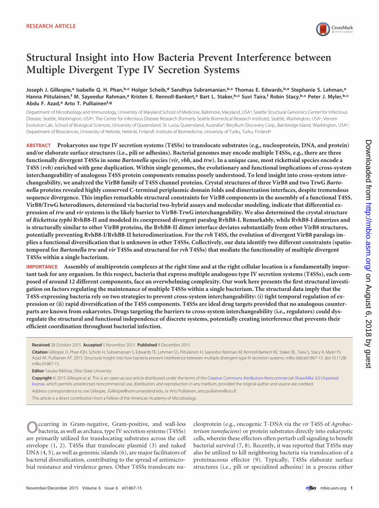
FIG 1 Architecture of P-type type IV secretions systems (P-T4SSs). (A) Bird’s-eye (left) and side (right) views of the dodecameric P-T4SS core complex (CC)encoded by plasmid pKM101 of Escherichia coli (PDB ID 3JQO), adapted from the work of Chandran et al. (17). Colors for the CC subunits (VirB7, VirB9, andVirB10) are similar to the model in panel C. (B) Negative-station electron microscopy-generated structure of the P-T4SS encoded by the E. coli R388 conjugativeplasmid (EMD-2567) (adapted from the work of Low et al. [19]). Colors for the CC subunits and cytosolic/IM barrels (VirB4) are similar to the model in panelC. The putative positions of other IM channel (IMC) proteins are depicted with questions marks. VirB1, VirB2, VirB11, and VirD4 are not shown, as they werenot included in the original structure. (C) General model of the composition of P-T4SSs, with functions for all 12 components (VirB1 to VirB11 and VirD4) listedat bottom right. The purple star depicts the bitopic VirB8 IMC proteins, with a dashed box illustrating the monomeric (left) and dimeric (right) structures forVirB8 C-terminal domains of Agrobacterium tumefaciens (PDB ID 2CC3) (44).
Gillespie et al.
2 ® mbio.asm.org November/December 2015 Volume 6 Issue 6 e01867-15
on August 6, 2018 by guest
http://mbio.asm
.org/D
ownloaded from
during Bartonella host cell infection (40), the potential for cross-system interchangeability is currently unknown. Second, for spe-cies of Rickettsiales, an entirely different strategy has evolved witha single T4SS, the Rickettsiales vir homolog (rvh), which containsduplications of several components, including a VirB8 homolog(RvhB8) (23, 41). The functional significance of these duplicatervh components is unknown, particularly regarding whether a sin-gle rvh T4SS functions during the rickettsial life cycle or if multipledifferent T4SSs are assembled throughout the complex life cycle(usually involving arthropod and vertebrate hosts) (42).
Herein we report crystal structures for six proteins of the VirB8family: three VirB8 and two TrwG proteins of several Bartonella spe-cies, as well as the first crystal structure of an rvh scaffold component,RvhB8-II of Rickettsia typhi. Comparative structural analysis, in con-junction with biochemical assays, protein modeling, and bioinfor-matics, leads us to propose two distinct mechanisms (spatiotemporalfor Bartonella trw and vir T4SSs and structural for rvh T4SSs) thatprevent cross-system interchangeability between multiple divergentT4SSs encoded within single bacterial genomes.
RESULTSOrigin of multiple VirB8 proteins in Bartonella and Rickettsiagenomes. To understand the origin of multiple VirB8-like pro-teins within Bartonella and Rickettsia, we estimated a phylogeny ofselected P-T4SSs. The Bartonella vir and vbh T4SSs share commonancestry, probably arising from whole-system duplication (Fig. 2).Previously, the Bartonella vir and vbh T4SSs were hypothesized tooriginate via lateral gene transfer (LGT), as they are absent fromthe ancestral human pathogen Bartonella bacilliformis (29, 36).The Bartonella trw T4SSs are found in a different clade, whichincludes plasmid-encoded T4SSs of gammaproteobacterial spe-cies (Fig. 2). Thus, the Bartonella trw T4SSs were likely acquiredvia LGT from non-alphaproteobacterial sources, becoming incor-porated into the chromosomes, where they evolved to mediateadhesion to host erythrocytes (29, 36).
The rvh T4SS forms a lineage distinct from other P-T4SSs,though robust support for the relationship of this lineage to otherP-T4SSs is lacking (Fig. 2; also see Text S1 in the supplementalmaterial). We considered the paralogous genes encoding RvhB4,RvhB8, and RvhB9 to comprise different structural and functionalmachines, as previous phylogeny estimation supported thesegenes as ancient duplications that arose early in Rickettsiales evo-lution (23, 41). The genes encoding RvhB4-I, RvhB8-I, andRvhB9-I (collectively referred to as rvh-I) are conserved relative toanalogs in other P-T4SSs and thus are anticipated to assemble intothe rvh T4SS while it functions in protein translocation. Alterna-tively, the genes encoding RvhB4-II, RvhB8-II, and RvhB9-II (col-lectively referred to as rvh-II) have evolved atypical features thatdeviate substantially from counterparts in other P-T4SSs (see Dis-cussion). The structural and functional significance of rvh-II isunknown, though a substantially higher divergence of rvh-II thanrvh-I indicates different selective pressures operating on theseparalogs (Fig. 2). Remarkably, this odd paralogy within the rvhT4SS is conserved across the three derived families of Rickettsiales(Rickettsiaceae, Anaplasmataceae, and “Candidatus Midichlori-aceae”), which comprise a large, diverse assemblage of symbioticand pathogenic species (43).
Divergent Bartonella VirB8 and TrwG proteins ho-modimerize via interactions at an NPXG motif highly con-served across T4SSs. We determined crystal structures of the sol-
uble C-terminal periplasmic domains of three VirB8 and twoTrwG proteins of several Bartonella species (see Text S2 in thesupplemental material). As expected by the highly conserved se-quences of C-terminal periplasmic domains of Bartonella VirB8(~90%) and TrwG (~90%) proteins (data not shown), the struc-tures within each protein family are highly similar, with 5�-helices and 4 �-sheets (Fig. 3A and B). Within Bartonella VirB8,the pairwise root mean square deviation (RMSD) values, or mea-sures of the average distance between the atoms of superimposedproteins, range from 0.33 to 0.60 Å for similar C� atoms, andwithin Bartonella TrwG, the two structures have a pairwise RMSDvalue of 0.88 Å. The structures of Bartonella VirB8 and TrwGproteins are also highly similar to each other (Fig. 3C), despiteextremely low sequence conservation (~28% identity overall,~34% identity for C-terminal periplasmic domains) (Fig. 3D; seeText S3 in the supplemental material). The pairwise RMSD valuesbetween Bartonella VirB8-TrwG sets range from 1.57 to 1.78 Å.The Bartonella VirB8 and TrwG protein structures are also similarto the Agrobacterium tumefaciens (44) (RMSD ranges of 1.28 to1.33 Å and 1.52 to 1.63 Å for VirB8 and TrwG, respectively) andBrucella suis (45) (RMSD ranges of 1.73 to 1.78 Å and 0.98 to1.18 Å for VirB8 and TrwG, respectively) VirB8 protein structures(Fig. 3C). This implies remarkable structural constraints for VirB8components in the assembly of functional P-T4SSs.
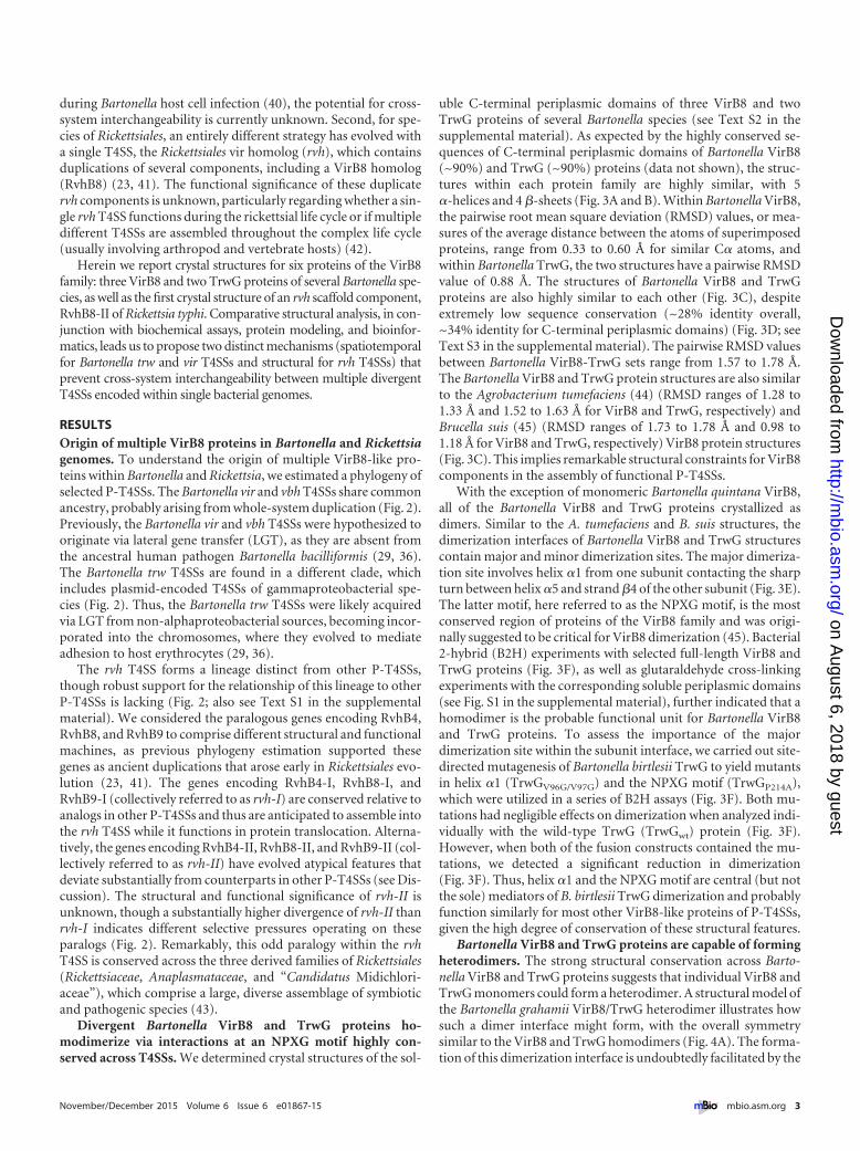
With the exception of monomeric Bartonella quintana VirB8,all of the Bartonella VirB8 and TrwG proteins crystallized asdimers. Similar to the A. tumefaciens and B. suis structures, thedimerization interfaces of Bartonella VirB8 and TrwG structurescontain major and minor dimerization sites. The major dimeriza-tion site involves helix �1 from one subunit contacting the sharpturn between helix �5 and strand �4 of the other subunit (Fig. 3E).The latter motif, here referred to as the NPXG motif, is the mostconserved region of proteins of the VirB8 family and was origi-nally suggested to be critical for VirB8 dimerization (45). Bacterial2-hybrid (B2H) experiments with selected full-length VirB8 andTrwG proteins (Fig. 3F), as well as glutaraldehyde cross-linkingexperiments with the corresponding soluble periplasmic domains(see Fig. S1 in the supplemental material), further indicated that ahomodimer is the probable functional unit for Bartonella VirB8and TrwG proteins. To assess the importance of the majordimerization site within the subunit interface, we carried out site-directed mutagenesis of Bartonella birtlesii TrwG to yield mutantsin helix �1 (TrwGV96G/V97G) and the NPXG motif (TrwGP214A),which were utilized in a series of B2H assays (Fig. 3F). Both mu-tations had negligible effects on dimerization when analyzed indi-vidually with the wild-type TrwG (TrwGwt) protein (Fig. 3F).However, when both of the fusion constructs contained the mu-tations, we detected a significant reduction in dimerization(Fig. 3F). Thus, helix �1 and the NPXG motif are central (but notthe sole) mediators of B. birtlesii TrwG dimerization and probablyfunction similarly for most other VirB8-like proteins of P-T4SSs,given the high degree of conservation of these structural features.
Bartonella VirB8 and TrwG proteins are capable of formingheterodimers. The strong structural conservation across Barto-nella VirB8 and TrwG proteins suggests that individual VirB8 andTrwG monomers could form a heterodimer. A structural model ofthe Bartonella grahamii VirB8/TrwG heterodimer illustrates howsuch a dimer interface might form, with the overall symmetrysimilar to the VirB8 and TrwG homodimers (Fig. 4A). The forma-tion of this dimerization interface is undoubtedly facilitated by the
November/December 2015 Volume 6 Issue 6 e01867-15 ® mbio.asm.org 3
on August 6, 2018 by guest
http://mbio.asm
.org/D
ownloaded from

bootstrap.91-1 .81-.90 .71-.80 .61-.70 .51-.60
Bordetella pertussis str. Tohama I
Shinella zoogloeoides str. DD12
Vibrio fischeri str. ES114 pES100
Salmonella enterica subsp. enterica serovar Typhimurium pR46 (IncN)
Gilvimarinus chinensis (unknown strain)
Mesorhizobium loti str. MAFF303099 pMLaSandarakinorhabdus limnophila (unknown strain)
Rhizosphere of Medicago sativa (alfalfa) pSB102
Pseudomonas putida (unknown strain) pWW0
Legionella pneumophila str. Lens
Phenylobacterium zucineum str. HLK1
Ochrobactrum anthropi str. ATCC 49188
Pseudomonas syringae pv. syringae str. A2 pPSR1
Campylobacter jejuni subsp. jejuni 81-176 pVir
Ralstonia solanacearum str. CMR15 pRSC35
Brucella suis str. ATCC 23445
Legionella pneumophila str. Paris
Agrobacterium fabrum str. C58 pAt
Cupriavidus basilensis str. OR16Xanthomonas citri subsp. citri pXcB
Sinorhizobium meliloti str. 1021 pSymA
Burkholderia multivorans str. CGD1
Sphingobium chlorophenolicum str. L-1
Helicobacter pylori str. J99
Erwinia amylovora str. UTRJ2 pEU30
Mesorhizobium loti str. MAFF303099 pMLb
Legionella pneumophila str. Corby
Stenotrophomonas maltophilia str. EPM1
Campylobacter coli str. CVM N29710 pN29710-1
Mesorhizobium loti str. MAFF303099
Legionella pneumophila subsp. pneumophila str. Philadelphia 1Legionella longbeachae str. D-4968
Sphingomonas phyllosphaerae (unknown strain)
Helicobacter pylori str. PeCan18B
Nitrobacter hamburgensis str. X14 “plasmid 2”
Wolinella succinogenes str. DSM 1740
Aeromonas caviae str. HGB5 pFBAOT6
Escherichia coli O25b:H4-ST131 str. EC958 pKC394
Pseudomonas fluorescens str. HK44 pUTK21
Xylella fastidiosa str. 9a5c pXF51
Agrobacterium tumefaciens str. LBA4213 (Ach5) pTi
Xanthomonas campestris pv. campestris str. 8004
Photobacterium profundum str. SS9
Yersinia pestis biovar Microtus str. 91001 pCRY
Pseudomonas putida str. LD209 pLD209
Epsilonproteobacteria
Gammaproteobacteria
Alphaproteobacteria
Betaproteobacteria
Bartonella birtlesii str. LL-WM9Bartonella henselae str. Houston-1
Bartonella tribocorum str. CIP 105476Bartonella grahamii str. as4aup
Bartonella tribocorum str. CIP 105476
Bartonella grahamii str. as4aup
Bartonella birtlesii str. LL-WM9
Bartonella quintana str. Toulouse
Bartonella grahamii str. as4aup
Bartonella quintana str. ToulouseBartonella henselae str. Houston-1Bartonella birtlesii str. LL-WM9
Bartonella tribocorum str. CIP 105476Bartonella grahamii str. as4aup
lvh
vbh
vir
Campylobacter jejuni subsp. jejuni 81-176 pTet
trw0.1 sub/site
viravh
Wolbachia endosymbiont (Drosophila melanogaster)
“Candidatus Xenolissoclinum pacificiensis” str. L6Orientia tsutsugamushi str. Boryong
Anaplasma phagocytophilum str. HZ
Ehrlichia chaffeensis str. Arkansas
Orientia tsutsugamushi str. Ikeda
Ehrlichia canis str. Jake
“Candidatus Midichloria mitochondrii” str. IricVA
Anaplasma marginale str. St. Maries
Rickettsiaceae bacterium str. Os18Rickettsia typhi str. Wilmington
Neorickettsia sennetsu str. Miyayama
Rickettsia bellii str. RML369-C
Neorickettsia risticii str. IllinoisWolbachia endosymbiont str. TRS (Brugia malayi)
rvh-II
Rickettsia bellii str. RML369-C
Anaplasma marginale str. St. Maries
“Candidatus Midichloria mitochondrii” str. IricVA
Ehrlichia canis str. JakeEhrlichia chaffeensis str. Arkansas
Rickettsia typhi str. Wilmington
Wolbachia endosymbiont (Drosophila melanogaster)
Anaplasma phagocytophilum str. HZ
Neorickettsia sennetsu str. Miyayama
Orientia tsutsugamushi str. Ikeda“Candidatus Xenolissoclinum pacificiensis” str. L6
Wolbachia endosymbiont str. TRS (Brugia malayi)
Neorickettsia risticii str. Illinois
Rickettsiaceae bacterium str. Os18Orientia tsutsugamushi str. Boryong
rvh-I
Midichloriaceae
Anaplasmataceae
Rickettsiaceae
ptl
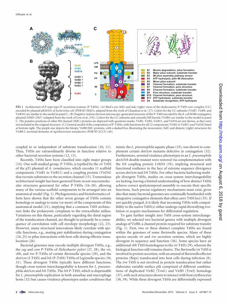
FIG 2 Phylogeny estimation of P-type type IV secretion systems (P-T4SSs). Phylogeny was estimated from concatenated alignments of five components (VirB4and VirB8 to VirB11), except for rvhB-II, which contains homologs to VirB4, VirB8, and VirB9 only (see the text for further details on alignment and data setconstruction). ML-based phylogeny was estimated with RAxML on the unmasked alignment (LG � gamma � �). Branch support was assessed with 1,000bootstrap pseudoreplications. A tree with specific bootstrap values, as well as additional phylogenies estimated from alternative alignments/models/optimality
(Continued)
Gillespie et al.
4 ® mbio.asm.org November/December 2015 Volume 6 Issue 6 e01867-15
on August 6, 2018 by guest
http://mbio.asm
.org/D
ownloaded from

high degree of sequence conservation of contacting residueswithin helix �1 and the NPXG motif, four of which are invariantacross VirB8 and TrwG sequences (Fig. 4B). Using the B2H assay,we analyzed the potential cross-system interchangeability be-tween B. grahamii VirB8 and TrwG (Fig. 4C). We initially estab-lished a reference of interaction strength for B. birtlesii and B. gra-hamii TrwG homodimers, as well as for the B. grahamii VirB8homodimer. TrwG interaction signals were much higher thanVirB8 homodimerization, possibly reflecting the known biologi-cal functions of these divergent systems, i.e., rigid adhesive func-tion of the trw T4SS compared to dynamic effector translocationof the vir T4SS (36). Subsequently, we evaluated the potential forheterodimeric interactions between B. grahamii VirB8 and TrwGproteins. These assays indicate the ability of VirB8 and TrwG toform heterodimers (Fig. 4C), with a strength of interactions notsignificantly lower than that observed for the B. grahamii VirB8homodimer. Thus, it appears that in the absence of spatiotempo-ral barriers (e.g., differential expression), VirB8 and TrwG pro-teins have the potential to interfere with one another during as-sembly into their respective T4SSs.
Duplicate, coexpressed VirB8 proteins encoded within theRickettsia typhi genome are structurally divergent. To lend in-sight into the duplicate VirB8-like proteins encoded within Rick-ettsia genomes, we set out to determine the crystal structures forthese divergent proteins from the murine typhus agent, R. typhistrain Wilmington (RvhB8-I and RvhB8-II, which share only 18%amino acid identity). Attempts to generate a crystal structure forRvhB8-I failed; thus, we modeled both the monomer and dimer ofthis protein by homology, using a multitemplate approach(Fig. 5A). Because RvhB8-I contains residues conserved in otherVirB8 family proteins, such as the NPXG motif (41), its sequencecould be reasonably modeled to the Bartonella spp., B. suis, andA. tumefaciens VirB8 structures, as well as the Bartonella TrwGstructures. Thus, the RvhB8-I structure is anticipated to fold sim-ilarly to these proteins. The protomeric structure of R. typhiRvhB8-I is quite similar to that of Bartonella VirB8 and TrwGproteins, with pairwise RMSD values of 1.46 to 1.64 Å. In contrast,the biological dimeric structure determined for R. typhi RvhB8-IIdeviates substantially from the RvhB8-I model and all sevenVirB8/TrwG dimeric structures, with pairwise RMSD values of3.20 to 3.79 Å (Fig. 5B). We previously observed that the lack ofconservation in the NPXG motif of RvhB8-II, coupled with rap-idly evolving sites throughout the protein, indicated a possibledivergent structure and function for this protein (41). All otherVirB8 and TrwG T4SS proteins with known dimeric structuresexhibit the NPXG motif posterior to helix �5 that interacts withhelix �1= of the other protomer. The RvhB8-II sequence differssubstantially in this region, and in the structure, this region doesnot form helix �5. Part of this region is disordered in the crystalstructure; one potential explanation could be in situ cleavage ofthis region during crystallization, although mass spectrometryanalysis of the protein stock solution revealed an intact protein(~21 kDa) prior to crystallization (see Text S2 in the supplemental
material). The divergent sequence and lack of helix �5 perturbsthe major dimerization site to generate an asymmetrical subunitinterface, unlike other VirB8 and TrwG structures that had sym-metric dimers. To compensate for disruption of the majordimerization site, a minor dimerization site is observed involvingresidues between �-strands �2 and �3 (Fig. 5C). Glutaraldehydecross-linking experiments with the soluble periplasmic domainsof RvhB8-I and RvhB8-II demonstrated that both proteins formhomodimers in solution (see Fig. S1 in the supplemental mate-rial). Thus, despite a divergent dimeric structure adopted byRvhB8-II, both VirB8-like proteins of R. typhi appear to formfunctional dimers.
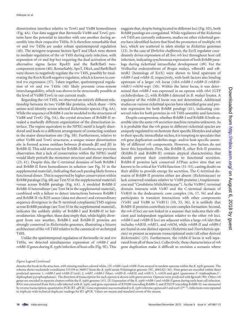
The significance of duplicate VirB8-like proteins carried by allspecies of Rickettsia is unclear. The arrangement of rvhB8-I andrvhB8-II in the R. typhi genome is odd (46), with the genes foundin adjacent operons that encode other rvh genes (Fig. 5D). Theconservation of these loci across all sequenced Rickettsia genomesimplies a conserved function (41). Reverse transcription-quantitative PCR (RT-qPCR) analysis of R. typhi throughout oneday of host cell infection indicates that rvhB8-I and rvhB8-II genesare simultaneously expressed (Fig. 5E). Whether RvhB8-I andRvhB8-II both assemble into the same rvh secretion machine re-mains unknown. A modeled RvhB8-I/RvhB8-II heterodimer sug-gests that such an interaction would be highly divergent fromother VirB8 and TrwG homodimers, as well as the VirB8/TrwGheterodimer (see Text S4 in the supplemental material). Asidefrom an entirely skewed major dimerization site, there are noresidues contacting across the minor dimerization site within themodeled heterodimer. Supporting these observations, B2H assaysfailed to detect interactions between RvhB8-I and RvhB8-II (datanot shown). Furthermore, the N-terminal cytoplasmic andtransmembrane-spanning (TMS) regions of RvhB8-I andRvhB8-II are extraordinarily divergent from one another (41).Collectively, if RvhB8-I and RvhB8-II both simultaneously assem-ble into one rvh T4SS, there is little support for these proteinsforming heterodimers.
Divergent RvhB8-II proteins are conserved across species ofRickettsiales but not found in other bacteria. The presence of twoVirB8-like genes in all species of Rickettsiales that carry the rvhT4SS prompted us to evaluate if the odd R. typhi RvhB8-II char-acteristics were present in other RvhB8-II proteins. Analysis ofselected rickettsial species revealed that RvhB8-I and RvhB8-IIproteins encoded within the same genome never share more than19% amino acid identity (Fig. 6A, yellow highlighting). Acrossspecies within the six major rickettsial genera, RvhB8-I is moreconserved relative to RvhB8-II (Fig. 6B). This is further witnessedby phylogeny estimation of RvhB8 proteins, which shows substan-tially higher divergence of RvhB8-II than RvhB8-I (Fig. 6C). Col-lectively, these data indicate that an ancient duplication of rvhB8quickly led to diversification of rvhB8 paralogs under differentselective constraints. Considering these results with the similardivergence patterns witnessed for RvhB4 and RvhB9 paralogs
Figure Legend Continued
criteria, is provided in Text S1 in the supplemental material. The tree was rooted with four P-T4SSs from species of Epsilonproteobacteria. rvh-I and rvh-II T4SSsare in light and dark gray, respectively. The Bartonella vir, vbh, and trw T4SSs are colored different shades of green. Red stars depict sequences used to generateVirB8 structures in this study; blue stars depict sequences previously used to determine VirB8 structures. Plasmid-encoded T4SSs are depicted with open circles,with plasmid names provided. NCBI GenBank accession numbers for all proteins are provided in Text S1 in the supplemental material.
November/December 2015 Volume 6 Issue 6 e01867-15 ® mbio.asm.org 5
on August 6, 2018 by guest
http://mbio.asm
.org/D
ownloaded from
FIG 3 Bartonella VirB8 and TrwG proteins form structurally conserved homodimers. (A) Superimposition of the ribbon representations of the VirB8 crystalstructures from B. quintana strain Toulouse (dark green) (PDB ID 4LSO), B. grahamii strain as4aup (yellow) (PDB ID 4KZ1) and B. tribocorum strain CIP 105476
(Continued)
Gillespie et al.
6 ® mbio.asm.org November/December 2015 Volume 6 Issue 6 e01867-15
on August 6, 2018 by guest
http://mbio.asm
.org/D
ownloaded from

(Fig. 2), it is probable that rvh-I and rvh-II contribute differentlyto the function and regulation of the rvh T4SS.
Comparison of full-length RvhB8 paralogs across selected rick-ettsial species provides further insight into the pattern of rvhB8diversification (Fig. 6D). Four residues within the periplasmicC-terminal domain are conserved across RvhB8-I and RvhB8-IIproteins, as they are in most other VirB8 proteins. One of theseresidues, a Tyr within �-strand �4, has been implicated in aninteraction with VirB4 (47), suggesting that both RvhB8-I andRvhB8-II have the potential to interact with one or both of theRvhB4 paralogs. A Trp residue within the cytoplasmic domain isalso conserved across RvhB8-I and RvhB8-II proteins. The func-tional significance of this Trp residue is unknown, though it de-marcates the highly variable N-terminal sequences from a con-served cytoplasmic region proximal to the predicted TMS region(see Text S5 in the supplemental material). RvhB8-I proteins share15 conserved residues; ten of these are found in the globularC-terminal domain and are also highly conserved across VirB8proteins from other bacteria. In contrast, RvhB8-II proteins shareonly one conserved residue, a Tyr residue within the N-terminalcytoplasmic domain. Collectively, RvhB8-I proteins are conservedrelative to other VirB8 proteins, while RvhB8-II proteins haveretained only a minimal set of the conserved residues that definethe VirB8 structural architecture.
Like R. typhi RvhB8-II, the other rickettsial RvhB8-II proteinscontain a divergent NPXG motif (Fig. 6E). Specifically, the Asnand Gly residues are replaced with other residues that are notconserved across RvhB8-II homologs, with the Pro residue pres-ent only in ~60% of the proteins. This affects the interactionswithin the major dimerization site, suggesting that all RvhB8-IIproteins form atypical dimer interfaces relative to the conservedVirB8 structure. To ascertain how conserved the NPXG motif isand, specifically, whether there is flexibility in this motif in otherVirB8 proteins, we evaluated this region in 1,239 nonredundantVirB8 proteins. Only ~10% of these proteins contained divergentNPXG motifs, which were assembled into groups based on theprimary sequence of their respective NPXG motifs (Fig. 6F). Wethen modeled the monomer and dimer structures for at least onerepresentative from each of these groups (see Text S4 in the sup-plemental material). All of these predicted structures involvedconserved contacts within the major dimerization site typical ofVirB8 proteins but not RvhB8-II. Even replacement of the Proresidue with bulky aromatic residues does not alter the dimeriza-tion interface (Fig. 6G). Altogether, these observations suggestthat the structure of RvhB8-II proteins is atypical relative to otherVirB8 proteins, with the deviant NPXG motif skewing thedimerization interface, resulting in an asymmetrical dimer.
DISCUSSION
In this study, we set out to identify potential barriers to within-genome T4SS cross-system interchangeability, focusing on the virand trw T4SSs carried by Bartonella species and the duplication-laden rvh T4SS of Rickettsiales species. Our data imply that thesebacteria either rely on robust spatiotemporal regulation or struc-tural constraints to maintain the cooccurrence of these analogousmultiprotein complexes.
For Bartonella VirB8 and TrwG proteins, which share overall~28% identity, the determined structures are highly similar toVirB8 structures previously determined for A. tumefaciens (44)and B. suis (45) (Fig. 3A to D). With the exception of monomericB. quintana VirB8, all of the Bartonella VirB8 and TrwG proteinscrystallized as dimers. Using site-directed mutagenesis of residueswithin the dimer interface, we demonstrate by B2H assays thatcontacts between helix �1 of one subunit and the NPXG motif ofthe other subunit are important for dimerization of B. birtlesiiTrwG (Fig. 3F). It is probable that this dimer interface area func-tions similarly in most other VirB8-like proteins of P-T4SSs, giventhe high conservation of the NPXG motifs. Strikingly, TrwGwt-TrwGV96G/V97G and TrwGwt-TrwGP214A protein pairs had astrength of interaction that was indistinguishable from thoseformed by the wild-type proteins. Also, the interaction signal wasnot completely abolished when mutations were introduced toboth of the interacting proteins. These data indicate either that acertain degree of flexibility is allowed across the B. birtlesii TrwGdimer interface or that other areas outside the periplasmic domainof B. birtlesii TrwG, such as the TMS region, are important for thedimerization process. Indeed, an M102R mutation in B. suis VirB8(Met102 corresponds to the Val97 of B. birtlesii TrwG) (Fig. 3E)abolished the homodimer formation when soluble periplasmicdomains were analyzed but had only a partially attenuating effectin an intracellular macrophage survival assay that employed full-length VirB8 (47). The partial M102R mutation-mediated reduc-tion of B. suis VirB8 homodimer formation has also been wit-nessed in B2H assays with full-length proteins (48). Takentogether, our data indicate that the periplasmic domains of VirB8proteins, specifically via the NPXG motif, are important but notthe sole determinants of dimerization.
To further explore the VirB8 dimerization process, we usedB2H assays to assess the possibility that the structurally conservedB. grahamii VirB8 and TrwG form heterodimers (Fig. 4C). Theseassays indicate that, while weaker than TrwG-TrwG interactions,TrwG-VirB8 interactions are not significantly different in strengthfrom VirB8-VirB8 interactions. A generated structural model ofthe B. grahamii TrwG-VirB8 heterodimer also indicated a similar
Figure Legend Continued
(light green) (PDB ID 4MEI). (B) Superimposition of the ribbon representations of the TrwG crystal structures from B. birtlesii strain LL-WM9 (burgundy) (PDBID 4JF8) and B. grahamii strain as4aup (pink) (PDB ID 4NHF). (C) Superimposition of the ribbon representations for the crystal structures of B. grahamii VirB8,B. birtlesii TrwG, Agrobacterium tumefaciens strain C58 VirB8 (dark blue) (PDB ID 2CC3) (44), and Brucella suis biovar 1 (strain 1330) VirB8 (aquamarine) (PDBID 2BHM) (45). (D) Sequence alignment of VirB8/TrwG proteins and secondary structure assignment. Sequences were extracted from a larger alignment (seeText S4 in the supplemental material). Sequences are the globular domains depicted in panel C. For each protein, residues involved in the major dimerization siteare boxed. Invariant residues are highlighted in yellow. Magenta bars and gray arrows depict the �-helices and �-strands for the VirB8/TrwG structure, withcolored residues in the proteins corresponding to these structural features. Green dots mark the residues mutated within the dimerization interface of B. birtlesiiTrwG. (E) High-resolution depiction of the major dimerization sites for the proteins illustrated in panels C and D. From left to right: B. grahamii VirB8, B. birtlesiiTrwG, A. tumefaciens VirB8, and B. suis VirB8. (F) Analysis of B. birtlesii TrwG-TrwG interactions using the bacterial two-hybrid system. Different plasmidcombinations were transformed into adenylate cyclase deficient and cAMP-specific-phosphodiesterase-deficient E. coli strain APE304. After overnight growth inliquid Luria-Bertani medium, �-galactosidase activity was measured and calculated (in Miller units). T25-ZIP and T18-ZIP are positive-interaction controlplasmids encoding dimer-forming yeast transcription factor GCN4 (89). Values are from three independent experiments, all analyzed in triplicate. Blue barsdepict wild-type-mutant interactions, while yellow bars depict mutant-mutant interactions.
November/December 2015 Volume 6 Issue 6 e01867-15 ® mbio.asm.org 7
on August 6, 2018 by guest
http://mbio.asm
.org/D
ownloaded from

FIG 4 In vitro heterodimerization of Bartonella VirB8 and TrwG proteins. (A) Ribbon representation of a modeled heterodimer comprised of the VirB8 (PDBID 4KZ1) and TrwG (PDB ID 4NHF) crystal structures from B. grahamii strain as4aup. Regions involved in dimerization are boxed and enlarged at right. Greenindicates a minor dimerization site involving residues within the loop between �-helices �1 and �2. Red indicates a major dimerization site involving residuesof �-helix �1 and the NPXG motif. TrwG residues are denoted with primes. (B) Sequence alignment of B. grahamii VirB8 and TrwG proteins, with secondarystructure assignment. Sequences were extracted from a larger alignment (see Text S4 in the supplemental material). Sequences are the globular domains depictedin panel A. For each protein, residues involved in the dimerization interface are within black boxes. Invariant residues are highlighted in yellow. Magenta bars andgray arrows depict the �-helices and �-strands for the VirB8 and TrwG structures. (C) Analysis of B. birtlesii strain LL-WM9 and B. grahamii TrwG-TrwGinteractions, the B. grahamii VirB8-VirB8 interaction, and the B. grahamii VirB8-TrwG interaction using the bacterial two-hybrid system. See the legend toFig. 3F and the text for descriptions of the bacterial two-hybrid assay. Yellow bars depict the interactions tested for the B. grahamii VirB8-TrwG heterodimer.
Gillespie et al.
8 ® mbio.asm.org November/December 2015 Volume 6 Issue 6 e01867-15
on August 6, 2018 by guest
http://mbio.asm
.org/D
ownloaded from

FIG 5 Rickettsia typhi expresses two structurally divergent VirB8-like proteins. (A) Structural model for R. typhi RvhB8-1 (RT0280; YP_067242). (Left) RvhB8-Imonomer in ribbon representation with nine residues involved in the dimerization interface shown in stick representation. For clarity, residues colored red arenot shown in the dimer. (Center) RvhB8-I dimer in ribbon representation with seven residues involved in the dimerization interface shown in stick represen-tation. Green indicates a minor dimerization site involving residues within the loop between �-helices �1 and �2. Red indicates a major dimerization siteinvolving residues of �-helix �1 and the NPXG motif. (Right) Higher magnification of the RvhB8-I subunit interface. (B) Ribbon representation for themonomer and dimer of the crystal structure (PDB ID 4O3V) of R. typhi RvhB8-II (RT0278; YP_067240). Depiction of dimerization scheme follows the layoutshown for RvhB8-I in panel A, except that the minor dimerization site (green) involved residues between �-strands �2 and �3. The brown star denotes the breakin the structure. (C) Sequence alignment of RvhB8-I and RvhB8-II proteins and secondary structure assignment. Sequences are those of the globular domainsdepicted in panels A and B. For each protein, residues involved in the dimerization interface are within black (or red) boxes. Invariant residues are highlightedyellow. Magenta cylinders and gray arrows depict the �-helices and �-strands, respectively, for structures shown in panels A and B. For RvhB8-II, the brown star
(Continued)
November/December 2015 Volume 6 Issue 6 e01867-15 ® mbio.asm.org 9
on August 6, 2018 by guest
http://mbio.asm
.org/D
ownloaded from
dimerization interface relative to TrwG and VirB8 homodimers(Fig. 4A). Our data suggest that Bartonella VirB8 and TrwG pro-teins have the potential to interfere with one another during as-sembly into their respective T4SSs. It is therefore remarkable thatvir and trw T4SSs are under robust spatiotemporal regulation(40). The stringent response factors SpoT and DksA were shownto mediate regulation of the vir T4SS during early infection, withexpression of vir and bep loci requiring the dual activation of thealternative sigma factor RpoH1 and the BatR/BatS two-component system (40). Remarkably, SpoT, DksA, and BatR/BatSwere shown to negatively regulate the trw T4SS, possibly by mod-erating the KorA/KorB negative regulator, which is known to con-trol trw expression (37). Taken together, spatiotemporal regula-tion of vir and trw T4SSs (40) likely prevents cross-systeminterchangeability, which was shown to be structurally possible atthe level of VirB8/TrwG in our current study.
Regarding the rvh T4SS, we observed an entirely different rela-tionship between its two VirB8-like proteins, which share ~16%amino acid identity across selected Rickettsiales species (Fig. 6A).While the sequence of RvhB8-I can be modeled to the structures ofVirB8 and TrwG (Fig. 5A), the crystal structure of RvhB8-II re-vealed a markedly different organization of the dimerization in-terface. The region equivalent to the NPXG motif is largely disor-dered and leads to a different arrangement of contacting residuesin the major dimerization site (Fig. 5B). Furthermore, relative toother VirB8 and TrwG structures, a unique minor dimerizationsite is formed across residues between �-strands �2 and �3 inRvhB8-II. This odd structure for RvhB8-II confirms our previousobservation that a lack of conservation within the NPXG motifwould likely perturb the monomer structure and dimer interface(23, 41). Despite this, the C-terminal domains of both RvhB8-Iand RvhB8-II form homodimers in solution (see Fig. S1 in thesupplemental material), indicating that each paralog likely forms afunctional dimer. This is supported by higher conservation withinRvhB8 paralogs (Fig. 6B; see Text S5 in the supplemental material)versus across RvhB8 paralogs (Fig. 6A). A modeled RvhB8-I/RvhB8-II heterodimer (see Text S4 in the supplemental material),combined with a failure to detect interactions between RvhB8-Iand RvhB8-II via B2H assays (data not shown) and extraordinarysequence divergence in the N-terminal cytoplasmic/TMS regionsacross RvhB8 paralogs (see Text S5 in the supplemental material),indicates the unlikely ability of RvhB8-I and RvhB8-II to het-erodimerize. Altogether, these data imply that, while highly diver-gent from one another, RvhB8-I and RvhB8-II proteins arestrongly conserved in Rickettsiales genomes, illustrating a bizarrearchitecture of the rvh T4SS relative to the canonical vir archetypalT4SS.
Unlike the spatiotemporal regulation of Bartonella vir and trwT4SSs, we detected simultaneous expression of rvhB8-I andrvhB8-II genes during R. typhi infection of host cells (Fig. 5E). This
suggests that, despite being located in different loci (Fig. 5D), bothRvhB8 paralogs are coregulated. While regulators of the Rickettsiarvh T4SS are currently unknown, studies on other rickettsial gen-era have identified factors that bind the promoters of multiple rvhloci, which are scattered in islets similar to Rickettsia genomes(23). In the case of Ehrlichia chaffeensis, the EcrX regulator coor-dinately drives expression of all five rvh loci throughout host cellinfection, indicating synchronous expression of both RvhB8 para-logs during rickettsial intracellular development (49). For theWolbachia endosymbiont of Brugia malayi, wBmxR1 and wB-mxR2 (homologs of EcrX) were shown to bind upstream ofrvhB9-I and rvhB4-II, respectively, with both factors also bindingupstream of a larger rvh locus (ribA-rvhB8-I-rvhB9-II-rvhB10-rvhB11-rvhD4-wsp) (50). Within the latter locus, it was deter-mined that rvhB8-I was expressed in an operon with ribA (GTPcyclohydrolase-2), with RvhB8-I detected in B. malayi lysates. Aregulator of the rvhB8-II locus was not determined. Additionalstudies on various rickettsial species have identified gene and pro-tein expression for both RvhB8 paralogs (41), indicating con-served roles for both proteins in rvh T4SS assembly and function.
Despite coexpression, whether RvhB8-I and RvhB8-II both as-semble into the same rvh secretion machine remains unknown. Asit is probable that the rvh genes in different rickettsial species areuniquely regulated to orchestrate their specific lifestyles and adaptto their specific intracellular niches, it is tempting to speculate thatrvh gene duplication underlies stage- and/or host-specific assem-bly of different rvh components. However, two factors do notfavor this hypothesis. First, like RvhB8-II, other Rvh-II proteins(RvhB4-II and RvhB9-II) contain atypical characteristics thatshould prevent their contribution to functional secretion.RvhB4-II proteins lack conserved ATPase active sites that areknown to be critical for VirB4 function (51–53), casting doubt ontheir ability to provide energy for secretion. The C-terminal do-mains of RvhB9-II proteins either are absent (Rickettsiaceae) orlack conserved residues relative to VirB9 proteins (Anaplasmata-ceae and “Candidatus Midichloriaceae”). As the VirB9 C-terminaldomain interacts with VirB7 and the C-terminal domain ofVirB10 in the dodecameric core complex (16, 17, 54) and alsoparticipates in transient interactions with other components(VirB1 and VirB8 to VirB11) (10, 55, 56), it is unlikely thatRvhB9-II proteins contribute to core complex formation. Second,the rvh-II loci are not linked in a manner that indicates their effi-cient and independent regulation relative to the other rvh loci.rvhB8-I and rvhB9-II loci are adjacent within a large rvh islet thatincludes rvhB10, rvhB11, and rvhD4, while rvhB8-II and rvhB9-Iare found in one distinct operon (Rickettsia and Neorickettsia spe-cies) or present as separate transcriptional units (all other derivedRickettsiales) (23). Furthermore, the rvhB4-II locus is well sepa-rated from all of these loci. Collectively, these characteristics of rvhgene duplication make it difficult to envision a scenario where
Figure Legend Continued
denotes the break in the structure, with missing residues colored white. (D) rvhB8-I and rvhB8-II are arrayed in tandem operons within the R. typhi genome. Theschema shows nucleotide coordinates 351539 to 360917 from the R. typhi strain Wilmington genome (NC_006142) (46). Nine genes are encoded within threepredicted operons: 1, rvhB9-I and rvhB8-II (red); 2, rvhB7, rvhB8-I (blue), rvhB9-II, rvhB10, and rvhB11; 3, rvhD4 and gppA (guanosine-5=-triphosphate,3=-diphosphate pyrophosphatase). The direction of transcription for each operon is shown with green arrows. Operons were predicted with fgenesb (90). Other rvhgenes are encoded in separate clusters within the R. typhi genome (41). (E) Expression of the R. typhi rvhB8-I and rvhB8-II genes during early host cell infection.RNA was extracted from HeLa cells infected with R. typhi, and gene expression of RT0280 (encoding RvhB8-I) and RT0278 (encoding RvhB8-II) was measuredby reverse transcription-quantitative PCR (RT-qPCR). Gene expression was normalized to R. typhi reference genes adr1 and sca5 (2�CT). Infections were repeatedin triplicate with technical duplicate readings for RT-qPCR. Values are means and standard errors of the means.
Gillespie et al.
10 ® mbio.asm.org November/December 2015 Volume 6 Issue 6 e01867-15
on August 6, 2018 by guest
http://mbio.asm
.org/D
ownloaded from

FIG 6 Conserved RvhB8-I and RvhB8-II paralogs are highly divergent from one another. (A) Pairwise divergence between RvhB8-I and RvhB8-II proteins fromselect rickettsial species. Numbers are amino acid identity (percent) as calculated across a global RvhB8 alignment (see the text for details). Highlighted values onthe diagonal depict divergences between paralogs encoded within the same genome. Full species names and NCBI GenBank accession numbers for all proteinsare provided in Text S1 in the supplemental material. (B) Across species and strains within the same genus, RvhB8-I is more conserved than RvhB8-II. Numbersare amino acid identity (percent) as described for panel A. Complete percent identity matrices used to estimate protein divergence are provided in Text S5 in thesupplemental material. (C) Phylogeny estimation of RvhB8 proteins reveals higher divergence within the RvhB8-II clade than the RvhB8-I clade. ML-basedphylogeny was estimated with RAxML on the unmasked global RvhB8 alignment (WAG � gamma � �). A complete tree, as well as phylogenies estimated fromthe masked alignment with other substitution models, is provided in Text S5 supplemental material. (D) Comparison of R. typhi RvhB8-I and RvhB8-II proteins.Sequences were aligned according to structure in SPDBV using “magic fit” followed by “improved fit” algorithms. The alignment shows predicted (top) andsolved (bottom) structures for RvhB8-I and RvhB8-II, respectively. Predicted transmembrane-spanning regions (76) are colored blue. Five residues conserved
(Continued)
November/December 2015 Volume 6 Issue 6 e01867-15 ® mbio.asm.org 11
on August 6, 2018 by guest
http://mbio.asm
.org/D
ownloaded from

Rvh-II proteins assemble into a functional T4SS independent ofRvh-I proteins.
With their strict conservation across the derived rickettsialfamilies and strikingly higher rate of evolution than Rvh-I pro-teins (Fig. 2), it is likely that the functions of Rvh-II proteins arelinked. It is also probable that these proteins assemble in somefashion into the rvh T4SS. Despite defective ATPase active sites,RvhB4-II proteins are similar in size to RvhB4-I and other VirB4proteins. Curiously, the N-terminal domains of RvhB9-II proteinsare conserved, indicating that they may make contacts with RvhB4proteins given that the N-terminal domain of VirB9 interacts withVirB4 in the periplasm near the inner membrane (18). ForRvhB8-II proteins, dimers may occupy half of the inner mem-brane channel (IMC) in conjunction with RvhB8-I dimers, per-haps with an as-yet-unknown interaction across these complexes.In accordance with other P-T4SSs, it can be expected that 12RvhB8 proteins occupy the IMC, as revealed in the electron-microscopic structure of the R388 P-T4SS VirB3-VirB10 complex(19). However, recent structures for VirB8-like proteins from twoconjugative T4SSs of Gram-positive bacterial species, TcpC ofClostridium perfringens (57) and TraM of Enterococcus faecalis(58), revealed trimers as functional units, indicating that the man-ner by which VirB8 proteins oligomerize in the IMC differs acrossdivergent T4SSs. Furthermore, for I-T4SSs, recent crystal struc-tures of DotI (L. pneumophila) and TraM (IncI plasmid R64) re-vealed octomers and hexamers, respectively, with DotI also oli-gomerizing with its truncated paralog DotJ (59). Curiously, likeRvhB8-II, the structures of DotI, TraM (R64), TcpC, and TraM(E. faecalis) all lack helix �5, which is highly conserved in RvhB8-I,VirB8, and TrwG structures. Thus, it can be anticipated thatRvhB8-II might adopt a different oligomerization scheme relativeto RvhB8-I within the rvh IMC. Elucidation of the composition ofthe IMC within the rvh T4SS will help resolve the structural andfunctional significance of two divergent RvhB8 proteins.
Conclusion. Our work here presents the first structural inves-tigation on factors regulating the maintenance of multiple diver-gent T4SSs (or duplicate T4SS components) within a single bac-terium. We identified the ability of VirB8 and TrwG offunctionally divergent Bartonella T4SSs (vir and trw) to interactand therefore potentially compete in T4SS assembly. This corrob-orates previous studies demonstrating tight differential expres-sion of these T4SSs (40). For the Rickettsia rvh T4SS, the simulta-neously expressed RvhB8 proteins were shown to be structurallydivergent, casting doubt on their ability to form heterodimers.Taken together, our data indicate that two distinct mechanisms(spatiotemporal for Bartonella trw and vir T4SSs, structural forrvh T4SSs) prevent cross-system interchangeability between di-vergent T4SSs encoded within a single bacterium.
T4SSs are ideal drug targets provided that no analogous coun-terparts are known from eukaryotes. Compounds inhibiting thetransfer of broad-host-range plasmids (60) and the ATPase activ-
ity of H. pylori VirB11 (61) prove the efficacy of such therapeuticapproaches. As an assembly factor that interacts with half of theT4SS scaffold components, VirB8 has been suggested as an appro-priate target for drugs inhibiting its many interactions (62). In-deed, several inhibitors of Brucella abortus VirB8 have been iden-tified, with one compound significantly limiting bacterial growthin vivo (63, 64). Our work in this study expands the modality fordrug targeting of T4SSs, applicable to bacterial pathogens thatharbor multiple T4SSs. Specifically, drugs targeting the barriers tocross-system interchangeability (i.e., regulators) could dysregu-late the structural and functional independence of discrete sys-tems, creating interference that prevents their efficient coordina-tion throughout bacterial infection.
MATERIALS AND METHODSPhylogeny estimation of P-T4SSs. For 75 P-T4SSs encoded within di-verse proteobacterial genomes, five proteins (VirB4 and VirB8 to VirB11)were selected for analysis. Additionally, second copies of RvhB4, RvhB8,and RvhB9 encoded within Rickettsiales genomes were treated as a mini-mal T4SS (rvh-II), resulting in 90 P-T4SSs in total (for all NCBI GenBankprotein accession numbers, see Text S1 in the supplemental material).Individual proteins were separately aligned with MUSCLE (default pa-rameters) (65) and subsequently concatenated into a single data set (un-masked data set; 3,752 amino acids [aa]). Protein alignments were alsotrimmed of less-conserved regions using Gblocks (66), with these maskedalignments also concatenated into a single data set (masked data set; 818characters). For both datasets, phylogenies were estimated under maxi-mum likelihood (ML) using RAxML v.7.2.8 (67), implementing a gammamodel of rate heterogeneity and estimation of the proportion of invariablesites. Two separate analyses for each data set employed the WAG or LGamino acid substitution model, resulting in four total ML-based phylog-eny estimations. For all ML analyses, branch support was assessed with1,000 bootstrap pseudoreplications.
We also analyzed the masked data set with the CAT substitutionmodel, a nonparametric method for modeling site-specific features ofsequence evolution (68, 69). The CAT model, as implemented in Phylo-Bayes v3.3 (70), accommodates saturation caused by convergences andreversions (71). The strong base compositional biases of Rickettsiaceaegenomes (~30% GC) makes the CAT model highly amenable to estimat-ing rickettsial phylogeny (72–74). Two independent Markov chains wererun in parallel using PhyloBayes MPI v.1.2e (75) under the CAT-GTRmodel, with the bipartition frequencies analyzed at various time pointsusing the bpcomp program. For tree building, appropriate burn-in valueswere determined by plotting the log likelihoods for each chain over sam-pled generations (time). Analyses were considered complete when themaximum difference in bipartition frequencies between the two chainswas less than 0.1. Ultimately, a burn-in value of 1,000, with sampling every2 trees, was used to build a consensus tree.
Comparative analyses of VirB8 proteins. For Bartonella and Rickett-siales VirB8 family proteins, predicted TMS regions were determined withTMHMM v.2.0 (76). TMS regions were used to delineate all proteins intoNT and CT domains. Bartonella VirB8, VbhB8, and TrwG proteins werealigned with MUSCLE (default parameters), with percent identity matri-ces used to estimate the divergence across all sequences. The NT and CT
Figure Legend Continued
across all RvhB8 proteins are in black, with residues conserved only in RvhB8-I (n � 15) or RvhB8-II (n � 1) highlighted in yellow (see Text S5 in thesupplemental material). The NPXG motif is in a red box (see the description of panel E below). For RvhB8-II, the region of proteolysis (STLH) that occurredduring crystallization is in a brown box. (E) Composition of the NPXG motif across 15 RvhB8-I (top) and 15 RvhB8-II (bottom) proteins. Sequence logos weregenerated using WebLogo v.3.3 (77). (F) Analysis of the conservation of the NPXG motif across 1,239 nonredundant proteobacterial VirB8 proteins (excludingRickettsiales). Proteins lacking the conserved NPXG motif (10.6%) were placed in 14 categories based on their alternative sequences and ranked by their frequency(see Text S4 in the supplemental material for structural modeling of proteins within each category). (G) Example of a canonical interaction across the NPXGmotif for Yersinia pestis biovar microtus strain 91001 (NP_995427), which contains the alternative sequence NYFG.
Gillespie et al.
12 ® mbio.asm.org November/December 2015 Volume 6 Issue 6 e01867-15
on August 6, 2018 by guest
http://mbio.asm
.org/D
ownloaded from

domains were also separately aligned to determine their percent diver-gence relative to full-length proteins.
Rickettsiales RvhB8-I and RvhB8-II sequences were separately aligned,as well as being combined in one global RvhB8 alignment (see Text S5 inthe supplemental material), with percent identity matrices used to esti-mate the divergence across all sequences. Phylogenies were estimated onthe global RvhB8 alignment, which included one outgroup VirB8 proteinfrom Yersinia frederiksenii (WP_042562314). Phylogenies of the un-masked (295 aa) and masked (87 aa) alignments were estimated undermaximum likelihood (ML) using RAxML, implementing a gamma modelof rate heterogeneity and estimation of the proportion of invariable sites.Two separate analyses for each alignment employed the WAG or LGamino acid substitution models, resulting in four total ML-based phylog-eny estimations. Branch support was assessed with 1,000 bootstrap pseu-doreplications.
A structural alignment of R. typhi RvhB8-I and RvhB8-II was con-structed, with conserved residues from the Rickettsiales RvhB8-I andRvhB8-II alignments superimposed to illustrate the divergent selectiveconstraints operating on each paralog. Sequence logos depicting the con-servation of the NPXG motif for RvhB8-I and RvhB8-II proteins weregenerated using WebLogo v.3.3 (77). To gain insight into the conservationof the NPXG motif across a broader set of VirB8 family proteins, BLASTPsearches (using A. tumefaciens VirB8 as a query [GenBank no.AHK05288]) were conducted across the NR (all GenBank � RefSeq Nu-cleotides � EMBL � DDBJ � PDB) database, coupled with a searchagainst the Conserved Domains database (78). Searches were performedwith composition-based statistics across four specific databases: (i) Alp-haproteobacteria (taxid 28211) excluding Rickettsiales (taxid 766); (ii)Gammaproteobacteria (taxid 1236); (iii) Betaproteobacteria (taxid 28216);and (iv) Deltaproteobacteria (taxid 28221) � Epsilonproteobacteria (taxid29547). No filter was used. Default matrix parameters (BLOSUM62) andgap costs (existence, 11; extension, 1) were implemented, with an inclu-sion threshold of 0.005. A maximum of 500 unique VirB8 family proteinsper database were retrieved, with all collected sequences aligned usingMUSCLE (default parameters) and manually evaluated for compositionof the NPXG motif within the VirB8 structure. Sequences lacking theconserved NPXG motif were selected for further analysis (see “Proteinmodeling” below).
High-throughput protein expression, purification, crystallization,and structure determination. PCR, cloning, screening, sequencing, ex-pression screening, scale-up, and purification of proteins were performedas described previously (79, 80). DNA templates for PCR amplificationwere obtained from Donald Bouyer (University of Texas Medical Branch,Galveston, TX, USA) for R. typhi strain Wilmington and from ChristophDehio (Biozentrum, University of Basel, Basel, Switzerland) for Barto-nella tribocorum strain CIP 105476, B. quintana strain Toulouse, B. gra-hamii strain as4aup, and B. birtlesii strain LL-WM9. Crystal trials, diffrac-tion, and structure solution were performed as described previously (81,82). Further details and all associated data and summary statistics areprovided in a description of the gene-to-structure pipeline (see Text S2 inthe supplemental material).
Protein-protein interactions. For experiments measuring protein-protein interactions, all oligonucleotide primers used for generating con-structs, and a description of each plasmid, see Text S6 in the supplementalmaterial.
(i) Cloning of bacterial two-hybrid plasmid constructs. (a) Wild-type genes. The genes of interest were fused to the 3= end of the T25fragment (pKT25) and the 3= end of the T18 fragment (pUT18c) of theBordetella pertussis adenylate cyclase. Full-length trwG of B. birtlesii wasPCR amplified with oligonucleotides prAPV-36 and prAPV-37 usingchromosomal DNA of strain IBS325 as the template. The PCR fragmentwas digested with BamHI and KpnI and ligated into BamHI- and KpnI-digested pKT25, as well as pUT18c, to acquire prAPV001 and prAPV002,respectively. Full-length trwG of B. grahamii was PCR amplified with oli-gonucleotides prAPV-52 and prAPV-53 using chromosomal DNA of
strain as4aup as the template. The PCR fragment was digested withBamHI and KpnI and ligated into BamHI- and KpnI-digested pKT25, aswell as pUT18c, to acquire prAPV003 and prAPV004, respectively. Full-length virB8 of B. grahamii was PCR amplified with oligonucleotidesprAPV-54 and prAPV-55 using chromosomal DNA of strain as4aup asthe template. The PCR fragment was digested with BamHI and KpnI andligated into BamHI- and KpnI-digested pKT25, as well as pUT18c, toacquire prAPV005 and prAPV006, respectively.
(b) Mutant genes. The pAPV001 plasmid encoding T25-TrwG ofB. birtlesii was linearized with PCR using mutagenic 5=-phosphorylatedoligonucleotide primers prAPV-38 and prAPV-39 with the mutationP214A and prAPV-40 and prAPV-41 with the double mutation V96G/V97G. The PCR products were gel isolated and religated to acquire pAPV-007 and pAPV-008, respectively. After the mutant genes were verified viasequencing, the inserts were shuttle cloned into BamHI- and KpnI-digested pUT18c to acquire pAPV-009 and pAPV-010, respectively.
(ii) Bacterial two-hybrid experiments. The plasmids were introducedinto the adenylate cyclase-deficient and cyclic AMP (cAMP)-specific-phosphodiesterase-deficient Escherichia coli strain APE304 (83) by thepolyethylene glycol method. Colonies (5 to 10) from freshly transformedplates were pooled and subcultured overnight in 5 ml of Luria-Bertanimedium with 50 �g/ml of kanamycin, 200 �g/ml of ampicillin, and100 �M isopropyl-�-D-thiogalactopyranoside (IPTG) at 37°C under vig-orous shaking (250 rpm). The following day, the strains were dilutedeither 1:5 or 1:10 in phosphate-buffered saline (PBS) for the measurementof �-galactosidase activities. First, 10 �l of chloroform and 10 �l of 0.1%(wt/vol) sodium dodecyl sulfate (SDS) were added to 1 ml of the dilutedbacterial suspensions. Next, 20 �l of the vortexed bacterial suspensionswere mixed in triplicate with 180 �l of �-galactosidase substrate solution(1 mg/ml of 2-nitrophenyl �-D-galactopyranoside [catalogue no. N1127;Sigma-Aldrich] in 60 mM Na2HPO4, 40 mM NaH2PO4, 10 mM KCl,1 mM MgSO4, 0.01% [wt/vol] SDS, and 40 mM �-mercaptoethanol). Thereaction mixtures were incubated at room temperature for 20 to 40 min.The reactions were stopped by adding 100 �l of 1 M Na2CO3. The endproducts were measured at 420 nm and 550 nm. Specific �-galactosidaseactivities (in Miller units) were calculated as follows: 1,000 � [A420 �(1.75 � A550)/t � V � A600], where t is the reaction mixture incubationtime, V is the volume of bacterial suspension in the reaction mixture(20 �l), and A600 equals the optical density of the 1:5- or 1:10-dilutedbacterial overnight cultures measured at 600 nm.
(iii) Glutaraldehyde cross-linking experiments. RecombinantN-terminally His-tagged proteins (10 �g in 70 �l of 20 mM HEPES,300 mM NaCl, 5% glycerol, and 2 mM dithiothreitol [DTT]) were incu-bated for 20 min at room temperature in the presence of different con-centrations of glutaraldehyde (0, 0.005, 0.01, 0.1, 0.5, 1, and 5%). Thereactions were quenched by adding 20 �l of 1 M Tris-HCl, pH 7.5. Laem-mli loading dye (3�, 45 �l) was added, and the samples were incubated at95°C for 10 min. Proteins from 5 �l of the samples were separated bySDS-PAGE using 10% SDS gels and transferred onto Protran nitrocellu-lose transfer membranes (Whatman). The membrane was examined forthe His epitope using primary mouse monoclonal anti-His antibody (1:1,000; H1029; Sigma-Aldrich) and secondary HRP-conjugated goat anti-mouse IgG (1:5,000; sc-2005; Santa Cruz Biotechnology) with the en-hanced chemiluminescence (ECL) system (SuperSignal West Picochemiluminescent substrate; Thermo Scientific).
Protein modeling. (i) Building 3D models. Three-dimensional (3D)structural models were built for R. typhi RvhB8-I and 16 VirB8 familyproteins containing divergent NPXG motifs (for complete species namesand sequence information, see Text S4 in the supplemental material). 3Dmodels were generated applying the fragment assembly approach of ho-mology modeling (84) using SPDBV/DeepView (85) and the Swiss-Modelserver (85, 86). Each target sequence was modeled to eight templates,including six VirB8 structures (PDB codes 4MEI from Bartonella triboco-rum, 4LSO from Bartonella quintana, 4KZ1 from Bartonella grahamii,2BHM from Brucella suis [45], 2CC3 from Agrobacterium tumefaciens
November/December 2015 Volume 6 Issue 6 e01867-15 ® mbio.asm.org 13
on August 6, 2018 by guest
http://mbio.asm
.org/D
ownloaded from
[44], and 4O3V from Rickettsia typhi) and two TrwG structures (4JF8from Bartonella birtlesii and chain A of 4NHF from Bartonella grahamii).All structures were downloaded from the Protein DataBank (87). Tem-plate structures were structurally aligned using SPDBV/Deep View. Thetarget sequences were computationally aligned to this set of templates andmanually curated with the following criteria (position numbers refer tothe X-ray structure 4MEI, VirB8 from Bartonella tribocorum; for a fullalignment of template structures see Text S4 in the supplemental mate-rial). (i) In �-helix 1, there is a semiconserved motif, e.g., EAIT in 4MEI,4LSO, and 4KZ1. (ii) In position 5 of �-helix 2, an aromatic amino acidcan often be found. (iii) In position 8 of �-helix 3, there is often anaromatic residue. In positions 11 and 12 of the same �-helix, there is atendency to have aromatic residues. (iv) In �-helix 4, there is a tendency tohave a proline in position 1. (v) In �-strand 1, a moderately conservedmotif exists starting at position 4: hydrophobic-X-hydrophobic-X-X-hydrophobic-polar. In 4MEI, this motif is V151-T152-I153-K154-S155-I156-S157. (vi) In �-strand 2, in many cases there is the triad motifQ-hydrophobic-charged. In 4MEI, it is Q165-V166-R167. (vii) In�-strand 3, alignments were anchored using a moderately conserved mo-tif from positions �-4 to �-2: aromatic-polar/charged-aromatic. (viii) Inthe loop between �-helix 5 and �-strand 4, a conserved NPXG motifexists. (ix) Insertions and deletions were moved from inside secondarystructure elements to surface loops in order to preserve the fold. Thisstrategy followed the principle that fold is conserved over sequence.
Conserved motifs at the N and C termini were used as main alignmentanchors (points 1 and 8 above). Since target sequences share low (or evenvery low) similarity to the sequences of the templates, only selected criteriafor target to template alignment were applied. In some cases (Colwelliapsychrerythrea, Hydrogenophaga species, Legionella longbeachae, Legion-ella pneumophila, Pseudomonas putida, Pseudomonas syringae, and Ralsto-nia solanacearum), one or several of these alignment “anchors” were miss-ing. This was especially the case for the only moderately conserved motifsin �-strands.
Target to template alignments in SPDBV/DeepView were saved as“projects” and submitted to the Swiss-Model server via the DeepViewproject mode. Although the overall quality of the models was limited dueto low homology, the structures were of sufficient quality for (i) studyingstructural properties (i.e., neighboring residues, positions of conservedmotifs, etc.) and (ii) investigating dimer interfaces.
(ii) Building dimers. (a) Homomeric models. In SPDBV/DeepView,the dimeric structure 2CC3 from Agrobacterium tumefaciens was used as atemplate for dimer modeling. Two versions of the same monomericmodel were loaded into SPDBV/DeepView as different layers. One ofthese monomers was fitted to chain A of 2CC3, the other was fitted tochain B. Superposition of the monomers with the respective chains of2CC3 was optimized using the “Improved Fit” option. The monomerswere merged into a new layer to obtain a homodimer.
(b) Heterodimeric models. For the Rickettsia heterodimeric model,the structure of R. typhi RvhB8-II (4O3V) was used as a template. Chain Acorresponded already to RvhB8-II, with chain B replaced by the 3D modelgenerated for RvhB8-I. The TrwG/VirB8 heterodimeric structure for Bar-tonella grahamii was generated using chains A and B of the X-ray structure4NHF, which corresponds to the TrwG domain of B. grahamii. Chain Bwas then replaced by the X-ray structure of B. grahamii VirB8 (4KZ1).
The dimer interface was optimized with SCWRL4 (88). Models weresubsequently energy minimized by applying 20 steps of the Steepest De-scent algorithm, as implemented in SPDBV/DeepView.
RT-qPCR for RvhB8 proteins. HeLa cells (~1 � 106 cells) were in-fected with R. typhi at a multiplicity of infection (MOI) of ~100 andincubated at 34°C with 5% CO2 for 10 min, 30 min, 2 h, and 24 h. At eachtime point, cells were washed with phosphate-buffered saline (pH 7.4)and RNA was extracted using a Quick-RNA miniprep kit (Zymo Re-search). IScript reverse transcription supermix for RT-qPCR (Bio-Rad)was used to synthesize cDNA from 500 ng of purified RNA. qPCR wasperformed with VeriQuest SYBR green qPCR master mix (Affymetrix) in
a CFX384 Multicycler (Bio-Rad) (for primer sequences, see Text S6 in thesupplemental material). The thermal cycling conditions included 95°C for3 min followed by 40 cycles of amplification at 95°C for 10 s and 55°C for60 s. A melting curve analysis was performed to confirm amplification ofa single product for each primer pair. A panel of 6 reference genes (seeText S6 in the supplemental material) was tested to determine whichgenes were stably expressed (geNorm M score � 0.5; Qbase Plus; Bioga-zelle) with infection. rvhB8-I and rvhB8-II gene expression was normal-ized to the average cycle threshold (CT) of R. typhi reference genes adr1and sca5 (2�CT).
SUPPLEMENTAL MATERIALSupplemental material for this article may be found at http://mbio.asm.org/lookup/suppl/doi:10.1128/mBio.01867-15/-/DCSupplemental.
Text S1, PDF file, 2.1 MB.Text S2, PDF file, 1.6 MB.Text S3, PDF file, 0.5 MB.Text S4, PDF file, 0.8 MB.Text S5, PDF file, 1.7 MB.Text S6, PDF file, 0.2 MB.Figure S1, PDF file, 0.1 MB.
ACKNOWLEDGMENTS
This work was supported by National Institutes of Health/National Insti-tute of Allergy and Infectious Diseases (grants R01AI017828,R01AI043006, and R01AI59118 to A.F.A. and contract no.HHSN272200700057C and HHSN272201200025C to P.J.M.) and a grantfrom the Sigrid Jusélius Foundation (to A.T.P.). S.S.L. and K.E.R-B. weretrainees under Institutional Training Grants T32AI007540 andT32AI095190, respectively, from the National Institute of Allergy andInfectious Diseases.
We thank the SSGCID cloning and protein production groups at theCenter for Infectious Disease Research and at the University of Washing-ton. For the mass spectrometry analyses we thank members of the Uni-versity of Washington Medicinal Chemistry Mass Spectrometry Center.
The content is solely the responsibility of the authors and does notnecessarily represent the official views of the funding agencies. Thefunders had no role in study design, data collection and analysis, decisionto publish, or preparation of the manuscript.
REFERENCES1. Alvarez-Martinez CE, Christie PJ. 2009. Biological diversity of prokary-
otic type IV secretion systems. Microbiol Mol Biol Rev 73:775– 808. http://dx.doi.org/10.1128/MMBR.00023-09.
2. Christie PJ, Atmakuri K, Krishnamoorthy V, Jakubowski S, Cascales E.2005. Biogenesis, architecture, and function of bacterial type IV secretionsystems. Annu Rev Microbiol 59:451– 485. http://dx.doi.org/10.1146/annurev.micro.58.030603.123630.
3. Lawley TD, Klimke WA, Gubbins MJ, Frost LS. 2003. F factor conjuga-tion is a true type IV secretion system. FEMS Microbiol Lett 224:1–15.http://dx.doi.org/10.1016/S0378-1097(03)00430-0.
4. Hofreuter D, Odenbreit S, Haas R. 2001. Natural transformation com-petence in Helicobacter pylori is mediated by the basic components of atype IV secretion system. Mol Microbiol 41:379 –391. http://dx.doi.org/10.1046/j.1365-2958.2001.02502.x.
5. Hamilton HL, Domínguez NM, Schwartz KJ, Hackett KT, Dillard JP.2005. Neisseria gonorrhoeae secretes chromosomal DNA via a novel typeIV secretion system. Mol Microbiol 55:1704 –1721. http://dx.doi.org/10.1111/j.1365-2958.2005.04521.x.
6. Juhas M, Crook DW, Dimopoulou ID, Lunter G, Harding RM, Fergu-son DJP, Hood DW. 2007. Novel type IV secretion system involved inpropagation of genomic islands. J Bacteriol 189:761–771. http://dx.doi.org/10.1128/JB.01327-06.
7. Cascales E, Christie PJ. 2003. The versatile bacterial type IV secretionsystems. Nat Rev Microbiol 1:137–149. http://dx.doi.org/10.1038/nrmicro753.
8. Siamer S, Dehio C. 2015. New insights into the role of Bartonella effector
Gillespie et al.
14 ® mbio.asm.org November/December 2015 Volume 6 Issue 6 e01867-15
on August 6, 2018 by guest
http://mbio.asm
.org/D
ownloaded from

proteins in pathogenesis. Curr Opin Microbiol 23:80 – 85. http://dx.doi.org/10.1016/j.mib.2014.11.007.
9. Souza DP, Oka GU, Alvarez-Martinez CE, Bisson-Filho AW, Dunger G,Hobeika L, Cavalcante NS, Alegria MC, Barbosa LRS, Salinas RK,Guzzo CR, Farah CS. 2015. Bacterial killing via a type IV secretion system.Nat Commun 6:6453. http://dx.doi.org/10.1038/ncomms7453.
10. Jakubowski SJ, Cascales E, Krishnamoorthy V, Christie PJ. 2005. Agro-bacterium tumefaciens VirB9, an outer-membrane-associated compo-nent of a type IV secretion system, regulates substrate selection andT-pilus biogenesis. J Bacteriol 187:3486 –3495. http://dx.doi.org/10.1128/JB.187.10.3486-3495.2005.
11. Kerr JE, Christie PJ. 2010. Evidence for VirB4-mediated dislocation ofmembrane-integrated VirB2 pilin during biogenesis of the AgrobacteriumVirB/VirD4 type IV secretion system. J Bacteriol 192:4923– 4934. http://dx.doi.org/10.1128/JB.00557-10.
12. Voth DE, Broederdorf LJ, Graham JG. 2012. Bacterial type IV secretionsystems: versatile virulence machines. Future Microbiol 7:241–257. http://dx.doi.org/10.2217/fmb.11.150.
13. Wallden K, Rivera-Calzada A, Waksman G. 2010. Type IV secretionsystems: versatility and diversity in function. Cell Microbiol 12:1203–1212. http://dx.doi.org/10.1111/j.1462-5822.2010.01499.x.
14. Guglielmini J, de la Cruz F, Rocha EPC. 2013. Evolution of conjugationand type IV secretion systems. Mol Biol Evol 30:315–331. http://dx.doi.org/10.1093/molbev/mss221.
15. Christie PJ. 2004. Type IV secretion: the Agrobacterium VirB/D4 andrelated conjugation systems. Biochim Biophys Acta 1694:219 –234. http://dx.doi.org/10.1016/j.bbamcr.2004.02.013.
16. Fronzes R, Schäfer E, Wang L, Saibil HR, Orlova EV, Waksman G.2009. Structure of a type IV secretion system core complex. Science 323:266 –268. http://dx.doi.org/10.1126/science.1166101.
17. Chandran V, Fronzes R, Duquerroy S, Cronin N, Navaza J, WaksmanG. 2009. Structure of the outer membrane complex of a type IV secretionsystem. Nature 462:1011–1015. http://dx.doi.org/10.1038/nature08588.
18. Walldén K, Williams R, Yan J, Lian PW, Wang L, Thalassinos K, OrlovaEV, Waksman G. 2012. Structure of the VirB4 ATPase, alone and boundto the core complex of a type IV secretion system. Proc Natl Acad Sci U SA 109:11348 –11353. http://dx.doi.org/10.1073/pnas.1201428109.
19. Low HH, Gubellini F, Rivera-Calzada A, Braun N, Connery S, Dujean-court A, Lu F, Redzej A, Fronzes R, Orlova EV, Waksman G. 2014.Structure of a type IV secretion system. Nature 508:550 –553. http://dx.doi.org/10.1038/nature13081.
20. Rivera-Calzada A, Fronzes R, Savva CG, Chandran V, Lian PW, Laere-mans T, Pardon E, Steyaert J, Remaut H, Waksman G, Orlova EV. 2013.Structure of a bacterial type IV secretion core complex at subnanometreresolution. EMBO J 32:1195–1204. http://dx.doi.org/10.1038/emboj.2013.58.
21. Guglielmini J, Neron B, Abby SS, Garcillan-Barcia MP, la Cruz Fd,Rocha EPC. 2014. Key components of the eight classes of type IV secretionsystems involved in bacterial conjugation or protein secretion. NucleicAcids Res 42:5715–5727. http://dx.doi.org/10.1093/nar/gku194.
22. Bhatty M, Laverde Gomez JA, Christie PJ. 2013. The expanding bacterialtype IV secretion lexicon. Res Microbiol 164:620 – 639. http://dx.doi.org/10.1016/j.resmic.2013.03.012.
23. Gillespie JJ, Brayton KA, Williams KP, Diaz MA, Brown WC, Azad AF,Sobral BW. 2010. Phylogenomics reveals a diverse Rickettsiales type IVsecretion system. Infect Immun 78:1809 –1823. http://dx.doi.org/10.1128/IAI.01384-09.
24. Arutyunov D, Arenson B, Manchak J, Frost LS. 2010. F plasmid TraFand TraH are components of an outer membrane complex involved inconjugation. J Bacteriol 192:1730 –1734. http://dx.doi.org/10.1128/JB.00726-09.
25. Silverman PM, Clarke MB. 2010. New insights into F-pilus structure,dynamics, and function. Integr Biol (Camb) 2:25–31. http://dx.doi.org/10.1039/b917761b.
26. Shaffer CL, Gaddy JA, Loh JT, Johnson EM, Hill S, Hennig EE, McClainMS, McDonald WH, Cover TL. 2011. Helicobacter pylori exploits aunique repertoire of type IV secretion system components for pilus assem-bly at the bacteria-host cell interface. PLoS Pathog 7:e1002237. http://dx.doi.org/10.1371/journal.ppat.1002237.
27. Kutter S, Buhrdorf R, Haas J, Schneider-Brachert W, Haas R, FischerW. 2008. Protein subassemblies of the Helicobacter pylori Cag type IVsecretion system revealed by localization and interaction studies. J Bacte-riol 190:2161–2171. http://dx.doi.org/10.1128/JB.01341-07.
28. Karnholz A, Hoefler C, Odenbreit S, Fischer W, Hofreuter D, Haas R.2006. Functional and topological characterization of novel components ofthe comB DNA transformation competence system in Helicobacter py-lori. J Bacteriol 188:882– 893. http://dx.doi.org/10.1128/JB.188.3.882-893.2006.
29. Saenz HL, Engel P, Stoeckli MC, Lanz C, Raddatz G, Vayssier-TaussatM, Birtles R, Schuster SC, Dehio C. 2007. Genomic analysis of Bartonellaidentifies type IV secretion systems as host adaptability factors. Nat Genet39:1469 –1476. http://dx.doi.org/10.1038/ng.2007.38.
30. Vogel JP, Isberg RR. 1999. Cell biology of Legionella pneumophila. CurrOpin Microbiol 2:30 –34. http : / /dx.doi .org/10.1016/S1369-5274(99)80005-8.
31. Segal G, Shuman HA. 1998. How is the intracellular fate of the Legionellapneumophila phagosome determined? Trends Microbiol 6:253–255.http://dx.doi.org/10.1016/S0966-842X(98)01308-0.
32. Segal G, Russo JJ, Shuman HA. 1999. Relationships between a new typeIV secretion system and the icm/dot virulence system of Legionella pneu-mophila. Mol Microbiol 34:799 – 809. http://dx.doi.org/10.1046/j.1365-2958.1999.01642.x.
33. Bandyopadhyay P, Liu S, Gabbai CB, Venitelli Z, Steinman HM. 2007.Environmental mimics and the Lvh type IVA secretion system contributeto virulence-related phenotypes of Legionella pneumophila. Infect Im-mun 75:723–735. http://dx.doi.org/10.1128/IAI.00956-06.
34. Bandyopadhyay P, Lang EAS, Rasaputra KS, Steinman HM. 2013.Implication of the VirD4 coupling protein of the Lvh type 4 secretionsystem in virulence phenotypes of Legionella pneumophila. J Bacteriol195:3468 –3475. http://dx.doi.org/10.1128/JB.00430-13.
35. Wozniak RAF, Waldor MK. 2010. Integrative and conjugative elements:mosaic mobile genetic elements enabling dynamic lateral gene flow. NatRev Microbiol 8:552–563. http://dx.doi.org/10.1038/nrmicro2382.
36. Dehio C. 2008. Infection-associated type IV secretion systems of Barto-nella and their diverse roles in host cell interaction. Cell Microbiol 10:1591–1598. http://dx.doi.org/10.1111/j.1462-5822.2008.01171.x.
37. Seubert A, Hiestand R, de la Cruz F, Dehio C. 2003. A bacterial conju-gation machinery recruited for pathogenesis. Mol Microbiol 49:1253–1266. http://dx.doi.org/10.1046/j.1365-2958.2003.03650.x.
38. Vayssier-Taussat M, Le Rhun D, Deng HK, Biville F, Cescau S, Danchin A,Marignac G, Lenaour E, Boulouis HJ, Mavris M, Arnaud L, Yang H, WangJ, Quebatte M, Engel P, Saenz H, Dehio C. 2010. The Trw type IV secretionsystem of Bartonella mediates host-specific adhesion to erythrocytes. PLoSPathog 6:e1000946. http://dx.doi.org/10.1371/journal.ppat.1000946.
39. Deng HK, Le Rhun D, Le Naour E, Bonnet S, Vayssier-Taussat M. 2012.Identification of Bartonella Trw host-specific receptor on erythrocytes.PLoS One 7:e41447. http://dx.doi.org/10.1371/journal.pone.0041447.
40. Québatte M, Dick MS, Kaever V, Schmidt A, Dehio C. 2013. Dual inputcontrol: activation of the Bartonella henselae VirB/D4 type IV secretionsystem by the stringent sigma factor RpoH1 and the BatR/BatS two-component system. Mol Microbiol 90:756 –775. http://dx.doi.org/10.1111/mmi.12396.
41. Gillespie JJ, Ammerman NC, Dreher-Lesnick SM, Rahman MS, WorleyMJ, Setubal JC, Sobral BS, Azad AF. 2009. An anomalous type IV secre-tion system in rickettsia is evolutionarily conserved. PLoS One 4:e4833.http://dx.doi.org/10.1371/journal.pone.0004833.
42. Gillespie JJ, Kaur SJ, Rahman MS, Rennoll-Bankert K, Sears KT,Beier-Sexton M, Azad AF. 2015. Secretome of obligate intracellular rick-ettsia. FEMS Microbiol Rev 39:47– 80. http://dx.doi.org/10.1111/1574-6976.12084.
43. Gillespie J, Nordberg E, Azad A, Sobral B. 2012. Phylogeny and com-parative genomics: the shifting landscape in the genomics era, p 84 –141.In Azad AF, Palmer GH (ed), Intracellular pathogens II: Rickettsiales.A S M P r e s s , W a s h i n g t o n , D C . h t t p : / / d x . d o i . o r g / 1 0 . 1 1 2 8 /9781555817336.ch3.
44. Bailey S, Ward D, Middleton R, Grossmann JG, Zambryski PC. 2006.Agrobacterium tumefaciens VirB8 structure reveals potential protein-protein interaction sites. Proc Natl Acad Sci U S A 103:2582–2587. http://dx.doi.org/10.1073/pnas.0511216103.
45. Terradot L, Bayliss R, Oomen C, Leonard GA, Baron C, Waksman G.2005. Structures of two core subunits of the bacterial type IV secretionsystem, VirB8 from Brucella suis and ComB10 from Helicobacter pylori.Proc Natl Acad Sci U S A 102:4596 – 4601. http://dx.doi.org/10.1073/pnas.0408927102.
46. McLeod MP, Qin X, Karpathy SE, Gioia J, Highlander SK, Fox GE,McNeill TZ, Jiang H, Muzny D, Jacob LS, Hawes AC, Sodergren E, Gill
November/December 2015 Volume 6 Issue 6 e01867-15 ® mbio.asm.org 15
on August 6, 2018 by guest
http://mbio.asm
.org/D
ownloaded from
R, Hume J, Morgan M, Fan G, Amin AG, Gibbs RA, Hong C, Yu X-,Walker DH, Weinstock GM. 2004. Complete genome sequence of Rick-ettsia typhi and comparison with sequences of other rickettsiae. J Bacteriol186:5842–5855. http://dx.doi.org/10.1128/JB.186.17.5842-5855.2004.
47. Paschos A, Patey G, Sivanesan D, Gao C, Bayliss R, Waksman G,O’Callaghan D, Baron C. 2006. Dimerization and interactions of Brucellasuis VirB8 with VirB4 and VirB10 are required for its biological activity.Proc Natl Acad Sci U S A 103:7252–7257. http://dx.doi.org/10.1073/pnas.0600862103.
48. Sivanesan D, Hancock MA, Villamil Giraldo AM, Baron C. 2010. Quan-titative analysis of VirB8-VirB9-VirB10 interactions provides a dynamicmodel of type IV secretion system core complex assembly. Biochemistry49:4483– 4493. http://dx.doi.org/10.1021/bi902201y.
49. Cheng Z, Wang X, Rikihisa Y. 2008. Regulation of type IV secretionapparatus genes during Ehrlichia chaffeensis intracellular development bya previously unidentified protein. J Bacteriol 190:2096 –2105. http://dx.doi.org/10.1128/JB.01813-07.
50. Li Z, Carlow CKS. 2012. Characterization of transcription factors thatregulate the type IV secretion system and riboflavin biosynthesis inWolbachia of Brugia malayi. PLoS One 7:e51597. http://dx.doi.org/10.1371/journal.pone.0051597.
51. Berger BR, Christie PJ. 1993. The Agrobacterium tumefaciens virB4 geneproduct is an essential virulence protein requiring an intact nucleosidetriphosphate-binding domain. J Bacteriol 175:1723–1734.
52. Fullner KJ, Stephens KM, Nester EW. 1994. An essential virulence pro-tein of Agrobacterium tumefaciens, VirB4, requires an intact mononucle-otide binding domain to function in transfer of T-DNA. Mol Gen Genet245:704 –715. http://dx.doi.org/10.1007/BF00297277.
53. Watarai M, Makino S, Shirahata T. 2002. An essential virulence proteinof Brucella abortus, VirB4, requires an intact nucleoside-triphosphate-binding domain. Microbiology 148:1439 –1446. http://dx.doi.org/10.1099/00221287-148-5-1439.
54. Bayliss R, Harris R, Coutte L, Monier A, Fronzes R, Christie PJ, DriscollPC, Waksman G. 2007. NMR structure of a complex between the VirB9/VirB7 interaction domains of the pKM101 type IV secretion system. ProcNatl Acad Sci U S A 104:1673–1678. http://dx.doi.org/10.1073/pnas.0609535104.
55. Höppner C, Carle A, Sivanesan D, Hoeppner S, Baron C. 2005. Theputative lytic transglycosylase VirB1 from Brucella suis interacts with thetype IV secretion system core components Vir B8, VirB9 and VirB11.Microbiology 151:3469 –3482. http://dx.doi.org/10.1099/mic.0.28326-0.
56. Das A, Xie Y-. 2000. The Agrobacterium T-DNA transport pore proteinsVirB8, VirB9, and VirB10 interact with one another. J Bacteriol 182:758 –763. http://dx.doi.org/10.1128/JB.182.3.758-763.2000.
57. Porter CJ, Bantwal R, Bannam TL, Rosado CJ, Pearce MC, Adams V,Lyras D, Whisstock JC, Rood JI. 2012. The conjugation protein TcpCfrom Clostridium perfringens is structurally related to the type IV secre-tion system protein VirB8 from Gram-negative bacteria. Mol Microbiol83:275–288. http://dx.doi.org/10.1111/j.1365-2958.2011.07930.x.
58. Goessweiner-Mohr N, Grumet L, Arends K, Pavkov-Keller T, GruberCC, Gruber K, Birner-Gruenberger R, Kropec-Huebner A, Huebner J,Grohmann E, Keller W. 2013. The 2.5 Å structure of the enterococcusconjugation protein TraM resembles VirB8 type IV secretion proteins. JBiol Chem 288:2018 –2028. http://dx.doi.org/10.1074/jbc.M112.428847.
59. Kuroda T, Kubori T, Thanh Bui X, Hyakutake A, Uchida Y, Imada K,Nagai H. 2015. Molecular and structural analysis of Legionella DotI givesinsights into an inner membrane complex essential for type IV secretion.Sci Rep 5:10912. http://dx.doi.org/10.1038/srep10912.
60. Fernandez-Lopez R, Machón C, Longshaw CM, Martin S, Molin S,Zechner EL, Espinosa M, Lanka E, de la Cruz F. 2005. Unsaturated fattyacids are inhibitors of bacterial conjugation. Microbiology 151:3517–3526. http://dx.doi.org/10.1099/mic.0.28216-0.
61. Hilleringmann M, Pansegrau W, Doyle M, Kaufman S, MacKichan ML,Gianfaldoni C, Ruggiero P, Covacci A. 2006. Inhibitors of Helicobacterpylori ATPase Cagalpha block CagA transport and cag virulence. Micro-biology 152:2919 –2930. http://dx.doi.org/10.1099/mic.0.28984-0.
62. Baron C. 2006. VirB8: a conserved type IV secretion system assemblyfactor and drug target. Biochem Cell Biol 84:890 – 899. http://dx.doi.org/10.1139/o06-148.
63. Paschos A, den Hartigh A, Smith MA, Atluri VL, Sivanesan D, TsolisRM, Baron C. 2011. An in vivo high-throughput screening approachtargeting the type IV secretion system component VirB8 identified inhib-
itors of Brucella abortus 2308 proliferation. Infect Immun 79:1033–1043.http://dx.doi.org/10.1128/IAI.00993-10.
64. Smith M, Coinçon M, Paschos A, Jolicoeur B, Lavallée P, Sygusch J,Baron C. 2012. Identification of the binding site of Brucella VirB8 inter-action inhibitors. Chem Biol 19:1041–1048. http://dx.doi.org/10.1016/j.chembiol.2012.07.007.
65. Edgar RC. 2004. MUSCLE: multiple sequence alignment with high accu-racy and high throughput. Nucleic Acids Res 32:1792–1797. http://dx.doi.org/10.1093/nar/gkh340.
66. Talavera G, Castresana J. 2007. Improvement of phylogenies after re-moving divergent and ambiguously aligned blocks from protein sequencealignments. Syst Biol 56:564 –577. http://dx.doi.org/10.1080/10635150701472164.
67. Stamatakis A. 2014. RAxML version 8: A tool for phylogenetic analysisand post-analysis of large phylogenies. BioInformatics 30:1312–1313.http://dx.doi.org/10.1093/bioinformatics/btu033.
68. Lartillot N. 2004. A Bayesian mixture model for across-site heterogene-ities in the amino-acid replacement process. Mol Biol Evol 21:1095–1109.http://dx.doi.org/10.1093/molbev/msh112.
69. Lartillot N, Philippe H. 2006. Computing Bayes factors using thermody-namic integration. Syst Biol 55:195–207. http://dx.doi.org/10.1080/10635150500433722.
70. Lartillot N, Lepage T, Blanquart S. 2009. PhyloBayes 3: a Bayesian soft-ware package for phylogenetic reconstruction and molecular dating.BioInformatics 25:2286 –2288. http://dx.doi.org/10.1093/bioinformatics/btp368.
71. Lartillot N, Brinkmann H, Philippe H. 2007. Suppression of long-branchattraction artefacts in the animal phylogeny using a site-heterogeneousmodel. BMC Evol Biol 7(Suppl 1):S4. http://dx.doi.org/10.1186/1471-2148-7-S1-S4.
72. Driscoll T, Gillespie JJ, Nordberg EK, Azad AF, Sobral BW. 2013.Bacterial DNA sifted from the Trichoplax adhaerens (Animalia: Placozoa)genome project reveals a putative rickettsial endosymbiont. Genome BiolEvol 5:621– 645. http://dx.doi.org/10.1093/gbe/evt036.
73. Rodríguez-Ezpeleta N, Embley TM. 2012. The SAR11 group of alpha-proteobacteria is not related to the origin of mitochondria. PLoS One7:e30520. http://dx.doi.org/10.1371/journal.pone.0030520.
74. Viklund J, Ettema TJG, Andersson SGE. 2012. Independent genomereduction and phylogenetic reclassification of the oceanic SAR11 clade.Mol Biol Evol 29:599 – 615. http://dx.doi.org/10.1093/molbev/msr203.
75. Lartillot N, Rodrigue N, Stubbs D, Richer J. 2013. PhyloBayes MPI:phylogenetic reconstruction with infinite mixtures of profiles in a parallelenvironment. Syst Biol 62:611– 615. http://dx.doi.org/10.1093/sysbio/syt022.
76. Krogh A, Larsson B, von Heijne G, Sonnhammer ELL. 2001. Predictingtransmembrane protein topology with a hidden Markov model: applica-tion to complete genomes. J Mol Biol 305:567–580. http://dx.doi.org/10.1006/jmbi.2000.4315.
77. Crooks GE, Hon G, Chandonia J-M, Brenner SE. 2004. WebLogo: asequence logo generator. Genome Res 14:1188 –1190. http://dx.doi.org/10.1101/gr.849004.
78. Marchler-Bauer A, Lu S, Anderson JB, Chitsaz F, Derbyshire MK,DeWeese-Scott C, Fong JH, Geer LY, Geer RC, Gonzales NR, Gwadz M,Hurwitz DI, Jackson JD, Ke Z, Lanczycki CJ, Lu F, Marchler GH,Mullokandov M, Omelchenko MV, Robertson CL, Song JS, Thanki N,Yamashita RA, Zhang D, Zhang N, Zheng C, Bryant SH. 2011. CDD: aconserved domain database for the functional annotation of proteins. Nu-cleic Acids Res 39:D225–D229. http://dx.doi.org/10.1093/nar/gkq1189.
79. Bryan CM, Bhandari J, Napuli AJ, Leibly DJ, Choi R, Kelley A, VanVoorhis WC, Edwards TE, Stewart LJ. 2011. High-throughput proteinproduction and purification at the Seattle Structural Genomics Center forInfectious Disease. Acta Crystallogr Sect F Struct Biol Cryst Commun67:1010 –1014. http://dx.doi.org/10.1107/S1744309111018367.
80. Choi R, Kelley A, Leibly D, Hewitt S, Napuli A, Van Voorhis W. 2011.Immobilized metal-affinity chromatography protein-recovery screeningis predictive of crystallographic structure success. Acta Crystallogr F StructBiol Cryst Commun 67:998 –1005. http://dx.doi.org/10.1107/S1744309111017374.
81. Myler P, Stacy R, Stewart L, Staker B, Van Voorhis W, Varani G,Buchko G. 2009. The Seattle Structural Genomics Center for InfectiousDisease (SSGCID). Infect Disord Drug Targets 9:493–506. http://dx.doi.org/10.2174/187152609789105687.
82. Begley DW, Hartley RC, Davies DR, Edwards TE, Leonard JT, Aben-
Gillespie et al.
16 ® mbio.asm.org November/December 2015 Volume 6 Issue 6 e01867-15
on August 6, 2018 by guest
http://mbio.asm
.org/D
ownloaded from
droth J, Burris CA, Bhandari J, Myler PJ, Staker BL, Stewart LJ. 2011.Leveraging structure determination with fragment screening for infec-tious disease drug targets: MECP synthase from Burkholderia pseudomal-lei. J Struct Funct Genomics 12:63–76. http://dx.doi.org/10.1007/s10969-011-9102-6.
83. Pulliainen AT, Pieles K, Brand CS, Hauert B, Böhm A, Quebatte M,Wepf A, Gstaiger M, Aebersold R, Dessauer CW, Dehio C. 2012.Bacterial effector binds host cell adenylyl cyclase to potentiate G�s-dependent cAMP production. Proc Natl Acad Sci U S A 109:9581–9586.http://dx.doi.org/10.1073/pnas.1117651109.
84. Greer J. 1981. Comparative model-building of the mammalian serineproteases. J Mol Biol 153:1027–1042. http://dx.doi.org/10.1016/0022-2836(81)90465-4.
85. Guex N, Peitsch MC. 1997. SWISS-MODEL and the Swiss-PdbViewer: anenvironment for comparative protein modeling. Electrophoresis 18:2714 –2723. http://dx.doi.org/10.1002/elps.1150181505.
86. Biasini M, Bienert S, Waterhouse A, Arnold K, Studer G, Schmidt T,Kiefer F, Cassarino TG, Bertoni M, Bordoli L, Schwede T. 2014. SWISS-
MODEL: modelling protein tertiary and quaternary structure using evo-lutionary information. Nucleic Acids Res 42:W252–W258. http://dx.doi.org/10.1093/nar/gku340.
87. Berman HM, Westbrook J, Feng Z, Gilliland G, Bhat TN, Weissig H,Shindyalov IN, Bourne PE. 2000. The Protein Data Bank. Nucleic AcidsRes 28:235–242. http://dx.doi.org/10.1093/nar/28.1.235.
88. Krivov GG, Shapovalov MV, Dunbrack RL. 2009. Improved predictionof protein side-chain conformations with SCWRL4. Proteins 77:778 –795.http://dx.doi.org/10.1002/prot.22488.
89. Karimova G, Pidoux J, Ullmann A, Ladant D. 1998. A bacterial two-hybrid system based on a reconstituted signal transduction pathway. ProcNatl Acad Sci U S A 95:5752–5756. http://dx.doi.org/10.1073/pnas.95.10.5752.
90. Tyson GW, Chapman J, Hugenholtz P, Allen EE, Ram RJ, RichardsonPM, Solovyev VV, Rubin EM, Rokhsar DS, Banfield JF. 2004. Commu-nity structure and metabolism through reconstruction of microbial ge-nomes from the environment. Nature 428:37– 43. http://dx.doi.org/10.1038/nature02340.
November/December 2015 Volume 6 Issue 6 e01867-15 ® mbio.asm.org 17
on August 6, 2018 by guest
http://mbio.asm
.org/D
ownloaded from