structural colour from helicoidal cell-wall architecture ...€¦ · structural colour from...
TRANSCRIPT

Structural colour from helicoidal cell-wall architecture in
fruits of Margaritaria nobilis
Silvia Vignolini1, Tom Gregory2,Mathias Kolle4, Alfie Lethbridge3,Edwige Moyroud5, Ullrich Steiner6,Beverley J. Glover5,∗, Peter Vukusic3,∗, Paula Rudall2
1Chemistry Department, University of Cambridge, Lensfield Road , Cambridge CB2 1EW, UK2Jodrell Laboratory, Royal Botanic Gardens Kew, Richmond, Surrey TW9 3AB, UK3Thin Film Photonics, School of Physics, Exeter University, Exeter EX4 4QL, UK
4 Massachusetts Institute of Technology 77 Massachusetts Avenue , Cambridge MA 02139-4307, USA5Department of Plant Sciences, University of Cambridge, Downing Street, Cambridge CB2 3EA, UK
6Adolphe Merkle Institute, Chemin des Verdiers 4, 1700 Fribourg, CH
October 23, 2016
Supplementary Material
Transmission Electron Microscopy
Imaging the helicoidal structure with high resolutionwith TEM can be challenging. Here in Figure S1 weshow how the same fixation procedure can fail to revealthe helicoidal structure of the cell wall of Margaritarianobilis fruits when as stained fresh.
Figure S 1: TEM cross sectional images of Margaritarianobilis fruit: (a) post stained cell wall of fresh Margar-itaria nobilis specimen from herbarium compared with(b) specimen collected by Spruce in 1855.
Margaritaria nobilis fruit anatomy
As described in the main text, each fruit of Margaritarianobilis consists of several (4 to 6) segments, each con-taining a single seed. The entire structure is enclosedin a pericarp that consists of two layers: an outer pa-pery exocarp that dehisces at fruit maturity and an en-docarp consisting of three or four layers of thick-walledcells. The endocarp is about 1mm thick, and the averagethickness of the cell wall is about 10− 15µm. When thefruit is fresh or well hydrated the colour of the remainingfruit is metallic blue or green.
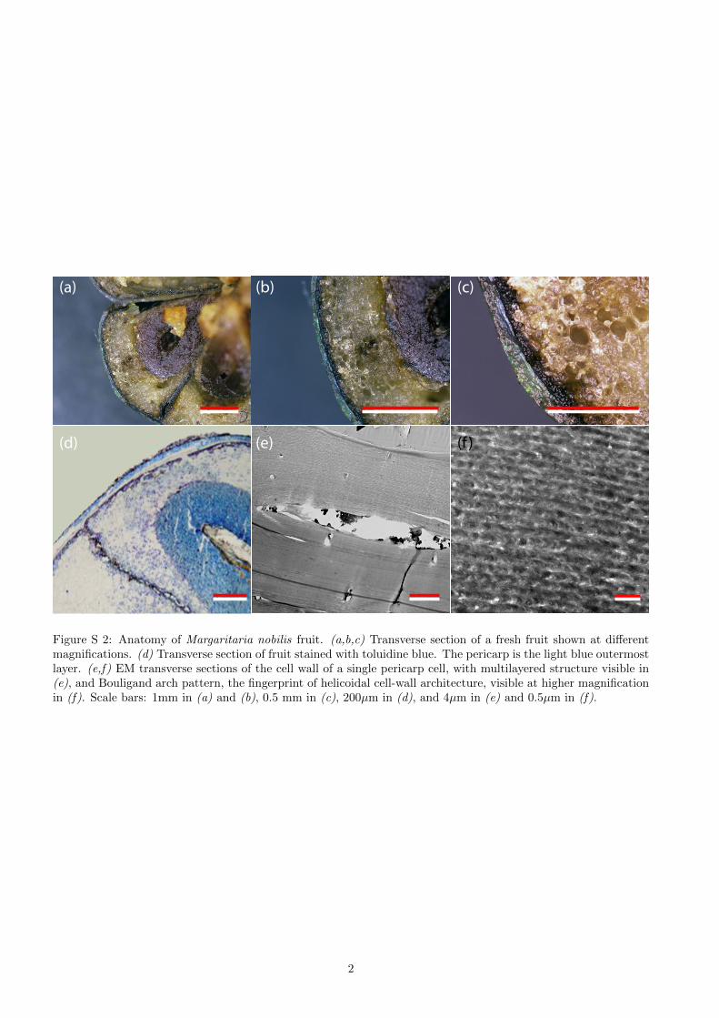
The blue-green coloration of the fruits comes from theendocarp, which consists of thick-walled cells (Figure S2,(d,e,f)). When the fruit is fresh, the seeds are hydratedand adhere perfectly to the endocarp. In the dry state,the seeds shrink, and the endocarp is separated fromthe seeds by an air layer that prevents light absorptionand therefore decreases the contrast and the saturationof the structural coloration, see Figure S2. Transversesections of fresh fruits are shown in (Figure S3(a,b,c))for different magnifications and methods, as describedin the caption.
Hyperspectral Microscopy
Hyperspectral microscopy allows to visualise how themaximum of reflectivity is spatially distributed on thesurface. Due to the fact that the shape of the helicoidalstructure is not flat but is ”wrap around” the cell thehyperspectral imaging can show how the colour shift infunction of the position in the same cell.
1
(a) (b) (c)
(d) (e) (f )
Figure S 2: Anatomy of Margaritaria nobilis fruit. (a,b,c) Transverse section of a fresh fruit shown at differentmagnifications. (d) Transverse section of fruit stained with toluidine blue. The pericarp is the light blue outermostlayer. (e,f) EM transverse sections of the cell wall of a single pericarp cell, with multilayered structure visible in(e), and Bouligand arch pattern, the fingerprint of helicoidal cell-wall architecture, visible at higher magnificationin (f). Scale bars: 1mm in (a) and (b), 0.5 mm in (c), 200µm in (d), and 4µm in (e) and 0.5µm in (f).
2
PERICA
RPSEED
PERICA
RP
SEED
COAT
SEEDSEED
COAT
(a)
(b)
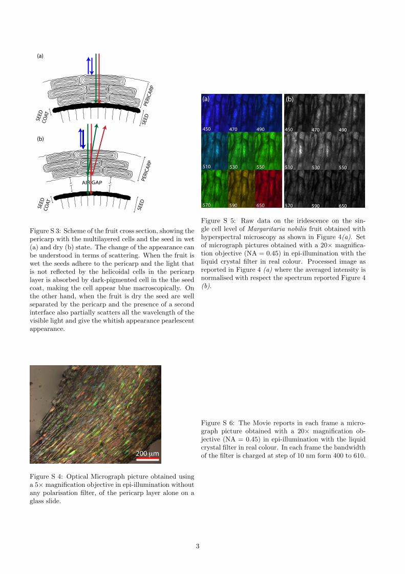
Figure S 3: Scheme of the fruit cross section, showing thepericarp with the multilayered cells and the seed in wet(a) and dry (b) state. The change of the appearance canbe understood in terms of scattering. When the fruit iswet the seeds adhere to the pericarp and the light thatis not reflected by the helicoidal cells in the pericarplayer is absorbed by dark-pigmented cell in the the seedcoat, making the cell appear blue macroscopically. Onthe other hand, when the fruit is dry the seed are wellseparated by the pericarp and the presence of a secondinterface also partially scatters all the wavelength of thevisible light and give the whitish appearance pearlescentappearance.
200 μm
Figure S 4: Optical Micrograph picture obtained usinga 5× magnification objective in epi-illumination withoutany polarisation filter, of the pericarp layer alone on aglass slide.
450 470 490
510 530 550
570 590 650
450 470 490
510 530 550
570 590 650
(a) (b)
Figure S 5: Raw data on the iridescence on the sin-gle cell level of Margaritaria nobilis fruit obtained withhyperspectral microscopy as shown in Figure 4(a). Setof micrograph pictures obtained with a 20× magnifica-tion objective (NA = 0.45) in epi-illumination with theliquid crystal filter in real colour. Processed image asreported in Figure 4 (a) where the averaged intensity isnormalised with respect the spectrum reported Figure 4(b).
Figure S 6: The Movie reports in each frame a micro-graph picture obtained with a 20× magnification ob-jective (NA = 0.45) in epi-illumination with the liquidcrystal filter in real colour. In each frame the bandwidthof the filter is charged at step of 10 nm form 400 to 610.
3