structural biology and evolution of the tgf-b family
TRANSCRIPT

Structural Biology and Evolution of theTGF-b Family
Andrew P. Hinck,1 Thomas D. Mueller,2 and Timothy A. Springer3,4
1Department of Structural Biology, University of Pittsburgh School of Medicine, Pittsburgh, Pennsylvania 152602Department of Plant Physiology and Biophysics, Julius-von-Sachs Institute of the University Wuerzburg,D-97082 Wuerzburg, Germany
3Program in Cellular and Molecular Medicine and Division of Hematology, Department of Medicine, BostonChildren’s Hospital, Boston, Massachusetts 02115
4DepartmentofBiologicalChemistryandPharmacology,HarvardMedicalSchool,Boston,Massachusetts02115
Correspondence: [email protected]
We review the evolution and structure of members of the transforming growth factorb (TGF-b) family, antagonistic or agonistic modulators, and receptors that regulate TGF-bsignaling in extracellular environments. The growth factor (GF) domain common to all familymembers and many of their antagonists evolved from a common cystine knot growth factor(CKGF) domain. The CKGF superfamily comprises six distinct families in primitive metazo-ans, including the TGF-b and Dan families. Compared with Wnt/Frizzled and Notch/Deltafamilies that also specify body axes, cell fate, tissues, and other families that contain CKGFdomains that evolved in parallel, the TGF-b family was the most fruitful in evolution.Complexes between the prodomains and GFs of the TGF-b family suggest a new paradigmfor regulating GF release by conversion from closed- to open-arm procomplex confor-mations. Ternary complexes of the final step in extracellular signaling show how TGF-b GFdimers bind type I and type II receptors on the cell surface, and enable understanding of muchof the specificity and promiscuity in extracellular signaling. However, structures suggest thatwhen GFs bind repulsive guidance molecule (RGM) family coreceptors, type I receptorsdo not bind until reaching an intracellular, membrane-enveloped compartment, blurringthe line between extra- and intracellular signaling. Modulator protein structures show howstructurally diverse antagonists including follistatins, noggin, and members of the chordinfamily bind GFs to regulate signaling; complexes with the Dan family remain elusive. Muchwork is needed to understand how these molecular components assemble to form signalinghubs in extracellular environments in vivo.
Multicellular organisms require an elaboratesystem of intercellular communication to
coordinate cellular actions. In developing em-bryos, the ability of cells to sense and respond tosuch communication is vital for the establish-ment of the overall body plan and to patterntissues, whereas, in adults, it is required for
diverse processes including repair of damagedtissues and regulation of immune responses.In mammals, the 33 genes of the transforminggrowth factor b (TGF-b) family each encode apolypeptide comprising a secretion signal pep-tide, a �250-residue prodomain, and a �110-residue growth factor (GF) domain. Family
Editors: Rik Derynck and Kohei Miyazono
Additional Perspectives on The Biology of the TGF-b Family available at www.cshperspectives.org
Copyright # 2016 Cold Spring Harbor Laboratory Press; all rights reserved; doi: 10.1101/cshperspect.a022103
Cite this article as Cold Spring Harb Perspect Biol 2016;8:a022103
1
on January 5, 2022 - Published by Cold Spring Harbor Laboratory Press http://cshperspectives.cshlp.org/Downloaded from

members include bone morphogenetic pro-teins (BMPs), growth and differentiation fac-tors (GDFs), activins, and TGF-bs. We discussthe emerging concept that the distinctive activ-ities of TGF-b family GFs are determined notjust by signaling type I and type II receptors,which show varying degrees of promiscuityfor GFs, but also by multiple binding pro-teins and enzymes. These modulators greatlydiversify signaling activity by adding anotherlayer of signaling that occurs in extracellularenvironments. They control not only whetherGFs reach their receptors on cells, but alsowhether additional components are presentwithin GF-receptor complexes. Receptors in-clude not only the classical type I and type IIreceptors, but also type III coreceptors and re-pulsive guidance molecule (RGM) coreceptors.Binding proteins comprise the cognate prodo-mains, antagonists such as noggin, follistatin,chordin, and Dan proteins, anchoring mole-cules such as latent TGF-b binding protein(LTBP), and activators such as integrins. En-zymes include proprotein convertases (PCs)that cleave the prodomain from the GF, eitherintracellularly during biosynthesis or extracellu-larly, and tolloid and matrix metalloproteases.Thus, molecular recognition in the TGF-b fam-ily is not singularly achieved by GF-receptorinteractions, but by a network of interactionswith multiple partners. Box 1 summarizes theproteins, interactions, relationships, and termsused in this review.
The complex network of interactions de-scribed here parallels that found through high-throughput mapping of interactomes (Li et al.2004; Rual et al. 2005; Guruharsha et al. 2011),which has shown that regulation is maintainedby a protein–protein interaction network inwhich some proteins serve as hubs and interactwith a larger number of interaction partnersto regulate activity (Jeong et al. 2000). Thesefindings shift our understanding of “linear” sig-naling cascades toward complex signaling net-works, which may better explain the functionaldiversity encoded by TGF-b family proteins inmulticellular animals.
Our review has four sections following theintroduction. The protein domain from which
TGF-b GFs and many of their inhibitors areconstructed evolved in early metazoans beforethe development of bilateral symmetry (seesection on a Brief History of TGF-b FamilyEvolution). GFs are biosynthesized as dimericprodomain–GF complexes, and the prodomainhas an important role in storage and release(activation) of the GF (see section on Structuresand Functions of TGF-b Family Procomplexes).Type I and type II receptors and coreceptorsbind and discriminate between GFs and signalinto cells (see section on Specificity and Promis-cuity in GF-Receptor Interactions). Finally, alarge number of inhibitors form complexeswith the GFs to antagonize or modulate signal-ing (see section on Regulation of TGF-b/BMPSignaling by Modulator Proteins—BMPs asSignaling Hubs).
A BRIEF HISTORY OF TGF-b FAMILYEVOLUTION
Function, three-dimensional structure, andamino acid sequence are all related. Thus, trac-ing the evolution of the TGF-b family throughits sequence relationships is an excellent intro-duction to function. The TGF-b family and itsreceptors and antagonists evolved in parallelwith the Wnt/Frizzled and Delta/Notch path-ways. Together, these extracellular ligands andreceptors create the networks that establish mul-ticellular animal body plans. They specify ante-roposterior, dorsoventral (bilateral), and left–right axes, details of individual organs, and reg-ulate development and homeostasis.
The CKGF Domain and Its Families
The cystine knot (CK) growth factor (CKGF)domain has a specific three-dimensional foldand sequence. Other proteins contain CKs andthus the CK alone does not define the CKGFdomain; the definition of this domain alsoincludes the topological relationship betweenb-strand order in b-sheets and amino acid se-quence. The CKGF domain can thus be recog-nized both by sequence and by structure (Rochand Sherwood 2014), similarly to epidermalgrowth factor (EGF) or immunoglobulin (Ig)
A.P. Hinck et al.
2 Cite this article as Cold Spring Harb Perspect Biol 2016;8:a022103
on January 5, 2022 - Published by Cold Spring Harbor Laboratory Press http://cshperspectives.cshlp.org/Downloaded from

BOX 1. PRODOMAIN, GROWTH FACTOR (GF), AND RECEPTOR STRUCTURES AND EVOLUTIONARY
TREES
Structures are described in sections on Structures and Functions of TGF-b Family Procomplexes, andSpecificity and Promiscuity in GF-Receptor Interactions. For evolutionary trees, sequences were alignedusing MAFFT version 7 and the E-INS-i strategy with BLOSUM30 matrices and gap penalties of 1.53 to 2.5(Katoh and Standley 2013). Trees were calculated as implemented on the same MAFFT server with the NJmethod using all gap-free sites, the JTT model, estimation of a, and 1000 bootstrap samples. In each tree,branch lengths are to the scale shown in the bars.
GF monomerGF homodimer
RII RI Cystine knot
α1
α2
α1
α5
α2
RGDRGD
Cyslinkage
Associationregion
Latencylasso
Latencylasso
Fastener
Armdomain
Bowtie
Armdomain
Strait-jacket
Prodomain 2
GF1
Prodomain 1
GF2
TGF-β1 homodimer
α5
α2
α1
RGD
Cyslinkage
Associationregion
Latencylasso
Bowtie
Armdomain
Strait-jacket
GF1
Prodomain 1
Fastener
TGF-β1 monomer
Furincleavage
C4C2
C3C1
C5C6
Finger 1
Finger 2
Finger 3
Finger 1
Finger 2
Finger 3
GF monomer 2
GF monomer 1
β4-β5 loop
β4-β5loop
Fingers
Knuckle
Thumb
Wrist
Heel
Finger, loop 3Heel, loop 2
Palm
ThumbCysteine knotFinger, loop 1
GDF7
ALK3 ALK6
ThickVeins_Dm
ALK2 ALK1
ALK5
Sax_Dm
BMPRII
TβRII
ActRII ActRIIB
Punt_Dm
0.2
Baboon_Dm
InhβB
Myostatin
InhβE
InhβCInhβA
Activin_DmBMP5BMP6
BMP7BMP8
GBB_Dm
SCW_Dm Dawdle_DmBMP2BMP4
BMP3
GDF10GDF3
GDF15Inhα
TGFβ1
TGFβ3
0.2
LEFTY2GDF1
LEFTY1 NodalBMP10BMP9
BMP8BMP6
BMP5BMP7
BMP9BMP10
NodalGDF6
GDF5
GDF3
InhβEInhβC
InhβB
GDF11Myostatin
BMP3
Dawdle_Dm
TGFβ2TGFβ3
TGFβ1AMH
GDF15
LEFTY1LEFTY2
Maverick_Dm
0.2
Activin clade
DPP_Dm
AMHRII
BMP clade
Type II Type IWishful Thinking_Dm
ALK4 ALK7
GDF10
Activin_Dme
Myoglianin_Dm GDF1
GDF9
BMP15
BMP2
TGFβ2
Screw_Dm
Myoglianin_DmGDF11
Maverick_DmAMH
GDF9
DPP_Dm
GDF7
GDF5GDF6
GBB_DmInhα TGF-β GFs
TGF-β prodomains
BMP antagonists
TGF-β receptors
CerberusNoggin
Coco Sclerostin
Nbl1/Dan
GremlinPRDC
USAG1
0.2
BMP15
InhβA
BMP4
Palm
Structural Biology and Evolution of the TGF-b Family
Cite this article as Cold Spring Harb Perspect Biol 2016;8:a022103 3
on January 5, 2022 - Published by Cold Spring Harbor Laboratory Press http://cshperspectives.cshlp.org/Downloaded from
domains. And just as the latter families havediverse functions, many proteins containingCKGF domains lack growth factor activity.Most TGF-b family members act as differenti-ation factors rather than growth factors andsome such as inhibins and Leftys antagonizeactivities of other members. However, just asTGF-b is eponymous for the family as a whole,its signaling moiety is also eponymous for whatwe term the GF domain. All family memberscontain a GF domain that can form a GF dimer;thus, we term this the GF domain whether ornot it has signaling activity.
The CKGF domain contains two longb-rib-bons (b-sheets with two antiparallel b-strands)and a CK (GF monomer, Box 1). Two closelyspaced pairs of cysteines in adjacent, parallelb-strands disulfide-link to form a ring com-posed of two peptide backbone segments andtwo disulfides. Another disulfide passes throughthe ring, linking two additional polypeptide seg-ments (cystine knot, Box 1). Thus, the knot tiestogether four polypeptide segments that aredistal in sequence, and forms a highly stableCK core from which three long loops emanate(GF monomer, Box 1).
The CKGF domain superfamily is present inthe earliest metazoans. Like the Wnt/Frizzledand Delta/Notch families, the CKGF family isnot found in protists, and first appears in prim-itive metazoans that lack a bilateral axis. Repre-sentatives are present in four distinct prebilater-ian phyla, that is, Porifera (sponges), Cnidaria(coral and hydra), Ctenophora (comb jellies),and Placozoa (Trichoplax) (Roch and Sherwood2014). The CKGF superfamily is composedof six groups (families) with the CKGF domainas the primary structural feature, that is, withor without a prodomain and without any otherfolded domain (Adamska et al. 2007; Roch andSherwood 2014). These are (1) the TGF-b fam-ily, (2) the Dan family of BMP antagonists, (3)the glycoprotein hormone family (GPH), forexample, the pituitary hormones follicle-stim-ulating hormone, luteinizing hormone, andthyroid-stimulating hormone, (4) bursiconhormones, which are limited to invertebrates,(5) the platelet-derived growth factor (PDGF)family, including vascular endothelial growth
factors (VEGFs), and (6) the nerve growth fac-tors (NGFs) (Roch and Sherwood 2014). CKGFdomains are also found in noggin, in the CCNfamily, and as carboxy-terminal CK domains.
From Prebilaterians to the Emergenceof TGF-b Itself in Deuterostomes
Of these six families, the first five are represent-ed in prebilaterian phyla. Moreover, sequencescharacteristic of specific Dan family membersrepresented in Cnidaria, Porifera, Ctenophora,and Placazoa are detectable in humans. In otherwords, statistically significant relationships be-tween sequences can be used to infer orthologyso that prebilaterian Dan family members canbe linked to specific proteins or subfamilies ofrelated proteins in humans, including gremlin,sclerostin, and uterine sensitization-associatedgene 1 (USAG1) (Roch and Sherwood 2014).TGF-b family members and their type I andtype II receptors are also present in nonbilater-ians, but orthology with particular familymembers in chordates is unclear. In contrast,Smad orthologues corresponding to Smad 1or 5, Smad 2 or 3, the co-Smad Smad4, andinhibitory Smads (I-Smads) can be identifiedin nonbilaterians (Herpin et al. 2004; Humi-niecki et al. 2009; Pang et al. 2011).
The next step in evolution is marked by thedefinition of a new body axis, the bilateral axis,which creates dorsoventral asymmetry and alsoenables development of left–right asymmetry.Bilaterian phyla are grouped by nucleotide andprotein sequence into three branches that ex-tend previous embryological classification(Blair and Hedges 2005; Dunn et al. 2008; Hej-nol et al. 2009). The protostome branch is nowsubdivided into the Ecdysozoa (e.g., Caenorhab-ditis elegans and Drosophila melanogaster) andLophotrochozoa (e.g., molluscs and annelids)branches. Deuterostomes represent a laterevolving group of phyla. Molecular phylogenyconfirms deuterostomes as a branch with threephyla, Echinodermata (e.g., sea urchins), Chor-data (vertebrates, urochordates, and cephalo-chordates), and Hemichordata (acorn worms).
Key innovations in the evolution of deutero-stomes from lower bilaterians include the emer-
A.P. Hinck et al.
4 Cite this article as Cold Spring Harb Perspect Biol 2016;8:a022103
on January 5, 2022 - Published by Cold Spring Harbor Laboratory Press http://cshperspectives.cshlp.org/Downloaded from

gence of pharyngeal gill slits and an inversion inthe dorsoventral axis of the body plan (Hollandet al. 2015; Lowe et al. 2015). Notably, the BMPand chordin gradients that establish dorsoven-tral polarity also invert in deuterostomes (Loweet al. 2015).
Despite the presence of multiple TGF-bfamily members, TGF-b family receptors, andSmads, TGF-b is clearly absent from early bilat-erians. TGF-b itself first appears in the genome-based evolutionary record with the emergence ofdeuterostomes. Lowerdeuterostomes, includingsea urchins, tunicates, and hemichordates, haveonly a single TGF-b, and in all cases its prodo-main has an RGD motif (Robertson and Rifkin2013). Mammals have three TGF-bs. TGF-b1and -b3 contain RGD motifs and bind and areactivated by integrins. TGF-b2 diverged fromTGF-b3 after the divergence of TGF-b1. Thus,the lackof the Arg of the RGD motif in TGF-b2 isan acquired, not ancestral, characteristic. Fur-thermore, the acidic Asp residue retained inTGF-b2 is the only key residue required for in-tegrin binding, and thus it is possible that TGF-b2 is activated by a yet unidentified integrin.
These evolutionary events shed importantlight on the TGF-b family and answer one ofthe commonly asked questions in the field,“Why are there so many antagonists of BMPsand activins but not TGF-bs?” Currently se-quenced nonbilaterian metazoans have asmany or more members of the CKGF Danfamily than of the CKGF TGF-b family itself.Therefore, in prebilaterians, BMP antagonistand TGF-b family members coevolved in thepresence of one another (Roch and Sherwood2014), and in the presence of other develop-mental pathways such as Wnt (Adamska et al.2007). These signaling families matured anddiversified much further in bilaterian inverte-brates, to the point where members in Droso-phila can be grouped with specific TGF-b sub-families in humans (TGF-b GFs, Box 1). Incontrast, TGF-b itself evolved much later, longafter the diversification of bilaterian phyla,at the emergence of deuterostomes. Integrins,like the CKGF superfamily, are present in bothprebilaterian and early bilaterian metazoans, inwhich they are already well diversified. Thus,
TGF-b emerged in a context in which the keytasks of bilaterian axis and organ specificationby complex signaling networks were well estab-lished. Within this context, TGF-b evolved totake advantage of a new regulatory mechanism,in which integrins activate signaling. Such anew mechanism may have been required be-cause the regulatory hubs involving BMP antag-onists were already well established in the spec-ification of body axes and organs, and their useby a radical new CKGF member might havethrown a wrench into this machinery.
Family Trees
The family trees in Box 1 show the diversifi-cation of family members in humans andD. melanogaster for TGF-b family GFs and pro-domains (separately), BMP antagonists, andTGF-b family type I and type II receptors. Thesetrees were constructed using rigorous methodsappropriate for defining phylogenetic relation-ships, as implemented in MAFFT (Katoh andStandley 2013). Scale bars show the extent ofsequence divergence, which for any two mem-bers is given by adding the lengths of the branch-es that separate them. However, the trees showontogenetic relationships, not phylogenetic re-lationships. Thus, the trees in Box 1 show se-quence divergence as aconsequence of function-al and structural diversification, rather than as aconsequence of separation of animal species inevolutionary time. Furthermore, the shapes ofthe trees in Box 1 provide insights into how func-tion and structure have driven diversification ofthe subfamilies. Separate trees for the TGF-bfamily prodomains and GFs emphasize that theprodomains are more divergent; when the scalebars in Box 1 are taken into account, the prodo-mains are about fourfold more divergent thanthe GFs. Thus, the prodomains have been undergreater functional or structural pressure for di-vergencethan theGFs. Also, thesepressuresmustdiffer, because branch lengths and clusteringwithin subfamilies differ; however, because ofhigher sequence identity for the GFs, subfamiliescan be defined with more statistical significance.
Clustering using neighbor joining withbootstrap calculations (here) or Bayesian anal-
Structural Biology and Evolution of the TGF-b Family
Cite this article as Cold Spring Harb Perspect Biol 2016;8:a022103 5
on January 5, 2022 - Published by Cold Spring Harbor Laboratory Press http://cshperspectives.cshlp.org/Downloaded from
ysis (Pang et al. 2011) support significant rela-tionships among TGF-b family GF domains(color-coded in Box 1). A BMP clade includessix subfamilies. Human BMP-5, 6, 7, and 8 andDrosophila Glass bottom boat (Gbb) and Screwcluster in one group that is significantly relatedto the cluster with human BMP-2 and 4 andDrosophila Decapentaplegic. Four other clusterscontain BMP-9 and 10; GDF-5, 6, and 7; GDF-9and BMP-15; and GDF-1 and 3. Nodal falls inthe same BMP clade. An activin clade includesthree subgroups: Drosophila activin and humaninhibin b-subunits, which dimerize to formactivins, GDF-11, myostatin (GDF-8) and pos-sibly Drosophila Myoglianin, and BMP-3 andGDF-10. Further family members in the leftside of the GF family tree in Box 1 are difficultto place, but lie closer to the activin than BMPclade. All have unique features. TGF-bs andanti-Mullerian hormone (AMH) each haveunique type II receptors. AMH and GDF-15have atypical prodomains that are the longestand shortest in the family, with 434 and 170residues, respectively. Lefty 1 and 2 are cleavedby furin after the a2-helix in the prodomain,rather than between the prodomain and GF do-main (Fig. 1), and are thus predicted to havetheir type II receptor binding sites permanentlyblocked (Shi et al. 2011; Mi et al. 2015). Inhibin-a is inhibitory; when it heterodimerizes withinhibin b-subunits it forms inhibin.
Type I and type II receptors for TGF-b familyGFs have structurally homologous ligand-bind-ing ectodomains and homologous dual specific-ity kinase cytoplasmic domains, and thus may becompared in the same tree (TGF-b receptors,Box 1). The longer branches of type II receptorsshow that they are more diverse than the type Ireceptors; the receptor trees reflect primarily thecytoplasmic domain because of its much greaternumber of aligned positions. The human type Ireceptors show three clades, each with a Droso-phila relative. Activin receptor-like kinase (ALK)1 and 2 are closely related to Saxophone (Sax),ALK3 and 6 are related to Thickveins, and ALK4,5 and 7 are relatives to Baboon. The ALK1/2,ALK3/6, and ALK4–7 subfamilies couple totwo distinct groups of receptor-regulated Smads(R-Smads), with the ALK1/2 and ALK3/6 sub-
families coupling to R-Smad1, 5, 8 and theALK4/5/7 subfamily coupling to R-Smad2and 3. Type II receptors also show three groups.BMPRII and AMHRII relate to Wishful think-ing, and ActRII and ActRIIB ally with Punt. Incontrast, among all type I and type II receptors,only TbRII lacks a Drosophila relative, consistentwith the recent deuterostome origin of TGF-b.Moreover, TbRII has no close human relatives.
The Dan family of antagonists against BMPsand Wnts has seven members in humans andshares some similarity with noggin in its CKGFdomain (BMP antagonists, Box 1). Althoughpresent in many prebilaterians, the Dan familywas lost in insects.
Did the Prodomain Contribute to theEvolutionary Success of the TGF-b Family?
Although the origins of the prodomain of TGF-b remain unclear, it may have been an impor-tant contributor to the success of the TGF-bfamily branch of the CKGF superfamily. In evo-lutionary terms, success may be defined notonly as lack of extinction, but also as diversifi-cation and radiation into a protein family with alarge number of members in higher organisms.By the criterion of number of members alone,the TGF-b family has been more successfulthan any of its kindred hormone or GF familiesin the CKGF superfamily (Roch and Sherwood2014). The TGF-b family also radiated morethan other extracellular protein families of par-allel importance in developmental specification.With 33 gene family members in mammals,the TGF-b family is more numerous than the19 Wnts, which have 10 Frizzled receptors, orthe total of five Deltas and Jaggeds, which havefour Notch receptors.
The prodomain, including its arm domainand straitjacket appendages (TGF-b1 homo-dimer, Box 1) is clearly defined in the sequencesof prebilaterian TGF-b family members(Adamska et al. 2007; Roch and Sherwood2014). The CKGF domains of the TGF-b familydimerize in a specific manner and associate withtwo prodomain monomers; the prodomainarm domain and straitjacket extensions togeth-er wrap around the GF and block receptor and
A.P. Hinck et al.
6 Cite this article as Cold Spring Harb Perspect Biol 2016;8:a022103
on January 5, 2022 - Published by Cold Spring Harbor Laboratory Press http://cshperspectives.cshlp.org/Downloaded from

Dpp_Dr GDF-5 AMH
TGF-β1 TGF-β2 TGF-β3 myosta GDF-11 Inh-βA Inh-βB Inh-βC Inh-βE Inh-α BMP-3 GDF-10 BMP-15 GDF-9 GDF-1 GDF-3 BMP-9 BMP-10 Nodal BMP-2 BMP-4 Dpp_Dr Scw_Dr Gbb_Dr BMP-5 BMP-7 BMP-6 BMP-8 GDF-5 GDF-6 GDF-7 GDF-15 AMH LEFTY1 LEFTY2
TGF-β1 TGF-β2 TGF-β3 myosta GDF-11 Inh-βA Inh-βB Inh-βC Inh-βE Inh-α BMP-3 GDF-10 BMP-15 GDF-9 GDF-1 GDF-3 BMP-9 BMP-10 Nodal BMP-2 BMP-4 Dpp_Dr Scw_Dr Gbb_Dr BMP-5 BMP-7 BMP-6 BMP-8 GDF-5 GDF-6 GDF-7 GDF-15 AMH LEFTY1 LEFTY2
TGF-β1 TGF-β2 TGF-β3 myosta GDF-11 Inh-βA Inh-βB Inh-βC Inh-βE Inh-α BMP-3 GDF-10 BMP-15 GDF-9 GDF-1 GDF-3 BMP-9 BMP-10 Nodal BMP-2 BMP-4 Dpp_Dr Scw_Dr Gbb_Dr BMP-5 BMP-7 BMP-6 BMP-8 GDF-5 GDF-6 GDF-7 GDF-15 AMH LEFTY1 LEFTY2
ED I SQRF I AA I APVAAH I PLASASGSGSGRSGSRSVGASTSTALAKAFNPFSEPASFSDSDKSHRSKTNKKPSKSDANRQFNEVHKPRTDQLENS 95- - - - - - - - - - - - - - - - - - - - - - - - - - - - - - - - - - - - - - - - APDLGQRPQGTRPGLAKAEAKERPPLARNVFRPGGHSYGGGATNANARAKGGTGQ 55- - - - - - - - - - - - - - - - - - - - - - - - - - - - - - - - - - L LGTEALRAEEPAVGTSGL I FREDLDWPPG I PQEPLCLVALGGDSNGSSSPLRVVGALSAY 61
E 110
- - - - - - - - - - - - - - - - - - - - - - - - - - - - - - - - - - - - - - - LSTCKT I DMELVKRKR I EA I RGQ I LSKLRLASPPSQGEV - - - - - - - - - - - - - - - - - - - - - PP - - - - - - - - - GPLPEAVLA 50- - - - - - - - - - - - - - - - - - - - - - - - - - - - - - - - - - - - - - SLSTCST LDMDQFMRKR I EA I RGQ I LSKLKL TSPPEDY - - - - - - - - - - - - - - - - - - - - - - - PE - - - - - - - - PEEVPPEV I S 50- - - - - - - - - - - - - - - - - - - - - - - - - - - - - - - - - - - - SLSLSTCT T LDFGH I KKKRVEA I RGQ I LSKLRL TSPPE - - - - - - - - - - - - - - - - - - - - - - - - - PT - - - - - - - VMTHVPYQVLA 51- - - - - - - - - - - - - - - - - - - - - - - - - - NENSEQKENVEKEGLCNACTWRQNTKSSR I EA I K I Q I LSKLRLETAPN I SKDV I RQL L - - - - - - - - - - - - - - - PK - - - - - - - - - APPLREL I D 69- - - - AEGPAAAAAAAAAAAAAGVGGERSSRPAPSVAPEPDGCPVCVWRQHSRELRLES I KSQ I LSKLRLKEAPN I SREVVKQL L - - - - - - - - - - - - - - - PK - - - - - - - - - APPLQQ I LD 91- - - - - - - - - - - - - - - - - - - - - SPTPGSEGHSAAPDCPSCALAALPKDVPNSQPEMVEAVKKH I LNMLHLKKRPDVTQPV - - - - - - - - - - - - - - - - - - - - PK - - - - - - - - - AAL LNA I RK 69- - - - - - - - - SPTPPPTPAAPPPPPPPGSPGGSQDTCTSCGGFRRPEELGRVDGDF LEAVKRH I LSRLQMRGRPN I THAV - - - - - - - - - - - - - - - - - - - - PK - - - - - - - - - AAMVTALRK 81- - - - - - - - - - - - - - - - - - - - - - - - - - - - TPRAGGQCPACGG - - PT LELESQREL L LDLAKRS I LDKLHL TQRPT LNRPV - - - - - - - - - - - - - - - - - - - - SR - - - - - - - - - AALRTALQH 60- - - - - - - - - - - - - - - - - - - - - - - - - - - - - QGTGSVCPSCGG - - SKLAPQAERALVLELAKQQ I LDGLHL TSRPR I THPP - - - - - - - - - - - - - - - - - - - - PQ - - - - - - - - - AAL TRALRR 59- - - - - - - - - - - - - - - - - - - - - - - - - - - - - - - - - - - - - - - - - - - - CQGLELARELVLAKVRAL F LDAL - - - GPPAVTREGGD - - - - - - - - - - - - - - - - - - PG - - - - - - - - - - - - - - - VRR 39- - - - - - - - - - - - - - - - - - - - - - - - - - - - - - - - - - - - - - - - - - - - - - - - - ERPKPPFPELRKAVPGDRTAGGGPDSELQ - - - - - - - - - - - - - - - - - - - - - PQ - - - - - - - - - DKVSEHMLR 40- - - - - - - - - - - - - - - - - - - - - - - - - - - - - - - - - - - - - - - - - - - - - - SHRAPAWSALPAAADGLQGDRDLQRHPGDAAAT LG - - - - - - - - - - - - - - - - - - PS - - - - - - - AQDMVAVHMHR 48- - - - - - - - - - - - - - - - - - - - - - - - - - - - - - - - - - - - - MEHRAQMAEGGQSS I AL LAEAPT LPL I EEL - LEESPGEQPRK - - - - - - - - - - - - - - - - - - - - PR - - - - - - - L LGHSLRYMLE 54- - - - - - - - - - - - - - - - - - - - - - - - - - SQASGGEAQ I AASAELESGAMPWSL LQH I DERDRAGL LPAL FKVLSVGRGGSPRLQ - - - - - - - - - - - - - - - - - PD - - - - - - - - - SRALHYMKK 67- - - - - - - - - - - - - - - - - - - - - - - - - - - - - - - - - - - - - - - - - - - - - - - - - - - - - PVPPGPAAAL LQALGLRDEPQGA - - - - - - - - - - - - - - - - - - - - - - - PR - - - - - - - - LRPVPPVMWR 35- - - - - - - - - - - - - - - - - - - - - - - - - - - - - - - - - - - - - - - - - - - - - - - - - - - - - - - - - - QEYVF LQF LGLDKAPS - - - - - - - - - - - - - - - - - - - - - - - - - PQ - - - - - - - KFQPVPY I LKK 29- - - - - - - - - - - - - - - - - - - KPLQSWGRGSAGGNAHSPLGVPGGGLPEHT FNLKMF LENVKVDF LRSLNLSGVPSQDKTRVE - - - - - - - - - - - - - - - - - - P - - - - - - - - - - - - - PQYM I D 69- - - - - - - - - - - - - - - - - - - - - - SP I MNLEQSPLEEDMSL FGDVFSEQDGVDFNT L LQSMKDEF LKT LNLSD I PTQDSAKVD - - - - - - - - - - - - - - - - - - P - - - - - - - - - - - - - PEYMLE 66- - - - - - - - - - - - - - - - - - - - - - - - - - - - - - - - - - - - - - - - - - - - - - - - - - - - - - - - - TVATAL LRTRGQPSSPS - - - - - - - - - - - - - - - - - - - - - - - - - PL - - - - - - - - - - - - - AYMLS 24- - - - - - - - - - - - - - - - - - - - - - - - - - - - - LVPELGRRKFAAASSGRPSSQPSDEVLSEFELRL LSMFGLKQRPT - - - - - - - - - - - - - - - - - - - - - - - - - PS - - - - - - - RDAVVPPYMLD 58- - - - - - - - - - - - - - - - - - - - GASHASL I PETGKKKVAE I QGHAGGRRSGQ - SHEL LRDFEAT L LQMFGLRRRPQ - - - - - - - - - - - - - - - - - - - - - - - - - PS - - - - - - - KSAV I PDYMRD 66KNKSKQLVNKPNHNKMAVKEQRSHHKKSHHHRSHQPKQASASTESHQSSS I ES I FVEEPT LVLDREVAS I NVPANAKA I I AEQGPSTYSKEAL I KDKLKPD - - - - - PST LVE I EKSL LS 209- - - - - - - - - - - - - - - - - - - - - - - - - - - - - - - - - - - - - T TYVT TNNH I EMP I YQKRPLSEQMEM I D I LDLGDRPRRQAEPN - - - - - - - - - - - - - - - - - - - - - - - - - - - - LHNSASKF L LE 54- - - - - - - - - - - - - - - - - - - - - - - - - - - - - - - TQSG I Y I DNGKDQT I MHRVLSEDDKLDVSYE I LEF LG I AERPTHLSSHQLSLR - - - - - - - - - - - - - - - - - - - - - - - - - - KSAPKF L LD 62- - - - - - - - - - - - - - - - - - - - - - - - - - - - - - - - - - - - - - DNHVHSSF I YRRLRNHERRE I QRE I LS I LGLPHRPR - - - - - - - - - - - - - - - - - - - - - - - - - PF - - - SPGKQASSAPL FMLD 53- - - - - - - - - - - - - - - - - - - - - - - - - - - - - - - - - - DFSLDNEVHSSF I HRRLRSQERREMQRE I LS I LGLPHRPR - - - - - - - - - - - - - - - - - - - - - - - - - PH - - - LQGKH - NSAPMFMLD 56CCGPPPLRPPLPAAAAAAAGGQL LGDGGSPGRTEQPPPSPQSSSGF LYRRLKTQEKREMQKE I LSVLGLPHRPRPLHGLQQPQPPALRQQEEQQQQQQLPRGEPPPGRL - KSAPL FMLD 118- - - - - - - - - - - - - - - - - - - - - - - - - - - - - - - - - - GGGPGLRPPPGCPQRRLGARERRDVQRE I LAVLGLPGRPR - - - - - - - - - - - - - - - - - - - - - - - - - PRAPPAASRLPASAPL FMLD 60TGGL TQPKKDEPKKLPPRPGGPEPKPGHPPQTRQATARTVTPKGQLPGGKAPPKAGSVPSSF L LKKAREPGPPREPKEPFRPPP I T - - - - - - - - - - - - - PH - - - - - - - - - - - - - EYMLS 148- - - - - - - - - - - - - - - FQQAS I SSSSSSAELGSTKGMRSRKEGKMQRAPRDSDAGREGQEPQPRPQDEPRAQQPRAQEPPGRGPRVV - - - - - - - - - - - - - PH - - - - - - - - - - - - - EYMLS 78- - - - - - - - - - - - - - - - - - - - - - - - - - - - - RDGLEAAAVLRAAGAGPVRSPGGGGGGGGGGRT LAQAAGAAAVPAAAVPRARAARRAAGSGFRNGSVV - - PH - - - - - - - - - - - - - HFMMS 75- - - - - - - - - - - - - - - - - - - - - - - - - - - - - - - - - - - - - - - - - - - - - - - - - - - - - - - - - - - - - - - - - - LSLAEASRASFPG - - - - - - - - - - - - - - - - - - - - PS - - - - - - - ELHSEDSRFRE 26EQAF LGAVQRARWGPRDLAT FGVCNTGDRQAALPSLRRLGAWLRDPGGQRLVVLHLEEVTWEPTPSLRFQEPP - - - - - - - - - - - - - - - - - - - - - - - - - - PG - - - - - - - GAGPPELAL LV 147- - - - - - - - - - - - - - - - - - - - - - - - - - - - - - - - - - - - - - - - - - - - - - - - - - - - - L TGEQL LGSL LRQLQLKEVPT LDRADMEELV I - - - - - - - - - - - - - - PT - - - - - - - - - HVRAQYVAL 43- - - - - - - - - - - - - - - - - - - - - - - - - - - - - - - - - - - - - - - - - - - - - - - - - - - - - L TEEQL LGSL LRQLQLSEVPVLDRADMEKLV I - - - - - - - - - - - - - - PA - - - - - - - - - HVRAQYVVL 43
LYNSTRDRVAGESAEPE - - - - - - - - - - - - - - - - - - - - - - - - - - - - - - - - - - - - - - - - - - - - - - - - PEPEADYYAKEVTRVLMVETHNE I YDKFKQS - - - - - - - - - - - THS I YMF FNTSI YNSTRDL LQEKASRRA - - - - - - - - - - - - - - - - - - - - - - - - - - - - - - - - - - - - - - - - AAC - - - ERERSDEEYYAKEVYK I DMPPF FPSENA I PPT FYR - - - - - - - - - PYFR I VRFDVSA 117LYNSTREL LEEMHGERE - - - - - - - - - - - - - - - - - - - - - - - - - - - - - - - - - - - - - - - - EGC - - - TQENTESEYYAKE I HKFDM I QGLAEHNELAVCPKG - - - - - - - - - I TSKVFRFNVSS 118QYDVQRDD - SSDGSLED - - - - - - - - - - - - - - - - - - - - - - - - - - - - - - - - - - - - - - - - - - - - - - - - - - - - DDYHAT TET I I TMPTESDF LMQVD - - - - - - - - - - - - - - GKPKCCF FKFSS 121LHDFQGDALQPEDF LEE - - - - - - - - - - - - - - - - - - - - - - - - - - - - - - - - - - - - - - - - - - - - - - - - - - - - DEYHAT TETV I SMAQETDPAVQTD - - - - - - - - - - - - - - GSPLCCHFHFSP 144LHVGKVGENGYVE I - - - - - - - - - - - - - - - - - - - - - - - - - - - - - - - - - - - - - - - - - - - EDD I GRRAEMNELMEQTSE I I T FAESG - - - - - - - - - - - - - - - - - - - - - - - TARKT LHFE I SK 122LHAGKVREDGRVE I - - - - - - - - - - - - - - - - - - - - - - - - - - - - - - - - - - - - - - - - - - - PHLDGHASPGADGQERVSE I I SFAETDGLA - - - - - - - - - - - - - - - - - - - - SSRVRLYF F I SN 137LHGVPQGAL LEDNR - - - - - - - - - - - - - - - - - - - - - - - - - - - - - - - - - - - - - - - - - - - - - - - - - - - - - - - - - EQECE I I SFAETGLST - - - - - - - - - - - - - - - - - - - - I NQTRLDFHFSS 102LQPGSVAPGN - - - - - - - - - - - - - - - - - - - - - - - - - - - - - - - - - - - - - - - - - - - - - - - - - - - - - - - - - - - - - - - GEEV I SFATVTDSTS - - - - - - - - - - - - - - - - - - - AYSSL L T FHLST 96LPRRHALGGF THRGSEP - - - - - - - - - - - - - - - - - - - - - - - - - - - - - - - - - - - - - - - - - - - - - - - - - - - EEEEDVSQA I L FPATDASCEDKSAARGLAQEA - - - - - E - EGL FRYMFRPSQ 101LYDRYSTVQAARTPGSL - - - - - - - - - - - - - - - - - - - - - - - - - - - - - - - - - - - - - - - - - - EGGSQPWRPRL LREGNTVRSFRAAAAET L - - - - - - - - - - - - - - - - - - - ERKGLY I FNL TS 98LYEKYSRQGARPGG - - - - - - - - - - - - - - - - - - - - - - - - - - - - - - - - - - - - - - - - - - - - - - - - - - - - - - - - - - - GNTVRSFRARLEVV - - - - - - - - - - - - - - - - - - - - DQKAVYF FNL TS 88LYRRSADSHGHPRENRT - - - - - - - - - - - - - - - - - - - - - - - - - - - - - - - - - - - - - - - - - - - - - - - - - - - - - - I GATMVRLVKPL TSVARPHRGTWH I Q I LG - - - - - FPLRPNRGLYQLVR 114LYKTYATKEG I PKSNRS - - - - - - - - - - - - - - - - - - - - - - - - - - - - - - - - - - - - - - - - - - - - - - - - - - - - - - HLYNTVRL F TPCTRHKQAPGDQVTG I - - - - - - - - - - LPSVEL L FNLDR 122L FRRRDPQETRSGSRRT - - - - - - - - - - - - - - - - - - - - - - - - - - - - - - - - - - - - - - - SPGVT LQPCHVEELGVAGN I VRH I PDRGAPTRASEPASAAGH - - - - - - - - - CPEWTVVFDLSA 106I FQDREAAAT TGVSRDL - - - - - - - - - - - - - - - - - - - - - - - - - - - - - - - - - - - - - - - - - - - - - - - CYVKELGVRGNVLRF LPDQGF F LYPKK I SQASS - - - - - - - - - - CLQKL LYFNLSA 91LYNRYTSDKST T - - - - - - - - - - - - - - - - - - - - - - - - - - - - - - - - - - - - - - - - - - - - - - - - - - - - - - - - - - - PASN I VRSFSMEDA I S I TATEDFP - - - - - - - - - - - - FQKH I L L FN I S- 116LYNKFATDRTS - - - - - - - - - - - - - - - - - - - - - - - - - - - - - - - - - - - - - - - - - - - - - - - - - - - - - - - - - - - MPSAN I I RSFKNEDL FSQPVSFNG - - - - - - - - - - - - - LRKYPL L FNVS- 112LYR - - - - - - - - - - - - - - - - - - - - - - - - - - - - - - - - - - - - - - - - - - - - - - - - - - - - - - - - - - - - - - - - - DPLPRAD I I RSLQAEDVAVD - - - - - - - - - - - - - - - - - - - GQNWT FAFDFSF 59LYRRHSGQPGSPAPDHR - - - - - - - - - - - - - - - - - - - - - - - - - - - - - - - - - - - - - - - - - - - - - - - - - LERAASRANTVRSFHHEESLEELPETSG - - - - - - - - - - - - - KT TRRF F FNLSS 115LYRLQSGEEEEEQ I HST - - - - - - - - - - - - - - - - - - - - - - - - - - - - - - - - - - - - - - - - - - - - - GLEYPERPASRANTVRSFHHEEHLEN I PGTSE - - - - - - - - - - - - - NSAFRF L FNLSS 127L FNMKRPPK I DRSK I I I PEPMKKLYAE I MG - - - - - - - - - - - - - - - - - - - - - - - - - - HELDSVN I PKPGL L TKSANTVRSF THKDSK I DDRFP - - - - - - - - - - - - - - HHHRFRLHFDVKS 288VYNE I SEDQEPKEVLHQRHKR - - - - - - - - - - - - - - - - - - - - - - - - - - - - - - - - - - SLDDD I L I SNEDRQE I ASCNS I L T FSSRLKPEQLDN - - - - - - - - - - - - - - - - ELDMH I T FNTND 123VYHR I TAEEGLSDQDEDDDYERG - - - - - - - - - - - - - - - - - - - - - - - - - - - - - - - - - HRSRRSADLEEDEGEQQKNF I TDLDKRA I DESD I I MT F LNKRHHNVDELRHEHGRRLWFDVSN 148LYNAMTN - - - EENPEESEYSVRASLAEETRGARKGYPASPNGYPRR I QLSRT TPL T TQSPPLASLHDTNF LNDADMVMSFVNLVERDKDFSHQR - - - - - - - - - - - - - RHYKEFRFDL TQ 156LYNAMAV - - - EEGGGPGGQGFSYPYKAVFS - - - - - - - - - - - - - - - - - - - - - - - - - - TQGPPLASLQDSHF L TDADMVMSFVNLVEHDKEF FHPR - - - - - - - - - - - - - YHHREFRFDLSK 133LYNALSADNDEDGASEGERQQSWPHEAASSSQRRQPPPGAAHPLNRKSL LAPGSGSGGASPL TSAQDSAF LNDADMVMSFVNLVEYDKEFSPRQ - - - - - - - - - - - - - RHHKEFKFNLSQ 224LYHAMAGDDDEDGAPAE - - - - - - - - - - - - - - - - - - - - - - - - - - - - - - - - - - - - - - - - - - - - - - - - - - - QRLGRADLVMSFVNMVERDRALGHQE - - - - - - - - - - - - - PHWKEFRFDL TQ 115LYRT LSDADRKGGNSSV - - - - - - - - - - - - - - - - - - - - - - - - - - - - - - - - - - - - - - - - - - - - - - - - - - KLEAGLANT I TSF I DKGQDDRGPV - - - - - - - - - - - - - - - - VRKQRYVFD I SA 201I YRTYS I AEKLG I NASF - - - - - - - - - - - - - - - - - - - - - - - - - - - - - - - - - - - - - - - - - - - - - - - - - - FQSSKSANT I TSFVDRGLDDLSHTP - - - - - - - - - - - - - - - LRRQKYL FDVSM 132LYRSLAGRAPAGAAAVS - - - - - - - - - - - - - - - - - - - - - - - - - - - - - - - - - - - - - - - - - - - - - - - - - - ASGHGRADT I TGF TDQATQDESAA - - - - - - - - - - - - - - - - ETGQSF L FDVSS 128LRKRYEDL L TRLRANQS - - - - - - - - - - - - - - - - - - - - - - - - - - - - - - - - - - - - - - - - - - - - - - - - - - - - - - -WEDSNTDLVPAPAVR I L T - - - - - - - PEV - - - - - RLGSGGHLHLR I SR 78LYPGPGPEVTVTRAGLP - - GAQSLCPSRD - - - - - - - - - - - - - - - - - - - - - - - - - - - TRYLVLAVDRPAGAWRGSGLAL T LQPRGEDSRLST - - - - - - - - - - - - - - - - ARLQAL L FGDDH 221LQRSHGDRSRGKRFSQS - - - - - - - - - - - - - - - - - - - - - - - - - - - - - - - - - - - - - - - - - - - - - - - - - - - - - - - FREVAGRF LALE - - - - - - - - - - - - - - - - - - - - - - - ASTHL LVFGMEQ 84LRRSHGDRSRGKRFSQS - - - - - - - - - - - - - - - - - - - - - - - - - - - - - - - - - - - - - - - - - - - - - - - - - - - - - - - FREVAGRF LASE - - - - - - - - - - - - - - - - - - - - - - - ASTHL LVFGMEQ 84
LREAVPEP - - VL LSRAELRL LRLK - - - - - - - - - - - - - - LKVEQHVELYQKYSNN - - - - - - - - - - - - - - - - - - - - - SWRYLSNRL LA - - - - PSDSPEWLSFDVTGVVRQWLSRGG - - E I E 186MEKNA - - - - - SNLVKAEFRVFRLQ - - - - - - - - - - NPKARVPEQR I ELYQ I LKSKDL T - - - - - - - - - - - - - - - - SPTQRY I DSKVVK - - - - TRAEGEWLSFDVTDAVHEWLHHKD - - RNL 199VEKNR - - - - - TNL FRAEFRVLRVP - - - - - - - - - - NPSSKRNEQR I EL FQ I LRPDEH I - - - - - - - - - - - - - - - - - AKQRY I GGKNLP - - - - TRGTAEWLSFDVTDTVREWL LRRE - - SNL 199K I QY - - - - - - NKVVKAQLW I YL - - - - - - - - - - - - RPVETPT TVFVQ I LRL I KPMKDG - - - - - - - - - - - - - - - - - - - TRYTG I RSLKLD - MNPGTG I WQS I DVKTVLQNWLKQPE - - SNL 200KVMF - - - - - - TKVLKAQLWVYL - - - - - - - - - - - - RPVPRPATVYLQ I LRLKPL TGEGTA - - - - - - - - - - - GGGGGGRRH I R I RSLK I E - LHSRSGHWQS I DFKQVLHSWFRQPQ - - SNW 231EGSDL - - - - - SVVERAEVWL F L - - - - - - - - - - - KVPKANRTRTKVT I RL FQQQKHPQGSLDTGEEAEEVGLKGERSEL L LSEKVVD - - - - - ARKSTWHVFPVSSS I QRL LDQGK - - SSL 218EGNQN - - - - - L FVVQASLWLYL - - - - - - KL LPYVLEKGSRRKVRVKVYFQEQGH - - - - - - - - - - - - - - - - - - - - GDRWNMVEKRVD - - - - - LKRSGWHT FPL TEA I QAL FERGE - - RRL 218DRTAGD - - - - REVQQASLMF FV - - - - - - - - - - - QLPSNT TWT LKVRVLVLGPHN - - - - - - - - - - - - - - - - - - - - TNL T LATQYL LE - - - - - VDASGWHQLPLGPEAQAACSQGH - - L TL 179PRSH - - - - - - - HLYHARLWLHV - - - - - - - - - - - - - LPT LPGT LCLR I FRWGPRRRRQ - - - - - - - - - - - - - - - - - GSRT L LAEHH I - - - - - - - TNLGWHT L T L - - - PSSGLRGEKS - GVL 167HTRS - - - - - - RQVTSAQLWFHT - - - - - - GLDRQGTAASNSSEPL LGL LALSPG - - - - - - - - - - - - - - - - - - - - - - - GPVAVPMSLG - - - - - HAPPHWAVLHLATSALSL L THPV - - LVL 178L TKS - - - - - - EN I LSAT LYFC I GELGN I SLSCPVSGGCSHHAQRKH I Q I DLSAWT LK - - - - - - - - - - - - - - FSRNQSQL LGHLSVDMAKSHRD I MSWLSKD I TQL LRKAKENEE - - F LI 195MQDS - - - - - - EM I L TAT FHFYS - EPPRWPRALEVLCKPRAKNASGRPLPLGPPTRQHL L FR - - - - - - - - SLSQNTATQGL LRGAMAL - - APPPRGLWQAKD I SP I VKAARRDGE - - L LL 188ATV - - - - - - - - - VYRHHLQL TR - - - - - - - - - - - - - - - - - - FNLSCHVEPWVQKNPTN - - - - - - - - - - - - - - - - - - - - - HFPSSEGDSSKPSLMSNAWKEMD I TQLVQQRFWNNKGHR I L 185I T TV - - - - - - EHL LKSVL LYN I - - - - - - - - - NNSVSFSSAVKCVCNLM I KEPKSSSR - - - - - - - - - - - - T LGRAPYSF T FNSQFEF - - - - - GKKHKW I Q I DVTSL LQPLVASNK - - RSI 207VEPA - - - - - - ERPSRARLELRFAAA - - - - - - - - - - - AAAAPEGGWELSVAQAGQ - - - - - - - - - - - - - - - GAGADPGPVL LRQLV - - P - - - - - - - ALGPPVRAEL LGAAWARNASWPRSL 184I KER - - - - - - EQL T LAQLGLDL - - - - - - - - - GPNSYYNLGPELELAL F LVQEPHVWGQ - - - - - - - - - - - T TPKPGKMFVLRSVPWPQ - - - - - GAVHFNL LDV - - - AKDWNDNPR - - KNF 174I PRH - - - - - - EQ I TRAELRLYVSCQ - - - - - - - NHVDPSHDLKGSVV I YDVLDGTDAW- - - - - - - - - - - - - DSATETKT F LVSQD I QD - - - - - - - EGWET LEVSSAVKRWVRSDSTKSKN 202I PHH - - - - - - EEV I MAELRLYT LVQ - - - - - - - RDRM I YDGVDRK I T I FEVLESKGDN - - - - - - - - - - - - - - EGERNMLVLVSGE I YG - - - - - TNSEWET FDVTDA I RRWQKSGS - - STH 197LSQQ - - - - - - EDLAWAELRLQLS - - - - - - - - - SPVDLPTEGSLA I E I FHQPKPDTEQ - - - - - - - - - - - - - ASDSCLERFQMDL F TVT LSQVT FSLGSMVLEVTRPLSKWLKR - - - - - PG 145I PTE - - - - - - EF I TSAELQVFR - - - - - - EQMQDALGNNSSFHHR I N I YE I I KPATAN - - - - - - - - - - - - - - SKFPVTRL LDTRLVN - - - - - QNASRWESFDVTPAVMRWTAQGH - - ANH 201I PEN - - - - - - EV I SSAELRL FR - - - - - - EQVDQGPDWERGF - HR I N I YEVMKPPAEV - - - - - - - - - - - - - VPGHL I TRL LDTRLVH - - - - - HNVTRWET FDVSPAVLRWTREKQ - - PNY 213I PAD - - - - - - EKLKAAELQL TRDALS - - QQVVASRSSANRTRYQVLVYD I TRVGVRG - - - - - - - - - - - - - - QREPSYL L LDTKTVR - - - - - LNSTDTVSLDVQPAVDRWLASPQ - - RNY 378VPVD - - - - - - LSLVQAMLR I YK - - - - - - - - - - QPSLVDRRANF TVSVYRKLDNRQDFS - - - - - - - - - - - - - - - - - - YR I LGSVNT TS - - - - - SQRGWLEFNL TDT LRYWLHNKGLQRRN 203VPND - - - - - - NYLVMAELR I YQNAN - - - - - - - EGKWL TANREF T I TVYA I GTGT LGQ - - - - - - - - - - - - - - - - - HTMEPLSSVNT TG - - - - - DYVGWLELNVTEGLHEWLVKSK - - DNH 230I PHG - - - - - - EAVTAAEFR I YK - - - - - - - - - DRSNNRFENET I K I S I YQ I I KEYTNR - - - - - - - - - - - - - - - - DADL F L LDTRKAQ - - - - - ALDVGWLVFD I TVTSNHWV I NPQ - - NNL 237I PEG - - - - - - EAVTAAEFR I YK - - - - - - - - - DY I RERFDNET FR I SVYQVLQEHLGR - - - - - - - - - - - - - - - - ESDL F L LDSRT LW- - - - - ASEEGWLVFD I TATSNHWVVNPR - - HNL 214I PEG - - - - - - EVVTAAEFR I YK - - - - - - - - - DCVMGSFKNQT F L I S I YQVLQEHQHR - - - - - - - - - - - - - - - - DSDL F L LDTRVVW- - - - - ASEEGWLEFD I TATSNLWVVTPQ - - HNM 305I PAG - - - - - - EAVTAAEFR I YK - - - - - - - - - - VPS I HL LNRT LHVSMFQVVQEQSNR - - - - - - - - - - - - - - - - ESDL F F LDLQT LR - - - - - AGDEGWLVLDVTAASDCWL LKRH - - KDL 195LEKD - - - - - - G - L LGAELR I LRKKPSDTAK - PAAPGGGRAAQLKLSSCPSGR - - - - - - - - - - - - - - - - - - - - - - QPASL LDVRSVPG - - - - LDGSGWEVFD I WKL FRNFKNS - - - - - - A 280LSDK - - - - - - EELVGAELRL FR - - - - - - - QAPSAPWGPPAGPLHVQL FPCLSP - - - - - - - - - - - - - - - - - - - - - - - - L L LDART LDPQ - - GAPPAGWEVFDVWQGLRHQPWK - - - - - - - 205LNDA - - - - - - DEVVGAELRVLRRGSPE - - - - SGPGSWTSPPL L L LSTCPGAA - - - - - - - - - - - - - - - - - - - - - - RAPRL LYSRAAEP - - - - LVGQRWEAFDVADAMRRHRREPR - - PPR 209AALPEGLPEASRLHRAL FRLS - - - - - - - - - - - - - - - - - - - - - - - - - - - - - - - - - - - - - - - - - - - - - - - - - - - - - - - - - - - - - - - - - - - - - PTASRSWDVTRP - - LRRQLSLARPQAPAL 126RCF TR - - - - - - - MTPAL L L LPR - - - - - - - - - SEPAPLPAHGQLDTVPFPPPR - - - - - - - - - - - - - - - - - - - - - - - PSAELEESPPS - - - - - - - ADPF LET L - TRLVRALRVPPARASAP 293RLPPNS - - - - - ELVQAVLRL FQEPVPKAALHRHGRLSPRSARARVTVEWLRVRDDGS - - - - - - - - - - - - - - - - - NRTSL I DSRLVS - - - - - VHESGWKAFDVTEAVNFWQQLSRPRQPL 176RLPPNS - - - - - ELVQAVLRL FQEPVPKAALHRHGRLSPRSAQARVTVEWLRVRDDGS - - - - - - - - - - - - - - - - - NRTSL I DSRLVS - - - - - VHESGWKAFDVTEAVNFWQQLSRPRQPL 176
α1 Latency lasso α2
α2 β1 β1′ β2 α3
α3 β3 α3′ β4 β5
Fastener
Prodomain
Association
Figure 1. Sequence alignment of the transforming growth factor b (TGF-b) family. Sequences were aligned withMAFFT (Katoh and Standley 2013), as described (Shi et al. 2011), with slight structural realignment of TGF-b1and bone morphogenetic protein (BMP)-9 procomplexes (Mi et al. 2015). Structure elements of pro-TGF-b1(Shi et al. 2011) and pro-BMP-9 (Mi et al. 2015) are labeled and shown as colored lines (upper and lower lines,respectively). (Legend continues on following page.)
Structural Biology and Evolution of the TGF-b Family
Cite this article as Cold Spring Harb Perspect Biol 2016;8:a022103 7
on January 5, 2022 - Published by Cold Spring Harbor Laboratory Press http://cshperspectives.cshlp.org/Downloaded from

TGF-β1 TGF-β2 TGF-β3 myosta GDF-11 Inh-βA Inh-βB Inh-βC Inh-βE Inh-α BMP-3 GDF-10 BMP-15 GDF-9 GDF-1 GDF-3 BMP-9 BMP-10 Nodal BMP-2 BMP-4 Dpp_Dr Scw_Dr Gbb_Dr BMP-5 BMP-7 BMP-6 BMP-8 GDF-5 GDF-6 GDF-7 GDF-15 AMH LEFTY1 LEFTY2
TGF-β1 TGF-β2 TGF-β3 myosta GDF-11 Inh-βA Inh-βB Inh-βC Inh-βE Inh-α BMP-3 GDF-10 BMP-15 GDF-9 GDF-1 GDF-3 BMP-9 BMP-10 Nodal BMP-2 BMP-4 Dpp_Dr Scw_Dr Gbb_Dr BMP-5 BMP-7 BMP-6 BMP-8 GDF-5 GDF-6 GDF-7 GDF-15 AMH LEFTY1 LEFTY2
TGF-β1 TGF-β2 TGF-β3 myosta GDF-11 Inh-βA Inh-βB Inh-βC Inh-βE Inh-α BMP-3 GDF-10 BMP-15 GDF-9 GDF-1 GDF-3 BMP-9 BMP-10 Nodal BMP-2 BMP-4 Dpp_Dr Scw_Dr Gbb_Dr BMP-5 BMP-7 BMP-6 BMP-8 GDF-5 GDF-6 GDF-7 GDF-15 AMH LEFTY1 LEFTY2
GFRLSAHCSCDSRD - - - - - - - - - - - NT LQVD I NGF T - - - - - - - - - TGRRGDLAT I HGMNRPF L L LMATPLERAQ - - - - - - - - - - - - - - - - - - - - - - - - - - - - - - - - - - - - - - - - - - - - - 240GFK I SLHCPCCT FVPSNNY I I PNKSEELEARFAG I DGTSTYTSGDQKT I KSTRKKNSGKTPHL L LML LPSYRLE - - - - - - - - - - - - - - - - - - - - - - - - - - - - - - - - - - - - - - - - - - - - - 273GLE I S I HCPCHT FQP - NGD I LEN I HEVME I KFKGVDNED - - - DHGRGDLGRLKKQKDHHNPHL I LMM I PPHRLD - - - - - - - - - - - - - - - - - - - - - - - - - - - - - - - - - - - - - - - - - - - - - 269G I E I KALDENGHDLAVT FPG - - - - - - - - - - - - - - - - - - - - - - - - - - - - - - - - - PGEDGLNPF LEVKVTDT - - - - - - - - - - - - - - - - - - - - - - - - - - - - - - - - - - - - - - - - - - - - - - - - - 237G I E I NAFDPSGTDLAVTSLG - - - - - - - - - - - - - - - - - - - - - - - - - - - - - - - - - PGAEGLHPFMELRVLEN - - - - - - - - - - - - - - - - - - - - - - - - - - - - - - - - - - - - - - - - - - - - - - - - - 268DVR I ACEQCQESGASLVL LGKKKKKEE - - - - - - - - EGEGKKKGGGEGGAGADEEKEQSHRPF LMLQARQSEDHP - - - - - - - - - - - - - - - - - - - - - - - - - - - - - - - - - - - - - - - - - - - - - 284NLDVQCDSCQELAVVPVFV - - - - - - - - - - - - - - - - - - - - - - - - - - - - - - - - - DPGEESHRPFVVVQARLGDSR - - - - - - - - - - - - - - - - - - - - - - - - - - - - - - - - - - - - - - - - - - - - - - 258ELVLEGQVAQSSV I L - - - - - - - - - - - - - - - - - - - - - - - - - - - - - - - - - - - - - - - GGAAHRPFVAARVRVGGK - - - - - - - - - - - - - - - - - - - - - - - - - - - - - - - - - - - - - - - - - - - - - - - 212KLQLDCRPLEGNSTV - - - - - - - - - - - - - - - - - - - - - - - - - - - - - - TGQPRRL LDTAGHQQPF LELK I RANEPGA - - - - - - - - - - - - - - - - - - - - - - - - - - - - - - - - - - - - - - - - - - - - - 211L LRCPLCTCSA - - - - - - - - - - - - - - - - - - - - - - - - - - - - - - - - - - - - - - - - - - - - RPEATPF LVAHTRTRPPSG - - - - - - - - - - - - - - - - - - - - - - - - - - - - - - - - - - - - - - - - - - - - - 208GFN I TSK - - - - - - - - - - - - - - - - - - - - - - - - - - - - - - - - - - - - - - - - GRQLPKRRLPFPEPY I LVYANDAA I SEPESVVSSLQGHRNFPTGTVPKWDSH I RAALS I ERRKKRSTGVL LP 274SAQLDSE - - - - - - - - - - - - - - - - - - - - - - - - - - - - - - - - - - - - - - - - ERDPGVPRPSPYAPY I LVYANDLA I SEPNSVAVT LQRYDPFPA - - - GDPEPRAAPNNSADPRVRRAAQATGP 264RLRFMCQQQKD - - - - - - - - - - - - - - - - - - - - - - - - - - - - - - - - - - SGGLELWHGTSSLD I AF L L LYFNDTHKS I RKAK - - - - - - - - - - - - - - - - - - - - - - - - - - - - - - - - - - - - - - - - - 229HMS I NF TCMKDQLEHP - - - - - - - - - - - - - - - - - - - - - - - - - - - - - - - SAQNGL FNMT LVSPSL I LYLNDTSAQAYHSW- - - - - - - - - - - - - - - - - - - - - - - - - - - - - - YSLHYKRRPSQ 265RLALALR - - - - - - - - - - - - - - - - - - - - - - - - - - - - - - - - - - - - - - - - PRAPAACARLAEASL L LVT LDPRLCHP - - - - - - - - - - - - - - - - - - - - - - - - - - - - - - - - - - - - - - - - - - - - - 218GL F LE I LVKEDRDSGVNFQPEDT - - - - - - - - - - - - - - - - - RCSLHASL LVVT LNPDQCH - - - - - - - - - - - - - - - - - - - - - - - - - - - - - - - - - - - - - - - - - - - - - - 220KLEVTVE - - - - - - - - - - - - - - PGSRNLPF FVVFSNDHSSG - - - - - - - - - - - - - - - ELREM I SHEQESVLKKLSK 267QLEVH I ESKHDEAEDAS - - - - AQNKHNPL L I VFSDDQSSD - - - - - - - - - - - - - - - - - ELNEM I SHEQLPELDNLGL 266ALEKQMS - - - - - - - - - - - - - - TPPATNVL LMLYSNLSQEQRQLG - - - - - - - - - - - - - - - - - - - - - - - - - - - - - - - - - - - - - - - - - - 186GFVVEVAHLEEKQGVSK - - - - SWSQ I RPL LVT FGHDGKGHP - - - - - - - - - - - - - - - - - - - - - - - - - - - - - - - - - - - - - - - - - - - - - 252GLA I EVTHLHQTRTHQG - - - - NWAQLRPL LVT FGHDGRGHA - - - - - - - - - - - - - - - - - - - - - - - - - - - - - - - - - - - - - - - - - - - - - 264GL LVEVRTVRSLKPAPHHHVR RWQHKQPL L F TYTDDGRHKARS I R - - - - - - - - - - - - - - - - - - - - - - - - - - - - - - - - - - - - - - - - - 433ELR I S I GDSQLST FAAG - - - -
- - - - - - - - - - - - - - - - - - -- - - - - - - - - - - - - - - - - - -- - - - - - - - - - - - - - - - - - -- - - - - - - - - - - - - - - - - - -- - - - - - - - - - - - - - - - - - -- - - - - - - - - - - - - - - - - - -- - - - - - - - - - - - - - - - - - - SRTSLEPF I VGYFNGPEL LVK I QKLR - - - - - - - - - - - - - - - - - - - - - - - - - - - - - - - - - - - - - - - 252
G I Y I GAHAVNRPDREVKLDD I G - - - - - - - - - - - - - - - - - - - - - - - - - - - L I HRKVDDEFQPFM I GF FRGPEL I KATAHS - - - - - - - - - - - - - - - - - - - - - - - - - - - - - - - - - - - - - - - - 282GLQLCAETGDGRS I NVKSAGL - - - - - - - - - - - - - - - - - - - - - - - - - - - - - VGRQGPQSKQPFMVAF FKASEVL LRSV - - - - - - - - - - - - - - - - - - - - - - - - - - - - - - - - - - - - - - - - - - 285GLQLSVET LDGQS I NPKLAGL - - - - - - - - - - - - - - - - - - - - - - - - - - - - - I GRHGPQNKQPFMVAF FKATEVHFRS I - - - - - - - - - - - - - - - - - - - - - - - - - - - - - - - - - - - - - - - - - - 262GLQLSVVTRDGVHVHPRAAGL - - - - - - - - - - - - - - - - - - - - - - - - - - - - - VGRDGPYDKQPFMVAF FKVSEVHVRT T - - - - - - - - - - - - - - - - - - - - - - - - - - - - - - - - - - - - - - - - - - 353GLRLYVETEDGHSVDPGLAGL - - - - - - - - - - - - - - - - - - - - - - - - - - - - - LGQRAPRSQQPFVVT F FRASPSP I RTP - - - - - - - - - - - - - - - - - - - - - - - - - - - - - - - - - - - - - - - - - - 243QLCLELEAWERGRAV - - - - - - - - - - - - - - - - - - - - - - - - - DLRGLGF - - - DRAARQVHEKAL F LVFGRTKKRDL F FN - - - - - - - - - - - - - - - - - - - - - - - - - - - - - - - E I KARSGQDDK 340QLCLELRAAWGELDAGEAEARARGPQQPPPP - - - - - - - - - DLRSLGF - - - GRRVRPPQERAL LVVF TRSQRKNL FAEMREQL - - - - - - - - - - - - GSAEAAGPGAGAEGSWPPPSGAPDA 300AFCL L LRAVAGPVPSPL - - - - - - - - - - - - - - - - - - - - - - - ALRRLGFGWPGGGGSAAEERAVLVVSSRTQRKESL FRE I RAQ - - - - - - - - - - - - - - - - ARALGAALASEPLPDPGTGTA 289HLRLSPPPSQSDQL L - - - - - - - - - - - - - - - - - - - - - - - - - - - - - - - - - - - - - - AESSSARPQLELHLRPQAARG - - - - - - - - - - - - - - - - - - - - - - - - - - - - - - - - - - - - - - - - - - - - - 162RLALDPDALAGFPQGLVNLSDPAALERL LDG - - - - - - - - - - - - - - - - - - - - - - - - - - - EEPL L L L LRPTAAT TGDPAPLHDP - - - - - - - - - - - TSAPWATALARRVAAELQAAAAELRS 374L LQVSVQREHLGPLASGAHKLV - - - - - - - - - - - - - - - - - - - - - - - - - RFASQGAPAGLGEPQLELHT LDLGD - - - - - - - - - - - - - - - - - - - - - - - - - - - - - - - - - - - - - - - - - - - - - - - 223L LQVSVQREHLGPLASGAHKLV - - - - - - - - - - - - - - - - - - - - - - - - - RFASQGAPAGLGEPQLELHT LDLRD - - - - - - - - - - - - - - - - - - - - - - - - - - - - - - - - - - - - - - - - - - - - - - - 223
- - - - - - - - - - - - - - - - - - - - - - - - - - - - - - - - - - - - - - - - - - - - - - - - - - - - - - - - - - - - - - - - HLQSSRHRR - - - - - - - - - - - - - - - - - - - ALDTNYCFSSTEKNCCVRQLY I DFRKD 276- - - - - - - - - - - - - - - - - - - - - - - - - - - - - - - - - - - - - - - - - - - - - - - - - - - - - - - - - - - - - - - SQQTNRRKKR - - - - - - - - - - - - - - - - - - - ALDAAYCFRNVQDNCCLRPLY I DFKR D 310- - - - - - - - - - - - - - - - - - - - - - - - - - - - - - - - - - - - - - - - - - - - - - - - - - - - - - - - - - - - - - NPGQGGQRKKR - - - - - - - - - - - - - - - - - - - ALDTNYCFRNLEENCCVRPLY I DFRQD 307- - - - - - - - - - - - - - - - - - - - - - - - - - - - - - - - - - - - - - - - - - - - - - - - - - - - - - - - - - - - - - - - - - - PKRSRR - - - - - - - - - - - - - - - - - - - DFGLDCDEHSTESRCCRYPL TVDFE - A 269- - - - - - - - - - - - - - - - - - - - - - - - - - - - - - - - - - - - - - - - - - - - - - - - - - - - - - - - - - - - - - - - - - - TKRSRR - - - - - - - - - - - - - - - - - - - NLGLDCDEHSSESRCCRYPL TVDFE - A 300- - - - - - - - - - - - - - - - - - - - - - - - - - - - - - - - - - - - - - - - - - - - - - - - - - - - - - - - - - - - - - - - - - - HRRRRR - - - - - - - - - - - - - - - - - - - - - - - GLECDGKVN I CCKKQF FVSFK - D 312- - - - - - - - - - - - - - - - - - - - - - - - - - - - - - - - - - - - - - - - - - - - - - - - - - - - - - - - - - - - - - - - - - - HR I RKR - - - - - - - - - - - - - - - - - - - - - - - GLECDGRTNLCCRQQF F I DFR - L 286- - - - - - - - - - - - - - - - - - - - - - - - - - - - - - - - - - - - - - - - - - - - - - - - - - - - - - - - - - - - - - - - - - - HQ I HRR - - - - - - - - - - - - - - - - - - - - - - - G I DCQGGSRMCCRQEF FVDFR - E 240- - - - - - - - - - - - - - - - - - - - - - - - - - - - - - - - - - - - - - - - - - - - - - - - - - - - - - - - - - - - - - - - - - - GRARRR - - - - - - - - - - - - - - - - - - - - - - - TPTCEPATPLCCRRDHYVDFQ - E 239- - - - - - - - - - - - - - - - - - - - - - - - - - - - - - - - - - - - - - - - - - - - - - - - - - - - - - - - - - - - - - - - - - - GERARR - - - - STPLMSWPWSPSALRL LQRPPEEPAAHANCHRVALN I SFQ - E 255LQNNELPGAE - - - - - - - - YQYKKDEVWEERKPYKT LQAQAPEKSKNKKKQRKGPHRKSQT LQFDEQT LKKARR - - - - - - - - - - - - - - - - - - - - - - - - - KQW I EPRNCARRYLKVDFA - D 359LQDNELPGLDERPPRAHAQHFHKHQLWP - - SPFRALKPRPGRKDRRKKGQEV - FMAASQVLDFDEKTMQKARR - - - - - - - - - - - - - - - - - - - - - - - - - KQWDEPRVCSRRYLKVDFA - D 354- - - - - - - - - - - - - - - - - - - - - - - - - - - - - - - - - - - - - - - - - - - - - - - - - - - - - F LPRGMEEFMERESL LRRTRQADG I SAEVTA - - - - - - - - - - SSSKHSGPENNQCSLHPFQ I SFR - Q 284GPDQERSLSA - - - - - - - - - - - - - - - - - - - - - - - - - - - - - - - - - - - - - - - - - - - YPVGEEAAEDGRSSHHRHRRGQETVSSELKKPLGPASFNLSEYFRQF L LPQNECELHDFRLSFS - Q 340- - - - - - - - - - - - - - - - - - - - - - - - - - - - - - - - - - - - - - - - - - - - - - - - - - - - - - - - - - - - - - - - - - - LARPRR - - - - - - - - - - - - - - - - - - - - DAEPVLGGGPGGACRARRLYVSFR - E 249- - - - - - - - - - - - - - - - - - - - - - - - - - - - - - - - - - - - - - - - - - - - - - - - - - - - - - - - - - - - - - - - - - - PSRKRR - - - - - - - - - - - - - - - - - - - - AA I PVPKLSCKNLCHRHQL F I NFR - D 251DGSTEAGESS - - - - - - - - - - - - - - - - - - - - - - - - - - - - - - - - - - - - - - - - - - - HEEDTDGHVAAGST LARRKR - - - - - - - - - - - - - - - - - - - - - - - - - - SAGAGSHCQKTSLRVNFE - D 316DSFSSGPGEE - - - - - - - - - - - - - - - - - - - - - - - - - - - - - - - - - - - - - - - - - - - - AL LQMRSN I I YDSTAR I RR - - - - - - - - - - - - - - - - - - - - - - - - - - - NAKGNYCKRTPLY I DFK - E 313- GST L LWEAE - - - - - - - - - - - - - - - - - - - - - - - - - - - - - - - - - - - - - - - - - - - SSWRAQEGQLSWEWGKRHRR - - - - - - - - - - - - - - - - - - - - - - - - HHLPDRSQLCRKVKFQVDFN - L 236- - - - - - - - - - - - - - - - - - - - - - - - - - - - - - - - - - - - - - - - - - - - - - - - - - - - - - - - - - - - - - - - - - LHKREKR - - - - - - - - - - - - - - - - - - - - QAKHKQRKRLKSSCKRHPLYVDFS - D 284- - - - - - - - - - - - - - - - - - - - - - - - - - - - - - - - - - - - - - - - - - - - - - - - - - - - - - - - - - - - - - - - L TRRRRAKR - - - - - - - - - - - - - - - - - - SPKHHSQRARKKNKNCRRHSLYVDFS - D 300- - - - - - - - - - - - - - - - - - - - - - - - - - - - - - - - - - - - - - - - - - - - - - - - - - - - - - - - - DVSGGEGGGKGGRNKRQP - - - - - - - - - - - - - - - - - - - RRPTRRKNHDDTCRRHSLYVDFS - D 475- - - - - - - - - - - - - - - - - - - - - - - - - - - - - - - - - - - - - - - - - - - - - - - - - - - - - - - - - - - - - - - - FKRDLEKRRAGGGS - - - - - - - - - - - PPPPPPPPVDLYRPPQSCERLNF TVDFK - E 295- - - - - - - - - - - - - - - - - - - - - - - - - - - - - - - - - - - - - - - - - - - - - - - - - - - - - - - - SHHRSKRSASHPRKRKKSVS - - - - - - - - - - - - - - - PNNVPL LEPMESTRSCQMQT LY I DFK - D 329- - - - - - - - - - - - - - - - - - - - - - - - - - - - - - - - - - - - - - - - - - - - - - - - - - - - - - - - - - - - - R - AANKRKNQNRNK - - - - - - SSSHQDSSRMSS - VGDYNTSEQKQACKKHELYVSFR - D 334- - - - - - - - - - - - - - - - - - - - - - - - - - - - - - - - - - - - - - - - - - - - - - - - - - - - - - - - - - - - - RSTGSKQRSQNRSK - - - - - - TPKNQEALRMAN - VAENSSSDQRQACKKHELYVSFR - D 312- - - - - - - - - - - - - - - - - - - - - - - - - - - - - - - - - - - - - - - - - - - - - - - - - - - - - - - - - - - - - RSASSRRRQQSRNR - - - - - - STQSQDVARVSS - ASDYNSSELKTACRKHELYVSFQ - D 403- - - - - - - - - - - - - - - - - - - - - - - - - - - - - - - - - - - - - - - - - - - - - - - - - - - - - - - - - - - - - RAVRPLRRRQPK - K - - - - - - SNELPQANRLPG I FDDVRGSHGRQVCRRHELYVSFQ - D 293TVYEYL FSQR - - - - - - - - - - - - - - - - - - - - - - - - - - - - - - - - - - - - - - - - - - - RKRRAPLATRQGKRPSKNLK - - - - - - - - - - - - - - - - - - - - - - - - - - - - - - - ARCSRKALHVNFK - D 384RPWLPSPGRR - - - - - - - - - - - - - - - - - - - - - - - - - - - - - - - - - - - - - - - - - - - - RRRTAFASRHGKRHGKKSR - - - - - - - - - - - - - - - - - - - - - - - - - - - - - - - LRCSKKPLHVNFK - E 343SPRAV I GGRR - - - - - - - - - - - - - - - - - - - - - - - - - - - - - - - - - - RRRTALAG - TRTAQGSGGGAGRGHGRRGR - - - - - - - - - - - - - - - - - - - - - - - - - - - - - - - SRCSRKPLHVDFK - E 341- - - - - - - - - - - - - - - - - - - - - - - - - - - - - - - - - - - - - - - - - - - - - - - - - - - - - - - - - - - - - - - - - - RRRARAR - - - - - - - - - - - - - - - - - - - - - NGDHCPLGPGRCCRLHTVRASLE - D 193LPGLPPATAP - - - - - - - - L LARL LALCP - - - - - - GGPGGLGDPLRAL L L LKALQGLRVEWRGRDPRGPGRAQR - - - - - - - - - - - - - - - - - - - - - SA - - GATAADGPCALRELSVDLR - A 455- - - - - - - - - - - - - - - - - - - - - - - - - - - - - - - - - - - - - - - - - - - - - - - - - - - - - - - - - - - - - - - - - - - YGAQGD - - - - - - - - - - - - - - - - - - - - - CDPEAPMTEGTRCCRQEMY I DLQ - G 253- - - - - - - - - - - - - - - - - - - - - - - - - - - - - - - - - - - - - - - - - - - - - - - - - - - - - - - - - - - - - - - - - - - YGAQGD - - - - - - - - - - - - - - - - - - - - - CDPEAPMTEGTRCCRQEMY I DLQ - G 253
LGW- - KW I HEPKGYHANFCLGPCPY I WSLD - - - - - T - - - - QYSKVLALYNQH - - N - PGASAAPC - - CVPQALEPLP I VYYVG - - - - RKPKVEQLSNM I VRSCKCS - - - - - - - - - - - - 361LGW- - KW I HEPKGYNANFCAGACPYLWSSD - - - - - T - - - - QHSRVLSLYNT I - - N - PEASASPC - - CVSQDLEPL T I LYY I G - - - - KTPK I EQLSNM I VKSCKCS - - - - - - - - - - - - 395LGW- - KWVHEPKGYYANFCSGPCPYLRSAD - - - - - T - - - - THSTVLGLYNT L - - N - PEASASPC - - CVPQDLEPL T I LYYVG - - - - RTPKVEQLSNMVVKSCKCS - - - - - - - - - - - - 392FGW- - DW I I APKRYKANYCSGECEFVF LQK - - YPHT - - - - - - - - - - HLVHQA - - N - PRGSAGPC - - CTPTKMSP I NMLYFNGK - - - EQ I I YGK I PAMVVDRCGCS - - - - - - - - - - - - 352FGW- - DW I I APKRYKANYCSGQCEYMFMQK - - YPHT - - - - - - - - - - HLVQQA - - N - PRGSAGPC - - CTPTKMSP I NMLYFNDK - - - QQ I I YGK I PGMVVDRCGCS - - - - - - - - - - - - 383I GW- NDW I I APSGYHANYCEGECPSH I AGTSGSSLS - - - - FHSTV I NHYRMRGHS - PFANLKSC - - CVPTKLRPMSMLYYDDG - - - QN I I KKD I QNM I VEECGCS - - - - - - - - - - - - 406I GW- NDW I I APTGYYGNYCEGSCPAYLAGVPGSASS - - - - FHTAVVNQYRMRGLN - P - GTVNSC - - C I PTKLSTMSMLYFDDE - - - YN I VKRDVPNM I VEECGCA - - - - - - - - - - - - 379I GW- HDW I I QPEGYAMNFC I GQCPLH I AGMPG I AAS - - - - FHTAVLNL LKANTAA - GT TGGGSC - - CVPTARRPLSL LYYDRD - - - SN I VKTD I PDMVVEACGCS - - - - - - - - - - - - 334LGW- RDW I LQPEGYQLNYCSGQCPPHLAGSPG I AAS - - - - FHSAVFSL LKAN - - N - PWPASTSC - - CVPTARRPLSL LYLDHN - - - GNVVKTDVPDMVVEACGCS - - - - - - - - - - - - 331LGW- ERW I VYPPSF I FHYCHGGCGLH I PPN - - LSLP - - - - VPGAPPTPAQPY - - S - L LPGAQPCCAALPGTMRPLHVRT TSDGG - - YSFKYETVPNL L TQHCAC I - - - - - - - - - - - - 348I GW- SEW I I SPKSFDAYYCSGACQFPMPKS - - LKPS - - - - NHAT I QS I VRAV - - GVVPG I PEPC - - CVPEKMSSLS I L F FDEN - - - KNVVLKVYPNMTVESCACR - - - - - - - - - - - - 450I GW- NEW I I SPKSFDAYYCAGACEFPMPK I - - VRPS - - - - NHAT I QS I VRAV - - G I I PG I PEPC - - CVPDKMNSLGVL F LDEN - - - RNVVLKVYPNMSVDTCACR - - - - - - - - - - - - 445LGW- DHW I I APPFYTPNYCKGTCLRVLRDG - - LNSP - - - - NHA I I QNL I NQL - - V - DQSVPRPS - - CVPYKYVP I SVLM I EAN - - - GS I LYKEYEGM I AESCTCR - - - - - - - - - - - - 374LKW- DNW I VAPHRYNPRYCKGDCPRAVGHR - - YGSP - - - - VHTMVQN I I YEK - - L - DSSVPRPS - - CVPAKYSPLSVL T I EPD - - - GS I AYKEYEDM I ATKCTCR - - - - - - - - - - - - 430VGW- HRWV I APRGF LANYCQGQCALPVALS - - GSGGPPALNHAVLRALMHAA - - A - PGAADLPC - - CVPARLSP I SVL F FDNS - - - DNVVLRQYEDMVVDECGCR - - - - - - - - - - - - 343LGW- HKW I I APKGFMANYCHGECPFSL T I S - - LNSS - - - - NYAFMQALMHAV - - D - P - E I PQAV - - C I PTKLSP I SMLYQDNN - - - DNV I LRHYEDMVVDECGCG - - - - - - - - - - - - 340I GW- DSW I I APKEYEAYECKGGCF FPLADD - - VTPT - - - - KHA I VQT LVHLK - - F - PTKVGKAC - - CVPTKLSP I SVLYKDDM - - GVPT LKYHYEGMSVAECGCR - - - - - - - - - - - - 407I GW- DSW I I APPGYEAYECRGVCNYPLAEH - - L TPT - - - - KHA I I QALVHLK - - N - SQKASKAC - - CVPTKLEP I S I LYLDKG - - - VVTYKFKYEGMAVSECGCR - - - - - - - - - - - - 403I GW- GSW I I YPKQYNAYRCEGECPNPVGEE - - FHPT - - - - NHAY I QSL LKRY - - Q - PHRVPSTC - - CAPVKTKPLSMLYVDN - - - - GRVL LDHHKDM I VEECGCL - - - - - - - - - - - - 325VGW- NDW I VAPPGYHAFYCHGECPFPLADH - - LNST - - - - NHA I VQT LVNSV - - - - NSK I PKAC - - CVPTELSA I SMLYLDE - - - NEKVVLKNYQDMVVEGCGCR - - - - - - - - - - - - 373VGW- NDW I VAPPGYQAFYCHGDCPFPLADH - - LNST - - - - NHA I VQT LVNSV - - - - NSS I PKAC - - CVPTELSA I SMLYLDE - - - YDKVVLKNYQEMVVEGCGCR - - - - - - - - - - - - 389VGW- DDW I VAPLGYDAYYCHGKCPFPLADH - - FNST - - - - NHAVVQT LVNNMN - - - PGKVPKAC - - CVPTQLDSVAMLYLNDQ - - - STVVLKNYQEMTVVGCGCR - - - - - - - - - - - - 565LHM - HNWV I APKKFEAYFCGGGCNFPLGTK - - MNAT - - - - NHA I VQT LMHLK - - - - QPHLPKPC - - CVPTVLGA I T I LRYLN - - - ED I I DL TKYQKAVAKECGCH - - - - - - - - - - - - 384LGW- HDW I I APEGYGAFYCSGECNFPLNAH - - MNAT - - - - NHA I VQT LVHL LE - - - PKKVPKPC - - CAPTRLGALPVLYHLND - - - ENVNLKKYRNM I VKSCGCH - - - - - - - - - - - - 419LGW- QDW I I APEGYAAFYCDGECSFPLNAH - - MNAT - - - - NHA I VQT LVHLM - - F - PDHVPKPC - - CAPTKLNA I SVLYFDD - - - SSNV I LKKYRNMVVRSCGCH - - - - - - - - - - - - 424LGW- QDW I I APEGYAAYYCEGECAFPLNSY - - MNAT - - - - NHA I VQT LVHF I - - N - PETVPKPC - - CAPTQLNA I SVLYFDD - - - SSNV I LKKYRNMVVRACGCH - - - - - - - - - - - - 402LGW- QDW I I APKGYAANYCDGECSFPLNAH - - MNAT - - - - NHA I VQT LVHLM - - N - PEYVPKPC - - CAPTKLNA I SVLYFDD - - - NSNV I LKKYRNMVVRACGCH - - - - - - - - - - - - 493LGW- LDWV I APQGYSAYYCEGECSFPLDSC - - MNAT - - - - NHA I LQSLVHLM - - K - PNAVPKAC - - CAPTKLSATSVLYYDS - - - SNNV I LRKHRNMVVKACGCH - - - - - - - - - - - - 383MGW- DDW I I APLEYEAFHCEGLCEFPLRSH - - LEPT - - - - NHAV I QT LMNSM - - D - PESTPPTC - - CVPTRLSP I S I L F I DS - - - ANNVVYKQYEDMVVESCGCR - - - - - - - - - - - - 474LGW- DDW I I APLEYEAYHCEGVCDFPLRSH - - LEPT - - - - NHA I I QT LMNSM - - D - PGSTPPSC - - CVPTKL TP I S I LY I DA - - - GNNVVYKQYEDMVVESCGCR - - - - - - - - - - - - 433LGW- DDW I I APLDYEAYHCEGLCDFPLRSH - - LEPT - - - - NHA I I QT L LNSM - - A - PDAAPASC - - CVPARLSP I S I LY I DA - - - ANNVVYKQYEDMVVEACGCR - - - - - - - - - - - - 431LGW- ADWVLSPREVQVTMC I GACPSQFRAA - - - - - N - - - - MHAQ I KTSLHRL - - K - PDTVPAPC - - CVPASYNPMVL I QKTD - - - - TGVSLQTYDDL LAKDCHC I - - - - - - - - - - - - 279- - - - ERSVL I PETYQANNCQGVCGWPQSDR - - - - - NPRYGNHVVL L LKMQVR - - G - AALARPPC - - CVPTAY - AGKL L I SLSE - - - ER I SAHHVPNMVATECGCR - - - - - - - - - - - - 542MKWAENWVLEPPGF LAYECVGTCRQPPEAL - - - - - - - - - - - - - - - - - - - - - - AFKWPF LGPRQC - - - I ASETDSLPM I VS I KEGGRTRPQVVSLPNMRVQKCSCASDGALVPRRLQP 345MKWAKNWVLEPPGF LAYECVGTCQQPPEAL - - - - - - - - - - - - - - - - - - - - - - AFNWPF LGPRQC - - - I ASETASLPM I VS I KEGGRTRPQVVSLPNMRVQKCSCASDGALVPRRLQP 345
- - - - - -- - - - -
TKETRLKERKE
β7 β9′ β10 α5
α1 β1 β2
α2 β3 β4 α3′ α3 β5 β6 β7 β8
α5
Growth factor domain
- - - - - - - - - - CARLSHRKGCDT LD I SVP- - - - - SGRLE I DTS- - - RVAGECWPRPPRHVR I SRSLHQDEHQHVR I SRSLPQGSG- - - - LRRSADEAHE- - - - - - - - LVTPQA
Figure 1. (Continued) Potential proprotein convertase (PC) and tolloid cleavage sites are marked with green andred circles, respectively. Potentially N-glycosylated asparagines in all family members and known phosphory-lated serines in mature BMP-15 and growth and differentiation factor 9 (GDF-9) (Tibaldi et al. 2010) are shownin bold red. All 33 human TGF-b family polypeptides are shown, except BMP-8B; the almost identical BMP-8Ais shown as BMP-8. Three well-characterized Drosophila melanogaster BMPs are included: Decapentaplegic(Dpp), Screw (Scw), and Glass bottom boat (Gbb). Closely related family members appear in adjacent rows.
A.P. Hinck et al.
8 Cite this article as Cold Spring Harb Perspect Biol 2016;8:a022103
on January 5, 2022 - Published by Cold Spring Harbor Laboratory Press http://cshperspectives.cshlp.org/Downloaded from

antagonist binding (TGF-b1 homodimer andGF homodimer, Box 1).
The common focus in the TGF-b familyfield on the structures and activities of the ma-ture proteins might suggest that the prodomainfunctions primarily in proper folding and dime-rization of GF domains during biosynthesis;however, evolution suggests otherwise. Amongthe six CKGF groups described above, theGPHs, bursicons, and BMP antagonists haveno prodomains and many are disulfide-linkeddimers. Thus, prodomains are not essentialfor folding or dimerization of proteins in theCKGF superfamily. Moreover, the glial-derivedneurotrophic factors (GDNFs), including neur-turin, artemin, and persephin, have GF domainsthat are very similar in sequence to those of theTGF-b family, dimerize through the equivalentcysteine, and have similar structures (Wang et al.2006). Yet, GDNF prodomains are much shorter(41–75 residues) than TGF-b family prodo-mains (�250 residues), and have no detectablesequence homology (Shi et al. 2011). Two otherCKGF families, the nerve growth factor (NGF)and PDGF families, have prodomains of �100residues that are functionally important andstructurally characterized. The PDGF prodo-main remains bound after cleavage and shieldsthe receptor binding site (Shim et al. 2010). Incontrast, the NGF prodomain is poorly orderedand dissociates from the GF after cleavage;nonetheless, the prodomain has an importantregulatory role because it alters the stoichiom-etry and symmetry of NGF binding to its recep-tor p75NTR (Feng et al. 2010).
These evolutionary comparisons show thatCKGF domains do not inherently require pro-domains for biosynthesis or dimerization andthat prodomains of even smaller size than thoseof TGF-b have important functional roles in thePDGF and NGF families. Among the six CKGFprotein families (see section on CKGF Domainand Its Families), the TGF-b gene family hasboth the largest prodomain and the largestnumber of members in vertebrates (33 in hu-mans), whereas bursicons lack prodomains andare extinct in deuterostomes. It is thus temptingto propose a prodomain-centric view that theevolutionary success of the TGF-b family is
in part a result of its large and complex pro-domain, which enables complex regulation ofbiological function during signaling in extra-cellular environments that can be layered ontocell-surface and intracellular signaling in con-trol of agonism and antagonism. The followingsection highlights the important functional roleof the prodomains in the TGF-b family.
STRUCTURES AND FUNCTIONS OF TGF-bFAMILY PROCOMPLEXES
Biosynthesis and Latency
TGF-b family members are synthesized withlarge, �250 residue amino-terminal prodo-mains that are required for proper folding anddimerization of the smaller, �110 residue car-boxy-terminal GF domains (Fig. 1) (Gentry andNash 1990; Gray and Mason 1990). During pro-domain and GF domain folding and disulfidebond formation in the endoplasmic reticulum(ER), TGF-bs 1–3 also become disulfide-linkedto LTBPs. Studies on TGF-b1, activin, andAMH suggest that the carboxy-terminal GF do-main folds either concomitantly with, or sub-sequently to, the amino-terminal prodomain(Gray and Mason 1990; Belville et al. 2004; Wal-ton et al. 2009).
Processing by PCs (Miyazono et al. 1991)occurs following transit out of the ER. AllTGF-b family members have one or more PCcleavage sites (green dots, Fig. 1) (Constam2014). PCs are a family of secreted, or morecommonly membrane-bound, Golgi serineproteases. Seven PCs cleave after a basic residue(Arg or Lys) (Seidah and Prat 2012). These in-clude PC1, PC2, furin, PC4, PC5, PACE4, andPC7 (Seidah and Prat 2012). Most PC familymembers reside in the Golgi and most TGF-bfamily members are cleaved between the prodo-main and GF domain in the Golgi prior to se-cretion. However, some PC family members aresecreted, and some TGF-b family members, in-cluding nodal and myostatin, are cleaved extra-cellularly (Anderson et al. 2008; Blanchet et al.2008; Constam 2014).
PC cleavage is regulated by many fac-tors, including association with other proteins
Structural Biology and Evolution of the TGF-b Family
Cite this article as Cold Spring Harb Perspect Biol 2016;8:a022103 9
on January 5, 2022 - Published by Cold Spring Harbor Laboratory Press http://cshperspectives.cshlp.org/Downloaded from

(Blanchet et al. 2008), but most PCs cleave afterthe motif (R/K)-Xn-(R/K), in which Xn is a 0, 2,4, or 6 amino acid spacer, and R (Arg) is highlyfavored over K (Lys) immediately before thecleavage site (Seidah and Prat 2012). Often,multiple PC cleavage motifs are present (Fig.1) and are used (Israel et al. 1992; Akiyamaet al. 2012; Tilak et al. 2014).
The tendency of the prodomains and GFs todissociate after secretion varies greatly amongthe TGF-b family. TGF-b, GDF-8 (myostatin),and GDF-11 procomplexes are so stable thattheir GFs are kept latent by the associating pro-domain, whereas the BMP-2 prodomain readilydissociates during GF purification (Hammondset al. 1991; Israel et al. 1992). On the other hand,the prodomains and GFs of BMP-7 and BMP-9largely remain associated during purification(Brown et al. 2005; Gregory et al. 2005), andmany BMP prodomains bind their GFs withhigh affinity (Sengle et al. 2008).
Structures of Prodomain–GF Complexes
Structures of TGF-b1 (Shi et al. 2011) and BMP-9 (Mi et al. 2015) procomplexes, which shareonly 11% prodomain sequence identity, illumi-nate TGF-b family diversity (Fig. 2A,B). Theprodomain contains an arm domain flankedby shorter, partially a-helical, amino- and car-boxy-terminal straitjacket elements (Fig. 2C,D).TGF-b1 and BMP-9 procomplexes reveal over-all crossed-arm and open-arm conformations,respectively, with markedly different orienta-tions between arm domain monomers (Fig.2A,B). In the open-arm conformation of pro-BMP-9, the two arm domains point away fromone another and are not in contact (Fig. 2B). Inthe crossed-arm conformation of pro-TGF-b1,the arm domains point toward one another anddimerize at the bowtie (Fig. 2A). In contrast tothe arm domain, the segments amino-terminal(a1-helix, latency lasso, and fastener) and car-boxy-terminal (a5-helix) to the arm domaininteract differently with the GF domain in thetwo conformations (Fig. 2A–F).
Despite differences in orientation, the armdomains of TGF-b1 and BMP-9 show overallsimilar folds. TGF-b1 and BMP-9 arm do-
mains have two b-sheets that partially over-lap in the hydrophobic core. Long meanderingloops and a substantial a4-helix cover the re-mainder of the core (Fig. 2C,D). The b1-strandof the arm domain hydrogen bonds to the b7-strand in the GF finger to form a super-b-sheet(Fig. 2E,F). This super-b-sheet knits the pro-domain and GF together. Substantial twistingof the arm b1-strand between the crossed-armand open-arm conformations enables the armdomain to reorient by �90˚, while nonethelessmaintaining equivalent super-b-sheet hydro-gen bonds in TGF-b and BMP-9 procomplexes(Fig. 2A,B,E,F).
A long loop at the opposite end of the armdomain special to pro-TGF-b1,b2, andb3 con-tains cysteines that disulfide link the two armdomains together to stabilize the crossed-armprocomplex conformation (Fig. 2A). The disul-fide-linked region is termed the bowtie knot; theregion that follows the knot and includes theRGD motif is termed the bowtie tail. The bowtietail greatly changes in conformation whenbound to integrins (Dong et al. 2014; X Dong,B Zhao, and TA Springer, unpubl.).
Elements amino terminal to the arm do-main surround the GF dimer in the crossed-arm conformation of pro-TGF-b and form astraitjacket (Fig. 2A,E) (Shi et al. 2011). Thea1-helix is buried between the two GF mono-mers in an extensive interface that includesthe amphipathic, carboxy-terminal end of thea1-helix (Fig. 2A,E). Hydrophobic residues inthree turns of the a1-helix interface with aro-matic residues on one GF monomer. a1-helixburial helps stabilize the crossed-arm confor-mation with its unique arm domain orientation,and displaces the GF a3-helix from its usualposition in the interface between the two mono-mers. The latency lasso loosely surrounds theGF finger (Fig. 2A,E). The fastener, a short seg-ment just before the arm domain, binds to thea1-helix and completes GF encirclement (TGF-b1 homodimer, Box 1, Fig. 2A,C,E). These ele-ments form a straitjacket that prevents receptorbinding (Fig. 2G) and maintains TGF-b latency,as verified by mutation (Shi et al. 2011).
In native pro-TGF-b complexes with LTBPand the cell-surface leucine-rich repeat protein
A.P. Hinck et al.
10 Cite this article as Cold Spring Harb Perspect Biol 2016;8:a022103
on January 5, 2022 - Published by Cold Spring Harbor Laboratory Press http://cshperspectives.cshlp.org/Downloaded from

α3
β6
β1β10
β7β7
β6
α2
α5
β1
α1
α3
α4
β10
β3
β6
β2 β7
β4
β5
α5 α5 α2α2
α4
β1
β5
β6
β2
β3
β7
β10
β4β3
β9′
β1′
α2
α1
α2
α1
α5
RGDRGD
Cyslinkage
RGD
Fastenerα2
α5
Fastener
β1α2
α5
α1
α2
α5
β10
α2
α5
α2
α5
α2
α1
Latency lasso
Latency lasso
Latency lasso
Latency lasso
Latency lasso
Fastener
Finger
Finger
Armdomain
Armdomain
Armdomain
Bowtie
Armdomain
Strait-jacket
Bowtie
VWCdomain
Clip
ArmdomainArm
domain
Armdomain
Armdomain
Armdomain
Association region
Prodomain 1
Pro-domain 1
Prodomain 2Pro-
domain 2
GF 1
Pro-domain 1
GF 2Growthfactor 1
Growthfactor 2
Pro-domain 1
Growthfactor 1
Growthfactor 2 Growth
factor 1
Growthfactor 2
Pro-domain
R type II
R type I
GF
GF
Pro-domain
R type II
R type I
GF
GF
Pro-domain
CV2
GF
GF
BA
FE
C TGF-β1 prodomain
D BMP-9 prodomain
H
I
G
Pro-TGF-β1 Pro-BMP-9
Pro-TGF-β1, receptors
Pro-BMP-9, receptors
Pro-BMP-9, inhibitor
Pro-TGF-β1
Pro-TGF-β1
Pro-TGF-β1
Pro-BMP-9
Pro-BMP-9
Pro-BMP-9
Figure 2. Procomplexes of transforming growth factor (TGF)-b1 and bone morphogenetic protein 9 (BMP-9)have crossed-arm and open-arm conformations, respectively. Crystal structures are shown in cartoon represen-tation. Some labels are shown in the color of the corresponding proteins or protein segment. Cysteine side chainsare shown as yellow sticks and in some cases with sulfur atom spheres. (A,B) Overall structures of pro-TGF-b1(Shi et al. 2011) (A), and pro-BMP-9 (Mi et al. 2015) (B) in identical orientations with superimposition on GFdimers. Yellow spheres in A show Cys residues that disulfide-bond to latent TGF-b binding protein (LTBP) orGARP. (C,D) The prodomains in identical orientations after superimposition on the arm domains. Arm domainsecondary structural elements common or unique to TGF-b1 and BMP-9 are labeled in black and red, respec-tively. Amino-terminal and carboxy-terminal appendages to the arm domain are labeled and are, in order, thea1-helix, latency lasso, a2-helix, and fastener (all amino-terminal) and the a5-helix (carboxy-terminal). (E,F)Interacting regions of the prodomain and GF shown in identical orientations after superimposition on GFdimers. (G–I) Competition of the prodomain with receptor and inhibitor binding. Complexes are shown inidentical orientations after superimposition on GF dimers. For clarity, GFs in receptor and inhibitor complexesare omitted. (G) TGF-b1 procomplex (Shi et al. 2011) superimposed on TGF-b1 complex with ALK5 (R type I)and TbRII (R type II) (Radaev et al. 2010). (H,I) BMP-9 procomplex (Mi et al. 2015) superimposed on (H )BMP-9 complex with ALK1 (R type I) and ActRIIB (R type II) (Townson et al. 2012) or superimposed on (I)BMP-2 complex with a crossveinless 2(CV2)VWC domain fragment (Zhang et al. 2008).
Structural Biology and Evolution of the TGF-b Family
Cite this article as Cold Spring Harb Perspect Biol 2016;8:a022103 11
on January 5, 2022 - Published by Cold Spring Harbor Laboratory Press http://cshperspectives.cshlp.org/Downloaded from

32 (LRRC32 or GARP), cysteines in an “associ-ation region” that comprises the most amino-terminal portion of each prodomain (Figs.1 and2A) link to two cysteines in a single LTBP orGARP molecule in a 2:1 complex (Shi et al.2011; Wang et al. 2012). Thus, the prodomainsare covalently linked together, directly throughthe bowtie knot at one end of the wreath-shapedprocomplex, and indirectly through an inter-mediate protein on the opposite side of theprocomplex (Fig. 2A). Unpublished pro-TGF-b crystal structures show that, in the absence ofan LTBP or GARP partner, the association re-gion of �10 residues is unstructured or variesin structure depending on the crystal lattice(X Dong, B Zhao, and TA Springer, unpubl.).Proper disulfide bond formation is guided bynoncovalent interactions surrounding the cys-teines. Because the TGF-b binding-like (TB3)domain in LTBP and the LRRC domain inGARP that disulfide-link to pro-TGF-b haveno structural similarity, the association regionmust adopt different conformations whenlinked to these different partners. Furthermore,because the complex is 2:1, each prodomainmonomer must associate with a different cys-teine and surrounding region in the bindingpartner, resulting in a different structure in theassociation region of each monomer (shown inFig. 3A,B).
In the open-arm conformation of pro-BMP-9, the prodomain a1-helix and latency lasso arenot visualized (Fig. 2B) (Mi et al. 2015). Provoc-atively, the a1-helix sequence and especially itsamphipathic signature are well conserved be-tween BMP-9 and TGF-b procomplexes andamong the majority of TGF-b family members(Fig. 1). This suggests that pro-BMP-9 may as-sume an alternative, crossed-arm conforma-tion with an ordered a1-helix that resemblespro-TGF-b. Models show that such a confor-mation is plausible for pro-BMP-9 (schematizedin Fig. 3D), and movies show a pathway for in-terconversion between open-arm and crossed-arm conformations of pro-BMP-9 (see onlineMovie 1 at cshperspectives.cshlp.org) (Mi et al.2015).
The sequence of the prodomain a2-helix isconserved in all 33 TGF-b family polypeptides
(Fig. 1). The a2-helix has a unique role at theinterface between the arm and GF domains.The a2-helix is amphipathic, and its hydropho-bic face shields hydrophobic residues in theGF fingers from solvent. Between the crossed-arm and open-arm conformations, thea2-helixmaintains its position relative to the GF fingers(Fig. 2A,B,E,F). In contrast, the a2-helix moves�90˚ with respect to the arm domain betweencrossed- and open-arm conformations (Fig.2C,D). Reorientation of the a2-helix in pro-BMP-9 allows it to interact with the arm do-main in a way not seen in pro-TGF-b1 (Miet al. 2015).
The a5-helix in pro-BMP-9 is much longerthan in pro-TGF-b1 (Fig. 1), orients differentlyrelative to the arm domain, and binds to a sim-ilar region of the GF domain as the a1-helix inpro-TGF-b1 (Fig. 2A,B,E,F) (Mi et al. 2015).The BMP-9 prodomain a5 helix inserts intothe hydrophobic groove formed by the fingersof one GF monomer and a3-helix of the otherGF monomer (Fig. 2F). Thus, instead of dis-placing the GF a3-helix as the prodomain a1-helix does in pro-TGF-b1, the a5-helix in pro-BMP-9 binds adjacent to the GF a3-helix (Fig.2F), enabling a relaxed, mature-like GF confor-mation in pro-BMP-9. In pro-TGF-b1 the a5-helix also binds the GF; it fills a small gap be-tween the arm domain and a1-helix (Fig. 2E)(Shi et al. 2011).
A remarkable feature of the pro-TGF-bdimer is a swap in the GF monomer that eachprodomain monomer embraces. The swap wassuggested based on the structure of PC-cleavedpro-TGF-b1, in which prodomain residues pre-ceding the PC cleavage site are disordered (Shiet al. 2011). In a structure with the PC site mu-tated (B Zhao, X Dong, and TA Springer, un-publ.), the linkage is also flexible, as expectedfor a polypeptide that is susceptible to PC cleav-age. However, the disordered polypeptide isonly long enough to span one of the two possi-ble prodomain–GF connections (TGF-b1monomer, Box 1). This establishes that themost intimate contacts are between pro- andGF domains of different polypeptide chainmonomers in the precursor in the ER. Thus,the straitjacket, a2-helix, arm domain, and
A.P. Hinck et al.
12 Cite this article as Cold Spring Harb Perspect Biol 2016;8:a022103
on January 5, 2022 - Published by Cold Spring Harbor Laboratory Press http://cshperspectives.cshlp.org/Downloaded from

A B
...... 70 residuesTB3
LTBP
Extracellular matrix
GF
SS
SS
TGF-β
GF
SS
SS
600 residues
Associationregion
C TGF-β activation by cytoskeletal force transmitted by integrins
LTBP or GARP
Cytoskeletal force
Active TGF-β
Armdomain
GF
LTBP or GARP
αvβ6 αvβ6
SS
SS
SS
SS
GF
P
+
α1
α1
α5
α5
α1
α5
(c)(a)
(2)ECM?
(1)
(3) (4)
(b)Cell
Pro-BMP + matrix ?
Pro-BMP
Type I
Pro-BMP
GF
Prodomains
P
Type II
Prodomain
GFStrait-jacket
GARP
Plasma membrane
Armdomain
Latent TGF-β
GFStrait-jacket
Armdomain
Armdomain
Armdomain
Armdomain
Armdomain
Armdomain
RGD RGD
RGD RGD
RGD RGD RGD RGD
Latent TGF-β
D Model for exchange between crossed and open-arm conformations
Prodomain
Figure 3. Models for growth factor (GF) latency, activation, and binding to receptors. (A,B) Schematic repre-sentation of how anchoring molecules present pro-transforming growth factor b1 (TGF-b1) in the extracellularmatrix (ECM) or on cell surfaces, and how the association region at the amino terminus of the prodomain musttake on different conformations to bind distinct anchoring molecules and two different sites within each anchormolecule. (C) Schematic representation of pro-TGF-b1 activation (Shi et al. 2011). Integrins, such as avb6,attached to the actin cytoskeleton exert traction force against RGDLXXL/I motifs in pro-TGF-b1 and -b3,which are covalently linked to anchor molecules in the ECM or on the cell surface. The most force-susceptiblestructural region is the straitjacket and its latency lasso, which will be elongated. Consequently, TGF-b1 isreleased, and shifts conformation to its free form. (D) Models for conformational change between crossed-arm, latent, and open-arm nonlatent conformations of pro-BMPs (Mi et al. 2015). (1) Open-arm, nonlatentconformation; (2) putative crossed-arm conformation of pro-BMPs stabilized by association with ECM com-ponents; (3,4) step-wise binding to type I (3) and type II (4) receptors. Putative pathways for secretion (a,b) andconformational interconversion (c) are marked.
Structural Biology and Evolution of the TGF-b Family
Cite this article as Cold Spring Harb Perspect Biol 2016;8:a022103 13
on January 5, 2022 - Published by Cold Spring Harbor Laboratory Press http://cshperspectives.cshlp.org/Downloaded from

a5-helix from prodomain monomer 1 sur-round the GF from monomer 2 (Fig. 2A).
Prodomain–GF swapping has importantbiological implications, because it can providea mechanism for preferential formation of het-erodimers over homodimers when a cell syn-thesizes monomers for two different TGF-bfamily members. In zebrafish embryos, BMP-2/7 heterodimers pattern the dorsoventralaxis, whereas the respective BMP-2 and BMP-7homodimers are insufficient (Little and Mullins2009). Overexpression of recombinant humanBMPs also suggests greater activity of BMP-2/5,2/6, 2/7, and 4/7 heterodimers than the corre-sponding BMP homodimers (Israel et al. 1996).Complementary interactions between swappedprodomains and GF domains must also beimportant among inhibin a- and b-subunits,which form both activating activin b homo-dimers and inhibitory inhibin ab heterodimers(Walton et al. 2009).
Competition between Prodomains,Receptors, and Antagonists for GF Binding
Prodomains encoded by the 33 TGF-b familygenes have important yet poorly understoodroles, including extending the range of BMPsignaling in vivo (Cui et al. 2001; Harrison etal. 2011; Akiyama et al. 2012; Constam 2014).Many prodomains form strong complexeswith their GFs (Brown et al. 2005; Sengleet al. 2008), and some of these bind tightlyenough to compete for binding of the GFs totheir cognate receptors (Sengle et al. 2008; Wal-ton et al. 2009; Mi et al. 2015). Interestingly, theprodomains seem to compete mainly with typeII receptors but rarely with type I receptors(Sengle et al. 2008; Hauburger et al. 2009; Miet al. 2015).
The strong effect of BMP prodomains ontype II receptor binding can be explained bythe pro-BMP-9 structure (Fig. 2H). The prodo-main arm domain anda2-helix occupy the typeII receptor-binding site. In contrast, only theprodomain a5-helix blocks the type I receptorbinding site (Fig. 2H). The a5-helix bindsweakly as suggested by the relatively small sur-face on the GF that it buries, and by its weak
electron density in certain crystallization con-ditions (Mi et al. 2015). These structural resultstogether with receptor competition experi-ments suggest that, among BMPs, type I recep-tors may displace the prodomain a5-helix fromthe GF, whereas the prodomain a2-helix andarm domain remain bound to the GF and in-hibit type II receptor binding (Sengle et al. 2008;Mi et al. 2015).
Chordin and chordin-related BMP inhib-itors contain one or several von Willebrandfactor C (VWC) domains as BMP-inhibitingmoieties. Most interestingly, the binding ofsuch a domain of the chordin-member cross-veinless 2 to BMP-2 (Zhang et al. 2008) is rem-iniscent of prodomain binding to BMP-9(Fig. 2I). The VWC domain binds to the GFfingers similarly to the a2-helix and arm do-main, whereas an amino-terminal appendagecalled clip binds to the same site as the prodo-main carboxy-terminal appendage, thea5-helix(Fig. 2I, further described in section on ChordinFamily Members). It is, therefore, theoreticallypossible that prodomains could help deliverGFs to receptors by shielding GFs from inhibi-tors; a similar mechanism has been shown forchordins (Ashe and Levine 1999; De Robertiset al. 2000; Harland 2001).
Integrin- and Force-Dependent Releaseof TGF-b1 from the Prodomain
TGF-bs evolved in deuterostomes with a uniqueintegrin-dependent mechanism of activation.In the crossed-arm conformation of pro-TGF-b1, binding to both type I and type II receptorsis completely blocked (Fig. 2G) (Shi et al. 2011).An integrin-binding RGDLXX(L/I) motif inthe prodomains of TGF-b1 and 3 locates tothe shoulder region of their arm domains, be-tween the b7- and b10-strands of the arm do-main (Fig. 1). Integrins avb6 and avb8 bindthis motif with �10 nM affinity, which is ex-tremely high for integrins (Dong et al. 2014).Integrin binding is not sufficient to release andthus to activate TGF-b (Shi et al. 2011). An-choring pro-TGF-b covalently to LTBP in theextracellular matrix (ECM) or covalently toGARP on cell surfaces is required for activation
A.P. Hinck et al.
14 Cite this article as Cold Spring Harb Perspect Biol 2016;8:a022103
on January 5, 2022 - Published by Cold Spring Harbor Laboratory Press http://cshperspectives.cshlp.org/Downloaded from

(Munger et al. 1999; Annes et al. 2004; Yoshi-naga et al. 2008; Wang et al. 2012). Furthermore,activation requires the cytoplasmic domain ofthe integrin b-subunit, which connects to theactin cytoskeleton and exerts traction (Mungeret al. 1999). These and other experiments(Wipff et al. 2007; Wipff and Hinz 2008) suggestthat traction force exerted by integrins on theRGDLXX(L/I) motif in the arm domain, andits resistance on the opposite side of the pro-domain by association region cysteines that linkto LTBP and GARP, is required for activation(Fig. 3C). The results suggest a model in whichapplied traction force distorts the straitjacketregion and results in release of TGF-b (Shiet al. 2011).
Conformational Change in Procomplexesas a Mechanism for Regulating Activityin the TGF-b Family
Some members of the TGF-b family may beable to access both open-arm and crossed-armprocomplex conformations (Fig. 3D). BMP-9may be one of these; its procomplex structureshows an open-arm conformation (Fig. 3D,structure 1), whereas strong conservation of itsa1-helix sequence suggests the presence of adistinct, putative crossed-arm conformation(Fig. 3D, structure 2) (Mi et al. 2015). Modelingof a crossed-arm BMP-9 procomplex shows thatthe prodomaina1-helix and GF can associate ina crossed-arm conformation that recapitulatesthe hydrophobic interface in pro-TGF-b1 (seeonline Movie 1 at cshperspectives.cshlp.org)(Mi et al. 2015). BMP-9 regulates vascular de-velopment. Mutations in its prodomain cancause syndromes resembling hereditary hemor-rhagic telangiectasia (HHT) caused by muta-tions in receptors for BMP-9 (David et al.2009; Castonguay et al. 2011; Wooderchak-Donahue et al. 2013). The location of mutationsassociated with an HHT-like condition in theBMP-9 prodomain a1-helix and in the sub-sequent loop supports the hypothesis of acrossed-arm conformation. A similar embracebetween the prodomain a1-helix and maturedomains in inhibin a and bA is shown bymapping of disruptive mutations to the hy-
drophobic face of the amphipathic a1-helix(Walton et al. 2009). Thus, multiple familymembers besides pro-TGF-b1 may adopt ana1-helix–associated, putative crossed-armconformation.
Conformational changes between latent,crossed-arm conformations and nonlatent,open-arm conformations may regulate whetherprocomplexes are stored in the ECM in a latentform or released in a nonlatent form for sig-naling (Fig. 3D) (Mi et al. 2015). Because thecrossed-arm conformation is tensed with moresurface area buried between the GF and prodo-main, and with the GF surrounded by a strait-jacket, it may correspond to a latent conforma-tion. In contrast, the open-arm conformationmay correspond to a nonlatent conformation.This idea is consistent with latency of crossed-arm pro-TGF-b1 (Shi et al. 2011) and nonla-tency of open-arm pro-BMP-7 and -9 (Mi et al.2015). Although most members of the TGF-bfamily are nonlatent when overexpressed as re-combinant proteins (Fig. 3D, pathway a, struc-ture 1) (Brown et al. 2005; Sengle et al. 2011; Miet al. 2015), latency may differ in vivo, in whichcells cosynthesize TGF-b family members withcomponents of the ECM (Fig. 3D, pathway b).Prodomains can bind to ECM components,including heparin, proteoglycans, LTBP, andfibrillin (Gregory et al. 2005; Anderson et al.2008; Sengle et al. 2008, 2011; Li et al. 2010);one or more of these may bind to procomplexes,and thereby stabilize the crossed-arm con-formation (Fig. 3D, structure 2) and enablethe GF domain, which is very short-lived invivo, to reach storage concentrations as highas 100 ng/g in demineralized bone (Pietrzaket al. 2006). The short lifetimes of free BMPs,together with significant storage depots ofBMPs in bone, and colocalization of GFs withprodomains (Sampath and Reddi 1981, 1984;Gregory et al. 2005; Pietrzak et al. 2006), suggestlatency in vivo. Furthermore, purification ofBMPs showed a 60-fold increase in total activityin the first two steps, consistent with possiblepurification away from an inhibitor such as theprodomain (Luyten et al. 1989). Release fromstorage depots in vivo may be associated withconformational change to the open-arm con-
Structural Biology and Evolution of the TGF-b Family
Cite this article as Cold Spring Harb Perspect Biol 2016;8:a022103 15
on January 5, 2022 - Published by Cold Spring Harbor Laboratory Press http://cshperspectives.cshlp.org/Downloaded from

formation, which is ready for receptor or inhib-itor binding (Fig. 3D, pathway c).
Among different family members, differ-ences in structural elements, including thosecorresponding to the straitjacket and fastener,may regulate whether crossed-arm or open-arm conformations predominate. The prodo-main a1 and a5-helices are likely to competefor overlapping binding sites on the GF inpro-BMP-9 (Mi et al. 2015). Therefore, therelative strengths of a1- and a5-helix bind-ing among different TGF-b family mem-bers may regulate which conformation is pre-ferred (Fig. 3D, pathway c; see online Movie 1 atcshperspectives.cshlp.org). In a single family mem-ber, binding to ECM components, and proteo-lytic removal by PC or tolloid proteases ofhelical segments amino- or carboxy-terminalto the arm domain may regulate which statepredominates. Many family members containknown or putative PC or tolloid/BMP proteasecleavage sites in these segments (Fig. 1). Theopen-arm conformation of BMP-9 can readilybind to type I receptors, with displacement ofthe a5-helix (Fig. 3D, structure 3) (Mi et al.2015). The final step in signaling could thenbe complete prodomain dissociation followedby binding to type II receptors (Fig. 3D, struc-ture 4).
Implications of Prodomain Diversityfor Structure and Function of the TGF-bFamily
What can we deduce from sequence (Shi et al.2011) and structural alignment (Mi et al. 2015)of all 33 human TGF-b family polypeptides(Fig. 1)? The minimum TGF-b family prodo-main, exemplified by the short (169 residues)prodomain of GDF-15, contains an a2-helixand an arm domain core containing the b1,b2, b3, b6, b7, and b10-strands and the a4-helix (Figs. 1 and 2C,D). The b4 and b5-strands on the edge of the arm domain b-sand-wich closest to the arm–arm interface in pro-TGF-b1 are dispensable, as they are absent inGDF-15 (Fig. 1). All members except GDF-6,GDF-15, and possibly AMH contain an a1-he-lix with an amphipathic signature (Fig. 1). A
third of the family contains substantial se-quence carboxy-terminal to the arm domain(Fig. 1) that might fulfill a function similar tothe a5-helix in competing with the a1-helixfor GF binding.
Prodomain sequences corresponding to theassociation region in TGF-b1 reveal interestingpeculiarities (Fig. 1). An abundance of Arg res-idues in BMP-5, 6, 7, and 8 suggests possiblebinding to heparan sulfate proteoglycans.Many members have long, hydrophilic associa-tion regions that are likely unfolded, unless theyassociate with another macromolecule. Unusu-al features include continuous repeats of 7 to 11Ala residues (GDF-11 and BMP-6), 7 Pro resi-dues (inhibin bB) or 10 Gly residues (GDF-7).In similar positions to the LTBPor GARP-linkedCys of TGF-b1, one or two Cys are present in theassociation regions of myostatin, GDF-11, in-hibin-bA, bB, bC, and bE, inhibin-a, BMP-6,and BMP-8. These unexplained features high-light how much remains to be learned about theprodomains of the TGF-b family, both as re-combinant proteins, and in their native contextin tissues in which our understanding of theirbinding partners remains incomplete. The GFdomains in the family also show great variationin sequence and structure and can bind to ECMcomponents as well as to receptors, as describedin the next section.
SPECIFICITY AND PROMISCUITYIN GF-RECEPTOR INTERACTIONS
GF Structures
Three-dimensional structures have been re-ported for more than ten different TGF-b fam-ily GFs (Table 1, Fig. 4). The GFs consist of twoextended monomers joined together in most,but not all cases, by a single interchain disulfidebond. The monomers adopt the shape of acurled left hand, with the heel of one hand pack-ing into the palm of the other (GF homodimer,Box 1). The heel is formed by a 3-4 turn a-helix(GF monomer, Box 1). The fingers are formedby two loops that extend from the CK and adopta b-strand conformation in most family mem-bers (GF monomer, Box 1). Finger nomencla-
A.P. Hinck et al.
16 Cite this article as Cold Spring Harb Perspect Biol 2016;8:a022103
on January 5, 2022 - Published by Cold Spring Harbor Laboratory Press http://cshperspectives.cshlp.org/Downloaded from

ture differs in the family. For TGF-bs eachb-strand is counted as one finger, and thusTGF-bs are described as having four fingers;whereas for BMP, GDF, and activin GFs, andDAN family antagonists (see section on TheDan Family), each two-stranded b-ribbon iscounted as one finger, and thus these CKGFdomains are described as having two fingers.The thumb, when present, is formed by a shortamino-terminala-helix. TGF-bs, AMH and ac-tivins have a fourth disulfide that tethers theone-turn amino-terminal thumb a-helix tothe amino-terminal end of b-strand 1. This di-sulfide causes the amino-terminal a-helix toextend away from the fingers to form a pro-nounced thumb (Fig. 4A, TGF-b3). The thumbregion of BMP-2 has basic, heparin-binding res-idues and is disordered in crystal structures(Scheufler et al. 1999); GDF-5 lacks sequencefor the thumb (Figs. 1 and 4A).
TGF-b family proteins can be expressed asboth homodimers and heterodimers. When acell synthesizes two polypeptide subunits thatthen heterodimerize, prodomain-GF swapping(see section on Structures of Prodomain–GFComplexes) provides a mechanism for facilitat-ing heterodimer formation (Israel et al. 1996).Inhibin subunits are interesting because dime-rization of their b-subunits generates activins,whereas b-subunit heterodimerization with theinhibin a-subunit forms inhibins. Recombi-nant BMP heterodimers, including BMP-4/7(Aono et al. 1995; Suzuki et al. 1997; Nishi-matsu and Thomsen 1998) and BMP-2/7 (Buijset al. 2012; Zheng et al. 2012), are more activethan their homodimers both in cultured cellsand in animals (Aono et al. 1995; Israel et al.1996; Suzuki et al. 1997; Nishimatsu and Thom-sen 1998; Zhu et al. 2006; Buijs et al. 2012;Zheng et al. 2012). However, we know littleabout the relative abundance of BMP homo-dimers and heterodimers in vivo, and structuresfor biologically relevant TGF-b family hetero-dimers are currently lacking.
The GF domains of GDF-3, GDF-9, andBMP-15 lack the cysteine residue that formsthe interchain disulfide, but they can still formstable homodimers. BMP-15 and GDF-9 canform heterodimers either during biosynthesis
or by exchange between preformed homo-dimers (Liao et al. 2003; McNatty et al. 2005).Their heterodimers have more pronounced bi-ological activities than the homodimers (Mot-tershead et al. 2013, 2015; Peng et al. 2013).Curiously, some GFs including BMP-9 containthe cysteine for the interchain disulfide, yet theirhomodimers fail to be quantitatively disulfide-linked (Wei et al. 2014).
GF homodimers within the same subfamilyhave similar backbone conformations, as illus-trated with TGF-b2 overlayed on TGF-b3, orBMP-2 overlayed on BMP-9 (Fig. 4B). GFs be-longing to different subfamilies also share sim-ilarity, especially in the region of the CK and thefingers extending to the knuckle, but tend tohave significant differences in the heel a3-helix,the “prehelix extension,” and the fingertips(Fig. 4B, overlay of TGF-b3 and BMP-2). Suchdifferences correlate with sequence differences;for example, in the prehelix extension beforethe heel a3-helix, inhibin-bs are five residueslonger, and BMPs and GDFs are three residueslonger than TGF-bs (Fig. 1). Furthermore, thestructural differences in the heel, prehelix ex-tension, and fingertips are functionally impor-tant, because these regions comprise the prima-ry binding sites, not only for the ectodomainsof the signaling receptors, but also for prodo-mains and modulator proteins, which have dis-tinct preferences for specific subclasses of GFs,or, in some cases, even for single GFs.
Structural studies have shown that not allfamily members are stable as “closed” dimers(Fig. 4A,B) in which the heel region of onemonomer extensively packs onto the palm re-gion of the opposing monomer. NMR showsthat TGF-b3 is very dynamic (“open”), withlittle order in the regions including the heela3-helix that are important in the dimer inter-face (Bocharov et al. 2000, 2002; Huang et al.2014). Although extensive NMR data havebeen collected on both TGF-b1 and TGF-b3,an NMR model of the dimer could only beconstructed for TGF-b1 (Hinck et al. 1996).In agreement, the TGF-b1 solution structureis closed, whereas TGF-b3 can crystallize in anopen conformation, with little contact betweenmonomers except near the interchain disulfide
Structural Biology and Evolution of the TGF-b Family
Cite this article as Cold Spring Harb Perspect Biol 2016;8:a022103 17
on January 5, 2022 - Published by Cold Spring Harbor Laboratory Press http://cshperspectives.cshlp.org/Downloaded from

Table 1. Structures that illuminate transforming growth factor b (TGF-b) family signaling
Molecules
NMR or X-ray
(resolution, A) PDB entry References
Growth factorsTGF-b1 NMR 1KLC Hinck et al. 1996TGF-b2 X-ray (2.0, 1.95) 2TGI, 1TFG Daopin et al. 1992; Schlunegger and
Grutter 1992; Daopin et al. 1993TGF-b3 X-ray (3.3, 2.0) 1TGK, 1TGJ Mittl et al. 1996BMP-2 X-ray (2.7, 2.65) 3BMP, 1REU Scheufler et al. 1999; Keller et al. 2004BMP-3 X-ray (2.21) 2QCQ Allendorph et al. 2007BMP-6 X-ray (2.49, 2.50, 2.1) 2QCW, 2R52,
2R53Allendorph et al. 2007; Saremba et al.
2008BMP-7 X-ray (2.8, 2.0) 1BMP, 1LXI Griffith et al. 1996; Greenwald et al. 2003BMP-9 X-ray (2.33) 1ZKZ, 4MPL Brown et al. 2005; Wei et al. 2014Activin A X-ray (2.0) 2ARV Harrington et al. 2006GDF-5 X-ray (2.28, 2.40) 1WAQ, 2BH5 Nickel et al. 2005; Schreuder et al. 2005Nodal/BMP-2 chimera X-ray (1.91) 4N1D Esquivies et al. 2014Activin A/BMP-2
chimeraX-ray (2.14) 4MID Yoon et al. 2014
ProcomplexesPro-TGF-b1 X-ray (3.05 A) 3RJR Shi et al. 2011Pro-BMP-9 X-ray (3.3 A, 3.25 A) 4YCG, 4YCI Mi et al. 2015
ReceptorsActRII X-ray (1.5) 1BTE Greenwald et al. 1999TbRII NMR, X-ray (1.05) 1PLO, 1KS6,
1M9ZBoesen et al. 2002; Deep et al. 2003;
Marlow et al. 2003BMPRII X-ray (1.2, 1.45) 2HLR, 2HLQ Mace et al. 2006ALK1 NMR 2LCR Mahlawat et al. 2012ALK3 NMR, X-ray (2.7) 3K3G, 3NH7 Klages et al. 2008; Harth et al. 2010ALK5 NMR 2L5S Zuniga et al. 2011
AntagonistsSclerostin NMR 2K8P, 2KD3 Veverka et al. 2009; Weidauer et al. 2009PRDC X-ray (2.25) 4JPH Nolan et al. 2013Nbl1/Dan X-ray (2.5, 1.8) 4X1J, 4YU8a Nolan et al. 2013CV2 fragment NMR 2MBK Fiebig et al. 2013
CoreceptorsBetaglycan fragment X-ray (2.0, 2.7) 3QW9, 4AJV Lin et al. 2011; Diestel et al. 2013Cripto fragment NMR 2J5H Calvanese et al. 2006
ComplexesBMP-2:ALK3 X-ray (2.90, 1.86) 1ES7, 1REW Kirsch et al. 2000; Keller et al. 2004BMP-2:ALK3/ALK6
chimeraX-ray (2.5, 2.4, 2.6) 2QJB, 2QJ9,
2QJAKotzsch et al. 2008
TGF-b3:TbRII X-ray (2.15) 1KTZ Hart et al. 2002Activin A:ActRIIB X-ray (3.1, 2.3) 1NYU, 1S4Y Thompson et al. 2003; Greenwald
et al. 2004BMP-7:ActRII X-ray (3.3) 1LX5 Greenwald et al. 2003GDF-5:ALK6 X-ray (2.1) 3EVS Kotzsch et al. 2009GDF-5:ALK3 X-ray (2.28) 3QB4 Klammert et al. 2015BMP-2:ActRII:ALK3 X-ray (2.2) 2GOO Allendorph et al. 2006
Continued
A.P. Hinck et al.
18 Cite this article as Cold Spring Harb Perspect Biol 2016;8:a022103
on January 5, 2022 - Published by Cold Spring Harbor Laboratory Press http://cshperspectives.cshlp.org/Downloaded from
bond (Fig. 4C) (Hart et al. 2002). However,TGF-b3 also crystallizes in the closed confor-mation (Mittl et al. 1996). Similarly, activin Ahas been captured in alternative conformationsthat show large variation and asymmetry inmonomer–monomer orientation (Fig. 4D)(Thompson et al. 2003; Greenwald et al.2004). For both TGF-b3 and activin A, theless ordered, “open” conformation shows disor-der at the dimerization-proximal end of themonomer. The other end of the monomer,with its b-sheets and CK is more stable, andmakes the overall shapes of closed and openmonomers similar (Fig. 4C,D). TGF-b3 incomplex with type I and type II receptors showsa closed structure, in agreement with bindingof type I receptors near the monomer–mono-mer interface, requiring ordering of this region(Groppe et al. 2008). Similarly, when activinA is surrounded by two follistatin monomers,it assumes a symmetric, closed conformation(see section on follistatin) (Thompson et al.2005; Harrington et al. 2006).
Open forms may represent the preferredconformation for several family TGF-b familyproteins, and might impart them with theirdistinctive activities, for example by alteringthe kinetics and/or affinities of binding to thesignaling receptors or by exposing residues thatallow recognition by a secreted antagonist. Thelatter was suggested by a study of TGF-b chime-ras, in which cultured dermal fibroblasts mi-grate in response to TGF-b3 or a TGF-b3-b1-b3 chimera that adopted the open form, butnot in response to TGF-b1 or a TGF-b1-b3-b1 chimera that adopted the closed form(Huang et al. 2014).
Structures of Type I and Type II ReceptorEctodomains
TGF-b family GF dimers initiate signaling bybinding to type I and type II receptors. Thereceptor ectodomains of �100 residues are cys-teine-rich. Homology in sequence and structureis detectable between type I and type II recep-
Table 1. Continued
Molecules
NMR or X-ray
(resolution, A) PDB entry References
BMP-2:ActRIIB:ALK3 X-ray (1.85, 1.92) 2H62, 2H64 Weber et al. 2007TGF-b3:TbRII:ALK5 X-ray (3.0) 2PJY Groppe et al. 2008TGF-b3:Antibody Fab X-ray (3.1) 3EO1 Grutter et al. 2008TGF-b1:TbRII:ALK5 X-ray (3.0) 3KFD Radaev et al. 2010BMP-9:ActRIIB:ALK1 X-ray (3.36) 4FAO Townson et al. 2012BMP-2:CV2 fragment X-ray (2.7 A) 3BK3 Zhang et al. 2008BMP-7:Noggin X-ray (2.42) 1M4U Groppe et al. 2002GDF-8:Follistatin 288 X-ray (2.15 A) 3HH2 Cash et al. 2009Activin A:Follistatin 288 X-ray (2.8 A) 2B0U Thompson et al. 2005Activin A:Follistatin-
like 3X-ray (2.48 A) 3B4V Stamler et al. 2008
Activin A:Follistatin Fs12 X-ray (2.0 A) 2ARP Harrington et al. 2006Activin A:Follistatin 315 X-ray (3.40) 2P6A Lerch et al. 2007GDF-8:Follistatin-like 3 X-ray (2.4 A) 3SEK Cash et al. 2012RGMa:BMP-2 X-ray (3.2) 4UHY Healey et al. 2015RGMb:BMP-2 X-ray (2.85, 2.8) 4UHZ, 4UIO Healey et al. 2015RGMc:BMP-2 X-ray (2.35) 4UI1 Healey et al. 2015RGMb:BMP-2:Neogenin X-ray (3.15) 4UI2 Healey et al. 2015RGMb:Neogenin X-ray (2.3, 6.6, 2.8) 4BQ6, 4BQ7,
4BQ8Bell et al. 2013
Norrin:Frizzled4 X-ray (3.0) 5BQC Chang et al. 2015
NMR, Nuclear magnetic resonance; BMP, bone morhphogenetic factor; GDF, growth and differentiation factor.aR.J. Owens, Oxford Protein Production Facility.
Structural Biology and Evolution of the TGF-b Family
Cite this article as Cold Spring Harb Perspect Biol 2016;8:a022103 19
on January 5, 2022 - Published by Cold Spring Harbor Laboratory Press http://cshperspectives.cshlp.org/Downloaded from

TG
F-β
3
AC
lose
d G
F h
omod
imer
sC
lose
d G
F h
omod
imer
ove
rlays
BM
P-2
GD
F-5
TG
F-β
3/T
GF
-β2
TG
F-β
3/T
GF
-β2
BM
P-2
/BM
P-9
BM
P-2
/BM
P-9
B
TG
F-β
3/B
MP
-2T
GF
-β3/
BM
P-2
CO
pen
TG
F-β
3 ho
mod
imer
O
pen
Act
A h
omod
imer
D
Clo
sed
and
open
TG
F-β
3 m
onom
ers
Mon
omer
2M
onom
er 1
Fin
gers K
nuck
le
Fin
gers
Knu
ckle
Cys
tine
knot
Heel
Helix
Thu
mb
Ope
nm
onom
er 1
Clo
sed
and
open
Act
A m
onom
ers
TG
F-β
3 ho
mod
imer
s cl
osed
and
ope
n
Act
A h
omod
imer
s cl
osed
and
ope
n
Clo
sed
mon
omer
1
Ope
nm
onom
er 2
Clo
sed
mon
omer
2
Ope
nm
onom
er 1
Clo
sed
mon
omer
1
Ope
nm
onom
er 2
Clo
sed
mon
omer
2
Mon
omer
2M
onom
er 1
Fin
gers
Knu
ckle
Cys
tine
knot
Heel
Helix
Thu
mb
N
NN
NN
Knu
ckle
Cys
tine
kno
Thu
mb
N
NN
N
N
Thu
mb
Fin
gers
Fin
gers
Fin
gers
Pre
helix
exte
nsio
nP
rehe
lixex
tens
ion
Pre
helix
exte
nsio
n
Knu
ckle
Knu
ckle
Knu
ckle
Thu
mb
Pre
helix
exte
nsio
n
Fin
gers
Knu
ckle
Pre
helix
exte
nsio
n
Fin
gers
Knu
ckle
Knu
ckle
Fin
gers
Pre
helix
exte
nsio
n
Thu
mb
Pre
helix
exte
nsio
n
Fin
gers
Knu
ck
Cys
tine
knot
Fou
rth
disu
lfide
αN
BM
P2
Fin
gers
Knu
ckle
Pre
helixF
ing
Knu
ckl
Knu
ckle
Fin
gP
rehe
lixt
i
Figu
re4.
Stru
ctu
res
of
tran
sfo
rmin
ggr
owth
fact
orb
(TG
F-b
)fa
mil
ygr
owth
fact
ors
(GF
s).A
llst
ruct
ure
sar
eo
fd
isu
lfid
e-li
nke
dG
Fh
om
od
imer
s.R
ibb
on
dia
gram
sar
eco
lor-
cod
edb
ym
on
om
er(w
ith
nam
esin
bla
ck)
or
colo
r-co
ded
by
mo
no
mer
or
GF
(wit
hco
rres
po
nd
ing
colo
r-co
ded
nam
es).
Cys
tein
esi
de
chai
ns
are
show
nas
yell
owst
icks
.(A
)C
lose
dG
Fst
ruct
ure
s.(B
)Su
per
po
siti
on
of
clo
sed
GF
s.(C
)C
om
par
iso
no
fo
pen
(1K
TZ
)an
dcl
ose
d(1
TG
K)
TG
F-b
3d
imer
san
dm
on
om
ers
fro
mh
om
od
imer
crys
tals
tru
ctu
res.
(D)
Co
mp
aris
on
ofo
pen
(1N
YU
)an
dcl
ose
d(2
B0U
)ac
tivi
nA
dim
ers
and
mo
no
mer
sfr
om
ho
mo
dim
ercr
ysta
lst
ruct
ure
s.St
ruct
ure
sar
eo
fT
GF
-b2
(Dao
pin
etal
.199
2),T
GF
-b3
(Mit
tlet
al.1
996;
Har
tet
al.
2002
),gr
owth
and
dif
fere
nti
atio
nfa
cto
r5
(GD
F-5
)(N
icke
let
al.2
005)
,bo
ne
mo
rph
oge
net
icp
rote
in2
(BM
P-2
)(S
cheu
fler
etal
.19
99),
BM
P-9
(Bro
wn
etal
.20
05),
and
acti
vin
A(T
ho
mp
son
etal
.20
03).
A.P. Hinck et al.
20 Cite this article as Cold Spring Harb Perspect Biol 2016;8:a022103
on January 5, 2022 - Published by Cold Spring Harbor Laboratory Press http://cshperspectives.cshlp.org/Downloaded from

tors in both their ectodomains and kinasedomains, and thus they arose from a commonancestor (Box 1, Fig. 5). Four of the seven mam-malian type I receptors and four of the fivemammalian type II receptors are structurallycharacterized (Table 1, Fig. 6). Because thereceptor ectodomain fold has three fingers(RI and RII, Box 1) and the superfamily includesneuro- and cardiotoxins, it is sometimes termedthe three-finger toxin fold (Greenwald et al.1999). The fold comprises a central three-strand-ed b4-b3-b5 antiparallel b-sheet flanked on itsconvex surface by a smaller two-stranded b1-b2antiparallel b-sheet, and along the edge of b-strand 5 by an extended loop of variable struc-ture that connects b-strands 4 and 5 (Fig. 6).
Type I and type II receptors all share a com-mon set of four disulfide bonds termed a, b,c, and d (Figs. 5 and 6). The e–g disulfides,which are present in only some receptors(Figs. 5 and 6), alter structures to enable distinc-tive GF binding modalities. Thus, the f disul-fide, which is present in ActRII, ActRIIB, andBMPRII (Figs. 5 and 6E–G), braces the amino-terminal portion of the b4-b5 loop so that itpoints away from the convex surface of the b4-b3-b5 sheet. This is important for the functionof ActRII (Greenwald et al. 2003; Allendorphet al. 2006) and ActRIIB (Thompson et al.2003; Weber et al. 2007), and likely BMPRII aswell (Yin et al. 2008), as these receptors use theexposed face of the b4-b3-b5 sheet, along withtheb4-b5 loop, to bind their cognate GFs. The edisulfide is present in all type I receptors, as wellas TbRII (Figs. 5 and 6A–D,H). The e disulfidein type I receptors fulfills an analogous role asthe f disulfide in type II receptors, as it precludesthe b1-b2 loop from occluding the convex sur-face of the b4-b3-b5 sheet, which along withthe critical b4-b5 loop, binds cognate GFs(Kirsch et al. 2000).
The TGF-b type II receptor, TbRII, isunique among the type I and type II receptorsof the family in that it binds its cognate GFsthrough a distinct interface, namely, an edgeb-strand on the smaller b1-b2-sheet, the b2-strand, and the flanking b1 and b10 strands(Figs. 6H and 7F) (Hart et al. 2002; Groppeet al. 2008; Radaev et al. 2010). Use of this al-
ternative interface is promoted by two impor-tant structural differences in TbRII comparedwith ActRII, ActRIIB, and BMPRII. First, theb4-b5 loop is extended by seven to eight resi-dues in TbRII relative to other type II receptorsand lacks the f disulfide (Figs. 5 and 6H). Theabsence of this disulfide allows the extendedb4-b5 loop to fold onto the concave surface ofthe b4-b3-b5 sheet, thus blocking GF bindingthrough this interface. Second, the b1-b2 loopin TbRII is extended by more than 10 residuescompared with all other type I and type II re-ceptors of the family, except AMHRII (Fig. 5).The b1-b2 loop is further stabilized by both thee disulfide at its base, as in type I receptors, andthe TbRII-specific g disulfide near its tip (Figs. 5and 6H). The extended loop wraps onto theconvex surface of the b4-b3-b5 sheet and packstightly against it. This unusual structural featureis likely important for proper positioning andbracing of the short b2-strand, which forms theprimary structural element used by TbRII tobind the three TGF-b isoforms.
The AMHRII type II receptor is unusual inthat it includes only one of the two cysteines thatform the f disulfide found in other family typeII receptors, including ActRII, ActRIIB, andBMPRII (Fig. 5). However, this cysteine is pre-dicted to form a disulfide unique to AMHRIIwith another cysteine that immediately pre-cedes the carboxy-terminal cysteine of the con-served a disulfide (Fig. 5). This putative h disul-fide is predicted by the structure and sequence-based alignment in Figure 5, because alignedresidues in homologous type II receptors haveCa atom separations and side chain orienta-tions consistent with disulfide bond formation.
Structures of GF Complexes with Receptors
TGF-b family GF dimers greatly outnumber thetype I and type II receptors, and thus, multipleGFs must share the same type I or II receptors,or in some cases, share both type I and type IIreceptors. Nonetheless, sequence and structureprovide a basis for understanding this sharing;particular subfamilies of type I and type II re-ceptors (TGF-b receptors, Box 1) preferentiallybind and transduce signals for particular sub-
Structural Biology and Evolution of the TGF-b Family
Cite this article as Cold Spring Harb Perspect Biol 2016;8:a022103 21
on January 5, 2022 - Published by Cold Spring Harbor Laboratory Press http://cshperspectives.cshlp.org/Downloaded from

AL
K1
4F
AO
L
VT
CT
CE
SP
HC
--
--
--
--
--
-K
--
--
--G
PT
CR
G-
--
--
--A
WC
TV
VL
VR
EE
GR
HP
QE
-H
RG
CG
NL
-H
--
--
--
RE
L-
--
--
-CR
GR
--
--
P-
-T
-E
--
-F
VN
HY
CC
D-
SH
LC
NH
NV
SL
AL
K2
LY
MC
VC
EG
LS
C-
--
--
--
--
--G
--
--
-N
ED
HC
EG
--
--
--
CF
SS
LS
IND
GF
HV
Y-
-QK
GC
FQ
V-
Y-
EQ
G-
-K
MT
--
--
--C
KT
P-
--
-P
--
S-
--
-P
GQ
AV
EC
CQ
-GD
WC
NR
NI
TA
AL
K3
2H
62
FL
KC
YC
SG
-H
C-
--
PD
--
--
-D
A-
--
--
INN
TC
IT
N-
--
--
-GH
CF
AI
IE
ED
DQ
GE
TT
L-
AS
GC
MK
Y-
--
-E
G-
-S
DF
Q-
--
--C
K-
-D
--
-S
P-
-K
A-Q
LR
RT
IE
CC
R-
TN
LC
NQ
YL
QP
AL
K6
3E
VS
I
LR
CK
CH
H-
HC
--
-P
E-
--
--
DS
--
--
-V
NN
ICS
TD
--
--
--G
YC
FT
MI
EE
DD
SG
MP
VV
-T
SG
CL
GL
--
--
EG
--
SD
FQ
--
--
-CR
--
--
--
D-
TP
-IP
HQ
RR
SI
EC
CT
ER
NE
CN
KD
LH
PA
LK
4 A
LL
CA
CT
S-
-C-
--
--
--
--
--
L-
--Q
-A
NY
TC
ET
D-
--
--
-GA
CM
VS
IF
NL
D-G
ME
HH
-V
RT
CI
PK
VE
LV
--
-P
AG
KP
--
-F
YC
L-
--
--
-S
-S
E-
--
-D
LR
NT
HC
CY
-T
DY
CN
RID
LR
AL
K5
3K
FD
A
LQ
CF
CH
L-
-C-
--
--
--
--
--
T-
--
-KD
NF
TC
VT
D-
--
--
-GL
CF
VS
VT
ET
T-D
KV
IH-
NS
MC
IAE
IDL
I-
--
PR
DR
P-
--
FV
CA
--
--
--
P-
SS
-KT
GS
VT
TT
YC
CN
-QD
HC
NK
IE
LP
AL
K7
GL
KC
VC
LL
--C
--
--
--
--
--
-D
--
--
SS
NF
TC
QT
E-
--
--
-GA
CW
AS
VM
LT
N-G
KE
QV
-IK
SC
VS
LP
EL
--
--
--
NA
Q-
--
VF
CH
--
--
--
S-
SN
--
--
NV
TK
TE
CC
F-
TD
FC
NN
IT
LH
TB
RII
1K
TZ
P
QL
CK
FC
DV
RF
--
--
ST
CD
NQ
KS
CM
SN
C-
SI
TS
ICE
K-
P-
-QE
VC
VA
VW
RK
ND
-E
NI
TL
-E
TV
CH
DP
KL
PY
HD
FI
LE
DA
A-
SP
KC
IMK
--
--
EK
-K
-K
PG
ET
FF
MC
SC
S-
SD
EC
ND
NI
IF
BM
PR
II 2H
LQ
E
RL
CA
FK
DP
YQ
QD
L-G
IGE
SR
IS
--
--H
EN
GT
IL
CS
K-
--
-GS
TC
YG
LW
EK
SK
G-
DIN
LV
KQ
GC
WS
H-
--
-IG
DP
QE
-C
H-
YE
EC
VV
TT
TP
PS
I-Q
-N-
-GT
YR
FC
CC
S-
TD
LC
NV
NF
TE
Act
RII
2G
OO
T
QE
CL
FF
NA
NW
ER
D-
RT
--
--
-N
--
--Q
-T
GV
EP
CY
GD
KD
KR
RH
CF
AT
WK
NI
SG
-S
IE
IV
KQ
GC
WL
D-
--
--
--
-D
INC
YD
RT
DC
VE
K-
--
-K
D-
S-
P-
--
EV
YF
CC
CE
-GN
MC
NE
KF
SY
Act
RIIB
2H
62
TR
EC
IY
YN
AN
WE
LE
-R
T-
--
--
N-
--
-Q-
SG
LE
RC
EG
EQ
DK
RL
HC
YA
SW
RN
SS
G-
TI
EL
VK
KG
CW
LD
--
--
--
--
DF
NC
YD
RQ
EC
VA
T-
--
-E
E-
N-
P-
--Q
VY
FC
CC
E-G
NF
CN
ER
FT
HR
RT
CV
FF
EA
PG
VR
GS
TK
TL
GE
LL
DT
GT
EL
PR
AIR
C-
--
-L
YS
RC
CF
GIW
NL
TQ
D-
RA
QV
EM
QG
CR
DS
D-
--
--
--
EP
GC
E-
SL
HC
DP
S-
--
-P
RA
HP
SP
GS
TL
FT
CS
CG
-T
DF
CN
AN
YS
H
31
00
00
12
37
*5
57
-6
6-
--
-4
5-
--
-7
-5
11
11
12
30
51
03
10
00
00
12
4-
--
-6
42
10
10
10
12
3-
--
--
--
-4
46
61
00
10
**
--
--
--
55
25
--
-3
52
11
10
00
0-
10
01
11
22
33
Alig
n.
RM
SD
eb
gh
c
ab
ec
dβ1
β2β3
β4β5
10 12 35 16 8 9 5 25 4 8 7 4
126
112
97 96 105
80 85 113
95 84 88 78
f
AM
HR
II
Figu
re5.
Ali
gnm
ent
oft
ype
Ian
dty
pe
IIre
cep
tor
ecto
do
mai
ns
by
stru
ctu
rean
dse
qu
ence
.Typ
eI
and
typ
eII
rece
pto
rsw
ith
stru
ctu
res
(lab
eled
wit
hP
DB
cod
es)
wer
eal
ign
edto
on
ean
oth
erst
ruct
ura
lly
usi
ng
Dee
pA
lign
(Wan
get
al.2
013)
;th
atis
,o
nly
resi
du
esth
atar
ein
equ
ival
ent
po
siti
on
sin
thre
e-d
imen
sio
nal
stru
ctu
res
are
alig
ned
inth
esa
me
colu
mn
inth
ese
qu
ence
alig
nm
ent.
Ca
ato
mro
ot
mea
nsq
uar
ed
evia
tio
n(R
MSD
,A
)o
fea
chty
pe
Ian
dty
pe
IIre
cep
tor
stru
ctu
reto
thei
rav
erag
ep
osi
tio
nin
the
stru
ctu
ral
sup
erp
osi
tio
nis
show
nat
each
po
siti
on
inth
eal
ign
men
t.G
aps
wer
ecl
ose
dto
min
imiz
eth
ele
ngt
ho
fth
eal
ign
men
t;co
rres
po
nd
ing
po
siti
on
sla
ckan
RM
SDva
lue.
Ast
eris
ksin
the
RM
SDro
wsh
owp
osi
tio
ns
inw
hic
hty
pe
Ire
cep
tor
cyst
ein
esar
ep
oo
rly
alig
ned
wit
ho
ne
ano
ther
stru
ctu
rall
yan
dw
ere
man
ual
lyal
ign
ed.
Hu
man
typ
eI
and
typ
eII
rece
pto
rsw
ere
also
alig
ned
sep
arat
ely
fro
mo
ne
ano
ther
by
seq
uen
ceu
sin
gM
AF
FT
(Kat
oh
and
Stan
dle
y20
13).
Seq
uen
ces
no
tre
pre
sen
ted
by
stru
ctu
res
wer
eth
enal
ign
edto
tho
sein
the
stru
ctu
rala
lign
men
t.C
olo
rco
de
ism
od
ified
fro
mC
lust
alX
.K
now
nd
isu
lfid
eb
on
ds
are
colo
rco
ded
and
lett
ered
,ex
cep
tfo
rth
e“h
”d
isu
lfid
ein
AM
HR
II,
wh
ich
isp
red
icte
db
yth
est
ruct
ura
lali
gnm
ent
(see
sect
ion
on
Stru
ctu
res
of
Typ
eI
and
Typ
eII
Rec
epto
rE
cto
do
mai
ns)
.b-
stra
nd
arro
ws
abov
eth
eal
ign
men
tsh
own
wit
hd
ark
or
ligh
tsh
adin
gsh
owp
osi
tio
ns
wh
ere
all
or
som
est
ruct
ure
s,re
spec
tive
ly,
hav
eb
-str
uct
ure
.
A.P. Hinck et al.
22 Cite this article as Cold Spring Harb Perspect Biol 2016;8:a022103
on January 5, 2022 - Published by Cold Spring Harbor Laboratory Press http://cshperspectives.cshlp.org/Downloaded from

families of GFs (TGF-b GFs, Box 1). Thus, theclosely related type I receptors ALK3 and ALK6bind and transduce signals for the BMP cladecomprising the BMP-5, 6, and 7 subfamily, theBMP-2 and 4 subfamily, or the GDF-5, 6, and 7subfamily, yet not for the activin or the myosta-tin and GDF-11 subfamilies in the activin cladeor for TGF-b (ten Dijke et al. 1994b). The type Ireceptors ALK4 and ALK5, which diverge fromALK3 and ALK6 (TGF-b receptors, Box 1),show the opposite GF binding properties; theybind and transduce signals for the activin sub-family and the myostatin and GDF-11 subfam-ily in the activin clade, and for TGF-b, but notfor BMP-5, 6, and 7, BMP-2 and 4, or GDF-5, 6,and 7 in the BMP clade (ten Dijke et al. 1994a).The three type II receptors ActRII, ActRIIB, andBMPRII bind and transduce signals for BMP-5,6, and 7, BMP-2 and 4, GDF-5, 6, and 7. Fur-thermore, the receptors ActRII and ActRIIB alsointeract with activin A and B (receptor bindingdata for activin C and E is unclear) as well asmyostatin, and thus act across clades, yet donot bind or signal for the TGF-bs or AMH
(Liu et al. 1995). In contrast, TbRII and AMH-RII are highly specific or exclusive for bindingand transducing signals for the TGF-bs andAMH, respectively (Baarends et al. 1994;Liu et al. 1995). TbRII does not ally with anyother type II receptors in the family tree and liesclosest to the type I receptors; however, AMH-RII is the most divergent from the consensustype II receptor sequence (TGF-b receptors,Box 1).
Among receptor ectodomain:GF complexes(Table 1), binary type I or II receptor:GF com-plexes and ternary type I receptor:type II recep-tor:GF complexes show nearly identical bindingpositions for type I and type II receptors (Fig.7). Type II receptors bind the distal ends of theGF dimer, at the knuckle (ActRII and ActRIIB)or fingertips (TbRII) of the GF. Thus, residueswithin a single monomer determine type IIreceptor binding specificity. In contrast, type Ireceptors bind across the dimer interface at thewrist, and thus can show selectivity for openversus closed homodimers or homodimer ver-sus heterodimer (Fig. 7). The GF:receptor struc-
ALK1 (AcvRL1) ALK5 (TβRI)
TβRIIBMPRIIActRIIE
abc d
e
ab c
d
fa
b cd
fa
b cd
ge
I53
D32
β1
β2
β3
β4
β5
β1
β2β3
β4
β5 β1
β2
β3β4
β5
β1
β2
β3β4
β5
β1'
Type I receptors
C D
G H
Type II receptors
abbbbbbbbbbbbbbbbbbbbbbbbbbbbbbbbbbbbbbbbbbbbbbbbbbbbbbbbbbbbb cccc
ddddddddddddddddddddddddddddddddddddddddddddddddddddddddd
fffffffffffa
bbbbbbbbbbbbbbbbbbbbbbbbbbbbbbbbbbbbbbbbbb cccccccccccccccccccccccccccccccccccdddddddddddddddddddddddddddddddddddddddddddddddddddd
faaaaaaaaaaaaaa
bbbbbbbbbbbbbbbbbbbbbbbbbbbbbbbbbbbbbbbbbbbbbbbbbbb ccccccccccccccccccccccccccccccccccccccccccccccccccccdddddddddddddddddddddddddddddddddddddddddddd
ggggggggggggggggggggggggggggggggeeeeeeeeeeeeee
I5333333333333333333
D3222222222222222222D3222222222222222222222222222222222222222222D 22222
β1
β2ββββββββββββββββββ3
β4
βββββββββββ5 βββββββββββββββββββββββββββββββββββββββββββββββ111111111
ββββ2
β3β44444444444
ββββββββββββββ5
β1
β22
ββββββββββββββββββ333333333333333333β4
ββββββββββββββββββββββ555555555555555555555555555
β1'
ActRIIBF
β4-β5loop a
b c
d
fβ1
β2 β3
β4
β5
β4-β5loop
β4-β5loop
β2'
β4-β5loop
ALK3 (BMPRIA)B
ab c d
e
β1
β2
β3β4
β5
abcd
e
β1
β2
β3
β4
β5
ALK6 (BMPRIB)A
abc d
e
β1
β2
β3
β4
β5
abcd
e
β1
β2
β3
β4
β5
β4-β5loop
β4-β5loop
β4-β5loop
β4-β5loop
Prehelixextension
Figure 6. Type I and type II receptors of the transforming growth factor b (TGF-b) family. Structures of type I(A–D) and II (E–H ) receptor ectodomains are shown as ribbon diagrams, with disulfide bonds depicted assticks with yellow sulfur atoms. Disulfide bond letters and b-strand numbers correspond to those in Fig. 5. Aloop without reliable electron density in ALK3 is shown as a dashed line. Structures are for ALK6 (Kotzsch et al.2009), ALK3 (Kirsch et al. 2000), ALK1 (Townson et al. 2012), ALK5 (Groppe et al. 2008), ActRII (Greenwaldet al. 1999), ActRIIB (Thompson et al. 2003), BMPRII (Mace et al. 2006), and TbRII (Boesen et al. 2002).
Structural Biology and Evolution of the TGF-b Family
Cite this article as Cold Spring Harb Perspect Biol 2016;8:a022103 23
on January 5, 2022 - Published by Cold Spring Harbor Laboratory Press http://cshperspectives.cshlp.org/Downloaded from

tures provide insight into many patterns of re-ceptor:GF binding that have been reported.
Promiscuous binding of many GFs from theBMP and activin clades by ActRII and ActRIIB,for example, is evident from their identical ori-entations for binding BMP-7, activin A, BMP-2,and BMP-9 dimers (Fig. 7B–E) (Greenwaldet al. 2003, 2004; Thompson et al. 2003; Allen-dorph et al. 2006; Weber et al. 2007; Townsonet al. 2012). Consistent with their similar b4-b3-b5 sheet concave surfaces and b4-b5 loopoccluding f disulfide (Fig. 6E,F), ActRII andActRIIB complement the convex knuckle ofthe GF in a manner that is nearly indistinguish-able (Fig. 7B–E). Superpositions of ActRIIBbinding to three different GFs and of ActRII
and ActRIIB binding to two different GFs fur-ther show essentially identical orientations in allcases (Fig. 8E–G). Promiscuity of ActRII andActRIIB for binding a large number of GFsagrees with the similarity in the receptor struc-tures and knuckle regions of the GFs, which areamong their most structurally conserved fea-tures. Promiscuity is further promoted by thepredominantly hydrophobic composition ofthe interface (Fig. 9E–G). Among 23 residuesin BMP-2 that directly contact ActRIIB in thestructure of the BMP-2:ActRIIB:ALK3 complex,substitution with alanine of only six residuesperturbed binding (Kirsch et al. 2000; Weberet al. 2007). Among these, only a Leu is con-served among the diverse GFs bound by these
ActRIIB
ActRIIB
ALK3
ALK3
BMP-2BMP-2
TβRII
TβRIIALK5
ALK5
TGF-β3
TGF-β3ActRIIB
ActRIIB
ALK1
ALK1
BMP-9
BMP-9
A B C
ActRIIB ActRIIB
ALK3ALK3
BMP-2 BMP-2
90°
ActRIIB ActRIIB
ALK1ALK1
BMP-9BMP-9
TβRII TβRII
ALK5ALK5
TGF-β3TGF-β3
90° 90°
D E F
ALK6
ALK6
GDF-5
GDF-5
ALK6
ALK6
GDF-5 GDF-5
90° 90°
ActRII
ActRIIBMP-7
BMP-7
ActRII ActRII
BMP-7 BMP-7
ActRIIB ActRIIB
ActA ActA
ActRIIB
ActRIIBActA
ActA
90°
ALK1 - ActRIIB - BMP-9 ALK5 - TβRII - TGF-β3
ALK6 - GDF-5 ActRII - BMP-7 ActRIIB - ActA
ALK3 - ActRIIB - BMP-2
RIIB
Act
ALK3
ALK3
BMP-2BMP-2
RIIB
ActRII
ALK1
ALK1
BMP-9
BMP-9
P55
R58
F24
K97
D118
D57
P59
TβRII N-term
TβRII N-term
TβRII N-term
Figure 7. Representative growth factor (GF) complexes with type I and type II receptors. Structures are shown forrepresentative binary (A–C) or ternary (D–F) complexes as ribbon diagrams with pink and light blue GFmonomers, and type I and type II receptor ectodomains in orange-red and green, respectively. Orthogonal viewsare shown above and below one another in each panel. The ActRIIB:activin A structure model with a closedactivin GF is made by superimposing open monomer complexes from the ActRIIB:activin A structure (Thomp-son et al. 2003) on the closed activin dimer from the activin A:Fst288 structure (Thompson et al. 2005). Panel F(inset) shows the interactions of Pro55, Asp57, Arg58, and Pro59 in the ALK5 pre-helix extension with Lys97 onTGF-b3 and Asp118 in TbRII, and of ALK5 with Phe24 in the amino-terminal tail of TbRII. Structures are forALK6:GDF-5 (Kotzsch et al. 2009), ActRII:BMP-7 (Greenwald et al. 2003), ActRIIB:activin A (Thompson etal. 2003), ActRIIB:ALK3:BMP-2 (Weber et al. 2007), ActRIIB:ALK1:BMP-9 (Townson et al. 2012), andTbRII:ALK5:TGF-b3 (Groppe et al. 2008) complexes. N-term, amino terminal.
A.P. Hinck et al.
24 Cite this article as Cold Spring Harb Perspect Biol 2016;8:a022103
on January 5, 2022 - Published by Cold Spring Harbor Laboratory Press http://cshperspectives.cshlp.org/Downloaded from

Typ
e I r
ecep
tor
bin
din
g m
od
es
Typ
e II
rece
pto
r b
ind
ing
mo
des
AL
K3
GD
F-5
AL
K6
GD
F-5
AL
K1
BM
P-9
AL
K5
TG
F-β
3
Act
RIIB
Act
AA
ctR
IIBB
MP
-9A
ctR
IIB
MP
-7T
βRII
TG
F-β
3
AL
K3
BM
P-2
AL
K3
BM
P-2
AL
K3
BM
P-2
AL
K3
BM
P-2
Act
RIIB
BM
P-2
Act
RIIB
BM
P-2
Act
RIIB
BM
P-2
Act
RIIB
BM
P-2
AB
CD
EF
GH
Figu
re8.
Rel
ativ
ep
osi
tio
nin
go
fty
pe
Ian
dty
pe
IIre
cep
tor
ecto
do
mai
ns
on
grow
thfa
cto
r(G
F)
dim
ers.
Po
siti
on
so
fty
pe
Ire
cep
tors
on
GF
dim
ers
inco
mp
lexe
sar
eco
mp
ared
by
sup
erim
po
siti
on
usi
ng
GF
dim
ers
on
the
AL
K3:
BM
P-2
com
ple
x(A
–D
);si
mil
arly
,po
siti
on
so
fty
pe
IIre
cep
tors
on
GF
dim
ers
inco
mp
lexe
sar
eco
mp
ared
by
sup
erim
po
siti
on
usi
ng
GF
dim
ers
on
the
Act
RII
B:B
MP
-2co
mp
lex
(E–
H).
Rec
epto
rn
ames
are
colo
r-co
ded
asin
thei
rri
bb
on
dia
gram
s.G
Fs
use
dfo
rsu
per
po
siti
on
are
show
nw
ith
bla
ckn
ames
bel
own
ames
of
thei
rb
ou
nd
rece
pto
rs.
GF
dim
ers
are
show
nas
surf
aces
,w
ith
mo
no
mer
ssh
aded
pin
kan
dli
ght
blu
e.O
verl
aid
typ
eI
and
typ
eII
rece
pto
rsar
ein
yell
ow,w
her
eas
AL
K3
and
Act
RII
Bar
ein
ora
nge
–re
dan
dgr
een
,res
pec
tive
ly.C
om
ple
xst
ruct
ure
sar
efo
rA
LK
3:G
DF
-5(K
lam
mer
tet
al.2
015)
,AL
K6:
GD
F-5
(Ko
tzsc
het
al.
2009
),A
LK
1:A
ctR
IIB
:BM
P-9
(Tow
nso
net
al.
2012
),A
LK
5:Tb
RII
:TG
F-b
3(G
rop
pe
etal
.20
08),
AL
K3:
BM
P-2
(Kir
sch
etal
.20
00),
Act
RII
B:A
ctA
(Th
om
pso
net
al.
2003
),A
ctR
II:B
MP
-7(G
reen
wal
det
al.
2003
),an
dA
LK
3:
Act
RII
B:B
MP
-2(W
eber
etal
.20
07).
Structural Biology and Evolution of the TGF-b Family
Cite this article as Cold Spring Harb Perspect Biol 2016;8:a022103 25
on January 5, 2022 - Published by Cold Spring Harbor Laboratory Press http://cshperspectives.cshlp.org/Downloaded from

BM
P-2
BM
P-2
AL
K3
W31
W28
Y10
3
M10
6
F85
F49
BM
P-9
BM
P-9
AL
K1
W25
W22
Y99 F
43
D23
R78
D89
E75
K53 H
73L7
6
GD
F-5
GD
F-5
AL
K6W36
W33
Y10
3
F66
I68
F54
R57
TG
F-β
3
TG
F-β
3
AL
K3
TβR
II
D57
Y90
P55
D11
8
R58
V22
K97
V92
L29
TG
F-β
3
BM
P-7
BM
P-7
F83
W60
F42
V81
L61
L11
5L
125 A
58S
113
A63
Act
RIIB
BM
P-7 B
MP
-7
Y42
W60
F83
V81
L61
V98
L90
A34
S88
H39
D32
R94
I53V
92
E11
9
L27
W32
W30
R25
Y90
A E
Act
RIIB
Act
A
Act
A
W60
F83
V81
L61
I100
L92
A34
S88
H39
Y42
I30
K10
2
D10
4
TβR
IIA
ctR
II
AL
K3
- B
MP
-2A
LK
1 -
BM
P-9
AL
K6
- G
DF
-5A
LK
5 - T
GF
-β3
TβR
II -
TG
F-β
3A
ctR
II -
BM
P-7
Act
RIIB
- B
MP
-2A
ctR
IIB -
Act
A
Typ
e I r
ecep
tor
inte
rfac
es
Typ
e II
rece
pto
r in
terf
aces
B F
C G
D H
β2
L27
β1
β1’
Fin
ger
1
Fin
ger
2
Fin
ger
3
Fin
ger
4
F24
Figu
re9.
Typ
eI
and
typ
eII
rece
pto
r–G
Fin
terf
aces
.In
terf
aces
show
grow
thfa
cto
rs(G
Fs)
bo
un
dto
typ
eI
rece
pto
rs(A
–D
)o
rty
pe
IIre
cep
tors
(E–
H).
Car
too
nd
iagr
ams
show
GF
sas
pin
kan
dli
ght
blu
em
on
om
ers,
and
typ
eI
and
typ
eII
rece
pto
rsin
red
–o
ran
gean
dgr
een
,res
pec
tive
ly,w
ith
key
sid
ech
ain
sin
stic
kw
ith
carb
on
sin
the
sam
eco
lors
,red
oxy
gen
san
db
lue
nit
roge
ns.
Stru
ctu
res
corr
esp
on
dto
tho
sein
the
Fig
ure
8le
gen
d.
A.P. Hinck et al.
26 Cite this article as Cold Spring Harb Perspect Biol 2016;8:a022103
on January 5, 2022 - Published by Cold Spring Harbor Laboratory Press http://cshperspectives.cshlp.org/Downloaded from

receptors (Leu residues 92, 90, and 125 in acti-vin A, BMP-2, and BMP-7, respectively, in Fig.9E–G). Thus, the similar overall shape and hy-drophobicity of the knuckle in GFs that bindActRII and ActRIIB enables the promiscuitythat is possible for predominantly hydrophobicinterfaces (Weber et al. 2007).
How TbRII specifically binds TGF-b1–3is evident from the structure of the TGF-b3:TbRII:ALK5 ternary complex (Figs. 6H, 7F,and 9H) (Groppe et al. 2008). TbRII binds inthe cleft between the fingers of the GF, usingprimarily the b2, but also the flanking b1 andb10 strands. Remarkably, the TbRII binding siteon the GF does not overlap with those of ActRIIand ActRIIB (Fig. 8H). The ability of TbRII tobind a distinct site on the GF is driven by dif-ferences in receptor structure that change theoverall shape of the binding surface (see sectionon structures of type I and type II receptor ec-todomains); sequence and loop shape differ-ences in the finger region of TGF-bs relative toother GFs enable specificity (Fig. 4B, lower) (DeCrescenzo et al. 2006; Baardsnes et al. 2009).The interface that stabilizes TbRII in the cleftbetween the TGF-b fingertips is unique relativeto that used by ActRII and ActRIIB, and is char-acterized by an inner hydrophobic portion,with Ile53 of TbRII packing into a shallowpocket in TGF-b formed by Trp32, Tyr90, andVal92 (Fig. 9H) (Groppe et al. 2002; Radaevet al. 2010). The inner hydrophobic portion isflanked on either edge by hydrogen-bondedion pairs between the side chain carboxylategroups of Asp32 and Glu119 on TbRII andthe side chain guanidinium groups of Arg25and Arg94 on the fingertip loops of TGF-b(Fig. 9H). The two arginine residues contribute.30% of the total binding energy and are in-variant in TGF-b1 and 3, which bind TbRIIwith high affinity (De Crescenzo et al. 2006;Baardsnes et al. 2009). The two arginine resi-dues are conservatively replaced by lysine inTGF-b2, which binds TbRII with low affinity,and substitution of these with arginine increasesthe affinity of TGF-b2 to that of TGF-b1 and 3(De Crescenzo et al. 2006; Baardsnes et al.2009). Relative to all other GFs, TGF-bs have aone-residue insertion in the loop bearing Arg25,
and differ in the length and sequence of the loopbearing Arg94 (Fig. 1), correlating with the dif-ferent shapes of fingertip loops (Fig. 4B, lower)and creating high specificity for TbRII.
Type I receptor ectodomain:GF complexesalso provide insights into specificity, but thedifferences are more subtle. Thus, complexescontaining ALK1, 3, 5, and 6 with GFs as diverseas GDF-5, BMP-2, BMP-9 and TGF-b3, showthat the type I receptors are positioned similarly,although not identically, at the wrists of the GFs(Fig. 7A,D–F) (Kirsch et al. 2000; Groppe et al.2008; Kotzsch et al. 2009; Townson et al. 2012).The closely related type I receptors ALK3 andALK6 (TGF-b receptors, Box 1) bind similarlyat the wrist and contact each GF monomerroughly equally (Kirsch et al. 2000; Keller et al.2004; Kotzsch et al. 2009; Klammert et al. 2015).ALK1 and ALK5 belong to subfamilies distinctfrom that containing ALK3 and ALK6 (Box 1).The structural features that cause ALK1 andALK5 binding positions to shift relative to theALK3/6 subfamily (Fig. 8A–D) are discussed inthe next two paragraphs.
The type I receptor ALK1 is divergent insequence from ALK3 and ALK6 (TGF-b recep-tors, Box 1) and binds and transduces signalsfor BMP-9 and BMP-10, but not other BMPsand GDFs (David et al. 2007). ALK1 binds bothmonomers at the wrist, but the directionality ofthe strands that comprise its central b4-b3-b5sheet is rotated relative to that of ALK3 andALK6 (Mahlawat et al. 2012; Townson et al.2012), as shown by the dashed lines in Figure7D,E. Repositioning ALK1 relative to ALK3 andALK6 is primarily driven by a two-residueshortening in the segment between the Cys ofthe c disulfide at the carboxy-terminal end of b-strand 4 and the Cys of the d disulfide in the 310
helix of the b4-b5 loop (Figs. 5 and 6). Short-ening causes this segment of ALK1 to be morerigid and shifted closer to b-strand 5 than inALK3 and ALK6 (Fig. 6A–C) (Mahlawat et al.2012; Townson et al. 2012). Loop shortening inALK1, together with conformational changes inthe prehelix extension and other loops in thewrist region of BMP-9 (Fig. 4B), leads to stericclashes when ALK1 is positioned onto BMP-9in the same manner as ALK3 on BMP-2 (Mah-
Structural Biology and Evolution of the TGF-b Family
Cite this article as Cold Spring Harb Perspect Biol 2016;8:a022103 27
on January 5, 2022 - Published by Cold Spring Harbor Laboratory Press http://cshperspectives.cshlp.org/Downloaded from

lawat et al. 2012; Townson et al. 2012). Thestructure of the BMP-9:ActRIIB:ALK1 complexshows that these clashes are alleviated whenALK1 binds BMP-9 by a rotation of �20˚ rela-tive to ALK3 bound to BMP-2 (Fig. 7D,E)(Townson et al. 2012).
ALK5 (also known as TbRI) is in a type Ireceptor subfamily distinct from the ALK1 andALK 3/6 subfamilies (TGF-b receptors, Box 1).Compared with these receptors, ALK5 bindsmore toward the TGF-b fingertips, which en-ables it to additionally contact TbRII (Fig. 7F,upper panel) (Groppe et al. 2008). TbRII has alonger amino-terminal segment than other typeII receptors (Fig. 5), and a portion of this seg-ment becomes ordered by this contact, withVal22 and Phe24 in TbRII binding to a hydro-phobic pocket in ALK5 (Fig. 7F, inset). Therepositioning of ALK5 relative to ALK3 andALK6 is, like ALK1 repositioning, driven bya modification of the segment between thec disulfide cysteine in the carboxy-terminalend of b-strand 4 and the d disulfide cysteinein the 310 helix within the b4-b5 loop, but inthis case with a five-residue extension ratherthan a two-residue shortening (Figs. 5 and 6).The PRDRP sequence motif in the prehelix ex-tension, between the c and d disulfide in Alk5,is rigid and forms a tight turn at its amino-terminal cis Pro-55 (Figs. 5, 6D, and 7F, inset)(Zuniga et al. 2011). The extension precludesALK5 from binding similarly to ALK3 due toclashes with hydrophobic residues in the wrist.These clashes cause ALK5 to reposition so that it
shifts toward the fingertips in which it binds inthe cleft between TGF-b and TbRII. Reposi-tioning orchestrates a number of interactionswith TGF-b and TbRII that are required forcooperative assembly of the TGF-b signalingcomplex (Fig. 7F, inset) (Groppe et al. 2008;Radaev et al. 2010; Zuniga et al. 2011). In sum-mary, alternative positioning of ALK1 andALK5 provide distinct interfaces and stabilizinginteractions that enable segregation of GF spec-ificities of the ALK1 and ALK5 subfamilies fromthe ALK3 and ALK6 subfamily.
Further details on amino acid sidechainand structural features that underlie the selec-tivities displayed by type I and type II recep-tors, despite promiscuity in binding multipleGFs, are summarized in Table 2. These detailsextend to differences among ActRII, ActRIIB,and BMPRII, and between ALK3 and ALK6(Table 2).
Coreceptors
Several families of coreceptors regulate GF sig-naling. One subfamily comprises betaglycanand endoglin, which show sequence homologywith one another. They contain amino-terminalorphan and carboxy-terminal zona pellucida(ZP) domains (Gougos and Letarte 1990;Bork and Sander 1992; Esparza-Lopez et al.2001), single-pass transmembrane domains,and short cytoplasmic domains of ,50 aminoacid residues. Both coreceptors can be shed
Table 2. Structural basis of GF binding preferences by ActRII, ActRIIB, ALK3, and ALK6
Preference Interactions that promote binding to preferred GF References
ActRII and ActRIIB bindactivins 100- to 1000-fold more avidly thanBMPs and GDFs
Intramolecular salt bridge between Lys102 and Asp104 onfinger 4 of activin A promotes hydrophobic andH-bond interactions with ActRIIB; ion pairs of Glu111and Arg87 of activin Awith Lys37 and Asp62 of ActRIIB
Thompson et al. 2003;Allendorph et al.2007; Weber et al.2007
BMPRII binds BMPs andGDFs, but does notbind activins
BMPRII Tyr113 promotes binding to BMP-2, BMP-7, andGDF-5, but inhibits binding to activin A
Greenwald et al. 2003;Yin et al. 2008
ALK6 binds GDF-5 10-to 20-fold more avidlythan ALK3
Rotation of ALK6 by about 9˚ brings its b1-b2 loop closeto Arg57, which protrudes from the pre-helical loop ofGDF-5; the equivalent loop of ALK3 has a differentconformation that cannot accommodate Arg57
Keller et al. 2004;Kotzsch et al. 2009
GDF, Growth differentiation factor; BMP, bone morphogenetic protein.
A.P. Hinck et al.
28 Cite this article as Cold Spring Harb Perspect Biol 2016;8:a022103
on January 5, 2022 - Published by Cold Spring Harbor Laboratory Press http://cshperspectives.cshlp.org/Downloaded from

from the cell surface as soluble fragments(Lamarre et al. 1994; Hawinkels et al. 2010).
Betaglycan interacts with TGF-bs, BMP-2and 4, GDF-5, and inhibins (Lewis et al. 2000;Esparza-Lopez et al. 2001; Kirkbride et al. 2008).Most importantly, betaglycan binds the threeTGF-bs and, hence, was identified as the TGF-b type III receptor. It was initially thought topredominantly function by presenting TGF-b2to TbRII, and thereby compensate for TGF-b2’slow intrinsic affinity for TbRII (Lopez-Casillaset al. 1993). In agreement, betaglycan knockoutand TGF-b2 knockout mice each show perinatallethality and display many phenotypic charac-teristics in common (Stenvers et al. 2003). Be-cause the cytoplasmic domain of betaglycan caninteract with b-arrestin and mediate internali-zation of TGF-bs and their receptors, betagly-can can also directly modulate TGF-b signaling(Chen et al. 2003; McLean et al. 2013).
Betaglycan’s ZP domain comprises amino-terminal (ZP-N) and larger carboxy-terminal(ZP-C) immunoglobulin-like subdomains. Ofthese, only ZP-C interacts with TGF-b (Wiateret al. 2006). Structural and functional analyses ofthe betaglycan ZP-C domain suggest that a shortpeptide segment, named EHP, harbors the mainbinding determinants of the betaglycan ZP-Csubdomain for TGF-b (Fig. 10A) (Lin et al.2011; Diestel et al. 2013). Understanding howbetaglycan interacts with TGF-b GFs to providehigh affinity (Mendoza et al. 2009) will requirestructures of its orphan domain, and GF com-plexes with the orphan and ZP-C domains.
Endoglin has unique biological functionscompared with betaglycan (Nassiri et al.2011). Its importance in vascular developmentis shown by loss-of-function mutations in thevascular disorder HHT type 1 (HHT1) (Arde-lean and Letarte 2015). BMP-9 and -10 are sim-ilarly important in vasculogenesis (Chen et al.2013), and mutations in the BMP-9 prodomainand ALK1 cause similar HHTsyndromes (Tilletand Bailly 2014). Within the TGF-b family, en-doglin has high affinity and specificity for BMP-9 and -10 (Castonguay et al. 2011). It binds bothBMPs through its orphan domain, does notcompete or synergize with ALK1, but competeswith type II receptors (Castonguay et al. 2011;
Alt et al. 2012). BMP-9, BMP-10, endoglin, andALK1 may form a regulatory circuit to controlangiogenesis (Tillet and Bailly 2014).
Cripto is the eponymous member of a smallprotein family (Ciardiello et al. 1991) and servesas a coreceptor for several TGF-b family GF di-mers (Yeo and Whitman 2001; Cheng et al. 2003,2004; Gray et al. 2003; Chen et al. 2006). Criptoand its family members are attached to the cellmembrane by a glycosylphosphatidylinositol(GPI)-moiety (Minchiotti et al. 2000). Like be-taglycan and endoglin, cripto can be shed, but byGPI phospholipase D rather than by proteolysis(Watanabe et al. 2007). Cripto functions inswitching between nodal and activin signaling,which use the same type I and type II receptors(Rosa 2002). As nodal requires cripto for bind-ing to ALK4, whereas activins do not, nodal sig-naling is repressed in cells lacking cripto, where-as activins can signal via ALK4 and ActRII/IIB(Yeo and Whitman 2001). Structurally, criptocomprises two consecutive small cysteine-richdomains, an amino-terminal EGF-like domainand a CFC-domain. Both domains contain threedisulfides required for folding. Although theEGF domain is involved in binding to nodal,the CFC domain interacts with the type I recep-tor ALK4 (Adkins et al. 2003; Calvanese et al.2006). A structure for the CFC domain in itsunbound form shows that two residues impli-cated in interaction with ALK4, His104 andTrp107, form a patch on the protein surface(Fig. 10B) (Calvanese et al. 2006). However, fur-ther insights into how cripto bridges nodal toALK4 require a structure of the nodal:ALK4:-cripto ternary complex.
RGMs, initially identified for their func-tions in guiding retinal axons and neural tubeclosure (Muller et al. 1996; Monnier et al. 2002)are BMP-specific coreceptors (Babitt et al. 2005;Samad et al. 2005). Mammals have three RGMfamily members, RGMa, RGMb/DRAGONand RGMc/hemojuvelin (HJV) (Camus andLambert 2007). They are highly conserved,with 40%–50% amino acid sequence identity,and thus have identical protein architectures.RGMs both interact with the type I transmem-brane protein neogenin, a netrin family recep-tor, to mediate neuronal functions (Matsunaga
Structural Biology and Evolution of the TGF-b Family
Cite this article as Cold Spring Harb Perspect Biol 2016;8:a022103 29
on January 5, 2022 - Published by Cold Spring Harbor Laboratory Press http://cshperspectives.cshlp.org/Downloaded from
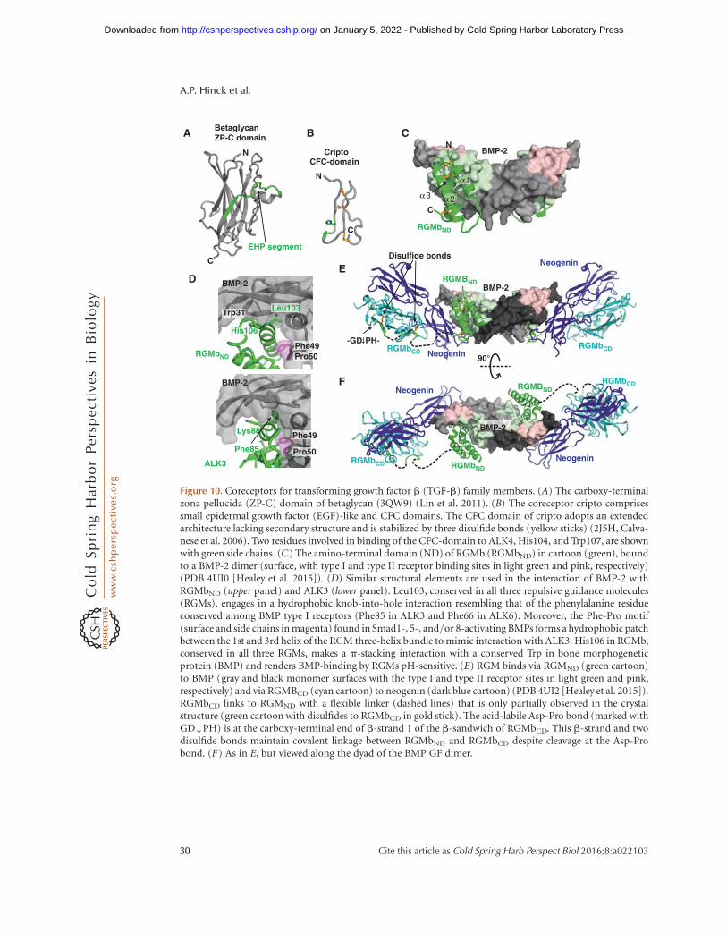
BetaglycanZP-C domain
CriptoCFC-domain
EHP segment
RGMbND
BMP-2N
C
N
C
N
Cα2α3
ALK3
BMP-2
BMP-2
Trp31
Phe49Pro50
Phe85
Lys88
Leu103
His106
BMP-2
Neogenin
RGMbND
RGMbNDRGMbCD
RGMbCD
RGMbCD
RGMbCD
RGMBND
RGMBND
Neogenin
BMP-2
Neogenin
α1
A B C
DE
F
90°
-GD�PH-
Disulfide bonds
Phe49
Pro50
Neogenin
Figure 10. Coreceptors for transforming growth factor b (TGF-b) family members. (A) The carboxy-terminalzona pellucida (ZP-C) domain of betaglycan (3QW9) (Lin et al. 2011). (B) The coreceptor cripto comprisessmall epidermal growth factor (EGF)-like and CFC domains. The CFC domain of cripto adopts an extendedarchitecture lacking secondary structure and is stabilized by three disulfide bonds (yellow sticks) (2J5H, Calva-nese et al. 2006). Two residues involved in binding of the CFC-domain to ALK4, His104, and Trp107, are shownwith green side chains. (C) The amino-terminal domain (ND) of RGMb (RGMbND) in cartoon (green), boundto a BMP-2 dimer (surface, with type I and type II receptor binding sites in light green and pink, respectively)(PDB 4UI0 [Healey et al. 2015]). (D) Similar structural elements are used in the interaction of BMP-2 withRGMbND (upper panel) and ALK3 (lower panel). Leu103, conserved in all three repulsive guidance molecules(RGMs), engages in a hydrophobic knob-into-hole interaction resembling that of the phenylalanine residueconserved among BMP type I receptors (Phe85 in ALK3 and Phe66 in ALK6). Moreover, the Phe-Pro motif(surface and side chains in magenta) found in Smad1-, 5-, and/or 8-activating BMPs forms a hydrophobic patchbetween the 1st and 3rd helix of the RGM three-helix bundle to mimic interaction with ALK3. His106 in RGMb,conserved in all three RGMs, makes a p-stacking interaction with a conserved Trp in bone morphogeneticprotein (BMP) and renders BMP-binding by RGMs pH-sensitive. (E) RGM binds via RGMND (green cartoon)to BMP (gray and black monomer surfaces with the type I and type II receptor sites in light green and pink,respectively) and via RGMBCD (cyan cartoon) to neogenin (dark blue cartoon) (PDB 4UI2 [Healey et al. 2015]).RGMbCD links to RGMND with a flexible linker (dashed lines) that is only partially observed in the crystalstructure (green cartoon with disulfides to RGMbCD in gold stick). The acid-labile Asp-Pro bond (marked withGD�PH) is at the carboxy-terminal end of b-strand 1 of the b-sandwich of RGMbCD. This b-strand and twodisulfide bonds maintain covalent linkage between RGMbND and RGMbCD despite cleavage at the Asp-Probond. (F) As in E, but viewed along the dyad of the BMP GF dimer.
A.P. Hinck et al.
30 Cite this article as Cold Spring Harb Perspect Biol 2016;8:a022103
on January 5, 2022 - Published by Cold Spring Harbor Laboratory Press http://cshperspectives.cshlp.org/Downloaded from

et al. 2004; Rajagopalan et al. 2004), and bindBMPs to modulate BMP signaling. The rela-tionship between these two functions is poorlyunderstood, but both neogenin and BMP bind-ing are required for the proper function of HJVin regulating systemic iron stores (Kuns-Hashi-moto et al. 2008; Zhang et al. 2009).
RGMs contain an amino-terminal domain(ND) with three disulfide-linked a-helices thatbinds BMPs, a flexible linker, a carboxy-termi-nal domain (CD) with two tightly packed b-sheets that contains an acid-labile Asp-Pro pep-tide bond and binds to neogenin, a PC cleavagesite, and a carboxy-terminal GPI membrane an-chor (Bell et al. 2013; Healey et al. 2015). Solubleforms of RGMa and RGMc result from process-ing by the PCs furin and/or SKI-1 (subtilisinkexin isozyme-1) (Tassew et al. 2012).
How do RGMs stimulate BMP signaling?Structural and microscopy studies suggest thatendocytosis of BMPs by RGMs and neogeninpromotes type I and type II receptor signalingin an intracellular compartment (Healey et al.2015). The first 120 amino-terminal residues ofRGMs, RGMND, fold into an antiparallel three-helical bundle that binds BMPs at the receptortype I site (wrist epitope) (Fig. 10C). When com-plexes of RGMs bound to BMP-2 are comparedwith ALK3 bound to BMP-2, the third helix ofRGMND mimics the interaction of the short a-helix of ALK3 with BMP-2 (Fig. 10D). One keyelement for the RGM–BMP interaction is a his-tidine residue conserved in all RGM proteins,His106 in RGMb, which forms a p-stacking in-teraction with the second Trp of the W-X-X-Wmotif in BMPs (Fig. 10D). Binding of RGMsto BMPs is inhibited at pH ,6.5, presumablybecause of histidine protonation. This featurepoints toward a mechanism by which RGMsmight stimulate BMP signaling. Because RGMsblock BMP binding to type I but not type IIreceptors, it is proposed that RGM–BMP–BMPRII complexes form at the cell surface,and that these are then endocytosed in a clath-rin-dependent manner. As type I receptors,including ALK3, are constitutively endocytosed,endosomes might form that contain RGM–BMP–type RII complexes as well as free ALK3.Because of endosomal acidification, the RGM
coreceptor would be released from its pH-de-pendent complex with BMP and the type II re-ceptor, enabling the type I receptor to replaceRGM in the complex, as type I receptor interac-tion with BMPs is not pH-sensitive (Heineckeet al. 2009; Healey et al. 2015). Signaling wouldthus occur in the cytoplasmicenvironment as forreceptors on the cell surface, but on the cytoplas-mic face of a distinct membrane compartmentwhere other colocalizing molecules might differ(Healeyet al. 2015). This might alter the shape ofsignaling as shown for some G protein-coupledreceptors, in which a second wave of signalingoccurs on endosomes that moves the site of sec-ond messenger production relative to effectors(Tsvetanova et al. 2015).
And how does neogenin influence the BMP-RGM interaction? The ternary complex struc-ture of BMP-2:RGMb:neogenin not only showsthat RGMs can simultaneously bind BMPs andneogenin (Fig. 10E,F), but also reveals that bind-ing of type II receptors to this complex is againnot blocked. Thus, formation of quaternarycomplexes consisting of RGMs, neogenin,BMP, and type II receptors is possible (Healeyet al. 2015). Although RGMCD binding to neo-genin brings neogenin into close proximity toBMP bound to RGMND, no direct interactionsbetween BMP and neogenin are observed (Fig.10E,F) (Healey et al. 2015). The modular archi-tecture of RGMs with their two domains inde-pendently binding two different partners, BMPsand neogenin, of which BMP is a dimer andneogenin is dimerized on RGM binding (Bellet al. 2013), suggest that RGMs could bridgeindividual complexes and facilitate formationof supramolecular clusters. Indeed, high-resolu-tion microscopy shows that BMP-2 mediatesclustering of RGM–neogenin complexes intolarge assemblies at the cell surface (Healey et al.2015). The physiological significance of supra-molecular cluster formation is unknown, al-though this may enable BMPs to modulate neo-genin signaling, and neogenin to modulate BMPsignaling, adding more complexity to BMP re-ceptor activation (Mueller 2015).
In summary, TGF-b and BMP coreceptorsare functionally and structurally diverse, andthe presence or absence of a coreceptor can
Structural Biology and Evolution of the TGF-b Family
Cite this article as Cold Spring Harb Perspect Biol 2016;8:a022103 31
on January 5, 2022 - Published by Cold Spring Harbor Laboratory Press http://cshperspectives.cshlp.org/Downloaded from

have large qualitative (changing gene expres-sion) and quantitative effects (sensitizing cellsfor a GF) on downstream signaling.
REGULATION OF TGF-b/BMP SIGNALINGBY MODULATOR PROTEINS—BMPs ASSIGNALING HUBS
Structural Diversity of TGF-b/BMPAntagonists
A hallmark of the TGF-b/BMP family is theplethora of extracellular modulator proteinsthat bind GFs and regulate signaling. Morethan 25 modulator proteins are known.Attempts have been made to classify TGF-b/BMP modulator proteins structurally. A com-mon hallmark is the large number of cysteineresidues. Although the diversity of these mod-ulators is stunning, they can be grouped, basedon structural features and sequence homology,into six distinct families: noggin, members ofthe Dan family (Fig. 11), the follistatin family,the CCN family, the chordin family, and twistedgastrulation (Tsg). The Dan and CCN familiesas well as noggin have CKGF domains. Notably,the diversity of BMP and activin antagonistscontrasts with the paucity or absence of modu-lators of TGF-b itself, perhaps correlating withthe distinct integrin-dependent mechanism ofTGF-b activation.
Noggin—The BMP Antagonist Paradigm
Noggin, a component of the Spemann organiz-er (Smith and Harland 1992), antagonizes theventralizing activity of BMPs in dorsoventralpatterning and regulates skeletogenesis (Zim-merman et al. 1996; Brunet et al. 1998). Nogginbinds to the same sites on BMP-7 as the BMPtype I and type II receptors, and thereby im-pedes BMP signaling (Fig. 12A–D) (Groppeet al. 2002). Noggin contains a CKGF domainlike TGF-b GF domains, but has a 10- ratherthan 8-membered CK ring (Fig. 12A). Further-more, its monomers dimerize through a Cysresidue in a position (cysteine 11 in Fig.11G,H) that differs from that in TGF-b familyproteins, and through an additional a-helicaldomain (Fig. 12A). The helices lie on the convex
side of noggin, opposite the concave BMP bind-ing site, and support noggin’s C-clamp-like cur-vature (Fig. 12A,B). Thus, whereas the TGF-bCKGF dimer is linked by monomers that over-lap in the palm and heel (GF homodimer, Box1), and dimerize through cysteine 6, the noggindimer links the two CKGF domain monomersmore distally toward their carboxy-terminalend at cysteine 11 (Fig. 11F,G), leading to a lon-ger and more curved dimer that straddles andclamps the BMP dimer (Fig. 12A,B).
Distinct structural elements in noggin blockthe BMP type I (wrist) and type II (knuckle)receptor epitopes (Fig. 12A–D). A 25-residuesegment within the amino-terminal region ofnoggin is spatially separated from the CKGFand a-helical domains in a complex withBMP, fastens to more than one BMP monomerin a U-shape, and is therefore termed the “clip”(Fig. 12A) (Groppe et al. 2002). In this segment,residues Pro35 to Ser38 snugly fold into thecenter of the wrist epitope, and block bindingto type I receptors. The clip then turns over thefingers of the BMP and runs over the peripheryof the knuckle epitope. In contrast, the nogginfingers at the end of its C-clamp extend over thefingertips of the BMP and shield a large part ofthe knuckle epitope, thereby blocking accessand binding of type II receptors. Hydrophobicinteractions dominate both the fingertip andclip interfaces, similarly to type I and type IIreceptor interfaces with BMPs. Moreover,Pro35 in the noggin clip, which is highly con-served among different species, extends into ahydrophobic pocket in the wrist epitope ofBMP-7, mimicking the knob-into-hole interac-tion found in various BMP-type I receptor in-teractions (Fig. 12A–D) (Hatta et al. 2000;Kirsch et al. 2000; Groppe et al. 2002; Kotzschet al. 2009). The importance of Pro35 for theBMP-neutralizing activity of noggin is high-lighted by its mutation in skeletal malformationdiseases (Gong et al. 1999; Mangino et al. 2002).
The mechanism of noggin binding to BMPspreserves much of the chemistry of receptorbinding to BMPs, including shared hydropho-bic interfaces and the knob-into-hole packingseen with most type I receptors; however, detailsof the distribution and arrangement of the
A.P. Hinck et al.
32 Cite this article as Cold Spring Harb Perspect Biol 2016;8:a022103
on January 5, 2022 - Published by Cold Spring Harbor Laboratory Press http://cshperspectives.cshlp.org/Downloaded from

98765 10
1 432
1
1
1
1
1
1
1
2
2
2
2
3
3
3
3
3
3
3
3
4
4
4
4
4
4
4
5
5
5
6-5’
6–6′
7
7
7
7
7
7
7
8
8
8
8
910
910
910
910
9
10
910
910
11
11
11–11′
11–11′
-6′
Loop 1 Loop 2
Loop 3Loop 2
1TFG DNCCLRP L Y I DFKRDLGWKW - I HEPKG - - YNANFCAG - A - C - PY LWSSDTQHSRV L S L
YNT I NPEASASPCCVSQDL EP L T I L YY I G - - - - KT P - - K I - EQL SNM I VKSCKCS - - -
4JPH DWCKTQP LRQT VS - - - - E - EGCRS - RT - - I LNRFCYG - Q - C - NS - F Y I - PRHVKKE - E
D S F- - -- Q- SCAFCKPQRV T SV I VE L ECPGLDPP - - - FR I - K - K I - QKVKHCRCMS - V
4X1J AWCEAKN I TQ I VG - H - - S - G - CEA - KS - - I QNRACLG - Q - C - F S - YSV - PN - T - - F PQ
- - S T ES L V - HCDSCMPAQSMWE I V T L ECPGHEE - - VPRVD - K - L V - EK I LHCSCQACG
2KD3 YSCRE LHY TRF L T - D - - G - P - C - R - SAKPV T E L VCSG - Q - C - GP - ARL - L - - - PNA I G
RVKWWRPNGPDFRC I PDRYRAQRVQL LCGG - - - - AAP - RS - R - KV - RL VASCKCKR - -
5BQC RRCMRHHYVDS I S - HP L Y - K - CSS - KM - - V L LARCEG - H - CSQA - SRS - EP - L S F S T V
LKQP FR - S - SCHCCRPQT SKLKALRLRCS - - - - GGMR - L T - A - T Y - RY I L SCHCEECN
1M4U T FCPV L YAWNDLG - S - - - - - - RFWPRY - - VKVGSCF SKRSC - SV - P - - - - - - - - - - - -
- - - - - - - - - EGMVCKPSKSVHL T V LRWRCQRRGGQRCGW I P I - QY - P I I SECKCSC - -
1QFW PRCRP I NAT LAVE - K - - EGC - PVC - I T - - VNT T I CAG - Y - C - P T - MTRV LQ - G - - V - -
- - - - L PAL - PQVVCNYRDVRF ES I RL PGCP - - - R - GVNPV - V - SY - AVAL SCQCALCR
5 3 2 1 2 1 1 1 1 2 3 2 3 - - - - - - 3 - 4 4 5 2 2 2 - - 2 2 2 1 1 1 1 2 - 2 - 1 - 3 - - - - - - - - - - - - - - -
- - - - - - - - - - 4 3 2 1 1 1 1 2 2 2 3 2 2 2 1 3 4 6 - - - - - - - - - 6 3 3 - 3 - 2 3 - 2 1 1 1 1 1 1 1 1 3 3 -
13
71335337
153
7
TGF-β2
PRDCNbl1SclerostinNorrinNoggin
HCGβAlignment RMSD
1TFG
4JPH4X1J2KD35BQC1M4U
1QFW
65
115739685
188
49TGF-β2
PRDCNbl1SclerostinNorrinNoggin
HCGβAlignment RMSD
PRDC
Nbl1
Sclerostin
Norrin
Noggin
TGF-β2
HCGβ
CK CKCK
A
H
G
F
E
D
C
BGlycan
Frizzled4CRD
Clip
Loop 1
Loop 2Loop 3
Loop 1
Loop 2Loop 3
Loop 1
Loop 2Loop 3
Loop 1
Loop 2 Loop 3
Loop 1
Loop
2
Loop 3
Loop 1
Loop 2Loop 3
Loop 1Loop 2
Loop 3
Figure 11. Structural alignment of Dan family monomers to cystine knot (CK) growth factor (CKGF) super-family representatives with similar monomer–monomer orientations. (A–G) CKGF monomers in cartoon witha-helices in cyan and b-strands in magenta. Disulfides are shown in stick and are color-coded with theircysteines numbered as in panel H. Cysteines in intermolecular disulfides are indicated by showing the partnerCys in the other monomer with its number preceded by a dash and followed by a hyphen. All monomers butsclerostin are from dimeric structures. A frizzled domain and glycan cocrystallized with norrin are shown incartoon and stick, respectively. Monomers are in identical orientations, and aligned in two columns. (H )Structure-based sequence alignment. Cysteines disulfide-bonded to one another are in identical colors, exceptthose that participate in interchain disulfides in some families and intrachain disulfides in others (positions 5, 6,and 11) are shown in red. Disulfides unique to particular families are shown in thinner silver stick and coloredsilver in panel H. Other residues are colored according to ClustalX. CK disulfides are labeled between the tworows of the alignment and link Cys residues distant in sequence; the three long loops between CK disulfides arealso marked. (Legend continues on following page.)
Structural Biology and Evolution of the TGF-b Family
Cite this article as Cold Spring Harb Perspect Biol 2016;8:a022103 33
on January 5, 2022 - Published by Cold Spring Harbor Laboratory Press http://cshperspectives.cshlp.org/Downloaded from

structural elements differ. Remarkably, the nog-gin–BMP complex partially shares its buildingprinciple with the TGF-b1 proprotein complex;the folded noggin CKGF domain, like the pro-domain arm domain, blocks the type II recep-tor site, and the single element clip in noggin,like the prodomain straitjacket, blocks type Ireceptor access to the GF (Fig. 12A; Box 1). Inboth noggin and the TGF-b1 prodomain, a cen-tral domain together with intermolecular disul-fide bonds forms a covalent homodimer, whichexplains the extremely tight binding to theGF. Moreover, both interact with the ECM; theTGF-b1 prodomain is covalently linked toLTBP, and noggin has heparin binding sites tointeract with the ECM (Groppe et al. 2002;Paine-Saunders et al. 2002) that can locally re-tain a bound GF to limit its radius of action.
Noggin binds in vitro to most BMPs withno apparent dissociation, which suggests thatthe concentration of active ligand would beeffectively decreased for all BMPs that it tar-gets (Zimmerman et al. 1996; Seemann et al.2009; Song et al. 2010; Schwaerzer et al. 2012).However, in cell-based assays some BMPs, forexample, BMP-6, have been described as nog-gin-resistant, although their affinity to nogginis in the same range as for nonresistant ligands(Song et al. 2010). Furthermore, mutations thatrender BMPs noggin-resistant only marginallydecrease their affinity to noggin (Song et al.2010; Schwaerzer et al. 2012), possibly indicat-ing that the inhibition mechanism is more com-plex in vivo.
The Dan Family—A Heterogeneous Group ofAntagonists for BMPs and/or Wnt Factors
With seven known members in mammals, Danmodulators are the largest family of TGF-b fam-
ily antagonists (Nolan and Thompson 2014).They include neuroblastoma suppressor of tu-morigenicity 1 (Nbl1, Dan, DAND1), gremlin/Drm (DAND2), protein related to Dan and cer-berus (PRDC, DAND3, gremlin2), cerberus(DAND4), and coco/dante (DAND5) (Fig.12E). These five members are the smallest pro-teins known to modulate BMP signaling. Scle-rostin and USAG1 (also known as wise, ectodin,and SOSD) also belong to the Dan modulatorfamily (BMP antagonists, Box 1, Figs. 11 and12E), but they antagonize Wnt and not BMPsignaling. Dan family members share a centralCKGF domain of 90 residues that may be sub-stantially elaborated with amino- and carboxy-terminal extensions that add another �100 res-idues (Fig. 11A–C, 12E, lower). Extensions in-cludea-helices in PRDC (Nolan et al. 2013) andunstructured regions in sclerostin (Veverka et al.2009; Weidauer et al. 2009). These might be-come structured when bound to GFs or ECMcomponents and might resemble in this respectthe long amino-terminal extensions in manyTGF-b family prodomains (Fig. 1).
All Dan and sclerostin family proteins con-tain an 8-membered CK ring with the samesequence motif as in TGF-b family CKGF do-mains (C-X-G-X-C and C-X-C) (Figs. 11 and12E). Besides the six CK cysteines, Dan proteinscontain two additional cysteines (designated C2and C8 in Fig. 11) that disulfide-link the two b-ribbon fingers at the fingertips (Figs. 11 and12F,G). The same eight Cys residues are sharedwith carboxy-terminal CK (CTCK) domains,which are closely related to CKGF domains.CTCK domains have been structurally charac-terized in von Willebrand factor (Zhou andSpringer 2014) and norrin (Ashe and Levine1999; Chang et al. 2015). Three long loops inCKGF and CTCK domains link the CK residues
Figure 11. (Continued) For alignments to CKGF families with monomers dimerized in different orientations(neurotrophin in the nerve growth factor [NGF] family, and platelet-derived growth factor [PDGF] in the PDGFfamily) (see Zhou and Springer 2014). CKGF and CTCK monomers (PDB codes are shown in panel H ) werealigned to one another structurally using DeepAlign (Wang et al. 2013). Structurally equivalent residues areshown aligned by sequence as described in Figure 5 legend. Ca atom root mean square deviation (RMSD, A) ofmonomers in each position relative to the average at each position are shown below the alignment. Gaps wereclosed to minimize the length of the alignment; corresponding positions lack an RMSD value.
A.P. Hinck et al.
34 Cite this article as Cold Spring Harb Perspect Biol 2016;8:a022103
on January 5, 2022 - Published by Cold Spring Harbor Laboratory Press http://cshperspectives.cshlp.org/Downloaded from
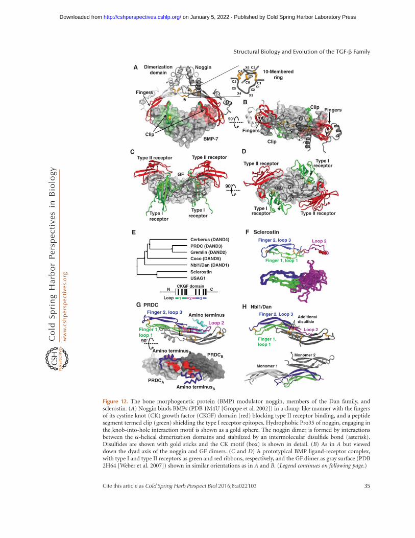
Type II receptor Type II receptorType II receptor
Type II receptorType Ireceptor
Type Ireceptor
Type I receptor
Type I receptor
GF
GF
Dimerizationdomain
Fingers
Clip
Clip
Clip
Fingers
Fingers
10-Memberedring
C1X1
X2
X3X4X5
C2
C3X6C4
C5
C6
Noggin
BMP-7
Cerberus (DAND4)
PRDC (DAND3)Gremlin (DAND2)Coco (DAND5)Nbl1/Dan (DAND1)
SclerostinUSAG1
CKGF domainN C
Finger 2, loop 3
Finger 1, loop 1
Loop 2
Sclerostin
Finger 2, loop 3
Finger 1,loop 1
Loop 2
Amino terminus
Amino terminusB
Amino terminusA
PRDCA
PRDCB
Finger 2, Loop 3
Finger 1,loop 1
Loop 2
PRDC Nbl1/Dan
Additionaldisulfide
90°
90°
90°
A
B
C D
E F
G H
Monomer 1
Monomer 2
*
Loop 1 32
Figure 12. The bone morphogenetic protein (BMP) modulator noggin, members of the Dan family, andsclerostin. (A) Noggin binds BMPs (PDB 1M4U [Groppe et al. 2002]) in a clamp-like manner with the fingersof its cystine knot (CK) growth factor (CKGF) domain (red) blocking type II receptor binding, and a peptidesegment termed clip (green) shielding the type I receptor epitopes. Hydrophobic Pro35 of noggin, engaging inthe knob-into-hole interaction motif is shown as a gold sphere. The noggin dimer is formed by interactionsbetween the a-helical dimerization domains and stabilized by an intermolecular disulfide bond (asterisk).Disulfides are shown with gold sticks and the CK motif (box) is shown in detail. (B) As in A but vieweddown the dyad axis of the noggin and GF dimers. (C and D) A prototypical BMP ligand-receptor complex,with type I and type II receptors as green and red ribbons, respectively, and the GF dimer as gray surface (PDB2H64 [Weber et al. 2007]) shown in similar orientations as in A and B. (Legend continues on following page.)
Structural Biology and Evolution of the TGF-b Family
Cite this article as Cold Spring Harb Perspect Biol 2016;8:a022103 35
on January 5, 2022 - Published by Cold Spring Harbor Laboratory Press http://cshperspectives.cshlp.org/Downloaded from
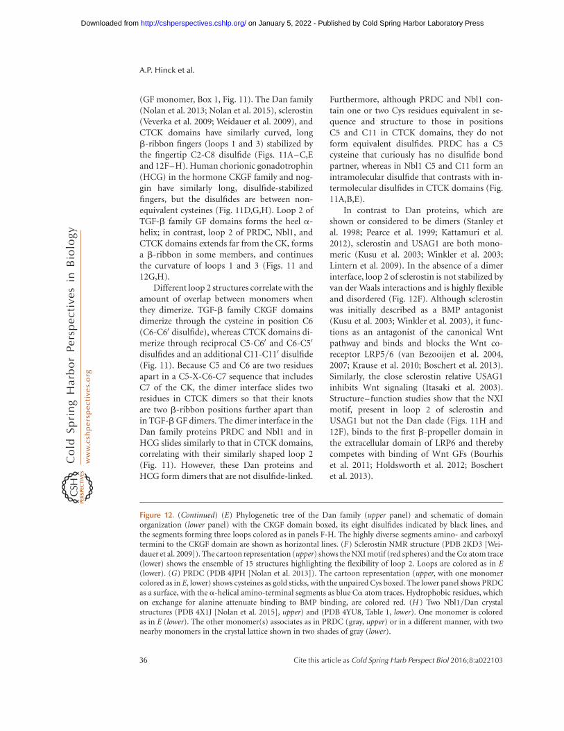
(GF monomer, Box 1, Fig. 11). The Dan family(Nolan et al. 2013; Nolan et al. 2015), sclerostin(Veverka et al. 2009; Weidauer et al. 2009), andCTCK domains have similarly curved, longb-ribbon fingers (loops 1 and 3) stabilized bythe fingertip C2-C8 disulfide (Figs. 11A–C,Eand 12F–H). Human chorionic gonadotrophin(HCG) in the hormone CKGF family and nog-gin have similarly long, disulfide-stabilizedfingers, but the disulfides are between non-equivalent cysteines (Fig. 11D,G,H). Loop 2 ofTGF-b family GF domains forms the heel a-helix; in contrast, loop 2 of PRDC, Nbl1, andCTCK domains extends far from the CK, formsa b-ribbon in some members, and continuesthe curvature of loops 1 and 3 (Figs. 11 and12G,H).
Different loop 2 structures correlate with theamount of overlap between monomers whenthey dimerize. TGF-b family CKGF domainsdimerize through the cysteine in position C6(C6-C60 disulfide), whereas CTCK domains di-merize through reciprocal C5-C60 and C6-C50
disulfides and an additional C11-C110 disulfide(Fig. 11). Because C5 and C6 are two residuesapart in a C5-X-C6-C7 sequence that includesC7 of the CK, the dimer interface slides tworesidues in CTCK dimers so that their knotsare two b-ribbon positions further apart thanin TGF-b GF dimers. The dimer interface in theDan family proteins PRDC and Nbl1 and inHCG slides similarly to that in CTCK domains,correlating with their similarly shaped loop 2(Fig. 11). However, these Dan proteins andHCG form dimers that are not disulfide-linked.
Furthermore, although PRDC and Nbl1 con-tain one or two Cys residues equivalent in se-quence and structure to those in positionsC5 and C11 in CTCK domains, they do notform equivalent disulfides. PRDC has a C5cysteine that curiously has no disulfide bondpartner, whereas in Nbl1 C5 and C11 form anintramolecular disulfide that contrasts with in-termolecular disulfides in CTCK domains (Fig.11A,B,E).
In contrast to Dan proteins, which areshown or considered to be dimers (Stanley etal. 1998; Pearce et al. 1999; Kattamuri et al.2012), sclerostin and USAG1 are both mono-meric (Kusu et al. 2003; Winkler et al. 2003;Lintern et al. 2009). In the absence of a dimerinterface, loop 2 of sclerostin is not stabilized byvan der Waals interactions and is highly flexibleand disordered (Fig. 12F). Although sclerostinwas initially described as a BMP antagonist(Kusu et al. 2003; Winkler et al. 2003), it func-tions as an antagonist of the canonical Wntpathway and binds and blocks the Wnt co-receptor LRP5/6 (van Bezooijen et al. 2004,2007; Krause et al. 2010; Boschert et al. 2013).Similarly, the close sclerostin relative USAG1inhibits Wnt signaling (Itasaki et al. 2003).Structure–function studies show that the NXImotif, present in loop 2 of sclerostin andUSAG1 but not the Dan clade (Figs. 11H and12F), binds to the first b-propeller domain inthe extracellular domain of LRP6 and therebycompetes with binding of Wnt GFs (Bourhiset al. 2011; Holdsworth et al. 2012; Boschertet al. 2013).
Figure 12. (Continued) (E) Phylogenetic tree of the Dan family (upper panel) and schematic of domainorganization (lower panel) with the CKGF domain boxed, its eight disulfides indicated by black lines, andthe segments forming three loops colored as in panels F-H. The highly diverse segments amino- and carboxyltermini to the CKGF domain are shown as horizontal lines. (F) Sclerostin NMR structure (PDB 2KD3 [Wei-dauer et al. 2009]). The cartoon representation (upper) shows the NXI motif (red spheres) and the Ca atom trace(lower) shows the ensemble of 15 structures highlighting the flexibility of loop 2. Loops are colored as in E(lower). (G) PRDC (PDB 4JPH [Nolan et al. 2013]). The cartoon representation (upper, with one monomercolored as in E, lower) shows cysteines as gold sticks, with the unpaired Cys boxed. The lower panel shows PRDCas a surface, with the a-helical amino-terminal segments as blue Ca atom traces. Hydrophobic residues, whichon exchange for alanine attenuate binding to BMP binding, are colored red. (H ) Two Nbl1/Dan crystalstructures (PDB 4X1J [Nolan et al. 2015], upper) and (PDB 4YU8, Table 1, lower). One monomer is coloredas in E (lower). The other monomer(s) associates as in PRDC (gray, upper) or in a different manner, with twonearby monomers in the crystal lattice shown in two shades of gray (lower).
A.P. Hinck et al.
36 Cite this article as Cold Spring Harb Perspect Biol 2016;8:a022103
on January 5, 2022 - Published by Cold Spring Harbor Laboratory Press http://cshperspectives.cshlp.org/Downloaded from
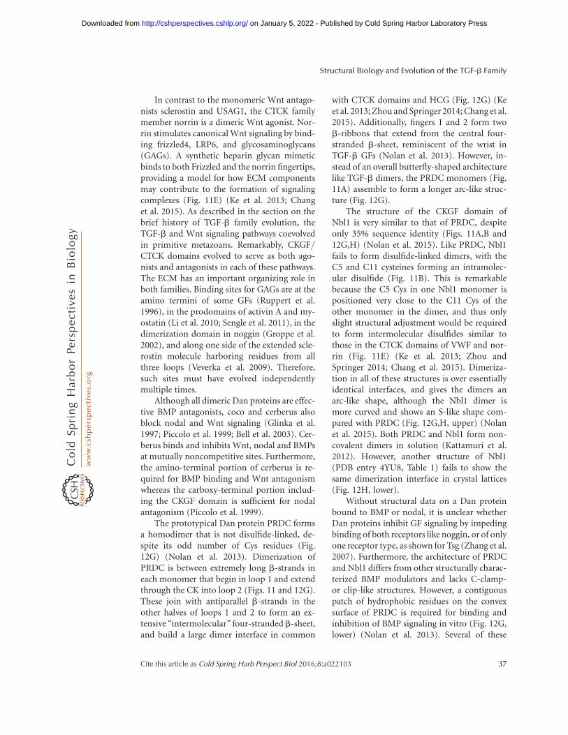
In contrast to the monomeric Wnt antago-nists sclerostin and USAG1, the CTCK familymember norrin is a dimeric Wnt agonist. Nor-rin stimulates canonical Wnt signaling by bind-ing frizzled4, LRP6, and glycosaminoglycans(GAGs). A synthetic heparin glycan mimeticbinds to both Frizzled and the norrin fingertips,providing a model for how ECM componentsmay contribute to the formation of signalingcomplexes (Fig. 11E) (Ke et al. 2013; Changet al. 2015). As described in the section on thebrief history of TGF-b family evolution, theTGF-b and Wnt signaling pathways coevolvedin primitive metazoans. Remarkably, CKGF/CTCK domains evolved to serve as both ago-nists and antagonists in each of these pathways.The ECM has an important organizing role inboth families. Binding sites for GAGs are at theamino termini of some GFs (Ruppert et al.1996), in the prodomains of activin A and my-ostatin (Li et al. 2010; Sengle et al. 2011), in thedimerization domain in noggin (Groppe et al.2002), and along one side of the extended scle-rostin molecule harboring residues from allthree loops (Veverka et al. 2009). Therefore,such sites must have evolved independentlymultiple times.
Although all dimeric Dan proteins are effec-tive BMP antagonists, coco and cerberus alsoblock nodal and Wnt signaling (Glinka et al.1997; Piccolo et al. 1999; Bell et al. 2003). Cer-berus binds and inhibits Wnt, nodal and BMPsat mutually noncompetitive sites. Furthermore,the amino-terminal portion of cerberus is re-quired for BMP binding and Wnt antagonismwhereas the carboxy-terminal portion includ-ing the CKGF domain is sufficient for nodalantagonism (Piccolo et al. 1999).
The prototypical Dan protein PRDC formsa homodimer that is not disulfide-linked, de-spite its odd number of Cys residues (Fig.12G) (Nolan et al. 2013). Dimerization ofPRDC is between extremely long b-strands ineach monomer that begin in loop 1 and extendthrough the CK into loop 2 (Figs. 11 and 12G).These join with antiparallel b-strands in theother halves of loops 1 and 2 to form an ex-tensive “intermolecular” four-strandedb-sheet,and build a large dimer interface in common
with CTCK domains and HCG (Fig. 12G) (Keet al. 2013; Zhou and Springer 2014; Chang et al.2015). Additionally, fingers 1 and 2 form twob-ribbons that extend from the central four-stranded b-sheet, reminiscent of the wrist inTGF-b GFs (Nolan et al. 2013). However, in-stead of an overall butterfly-shaped architecturelike TGF-b dimers, the PRDC monomers (Fig.11A) assemble to form a longer arc-like struc-ture (Fig. 12G).
The structure of the CKGF domain ofNbl1 is very similar to that of PRDC, despiteonly 35% sequence identity (Figs. 11A,B and12G,H) (Nolan et al. 2015). Like PRDC, Nbl1fails to form disulfide-linked dimers, with theC5 and C11 cysteines forming an intramolec-ular disulfide (Fig. 11B). This is remarkablebecause the C5 Cys in one Nbl1 monomer ispositioned very close to the C11 Cys of theother monomer in the dimer, and thus onlyslight structural adjustment would be requiredto form intermolecular disulfides similar tothose in the CTCK domains of VWF and nor-rin (Fig. 11E) (Ke et al. 2013; Zhou andSpringer 2014; Chang et al. 2015). Dimeriza-tion in all of these structures is over essentiallyidentical interfaces, and gives the dimers anarc-like shape, although the Nbl1 dimer ismore curved and shows an S-like shape com-pared with PRDC (Fig. 12G,H, upper) (Nolanet al. 2015). Both PRDC and Nbl1 form non-covalent dimers in solution (Kattamuri et al.2012). However, another structure of Nbl1(PDB entry 4YU8, Table 1) fails to show thesame dimerization interface in crystal lattices(Fig. 12H, lower).
Without structural data on a Dan proteinbound to BMP or nodal, it is unclear whetherDan proteins inhibit GF signaling by impedingbinding of both receptors like noggin, or of onlyone receptor type, as shown for Tsg (Zhang et al.2007). Furthermore, the architecture of PRDCand Nbl1 differs from other structurally charac-terized BMP modulators and lacks C-clamp-or clip-like structures. However, a contiguouspatch of hydrophobic residues on the convexsurface of PRDC is required for binding andinhibition of BMP signaling in vitro (Fig. 12G,lower) (Nolan et al. 2013). Several of these
Structural Biology and Evolution of the TGF-b Family
Cite this article as Cold Spring Harb Perspect Biol 2016;8:a022103 37
on January 5, 2022 - Published by Cold Spring Harbor Laboratory Press http://cshperspectives.cshlp.org/Downloaded from
residues are conserved in Nbl1, which bindsBMP-2 and 7 and GDF-5 with different affini-ties compared with PRDC. The importance ofthese residues in encoding BMP selectivity wasshown by exchange of patch residues fromPRDC into Nbl1, which yielded an Nbl1 variantmimicking PRDC (Nolan et al. 2015). Interest-ingly, in PRDC the hydrophobic patch respon-sible for BMP binding seems to be covered by adynamic a-helix in the amino-terminal seg-ment (Fig. 12G, lower). A similar helix is notobserved in the Nbl1 structure (Fig. 12H) (No-lan et al. 2015).
Follistatin Antagonizes Activins as Wellas BMPs
Follistatin (Fst) was initially described as a pro-tein that antagonizes activin signaling. How-ever, follistatin also binds BMPs and GDFs, in-cluding BMP-2, 4, 6, and 7 as well as myostatin/GDF-11, albeit with lower affinities (Iemuraet al. 1998; Amthor et al. 2002, 2004; Glisteret al. 2004; Schneyer et al. 2008). Fst is a mod-ular protein comprising a unique 65-residueamino-terminal domain (Fs0) followed by threefollistatin domains (Fs1, Fs2, Fs3) of 70 to 85residues each (Fig. 13A,B). Follistatin domainsare in turn each composed of EGF-like andKazal subdomains (Hohenester et al. 1997;Schneyer et al. 2004).
Fs domains together with other domains arepresent in follistatin-like (FSTL) proteins. Al-though FSTL-1 and 3 are known to regulateactivin and BMP signaling, FSTL-4, FSTL-5,SMOC1, and SMOC2 are not yet characterizedfor effects on activin and BMP signaling. Fur-thermore, SMOC1 and SMOC2 contain addi-tional domains characteristic of the SPARCfamily (Schneyer et al. 2004; Bradshaw 2012).The follistatin family thus includes modulatorswith similar mechanisms of action (binding toGFs and blocking receptor binding) and similarspecificities for activins and BMPs, yet its mem-bers are built from several types of domainsthat are assembled into distinctive architectures.Three isoforms of follistatin, Fst288, Fst303, andFst315 have identical amino-terminal regions,but end at carboxy-terminal residues 288, 303,
or 315, respectively, thus creating further com-plexity. They bind activin with different af-finities, and longer isoforms partially lack theability to bind to heparin and proteoglycans(Kumar 2005).
Structures of follistatin and FSTL-3 boundto activin A or myostatin provide insightsinto inhibitory mechanisms and specificity(Thompson et al. 2005; Harrington et al.2006; Cash et al. 2009, 2012). Although follista-tin and FSTL-3 block type I and type II receptorbinding sites, they bind the GF domain vastlydifferently from noggin (Figs. 12A and 13A).Follistatin wraps around activin on the perim-eter of the dyad (two-fold rotation axis) of theGF dimer (Fig. 13B). In contrast, noggin bindson one side of the dyad axis. Viewed normal tothis axis (as in Fig. 12A), noggin resembles aC-shaped clamp with the ends of the clampgripping the GF fingers. In the case of follistatin,two modulator molecules embrace the GF suchthat almost its entire surface seems covered (Fig.13A,B). In contrast to noggin, follistatin is amonomer when not bound to the GF. However,on binding, two follistatin monomers (struc-tures of follistatin 288 are shown) tightly inter-act in a head-to-tail fashion and form a stable,noncovalent dimer (Thompson et al. 2005).The type I receptor epitope of activin is blockedby the unique amino-terminal domain (Fs0) offollistatin (Fig. 13A). Here, the key element isPhe52 located in the second a-helix of the Fs0domain, which makes a knob-into-hole inter-action with the wrist epitope of activin A simi-larly to type I receptors that bind BMPs (Hattaet al. 2000; Keller et al. 2004). Fs1, and primarilyFs2, shield the type II receptor epitope by main-ly using hydrophobic interactions as found incomplexes of activins and BMPs bound to theirtype II receptors (Fig. 13A,B). The final domain,Fs3, wraps around the fingertips of activin Aand engages in predominantly polar contactswith the amino-terminal Fs0 domain of theother follistatin monomer, thus stabilizing en-circlement of the ligand (Fig. 13A,B).
The selectivity of follistatin for activins andBMPs is explained by a central core of GF aminoacids in the binding sites for the Fs0 and Fs1/2domains that are conserved in activins and
A.P. Hinck et al.
38 Cite this article as Cold Spring Harb Perspect Biol 2016;8:a022103
on January 5, 2022 - Published by Cold Spring Harbor Laboratory Press http://cshperspectives.cshlp.org/Downloaded from
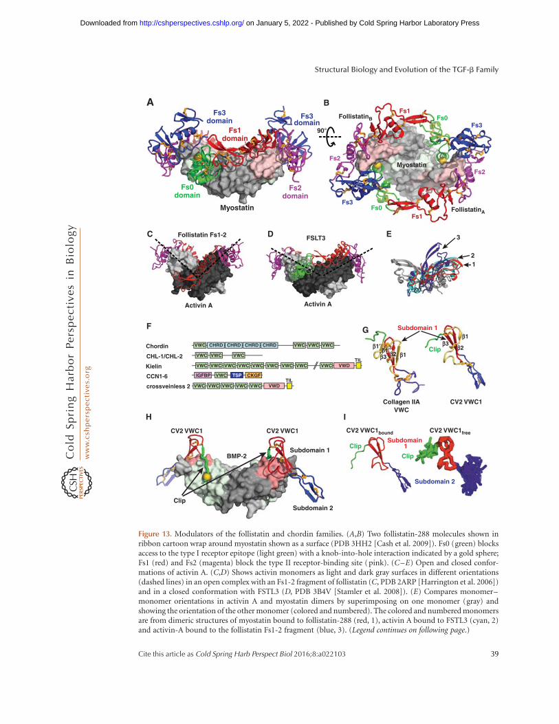
Fs0domain
Fs1domain
Fs2domain
Fs3domain
Fs3domain
Myostatin FollistatinA
FollistatinB
Activin A
Follistatin Fs1-2
Activin A
FSLT3
12
3
Clip
Subdomain 1
Subdomain 2
BMP-2
CV2 VWC1 CV2 VWC1 CV2 VWC1bound CV2 VWC1free
A B
C D E
F
Chordin
CHL-1/CHL-2
Kielin
CCN1-6
crossveinless 2
Subdomain 1
VWC CHRD
VWD
CHRD CHRD CHRD VWC VWC VWC
VWC VWC VWC
VWC VWC VWC VWC VWC VWC VWC VWC VWC
VWD
VWC
VWC VWC VWC VWC VWC
IGFBP TSP CKGF
TIL
TIL
Clip
Subdomain 2
CV2 VWC1Collagen IIAVWC
β1′
H I
90°
Myostatin
β3 β2 β1
β3β2
β1
Subdomain 1Clip
Clip
Fs3
Fs3
Fs0Fs1
Fs2
Fs0Fs1
Fs2
G
β1′′
Figure 13. Modulators of the follistatin and chordin families. (A,B) Two follistatin-288 molecules shown inribbon cartoon wrap around myostatin shown as a surface (PDB 3HH2 [Cash et al. 2009]). Fs0 (green) blocksaccess to the type I receptor epitope (light green) with a knob-into-hole interaction indicated by a gold sphere;Fs1 (red) and Fs2 (magenta) block the type II receptor-binding site (pink). (C–E) Open and closed confor-mations of activin A. (C,D) Shows activin monomers as light and dark gray surfaces in different orientations(dashed lines) in an open complex with an Fs1-2 fragment of follistatin (C, PDB 2ARP [Harrington et al. 2006])and in a closed conformation with FSTL3 (D, PDB 3B4V [Stamler et al. 2008]). (E) Compares monomer–monomer orientations in activin A and myostatin dimers by superimposing on one monomer (gray) andshowing the orientation of the other monomer (colored and numbered). The colored and numbered monomersare from dimeric structures of myostatin bound to follistatin-288 (red, 1), activin A bound to FSTL3 (cyan, 2)and activin-A bound to the follistatin Fs1-2 fragment (blue, 3). (Legend continues on following page.)
Structural Biology and Evolution of the TGF-b Family
Cite this article as Cold Spring Harb Perspect Biol 2016;8:a022103 39
on January 5, 2022 - Published by Cold Spring Harbor Laboratory Press http://cshperspectives.cshlp.org/Downloaded from
BMPs but not in TGF-bs (Thompson et al.2005). The contribution of individual follistatindomains to binding differs depending on theGF (Cash et al. 2009). For example, when boundto myostatin compared with activin A, theFs0 domain shifts more into the GF wrist epi-tope to better interact. To avoid a clash withFs0, Fs3 in the other monomer shifts awayfrom myostatin. Additionally, the ability ofactivin A to show both closed and open con-formations (see section on GF structures) is re-flected in differences in monomer–monomerorientation in complexes with different follista-tin fragments (Fig. 13C–E) (Thompson et al.2003, 2005; Greenwald et al. 2004; Harringtonet al. 2006).
The Fs0 domain in follistatin and FSTLs mayplay a role in discriminating between high-affin-ity ligands (i.e., activins, myostatin, and GDF-11), and low-affinity binders i.e., BMP-2, 4, 7,and others). The type I receptor ALK3 still in-teracts with BMP-2 when the latter is bound tofollistatin, suggesting that the Fs0 domain doesnot bind to or cannot block the wrist epitope inBMPs (Iemura et al. 1998). Lacking this interac-tion might also explain the lower affinities offollistatin binding to BMPs and indicate thatthe inhibition of BMPs by follistatin is function-ally different from that of activins (see also Stam-ler et al. 2008). A role for the Fs3 domain in BMPbinding is suggested by the finding that FSTL-3,which lacks Fs3 and thus cannot fully encircleactivin A and myostatin (Fig. 13D) (Stamleret al. 2008; Cash et al. 2012), binds activin Aand myostatin with high affinity, but cannotbind BMPs (Sidis et al. 2006).
Chordin Family Members Exert a DualModulator Function with Both Pro- andAnti-BMP Activities
Members of the chordin family are modularproteins with VWC domains (Fig. 13F). VWCdomains (Bork 1993), also confusingly knownas cysteine-rich domains in chordin (Francoiset al. 1994; Sasai et al. 1994), are 60 to 75 residueslong and are found, often in multiple tandemrepeats, in .70 human proteins, including theTGF-b family modulator proteins chordin, ven-troptin, chordin-like 2, kielin, and crossveinless2 (CV2, also known as BMP endothelial regu-lator or BMPER) (Fig. 13F) (Garcia Abreu et al.2002). Chordin is a large multidomain pro-tein that contains chordin (CHRD) domainsin addition to VWC domains (Fig. 13F). Expres-sion of chordin fragments in embryos showedthat BMP’s dorsalizing activity and BMP bind-ing reside in its VWC domains (Larrain et al.2000). Structures of VWC domains in collagenIIA and CV2 show two subdomains (Fig.13G,H) (O’Leary et al. 2004; Zhang et al.2008). Subdomain 2 is highly dynamic (Fig.13I, right) (O’Leary et al. 2004), which may berelated to the finding that six tandem VWC do-mains in von Willebrand factor give rise to aregion with an extended, highly flexible struc-ture (Springer 2014).
An amino-terminal fragment of CV2 boundto BMP-2 exemplifies how a chordin familymember binds and inhibits BMPs (Fig.13G,H) (Zhang et al. 2008). The CV2 fragmentcomprises an amino-terminal segment (termedclip, as in the structurally distinct noggin) and
Figure 13. (Continued) (F) Chordin family members are mosaics of tandem domains. Although von Willebrandfactor C (VWC) domain(s) mediate bone morphogenetic protein (BMP) binding, other domains presentinclude chordin (CHRD), von Willebrand type D (VWD), trypsin inhibitor-like (TIL), insulin-like growthfactor binding protein (IGFBP), thrombospondin (TSP) and cystine knot growth factor (CKGF). (G) VWCdomain of collagen IIA (left) and VWC domain 1 of CV2 (right). Amino-terminal peptide segments and VWCsubdomains 1 and 2 are green, red and blue, respectively. A structurally variable segment of VWC subdomain 1 isshown in yellow. (H ) The VWC1 domain of CV2 shown in ribbon with knob-into-hole residue shown as goldsphere bound to BMP-2 shown as surface with type I and type II receptor sites colored green and pink,respectively (PDB 3BK3 [Zhang et al. 2008]). (I) Similarity in architecture of VWC1 domain of CV2 boundto BMP-2 in crystals (left) and free in solution (right panel, NMR ensemble of 10 structures, PDB 2MBK [Fiebiget al. 2013]).
A.P. Hinck et al.
40 Cite this article as Cold Spring Harb Perspect Biol 2016;8:a022103
on January 5, 2022 - Published by Cold Spring Harbor Laboratory Press http://cshperspectives.cshlp.org/Downloaded from
VWC domain 1. In the 2:2 complex, the clipfolds into the type I receptor epitope, whereasthe amino-terminal subdomain of VWC1 bindsthe type II receptor site, showing how CV2 in-hibits BMP binding to both types of receptors(Zhang et al. 2007, 2008). The interaction moderesembles attachment of a paperclip (CV2) to asheet of paper (BMP) with the amino-terminalpeptide and VWC subdomain 1 folding overopposite sides of the GF finger (Fig. 13H), andshares some similarities with the noggin–BMPinteraction (Fig. 12A). In both cases, a linearpeptide in an amino-terminal segment, clip,efficiently interferes with type I receptor bind-ing, and a more globular domain occupies thetype II receptor binding site. If the clip werehighly flexible in the unbound state, it wouldbe puzzling how this short motif could contrib-ute significantly to BMP binding. However,NMR analysis of the VWC1 domain of CV2 inits unbound state reveals that the clip segmentis pre-oriented to form a hook that facilitatescooperative binding to BMP-2 (Fig. 13I) (Fiebiget al. 2013).
In contrast to Dan family members, mostVWC domains and most mosaic proteins withVWC domains do not act as BMP antagonists.Furthermore, the various VWC domains inchordin, chordin-like 2 and CV2 (Fig. 13F)bind BMPs with varying affinity, and some donot bind BMPs at all (Zhang et al. 2007). Amongthe five VWC domains of CV2, only VWC1binds BMPs, and among the four VWC domainsof chordin, only the first, third and the fourthinteract with BMPs (Zhang et al. 2007). Further-more, some VWC domains only bind to the typeI receptor site, and some interact with only thetype II receptor site, whereas others, like the firstVWC domain of CV2, block both sites (Zhanget al. 2007, 2008). Moreover, binding architec-tures differ. For instance, CV2 and chordin-like2 bind BMPs symmetrically in 2:2 complexes,whereas chordin interacts with BMPs in a 1:2asymmetric complex (Zhang et al. 2007).
Additionally, chordin family members reg-ulate BMP activity by a mechanism far morecomplex than for any other modulator family(Ashe and Levine 1999). Chordin and familymembers exert dual roles, displaying antagonis-
tic (anti-BMP) (Francois et al. 1994) as well asagonistic (pro-BMP) (Ross et al. 2001) rolesdepending on context (Larrain et al. 2001).For instance, very strong expression of CV2 inDrosophila occurs during formation of the wingcrossvein, which requires high BMP signalingactivity (Conley et al. 2000). This dual activitymight be explained for chordin by formationof ternary complexes with BMPs and Tsg,which by itself is a modulator protein that canblock BMP signaling (Oelgeschlager et al. 2000;Chang et al. 2001; Ross et al. 2001; Scott et al.2001). Ternary complexes might be transportedto locations with low BMP expression, in whichthe pro-BMP effect is then mediated when theGF is released from the complex (Ashe and Le-vine 1999; De Robertis et al. 2000; Harland2001). Localized release is achieved when chor-din is proteolytically cleaved by zinc metallo-proteases (Piccolo et al. 1997). In this context,Tsg binds and neutralizes the resulting chordinfragments, which otherwise would block BMPsignaling (Larrain et al. 2001). Thus, in vivo Tsgcan toggle chordin between a BMP-antagonisticand a BMP-agonistic function, and thereforefunctions as a switch rather than inhibitor asobserved in vitro. CV2 can similarly interactwith Tsg to yield a stronger BMP antagonist(Zakin et al. 2008). However, its pro-BMP roleis due to its ability to directly interact with chor-din and chordin-Tsg-BMP complexes (Ambro-sio et al. 2008). By binding to the ECM via itsheparin-binding site, CV2 does not diffuse farfrom its site of expression and can thus seques-ter and locally concentrate chordin–Tsg–BMPcomplexes, which when destroyed by tolloid,lead to a highly focused release of BMP ligand(Ambrosio et al. 2008; Zakin et al. 2010; forreview, see O’Connor et al. 2006). Binding ofTsg and CV2 to chordin fragments blunts vir-tually all residual BMP-antagonistic activities ofthese three modulator proteins. Notably, CV2binds to chordin’s second VWC domain, whichitself does not bind BMPs. In CV2 a complexepitope comprising the first four VWC domainsis responsible for binding to chordin (Zhanget al. 2010). These findings highlight the multi-faceted binding properties of the VWC domainswithin the chordin family.
Structural Biology and Evolution of the TGF-b Family
Cite this article as Cold Spring Harb Perspect Biol 2016;8:a022103 41
on January 5, 2022 - Published by Cold Spring Harbor Laboratory Press http://cshperspectives.cshlp.org/Downloaded from

This section has shown the high structuraldiversity of BMP modulators. Binding to ashared interaction partner, in this case BMPs,does not require identical protein structures,can be achieved by different domains andarchitectures, and has evolved independentlymultiple times. The diversity of interactionpartners also highlights the hub-like characterof the TGF-b/BMP GFs, which they recognize.As the structures of many modulator pro-teins including Tsg and members of the multi-domain CCN family have not yet been deter-mined, even greater structural diversity amongBMP binding partners is likely to be discoveredin the future.
PERSPECTIVE
This review summarized our knowledge ofthe structures of proteins that regulate TGF-bfamily signaling in extracellular environments,and illustrated how the CKGF domain evolvedto both agonize and antagonize TGF-b familyand Wnt signaling. Structures of prodomaincomplexes with GF dimers suggest a new para-digm for regulation of release of GFs by conver-sion from crossed-arm to open-arm procom-plex conformations. TGF-b family and otherpathway components evolved in the earliestmetazoans, and integrin- and force-dependentrelease of the TGF-b GF from the procomplexevolved in deuterostomes. However, how GF re-lease from other procomplexes occurs in vivoremains unclear. The conformational changemodel requires extension beyond the two exem-plary procomplexes reported, and hypothesesthat binding to ECM components or prodo-main cleavage by proteases regulate procomplexconformational changes and GF release need tobe tested. The final extracellular step in signalingactivation (i.e., binding of TGF-b family GFsto type I and type II receptors at the cell surface)is well characterized structurally with ternarycomplexes. These studies enable insight intothe specificity and promiscuity in extracellularsignaling, but await characterization of recep-tor complexes with biological GF heterodimers.Furthermore, structural understanding of howcoreceptors bind GFs remains incomplete. One
advance in this area shows how RGM familymembers bind GFs, yet lacks a bound type IIreceptor in the structure, and raises ques-tions by suggesting that type I receptors donot bind until reaching an intracellular, mem-brane-enveloped compartment. This findingblurs the line between extracellular and intra-cellular signaling. Work on modulator proteinshas revealed how structurally diverse antago-nists including follistatins, noggin, and chordinfragments can bind GFs, although complexeswith the Dan family remain elusive. We stilllack ternary complexes with other ECM com-ponents, and structural understanding of howchordin members can both antagonize and ag-onize BMP signaling.
One of the greatest strengths in the field,the use of recombinant proteins, also led toone of its greatest weaknesses, the current pau-city of work on physiological protein complex-es from tissues. Many TGF-b family and mod-ulator proteins were discovered by molecularcloning or genetics as cDNAs or genes, andthe glycoproteins they encode have not beencharacterized in vivo. Weaknesses in structuralcharacterization include a lack of complexstructures showing how procomplexes, GFs,and modulator proteins bind ECM compo-nents to form signaling hubs and regulate ex-tracellular signaling. Additionally, all structuralstudies to date on receptor complexes used ho-modimers, even though heterodimers are im-portant, and structural biology shows that thetype I receptor binds the interface between thetwo GF monomers. The two such type I siteson a heterodimer would be distinct from oneanother, as well as from those in homodimers.In vivo, the type I and type II receptors in thereceptor–ligand complex need not be the sameat the two sites on a GF dimer, even for homo-dimers, creating further complexity in signal-ing. Structures of such asymmetric complexeswith homo- and heterodimeric ligands are par-ticularly challenging. Furthermore, an under-standing of signaling hubs in the extracellularenvironment will require new tools and tech-niques for isolating hub proteins from, andcharacterizing them in, their native environ-ments.
A.P. Hinck et al.
42 Cite this article as Cold Spring Harb Perspect Biol 2016;8:a022103
on January 5, 2022 - Published by Cold Spring Harbor Laboratory Press http://cshperspectives.cshlp.org/Downloaded from

ACKNOWLEDGMENTS
T.A.S. is deeply grateful to Dr. Li-Zhi Mi for helpwith Figures 1 to 3 and is supported by NationalInstitutes of Health (NIH) Grants AR067288and CA210920. A.P.H is supported by NIHgrants GM58670 and CA054174. T.D.M. is sup-ported by Deutsche Forschungsgemeinschaft(DFG) grants MU1095/3-2 and MU1095/5-1.
REFERENCES
Adamska M, Degnan SM, Green KM, Adamski M, CraigieA, Larroux C, Degnan BM. 2007. Wnt and TGF-� ex-pression in the sponge Amphimedon queenslandica andthe origin of metazoan embryonic patterning. PLoS ONE2: e1031.
Adkins HB, Bianco C, Schiffer SG, Rayhorn P, Zafari M,Cheung AE, Orozco O, Olson D, De Luca A, Chen LL,et al. 2003. Antibody blockade of the Cripto CFC domainsuppresses tumor cell growth in vivo. J Clin Invest 112:575–587.
Akiyama T, Marques G, Wharton KA. 2012. A large bioac-tive BMP ligand with distinct signaling properties is pro-duced by alternative proconvertase processing. Sci Signal5: ra28.
Allendorph GP, Vale WW, Choe S. 2006. Structure of theternary signaling complex of a TGF-b superfamily mem-ber. Proc Natl Acad Sci 103: 7643–7648.
Allendorph GP, Isaacs MJ, Kawakami Y, Izpisua BelmonteJC, Choe S. 2007. BMP-3 and BMP-6 structures illumi-nate the nature of binding specificity with receptors. Bio-chemistry 46: 12238–12247.
Alt A, Miguel-Romero L, Donderis J, Aristorena M, BlancoFJ, Round A, Rubio V, Bernabeu C, Marina A. 2012.Structural and functional insights into endoglin ligandrecognition and binding. PLoS ONE 7: e29948.
Ambrosio AL, Taelman VF, Lee HX, Metzinger CA, CoffinierC, De Robertis EM. 2008. Crossveinless-2 Is a BMP feed-back inhibitor that binds chordin/BMP to regulate Xen-opus embryonic patterning. Dev Cell 15: 248–260.
Amthor H, Christ B, Rashid-Doubell F, Kemp CF, Lang E,Patel K. 2002. Follistatin regulates bone morphogeneticprotein-7 (BMP-7) activity to stimulate embryonic mus-cle growth. Dev Biol 243: 115–127.
Amthor H, Nicholas G, McKinnell I, Kemp CF, Sharma M,Kambadur R, Patel K. 2004. Follistatin complexes myo-statin and antagonises myostatin-mediated inhibition ofmyogenesis. Dev Biol 270: 19–30.
Anderson SB, Goldberg AL, Whitman M. 2008. Identifica-tion of a novel pool of extracellular pro-myostatin inskeletal muscle. J Biol Chem 283: 7027–7035.
Annes JP, Chen Y, Munger JS, Rifkin DB. 2004. IntegrinaVb6-mediated activation of latent TGF-b requires thelatent TGF-b binding protein-1. J Cell Biol 165: 723–734.
Aono A, Hazama M, Notoya K, Taketomi S, Yamasaki H,Tsukuda R, Sasaki S, Fujisawa Y. 1995. Potent ectopicbone-inducing activity of bone morphogenetic protein-4/7 heterodimer. Biochem Biophys Res Commun 210:670–677.
Ardelean DS, Letarte M. 2015. Anti-angiogenic therapeuticstrategies in hereditary hemorrhagic telangiectasia. FrontGenet 6: 35.
Ashe HL, Levine M. 1999. Local inhibition and long-rangeenhancement of Dpp signal transduction by Sog. Nature398: 427–431.
Baardsnes J, Hinck CS, Hinck AP, O’Connor-McCourt MD.2009. TbR-II discriminates the high- and low-affinityTGF-b isoforms via two hydrogen-bonded ion pairs. Bi-ochemistry 48: 2146–2155.
Baarends WM, van Helmond MJ, Post M, van der Schoot PJ,Hoogerbrugge JW, de Winter JP, Uilenbroek JT, Karels B,Wilming LG, Meijers JH, et al. 1994. A novel member ofthe transmembrane serine/threonine kinase receptorfamily is specifically expressed in the gonads and in mes-enchymal cells adjacent to the mullerian duct. Develop-ment 120: 189–197.
Babitt JL, Zhang Y, Samad TA, Xia Y, Tang J, Campagna JA,Schneyer AL, Woolf CJ, Lin HY. 2005. Repulsive guidancemolecule (RGMa), a DRAGON homologue, is a bonemorphogenetic protein co-receptor. J Biol Chem 280:29820–29827.
Bell E, Munoz-Sanjuan I, Altmann CR, Vonica A, BrivanlouAH. 2003. Cell fate specification and competence byCoco, a maternal BMP, TGFb and Wnt inhibitor. Devel-opment 130: 1381–1389.
Bell CH, Healey E, van Erp S, Bishop B, Tang C, Gilbert RJ,Aricescu AR, Pasterkamp RJ, Siebold C. 2013. Structureof the repulsive guidance molecule (RGM)-neogenin sig-naling hub. Science 341: 77–80.
Belville C, Van Vlijmen H, Ehrenfels C, Pepinsky B, RezaieAR, Picard JY, Josso N, di Clemente N, Cate RL. 2004.Mutations of the anti-mullerian hormone gene in pa-tients with persistent mullerian duct syndrome: Biosyn-thesis, secretion, and processing of the abnormal proteinsand analysis using a three-dimensional model. Mol En-docrinol 18: 708–721.
Blair JE, Hedges SB. 2005. Molecular phylogeny and diver-gence times of deuterostome animals. Mol Biol Evol 22:2275–2284.
Blanchet MH, Le Good JA, Mesnard D, Oorschot V, BaflastS, Minchiotti G, Klumperman J, Constam DB. 2008.Cripto recruits Furin and PACE4 and controls Nodaltrafficking during proteolytic maturation. EMBO J 27:2580–2591.
Bocharov EV, Blommers MJ, Kuhla J, Arvinte T, Burgi R,Arseniev AS. 2000. Sequence-specific 1H and 15N assign-ment and secondary structure of transforming growthfactor b3. J Biomol NMR 16: 179–180.
Bocharov EV, Korzhnev DM, Blommers MJ, Arvinte T, Ore-khov VY, Billeter M, Arseniev AS. 2002. Dynamics-mod-ulated biological activity of transforming growth factorb3. J Biol Chem 277: 46273–46279.
Boesen CC, Radaev S, Motyka SA, Patamawenu A, Sun PD.2002. The 1.1 A crystal structure of human TGF-b type IIreceptor ligand binding domain. Structure 10: 913–919.
Bork P. 1993. The modular architecture of a new familyof growth regulators related to connective tissue growthfactor. FEBS Lett 327: 125–130.
Bork P, Sander C. 1992. A large domain common to spermreceptors (Zp2 and Zp3) and TGF-b type III receptor.FEBS Lett 300: 237–240.
Structural Biology and Evolution of the TGF-b Family
Cite this article as Cold Spring Harb Perspect Biol 2016;8:a022103 43
on January 5, 2022 - Published by Cold Spring Harbor Laboratory Press http://cshperspectives.cshlp.org/Downloaded from

Boschert V, van Dinther M, Weidauer S, van Pee K, MuthEM, Ten Dijke P, Mueller TD. 2013. Mutational analysis ofsclerostin shows importance of the flexible loop and thecystine-knot for Wnt-signaling inhibition. PLoS ONE 8:e81710.
Bourhis E, Wang W, Tam C, Hwang J, Zhang Y, Spittler D,Huang OW, Gong Y, Estevez A, Zilberleyb I, et al. 2011.Wnt antagonists bind through a short peptide to thefirst b-propeller domain of LRP5/6. Structure 19:1433–1442.
Bradshaw AD. 2012. Diverse biological functions of theSPARC family of proteins. Int J Biochem Cell Biol 44:480–488.
Brown MA, Zhao Q, Baker KA, Naik C, Chen C, Pukac L,Singh M, Tsareva T, Parice Y, Mahoney A, et al. 2005.Crystal structure of BMP-9 and functional interactionswith pro-region and receptors. J Biol Chem 280: 25111–25118.
Brunet LJ, McMahon JA, McMahon AP, Harland RM. 1998.Noggin, cartilage morphogenesis, and joint formation inthe mammalian skeleton. Science 280: 1455–1457.
Buijs JT, van der Horst G, van den Hoogen C, Cheung H, deRooij B, Kroon J, Petersen M, van Overveld PG, PelgerRC, van der Pluijm G. 2012. The BMP2/7 heterodimerinhibits the human breast cancer stem cell subpopulationand bone metastases formation. Oncogene 31: 2164–2174.
Calvanese L, Saporito A, Marasco D, D’Auria G, MinchiottiG, Pedone C, Paolillo L, Falcigno L, Ruvo M. 2006. So-lution structure of mouse Cripto CFC domain and itsinactive variant Trp107Ala. J Med Chem 49: 7054–7062.
Camus LM, Lambert LA. 2007. Molecular evolution of he-mojuvelin and the repulsive guidance molecule family.J Mol Evol 65: 68–81.
Cash JN, Rejon CA, McPherron AC, Bernard DJ, ThompsonTB. 2009. The structure of myostatin:follistatin 288: In-sights into receptor utilization and heparin binding.EMBO J 28: 2662–2676.
Cash JN, Angerman EB, Kattamuri C, Nolan K, Zhao H,Sidis Y, Keutmann HT, Thompson TB. 2012. Structure ofmyostatin follistatin-like 3: N-terminal domains of folli-statin-type molecules exhibit alternate modes of binding.J Biol Chem 287: 1043–1053.
Castonguay R, Werner ED, Matthews RG, Presman E, Mu-livor AW, Solban N, Sako D, Pearsall RS, Underwood KW,Seehra J, et al. 2011. Soluble endoglin specifically bindsbone morphogenetic proteins 9 and 10 via its orphandomain, inhibits blood vessel formation, and suppressestumor growth. J Biol Chem 286: 30034–30046.
Chang C, Holtzman DA, Chau S, Chickering T, Woolf EA,Holmgren LM, Bodorova J, Gearing DP, Holmes WE,Brivanlou AH. 2001. Twisted gastrulation can functionas a BMP antagonist. Nature 410: 483–487.
Chang TH, Hsieh FL, Zebisch M, Harlos K, Elegheert J,Jones EY. 2015. Structure and functional properties ofNorrin mimic Wnt for signalling with Frizzled4, Lrp5/6, and proteoglycan. eLife 4: doi: 10.7554/eLife.06554.
Chen W, Kirkbride KC, How T, Nelson CD, Mo J, FrederickJP, Wang XF, Lefkowitz RJ, Blobe GC. 2003. b-arrestin 2mediates endocytosis of type III TGF-b receptor anddown-regulation of its signaling. Science 301: 1394–1397.
Chen C, Ware SM, Sato A, Houston-Hawkins DE, Habas R,Matzuk MM, Shen MM, Brown CW. 2006. The Vg1-re-lated protein Gdf3 acts in a Nodal signaling pathway inthe pre-gastrulation mouse embryo. Development 133:319–329.
Chen H, Brady Ridgway J, Sai T, Lai J, Warming S, Chen H,Roose-Girma M, Zhang G, Shou W, Yan M. 2013. Con-text-dependent signaling defines roles of BMP9 andBMP10 in embryonic and postnatal development. ProcNatl Acad Sci 110: 11887–11892.
Cheng SK, Olale F, Bennett JT, Brivanlou AH, Schier AF.2003. EGF-CFC proteins are essential coreceptors forthe TGF-b signals Vg1 and GDF1. Genes Dev 17: 31–36.
Cheng SK, Olale F, Brivanlou AH, Schier AF. 2004. Leftyblocks a subset of TGFb signals by antagonizing EGF-CFC coreceptors. PLoS Biol 2: E30.
Ciardiello F, Dono R, Kim N, Persico MG, Salomon DS.1991. Expression of cripto, a novel gene of the epidermalgrowth factor gene family, leads to in vitro transforma-tion of a normal mouse mammary epithelial cell line.Cancer Res 51: 1051–1054.
Conley CA, Silburn R, Singer MA, Ralston A, Rohwer-Nut-ter D, Olson DJ, Gelbart W, Blair SS. 2000. Crossveinless 2contains cysteine-rich domains and is required for highlevels of BMP-like activity during the formation of thecross veins in Drosophila. Development 127: 3947–3959.
Constam DB. 2014. Regulation of TGFb and related signalsby precursor processing. Semin Cell Dev Biol 32: 85–97.
Cui Y, Hackenmiller R, Berg L, Jean F, Nakayama T, ThomasG, Christian JL. 2001. The activity and signaling range ofmature BMP-4 is regulated by sequential cleavage at twosites within the prodomain of the precursor. Genes Dev15: 2797–2802.
Daopin S, Piez KA, Ogawa Y, Davies DR. 1992. Crystal struc-ture of transforming growth factor-b 2: An unusual foldfor the superfamily. Science 257: 369–373.
Daopin S, Li M, Davies DR. 1993. Crystal structure of TGF-b 2 refined at 1.8 A resolution. Proteins 17: 176–192.
David L, Mallet C, Mazerbourg S, Feige JJ, Bailly S. 2007.Identification of BMP9 and BMP10 as functional activa-tors of the orphan activin receptor-like kinase 1 (ALK1)in endothelial cells. Blood 109: 1953–1961.
David L, Feige JJ, Bailly S. 2009. Emerging role of bonemorphogenetic proteins in angiogenesis. CytokineGrowth Factor Rev 20: 203–212.
De Crescenzo G, Hinck CS, Shu Z, Zuniga J, Yang J, Tang Y,Baardsnes J, Mendoza V, Sun L, Lopez-Casillas F, et al.2006. Three key residues underlie the differential affinityof the TGFb isoforms for the TGFb type II receptor. J MolBiol 355: 47–62.
Deep S, Walker KP III, Shu Z, Hinck AP. 2003. Solutionstructure and backbone dynamics of the TGFb type IIreceptor extracellular domain. Biochemistry 42: 10126–10139.
De Robertis EM, Larrain J, Oelgeschlager M, Wessely O.2000. The establishment of Spemann’s organizer and pat-terning of the vertebrate embryo. Nat Rev Genet 1: 171–181.
Diestel U, Resch M, Meinhardt K, Weiler S, Hellmann TV,Mueller TD, Nickel J, Eichler J, Muller YA. 2013. Identi-fication of a novel TGF-b-binding site in the zona pellu-
A.P. Hinck et al.
44 Cite this article as Cold Spring Harb Perspect Biol 2016;8:a022103
on January 5, 2022 - Published by Cold Spring Harbor Laboratory Press http://cshperspectives.cshlp.org/Downloaded from

cida C-terminal (ZP-C) domain of TGF-b-receptor-3(TGFR-3). PLoS ONE 8: e67214.
Dong X, Hudson E, Lu C, Springer TA. 2014. Structuraldeterminants of integrin b-subunit specificity for latentTGF-b. Nat Struct Mol Biol 21: 1091–1096.
Dunn CW, Hejnol A, Matus DQ, Pang K, Browne WE, SmithSA, Seaver E, Rouse GW, Obst M, Edgecombe GD, et al.2008. Broad phylogenomic sampling improves resolu-tion of the animal tree of life. Nature 452: 745–749.
Esparza-Lopez J, Montiel JL, Vilchis-Landeros MM, Oka-dome T, Miyazono K, Lopez-Casillas F. 2001. Ligandbinding and functional properties of betaglycan, a co-receptor of the transforming growth factor-b superfam-ily. Specialized binding regions for transforming growthfactor-b and inhibin A. J Biol Chem 276: 14588–14596.
Esquivies L, Blackler A, Peran M, Rodriguez-Esteban C,Izpisua Belmonte JC, Booker E, Gray PC, Ahn C, Kwiat-kowski W, Choe S. 2014. Designer nodal/BMP2 chimerasmimic nodal signaling, promote chondrogenesis, andreveal a BMP2-like structure. J Biol Chem 289: 1788–1797.
Feng D, Kim T, Ozkan E, Light M, Torkin R, Teng KK,Hempstead BL, Garcia KC. 2010. Molecular and struc-tural insight into proNGF engagement of p75NTR andsortilin. J Mol Biol 396: 967–984.
Fiebig JE, Weidauer SE, Qiu LY, Bauer M, Schmieder P, Beer-baum M, Zhang JL, Oschkinat H, Sebald W, Mueller TD.2013. The clip-segment of the von Willebrand domain 1of the BMP modulator protein Crossveinless 2 is pre-formed. Molecules 18: 11658–11682.
Francois V, Solloway M, O’Neill JW, Emery J, Bier E. 1994.Dorsal-ventral patterning of the Drosophila embryo de-pends on a putative negative growth factor encoded bythe short gastrulation gene. Genes Dev 8: 2602–2616.
Garcia Abreu J, Coffinier C, Larrain J, Oelgeschlager M, DeRobertis EM. 2002. Chordin-like CR domains and theregulation of evolutionarily conserved extracellular sig-naling systems. Gene 287: 39–47.
Gentry LE, Nash BW. 1990. The pro domain of pre-pro-transforming growth factor b1 when independently ex-pressed is a functional binding protein for the maturegrowth factor. Biochemistry 29: 6851–6857.
Glinka A, Wu W, Onichtchouk D, Blumenstock C, Niehrs C.1997. Head induction by simultaneous repression ofBmp and Wnt signalling in Xenopus. Nature 389: 517–519.
Glister C, Kemp CF, Knight PG. 2004. Bone morphogeneticprotein (BMP) ligands and receptors in bovine ovarianfollicle cells: Actions of BMP-4, -6 and -7 on granulosacells and differential modulation of Smad-1 phosphory-lation by follistatin. Reproduction 127: 239–254.
Gong Y, Krakow D, Marcelino J, Wilkin D, Chitayat D, Ba-bul-Hirji R, Hudgins L, Cremers CW, Cremers FP, Brun-ner HG, et al. 1999. Heterozygous mutations in the geneencoding noggin affect human joint morphogenesis. NatGenet 21: 302–304.
Gougos A, Letarte M. 1990. Primary structure of endoglin,an RGD-containing glycoprotein of human endothelialcells. J Biol Chem 265: 8361–8364.
Gray AM, Mason AJ. 1990. Requirement for activin A andtransforming growth factor-b1 pro-regions in homo-dimer assembly. Science 247: 1328–1330.
Gray PC, Harrison CA, Vale W. 2003. Cripto forms a com-plex with activin and type II activin receptors and canblock activin signaling. Proc Natl Acad Sci 100: 5193–5198.
Greenwald J, Fischer WH, Vale WW, Choe S. 1999. Three-finger toxin fold for the extracellular ligand-binding do-main of the type II activin receptor serine kinase. NatStruct Biol 6: 18–22.
Greenwald J, Groppe J, Gray P, Wiater E, Kwiatkowski W,Vale W, Choe S. 2003. The BMP7/ActRII extracellulardomain complex provides new insights into the cooper-ative nature of receptor assembly. Mol Cell 11: 605–617.
Greenwald J, Vega ME, Allendorph GP, Fischer WH, Vale W,Choe S. 2004. A flexible activin explains the membrane-dependent cooperative assembly of TGF-b family recep-tors. Mol Cell 15: 485–489.
Gregory KE, Ono RN, Charbonneau NL, Kuo CL, KeeneDR, Bachinger HP, Sakai LY. 2005. The prodomain ofBMP-7 targets the BMP-7 complex to the extracellularmatrix. J Biol Chem 280: 27970–27980.
Griffith DL, Keck PC, Sampath TK, Rueger DC, CarlsonWD. 1996. Three-dimensional structure of recombinanthuman osteogenic protein 1: Structural paradigm for thetransforming growth factorb superfamily. Proc Natl AcadSci 93: 878–883.
Groppe J, Greenwald J, Wiater E, Rodriguez-Leon J, Econ-omides AN, Kwiatkowski W, Affolter M, Vale WW, Izpi-sua Belmonte JC, Choe S. 2002. Structural basis of BMPsignalling inhibition by the cystine knot protein Noggin.Nature 420: 636–642.
Groppe J, Hinck CS, Samavarchi-Tehrani P, Zubieta C,Schuermann JP, Taylor AB, Schwarz PM, Wrana JL, HinckAP. 2008. Cooperative assembly of TGF-b superfamilysignaling complexes is mediated by two disparate mech-anisms and distinct modes of receptor binding. Mol Cell29: 157–168.
Grutter C, Wilkinson T, Turner R, Podichetty S, Finch D,McCourt M, Loning S, Jermutus L, Grutter MG. 2008. Acytokine-neutralizing antibody as a structural mimetic of2 receptor interactions. Proc Natl Acad Sci 105: 20251–20256.
Guruharsha KG, Rual JF, Zhai B, Mintseris J, Vaidya P, Vai-dya N, Beekman C, Wong C, Rhee DY, Cenaj O, et al.2011. A protein complex network of Drosophila mela-nogaster. Cell 147: 690–703.
Hammonds RG Jr, Schwall R, Dudley A, Berkemeier L, LaiC, Lee J, Cunningham N, Reddi AH, Wood WI, MasonAJ. 1991. Bone-inducing activity of mature BMP-2b pro-duced from a hybrid BMP-2a/2b precursor. Mol Endo-crinol 5: 149–155.
Harland RM. 2001. Developmental biology. A twist on em-bryonic signalling. Nature 410: 423–424.
Harrington AE, Morris-Triggs SA, Ruotolo BT, RobinsonCV, Ohnuma S, Hyvonen M. 2006. Structural basis forthe inhibition of activin signalling by follistatin. EMBO J25: 1035–1045.
Harrison CA, Al-Musawi SL, Walton KL. 2011. Prodomainsregulate the synthesis, extracellular localisation and activ-ity of TGF-b superfamily ligands. Growth Factors 29:174–186.
Structural Biology and Evolution of the TGF-b Family
Cite this article as Cold Spring Harb Perspect Biol 2016;8:a022103 45
on January 5, 2022 - Published by Cold Spring Harbor Laboratory Press http://cshperspectives.cshlp.org/Downloaded from

Hart PJ, Deep S, Taylor AB, Shu Z, Hinck CS, Hinck AP.2002. Crystal structure of the human TbR2 ectodomain–TGF-b3 complex. Nat Struct Biol 9: 203–208.
Harth S, Kotzsch A, Hu J, Sebald W, Mueller TD. 2010.A selection fit mechanism in BMP receptor IA as a pos-sible source for BMP ligand-receptor promiscuity. PLoSONE 5: e13049.
Hatta T, Konishi H, Katoh E, Natsume T, Ueno N, KobayashiY, Yamazaki T. 2000. Identification of the ligand-bindingsite of the BMP type IA receptor for BMP-4. Biopolymers55: 399–406.
Hauburger A, von Einem S, Schwaerzer GK, Buttstedt A,Zebisch M, Schraml M, Hortschansky P, Knaus P,Schwarz E. 2009. The pro-form of BMP-2 interfereswith BMP-2 signalling by competing with BMP-2 forIA receptor binding. FEBS J 276: 6386–6398.
Hawinkels LJ, Kuiper P, Wiercinska E, Verspaget HW, Liu Z,Pardali E, Sier CF, ten Dijke P. 2010. Matrix metallopro-teinase-14 (MT1-MMP)-mediated endoglin sheddinginhibits tumor angiogenesis. Cancer Res 70: 4141–4150.
Healey EG, Bishop B, Elegheert J, Bell CH, Padilla-Parra S,Siebold C. 2015. Repulsive guidance molecule is a struc-tural bridge between neogenin and bone morphogeneticprotein. Nat Struct Mol Biol 22: 458–465.
Heinecke K, Seher A, Schmitz W, Mueller TD, Sebald W,Nickel J. 2009. Receptor oligomerization and beyond:A case study in bone morphogenetic proteins. BMCBiol 7: 59.
Hejnol A, Obst M, Stamatakis A, Ott M, Rouse GW, Edge-combe GD, Martinez P, Baguna J, Bailly X, Jondelius U,et al. 2009. Assessing the root of bilaterian animalswith scalable phylogenomic methods. Proc Biol Sci 276:4261–4270.
Herpin A, Lelong C, Favrel P. 2004. Transforming growthfactor-b-related proteins: An ancestral and widespreadsuperfamily of cytokines in metazoans. Dev Comp Immu-nol 28: 461–485.
Hinck AP, Archer SJ, Qian SW, Roberts AB, Sporn MB,Weatherbee JA, Tsang ML, Lucas R, Zhang BL, WenkerJ, et al. 1996. Transforming growth factor b1: Three-dimensional structure in solution and comparison withthe X-ray structure of transforming growth factor b2.Biochemistry 35: 8517–8534.
Hohenester E, Maurer P, Timpl R. 1997. Crystal structure ofa pair of follistatin-like and EF-hand calcium-bindingdomains in BM-40. EMBO J 16: 3778–3786.
Holdsworth G, Slocombe P, Doyle C, Sweeney B, Veverka V,Le Riche K, Franklin RJ, Compson J, Brookings D, TurnerJ, et al. 2012. Characterization of the interaction of scle-rostin with the low density lipoprotein receptor-relatedprotein (LRP) family of Wnt co-receptors. J Biol Chem287: 26464–26477.
Holland ND, Holland LZ, Holland PW. 2015. Scenarios forthe making of vertebrates. Nature 520: 450–455.
Huang T, Schor SL, Hinck AP. 2014. Biological activity dif-ferences between TGF-b1 and TGF-b3 correlate with dif-ferences in the rigidity and arrangement of their compo-nent monomers. Biochemistry 53: 5737–5749.
Huminiecki L, Goldovsky L, Freilich S, Moustakas A, Ou-zounis C, Heldin CH. 2009. Emergence, developmentand diversification of the TGF-b signalling pathway with-in the animal kingdom. BMC Evol Biol 9: 28.
Iemura S, Yamamoto TS, Takagi C, Uchiyama H, Natsume T,Shimasaki S, Sugino H, Ueno N. 1998. Direct binding offollistatin to a complex of bone-morphogenetic proteinand its receptor inhibits ventral and epidermal cell fatesin early Xenopus embryo. Proc Natl Acad Sci 95: 9337–9342.
Israel DI, Nove J, Kerns KM, Moutsatsos IK, Kaufman RJ.1992. Expression and characterization of bone morpho-genetic protein-2 in Chinese hamster ovary cells. GrowthFactors 7: 139–150.
Israel DI, Nove J, Kerns KM, Kaufman RJ, Rosen V, Cox KA,Wozney JM. 1996. Heterodimeric bone morphogeneticproteins show enhanced activity in vitro and in vivo.Growth Factors 13: 291–300.
Itasaki N, Jones CM, Mercurio S, Rowe A, Domingos PM,Smith JC, Krumlauf R. 2003. Wise, a context-dependentactivator and inhibitor of Wnt signalling. Development130: 4295–4305.
Jeong H, Tombor B, Albert R, Oltvai ZN, Barabasi AL. 2000.The large-scale organization of metabolic networks. Na-ture 407: 651–654.
Katoh K, Standley D. 2013. MAFFTmultiple sequence align-ment software version 7: Improvements in performanceand usability. Mol Biol Evol 30: 772–780.
Kattamuri C, Luedeke DM, Nolan K, Rankin SA, Greis KD,Zorn AM, Thompson TB. 2012. Members of the DANfamily are BMP antagonists that form highly stable non-covalent dimers. J Mol Biol 424: 313–327.
Ke J, Harikumar KG, Erice C, Chen C, Gu X, Wang L, ParkerN, Cheng Z, Xu W, Williams BO, et al. 2013. Structureand function of Norrin in assembly and activation of aFrizzled 4-Lrp5/6 complex. Genes Dev 27: 2305–2319.
Keller S, Nickel J, Zhang JL, Sebald W, Mueller TD. 2004.Molecular recognition of BMP-2 and BMP receptor IA.Nat Struct Mol Biol 11: 481–488.
Kirkbride KC, Townsend TA, Bruinsma MW, Barnett JV,Blobe GC. 2008. Bone morphogenetic proteins signalthrough the transforming growth factor-b type III recep-tor. J Biol Chem 283: 7628–7637.
Kirsch T, Sebald W, Dreyer MK. 2000. Crystal structure ofthe BMP-2-BRIA ectodomain complex. Nat Struct Biol 7:492–496.
Klages J, Kotzsch A, Coles M, Sebald W, Nickel J, Muller T,Kessler H. 2008. The solution structure of BMPR-IA re-veals a local disorder-to-order transition upon BMP-2binding. Biochemistry 47: 11930–11939.
Klammert U, Mueller TD, Hellmann TV, Wuerzler KK,Kotzsch A, Schliermann A, Schmitz W, Kuebler AC, Se-bald W, Nickel J. 2015. GDF-5 can act as a context-de-pendent BMP-2 antagonist. BMC Biol 13: 77.
Kotzsch A, Nickel J, Seher A, Heinecke K, van Geersdaele L,Herrmann T, Sebald W, Mueller TD. 2008. Structureanalysis of bone morphogenetic protein-2 type I receptorcomplexes reveals a mechanism of receptor inactivationin juvenile polyposis syndrome. J Biol Chem 283: 5876–5887.
Kotzsch A, Nickel J, Seher A, Sebald W, Muller TD. 2009.Crystal structure analysis reveals a spring-loaded latch asmolecular mechanism for GDF-5-type I receptor specif-icity. EMBO J 28: 937–947.
A.P. Hinck et al.
46 Cite this article as Cold Spring Harb Perspect Biol 2016;8:a022103
on January 5, 2022 - Published by Cold Spring Harbor Laboratory Press http://cshperspectives.cshlp.org/Downloaded from

Krause C, Korchynskyi O, de Rooij K, Weidauer SE, deGorter DJ, van Bezooijen RL, Hatsell S, EconomidesAN, Mueller TD, Lowik CW, et al. 2010. Distinct modesof inhibition by sclerostin on bone morphogenetic pro-tein and Wnt signaling pathways. J Biol Chem 285:41614–41626.
Kumar TR. 2005. Too many follistatins: Racing inside andgetting out of the cell. Endocrinology 146: 5048–5051.
Kuns-Hashimoto R, Kuninger D, Nili M, Rotwein P. 2008.Selective binding of RGMc/hemojuvelin, a key protein insystemic iron metabolism, to BMP-2 and neogenin. Am JPhysiol Cell Physiol 294: C994–C1003.
Kusu N, Laurikkala J, Imanishi M, Usui H, Konishi M,Miyake A, Thesleff I, Itoh N. 2003. Sclerostin is a novelsecreted osteoclast-derived bone morphogenetic proteinantagonist with unique ligand specificity. J Biol Chem278: 24113–24117.
Lamarre J, Vasudevan J, Gonias SL. 1994. Plasmin cleavesbetaglycan and releases a 60 kDa transforming growthfactor-b complex from the cell surface. Biochem J 302:199–205.
Larrain J, Bachiller D, Lu B, Agius E, Piccolo S, De RobertisEM. 2000. BMP-binding modules in chordin: A modelfor signalling regulation in the extracellular space. Devel-opment 127: 821–830.
Larrain J, Oelgeschlager M, Ketpura NI, Reversade B, ZakinL, De Robertis EM. 2001. Proteolytic cleavage of Chordinas a switch for the dual activities of Twisted gastrulationin BMP signaling. Development 128: 4439–4447.
Lerch TF, Shimasaki S, Woodruff TK, Jardetzky TS. 2007.Structural and biophysical coupling of heparin and acti-vin binding to follistatin isoform functions. J Biol Chem282: 15930–15939.
Lewis KA, Gray PC, Blount AL, MacConell LA, Wiater E,Bilezikjian LM, Vale W. 2000. Betaglycan binds inhibinand can mediate functional antagonism of activin signal-ling. Nature 404: 411–414.
Li S, Armstrong CM, Bertin N, Ge H, Milstein S, Boxem M,Vidalain PO, Han JD, Chesneau A, Hao T, et al. 2004. Amap of the interactome network of the metazoanC. elegans. Science 303: 540–543.
Li S, Shimono C, Norioka N, Nakano I, Okubo T, Yagi Y,Hayashi M, Sato Y, Fujisaki H, Hattori S, et al. 2010.Activin A binds to perlecan through its pro-region thathas heparin/heparan sulfate binding activity. J Biol Chem285: 36645–36655.
Liao WX, Moore RK, Otsuka F, Shimasaki S. 2003. Effect ofintracellular interactions on the processing and secretionof bone morphogenetic protein-15 (BMP-15) andgrowth and differentiation factor-9. Implication of theaberrant ovarian phenotype of BMP-15 mutant sheep.J Biol Chem 278: 3713–3719.
Lin SJ, Hu Y, Zhu J, Woodruff TK, Jardetzky TS. 2011.Structure of betaglycan zona pellucida (ZP)-C domainprovides insights into ZP-mediated protein polymeriza-tion and TGF-b binding. Proc Natl Acad Sci 108: 5232–5236.
Lintern KB, Guidato S, Rowe A, Saldanha JW, Itasaki N.2009. Characterization of wise protein and its molecularmechanism to interact with both Wnt and BMP signals.J Biol Chem 284: 23159–23168.
Little SC, Mullins MC. 2009. Bone morphogenetic proteinheterodimers assemble heteromeric type I receptor com-plexes to pattern the dorsoventral axis. Nat Cell Biol 11:637–643.
Liu F, Ventura F, Doody J, Massague J. 1995. Human type IIreceptor for bone morphogenic proteins (BMPs): Exten-sion of the two-kinase receptor model to the BMPs. MolCell Biol 15: 3479–3486.
Lopez-Casillas F, Wrana JL, Massague J. 1993. Betaglycanpresents ligand to the TGF-b signaling receptor. Cell73: 1435–1444.
Lowe CJ, Clarke DN, Medeiros DM, Rokhsar DS, Gerhart J.2015. The deuterostome context of chordate origins.Nature 520: 456–465.
Luyten FP, Cunningham NS, Ma S, Muthukumaran N,Hammonds RG, Nevins WB, Woods WI, Reddi AH.1989. Purification and partial amino acid sequenceof osteogenin, a protein initiating bone differentiation.J Biol Chem 264: 13377–13380.
Mace PD, Cutfield JF, Cutfield SM. 2006. High resolutionstructures of the bone morphogenetic protein type IIreceptor in two crystal forms: Implications for ligandbinding. Biochem Biophys Res Commun 351: 831–838.
Mahlawat P, Ilangovan U, Biswas T, Sun LZ, Hinck AP. 2012.Structure of the Alk1 extracellular domain and charac-terization of its bone morphogenetic protein (BMP)binding properties. Biochemistry 51: 6328–6341.
Mangino M, Flex E, Digilio MC, Giannotti A, DallapiccolaB. 2002. Identification of a novel NOG gene mutation(P35S) in an Italian family with symphalangism. HumMutat 19: 308.
Marlow MS, Brown CB, Barnett JV, Krezel AM. 2003.Solution structure of the chick TGFb type II receptorligand-binding domain. J Mol Biol 326: 989–997.
Matsunaga E, Tauszig-Delamasure S, Monnier PP, MuellerBK, Strittmatter SM, Mehlen P, Chedotal A. 2004. RGMand its receptor neogenin regulate neuronal survival. NatCell Biol 6: 749–755.
McLean S, Bhattacharya M, Di Guglielmo GM. 2013. bar-restin2 interacts with TbRII to regulate Smad-dependentand Smad-independent signal transduction. Cell Signal25: 319–331.
McNatty KP, Juengel JL, Reader KL, Lun S, Myllymaa S,Lawrence SB, Western A, Meerasahib MF, MottersheadDG, Groome NP, et al. 2005. Bone morphogenetic pro-tein 15 and growth differentiation factor 9 co-operateto regulate granulosa cell function. Reproduction 129:473–480.
Mendoza V, Vilchis-Landeros MM, Mendoza-Hernandez G,Huang T, Villarreal MM, Hinck AP, Lopez-Casillas F,Montiel JL. 2009. Betaglycan has two independent do-mains required for high affinity TGF-b binding: Proteo-lytic cleavage separates the domains and inactivates theneutralizing activity of the soluble receptor. Biochemistry48: 11755–11765.
Mi LZ, Brown CT, Gao Y, Tian Y, Le VQ, Walz T, Springer TA.2015. Structure of bone morphogenetic protein 9 pro-complex. Proc Natl Acad Sci 24: 3710–3715.
Minchiotti G, Parisi S, Liguori G, Signore M, Lania G,Adamson ED, Lago CT, Persico MG. 2000. Membrane-anchorage of Cripto protein by glycosylphosphatidylino-
Structural Biology and Evolution of the TGF-b Family
Cite this article as Cold Spring Harb Perspect Biol 2016;8:a022103 47
on January 5, 2022 - Published by Cold Spring Harbor Laboratory Press http://cshperspectives.cshlp.org/Downloaded from

sitol and its distribution during early mouse develop-ment. Mech Dev 90: 133–142.
Mittl PR, Priestle JP, Cox DA, McMaster G, Cerletti N, Grut-ter MG. 1996. The crystal structure of TGF-b 3 and com-parison to TGF-b 2: Implications for receptor binding.Protein Sci 5: 1261–1271.
Miyazono K, Olofsson A, Colosetti P, Heldin CH. 1991.A role of the latent TGF-b1-binding protein in the as-sembly and secretion of TGF-b1. EMBO J 10: 1091–1101.
Monnier PP, Sierra A, Macchi P, Deitinghoff L, Andersen JS,Mann M, Flad M, Hornberger MR, Stahl B, Bonhoeffer F,et al. 2002. RGM is a repulsive guidance molecule forretinal axons. Nature 419: 392–395.
Mottershead DG, Harrison CA, Mueller TD, Stanton PG,Gilchrist RB, McNatty KP. 2013. Growth differentiationfactor 9:bone morphogenetic protein 15 (GDF9:BMP15)synergism and protein heterodimerization. Proc NatlAcad Sci 110: E2257.
Mottershead DG, Sugimura S, Al-Musawi SL, Li JJ, RichaniD, White MA, Martin GA, Trotta AP, Ritter LJ, Shi J, et al.2015. Cumulin, an oocyte-secreted heterodimer of thetransforming growth factor-b family, is a potent activatorof granulosa cells and improves oocyte quality. J BiolChem 290: 24007–24020.
Mueller TD. 2015. RGM co-receptors add complexity toBMP signaling. Nat Struct Mol Biol 22: 439–440.
Muller BK, Jay DG, Bonhoeffer F. 1996. Chromophore-as-sisted laser inactivation of a repulsive axonal guidancemolecule. Curr Biol 6: 1497–1502.
Munger JS, Huang X, Kawakatsu H, Griffiths MJD, DaltonSL, Wu J, Pittet J-F, Kaminski N, Garat C, Matthay MA,et al. 1999. The integrin avb6 binds and activates latentTGFb1: A mechanism for regulating pulmonary inflam-mation and fibrosis. Cell 96: 319–328.
Nassiri F, Cusimano MD, Scheithauer BW, Rotondo F, FazioA, Yousef GM, Syro LV, Kovacs K, Lloyd RV. 2011. Endo-glin (CD105): A review of its role in angiogenesis andtumor diagnosis, progression and therapy. AnticancerRes 31: 2283–2290.
Nickel J, Kotzsch A, Sebald W, Mueller TD. 2005. A singleresidue of GDF-5 defines binding specificity to BMPreceptor IB. J Mol Biol 349: 933–947.
Nishimatsu S, Thomsen GH. 1998. Ventral mesoderm in-duction and patterning by bone morphogenetic proteinheterodimers in Xenopus embryos. Mech Dev 74: 75–88.
Nolan K, Thompson TB. 2014. The DAN family: Modula-tors of TGF-b signaling and beyond. Prot Sci 23: 999–1012.
Nolan K, Kattamuri C, Luedeke DM, Deng X, Jagpal A,Zhang F, Linhardt RJ, Kenny AP, Zorn AM, ThompsonTB. 2013. Structure of protein related to Dan and Cer-berus: Insights into the mechanism of bone morphoge-netic protein antagonism. Structure 21: 1417–1429.
Nolan K, Kattamuri C, Luedeke DM, Angerman EB, RankinSA, Stevens ML, Zorn AM, Thompson TB. 2015. Struc-ture of neuroblastoma suppressor of tumorigenicity1 (NBL1): Insights for the functional variability acrossbone morphogenetic protein (BMP) antagonists. J BiolChem 290: 4759–4771.
O’Connor MB, Umulis D, Othmer HG, Blair SS. 2006.Shaping BMP morphogen gradients in the Drosophilaembryo and pupal wing. Development 133: 183–193.
Oelgeschlager M, Larrain J, Geissert D, De Robertis EM.2000. The evolutionarily conserved BMP-binding pro-tein Twisted gastrulation promotes BMP signalling. Na-ture 405: 757–763.
O’Leary JM, Hamilton JM, Deane CM, Valeyev NV, SandellLJ, Downing AK. 2004. Solution structure and dynamicsof a prototypical chordin-like cysteine-rich repeat (vonWillebrand Factor type C module) from collagen IIA.J Biol Chem 279: 53857–53866.
Paine-Saunders S, Viviano BL, Economides AN, Saunders S.2002. Heparan sulfate proteoglycans retain Noggin atthe cell surface: A potential mechanism for shapingbone morphogenetic protein gradients. J Biol Chem277: 2089–2096.
Pang K, Ryan JF, Baxevanis AD, Martindale MQ. 2011. Evo-lution of the TGF-b signaling pathway and its potentialrole in the ctenophore, Mnemiopsis leidyi. PLoS ONE 6:e24152.
Pearce JJ, Penny G, Rossant J. 1999. A mouse cerberus/Dan-related gene family. Dev Biol 209: 98–110.
Peng J, Li Q, Wigglesworth K, Rangarajan A, Kattamuri C,Peterson RT, Eppig JJ, Thompson TB, Matzuk MM. 2013.Growth differentiation factor 9:bone morphogeneticprotein 15 heterodimers are potent regulators of ovarianfunctions. Proc Natl Acad Sci 110: E776–E785.
Piccolo S, Agius E, Lu B, Goodman S, Dale L, De RobertisEM. 1997. Cleavage of chordin by xolloid metallopro-tease suggests a role for proteolytic processing in theregulation of Spemann organizer activity. Cell 91: 407–416.
Piccolo S, Agius E, Leyns L, Bhattacharyya S, Grunz H,Bouwmeester T, De Robertis EM. 1999. The head inducerCerberus is a multifunctional antagonist of Nodal, BMPand Wnt signals. Nature 397: 707–710.
Pietrzak WS, Woodell-May J, McDonald N. 2006. Assay ofbone morphogenetic protein-2, -4, and -7 in human de-mineralized bone matrix. J Craniofac Surg 17: 84–90.
Radaev S, Zou Z, Huang T, Lafer EM, Hinck AP, Sun PD.2010. Ternary complex of transforming growth factor-b1reveals isoform-specific ligand recognition and receptorrecruitment in the superfamily. J Biol Chem 285: 14806–14814.
Rajagopalan S, Deitinghoff L, Davis D, Conrad S, Skutella T,Chedotal A, Mueller BK, Strittmatter SM. 2004. Neoge-nin mediates the action of repulsive guidance molecule.Nat Cell Biol 6: 756–762.
Robertson IB, Rifkin DB. 2013. Unchaining the beast; in-sights from structural and evolutionary studies on TGFbsecretion, sequestration, and activation. Cytokine GrowthFactor Rev 24: 355–372.
Roch GJ, Sherwood NM. 2014. Glycoprotein hormones andtheir receptors emerged at the origin of metazoans. Ge-nome Biol Evol 5: 1466–1479.
Rosa FM. 2002. Cripto, a multifunctional partner in signal-ing: Molecular forms and activities. Sci STKE 2002: e47.
Ross JJ, Shimmi O, Vilmos P, Petryk A, Kim H, Gaudenz K,Hermanson S, Ekker SC, O’Connor MB, Marsh JL. 2001.
A.P. Hinck et al.
48 Cite this article as Cold Spring Harb Perspect Biol 2016;8:a022103
on January 5, 2022 - Published by Cold Spring Harbor Laboratory Press http://cshperspectives.cshlp.org/Downloaded from

Twisted gastrulation is a conserved extracellular BMPantagonist. Nature 410: 479–483.
Rual JF, Venkatesan K, Hao T, Hirozane-Kishikawa T, DricotA, Li N, Berriz GF, Gibbons FD, Dreze M, Ayivi-Guede-houssou N, et al. 2005. Towards a proteome-scale map ofthe human protein–protein interaction network. Nature437: 1173–1178.
Ruppert R, Hoffmann E, Sebald W. 1996. Human bonemorphogenetic protein 2 contains a heparin-bindingsite which modifies its biological activity. Eur J Biochem237: 295–302.
Samad TA, Rebbapragada A, Bell E, Zhang Y, Sidis Y, JeongSJ, Campagna JA, Perusini S, Fabrizio DA, Schneyer AL,et al. 2005. DRAGON, a bone morphogenetic protein co-receptor. J Biol Chem 280: 14122–14129.
Sampath TK, Reddi AH. 1981. Dissociative extraction andreconstitution of extracellular matrix components in-volved in local bone differentiation. Proc Natl Acad Sci78: 7599–7603.
Sampath TK, Reddi AH. 1984. Distribution of bone induc-tive proteins in mineralized and demineralized extracel-lular matrix. Biochem Biophys Res Commun 119: 949–954.
Saremba S, Nickel J, Seher A, Kotzsch A, Sebald W, MuellerTD. 2008. Type I receptor binding of bone morphogenet-ic protein 6 is dependent on N-glycosylation of the li-gand. FEBS J 275: 172–183.
Sasai Y, Lu B, Steinbeisser H, Geissert D, Gont LK, De Ro-bertis EM. 1994. Xenopus chordin: A novel dorsalizingfactor activated by organizer-specific homeobox genes.Cell 79: 779–790.
Scheufler C, Sebald W, Hulsmeyer M. 1999. Crystal structureof human bone morphogenetic protein-2 at 2.7 A reso-lution. J Mol Biol 287: 103–115.
Schlunegger MP, Grutter MG. 1992. An unusual featurerevealed by the crystal structure at 2.2 A resolution ofhuman transforming growth factor-b2. Nature 358:430–434.
Schneyer A, Sidis Y, Xia Y, Saito S, del Re E, Lin HY, Keut-mann H. 2004. Differential actions of follistatin and fol-listatin-like 3. Mol Cell Endocrinol 225: 25–28.
Schneyer AL, Sidis Y, Gulati A, Sun JL, Keutmann H, Kras-ney PA. 2008. Differential antagonism of activin, myosta-tin and growth and differentiation factor 11 by wild-typeand mutant follistatin. Endocrinology 149: 4589–4595.
Schreuder H, Liesum A, Pohl J, Kruse M, Koyama M. 2005.Crystal structure of recombinant human growth and dif-ferentiation factor 5: Evidence for interaction of the type Iand type II receptor-binding sites. Biochem Biophys ResCommun 329: 1076–1086.
Schwaerzer GK, Hiepen C, Schrewe H, Nickel J, Ploeger F,Sebald W, Mueller T, Knaus P. 2012. New insights into themolecular mechanism of multiple synostoses syndrome(SYNS): Mutation within the GDF5 knuckle epitopecauses noggin-resistance. J Bone Miner Res 27: 429–442.
Scott IC, Blitz IL, Pappano WN, Maas SA, Cho KW, Green-span DS. 2001. Homologues of Twisted gastrulation areextracellular cofactors in antagonism of BMP signalling.Nature 410: 475–478.
Seemann P, Brehm A, Konig J, Reissner C, Stricker S, Kuss P,Haupt J, Renninger S, Nickel J, Sebald W, et al. 2009.
Mutations in GDF5 reveal a key residue mediating BMPinhibition by NOGGIN. PLoS Genet 5: e1000747.
Seidah NG, Prat A. 2012. The biology and therapeutic tar-geting of the proprotein convertases. Nat Rev Drug Discov11: 367–383.
Sengle G, Ono RN, Lyons KM, Bachinger HP, Sakai LY. 2008.A new model for growth factor activation: Type II recep-tors compete with the prodomain for BMP-7. J Mol Biol381: 1025–1039.
Sengle G, Ono RN, Sasaki T, Sakai LY. 2011. Prodomains oftransforming growth factorb (TGFb) superfamily mem-bers specify different functions: Extracellular matrix in-teractions and growth factor bioavailability. J Biol Chem286: 5087–5099.
Shi M, Zhu J, Wang R, Chen X, Mi L, Walz T, Springer TA.2011. Latent TGF-b structure and activation. Nature 474:343–349.
Shim A, Liu H, Focia P, Chen X, Lin P, He X. 2010. Structuresof a platelet-derived growth factor/propeptide complexand a platelet-derived growth factor/receptor complex.Proc Natl Acad Sci 107: 11307–11312.
Sidis Y, Mukherjee A, Keutmann H, Delbaere A, SadatsukiM, Schneyer A. 2006. Biological activity of follistatin iso-forms and follistatin-like-3 is dependent on differentialcell surface binding and specificity for activin, myostatin,and bone morphogenetic proteins. Endocrinology 147:3586–3597.
Smith WC, Harland RM. 1992. Expression cloning of nog-gin, a new dorsalizing factor localized to the Spemannorganizer in Xenopus embryos. Cell 70: 829–840.
Song K, Krause C, Shi S, Patterson M, Suto R, Grgurevic L,Vukicevic S, van Dinther M, Falb D, Ten Dijke P, et al.2010. Identification of a key residue mediating bone mor-phogenetic protein (BMP)-6 resistance to noggin inhibi-tion allows for engineered BMPs with superior agonistactivity. J Biol Chem 285: 12169–12180.
Springer TA. 2014. von Willebrand factor, Jedi knight of thebloodstream. Blood 124: 1412–1425.
Stamler R, Keutmann HT, Sidis Y, Kattamuri C, Schneyer A,Thompson TB. 2008. The structure of FSTL3.activin Acomplex. Differential binding of N-terminal domainsinfluences follistatin-type antagonist specificity. J BiolChem 283: 32831–32838.
Stanley E, Biben C, Kotecha S, Fabri L, Tajbakhsh S, WangCC, Hatzistavrou T, Roberts B, Drinkwater C, Lah M,et al. 1998. DAN is a secreted glycoprotein related toXenopus cerberus. Mech Dev 77: 173–184.
Stenvers KL, Tursky ML, Harder KW, Kountouri N, Ama-tayakul-Chantler S, Grail D, Small C, Weinberg RA, Size-land AM, Zhu HJ. 2003. Heart and liver defects and re-duced transforming growth factor b2 sensitivity intransforming growth factor b type III receptor-deficientembryos. Mol Cell Biol 23: 4371–4385.
Suzuki A, Kaneko E, Maeda J, Ueno N. 1997. Mesoderminduction by BMP-4 and -7 heterodimers. Biochem Bio-phys Res Commun 232: 153–156.
Tassew NG, Charish J, Seidah NG, Monnier PP. 2012. SKI-1and Furin generate multiple RGMa fragments that regu-late axonal growth. Dev Cell 22: 391–402.
ten Dijke P, Yamashita H, Ichijo H, Franzen P, Laiho M,Miyazono K, Heldin CH. 1994a. Characterization of
Structural Biology and Evolution of the TGF-b Family
Cite this article as Cold Spring Harb Perspect Biol 2016;8:a022103 49
on January 5, 2022 - Published by Cold Spring Harbor Laboratory Press http://cshperspectives.cshlp.org/Downloaded from

type I receptors for transforming growth factor-b andactivin. Science 264: 101–104.
ten Dijke P, Yamashita H, Sampath TK, Reddi AH, EstevezM, Riddle DL, Ichijo H, Heldin CH, Miyazono K. 1994b.Identification of type I receptors for osteogenic protein-1and bone morphogenetic protein-4. J Biol Chem 269:16985–16988.
Thompson TB, Woodruff TK, Jardetzky TS. 2003. Struc-tures of an ActRIIB:activin A complex reveal a novel bind-ing mode for TGF-b ligand:receptor interactions. EMBOJ 22: 1555–1566.
Thompson TB, Lerch TF, Cook RW, Woodruff TK, JardetzkyTS. 2005. The structure of the follistatin:activin complexreveals antagonism of both type I and type II receptorbinding. Dev Cell 9: 535–543.
Tibaldi E, Arrigoni G, Martinez HM, Inagaki K, ShimasakiS, Pinna LA. 2010. Golgi apparatus casein kinase phos-phorylates bioactive Ser-6 of bone morphogenetic pro-tein 15 and growth and differentiation factor 9. FEBS Lett584: 801–805.
Tilak A, Nelsen SM, Kim HS, Donley N, McKnite A, Lee H,Christian JL. 2014. Simultaneous rather than orderedcleavage of two sites within the BMP4 prodomain leadsto loss of ligand in mice. Development 141: 3062–3071.
Tillet E, Bailly S. 2014. Emerging roles of BMP9 and BMP10in hereditary hemorrhagic telangiectasia. Front Genet 5:456.
Townson SA, Martinez-Hackert E, Greppi C, Lowden P,Sako D, Liu J, Ucran JA, Liharska K, Underwood KW,Seehra J, et al. 2012. Specificity and structure of a highaffinity activin receptor-like kinase 1 (ALK1) signalingcomplex. J Biol Chem 287: 27313–27325.
Tsvetanova NG, Irannejad R, von Zastrow M. 2015. G pro-tein-coupled receptor (GPCR) signaling via heterotri-meric G proteins from endosomes. J Biol Chem 290:6689–6696.
van Bezooijen RL, Roelen BA, Visser A, van der Wee-Pals L,de Wilt E, Karperien M, Hamersma H, Papapoulos SE,ten Dijke P, Lowik CW. 2004. Sclerostin is an osteocyte-expressed negative regulator of bone formation, but not aclassical BMP antagonist. J Exp Med 199: 805–814.
van Bezooijen RL, Svensson JP, Eefting D, Visser A, van derHorst G, Karperien M, Quax PH, Vrieling H, PapapoulosSE, ten Dijke P, et al. 2007. Wnt but not BMP signaling isinvolved in the inhibitory action of sclerostin on BMP-stimulated bone formation. J Bone Miner Res 22: 19–28.
Veverka V, Henry AJ, Slocombe PM, Ventom A, Mulloy B,Muskett FW, Muzylak M, Greenslade K, Moore A, ZhangL, et al. 2009. Characterization of the structural featuresand interactions of sclerostin: Molecular insight into akey regulator of Wnt-mediated bone formation. J BiolChem 284: 10890–10900.
Walton KL, Makanji Y, Wilce MC, Chan KL, Robertson DM,Harrison CA. 2009. A common biosynthetic pathwaygoverns the dimerization and secretion of inhibin andrelated transforming growth factor b (TGFb) ligands.J Biol Chem 284: 9311–9320.
Wang X, Baloh RH, Milbrandt J, Garcia KC. 2006. Structureof artemin complexed with its receptor GFRa3: Conver-gent recognition of glial cell line-derived neurotrophicfactors. Structure 14: 1083–1092.
Wang R, Zhu J, Dong X, Shi M, Lu C, Springer TA. 2012.GARP regulates the bioavailability and activation of TGF-b. Mol Biol Cell 23: 1129–1139.
Wang S, Ma J, Peng J, Xu J. 2013. Protein structure alignmentbeyond spatial proximity. Sci Rep 3: 1448.
Watanabe K, Bianco C, Strizzi L, Hamada S, Mancino M,Bailly V, Mo W, Wen D, Miatkowski K, Gonzales M, et al.2007. Growth factor induction of Cripto-1 shedding byglycosylphosphatidylinositol-phospholipase D and en-hancement of endothelial cell migration. J Biol Chem282: 31643–31655.
Weber D, Kotzsch A, Nickel J, Harth S, Seher A, Mueller U,Sebald W, Mueller TD. 2007. A silent H-bond can bemutationally activated for high-affinity interaction ofBMP-2 and activin type IIB receptor. BMC Struct Biol7: 6.
Wei Z, Salmon RM, Upton PD, Morrell NW, Li W. 2014.Regulation of bone morphogenetic protein 9 (BMP9) byredox-dependent proteolysis. J Biol Chem 289: 31150–31159.
Weidauer SE, Schmieder P, Beerbaum M, Schmitz W,Oschkinat H, Mueller TD. 2009. NMR structure of theWnt modulator protein Sclerostin. Biochem Biophys ResCommun 380: 160–165.
Wiater E, Harrison CA, Lewis KA, Gray PC, Vale WW. 2006.Identification of distinct inhibin and transforminggrowth factor b-binding sites on betaglycan: Functionalseparation of betaglycan co-receptor actions. J Biol Chem281: 17011–17022.
Winkler DG, Sutherland MK, Geoghegan JC, Yu C, Hayes T,Skonier JE, Shpektor D, Jonas M, Kovacevich BR, Staeh-ling-Hampton K, et al. 2003. Osteocyte control of boneformation via sclerostin, a novel BMP antagonist. EMBOJ 22: 6267–6276.
Wipff PJ, Hinz B. 2008. Integrins and the activation of latenttransforming growth factor b1—An intimate relation-ship. Eur J Cell Biol 87: 601–615.
Wipff PJ, Rifkin DB, Meister JJ, Hinz B. 2007. Myofibroblastcontraction activates latent TGF-b1 from the extracellu-lar matrix. J Cell Biol 179: 1311–1323.
Wooderchak-Donahue WL, McDonald J, O’Fallon B, UptonPD, Li W, Roman BL, Young S, Plant P, Fulop GT, LangaC, et al. 2013. BMP9 mutations cause a vascular-anomalysyndrome with phenotypic overlap with hereditary hem-orrhagic telangiectasia. Am J Hum Genet 93: 530–537.
Yeo C, Whitman M. 2001. Nodal signals to Smads throughCripto-dependent and Cripto-independent mecha-nisms. Mol Cell 7: 949–957.
Yin H, Yeh LC, Hinck AP, Lee JC. 2008. Characterization ofligand-binding properties of the human BMP type IIreceptor extracellular domain. J Mol Biol 378: 191–203.
Yoon BH, Esquivies L, Ahn C, Gray PC, Ye SK, KwiatkowskiW, Choe S. 2014. An activin A/BMP2 chimera, AB204,displays bone-healing properties superior to those ofBMP2. J Bone Miner Res 29: 1950–1959.
Yoshinaga K, Obata H, Jurukovski V, Mazzieri R, Chen Y,Zilberberg L, Huso D, Melamed J, Prijatelj P, Todorovic V,et al. 2008. Perturbation of transforming growth factor(TGF)-b1 association with latent TGF-b binding proteinyields inflammation and tumors. Proc Natl Acad Sci 105:18758–18763.
A.P. Hinck et al.
50 Cite this article as Cold Spring Harb Perspect Biol 2016;8:a022103
on January 5, 2022 - Published by Cold Spring Harbor Laboratory Press http://cshperspectives.cshlp.org/Downloaded from

Zakin L, Metzinger CA, Chang EY, Coffinier C, De RobertisEM. 2008. Development of the vertebral morphogeneticfield in the mouse: Interactions between crossveinless-2and twisted gastrulation. Dev Biol 323: 6–18.
Zakin L, Chang EY, Plouhinec JL, De Robertis EM. 2010.Crossveinless-2 is required for the relocalization of Chor-din protein within the vertebral field in mouse embryos.Dev Biol 347: 204–215.
Zhang JL, Huang Y, Qiu LY, Nickel J, Sebald W. 2007. vonWillebrand factor type C domain-containing proteinsregulate bone morphogenetic protein signaling throughdifferent recognition mechanisms. J Biol Chem 282:20002–20014.
Zhang JL, Qiu LY, Kotzsch A, Weidauer S, Patterson L, Ham-merschmidt M, Sebald W, Mueller TD. 2008. Crystalstructure analysis reveals how the chordin family membercrossveinless 2 blocks BMP-2 receptor binding. Dev Cell14: 739–750.
Zhang AS, Yang F, Wang J, Tsukamoto H, Enns CA. 2009.Hemojuvelin-neogenin interaction is required for bonemorphogenic protein-4-induced hepcidin expression.J Biol Chem 284: 22580–22589.
Zhang JL, Patterson LJ, Qiu LY, Graziussi D, Sebald W, Ham-merschmidt M. 2010. Binding between Crossveinless-2
and Chordin von Willebrand factor type C domains pro-motes BMP signaling by blocking Chordin activity. PLoSONE 5: e12846.
Zheng Y, Wang L, Zhang X, Zhang X, Gu Z, Wu G. 2012.BMP2/7 heterodimer can modulate all cellular events ofthe in vitro RANKL-mediated osteoclastogenesis, respec-tively, in different dose patterns. Tissue Eng Part A 18:621–630.
Zhou YF, Springer TA. 2014. Highly reinforced structure of aC-terminal dimerization domain in von Willebrand fac-tor. Blood 123: 1785–1793.
Zhu W, Kim J, Cheng C, Rawlins BA, Boachie-Adjei O,Crystal RG, Hidaka C. 2006. Noggin regulation of bonemorphogenetic protein (BMP) 2/7 heterodimer activityin vitro. Bone 39: 61–71.
Zimmerman LB, De Jesus-Escobar JM, Harland RM.1996. The Spemann organizer signal noggin binds andinactivates bone morphogenetic protein 4. Cell 86: 599–606.
Zuniga JE, Ilangovan U, Mahlawat P, Hinck CS, Huang T,Groppe JC, McEwen DG, Hinck AP. 2011. The TbR-I pre-helix extension is structurally ordered in the unboundform and its flanking prolines are essential for binding.J Mol Biol 412: 601–618.
Structural Biology and Evolution of the TGF-b Family
Cite this article as Cold Spring Harb Perspect Biol 2016;8:a022103 51
on January 5, 2022 - Published by Cold Spring Harbor Laboratory Press http://cshperspectives.cshlp.org/Downloaded from

September 16, 20162016; doi: 10.1101/cshperspect.a022103 originally published onlineCold Spring Harb Perspect Biol
Andrew P. Hinck, Thomas D. Mueller and Timothy A. Springer
FamilyβStructural Biology and Evolution of the TGF-
Subject Collection The Biology of the TGF-β Family
Development Family Signaling in Early VertebrateβTGF-
Joseph Zinski, Benjamin Tajer and Mary C. MullinsDifferentiation
Family Signaling in MesenchymalβTGF-
Peterson, et al.Ingo Grafe, Stefanie Alexander, Jonathan R.
ApproachesBased Therapeutic−Bone Morphogenetic Protein
Jonathan W. Lowery and Vicki Rosen
1 Signaling and Tissue FibrosisβTGF-
ChapmanKevin K. Kim, Dean Sheppard and Harold A.
and Branching Morphogenesis Family Signaling in Ductal DifferentiationβTGF-
MoustakasKaoru Kahata, Varun Maturi and Aristidis
Homeostasis and DiseaseBone Morphogenetic Proteins in Vascular
et al.Marie-José Goumans, An Zwijsen, Peter ten Dijke,
Function Signaling in Control of CardiovascularβTGF-
Marie-José Goumans and Peter ten Dijke TransitionMesenchymal−Differentiation and Epithelial
Family Signaling in Epithelial βTGF-
MoustakasKaoru Kahata, Mahsa Shahidi Dadras and Aristidis
and Cancer Progression Family Signaling in Tumor SuppressionβTGF-
Joan Seoane and Roger R. GomisSkeletal Diseases
Family Signaling in Connective Tissue andβTGF-
Dietz, et al.Elena Gallo MacFarlane, Julia Haupt, Harry C.
Signaling for Therapeutic GainβTargeting TGF-Rosemary J. Akhurst
Family in the Reproductive TractβThe TGF-
A. PangasDiana Monsivais, Martin M. Matzuk and Stephanie
Family Signaling MoleculesβDisease by TGF-Regulation of Hematopoiesis and Hematological
Kazuhito Naka and Atsushi Hirao
Drosophila Family Signaling in βTGF-
Kim, et al.Ambuj Upadhyay, Lindsay Moss-Taylor, Myung-Jun
http://cshperspectives.cshlp.org/cgi/collection/ For additional articles in this collection, see
Copyright © 2016 Cold Spring Harbor Laboratory Press; all rights reserved
on January 5, 2022 - Published by Cold Spring Harbor Laboratory Press http://cshperspectives.cshlp.org/Downloaded from

Differentiation, Development, and Function Family Signaling in Neural and NeuronalβTGF-
Emily A. Meyers and John A. KesslerOther Signaling Pathways
/Smad andβSignaling Cross Talk between TGF-
Kunxin Luo
http://cshperspectives.cshlp.org/cgi/collection/ For additional articles in this collection, see
Copyright © 2016 Cold Spring Harbor Laboratory Press; all rights reserved
on January 5, 2022 - Published by Cold Spring Harbor Laboratory Press http://cshperspectives.cshlp.org/Downloaded from