structural basis for norovirus neutralization by an … basis for norovirus neutralization by an...
TRANSCRIPT
Structural basis for norovirus neutralization by anHBGA blocking human IgA antibodySreejesh Shankera, Rita Czakób, Gopal Sapparapuc, Gabriela Alvaradod, Maria Viskovskaa, Banumathi Sankarane,Robert L. Atmarb,f, James E. Crowe Jr.c,d, Mary K. Estesb,f,1, and B. V. Venkataram Prasada,b,1
aThe Verna and Marrs McLean Department of Biochemistry and Molecular Biology, Baylor College of Medicine, Houston, TX 77030; bDepartment ofMolecular Virology and Microbiology, Baylor College of Medicine, Houston, TX 77030; cDepartment of Pediatrics, Vanderbilt University Medical Center,Nashville, TN 37232; dDepartment of Pathology, Microbiology and Immunology, Vanderbilt University Medical Center, Nashville, TN 37232; eBerkeleyCenter for Structural Biology, Molecular Biophysics and Integrated Bioimaging, Lawrence Berkeley Laboratory, Berkeley, CA 94720; and fDepartment ofMedicine, Baylor College of Medicine, Houston, TX 77030
Contributed by Mary K. Estes, July 22, 2016 (sent for review June 20, 2016; reviewed by Kim Y. Green and Thilo Stehle)
Human noroviruses (HuNoVs) cause sporadic and epidemic gastroen-teritis worldwide. They are classified into two major genogroups (GIand GII), with each genogroup further divided into multiple geno-types. Susceptibility to these viruses is influenced by geneticallydetermined histo-blood group antigen (HBGA) expression. HBGAsfunction as cell attachment factors by binding to a surface-exposedregion in the protruding (P) domain of the capsid protein. Sequencevariations in this region that result in differential HBGA bindingpatterns and antigenicity are suggested to form a basis for straindiversification. Recent studies show that serum antibodies that blockHBGA binding correlate with protection against illness. Althoughgenogroup-dependent variation in HBGA binding specificity is struc-turally well characterized, an understanding of how antibodies blockHBGA binding and how genotypic variations affect such blockade islacking. Our crystallographic studies of the GI.1 P domain in complexwith the Fab fragment of a human IgAmonoclonal antibody (IgA 5I2)with HBGA blocking activity show that the antibody recognizes aconformational epitope formed by two surface-exposed loop clustersin the P domain. The antibody engulfs the HBGA binding site butdoes not affect its structural integrity. An unusual feature of theantigen recognition by IgA 5I2 is the predominant involvement of theCDR light chain 1 in contrast to the commonly observed CDR heavychain 3, providing a unique perspective into antibody diversity inantigen recognition. Identification of the antigenic site in the P do-main shows how genotypic variations might allow escape from an-tibody neutralization and exemplifies the interplay betweenantigenicity and HBGA specificity in HuNoV evolution.
norovirus | HBGA-blockade antibody | crystal structure |antibody neutralization | viral entry
Human noroviruses (NoVs; HuNoVs) are the leading cause ofviral gastroenteritis. They are associated with almost one
fifth of all cases of acute gastroenteritis worldwide (1). It is es-timated that ∼200,000 children under the age of 5 y die annuallyfrom HuNoV infections (2). Currently, there are no licensedvaccines or antiviral agents to treat the disease, although vaccinecandidates are being investigated (3, 4). Development of effi-cient vaccines is limited by a lack of understanding of the im-mune correlates of protection and rapid evolution of NoVsbased on antigenic variations and differential glycan binding.NoVs are nonenveloped positive-strand RNA viruses belonging
to the family Caliciviridae. They are phylogenetically classified intoat least six genogroups (GI–GVI), with each genogroup dividedinto several genotypes. Genogroups GI, GII, and GIV containhuman pathogens (5, 6). The prototype Norwalk virus (NV) isclassified as genogroup I genotype 1 (i.e., GI.1). NoVs belongingto genotype GII.4 are the most prevalent and are associated with∼70% of all HuNoV infections (7). HuNoVs recognize and bindto histo-blood group antigens (HBGAs) as receptors/coreceptorsfor cell entry. These glycoconjugates are also associated withsusceptibility to HuNoV infection (8–10). HuNoVs bind HBGAsthrough their major capsid protein VP1, which, as 90 dimers,
forms the T=3 icosahedral capsid (11). VP1, when expressed byitself in insect cells, self-assembles to form virus-like particles(VLPs) that are structurally and antigenically similar to the nativevirus. VP1 is composed of two principal domains, the shell domain,which is involved in the formation of the icosahedral shell, and theprotruding (P) domain that projects out from the shell (11). The Pdomain is further divided into P1 and P2 subdomains, with thelatter being an insertion in the P1 subdomain. Evolutionarily, theP2 subdomain is the least conserved and is implicated in straindiversity, differential HBGA binding, and antigenicity (12, 13).HuNoVs are suggested to evolve through a coordinated in-
terplay between differential HBGA binding specificities and anti-genic variations that allow emerging strains to escape hostimmunity. Differential HBGA binding has been previously wellcharacterized in GI and GII HuNoVs (14, 15). These studies showthat both genogroups have evolved distinct HBGA binding siteslocalized on the outermost hypervariable P2 subdomain of VP1(15–22). Human challenge studies show circulating serum anti-bodies that block HBGA binding correlate with protection fromclinical disease and infection, and these antibodies have beenproposed to serve as surrogate neutralizing Abs (NAbs) (4, 23–25).The presence of HBGA-blocking serum antibodies has also beenassociated with protection from infection in an i.v. challenge modelin chimpanzees (26) and in the resolution of diarrhea in an
Significance
Attachment to cellular glycans is a critical process in cell entry forseveral viruses. Antibodies that block this essential step can serveas neutralizing antibodies. Among human noroviruses (NoVs),serum antibodies that block histo-blood group antigen (HBGA)binding serve as correlates of protection. Escape from neutrali-zation with evolving human NoVs (HuNoVs) through antigenicvariation and differential HBGA binding is suggested to form abasis for the emergence of new strains. Currently, we are awareof no structural insights into antibody-mediated HBGA blockadeor neutralization, or how emerging strains escape such neutrali-zation. Our study reveals how a human IgA monoclonal antibodybinds and blocks HBGA binding and indicates how other strainsescape host immunity, laying the structural framework for un-derstanding the immune correlates of protection against HuNoVs.
Author contributions: S.S., G.S., R.L.A., J.E.C., M.K.E., and B.V.V.P. designed research; S.S.,R.C., G.S., G.A., M.V., and B.S. performed research; R.C., G.S., G.A., B.S., R.L.A., J.E.C., M.K.E.,and B.V.V.P. contributed new reagents/analytic tools; S.S., R.C., M.V., R.L.A., J.E.C., M.K.E., andB.V.V.P. analyzed data; and S.S., R.C., G.S., R.L.A., M.K.E., and B.V.V.P. wrote the paper.
Reviewers: K.Y.G., NIH National Institute of Allergy and Infectious Diseases; and T.S.,University of Tuebingen.
The authors declare no conflict of interest.
Data deposition: The atomic coordinates and structure factors have been deposited in theProtein Data Bank, www.pdb.org (PDB ID code 5KW9).1To whom correspondence may be addressed. Email: [email protected] or [email protected].
www.pnas.org/cgi/doi/10.1073/pnas.1609990113 PNAS Early Edition | 1 of 8
BIOCH
EMISTR
YPN
ASPL
US
immunocompromised patient with chronic gastroenteritis (27). Sur-rogate neutralization or HBGA-blockade assays have allowedidentification of critical residues on VP1 that may be involved inNAb recognition (28). However, the lack of an efficient cell cultureor small-animal model systems (29, 30) for HuNoVs has restrictedthe ability to define neutralization epitopes. This lack of in-formation is in contrast to the fields of study of other viruses such asinfluenza virus, HIV, and dengue virus, in which immune correlatesof protection and neutralization are better understood (31–33).In the absence of any structural studies of HuNoV in complex
with HBGA blockade antibodies, many critical questions remainunanswered, including how NAbs recognize and bind to VP1 andwhat the structural determinants of such binding are, what themechanism of HBGA blockade is, whether binding induces con-formational changes, and how antigenic variation allows escapefrom host immunity. Understanding the molecular basis ofHuNoV–antibody interactions is critical for the design and devel-opment of genotype-specific and broadly reactive immunothera-peutic agents in the form of antibody scaffolds, and can alsofacilitate the development of vaccine candidates that elicit block-ade antibodies. In this study, we determined the crystal structure ofthe Fab fragment of a potentially neutralizing human monoclonalantibody, IgA 5I2, in complex with the P domain of VP1 from NV.Our studies reveal that Fab 5I2 binds to a conformational epitopeon the P2 subdomain and elucidates the molecular determinants ofNV P domain–Fab 5I2 interactions. The work further providesstructural insights into the mechanism of HBGA blockade and howthe sequence and structural variations among the different GIgenotypes could allow escape from recognition by IgA 5I2.
ResultsInteraction of Fab 5I2 with the P Domain of NV. Among HuNoVs,the surface-exposed P2 subdomain in the P domain of the capsidprotein VP1 is implicated in differential HBGA binding andantigenicity. Although HBGA binding to the P domain has beencharacterized extensively, information about antigenicity, neu-tralization, and how these HuNoVs evolve to escape host im-munity remains limited. To understand the basis of antibodybinding and neutralization among HuNoVs, we purified the Pdomain of GI.1 NV and the Fab fragment of IgA 5I2, which wasselected from a panel of antibodies produced from a hybridoma
generated with B cells isolated from a person previously challengedwith GI.1 NV (34). By using ELISA-based assays, this monoclonalantibody is shown to be genotype-specific, to bind to the P domainof GI.1 NV, and to block hemagglutination of erythrocytesexpressing H type HBGA when preincubated with NV VLPs (34).To assess the suitability of the Fab 5I2 and NV P domain
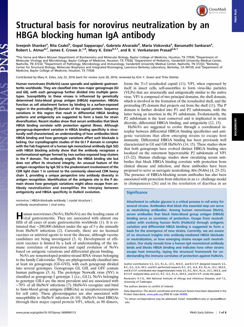
complex for crystallographic studies, we first carried out bindingstudies using biolayer interferometry (BLI). In these studies,biotinylated P domain was immobilized on a streptavidin bio-sensor and titrated against serial dilutions of Fab 5I2. Dataanalysis showed that Fab 5I2 binds to the P domain with an af-finity constant Kd of 20.5 nM and rate constants Kon of 2.04 × 10⁵
M1·s−1 and Koff of 4.01 × 10⁻³ s−1 for association and dissocia-tion, respectively (Fig. 1), indicating a tight interaction betweenFab 5I2 and NV P domain.
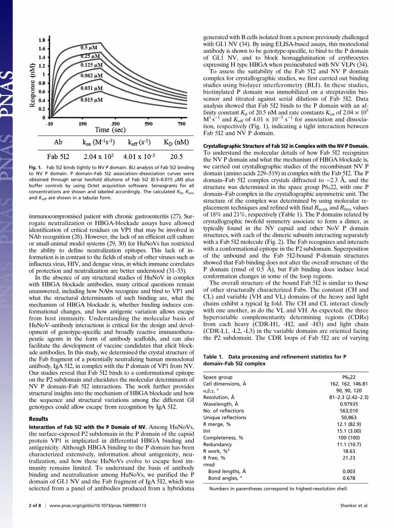
Crystallographic Structure of Fab 5I2 in Complex with the NV P Domain.To understand the molecular details of how Fab 5I2 recognizesthe NV P domain and what the mechanism of HBGA blockade is,we carried out crystallographic studies of the recombinant NV Pdomain (amino acids 229–519) in complex with the Fab 5I2. The Pdomain–Fab 5I2 complex crystals diffracted to ∼2.3 Å, and thestructure was determined in the space group P6522, with one Pdomain–Fab complex in the crystallographic asymmetric unit. Thestructure of the complex was determined by using molecular re-placement techniques and refined with final Rwork and Rfree valuesof 18% and 21%, respectively (Table 1). The P domains related bycrystallographic twofold symmetry associate to form a dimer, astypically found in the NV capsid and other NoV P domainstructures, with each of the dimeric subunits interacting separatelywith a Fab 5I2 molecule (Fig. 2). The Fab recognizes and interactswith a conformational epitope in the P2 subdomain. Superpositionof the unbound and the Fab 5I2-bound P-domain structuresshowed that Fab binding does not alter the overall structure of theP domain (rmsd of 0.5 Å), but Fab binding does induce localconformation changes in some of the loop regions.The overall structure of the bound Fab 5I2 is similar to those
of other structurally characterized Fabs. The constant (CH andCL) and variable (VH and VL) domains of the heavy and lightchains exhibit a typical Ig fold. The CH and CL interact closelywith one another, as do the VL and VH. As expected, the threehypervariable complementarity determining regions (CDRs)from each heavy (CDR-H1, -H2, and -H3) and light chain(CDR-L1, -L2, -L3) in the variable domains are oriented facingthe P2 subdomain. The CDR loops of Fab 5I2 are of varying
Fig. 1. Fab 5I2 binds tightly to NV P domain. BLI analysis of Fab 5I2 bindingto NV P domain. P domain–Fab 5I2 association–dissociation curves wereobtained through serial twofold dilutions of Fab 5I2 (0.5–0.015 μM) plusbuffer controls by using Octet acquisition software. Sensograms for allconcentrations are shown and labeled accordingly. The calculated KD, Kon,and Koff are shown in a tabular form.
Table 1. Data processing and refinement statistics for Pdomain–Fab 5I2 complex
Space group P6522Cell dimensions, Å 162, 162, 146.81α,β,γ, ° 90, 90, 120Resolution, Å 81–2.3 (2.42–2.3)Wavelength, Å 0.97935No. of reflections 563,010Unique reflections 50,863R merge, % 12.1 (82.9)I/σI 15.1 (3.00)Completeness, % 100 (100)Redundancy 11.1 (10.7)R work, %c 18.63R free, % 21.23rmsd
Bond lengths, Å 0.003Bond angles, ° 0.678
Numbers in parentheses correspond to highest-resolution shell.
2 of 8 | www.pnas.org/cgi/doi/10.1073/pnas.1609990113 Shanker et al.
lengths, with CDRH3 and CDRL1 being the longest, each con-sisting of 17 residues. Although the length of CDRH3 with 17residues is typical, the 17-residue length of CDRL1 is unusual, andanalyses of the interfacial interactions between the P domain andFab show that CDRL1 plays a dominant role in antigen recognition.
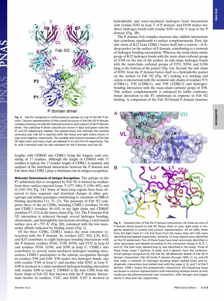
Molecular Determinants of Antigen Recognition. The epitope on theP2 subdomain that is recognized by Fab 5I2 is formed by residuesfrom three surface-exposed loops: T (377–386), U (394–405), andQ (345–354; Fig. 3A). Some of these loop regions have been ob-served to have sequence and structural changes in other gen-ogroups and within genotypes contributing to variations of HBGAbinding specificities (13, 15, 21). The paratope of Fab 5I2 com-prises three of the six CDRs, including CDRL1 (residues 24–40)and CDRL3 (residues 96–103) in the light chain and CDRH3(residues 97–113) in the heavy chain (Fig. 3A). The P domain–Fab5I2 interaction is achieved through several hydrogen bonding,electrostatic, and hydrophobic interactions involving a total buriedsurface area of ∼735 Å2 (Fig. 3B), consistent with the low nano-molar affinity indicated by binding assays (Fig. 1).Of the three CDRs, CDRL1 makes the most extensive in-
teraction with the P domain. Its residues Q27, S28, L30, K32,and K35 contribute to eight hydrogen bonding interactions withthe P domain residues N346, T348, D350, and F352 in loop Qand residues N394, G396, and S398 in loop U. CDRL1 alsocontributes to several water-mediated hydrogen bonding inter-actions. CDRL3 participates in the epitope recognition throughits residues Y98 and I100. Y98 makes two hydrogen bonds, onewith residue T348 in loop U and another with H381 in loop T.I100 is involved in a water-mediated hydrogen-bonding interactionwith residue S380 in loop T. CDRH3 is the lone CDR from theheavy chain of Fab 5I2 that interacts with the P domain. Interac-tions involve its residues, Y107, and D108. Y107 is involved in
hydrophobic and water-mediated hydrogen bond interactionswith residue S383 in loop T of P domain, and D108 makes twodirect hydrogen bonds with residue S383 on the T loop of the Pdomain (Fig. 3B).The P domain–Fab complex structure also exhibits interactions
that contribute significantly to surface complementarity. First, theside chain of K32 from CDRL1 buries itself into a narrow, ∼8-Å-deep pocket on the surface of P domain, contributing to a networkof hydrogen bonding interactions. Whereas the main-chain amidegroup of K32 hydrogen bonds with the main chain carbonyl groupof T348 on the rim of the pocket, its side-chain hydrogen bondswith the main-chain carbonyl groups of F352, N394, and G396lying at the bottom of the pocket (Fig. 4A). Second, the side chainof H381 from the P domain buries itself in a hydrophobic pocketon the surface of Fab 5I2 (Fig. 4C) making π–π stacking andcation–π interactions with the aromatic side chains of residues Y31(CDRL1), Y38 (CDRL1), and Y98 (CDRL3) and hydrogen-bonding interaction with the main chain carbonyl group of Y98.This surface complementarity is enhanced by subtle conforma-tional alterations in the P2 subdomain in response to Fab 5I2binding. A comparison of the Fab 5I2-bound P domain structure
Fig. 2. Fab 5I2 recognizes a conformational epitope on top of the NV P do-main. Cartoon representation of the overall structure of the Fab 5I2–P domaincomplex showing one Fab 5I2 molecule bound to each subunit of the P domaindimer. The individual P dimer subunits are shown in blue and green with theP1 and P2 subdomains labeled. The dashed black line indicates the twofoldsymmetry axis. Fab 5I2 is depicted with the heavy and light chains shown incyan and magenta, respectively. The variable and constant domains of the Fab5I2 light chain and heavy chain are labeled VL-CL and VH-CH respectively. TheN- and C-terminal ends are also indicated for the P domain and Fab 5I2.
Fig. 3. Detailed view of Fab 5I2–P domain interactions. (A) Close-up view ofP domain (blue) bound to Fab 5I2 (heavy chain in cyan; light chain in ma-genta) depicted in surface and cartoon representation. All six CDRs, threefrom the light chain (L1–L3) and three from the heavy chain (H1–H3), wereidentified and labeled respectively. Similarly, six loop regions were identifiedon the P2 subdomain. Five of these loops have been previously identified inother genotypes and labeled according to the convention (loops A, B, P, T,and U). The sixth loop, labeled loop Q, was identified in this study. Three ofthese loops, loops T (yellow), Q (red), and U (green), form the conforma-tional epitope recognized by the Fab 5I2. (B) Molecular details of Fab 5I2–Pdomain interactions. Fab 5I2 binds P domain through CDRs L1, L2, and H3that make a network of hydrogen bonding (black dashed lines) and hy-drophobic interactions (red dotted line) with the loops U, Q, and T of the Pdomain. CDRL1 makes the predominant interactions. All interacting loopsare shown in cartoon representation with interacting residues shown as stickmodel per the aforementioned color convention, with nitrogen and oxygenatoms in blue and red, respectively.
Shanker et al. PNAS Early Edition | 3 of 8
BIOCH
EMISTR
YPN
ASPL
US
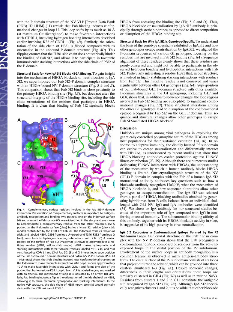
with the P domain structure of the NV VLP [Protein Data Bank(PDB) ID 1IHM] (11) reveals that Fab binding induces confor-mational changes in loop U. This loop shifts by as much as 10 Å(at maximum Cα divergence) to make favorable interactionswith CDRL1, including hydrogen bonding interactions describedearlier involving K32 of CDRL1 (Fig. 4B). Similarly, the orien-tation of the side chain of H381 is flipped compared with itsorientation in the unbound P domain structure (Fig. 4D). Thisflipping ensures that the H381 side chain does not sterically hinderthe binding of Fab 5I2, and allows it to participate in favorableintramolecular stacking interactions with the side chain of P382 ofthe P domain.
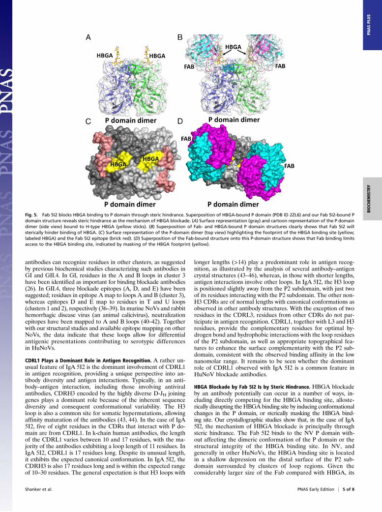
Structural Basis for How IgA 5I2 Blocks HBGA Binding. To gain insightinto the mechanism of HBGA blockade or neutralization by IgA5I2, we superimposed our Fab 5I2–P domain complex structurewith an HBGA-bound NV P-domain structure (Fig. 5 A and B).This comparison shows that Fab 5I2 binds in close proximity tothe primary HBGA binding site (Fig. 5B), but does not alter thestructural integrity of the HBGA binding site, including the sidechain orientations of the residues that participate in HBGAbinding. It is clear that binding of Fab 5I2 sterically blocks
HBGA from accessing the binding site (Fig. 5 C and D). Thus,HBGA blockade or neutralization by IgA 5I2 antibody is prin-cipally through steric hindrance as opposed to direct competitionor disruption of the HBGA binding site.
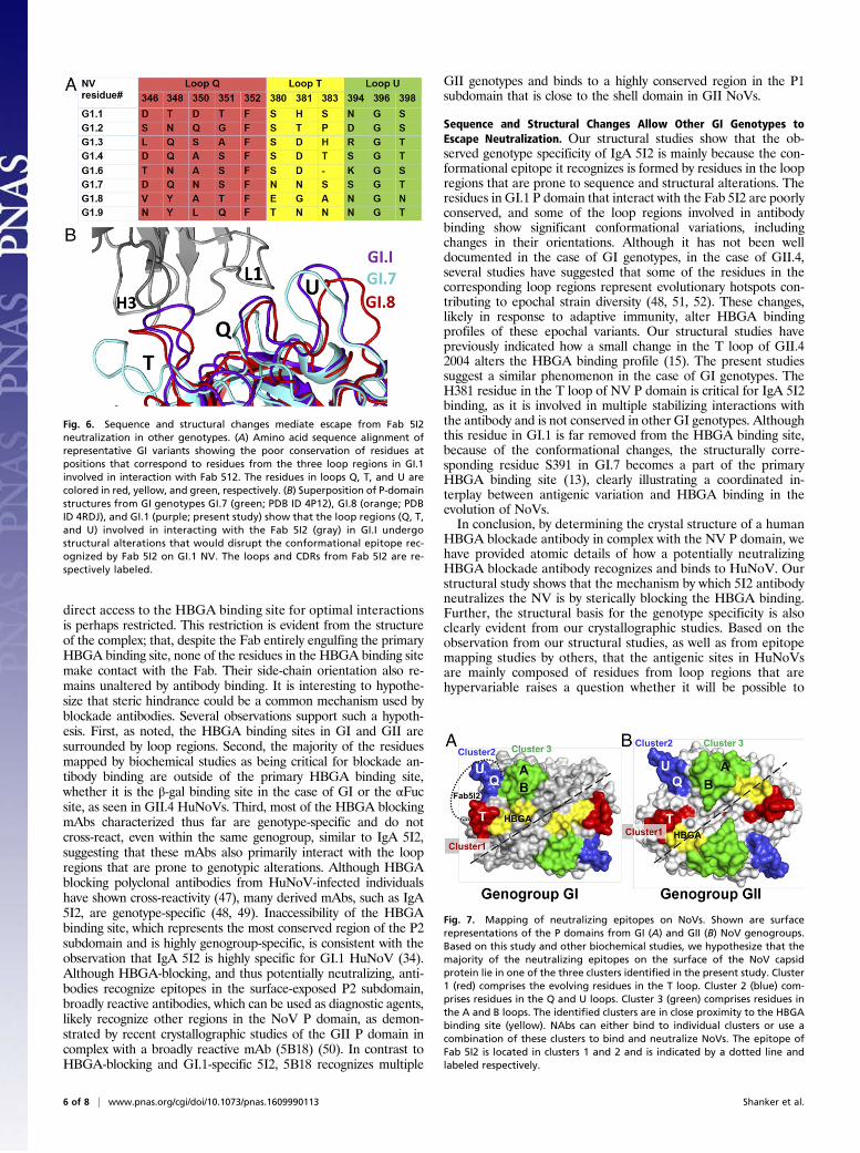
Structural Basis for Why IgA 5I2 Is Genotype-Specific. To understandthe basis of the genotype specificity exhibited by IgA 5I2 and howother genotypes escape neutralization by IgA 5I2, we aligned theP-domain sequences of various GI genotypes, focusing on theresidues that are involved in Fab 5I2 binding (Fig. 6A). Sequencealignment of these residues clearly shows that these residues arepoorly conserved and might not be able to participate in the ob-served hydrogen bonding and hydrophobic interactions with Fab5I2. Particularly interesting is residue H381 that, in our structure,is involved in highly stabilizing stacking interactions with residuesfrom Fab 5I2. This histidine residue is not conserved and variessignificantly between other GI genotypes (Fig. 6A). Superpositionof our Fab-bound GI.1 P-domain structure with other availableP-domain structures in the GI genogroup, including GI.7 andGI.8, shows that, in addition to sequence changes, the loop regionsinvolved in Fab 5I2 binding are susceptible to significant confor-mational changes (Fig. 6B). These structural alterations amongthe other GI genotypes lead to disruption of the conformationalepitope recognized by Fab 5I2 on the GI.1 P domain. Thus, se-quence and structural changes allow other genotypes to escapeFab 5I2-mediated HBGA blockade.
DiscussionHuNoVs are unique among viral pathogens in exploiting thegenetically controlled polymorphic nature of the HBGAs amonghost populations for their sustained evolution (14, 16). In re-sponse to adaptive immunity, the distally located P2 subdomaincan evolve to escape neutralization and differentially interactwith HBGAs, as underscored by recent studies that show thatHBGA-blocking antibodies confer protection against HuNoVillness or infection (23, 35). Although there are numerous studiesdelineating HuNoV interactions with HBGAs, the understandingof the mechanism by which a human antibody blocks HBGAbinding is limited. Our crystallographic structure of the NV(GI.1) P domain in complex with the Fab of a human IgA 5I2monoclonal antibody addresses key questions such as how ablockade antibody recognizes HuNoV, what the mechanism ofHBGA blockade is, and how sequence alterations allow othergenotypes to escape neutralization. The IgA 5I2 was selectedfrom a panel of HBGA blocking antibodies obtained by gener-ating hybridomas from B cells isolated from an individual chal-lenged with GI.1 NV. IgG and IgA antibodies were identified(34). We chose an IgA antibody for our structural studies be-cause of the important role of IgA compared with IgG in con-ferring mucosal immunity. The subnanomolar binding affinity ofthis antibody, together with its HBGA blockade activity in vitro,is suggestive of its high potency in virus neutralization.
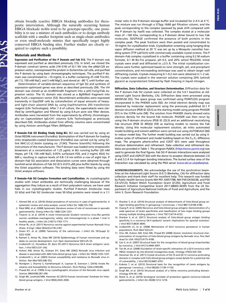
IgA 5I2 Recognizes a Conformational Epitope Formed by the P2Subdomain Loops. Our crystal structure of the Fab 5I2 in com-plex with the NV P domain shows that the Fab recognizes aconformational epitope composed of residues from the solvent-exposed loops in the distal portion of the P2 subdomain.Involvement of the surface loops in antibody recognition is acommon feature as observed in many antigen–antibody struc-tures. The distal surface of the P2 subdomain consists of six loopsthat project out into the solvent, which can be grouped into threeclusters, numbered 1–3 (Fig. 7A). Despite sequence changes,differences in their lengths and orientations, these loops aresimilarly clustered in GII.4 (Fig. 7B) as well as in murine NoVs.Residues from clusters 1 and 2 in GI.1 constitute the antigenicsite recognized by IgA 5I2 (Fig. 7A). Although IgA 5I2 specifi-cally recognizes clusters 1 and 2, it is possible that other blockade
Fig. 4. Complementary surface residues involved in the Fab 5I2–P domaininteraction. Presentation of complementary surfaces is important to antigen–antibody recognition and binding; two pockets, one on the P domain surface(A) and one on the Fab surface (C), were identified in the study and are shownto accommodate a complementary residue from the other molecule. (A) Apocket on the P domain surface (blue) buries a lysine 32 residue (pink stickmodel) contributed by the CDRL1 of Fab 5I2. The P domain residues, shown assticks and labeled N394, G396 from loop U (green) and T346, F352 from loop Q(red), contribute to hydrogen bonding interactions with K32. (C) A similarpocket on the surface of Fab 5I2 (magenta) is shown to accommodate a his-tidine residue (H381; yellow stick model). H381 makes hydrophobic andstacking interactions with three tyrosine residues labeled Y31, Y38, and Y98contributed by CDRs L1 and L3 of Fab 5I2. (B and D) Interestingly, superpositionof the Fab 5I2-bound P domain structure and native NV VLP structure (PDB ID1IHM; gray) shows that Fab binding induces local conformational changes onthe P domain to make favorable interactions. (B) Loop U moves approximately10 Å to make favorable interactions with CDRL1 and forms one side of thepocket that buries residue K32. Loop U from VLP is labeled in gray and markedwith an asterisk. The movement of loop U is indicated by an arrow. (D) Simi-larly, Fab binding induces a flip in the orientation of the side chain of an H381,allowing it to make favorable hydrophobic and stacking interactions. In thenative VLP structure, the side chain of H381 (gray; asterisk) would stericallyclash with the Y98 residue of CDRL3.
4 of 8 | www.pnas.org/cgi/doi/10.1073/pnas.1609990113 Shanker et al.
antibodies can recognize residues in other clusters, as suggestedby previous biochemical studies characterizing such antibodies inGI and GII.4. In GI, residues in the A and B loops in cluster 3have been identified as important for binding blockade antibodies(26). In GII.4, three blockade epitopes (A, D, and E) have beensuggested; residues in epitope A map to loops A and B (cluster 3),whereas epitopes D and E map to residues in T and U loops(clusters 1 and 2), respectively (36–39). In murine NoVs and rabbithemorrhagic disease virus (an animal calicivirus), neutralizationepitopes have been mapped to A and B loops (40–42). Togetherwith our structural studies and available epitope mapping on otherNoVs, the data indicate that these loops allow for differentialantigenic presentations contributing to serotypic differencesin HuNoVs.
CDRL1 Plays a Dominant Role in Antigen Recognition. A rather un-usual feature of IgA 5I2 is the dominant involvement of CDRL1in antigen recognition, providing a unique perspective into an-tibody diversity and antigen interactions. Typically, in an anti-body–antigen interaction, including those involving antiviralantibodies, CDRH3 encoded by the highly diverse D-JH joininggenes plays a dominant role because of the inherent sequencediversity and consequent conformational variability. The H3loop is also a common site for somatic hypermutations, allowingaffinity maturation of the antibodies (43, 44). In the case of IgA5I2, five of eight residues in the CDRs that interact with P do-main are from CDRL1. In k-chain human antibodies, the lengthof the CDRL1 varies between 10 and 17 residues, with the ma-jority of the antibodies exhibiting a loop length of 11 residues. InIgA 5I2, CDRL1 is 17 residues long. Despite its unusual length,it exhibits the expected canonical conformation. In IgA 5I2, theCDRH3 is also 17 residues long and is within the expected rangeof 10–30 residues. The general expectation is that H3 loops with
longer lengths (>14) play a predominant role in antigen recog-nition, as illustrated by the analysis of several antibody–antigencrystal structures (43–46), whereas, in those with shorter lengths,antigen interactions involve other loops. In IgA 5I2, the H3 loopis positioned slightly away from the P2 subdomain, with just twoof its residues interacting with the P2 subdomain. The other non-H3 CDRs are of normal lengths with canonical conformations asobserved in other antibody structures. With the exception of tworesidues in the CDRL3, residues from other CDRs do not par-ticipate in antigen recognition. CDRL1, together with L3 and H3residues, provide the complementary residues for optimal hy-drogen bond and hydrophobic interactions with the loop residuesof the P2 subdomain, as well as appropriate topographical fea-tures to enhance the surface complementarity with the P2 sub-domain, consistent with the observed binding affinity in the lownanomolar range. It remains to be seen whether the dominantrole of CDRL1 observed with IgA 5I2 is a common feature inHuNoV blockade antibodies.
HBGA Blockade by Fab 5I2 Is by Steric Hindrance. HBGA blockadeby an antibody potentially can occur in a number of ways, in-cluding directly competing for the HBGA binding site, alloste-rically disrupting the HBGA binding site by inducing conformationalchanges in the P domain, or sterically masking the HBGA bind-ing site. Our crystallographic studies show that, in the case of IgA5I2, the mechanism of HBGA blockade is principally throughsteric hindrance. The Fab 5I2 binds to the NV P domain with-out affecting the dimeric conformation of the P domain or thestructural integrity of the HBGA binding site. In NV, andgenerally in other HuNoVs, the HBGA binding site is locatedin a shallow depression on the distal surface of the P2 sub-domain surrounded by clusters of loop regions. Given theconsiderably larger size of the Fab compared with HBGA, its
Fig. 5. Fab 5I2 blocks HBGA binding to P domain through steric hindrance. Superposition of HBGA-bound P domain (PDB ID 2ZL6) and our Fab 5I2-bound Pdomain structure reveals steric hindrance as the mechanism of HBGA blockade. (A) Surface representation (gray) and cartoon representation of the P domaindimer (side view) bound to H-type HBGA (yellow sticks). (B) Superposition of Fab- and HBGA-bound P domain structures clearly shows that Fab 5I2 willsterically hinder binding of HBGA. (C) Surface representation of the P-domain dimer (top view) highlighting the footprint of the HBGA binding site (yellow;labeled HBGA) and the Fab 5I2 epitope (brick red). (D) Superposition of the Fab-bound structure onto this P-domain structure shows that Fab binding limitsaccess to the HBGA binding site, indicated by masking of the HBGA footprint (yellow).
Shanker et al. PNAS Early Edition | 5 of 8
BIOCH
EMISTR
YPN
ASPL
US
direct access to the HBGA binding site for optimal interactionsis perhaps restricted. This restriction is evident from the structureof the complex; that, despite the Fab entirely engulfing the primaryHBGA binding site, none of the residues in the HBGA binding sitemake contact with the Fab. Their side-chain orientation also re-mains unaltered by antibody binding. It is interesting to hypothe-size that steric hindrance could be a common mechanism used byblockade antibodies. Several observations support such a hypoth-esis. First, as noted, the HBGA binding sites in GI and GII aresurrounded by loop regions. Second, the majority of the residuesmapped by biochemical studies as being critical for blockade an-tibody binding are outside of the primary HBGA binding site,whether it is the β-gal binding site in the case of GI or the αFucsite, as seen in GII.4 HuNoVs. Third, most of the HBGA blockingmAbs characterized thus far are genotype-specific and do notcross-react, even within the same genogroup, similar to IgA 5I2,suggesting that these mAbs also primarily interact with the loopregions that are prone to genotypic alterations. Although HBGAblocking polyclonal antibodies from HuNoV-infected individualshave shown cross-reactivity (47), many derived mAbs, such as IgA5I2, are genotype-specific (48, 49). Inaccessibility of the HBGAbinding site, which represents the most conserved region of the P2subdomain and is highly genogroup-specific, is consistent with theobservation that IgA 5I2 is highly specific for GI.1 HuNoV (34).Although HBGA-blocking, and thus potentially neutralizing, anti-bodies recognize epitopes in the surface-exposed P2 subdomain,broadly reactive antibodies, which can be used as diagnostic agents,likely recognize other regions in the NoV P domain, as demon-strated by recent crystallographic studies of the GII P domain incomplex with a broadly reactive mAb (5B18) (50). In contrast toHBGA-blocking and GI.1-specific 5I2, 5B18 recognizes multiple
GII genotypes and binds to a highly conserved region in the P1subdomain that is close to the shell domain in GII NoVs.
Sequence and Structural Changes Allow Other GI Genotypes toEscape Neutralization. Our structural studies show that the ob-served genotype specificity of IgA 5I2 is mainly because the con-formational epitope it recognizes is formed by residues in the loopregions that are prone to sequence and structural alterations. Theresidues in GI.1 P domain that interact with the Fab 5I2 are poorlyconserved, and some of the loop regions involved in antibodybinding show significant conformational variations, includingchanges in their orientations. Although it has not been welldocumented in the case of GI genotypes, in the case of GII.4,several studies have suggested that some of the residues in thecorresponding loop regions represent evolutionary hotspots con-tributing to epochal strain diversity (48, 51, 52). These changes,likely in response to adaptive immunity, alter HBGA bindingprofiles of these epochal variants. Our structural studies havepreviously indicated how a small change in the T loop of GII.42004 alters the HBGA binding profile (15). The present studiessuggest a similar phenomenon in the case of GI genotypes. TheH381 residue in the T loop of NV P domain is critical for IgA 5I2binding, as it is involved in multiple stabilizing interactions withthe antibody and is not conserved in other GI genotypes. Althoughthis residue in GI.1 is far removed from the HBGA binding site,because of the conformational changes, the structurally corre-sponding residue S391 in GI.7 becomes a part of the primaryHBGA binding site (13), clearly illustrating a coordinated in-terplay between antigenic variation and HBGA binding in theevolution of NoVs.In conclusion, by determining the crystal structure of a human
HBGA blockade antibody in complex with the NV P domain, wehave provided atomic details of how a potentially neutralizingHBGA blockade antibody recognizes and binds to HuNoV. Ourstructural study shows that the mechanism by which 5I2 antibodyneutralizes the NV is by sterically blocking the HBGA binding.Further, the structural basis for the genotype specificity is alsoclearly evident from our crystallographic studies. Based on theobservation from our structural studies, as well as from epitopemapping studies by others, that the antigenic sites in HuNoVsare mainly composed of residues from loop regions that arehypervariable raises a question whether it will be possible to
Fig. 6. Sequence and structural changes mediate escape from Fab 5I2neutralization in other genotypes. (A) Amino acid sequence alignment ofrepresentative GI variants showing the poor conservation of residues atpositions that correspond to residues from the three loop regions in GI.1involved in interaction with Fab 512. The residues in loops Q, T, and U arecolored in red, yellow, and green, respectively. (B) Superposition of P-domainstructures from GI genotypes GI.7 (green; PDB ID 4P12), GI.8 (orange; PDBID 4RDJ), and GI.1 (purple; present study) show that the loop regions (Q, T,and U) involved in interacting with the Fab 5I2 (gray) in GI.I undergostructural alterations that would disrupt the conformational epitope rec-ognized by Fab 5I2 on GI.1 NV. The loops and CDRs from Fab 5I2 are re-spectively labeled.
Fig. 7. Mapping of neutralizing epitopes on NoVs. Shown are surfacerepresentations of the P domains from GI (A) and GII (B) NoV genogroups.Based on this study and other biochemical studies, we hypothesize that themajority of the neutralizing epitopes on the surface of the NoV capsidprotein lie in one of the three clusters identified in the present study. Cluster1 (red) comprises the evolving residues in the T loop. Cluster 2 (blue) com-prises residues in the Q and U loops. Cluster 3 (green) comprises residues inthe A and B loops. The identified clusters are in close proximity to the HBGAbinding site (yellow). NAbs can either bind to individual clusters or use acombination of these clusters to bind and neutralize NoVs. The epitope ofFab 5I2 is located in clusters 1 and 2 and is indicated by a dotted line andlabeled respectively.
6 of 8 | www.pnas.org/cgi/doi/10.1073/pnas.1609990113 Shanker et al.
obtain broadly reactive HBGA blocking antibodies for thera-peutic intervention. Although the naturally occurring humanHBGA blockade mAbs tend to be genotype-specific, one possi-bility is to use a mixture of such antibodies or to design antibodyscaffolds with a smaller footprint such as single-chain antibodiesor even small-molecule mimics that specifically target highlyconserved HBGA binding sites. Further studies are clearly re-quired to explore such a possibility.
Materials and MethodsExpression and Purification of the P Domain and Fab 512. The P domain wasexpressed and purified as described previously (15). In brief, we cloned theP-domain construct (amino acids 216–519) of GI.1 NV into the pMal-C2E ex-pression vector, overexpressed the protein in Escherichia coli cells, and purifiedthe P domain by using basic chromatography techniques. The purified P do-main was concentrated to ∼10 mg/mL in a buffer containing 25 mM Tris·HCl,pH 7.5, 150 mM NaCl, and 5 mM MgCl2 and stored at −80 °C until further use.
Determination of variable-domain sequences of IgA 5I2 and synthesis ofexpression-optimized genes was done as described previously (34). The VHdomain was cloned as an EcoRI/HindIII fragment into a pHC-huCg1Fab ex-pression vector. The VL domain was cloned as a BglII/NotI fragment intopML-huCk κ-expression vector (53). Recombinant antibodies were expressedtransiently in Expi293F cells by cotransfection of equal amounts of heavy-and light-chain plasmid DNA by using ExpiFectamine 293 transfectionreagent (Life Technologies). After 7 d of culture, the supernatants were clar-ified by centrifugation and filtered by using 0.4-μm pore size filter devices.Antibodies were harvested from the supernatants by affinity chromatogra-phy on CaptureSelect IgG-CH1 columns (Life Technologies) as previouslydescribed (54). Antibodies eluted from affinity columns were concentratedby using Amicon centrifugal filters (Millipore).
P Domain–Fab 5I2 Binding Study Using BLI. BLI was carried out by using anOctet RED96 instrument (ForteBio). Biotinylation of the P domain for loadingonto streptavidin-coated biosensors (ForteBio) was carried out by using EZ-link NHC-LC-LC-biotin (catalog no. 21343; Thermo Scientific) following theinstructions of the manufacturer. The P domain was loaded onto streptavidinbiosensors at a concentration of 1.25 μg/mL in BLI running buffer (20 mMHepes, pH 7.8, 150 mM NaCl, 0.05% surfactant P20, and 2 mg/mL BSA) for600 s, resulting in capture levels of 0.8–1.0 nm within a row of eight tips. Pdomain–Fab 5I2 association and dissociation curves were obtained throughtwofold serial dilutions of Fab 5I2 (0.5–0.015 μM) plus buffer blanks by usingthe Octet acquisition software. The binding data were fitted by using theOctet analysis software.
P Domain–Fab 5I2 Complex Formation and Crystallization. As crystallographicstudies with intact antibodies are technically challenging because of theaggregation they induce as a result of their polyvalent nature, we have usedFabs in our crystallographic studies. Purified P-domain (molecular mass32 kDa) and Fab 5I2 (molecular mass 50 kDa) proteins were mixed in a 1:1
molar ratio in the P-domain storage buffer and incubated for 2–4 h at 4 °C.The mixture was run through a S75pg 16/60 gel filtration column, and thepeak corresponding to the complex (assessed by peak shift compared withthe P domain by itself) was collected. The complex eluted at a molecularmass of ∼160 kDa, corresponding to a P-domain dimer bound to two Fabmolecules. SDS/PAGE confirmed the presence of both proteins in thecomplex peak. The peak fractions were then pooled and concentrated to10 mg/mL for crystallization trials. Crystallization screening using hanging-dropvapor diffusion method at 20 °C was set up by a Mosquito nanoliter han-dling system (TTP LabTech) with commercially available crystal screens. The Pdomain–Fab complex crystallized in a buffer containing using 0.2 M sodiumformate, 0.1 M Bis-Tris propane, pH 6.5, and 20% wt/vol PEG3350. Initialcrystals were small and diffracted to ≥3.5 Å. The initial crystallization con-ditions were further optimized based on ionic strength, pH, and precipitantconcentrations, and microseeding technique was used to obtain larger welldiffracting crystals. Crystals measuring 0.1–0.2 mm were obtained in 1–2 wk.The crystals were soaked in the reservoir solution containing 20% (wt/vol)glycerol as cryoprotectant followed by flash freezing in liquid nitrogen.
Diffraction, Data Collection, and Structure Determination. Diffraction data forthe P domain–Fab 5I2 crystals were collected on the 5.0.1 beamline at Ad-vanced Light Source (Berkeley, CA). Diffraction data were processed usingIMOSFLM (55). The space group was confirmed using POINTLESS programincorporated in the PHENIX suite (56). An initial electron density map wasobtained by molecular replacement using the previously published GI.1 Pdomain structure (PDB ID 2ZL5) as the starting model using program PHASER(57) in the CCP4i suite (58). The solution from PHASER clearly showed extraelectron density for the bound Fab molecule. PHASER was then rerun byusing the P-domain structure (PDB ID 2ZL5) and an additional neutralizingFab structure (PDB ID 4RQQ) (59) as starting models to resolve the Fabdensity. Using this molecular replacement solution, ab initio automatedmodel building and solvent addition were carried out using AUTOBUILD (60)to reduce model bias. The further model building was carried out by using it-erative cycles of refinement and model building based on the FO–FC differencemaps. The programs phenix.refine and COOT (61) were used throughoutstructure determination and refinement. Data collection and refinement sta-tistics are provided in Table 1. The program PyMOL (https://www.pymol.org) wasused to generate the final figures. Fab 5I2–P domain interactions were analyzedusing COOT and LIGPLOT (62) with the donor to acceptor distances between 2.6Å and 3.3 Å for hydrogen bonding interactions. The buried surface area of theinteraction was calculated by using the PISA server (www.ebi.ac.uk/pdbe/pisa).
ACKNOWLEDGMENTS. We acknowledge the use of the synchrotron beam-lines at the Advanced Light Source (5.0.1) (Berkeley, CA) for diffraction datacollection and thank their staff for excellent help. This research was fundedby Public Health Service Grants NIH P01 AI057788, NIH P30 DK56338, and P30CA125123; Robert Welch Foundation Grant Q1292; Agriculture and FoodResearch Initiative Competitive Grant 2011-68003-30395 from the US De-partment of Agriculture National Institute of Food and Agriculture; and theJohn S. Dunn Research Foundation.
1. Ahmed SM, et al. (2014) Global prevalence of norovirus in cases of gastroenteritis: A
systematic review and meta-analysis. Lancet Infect Dis 14(8):725–730.2. Patel MM, et al. (2008) Systematic literature review of role of noroviruses in sporadic
gastroenteritis. Emerg Infect Dis 14(8):1224–1231.3. Treanor JJ, et al. (2014) A novel intramuscular bivalent norovirus virus-like particle
vaccine candidate–reactogenicity, safety, and immunogenicity in a phase 1 trial in
healthy adults. J Infect Dis 210(11):1763–1771.4. Atmar RL, et al. (2011) Norovirus vaccine against experimental human Norwalk Virus
illness. N Engl J Med 365(23):2178–2187.5. Green KY, et al. (2000) Taxonomy of the caliciviruses. J Infect Dis 181(suppl 2):
S322–S330.6. Ramani S, Atmar RL, Estes MK (2014) Epidemiology of human noroviruses and up-
dates on vaccine development. Curr Opin Gastroenterol 30(1):25–33.7. Lindesmith LC, Donaldson EF, Baric RS (2011) Norovirus GII.4 strain antigenic varia-
tion. J Virol 85(1):231–242.8. Hutson AM, Atmar RL, Graham DY, Estes MK (2002) Norwalk virus infection and
disease is associated with ABO histo-blood group type. J Infect Dis 185(9):1335–1337.9. Lindesmith L, et al. (2003) Human susceptibility and resistance to Norwalk virus in-
fection. Nat Med 9(5):548–553.10. Nordgren J, Sharma S, Kambhampati A, Lopman B, Svensson L (2016) Innate Re-
sistance and Susceptibility to Norovirus Infection. PLoS Pathog 12(4):e1005385.11. Prasad BV, et al. (1999) X-ray crystallographic structure of the Norwalk virus capsid.
Science 286(5438):287–290.12. Singh BK, Leuthold MM, Hansman GS (2015) Human noroviruses’ fondness for histo-
blood group antigens. J Virol 89(4):2024–2040.
13. Shanker S, et al. (2014) Structural analysis of determinants of histo-blood group an-tigen binding specificity in genogroup I noroviruses. J Virol 88(11):6168–6180.
14. Huang P, et al. (2005) Norovirus and histo-blood group antigens: Demonstration of awide spectrum of strain specificities and classification of two major binding groupsamong multiple binding patterns. J Virol 79(11):6714–6722.
15. Shanker S, et al. (2011) Structural analysis of histo-blood group antigen bindingspecificity in a norovirus GII.4 epidemic variant: Implications for epochal evolution.J Virol 85(17):8635–8645.
16. Lindesmith LC, et al. (2008) Mechanisms of GII.4 norovirus persistence in humanpopulations. PLoS Med 5(2):e31.
17. Choi JM, Hutson AM, Estes MK, Prasad BV (2008) Atomic resolution structural char-acterization of recognition of histo-blood group antigens by Norwalk virus. Proc NatlAcad Sci USA 105(27):9175–9180.
18. Cao S, et al. (2007) Structural basis for the recognition of blood group trisaccharidesby norovirus. J Virol 81(11):5949–5957.
19. Tan M, et al. (2008) Elucidation of strain-specific interaction of a GII-4 norovirus withHBGA receptors by site-directed mutagenesis study. Virology 379(2):324–334.
20. Hansman GS, et al. (2011) Crystal structures of GII.10 and GII.12 norovirus protrudingdomains in complex with histo-blood group antigens reveal details for a potential siteof vulnerability. J Virol 85(13):6687–6701.
21. Kubota T, et al. (2012) Structural basis for the recognition of Lewis antigens bygenogroup I norovirus. J Virol 86(20):11138–11150.
22. Singh BK, et al. (2015) Structural analysis of a feline norovirus protruding domain.Virology 474:181–185.
23. Reeck A, et al. (2010) Serological correlate of protection against norovirus-inducedgastroenteritis. J Infect Dis 202(8):1212–1218.
Shanker et al. PNAS Early Edition | 7 of 8
BIOCH
EMISTR
YPN
ASPL
US

24. Ramani S, et al. (2015) Mucosal and cellular immune responses to Norwalk virus.J Infect Dis 212(3):397–405.
25. Lindesmith LC, et al. (2015) Broad blockade antibody responses in human volunteersafter immunization with a multivalent norovirus VLP candidate vaccine: Immuno-logical analyses from a phase I clinical trial. PLoS Med 12(3):e1001807.
26. Chen Z, et al. (2013) Development of Norwalk virus-specific monoclonal antibodieswith therapeutic potential for the treatment of Norwalk virus gastroenteritis. J Virol87(17):9547–9557.
27. Knoll BM, Lindesmith LC, Yount BL, Baric RS, Marty FM (2016) Resolution of diarrheain an immunocompromised patient with chronic norovirus gastroenteritis correlateswith constitution of specific antibody blockade titer. Infection 44(4):551–554.
28. Lindesmith LC, et al. (2012) Immunogenetic mechanisms driving norovirus GII.4 an-tigenic variation. PLoS Pathog 8(5):e1002705.
29. Lay MK, et al. (2010) Norwalk virus does not replicate in human macrophages ordendritic cells derived from the peripheral blood of susceptible humans. Virology406(1):1–11.
30. Herbst-Kralovetz MM, et al. (2013) Lack of norovirus replication and histo-bloodgroup antigen expression in 3-dimensional intestinal epithelial cells. Emerg Infect Dis19(3):431–438.
31. Kwong PD, Mascola JR, Nabel GJ (2011) Rational design of vaccines to elicit broadlyneutralizing antibodies to HIV-1. Cold Spring Harb Perspect Med 1(1):a007278.
32. Wei CJ, et al. (2010) Induction of broadly neutralizing H1N1 influenza antibodies byvaccination. Science 329(5995):1060–1064.
33. Pierson TC, Fremont DH, Kuhn RJ, Diamond MS (2008) Structural insights into themechanisms of antibody-mediated neutralization of flavivirus infection: Implicationsfor vaccine development. Cell Host Microbe 4(3):229–238.
34. Sapparapu G, et al. (2016) Frequent use of the IgA isotype in human B cells encodingpotent norovirus-specific monoclonal antibodies that block HBGA binding. PLoSPathog 12(6):e1005719.
35. Bok K, et al. (2011) Chimpanzees as an animal model for human norovirus infectionand vaccine development. Proc Natl Acad Sci USA 108(1):325–330.
36. Lindesmith LC, et al. (2012) Monoclonal antibody-based antigenic mapping of nor-ovirus GII.4-2002. J Virol 86(2):873–883.
37. Debbink K, Lindesmith LC, Donaldson EF, Baric RS (2012) Norovirus immunity and thegreat escape. PLoS Pathog 8(10):e1002921.
38. Allen DJ, et al. (2009) Characterisation of a GII-4 norovirus variant-specific surface-exposed site involved in antibody binding. Virol J 6:150.
39. Parra GI, et al. (2012) Multiple antigenic sites are involved in blocking the interactionof GII.4 norovirus capsid with ABH histo-blood group antigens. J Virol 86(13):7414–7426.
40. Taube S, et al. (2010) High-resolution x-ray structure and functional analysis of themurine norovirus 1 capsid protein protruding domain. J Virol 84(11):5695–5705.
41. Kolawole AO, et al. (2014) Flexibility in surface-exposed loops in a virus capsid me-diates escape from antibody neutralization. J Virol 88(8):4543–4557.
42. Wang X, et al. (2013) Atomic model of rabbit hemorrhagic disease virus by cryo-electron microscopy and crystallography. PLoS Pathog 9(1):e1003132.
43. Tsuchiya Y, Mizuguchi K (2016) The diversity of H3 loops determines the antigen-binding tendencies of antibody CDR loops. Protein Sci 25(4):815–825.
44. Shirai H, Kidera A, Nakamura H (1999) H3-rules: Identification of CDR-H3 structures in
antibodies. FEBS Lett 455(1-2):188–197.45. North B, Lehmann A, Dunbrack RL, Jr (2011) A new clustering of antibody CDR loop
conformations. J Mol Biol 406(2):228–256.46. Weitzner BD, Dunbrack RL, Jr, Gray JJ (2015) The origin of CDR H3 structural diversity.
Structure 23(2):302–311.47. Czakó R, et al. (2015) Experimental human infection with Norwalk virus elicits a
surrogate neutralizing antibody response with cross-genogroup activity. Clin Vaccine
Immunol 22(2):221–228.48. Lindesmith LC, et al. (2013) Emergence of a norovirus GII.4 strain correlates with
changes in evolving blockade epitopes. J Virol 87(5):2803–2813.49. Payne DC, Parashar UD, Lopman BA (2015) Developments in understanding acquired
immunity and innate susceptibility to norovirus and rotavirus gastroenteritis in chil-
dren. Curr Opin Pediatr 27(1):105–109.50. Hansman GS, et al. (2012) Structural basis for broad detection of genogroup II nor-
oviruses by a monoclonal antibody that binds to a site occluded in the viral particle.
J Virol 86(7):3635–3646.51. Bok K, et al. (2009) Evolutionary dynamics of GII.4 noroviruses over a 34-year period.
J Virol 83(22):11890–11901.52. Donaldson EF, Lindesmith LC, Lobue AD, Baric RS (2010) Viral shape-shifting: Nor-
ovirus evasion of the human immune system. Nat Rev Microbiol 8(3):231–241.53. McLean GR, Nakouzi A, Casadevall A, Green NS (2000) Human and murine immu-
noglobulin expression vector cassettes. Mol Immunol 37(14):837–845.54. Aiyegbo MS, et al. (2013) Human rotavirus VP6-specific antibodies mediate in-
tracellular neutralization by binding to a quaternary structure in the transcriptional
pore. PLoS One 8(5):e61101.55. Battye TG, Kontogiannis L, Johnson O, Powell HR, Leslie AG (2011) iMOSFLM: A new
graphical interface for diffraction-image processing with MOSFLM. Acta Crystallogr D
Biol Crystallogr 67(pt 4):271–281.56. Adams PD, et al. (2002) PHENIX: Building new software for automated crystallo-
graphic structure determination. Acta Crystallogr D Biol Crystallogr 58(pt 11):
1948–1954.57. McCoy AJ, et al. (2007) Phaser crystallographic software. J Appl Cryst 40(pt 4):
658–674.58. Collaborative Computational Project, Number 4 (1994) The CCP4 suite: Programs for
protein crystallography. Acta Crystallogr D Biol Crystallogr 50(pt 5):760–763.59. Sok D, et al. (2014) Recombinant HIV envelope trimer selects for quaternary-
dependent antibodies targeting the trimer apex. Proc Natl Acad Sci USA 111(49):
17624–17629.60. Terwilliger TC, et al. (2008) Iterative model building, structure refinement and density
modification with the PHENIX AutoBuild wizard. Acta Crystallogr D Biol Crystallogr
64(pt 1):61–69.61. Emsley P, Cowtan K (2004) Coot: Model-building tools for molecular graphics. Acta
Crystallogr D Biol Crystallogr 60(pt 12 pt 1):2126–2132.62. Wallace AC, Laskowski RA, Thornton JM (1995) LIGPLOT: A program to generate
schematic diagrams of protein-ligand interactions. Protein Eng 8(2):127–134.
8 of 8 | www.pnas.org/cgi/doi/10.1073/pnas.1609990113 Shanker et al.