structural and functional imaging functional images tend to be lower resolution and fail to convey...
Post on 19-Dec-2015
216 views
TRANSCRIPT

Structural and Functional Imaging• Functional images tend to be lower resolution and fail to convey spatial
information
Pixels

Structural and Functional Imaging
• Structural images have finer (smaller) pixels
Pixels

Structural and Functional Imaging
• Why? What’s wrong with the functional image alone?
• More subtly: a functional image typically isn’t a picture of the brain at all! It’s a picture of something else– PET, fMRI = oxygenated blood– EEG = electric fields– MEG = magnetic fields

Tools for measuring brain function• The main story about functional imaging is a trade-off between spatial
resolution and temporal resolution

Principles of MRI

Principles of MRI
• Some terms:– Nuclear Magnetic Resonance (NMR)
• quantum property of protons• energy absorbed when precession frequency matches radio
frequency
– Magnetic Resonance Imaging (MRI)• uses spatial differences in resonance frequencies to form an
image• basis of anatomical MRI
– functional Magnetic Resonance Imaging (fMRI)• exploits magnetic properties of hemaglobin to create images
changes in cortical blood flow
Principles of NMR
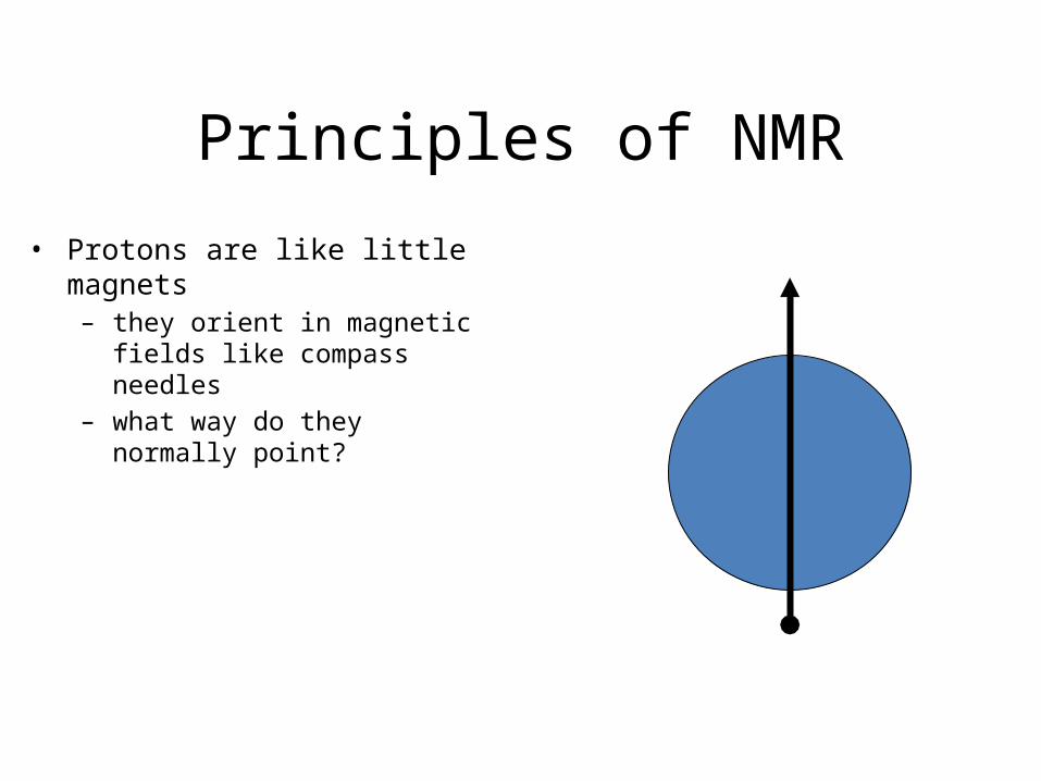
• Protons are like little magnets– they orient in magnetic fields like
compass needles– what way do they normally point?

Principles of NMR
• Protons are like little magnets– they orient in magnetic fields like
compass needles– what way do they normally point?– normally aligned with Earth’s
magnetic field

Principles of NMR
• Protons are like little magnets– they orient in magnetic fields like
compass needles– what way do they normally point?– normally aligned with Earth’s
magnetic field– NMR uses a big magnet to align all
the protons in a sample (e.g. brain tissue)

Principles of NMR
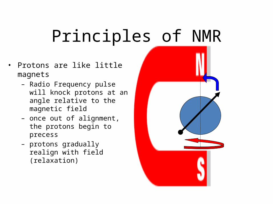
• Protons are like little magnets– Radio Frequency pulse will knock
protons at an angle relative to the magnetic field

Principles of NMR
• Protons are like little magnets– Radio Frequency pulse will knock
protons at an angle relative to the magnetic field
– once out of alignment, the protons begin to precess
Principles of NMR
• Protons are like little magnets– Radio Frequency pulse will knock
protons at an angle relative to the magnetic field
– once out of alignment, the protons begin to precess
– protons gradually realign with field (relaxation)

Principles of NMR
• Protons are like little magnets– Radio Frequency pulse will knock
protons at an angle relative to the magnetic field
– once out of alignment, the protons begin to precess
– protons gradually realign with field (relaxation)
– protons “echo” back the radio frequency that originally tipped them over
– That radio “echo” forms the basis of the MRI image

Principles of NMR
• Protons are like little magnets– The following simple equation
explains MRI image formation

Functional Imaging• Recall that precessing protons give
off a radio “echo” as they realign with the magnetic field
• We pick up the combined echo from many protons that are in phase

Functional Imaging
• Oxygenated hemoglobin is diamagnetic - it has no magnetic effects on surrounding molecules
• Deoxygenated hemoglobin is paramagnetic - it has strong magnetic effects on surrounding molecules!
Hemoglobin

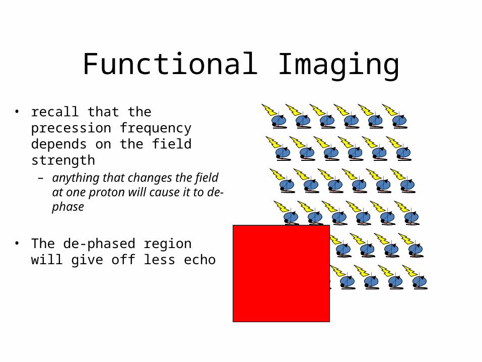
Functional Imaging• recall that the precession
frequency depends on the field strength– anything that changes the field at
one proton will cause it to de-phase
Functional Imaging• recall that the precession
frequency depends on the field strength– anything that changes the field at
one proton will cause it to de-phase
• The de-phased region will give off less echo

Functional Imaging
• blood flow overshoots baseline after a brain region is activated
• Deoxygenated blood in some region causes relatively less signal from that region
• More oxygenated blood in some region causes relatively more signal from that region