structural and functional analysis of the eukaryotic dna repair
TRANSCRIPT

Dissertation zur Erlangung des Doktorgrades
der Fakultät für Chemie und Pharmazie
der Ludwig-Maximilians-Universität München
Structural and functional analysis of the eukaryotic
DNA repair proteins Mre11 and Nbs1
Christian Bernd Schiller
aus
Kassel
2011

Erklärung
Diese Dissertation wurde im Sinne von § 13 Abs. 3 bzw. 4 der Promotionsordnung vom 29. Januar
1998 (in der Fassung der sechsten Änderungssatzung vom 16. August 2010)
von Herrn Prof. Dr. Karl-Peter Hopfner betreut.
Ehrenwörtliche Versicherung
Diese Dissertation wurde selbständig, ohne unerlaubte Hilfe erarbeitet.
München, am 07.06.2011
....................................................
(Christian Bernd Schiller)
Dissertation eingereicht am 07.06.2011
1. Gutachter: Herr Prof. Dr. Karl-Peter Hopfner
2. Gutachter: Herr Prof. Dr. Dietmar Martin
Mündliche Prüfung am 21.07.2011

During the work of this thesis, the following publication was published:
Lammens K., Bemeleit D. J., Möckel C., Clausing E., Schele A., Hartung S., Schiller C. B.,
Lucas M., Angermüller C., Soding J., Strässer K. and K. P. Hopfner (2011). "The
Mre11:Rad50 Structure Shows an ATP-Dependent Molecular Clamp in DNA Double-Strand
Break Repair." Cell 145(1): 54-66.
Parts of the present thesis will be submitted for publication:
Schiller C.B., Lammens K., Guerini I., Coordes B., Schlauderer F., Möckel C., Schele A.,
Sträßer K., Jackson S. P., Hopfner K.-P.:
“Insights into DNA double-strand break repair and ataxia-telangiectasia like disease from the
structure of an Mre11-Nbs1 complex“, manuscript in preparation.
Parts of this thesis have been presented at international conferences and workshops:
Talk and poster at the Biannual International Meeting of the German Society of DNA Repair
Research (DGDR) - Repair meets Replication, September 7-10, 2010 in Jena, Germany
Poster presentation at the Gordon Research Conference on Mutagenesis - Consequences of
Mutation and Repair for Human Disease, August 1-6, 2010 in Waterville, Maine, USA.
Poster presentation at the 2nd EU-IP DNA Repair Workshop for Young Scientists,
June 23-27, 2008 in Porto, Portugal.
Poster presentation at the 1st EU-IP DNA Repair Workshop for Young Scientists,
May 13-16, 2007, 2007 in Gent, Belgium.

TABLE OF CONTENTS
TABLE OF CONTENTS
1. SUMMARY ........................................................................................................................... 1
2. INTRODUCTION ................................................................................................................... 2
2.1 Biological roles of DNA double-strand breaks ............................................................ 2
2.1.1 DNA double strand breaks in cellular metabolism processes ........................................................... 2
2.1.2 Environmentally caused DNA double strand breaks ........................................................................ 3
2.2 DNA double-strand repair pathways - A short overview ............................................. 5
2.3 The Mre11-Rad50-Nbs1 complex - biochemistry and structural architecture............. 7
2.3.1 Biochemical in vitro activities of Mre11-Rad50-Nbs1 ..................................................................... 8
2.3.2 Structural architecture of the Mre11-Rad50-Nbs1 complex ............................................................. 9
2.4 The Mre11-Rad50-Nbs1 complex in double-strand break repair .............................. 13
2.4.1 The Mre11-Rad50-Nbs1 complex in homologous recombination.................................................. 13
2.4.2 The Mre11-Rad50-Nbs1 complex in meiotic recombination ......................................................... 15
2.4.3 The Mre11-Rad50-Nbs1 complex in telomere maintenance .......................................................... 16
2.4.4 The Mre11-Rad50-Nbs1 complex in non-homologous end joining pathways ............................... 17
2.5 The Mre11-Rad50-Nbs1 complex in DNA damage signaling ................................... 18
2.6 Diseases linked with mutations in Mre11-Rad50-Nbs1............................................. 20
2.7 Objectives ................................................................................................................... 22
3. MATERIALS AND METHODS .............................................................................................. 23
3.1 Materials ..................................................................................................................... 23
3.1.1 Antibodies....................................................................................................................................... 23
3.1.2 Oligonucleotides ............................................................................................................................. 24
3.1.3 Plasmids .......................................................................................................................................... 27
3.1.4 Strains ............................................................................................................................................. 29
3.2 Media and antibiotics ................................................................................................. 30
3.3 Methods ...................................................................................................................... 31
3.3.1 Molecular biology methods ............................................................................................................ 31
3.3.1.1 Molecular cloning ..................................................................................................................... 31
3.3.1.2 Site Directed Mutagenesis by Overlap Extension PCR............................................................. 32
3.3.1.3 Transformation in E. coli .......................................................................................................... 33
3.3.2 Protein biochemistry methods ........................................................................................................ 33
3.3.2.1 Protein expression in E. coli ..................................................................................................... 33

TABLE OF CONTENTS
3.3.2.2 Recombinant selenomethionine expression in E. coli ............................................................... 33
3.3.2.3 Purification of GST-labelled proteins ....................................................................................... 34
3.3.2.4 Purification of His-tag labeled proteins ................................................................................... 35
3.3.2.5 Discontinous Polyacrylamide Gel Electrophoresis (SDS-PAGE) ............................................ 36
3.3.2.6 Western blot analysis ................................................................................................................ 37
3.3.2.7 Analytical size exclusion chromatography ................................................................................ 38
3.3.2.8 Limited Proteolysis ................................................................................................................... 38
3.3.2.9 Nuclease activity assay ............................................................................................................. 38
3.3.2.10 EMSA (electrophoretic mobility shift assay) ............................................................................. 39
3.3.3 Structural biology methods ............................................................................................................. 39
3.3.3.1 Crystallization ........................................................................................................................... 39
3.3.3.2 Data collection, structure solution and model building ............................................................ 40
3.3.3.3 Small angle x-ray scattering ..................................................................................................... 41
3.3.4 Yeast specific methods ................................................................................................................... 42
3.3.4.1 Yeast transformation ................................................................................................................. 42
3.3.4.2 Plate survival assays ................................................................................................................. 42
3.3.4.3 Co-immunoprecipitation ........................................................................................................... 42
3.3.4.4 Indirect immunofluorescence .................................................................................................... 43
3.3.5 Bioinformatical methods ................................................................................................................ 44
3.3.5.1 Structure based sequence alignments ....................................................................................... 44
4. RESULTS ............................................................................................................................ 46
4.1 Cloning and expression of Mre11 and Nbs1 from S. pombe ..................................... 46
4.2 Crystallization, structure solution and refinement ..................................................... 49
4.2.1 Nbs1mir
-Mre11cd
complex ............................................................................................................... 49
4.2.2 Apo-Mre11cd
................................................................................................................................... 52
4.3 Analysis of the apo-Mre11cd
structure ....................................................................... 52
4.4 Analysis of the Nbs1mir
-Mre11cd
complex structure .................................................. 55
4.4.1 The structure of Nbs1mir
-Mre11cd
- An overview ........................................................................... 55
4.4.2 Analysis of protein interaction sites in the structure of Nbs1mir
-Mre11cd
....................................... 56
4.5 Conformational impact of Nbs1mir
binding on the Mre11cd
dimer configuration ...... 61
4.6 Comparison of Nbs1mir
-Mre11cd
structures with different metal coordinating states 63
4.7 SAXS analysis of Mre11cd
and comparison with Nbs1mir
-Mre11cd
........................... 64
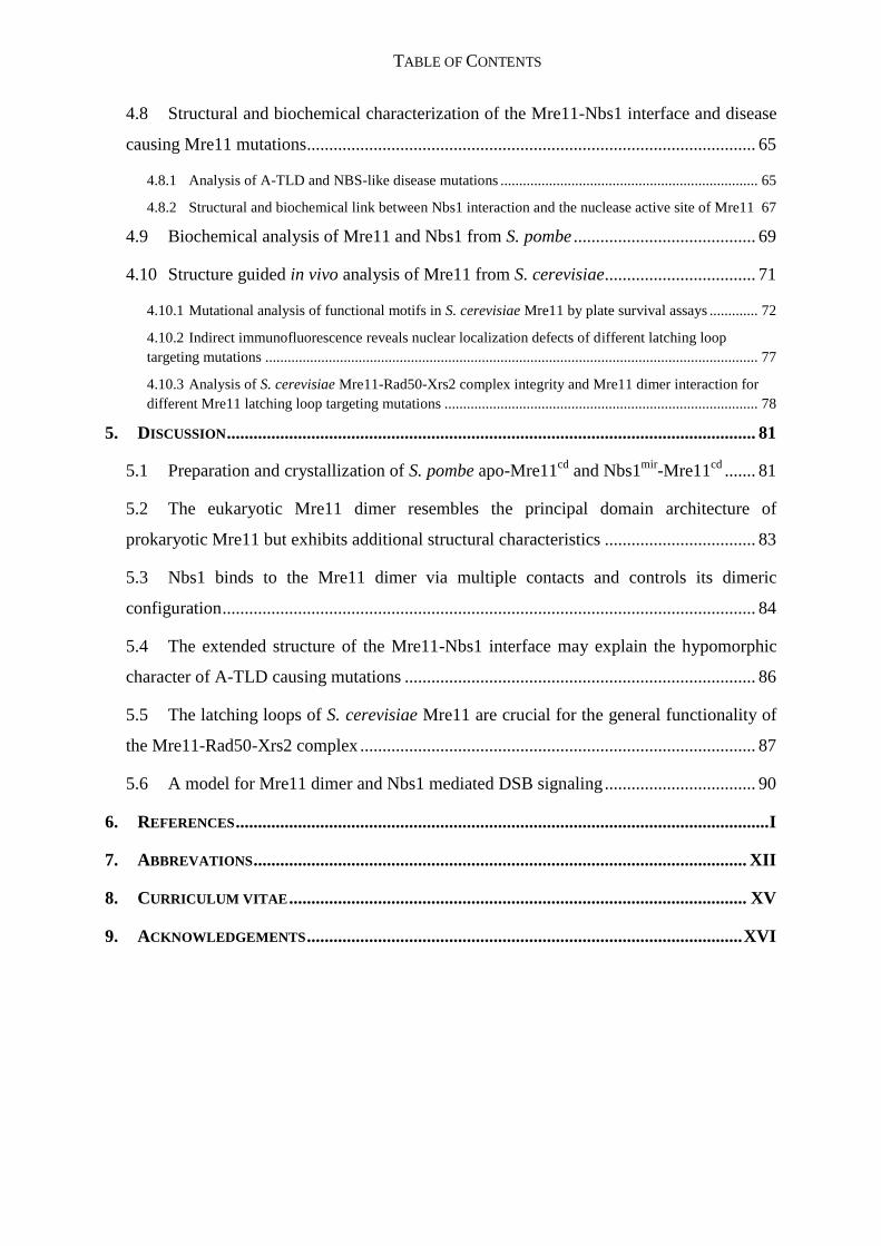
TABLE OF CONTENTS
4.8 Structural and biochemical characterization of the Mre11-Nbs1 interface and disease
causing Mre11 mutations..................................................................................................... 65
4.8.1 Analysis of A-TLD and NBS-like disease mutations ..................................................................... 65
4.8.2 Structural and biochemical link between Nbs1 interaction and the nuclease active site of Mre11 67
4.9 Biochemical analysis of Mre11 and Nbs1 from S. pombe ......................................... 69
4.10 Structure guided in vivo analysis of Mre11 from S. cerevisiae.................................. 71
4.10.1 Mutational analysis of functional motifs in S. cerevisiae Mre11 by plate survival assays ............. 72
4.10.2 Indirect immunofluorescence reveals nuclear localization defects of different latching loop
targeting mutations .................................................................................................................................... 77
4.10.3 Analysis of S. cerevisiae Mre11-Rad50-Xrs2 complex integrity and Mre11 dimer interaction for
different Mre11 latching loop targeting mutations .................................................................................... 78
5. DISCUSSION ....................................................................................................................... 81
5.1 Preparation and crystallization of S. pombe apo-Mre11cd
and Nbs1mir
-Mre11cd
....... 81
5.2 The eukaryotic Mre11 dimer resembles the principal domain architecture of
prokaryotic Mre11 but exhibits additional structural characteristics .................................. 83
5.3 Nbs1 binds to the Mre11 dimer via multiple contacts and controls its dimeric
configuration ........................................................................................................................ 84
5.4 The extended structure of the Mre11-Nbs1 interface may explain the hypomorphic
character of A-TLD causing mutations ............................................................................... 86
5.5 The latching loops of S. cerevisiae Mre11 are crucial for the general functionality of
the Mre11-Rad50-Xrs2 complex ......................................................................................... 87
5.6 A model for Mre11 dimer and Nbs1 mediated DSB signaling .................................. 90
6. REFERENCES ........................................................................................................................ I
7. ABBREVATIONS ............................................................................................................... XII
8. CURRICULUM VITAE ....................................................................................................... XV
9. ACKNOWLEDGEMENTS .................................................................................................. XVI

1. SUMMARY
1
1. SUMMARY
The integrity of the genome is constantly threatened by environmental influences and cellular
metabolism processes. DNA double strand breaks (DSBs) are among the most hazardous of
all DNA lesions and arise from failures in genome metabolism processes and from exogenous
sources. In addition they are important programmed intermediates in DNA metabolism. Cells
have evolved efficient pathways to repair DSBs and here the Mre11-Rad50-Nbs1 (MRN)
complex is a central key factor. Mre11 and Rad50 are conserved in all domains of life,
whereas Nbs1 is a eukaryote-specific protein and plays regulatory roles within the complex.
MRN senses and binds DSBs, recruits other repair factors and also stabilizes DSBs by its
tethering activity. Furthermore it processes DSB ends for repair and is involved in DNA
damage signaling by co-activating the checkpoint kinase ATM.
Null mutations of Mre11-Rad50-Nbs1 coding genes are lethal in higher eukaryotes, whereas
hypomorphic mutations induce different heredity diseases. Ataxia-telangiectasia like disorder
(A-TLD) and Nijmegen breakage syndrome (NBS) are linked to mutations in Mre11 and
Nbs1 respectively. However, also mutations in Mre11 and Rad50 may lead to an NBS-like
disorder. All diseases share genomic instability and delayed checkpoint activation.
The aim of this work was to characterize the structural and functional interplay between
eukaryotic Mre11 and Nbs1 and to analyze how it influences the role of the complex in repair
and checkpoint activation. For this purpose proteins form the fission yeast
Schizosaccharomyces pombe were studied and the Mre11 nuclease dimer alone and in
complex with the interacting region of Nbs1 determined as crystal structures. The Mre11-
Nbs1 structure reveals binding of two Nbs1 molecules as extended peptides to one Mre11
dimer at the outside of the nuclease domains. One Nbs1 molecule mediates also a second
interaction with Mre11 by asymmetrically binding across the Mre11 dimer and thereby
determining its dimeric conformation. The interfaces of Mre11 and Nbs1 were analyzed and
verified by mutational analysis in vitro using recombinant S. pombe proteins and in vivo in
Saccharomyces cerevisiae. The structures also allowed studying of the molecular basis for
several A-TLD and NBS-like disease mutations. As a result, all analyzed A-TLD mutations
exhibited a weakened but not abolished Nbs1 interaction, which might explain the
hypomorphic phenotype of A-TLD. Finally a model is proposed, in which a conformational
switch in the Mre11 dimer and modulated Nbs1 interactions permit subsequent DSB repair
and signaling.

2. INTRODUCTION
2
2. INTRODUCTION
2.1 Biological roles of DNA double-strand breaks
The maintenance of genomic stability is a fundamental problem for all living organisms, since
the integrity of every genome is constantly threatened by different sources of DNA damage.
DNA double strand breaks (DSBs) are among the most hazardous DNA lesions. They can
lead to chromosomal rearrangements and induction of cancerogentic diseases, if not repaired
properly. However, DSBs are also important intermediates in different DNA metabolism
processes where they are temporary inserted into the genome. The following chapter is giving
a short overview about the different sources of DSBs and their impact on the genomic
stability.
2.1.1 DNA double strand breaks in cellular metabolism processes
The majority of accidentally occurring DSBs in proliferating cells arise from aberrations in
DNA replication: Replication at blocking lesions or single-strand nicks can lead to a
replication fork collapse, which results in the generation of DSBs. (Costanzo et al. 2001;
Kuzminov 2001). But also (by)products of normal cellular metabolism processes like e.g.
reactive oxygen species (ROS) contribute significantly to the introduction of these blocking
lesions into the DNA (Cadet et al. 1997; Borde and Cobb 2009). Since most often a sister
chromatid is available as a repair template in S-phase, DSBs arising from collapsed
replication forks are mainly repaired by the homologous recombination (HR) machinery
(Errico and Costanzo 2010).
Importantly, DSBs are not solely harmful, but also play beneficial roles in the cell. During
various biological processes, DSBs are introduced transitionally into the genome in
programmed ways: One example is the switching of mating types in the budding yeast
S. cerevisiae: This process is initiated by a site specific cleavage of the HO endonuclease at
the MAT gene locus, which generates a DSB. Subsequently, the mating type gene is switched
by unidirectional gene conversation via recombination with the HML or HMR gene cassette,
which carry silenced copies of the mating types a and α respectively (Haber 1998; Coic et al.
2006).

2. INTRODUCTION
3
The programmed introduction of DSBs is also a crucial event during the generation of
immunoglobulins (Ig) and T cell receptors (TCR) by the vertebrate immune system. The
required diversity of these molecules is achieved by a process called V(D)J recombination.
Combination of Variable (V), Diversity (D) and Joining (J) encoding gene segments through a
specific DNA rearrangement mechanism leads to a broad diversity of proteins and allows the
recognition of many different antigens (Tonegawa 1983; Dudley et al. 2005). The process
begins with the introduction of DSBs by the RAG1/RAG2 proteins, which recognize
recombination signal sequences (RSS) at the borders of the V, D and J gene elements. This
results in two hairpin-sealed coding ends and two blunt signal ends. The following processing
steps are carried out by proteins of the non-homologous end joining (NHEJ) machinery,
which mediate the error prone repair of the breaks (Raghavan et al. 2005).
The specificity and efficiency of immunoglobulins is further increased after activation of the
humoral immune response by antigens via two different processes: Class switch
recombination (CSR) leads to the exchange of the Ig constant region of antibodies and allows
the generation of different antibody classes, whereas somatic hypermutation introduces
additional mutations into the Ig variable region. The activation-induced cytidine deaminase
(AID) in both processes initiates the introduction of DSBs which are joined and subsequently
repaired by NHEJ (Soulas-Sprauel et al. 2007; Dinkelmann et al. 2009; Zha et al. 2011).
Important roles of the Mre11-Rad50-Nbs1 complex in different NHEJ dependent repair
processes are discussed below (2.4.4).
In most sexually reproducing organisms programmed DSBs are also generated during the
process of meiosis. After the alignment of homologous chromosomes in meiotic prophase I,
DSBs are introduced at specific hot spot sites on the chromosomes by the type II
topoisomerase-like enzyme Spo11. The covalently bound Spo11 is then removed from the
DNA by the Mre11-Rad50-Nbs1 complex and resection of the 5`-strand takes place. Finally,
the DSBs are repaired by meiotic recombination between homologous chromosomes resulting
in gene conversion or chromosomal crossing over (Borde 2007; Inagaki et al. 2010).
2.1.2 Environmentally caused DNA double strand breaks
The genome is not only exposed to endogenous mutagens like oxidative byproducts of
cellular respiration, but also environmental agents like ionizing radiation, UV-light or

2. INTRODUCTION
4
genotoxic chemicals can cause various DNA damages. These include directly or indirectly
introduced DSBs (Hoeijmakers 2001).
Ionising radiation (IR) occurs naturally e.g. by radioactive decay of instable atomic nuclei or
by cosmic radiation. Besides, IR is used in medical procedures like X-ray inspections or
radiation therapy in cancer treatment (Ciccia and Elledge 2010). IR produces a broad
spectrum of different DNA damages, which are introduced via the production of reactive
oxygen species (Mahaney et al. 2009). Most often IR leads to DNA base damages or
introduction of DNA single-strand breaks (SSBs), which are repaired by base excision repair
(BER) or single strand repair pathways (Almeida and Sobol 2007; Dianov and Parsons 2007).
IR-caused DSBs occur when two SSBs are introduced in close proximity on opposite DNA
strands (Sutherland et al. 2000). Therefore, IR caused DSBs often possess single strand
overhangs. In addition IR produces DNA breaks with 3' termini carrying phosphate or
phosphoglycolate groups, which need to be removed before ligation of the breaks (Henner et
al. 1983).
UV light on the other hand can indirectly provoke DSB formation by introducing 6-4
photoproducts and cyclobutane pyrimidine dimers into the DNA. These bulky lesions may
induce replication-fork collapse and thereby DSBs if not repaired properly by the nucleotide
excision repair (NER) machinery (Limoli et al. 2002). Similar effects are induced by different
genotoxic chemicals, which also create replication blocking lesions like e.g. different
alkylating agents, the intrastrand crosslinking anti-cancer-drug cisplatin or the interstrand
crosslinking agent mitomycin (Bosco et al. 2004; Al-Minawi et al. 2009).
In addition, chemicals, which poison the topoisomerase enzymes, can promote DSB
formation by stabilizing the cleavage complex in which the topoisomerase is covalently
attached to the cleaved DNA (Degrassi et al. 2004). The Topoisomerase I (TopI) inhibitor
camptothecin (CPT) triggers the accumulation of TopI-bound SSBs, which may be converted
to DSBs when a replication fork collides with the cleavage complex (Jacob et al. 2005).
Topoisomerase II (TopII) enzymes introduce DSBs in the DNA during their catalytic cycle.
Top II inhibitors like etoposide increase the concentration of cleavage complexes, which can
be converted to permanent DSBs by collision with polymerases or helicases (Bromberg et al.
2003).

2. INTRODUCTION
5
2.2 DNA double-strand repair pathways - A short overview
To protect the genome and thus ensure the integrity of its coded information, cells have
evolved different sophisticated mechanisms to repair DSBs: The two major pathways here are
homologous recombination (HR) and non-homologous end joining (NHEJ) (Harper and
Elledge 2007).
HR enables the cell to repair DSBs in a relatively error free manner by using a sister- or
homologous chromatid as a template (Figure 2.1). The repair of DSBs by HR comprises
several sequential steps. First, 3´ ssDNA tails adjacent to the break have to be generated by
the combined action of several nucleases and helicases. The Mre11-Rad50-Nbs1 (MRN)
complex plays here crucial roles in the first steps of HR by sensing and tethering the break but
also in mediating the initiation of resection (2.4.1) (Huertas 2010). The resected 3´ ssDNA tail
is first coated by RPA, which is replaced by Rad51 (RecA in bacteria). Rad51 assembles on
the ssDNA to build a helical nucleoprotein filament, which in concert with proteins of the
Rad52 epistasis group and other HR proteins screens for a homologous sequence (New et al.
1998; Symington 2002). The nucleoprotein filament invades into the homologous donor
sequence and after removal of Rad51 hybridizes with it via normal base pairing, thereby
building a displacement loop (D-loop) structure (Sung and Klein 2006). After strand
extension by a DNA polymerase, using the donor strand as a template, the D-loop
intermediate can be repaired by two different HR mechanisms: (1) the double-strand break
repair (DSBR) pathway or (2) synthesis-dependent single strand annealing (SDSA). In DSBR
the second 3´ ssDNA tail, which was not involved in D-loop formation, is captured by
annealing to the extended D-loop and thereby an intermediate structure with two Holliday
junctions is formed (Bzymek et al. 2010). Depending on how the Holiday junctions are
resolved, DSBR results in crossover or non-crossover recombination products (Heyer 2004).
In the alternative SDSA pathway the invading 3´ ssDNA tail is displaced after its extension
and reanneals to the single-stranded DNA tail, that was not involved in D-loop formation.
Repair of DSBs by the SDSA results always in non-crosslinking recombination products (San
Filippo et al. 2008).
Sometimes DSBs are closely flanked by repeat sequences. In this case recombination repair
via single-strand annealing (SSA) may occur. Like in DSBR or SDSA also SSA is initiated by
end resection, which generates 3´ ssDNA tails, although it does not require a homologous
chromosome for recombination. Instead, the resected ends anneal via their repeat sequences,

2. INTRODUCTION
6
followed by nucleolytical removal of nonhomologous flap structures and ligation. SSA results
in the deletion of the sequence regions between the repeat elements and is therefore
considered to be potentially mutagenic (Ivanov et al. 1996; Mansour et al. 2008).
Figure 2.1: Overview of the major DSB repair pathways. Figure adapted from (Pandita and Richardson
2009). DSBs can be repaired by either non-homologous end joining (NHEJ) or homologous recombination (HR).
For details see text.
The more error-prone NHEJ pathway is utilized especially in G1 phase when no sister
chromatid is available for recombinational repair (Figure 2.1). Depending on the organism,
however it also occurs in S- and G2 phase cells. NHEJ promotes direct ligation of the two
ends. The main NHEJ pathway, also called classical NHEJ (c-NHEJ) pathway, is initiated
through binding of the Ku70/80 heterodimer to the DSB (Weterings and van Gent 2004).
Next, the Ku heterodimer recruits the catalytic subunit of the DNA-dependent protein kinase
(DNA-PK). The newly formed Ku/DNA-PK complex then places both DSBs into

2. INTRODUCTION
7
juxtaposition. DNA-PK gets autophosphorylated upon binding to Ku and the DNA. This is
required for the further recruitment of other NHEJ proteins to the DSB (Meek et al. 2007). If
the DSB ends do not require further processing, they are directly ligated by a complex
consisting of DNA ligase IV, XRCC4 and XLF/Cernunnos (Ahnesorg et al. 2006). Often, the
DNA ends are not compatible for direct ligation, for example if the ends possess ssDNA
overhangs or damaged bases. In this case different other NHEJ factors like the Artemis
nuclease, the Pol TdT (terminal deoxynucleotidyl transferase), pol lambda, pol mu and also
the Mre11-Rad50-Nbs1 complex process the end to allow ligation by the ligase IV/XRCC4
complex (Figure 2.1) (Lieber 2010).
Sometimes NHEJ employs also alternative pathways (a-NHEJ), in which additional factors
distinct from the c-NHEJ machinery facilitate the repair. One of these a-NHEJ pathways is
called microhomology-mediated endjoining (MMEJ) in which several bases are
nucleolytically removed from the DSB. This allows hybridization of single-stranded DNA
ends at short stretches of sequence homology. Therefore, MMEJ is considered to be highly
mutagenic (Haber 2008; Fattah et al. 2010). Mre11 was reported to be the major nuclease in
the resection procedure of MMEJ, which generates DSBs with compatible microhomology
sequences (Rahal et al. 2010).
2.3 The Mre11-Rad50-Nbs1 complex - biochemistry and structural architecture
The Mre11-Rad50-Nbs1/Xrs2 (MRN(X) complex plays various central roles in most, if not
all DNA double-strand break repair pathways. It senses and binds to DSBs and functions as a
recruiting platform for many other DNA repair proteins. Furthermore, it is a scaffold protein,
which stabilizes DSBs via its tethering activity. MRN is also involved in nucleolytic
processing of DNA ends and functions in DNA damage signaling by co-activating the
checkpoint kinase ATM. MRN comprises the Mre11 endo/exonuclease dimer, two Rad50
ATP-binding cassette proteins and contains in eukaryotic organisms also the third subunit
Nbs1 (Xrs2 in S. cerevisiae) as a regulatory factor (Assenmacher and Hopfner 2004; Williams
et al. 2010). The core complex consisting of the catalytic subunits Mre11 and Rad50 is
conserved from bacteria and archaea to eukaryotes and is even found in some viruses like the
bacteriophage T4 (Sharples and Leach 1995; Hopfner et al. 2000; Herdendorf et al. 2011).
Mre11, which stands for Meiotic recombination 11, was first identified in a genetic screen for
proteins functioning in meiosis in S. cerevisiae (Ajimura et al. 1993). Rad50 was discovered

2. INTRODUCTION
8
already earlier in genetic yeast studies were its deletion mutant strain was reported to be
sensitive to ionizing radiation and to produce inviable spores in meiosis (Game and Mortimer
1974). The human homologue of Nbs1, which is also called Nibrin, was functionally
identified in 1998, even though the genetic disease Nijmegen breakage syndrome, which is
linked to mutations in the Nbs1 gene, was known since much longer times (Weemaes et al.
1981; Varon et al. 1998).
2.3.1 Biochemical in vitro activities of Mre11-Rad50-Nbs1
Biochemical in vitro studies with bacterial, archaeal, yeast and human Mre11 and Rad50
proteins revealed that Mre11 possesses Mn2+
dependent nuclease activities: The dsDNA 5-3´
exonuclease activity of Mre11 is dependent on ATP binding by Rad50 in bacteria and
archaea, while being rather unaffected by ATP binding in eukaryotes. The ssDNA
endonuclease activity appears to be ATP independent (Furuse et al. 1998; Paull and Gellert
1998; Trujillo et al. 1998; Connelly et al. 1999; Hopfner et al. 2001). Mre11 exhibits also the
ability to open and process hairpin DNAs in an ATP dependent manner (Paull and Gellert
1998; Connelly et al. 1999; Trujillo and Sung 2001). Additionally, its nuclease activity can
remove covalently bound proteins from DNA ends (Connelly et al. 2003). The importance of
Mre11`s nuclease activity for its in vivo functions is discussed below (2.4).
The exact function of the ATPase activity of Rad50 was longtime puzzling. As already
mentioned, it stimulates the nucleolytic cleavage activity of Mre11 on hairpin structures.
Further, it was shown to be important for tethering DNA ends, and a Rad50 signature motif
mutant, which is impaired in ATP binding, lacks the ability to stimulate the checkpoint kinase
ATM in vitro (Lee and Paull 2005; Dupre et al. 2006). In addition, biochemical studies with
recombinant human MRN proteins indicated also a stimulatory role of Rad50 ATP binding
for melting and unwinding of DNA secondary structures (Paull and Gellert 1999). Recent data
from the Hopfner group revealed that ATP binding by Rad50 induces conformational changes
within the Mre11-Rad50 complex which promotes binding of DNA ends (Lammens et al.
2011).
Nbs1 possesses no own catalytic activity but plays regulatory roles within the eukaryotic
MRN complex. Human Nbs1 and the homologous Xrs2 protein from S. cerevisiae were
shown to stimulate DNA binding as well as nucleolytic hairpin processing by Mre11-Rad50
(Paull and Gellert 1999; Lee et al. 2003). Moreover, Nbs1 is a co-activator of the checkpoint

2. INTRODUCTION
9
kinase ATM in concert with Mre11-Rad50, which was also observed by in vitro reconstitution
assays with the recombinant human proteins (Falck et al. 2005; Lee and Paull 2005; You et al.
2005).
2.3.2 Structural architecture of the Mre11-Rad50-Nbs1 complex
The Mre11-Rad50 (MR) core complex is a heterotetramer consisting of two Mre11 and two
Rad50 molecules. In eukaryotes it associates furthermore with one or two molecules of Nbs1
(or the homologous Xrs2 protein in S. cerevisiae) to build the MRN(X) complex (Figure 2.2
A) (Paull and Gellert 1999; van der Linden et al. 2009). Atomic force microscopy data
(Figure 2.2 B) showed that MRN consists of a globular head region, which harbors the
catalytic functions of the complex and contains the Mre11 dimer, the ABC-ATPase domains
of Rad50 and the Nbs1 molecules. A large helical region, which links the N- and C-terminal
ATPase domains of Rad50, folds into a long coiled coil tail, which protrudes from the
catalytic head region (de Jager et al. 2001; Hopfner et al. 2002; Moreno-Herrero et al. 2005).
Figure 2.2: Structural organization of the MRN complex and DNA induced mesoscale conformational
changes: (A) Model of the eukaryotic MRN complex. MRN consists of a globular head which contains the
Mre11 dimer, two Rad50 ABC ATPases and two Nbs1 molecules. A long flexible coiled-coil region protrudes
from each Rad50 molecule. A Zinc-hook dimerization domain at the other end of the coiled-coil allows
interaction between different MRN complexes. The Figure was adapted from (Stracker and Petrini 2011).
(B) Atomic force microscopy (AFM) images of the human MRN complex in the presence and absence of DNA.
For each structural arrangement of MRN a schematic model is shown. Left figure: Image of the human MRN

2. INTRODUCTION
10
complex in the absence of DNA, resolving clearly the globular head and the coiled-coil region. Middle figure:
Upon binding of the head region to DNA (here 90bp dsDNA), the coiled-coils are oriented in a parallel
conformation. Left figure: MRN intercomplex interaction, mediated by the apical zinc-hook domain. All figures
from (B) are adapted from (Moreno-Herrero et al. 2005).
The Rad50 coiled-coil tail harbors an apical zinc-hook dimerization motif that allows the
interaction with other MRN complexes (Figure 2.2 A) (Hopfner et al. 2002).
The conserved N-terminus of Mre11 consists of a phosphodiesterase domain and a C-
terminally adjacent DNA capping domain (Figure 2.3). Yeast two hybrid studies with human
Mre11 and Nbs1 indicated that the phosphodiesterase domain harbors the interaction site for
Nbs1 (Desai-Mehta et al. 2001). However, the exact locations of Nbs1 interaction regions
within Mre11 were unknown and could be identified during the work for this thesis. The
interaction region(s) in Mre11 for Rad50 were first roughly mapped with deletion mutants in
S. cerevisiae and were recently confirmed by combined structural and biochemical studies
(Chamankhah and Xiao 1999; Lammens et al. 2011; Lim et al. 2011; Williams et al. 2011).
Eukaryotic Mre11 possesses two distinct DNA interaction motifs which flank the main
binding site for Rad50 (Figure 2.3). Whereas the DNA interaction motif adjacent to the
capping domain was reported to be important and sufficient for mitotic repair in S. cerevisiae,
the C-terminal motif is crucial for DSB formation and spore viability in meiosis (Furuse et al.
1998; Usui et al. 1998).
Rad50 contains a bipartite ATP-binding cassette - ATPase (ABC-ATPase), which is build up
by an N-terminal and a C-terminal domain, separated in the primary structure by a long
coiled-coil region (Figure 2.3). The N-terminal domain harbors the Walker A motif and the C-
terminal domain the Walker B and signature motifs. A highly conserved Cys-X-X-Cys
sequence maps to the center of the coiled-coil region. It folds into the MRN intercomplex
mediating zinc-hook motif. Rad50`s major binding sites for Mre11 map to the N- and C-
terminal ends of the coiled-coil region (Hopfner et al. 2000; Hopfner et al. 2002; Lammens et
al. 2011).
The eukaryotic Nbs1 /Xrs2 protein is the least conserved compound of the MRN(X) complex
(Figure 2.3). Its N-terminal region consists of a Forkhead domain and two BRCT domains,
which mediate binding of the MRN to different phosphoproteins in DNA repair (Palmbos et
al. 2005; Hari et al. 2010). The C-terminal region appears to be mainly unstructured as seen
by limited proteolysis analysis for the S. pombe protein homologue (Williams et al. 2009). It

2. INTRODUCTION
11
contains interaction motifs for Mre11 and the checkpoint kinase ATM (Desai-Mehta et al.
2001; Ueno et al. 2003; Falck et al. 2005; You et al. 2005).
Figure 2.3: Domain architecture of Mre11-Rads50-Nbs1: Mre11 consists of a conserved N-terminal
phosphodiesterase and an adjacent DNA capping domain. The C-terminal region contains a hydrophobic
interaction motif for Rad50, which is flanked by two DNA binding regions. Rad50 is build up of a bipartite
ABC-ATPase cassette, which is separated by a long coiled-coil region. An MRN intercomplex interaction
mediating Zn-hook maps to the central coiled-coil region. Nbs1 contains an N-terminal phosphoprotein binding
module, which comprises a Forkhead and two BRCT domains. The C-terminus possesses interaction sites for
both Mre11 and ATM. Domain maps are adapted from (Assenmacher and Hopfner 2004; Stracker and Petrini
2011).
Mre11 is a dimeric molecule in solution and the Mre11-Mre11 interaction is mediated via the
phosphodiesterase domains (Hopfner et al. 2001). Importantly, Mre11 dimerization is crucial
for the functionality of the MR(N) complex. It was e.g. observed in the archaeal Mre11-DNA
crystal structure from Pyrococcus furiosus that both molecules of the Mre11 dimer bind
cooperatively to one DNA molecule (Figure 2.4 C). The same authors showed also
biochemically, that mutations leading to Mre11 dimer disruption in the yeast
Schizosacchaormyces pombe render cells sensitive to different genotoxic agents (Williams et
al. 2008).
Recently, crystal structures of Mre11-Rad50 complexes from bacteria and archaea were
published by the Hopfner group and others. These structures reveal that the complex exhibits
an open form with a central Mre11 nuclease dimer and peripherial Rad50 molecules in the
absence of ATP or DNA (Figure 2.4 A). Binding of ATP leads to the dimerization of Rad50
molecules (Figure 2.4 B) and also increases - at least in the case of Thermotoga maritima
Mre11-Rad50 - the DNA affinity of the complex (Lammens et al. 2011; Lim et al. 2011;
Williams et al. 2011).

2. INTRODUCTION
12
Figure 2.4: Published crystal structures of Mre11-Rad50 and Nbs1. (A) Structure of the Mre11-Rad50
complex from the thermophilic bacterium Thermotoga maritima at 3.4 Å resolution (Lammens et al. 2011). (B)
Structure of Mre11-Rad50 from the archaeal organism Methanococcus jannaschii bound to ATPyS at 3.1 Å
resolution (Lim et al. 2011). (C) Structure of the Mre11 nuclease dimer from the archaeal organism Pyrococcus
furiosus in complex with a hairpin DNA at 2.2 Å resolution (Williams et al. 2008). (D) Structure of the N-
terminus of Nbs1 from Schizosaccharomyces pombe at 2.8 Å resolution. The structure contains the Forkhead
domain and the two BRCT domains, which are all involved in binding of phosphoproteins (Williams et al.
2009).
For Nbs1/Xrs2, only the N-terminal region is structurally characterized by crystal structures
of the Forkhead-domain and two BRCT domains from the fission yeast Schizosaccharomyces
pombe and an NMR-structure of the second BRCT domain from Xenopus laevis (Figure 2.4
D). The structures from S. pombe Nbs1 show a very compact arrangement of the Forkhead
and BRCT domains and were proposed to be linked to Mre11-Rad50 via the flexible C-
terminus of Nbs1 (Xu et al. 2008; Lloyd et al. 2009; Williams et al. 2009). However, the
interaction between Nbs1 and Mre11-Rad50 is not understood on a molecular level yet.
Therefore, an atomic structure of eukaryotic Nbs1 in complex with Mre11 or Rad50 would be

2. INTRODUCTION
13
highly valuable to help understanding the regulatory functions of Nbs1 within the MRN
complex.
2.4 The Mre11-Rad50-Nbs1 complex in double-strand break repair
2.4.1 The Mre11-Rad50-Nbs1 complex in homologous recombination
The Mre11-Rad50-Nbs1/Xrs2 MRN(X) complex is one of the first complexes which localize
to DSBs and plays various key roles in repair by sensing DSB ends, stabilizing breaks,
initiation of DNA resection and damage signaling. Several studies suggest antagonistic roles
for MRN(X) and the Ku complex, which is the second cellular DSB sensor, in the early phase
of repair events. Both complexes sense and bind to DSBs but whereas the MRN(X) complex
is the core initiation factor for HR, the Ku complex promotes NHEJ. However, MRN(X) is
also involved in different NHEJ repair pathways (2.4.4). It has been shown that the Ku
complex suppresses homologous recombination by inhibiting MRN(X) complex dependent
DNA end resection in G1 phase, but much less in S and G2 phase. How exactly the choice of
pathway is regulated in a cell-cycle dependent manner is only poorly understood yet, but it is
clear that DNA 5´-strand resection by MRN(X) and other nucleases shifts the balance towards
the HR pathway (Clerici et al. 2008; Zierhut and Diffley 2008; Shim et al. 2010).
Initiation of HR by resection depends strongly on the MRN(X) complex and the Sae2 protein
(in S. cerevisiae) or its homologues Ctp1 (in S. pombe) and CtIP (in metazoans), which are
poorly conserved in sequence (Figure 2.5 A). In addition, the phosphorylation of Sae2 or CtIP
by cyclin-dependent protein kinases is crucial for this step (Limbo et al. 2007; Huertas et al.
2008; Huertas and Jackson 2009). The dependence of initial DNA 5´ end resection on the
nuclease activity of Mre11 differs between organisms. Whereas the nuclease deficient Mre11
H125N mutant exhibits only a mild phenotype in S. cerevisae, the equivalent mutations in
S. pombe (H134S) or mouse (H129N) cause severe radiosensitivity and embryonic lethality,
respectively. One hypothesis which might explain these divergent phenotypes is that the
presence of Ku is more dominating in S. pombe and mice, thereby raising the barrier to
initiate resection. (Lewis et al. 2004; Limbo et al. 2007; Buis et al. 2008; Williams et al. 2008;
Mimitou and Symington 2011).

2. INTRODUCTION
14
The exact functional role of Sae2/Ctp1/CtIP in the resection procedure remains controversial.
The S. cerevisiae Sae2 protein was shown to possess an in vitro nuclease activity on its own,
which together with Mre11 might function in resection (Lengsfeld et al. 2007). However, no
nuclease activity was reported for S. pombe Ctp1 and human CtIP. For both organisms, the
proteins were suggested to act as co-factors of MRN and to stimulate the resection activity of
Mre11 (Limbo et al. 2007; Sartori et al. 2007). Resection by MRN(X) and Sae2/Ctp1/CtIP is
not processive but rather leads to the generation of a short 3´ ssDNA tail before other
nuclease/helicase complexes take over for processive long range resection of several
kilobases. In S. cerevisae, where the mechanistic details of resection are probably best
understood, the MRX complex stimulates the recruitment of the Sgs1/Top3/Rmi1
(STR)/Dna2 complex and Exo1 to the break, which are responsible for the majority of long
range resection (Shim et al. 2010). In addition, the Pso2 nuclease might play a backup role in
this process, since it promotes a residual resistance to IR in the absence of nuclease active
Mre11 and Exo1 in S. cerevisiae (Lam et al. 2008).
Figure 2.5: Roles of the MRN(X) complex in the resection of mitotic or meiotic DSBs and in telomere
processing. (A) Mitotic DSBs: MRX and Sae2 sense and bind to free DSBs. Sae2 is phosphorylated by Cdk1
which promotes resection initiation by MRX and Sae2, leading to the generation of short 3´ ssDNA tails. These
are then substrates for processive end resection carried out by Dna2/Sgs1 or Exo1. (B) Meiotic DSBs: MRX and
other proteins stimulate the generation of DSBs by Spo11. MRX and Sae2 then nucleolytically remove the

2. INTRODUCTION
15
covalently bound Spo11 from the DSBs. Cdk1 dependent phosphorylation of Sae2 is important for this
processing step. (C) Telomere replication: The G strand is replicated by the lagging strand machinery which
results in a short 3` ssDNA overhang after removal of the RNA primer. The leading strand machinery instead is
expected to generate blunt ends by replication of the C strand. To generate the 3´ ssDNA strand overhangs of
telomeres these blunt ends are resected by MRX and Sae2, similar to the situation in recombination resection.
Also here resection by MRX and Sae2 depends on Cdk1 phosphorylation of Sae2 and processive resection is
facilitated by Dna2/Sgs1 or Exo1. Figure from (Longhese et al. 2010).
Besides mediating DNA end resection, the MRN(X) complex is also important as a scaffold
factor in HR, which tethers the two DNA ends of a DSB and holds them in close proximity.
This function is dependent on the Rad50 zinc-hook domain, which mediates MRN(X)
intercomplex interactions (Hopfner et al. 2002). Atomic force microscopy studies monitored
this zinc-hook dependent MRN(X) intercomplex formation in the presence of DNA on a
molecular level (de Jager et al. 2001; Hopfner et al. 2002; Moreno-Herrero et al. 2005).
2.4.2 The Mre11-Rad50-Nbs1 complex in meiotic recombination
The MRN(X) complex had long been implicated to be important for the DNA end resection in
recombination repair since mutations in the genes encoding the complex cause a complete
block of 5´ strand removal in meiosis (Ivanov et al. 1992; Keeney and Kleckner 1995; Usui et
al. 1998). The roles of MRN(X) in meiosis are probably best understood for S. cerevisiae.
Here, the complex is important for several sequential steps of the process (Figure 2.5 B): (1)
First, MRX is recruited to meiotic DSB sites before the formation of DSBs, via its C-terminal
region (Furuse et al. 1998; Usui et al. 1998). It then facilitates the generation of DSBs by
Spo11, which is probably also mediated by the Mre11 C-terminus and is independent of
Mre11`s nuclease activity as well as stable complex formation with Rad50 and Xrs2. This was
shown by studies with the mre11-58 mutation, which is deficient for all of these functions, but
still functional in meiotic DSB formation (Usui et al. 1998). (2) The MRX complex is crucial
for the endonucleolytic removal of the covalently bound Spo11 from the 5´ strands of the
DSB ends. The nuclease activity of Mre11 is likely responsible for this processing step since
separation of function mutants (Mre11S), which have a defect in Spo11 removal, are nuclease
deficient in vitro (Furuse et al. 1998; Usui et al. 1998; Moreau et al. 1999). Furthermore,
nucleolytic removal of Spo11 depends on the Sae2/Com1 protein (McKee and Kleckner 1997;
Prinz et al. 1997). Remarkably, a specific class of Rad50 separation of function mutants,
named Rad50S, resembles the same phenotype as ∆sae2 in meiosis (Alani et al. 1990; Keeney

2. INTRODUCTION
16
et al. 1997). The Rad50S mutations cluster to the outer surface of the ABC-ATPase dimer in
the homologous archaeal P. furiosus crystal structure, suggesting that they might affect a Sae2
interaction site, which is important for Spo11 removal. However, a direct in vitro interaction
of MRX and Sae2 could not be observed (Hopfner et al. 2000; Lengsfeld et al. 2007). After
removal of Spo11, the MRX complex also stimulates recruitment of other nucleases, which
function in the processive generation of ssDNA tails for meiotic D-loop formation. Here
especially the Exo1 nuclease plays an important role, whereas the activity of the SGS1/DNA2
complex appears to be rather dispensable (Zakharyevich et al. 2010; Keelagher et al. 2011).
2.4.3 The Mre11-Rad50-Nbs1 complex in telomere maintenance
Beside its many functions in DSB repair and signaling, the MRN(X) complex plays also
crucial roles in telomere maintenance. Telomeres are specialized nucleoprotein structures that
protect the ends of eukaryotic chromosomes from degradation, fusion, recombination and
recognition by the DNA-damage repair machinery (Faure et al. 2010). They consist of several
G-rich sequence repeats and a terminal 3´ ssDNA tail, which is capped by specific protecting
factors like the CST (Cdc13-Stn1-Ten1) complex in S. cerevisiae. The length of telomeres is
maintained by the telomerase complex, which uses its RNA template to add G-rich telomeric
repeats to the terminal 3´ ssDNA tail (Hug and Lingner 2006). Recruitment of the telomerase
to 3´ ssDNA ends strongly depends on the MRX complex and Tel1 as studies in S. cerevisiae
showed. MRX recruits Tel1 to short telomeres, where its kinase activity stimulates telomerase
dependent telomere lengthening (Goudsouzian et al. 2006; Hector et al. 2007; Hirano et al.
2009). In addition, the MRN(X) complex is also important for the generation of 3´ ssDNA
overhangs after telomere leading strand replication (Figure 2.5 C). Replication at telomeres is
thought to result in blunt ends on the leading strand. In S. cerevisiae, the generation of
3´ ssDNA overhangs on the leading strand blunt end is carried out by the MRX complex and
Sae2, which initiate the resection of the 5´ strand. The thereby created short 3´ ssDNA
overhang is then extended by Sgs1/Dna2 or Exo1 and finally capped by the CST complex
(Bonetti et al. 2009; Mimitou and Symington 2009; Longhese et al. 2010).

2. INTRODUCTION
17
2.4.4 The Mre11-Rad50-Nbs1 complex in non-homologous end joining pathways
Studies with mammalian cells showed that the MRN complex is involved in both the classical
NHEJ (c-NHEJ) as well as alternative NHEJ pathways (a-NHEJ). Even though it is no core
factor of mammalian c-NHEJ, it is crucial for V(D)J recombination, which is strongly
dependent on c-NHEJ (Deriano et al. 2009; Helmink et al. 2009). In addition, depletion or
inhibition of Mre11 reduces the end-joining efficiency of I-SceI endonuclease-induced DSBs
up to 40% in both wild-type and Xrcc4-/-
cells, indicating a role of MRN in mitotic repair by
both c-NHEJ and a-NHEJ (Rass et al. 2009; Xie et al. 2009). However, the repair of I-SceI
endonuclease-induced DSBs requires the nuclease activity of Mre11 only in deleterious a-
NHEJ but not in c-NHEJ (Zhuang et al. 2009). The exact role of the MRN complex in c-
NHEJ is only poorly understood and needs further studying, but it was proposed that one of
the major roles for MRN in c-NHEJ is activation of the ATM checkpoint kinase, which is
required for efficient c-NHEJ dependent repair (Rass et al. 2009).
In recent years more and more evidence has accumulated for alternative end-joining
pathways, which can promote end-joining repair even in the absence of different c-NHEJ core
factors. One important pathway is the so called micro-homology pathway (MMEJ) in which
DSB ends are joined for ligation via short stretches of microhomology. The MRN complex is
an essential component of MMEJ and the nuclease activity of Mre11 catalyzes short range
resection at DSBs which generates ends with compatible microhomology sequences (Figure
2.6 A and B) (Rahal et al. 2010).
Figure 2.6: Functions of the MRN complex in non-homologous end joining pathways (A) Potential
functions of Mre11 in C-NHEJ and A-NHEJ pathways. Repair of DSBs by NHEJ can be executed via direct (left
side) and microhomology (MH)-mediated end joining (right side). Whereas blunt DNA ends can be ligated
directly, DNA ends with overhangs may require fill-in or end resection before ligation. In micro-homology

2. INTRODUCTION
18
mediated end-joining (MMEJ), the two ends are joined for ligation via short stretches of microhomologous
sequences. The nuclease function of Mre11 is important for the resection procedure which generates these
compatible ends. Figure from (Zha et al. 2009) (B) Schematic model for Mre11 dimer-mediated MMEJ: The
Mre11 homodimer may bind two DNA ends. The 3' ends are degraded by Mre11 3'-5' exonuclease activity in the
Mre11 active site whereas microhomology pairing of 5` tails takes place outside Mre11´s active site. Figure from
(Rahal et al. 2010).
In S. cerevisiae where NHEJ plays only a minor role for mitotic DSBR, all components of
MRX are essential core factors of the end-joining machinery and are e.g. required for the
efficient rejoining of linear plasmid DNA molecules with cohesive ends (Boulton and Jackson
1998; Critchlow and Jackson 1998). Since a nuclease deficient H125N Mre11 mutant has no
end-joining defect in S. cerevisiae, the complex is here assumed to rather play a role as a
structural scaffolding protein for other NHEJ factors (Moreau et al. 1999; Chen et al. 2001).
2.5 The Mre11-Rad50-Nbs1 complex in DNA damage signaling
The MRN(X) is not only a mediator of DSBR but also a DNA damage signal transducer and
promotes activation of cell-cycle arrest (and apoptosis in metazoan organisms) in response to
DSBs by recruiting and activating the checkpoint kinase ATM (Tel1 in yeast). The MRN
complex acts both upstream and downstream of ATM and there are two populations of the
complex at DSB sites. One population associates independent of ATM at a very early time
point to sites of DNA damage and acts upstream as a DNA damage sensor, which helps to
recruit and activate ATM. Localization of the second population to DSBs is instead dependent
on the phosporylation activity of ATM (Lavin 2008). Evidence for an upstream function of
MRN has been reported from various different studies: Cells from NBS and A-TLD patients,
which possess hypomorphic mutations in either the Nbs1 or Mre11 gene show decreased
ATM activation after irradiation (Uziel et al. 2003). ATM is also activated by MRN in vitro,
where it was observed to stimulate the dissociation of inactive ATM dimers into active
monomers (Lee and Paull 2005). Nbs1 and its S. cerevisiae homologue Xrs2 contain a
binding site for ATM at the C-terminal end of the protein close to the binding site for Mre11.
Human cells with a deletion of the C-terminal ATM binding site in Nbs1 exhibit decreased
phosphorylation of some ATM substrates and intra-S and G2/M checkpoint defects, although
ATM activation is normal. Similarly a C-terminal Xrs2 deletion mutant is deficient in Tel1

2. INTRODUCTION
19
dependent phosphorylation of the downstream kinase Rad53 in S. cerevisiae (Nakada et al.
2003; Falck et al. 2005).
In addition to monomerization, also ATM autophosphorylation at different sites is a hallmark
of its activation in vivo. Activated ATM phosphorylates a variety of substrates which
promotes both DNA damage checkpoint signaling as well as accumulation of repair proteins
at DSB sites. Importantly, also the MRN complex is a downstream factor of ATM in DSB
repair. Human Nbs1 is phosphorylated at residues 273 and 343, and once modified, facilitates
phosphorylation of other ATM targets like e.g. SMC1, which is an important event for
activation of the intra S-phase checkpoint. (Falck et al. 2002; Yazdi et al. 2002).
Phosphorylation of the histone variant H2AX by ATM (γH2AX) further facilitates the
accumulation of MRN at DSBs and repair foci formation (Figure 2.7A). γH2AX is bound by
the adaptor protein MDC1, which is additionally phosphorylated by the CK2 kinase. MDC1
then binds to MRN via the N-terminal Forkhead and BRCT domains of Nbs1 (Chapman and
Jackson 2008; Melander et al. 2008; Spycher et al. 2008). MRN in turn recruits and activates
more ATM molecules, which leads to an amplification of ATM promoted checkpoint
signaling via the downstream kinase Chk2 and effector molecules like p53 and p21 (Figure
2.7B). In addition, MDC1 recruits the ubiquitin ligase RNF8, which in cooperation with
UBC13 ubiquitinates H2AX, thereby leading to the accumulation of further repair proteins
and repair foci formation (Deribe et al. 2010).
DSB induced checkpoint signaling by the ATM homologue Tel1 in S. pombe and S. cerevisae
plays rather a minor role in addition to the activity of the Mec1 checkpoint kinase (ATR
homologue in yeast), which is activated by ssDNA/RPA during resection of DSBs. However,
Tel1 checkpoint signaling becomes important in the absence Mec1. In addition, MRX
dependent Tel1 activity is crucial for telomere maintenance and a deletion of the Tel1 binding
region in Xrs2 as well as functional mutations in all MRX proteins result in shortened
telomeres (Haber 1998; Shima et al. 2005; Tsukamoto et al. 2005).

2. INTRODUCTION
20
Figure 2.7: Mre11 is a co-activator of the cell-cycle checkpoint kinase ATM and promotes ATM
dependent foci formation at the site of DSBs (A) A model for MRN dependent recruitment and activation of
ATM at DSBs. After recruitment and co-activation by MRN the ATM kinase phosphorylates the histone variant
H2AX in DSB-flanking nucleosomes. The MDC1 adaptor protein binds to γ-H2AX and promotes accumulation
of more MRN and ATM molecules. By this, the repair machinery is spread at the site of break and the damage
signaling by ATM is amplified. Figure from (Misteli and Soutoglou 2009) (B) Schematic model for ATM
mediated checkpoint activation and repair foci formation. Activated ATM phosphorylates the downstream
checkpoint kinase Chk2 which gets thereby activated and promotes cell-cycle arrest via effector proteins like p53
and p21. Moreover MDC1, when bound to γ-H2AX, recruits the RNF8-Ubc13 complex which ubiquinitates
histones via Lys63 linkage. Ubiquitinated histones facilitate acummulation of Rap80, Abraxas and BRCA1,
which help to promote foci formation of the repair machinery. Figure from (Deribe et al. 2010).
2.6 Diseases linked with mutations in Mre11-Rad50-Nbs1
The functional association between MRN and ATM is shown by the closely related disease
syndromes linked with mutations in their genes: Ataxia telangiectasia (A-T) is caused by
disruption of ATM, while A-T like disease (A-TLD), Nijmegen breakage syndrome (NBS)
and NBS-like disease are caused by hypomorphic mutations in Mre11, Nbs1 and Rad50
respectively (Carney et al. 1998; Varon et al. 1998; Stewart et al. 1999; Frappart and
McKinnon 2006; Waltes et al. 2009). These diseases share radiation sensitivity and
chromosome instability of patient derived cells. In addition they are characterized by distinct
neuropathologic phenotypes and patients have an inherited cancer predisposition. Although in

2. INTRODUCTION
21
the case of A-TLD, cancer development was reported so far only for one specific subtype,
which in addition also causes mental retardation (Uchisaka et al. 2009). The characteristic
hallmark of AT and A-TLD is neurodegeneration, whereas NBS and NBS-like disease are
characterized by microcephaly, mental retardation, a bird like face and growth defects. Based
on in vivo studies with mouse models, this nonconformity of the neuropathology was
proposed to result from different impacts of the mutations on DNA DSB signaling. Both A-
TLD and NBS cells are partly defective in DSB repair and in the activation of the checkpoint
kinase ATM. However, the residual ATM activation is lower in A-TLD cells compared to
NBS cells. Therefore, A-TLD cells fail to efficiently induce ATM dependent apoptosis. As a
consequence more malfunctional cells may be incorporated into the nervous system where
they ultimately die and cause the observed neurodegeneration phenotype. NBS cells in
contrast can activate ATM more efficiently and possess a higher apoptosis rate. Therefore,
fewer brain cells survive initially, which may explain why NBS results in the development of
microcephaly. (Shull et al. 2009).
Recently, a heterozygous Mre11 mutation was reported, which causes an NBS-like disease
instead of A-TLD. Consistently, cells derived from NBS-like disease patients display a higher
rate of ATM activation compared to A-TLD cells, indicating that also here differences in the
apoptosis rate are the reason for the distinct neuropathological phenotypes (Matsumoto et al.
2011). While the somewhat related disease phenotypes demonstrate the tight functional
interconnection of the three MRN components in both DNA repair and DSB signaling, the
molecular basis for similarities and differences of A-T, A-TLD and NBS is poorly understood
and lacks a structural framework for the interaction of Nbs1 with Mre11.

2. INTRODUCTION
22
2.7 Objectives
DNA double-strand breaks (DSBs) are one of the most serious threats for the stability of the
genome. They can occur accidently by failures in genome metabolism and by exogenous
sources. In addition, they are also important intermediates in many DNA metabolism
processes. The Mre11-Rad50-Nbs1 complex is a central component of the cellular response,
which mediates the repair of DSBs. The complex senses, binds and stabilizes broken DSB
ends. It also initiates DSB repair by recruiting other repair factors and by nucleolytic
processing of the DNA ends. Importantly, it is also involved in cell-cycle checkpoint
signaling by activating the kinase ATM. Mre11 and Rad50 exist in all domains of live,
whereas Nbs1 is found only in eukaryotic organisms. Nbs1 is thought to play different
regulatory roles in the complex. Most importantly, it is crucial for the signaling of sensed
DNA damages to the cell cycle checkpoints. This function is mediated by direct binding of
Nbs1 to the checkpoint kinase ATM and additionally depends on the concerted action of all
three MRN proteins. Nbs1 also mediates important interactions of the MRN complex with
other repair factors via its N-terminal phosphoprotein binding modules. Moreover, Nbs1 was
reported to influence the DNA binding affinity and specificity of Mre11-Rad50 and to
stimulate the nucleolytic processing activity of Mre11 on DNA hairpin structures.
Many biochemical roles of the MRN complex have been revealed in the last years and
structures of the prokaryotic Mre11 and Rad50 proteins have been contributed importantly to
the understanding of principle functions of the catalytic Mre11-Rad50 core complex.
However, the structure of the eukaryotic MRN complex was only poorly characterized and
there were no high resolution structures of any eukaryotic MRN protein available, when the
work for this thesis was started. Therefore, it was not understood on a structural level how
Nbs1 interacts with Mre11 and Rad50 and how it can thereby mediate its regulatory
influences on the Mre11-Rad50 core complex. Thus, it was of high interest to gain insights
into the eukaryotic MRN complex by atomic high resolution structures. The aim of this thesis
was therefore to purify and crystallize a stable complex of Mre11-Nbs1 and to determine its
atomic structure. The crystal structure should then be validated by biochemical studies. Here,
one focus should also be on the structural characterization of disease causing Mre11
mutations. Furthermore, it was aimed to study motifs, which from the analysis of the structure
were suggested to be functionally important, in vivo using S. cerevisiae as a model system.

3. MATERIAL AND METHODS
23
3. MATERIALS AND METHODS
3.1 Materials
All chemicals used in this work were of the highest available grade obtained from Carl Roth,
Merck, or Sigma-Aldrich, unless otherwise stated. Crystallisation screens and tools were from
Hampton Research, NeXtal (QIAGEN), and Jena Bioscience. RP-HPLC purified
oligonucleotides for EMSAs and crystallization were purchased from Thermo Scientific.
Enzymes for molecular biology were obtained from Fermentas, Finnzymes, or New England
Biolabs.
3.1.1 Antibodies
Table 3.1: List of used antibodies:
Primary Antibodies
Antibody Source Dilution Company
α-c-Myc
(monoclonal - 9E10) Mouse
1:3000 for western blot
1:500 for immunofluorescence Sigma-Aldrich, Taufkirchen
α-HA
(monoclonal 12CA5) Mouse 1:1000 for western blot Abcam, Cambridge UK
α-Rad50 (S. cerevisiae) Rabbit 1:1000 for western blot Gift from J. Petrini,
New York, USA
α-Xrs2 (S. cerevisiae) Rabbit 1:1000 for western blot Gift from J. Petrini,
New York, USA
α-Actin
(monoclonal ab8224) Mouse 1:3000 for western blot Abcam, Cambridge UK
Secondary Antibodies Source Dilution Company
α-Mouse IgG - HRP Sheep 1:3000 for western blot GE Healthcare,
α -Mouse IgG (H+L)
Alexa Fluor 488 Goat 1:2000 for western blot Invitrogen, Darmstadt
α -Rabbit IgG
(H + L)-HRP Goat 1:2000 for western blot Biorad, München

3. MATERIAL AND METHODS
24
3.1.2 Oligonucleotides
Table 3.2: Oligonucleotide list - biochemical assays
Name Label Sequence
poly (T) 60mer ssDNA 5`-6-FAM TTTTTTTTTTTTTTTTTTTTTTTTTTTTTTTTTTTT
TTTTTTTTTTTTTTTTTTTTTTTT
13mer blunt end hairpin
dsDNA 5`-6-FAM CATCGTGACTTCGTTTTCGAAGTCACGATG
Table 3.3: Oligonucleotide list - S. pombe constructs:
SpNbs1 fwd TTTTTTTGGATCCATGTGGATAATTGAGGCTGAGGGTG
SpNbs1 rev AATTGCGGCCGCTTAAAAGTGAAACTTGAGATCATTAAATTCATCG
SpNbs1 E474 fwd TTTTTTTGGATCCGAATCTGAAGATGATAAAGCATTTGAGG
SpNbs1 K531 SalI rev TTTTTGTCGACTTTTTGTGAAGCTTTCTTTTG
SpNbs1 K531 NotI rev TTTTTTTGCGGCCGCTCATTATTTTTGTGAAGCTTTCTTTTGAAATTTTTTA
A
SpNbs1 E474 TEV f AAAAAGTCGACGAAAATCTTTACTTCCAAGGTGAATCTGAAGATGATAA
AGCATTTGAGG
SpNbs1 K428 fwd AAAAAGGATCCAAGACCAAGGTTGAGTATGTTTCC
SpNbs1 F524E fwd CGTAAAAATTTTAAAAAAGAGCAAAAGAAAGCTTCACA
SpNbs1 F524E rev TGTGAAGCTTTCTTTTGCTCTTTTTTAAAATTTTTACG
SpNbs1 K522E K526E
fwd
CAAAATATTATTCGGGCCGTAAAAATTTTGAAAAATTTCAAGAGAAAGCT
TCACAAAAAGCACCTTTACA
SpNbs1 K522E K526E
rev
TGTAAAGGTGCTTTTTGTGAAGCTTTCTCTTGAAATTTTTCAAAATTTTTA
CGGCCCGAATAATATTTTG
Sp Mre11 fwd TTTTTTGGATCCATGCCAAATGACCCCTCAGATATG
Sp Mre11 rev TTTTTTTGCGGCCGCTCATTAATCATCTAAAATTTCGTCATCCTCGTTATC
Sp Mre11 E15 fwd TTTAATGGATCCGAAAATACTATTAGAATCTTAATATCTTCTG
Sp Mre11 D7 fwd
Prescission
TTTTTGGATCCCTGGAGGTTCTGTTTCAAGGGCCCGATATGAATAATGAA
CTTCACAATGAAAATACTATTAG
SpMre11 E15 fwd
Linker8
AAAAAGTCGACGGTTCTGCTGGTTCTGCTGGTTCTGAAAATACTATTAGA
ATCTTAATATCTTCTG
SpMre11 K413 rev NotI TTTTTTTGCGGCCGCCTATTACTTTTTAAGATAAAATTGGACAACATCTG
SpMre11 K413 rev SalI TTTTTGTCGACCTTTTTAAGATAAAATTGGACAACATCTG

3. MATERIAL AND METHODS
25
SpMre11 N421 rev NotI TTTTTTTGCGGCCGCTTAGTTTCTTTTAGACCTAGTGTATTTTTTTTTAAG
SpMre11 R85A fwd CACTTCGCTCACTCGCATTAAACTGTTTAG
SpMre11 R85A rev CTAAACAGTTTAATGCGAGTGAGCGAAGTG
SpMre11 N122S fwd CCGAACATCAGCGTGGCTATAC
SpMre11 N122S rev GTATAGCCACGCTGATGTTCGG
SpMre11 W215C fwd CGGGATGAATGCTTCAACTTATTG
SpMre11 W215C rev CAATAAGTTGAAGCATTCATCCCG
SpMre11 W248R fwd GACTTCGTGTTAAGGGGACACGAAC
SpMre11 W248R rev GTTCGTGTCCCCTTAACACGAAGTC
SpMre11 H134S fwd CAATTCATGGTAATTCCGATGACCCTTCTG
SpMre11 H134S rev CAGAAGGGTCATCGGAATTACCATGAATTG
GST for TTTTTTTCATATGTCCCCTATACTAGGTTATTGG
GST rev AAAAAGGATCCCAGGGGCCC
Table 3.4: Oligonucleotide list - S. cerevisisae constructs:
HindIII_PAW8_13Myc_4417_f TTTTTTAAGCTTATGCATTTCTTTCCAGACTTG
NotI_ScMre11_cds53_r TTTTTTTGCGGCCGCTCTAGAACTAGTGG
P527 BamHI f (ScMre11end) TTTTTGGATCCGTTTTCTTTTCTTAGCAAG
ScMre11 L72F fwd GTCACTCTACCAAGTATTTAAGACTTTGAGATTATG
ScMre11 L72F rev CATAATCTCAAAGTCTTAAATACTTGGTAGAGTGAC
ScMre11 L72R fwd GTCACTCTACCAAGTAAGAAAGACTTTGAGATTATG
ScMre11 L72R rev CATAATCTCAAAGTCTTTCTTACTTGGTAGAGTGAC
ScMre11 R76A fwd GTACTGAAGACTTTGGCTTTATGTTGCATGGG
ScMre11 R76A rev CCCATGCAACATAAAGCCAAAGTCTTCAGTAC
ScMre11 R76K fwd CTGAAGACTTTGAAATTATGTTGCATGG
ScMre11 R76K rev CCATGCAACATAATTTCAAAGTCTTCAG
ScMre11 R76Qfwd GTACTGAAGACTTTGCAATTATGTTGCATGG
ScMre11 R76Q rev CCATGCAACATAATTGCAAAGTCTTCAGTAC
ScMre11 R76M fwd CTGAAGACTTTGATGTTATGTTGCATGGG
ScMre11 R76M rev CCCATGCAACATAACATCAAAGTCTTCAG

3. MATERIAL AND METHODS
26
ScMre11 R76F fwd GTACTGAAGACTTTGTTTTTATGTTGCATGGGTG
ScMre11 R76F rev CACCCATGCAACATAAAAACAAAGTCTTCAGTAC
ScMre11 S91A fwd CGAGTTAGAATTATTGGCTGATCCCTCACAAGTT
ScMre11 S91A rev AACTTGTGAGGGATCAGCCAATAATTCTAACTCG
ScMre11 S91E fwd TGCGAGTTAGAATTATTGGAAGATCCCTCACAAGTTTTTC
ScMre11 S91E rev GAAAAACTTGTGAGGGATCTTCCAATAATTCTAACTCGCA
ScMre11 D109N fwd CCAACGTTAACTATGAGAATCCCAACTTTAATATTTC
ScMre11 D109N rev GAAATATTAAAGTTGGGATTCTCATAGTTAACGTTGG
ScMre11 D109L fwd CCAACGTTAACTATGAGTTACCCAACTTTAATATTTC
ScMre11 D109L rev GAAATATTAAAGTTGGGTAACTCATAGTTAACGTTGG
ScMre11 D109M fwd CAACGTTAACTATGAGATGCCCAACTTTAATATTTC
ScMre11 D109M rev GAAATATTAAAGTTGGGCATCTCATAGTTAACGTTG
ScMre11 D109F fwd CCAACGTTAACTATGAGTTTCCCAACTTTAATATTTC
ScMre11 D109F rev GAAATATTAAAGTTGGGAAACTCATAGTTAACGTTGG
ScMre11 N113S fwd GGACCCCAACTTTTCTATTTCTATTCCCG
ScMre11 N113S rev CGGGAATAGAAATAGAAAAGTTGGGGTCC
ScMre11 D127A fwd GTAATCATGATGCTGCGTCGGGGGAC
ScMre11 D1127A rev GTCCCCCGACGCAGCATCATGATTAC
ScMre11 D127R fwd GGTAATCATGATAGAGCGTCGGGGGAC
ScMre11 D127R rev GTCCCCCGACGCTCTATCATGATTACC
ScMre11 D127R D131R fwd GGCATATCAGGTAATCATGATAGAGCGTCGGGGAGATCACTGTTG
TGTCCTATGGA
ScMre11 D127R D131R rev TCCATAGGACACAACAGTGATCTCCCCGACGCTCTATCATGATTAC
CTGATATGCC
ScMre11 I139F fwd GTGTCCTATGGATTTTCTTCATGCGACTGG
ScMre11 I139F rev CCAGTCGCATGAAGAAAATCCATAGGACAC
ScMre11 T143I fwd GATATACTTCATGCGATTGGTCTAATAAATCA
ScMre11 T143I rev TGATTTATTAGACCAATCGCATGAAGTATATC
ScMre11 P199A fwd GTCACTTTTGAAGTAGCTACTATGCGAGAAGG
ScMre11 P199A rev CCTTCTCGCATAGTAGCTACTTCAAAAGTGAC
ScMre11 F229E fwd CATTTTTACCTGAACAGGAGTTGCCAGATTTCCTGG

3. MATERIAL AND METHODS
27
ScMre11 F229E rev CCAGGAAATCTGGCAACTCCTGTTCAGGTAAAAATG
ScMre11 ∆127-133
Insert Pf_RTQRG fwd
CATGATAGAACTCAAAGAGGTTTGTGTCCTATGGATATACTTCATG
C
ScMre11 ∆127-133
Insert Pf_RTQRG rev
ACACAAACCTCTTTGAGTTCTATCATGATTACCTGATATGCCGAAT
AC
ScMre11 ∆127-134
Insert_ Pf_RTQRGP fwd
ATGATAGAACTCAAAGAGGTCCTTGTCCTATGGATATACTTCATGC
GAC
ScMre11 ∆127-134
Insert_ Pf_RTQRGP rev
GGACAAGGACCTCTTTGAGTTCTATCATGATTACCTGATATGCCGA
ATAC
3HA pYM-N12 BamHI f TTTTTGGATCCAATACCCATACGATGTTCCTGACTATG
3HA pYM-N12 EcoRI r TTTTTGAATTCACTAAGCGTAATCTGGAACGTCATATGG
NLS BamHI fwd TATAAGGATCCAATCTCCAAAAAAGAAGAGAAAGGTCGAAATCCC
CGGGTTAATTAACGGTG
3.1.3 Plasmids
Table 3.5: Plasmid list E. coli:
Construct name Encoded sequences Restriction
site Tag
Plasmid
name
Sp Mre11 1-649 His10TEV Sp Mre11 aa 1-649 Wt BamHI NotI N-10x His
+ TEV site
pKP29His10
TEV
pET29 SpMre11 1-478 (from Nora
Assenmacher, AG Hopfner)
Sp Mre11 aa 1-478 Wt NdeI NotI No Tag pKP29
pGEX-6P-II SpMre11 1-421 Sp Mre11 aa 1-421 BamHI NotI N-GST pGEX-6P-II
pGEX-6P-II SpMre11 1-421 R85A Sp Mre11 aa 1-421 R85A BamHI NotI N-GST pGEX-6P-II
pGEX-6P-II SpMre11 1-421 H134S Sp Mre11 aa 1-421 H134S BamHI NotI N-GST pGEX-6P-II
pET21-GST SpMre11 15-421 Sp Mre11 aa 15-421 BamHI NotI N-GST pET21-GST
pET21-GST SpMre11 15-421 H134S Sp Mre11 aa 15-421 H134S BamHI NotI N-GST pET21-GST
pGEX-6P-II SpMre11 1-413 Sp Mre11 aa 1-413 BamHI NotI N-GST pGEX-6P-II
pGEX-6P-II SpMre11 1-413 N122S Sp Mre11 aa 1-413 N122S BamHI NotI N-GST pGEX-6P-II
pGEX-6P-II SpMre11 1-413 W215C Sp Mre11 aa 1-413 W215C BamHI NotI N-GST pGEX-6P-II
pGEX-6P-II SpMre11 1-413 W248R Sp Mre11 aa 1-413 W248R BamHI NotI N-GST pGEX-6P-II
pGEX-6P-II SpMre11 1-413 H134S Sp Mre11 aa 1-413 H134S BamHI NotI N-GST pGEX-6P-II
pGEX-6P-II SpMre11 15-413 Sp Mre11 aa 15-413 BamHI NotI N-GST pGEX-6P-II

3. MATERIAL AND METHODS
28
Construct name Encoded sequences Restriction
site Tag
Plasmid
name
pGEX-6P-II SpNbs1 1-613 Sp Nbs1 aa 1-613 BamHI NotI N-GST pGEX-6P-II
pGEX-6P-II SpNbs1 428-613 Sp Nbs1 aa 428-613 BamHI NotI N-GST pGEX-6P-II
pGEX-6P-II SpNbs1 428-613 F524E Sp Nbs1 aa 428-613 F524E BamHI NotI N-GST pGEX-6P-II
pGEX-6P-II SpNbs1 428-613 K522E
K526E
Sp Nbs1 aa 428-613
K522E K526E
BamHI NotI N-GST pGEX-6P-II
pGEX-6P-II SpNbs1 474-613 SpNbs1 474-613 BamHI NotI N-GST pGEX-6P-II
pGEX-6P-II SpNbs1 474-531 SpNbs1 474-531 BamHI NotI N-GST pGEX-6P-II
pGEX-6P-II SpNbs1 474-531-L8-
SpMre11 15-413
Sp Nbs1 aa 474-531
Linker (GSAGSAGS)
SpMre11 aa 15-413
BamHI,
SalI, NotI
N-GST pGEX-6P-II
pET21-GST SpMre11 7-413
-L7TEV-SpNbs1 474-531
Sp Mre11 aa 7-413 TEV-site
Sp Nbs1 aa 474-531
BamHI,
SalI, NotI
N-GST pET21-GST
Table 3.6: Plasmid list S. cerevisiae:
Construct name Encoded sequences Restriction
site Tag
Plasmid
name
pRS416 ScMre11 Wt
(D´Amours and Jackson, 2001) Sc Mre11 Wt NotI, HindIII C-13myc pRS416
pRS416 ScMre11 Wt-NLS ScMre11 Wt-NLS NotI, HindIII C-13myc pRS416
pRS416 ScMre11 L72F ScMre11 L72F NotI, HindIII C-13myc pRS416
pRS416 ScMre11 L72R ScMre11 L72R NotI, HindIII C-13myc pRS416
pRS416 ScMre11 R76A ScMre11 R76A NotI, HindIII C-13myc pRS416
pRS416 ScMre11 R76A-NLS ScMre11 R76A-NLS NotI, HindIII C-13myc pRS416
pRS416 ScMre11 R76K ScMre11 R76K NotI, HindIII C-13myc pRS416
pRS416 ScMre11 R76K-NLS ScMre11 R76K-NLS NotI, HindIII C-13myc pRS416
pRS416 ScMre11 R76M ScMre11 R76M NotI, HindIII C-13myc pRS416
pRS416 ScMre11 R76M D109N ScMre11 R76M D109N
NotI, HindIII C-13myc pRS416
pRS416 ScMre11 R76Q D109N ScMre11 R76Q D109N NotI, HindIII C-13myc pRS416
pRS416 ScMre11 R76F D109M ScMre11 R76F D109M NotI, HindIII C-13myc pRS416
pRS416 ScMre11 R76M D109L ScMre11 R76M D109L NotI, HindIII C-13myc pRS416
pRS416 ScMre11 R76M D109M ScMre11 R76M D109M NotI, HindIII C-13myc pRS416
pRS416 ScMre11 R76M D109F ScMre11 R76M D109F NotI, HindIII C-13myc pRS416

3. MATERIAL AND METHODS
29
Construct name Encoded sequences Restriction
site Tag
Plasmid
name
pRS416 ScMre11 S91A ScMre11 S91A NotI, HindIII C-13myc pRS416
pRS416 ScMre11 S91A-NLS ScMre11 S91A-NLS NotI, HindIII C-13myc pRS416
pRS416 ScMre11 S91E ScMre11 S91E NotI, HindIII C-13myc pRS416
pRS416 ScMre11 S91E-NLS ScMre11 S91E-NLS NotI, HindIII C-13myc pRS416
pRS416 ScMre11 D109N ScMre11 D109N NotI, HindIII C-13myc pRS416
pRS416 ScMre11 N113S ScMre11 N113S NotI, HindIII C-13myc pRS416
pRS416 ScMre11 N113S-NLS ScMre11 N113S-NLS NotI, HindIII C-13myc pRS416
pRS416 ScMre11 D127A ScMre11 D127A NotI, HindIII C-13myc pRS416
pRS416 ScMre11 D127R ScMre11 D127R NotI, HindIII C-13myc pRS416
pRS416 ScMre11 D127R D131R ScMre11 D127R D131R NotI, HindIII C-13myc pRS416
pRS416 ScMre11 ∆127-133
Insert Pf_RTQRG
ScMre11 ∆127-133
Insert Pf_RTQRG NotI, HindIII C-13myc pRS416
pRS416 ScMre11 ∆127-134
Insert_ Pf_RTQRGP
ScMre11 ∆127-134
Insert_ Pf_RTQRGP NotI, HindIII C-13myc pRS416
pRS416 ScMre11 I139F ScMre11 I139F NotI, HindIII C-13myc pRS416
pRS416 ScMre11 T143I ScMre11 T143I NotI, HindIII C-13myc pRS416
pRS416 ScMre11 P199A ScMre11 P199A NotI, HindIII C-13myc pRS416
pRS416 ScMre11 F229E ScMre11 F229E NotI, HindIII C-13myc pRS416
pRS315 ScMre11 Wt ScMre11 Wt NotI, HindIII 3xHA Tag pRS315
pRS315 ScMre11 R76A ScMre11 R76A NotI, HindIII 3xHA Tag pRS315
3.1.4 Strains
Table 3.7: Escherichia coli strains
Strain Genotype Source
XL1 blue recA1 endA1 gyrA96 thi-1 hsdR17 supE44 relA1 lac [F´ proAB
lacIqZΔM15 Tn10 (Tetr)]
Stratagene,
Heidelberg
Rosetta
(DE3) F– ompT hsdSB(rB– mB-) gal dcm (DE3) pRARE2 (CamR)
Novagen,
Madison USA
B834(DE3) F– ompT hsdSB(rB– mB-) met gal dcm (DE3) Novagen,
Madison USA
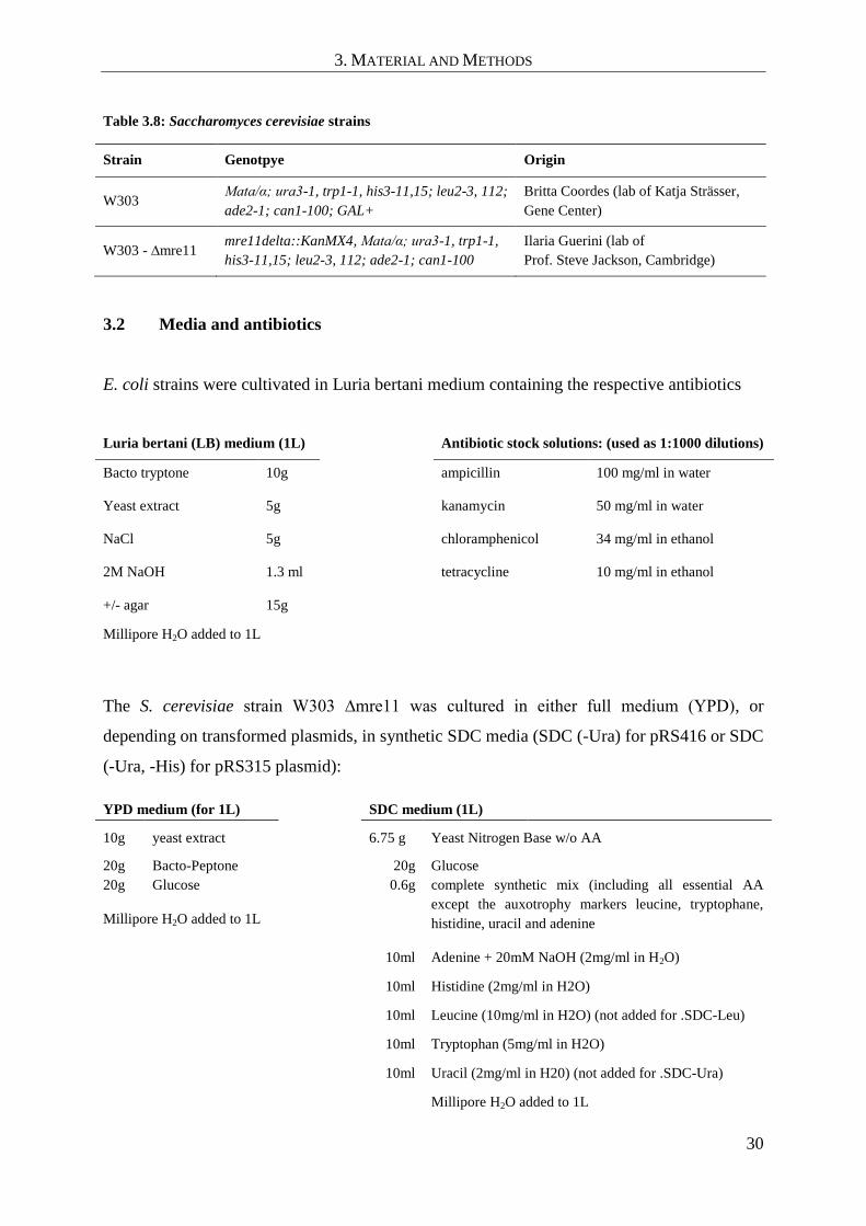
3. MATERIAL AND METHODS
30
Table 3.8: Saccharomyces cerevisiae strains
Strain Genotpye Origin
W303 Mata/α; ura3-1, trp1-1, his3-11,15; leu2-3, 112;
ade2-1; can1-100; GAL+
Britta Coordes (lab of Katja Strässer,
Gene Center)
W303 - ∆mre11 mre11delta::KanMX4, Mata/α; ura3-1, trp1-1,
his3-11,15; leu2-3, 112; ade2-1; can1-100
Ilaria Guerini (lab of
Prof. Steve Jackson, Cambridge)
3.2 Media and antibiotics
E. coli strains were cultivated in Luria bertani medium containing the respective antibiotics
Luria bertani (LB) medium (1L) Antibiotic stock solutions: (used as 1:1000 dilutions)
Bacto tryptone 10g ampicillin 100 mg/ml in water
Yeast extract 5g kanamycin 50 mg/ml in water
NaCl 5g chloramphenicol 34 mg/ml in ethanol
2M NaOH 1.3 ml tetracycline 10 mg/ml in ethanol
+/- agar 15g
Millipore H2O added to 1L
The S. cerevisiae strain W303 ∆mre11 was cultured in either full medium (YPD), or
depending on transformed plasmids, in synthetic SDC media (SDC (-Ura) for pRS416 or SDC
(-Ura, -His) for pRS315 plasmid):
YPD medium (for 1L) SDC medium (1L)
10g yeast extract 6.75 g Yeast Nitrogen Base w/o AA
20g Bacto-Peptone 20g Glucose
20g Glucose 0.6g complete synthetic mix (including all essential AA
except the auxotrophy markers leucine, tryptophane,
histidine, uracil and adenine Millipore H2O added to 1L
10ml Adenine + 20mM NaOH (2mg/ml in H2O)
10ml Histidine (2mg/ml in H2O)
10ml Leucine (10mg/ml in H2O) (not added for .SDC-Leu)
10ml Tryptophan (5mg/ml in H2O)
10ml Uracil (2mg/ml in H20) (not added for .SDC-Ura)
Millipore H2O added to 1L

3. MATERIAL AND METHODS
31
3.3 Methods
3.3.1 Molecular biology methods
If not stated differently molecular biology standard procedures were carried out according to
protocols by Sambrook, J. and Russell (Sambrook and Russell 2001). Commercially available
kits and enzymes were used following the manufacturer‟s instructions.
3.3.1.1 Molecular cloning
Constructs for the expression of S. pombe Mre11 and Nbs1 were designed with help of the
secondary structure prediction programs JPRED (Cole et al. 2008), PSIPred (Bryson et al.
2005), (Jones 1999) and HHPred (Soding et al. 2005) as well as by alignment of sequences
from different species using the program ClustalW (Larkin et al. 2007).
PCR primer were designed with a melting temperature (Tm) preferencially above 60 °C. For
this purpose the program GeneRunner was used (http://www.generunner.net/). The cloning
primers contained restriction sites for specific endonucleases with an overhang sequence
between 5 and 7 bases to assure efficient cleavage.
Full length constructs of Mre11 and Nbs1 were amplified by PCR from cDNA using Phusion
Flash Master Mix (Finnzymes, Espoo, Finnland). A typical PCR reaction contained 10-100 ng
template DNA and 50 pmol of each primer in 20µl of 1x PCR Mix. The following PCR
program was used:
1) 98 °C → 30 s
2) 98 °C → 10 s
3) 50-60 °C → 15 sec
(depending on melting temperature of used primers)
4) 72 °C 20 s / 1kb
repeat 2) – 4) 30 times 72 °C 120s

3. MATERIAL AND METHODS
32
PCR products were separated in agarose gels using a Gel Extraction Kit (Metabion,
Martinsried, Germany), digested with the corresponding restriction enzymens and ligated into
the vector of interest.
3.3.1.2 Site Directed Mutagenesis by Overlap Extension PCR
All Mre11 or Nbs1/Xrs2 mutants described in this thesis were genererated by site directed
mutagenesis using Overlap extension PCR as described in Sambrook and Russell (Sambrook
and Russell 2001). For the PCR reactions Phusion Flash Master Mix was used (Finnzymes,
Espoo, Finnland). Briefly described, the procedure consists of two PCR steps: In the first PCR
step, two fragments are generated which contain either the region 5´ or 3´ of the mutation site.
The primer pair for each reaction consists of a cloning primer hybridizing to either the 5´ or 3´
end of the gene and the corresponding mutagenesis primer which hybridizes to the opposite
strand at the site of mutation. Thereby two fragments with compatible ends around the
mutation site are generated. This allows hybridization of the fragments and subsequent
extension in the second PCR step, which yields the full length mutagenesis product. After 5
PCR cycles, the amount of full length PCR product is further amplified by the addition of the
cloning primers and additional 30 PCR cycles (see below):
1) 98 °C → 30 s
2) 98 °C → 10 s
3) 50-60 °C → 15 sec (depending on melting temperature of used primers)
4) 72 °C 20 s / 1kb
repeat 2) – 4) 5 times
5) Addition of 25 pmol forward and reverse cloning primers
6) 98 °C → 10 s
7) 50-60 °C → 15 sec (depending on melting temperature of used primers)
8) 72 °C 20 s / 1kb
repeat 6) – 8) 25-30 times followed by a final elongation step at 72 °C for 120-180 sec.
All following following cloning steps were carried out similar to cloning with normal PCR
products (3.3.1.1).

3. MATERIAL AND METHODS
33
3.3.1.3 Transformation in E. coli
For transformations chemically competent E. coli were used by performing the following standard
protocol (E. coli host strains are listed under 3.1.4): 10 μl ligation mixtures or 50-400 ng of
purified plasmid were added to 70 μl of competent cells and incubated on ice for 15 min. Cells
were then heat shocked for 1 min at 42 °C, followed by 2 min of incubation on ice, addition of
800 μl LB medium and subsequent incubation on a thermo shaker at 37 °C for 45 to 60 min to
establish antibiotic resistance. Afterwards cells were plated on LB agar plates containing the
respective antibiotics and incubated overnight at 37 °C.
3.3.2 Protein biochemistry methods
3.3.2.1 Protein expression in E. coli
To achieve overexpression of recombinant proteins, the plasmid containing the gene of
interest was transformed into E. coli Rosetta (DE3) (Novagen, Schwalbach, Germany). The
transformation was carried out analogous to 3.3.1.3, but with the addition of 34 mg/ml
Chloramphenicol. For pre-cultures 50 ml LB medium were inoculated with several colonies
from a fresh transformation plate (not older than 1 week) and grown overnight at 37 °C in a
shaking incubator. The next day 3L LB medium were inoculated 1:100 with the pre-culture and
grown at 37 °C to oD600 0.6 - 0.8. The culture was then cooled to 18 °C and further incubated for
additional 45 minutes in the shaker. Then protein production was induced by addition of 0.2 mM
IPTG. The culture was further shaken overnight at 18 °C. Cells were harvested the next morning,
frozen in liquid nitrogen and stored at 20 °C until further use.
3.3.2.2 Recombinant selenomethionine expression in E. coli
Expression of selenomethionine containing Nbsmir
-Mre11cd
fusion protein (3.3.3.1 and 4.2.1)
was performed in Escherichia coli B834 Rosetta (DE3) in a shaking culture of minimal
medium containing 50 mg/L selenomethionine with 250 µM IPTG at 18 °C overnight. All
amino acids were reagent-grade L-enantiomers purchased from Sigma (Deisenhofen,
Germany). After dissolving the components (Table 3.9) in 2000 ml FPLC grade water,
solution A was autoclaved, cooled to RT and supplemented with 200 ml filter-sterilized

3. MATERIAL AND METHODS
34
solution B plus 125 mg selenomethionine (Calbiochem, Schwalbach, Germany). Expression
was carried out analogous to native proteins (3.3.2.1).
Table 3.9: LeMaster’s medium (Hendrickson et al. 1990)
Compounds for autoclavable solution A (2000 ml)
Alanine 1.0 g Serine 4.166 g
Arginine hydrochloride 1.16 g Threonine 0.46 g
Aspartic acid 0.8 g Tyrosine 0.34 g
Cystine 0.066 g Valine 0.46 g
Glutamic acid 1.5 g Adenine 1.0 g
Glutamine 0.666 g Guanosine 1.34 g
Glycine 1.08 g Thymine 0.34 g
Histidine 0.12 g Uracil 1.0 g
Isoleucine 0.46 g Sodium acetate 3.0 g
Leucine 0.46 g Succinic acid 3.0 g
Lysine hydrochloride 0.84 Ammonium chloride 1.5 g
Phenylalanine 0.266 g Sodium hydroxide 1.7 g
Proline 0.2 g Dibasic potassium phosphate 21.0 g
Compounds for non-autoclavable solution B (200ml)
Glucose 20.0 g Concentrated sulfuric acid 16.0 μl
Magnesium sulphate 0.5 g Thiamine 10.0 mg
Iron sulphate 8.4 g
3.3.2.3 Purification of GST-labelled proteins
Cells from 3-12 L expression culture were resuspended in buffer A (Table 3.10) and lysed by
sonication followed by centrifugation at 30,000 x g, 4 °C for 45 min. The supernatant was
loaded onto a Glutathion-Sepharose affinity column (GE Healthcare) and washed with 5-10
column volumes of buffer A. Afterwards a high salt wash using 5-10 column volumes of
buffer B was performed and the column then equilibrated again with buffer A. The GST-
fusion proteins was eluted in buffer C containing 20 mM reduced Glutathione. Proteolytic
cleavage was achieved by addition of Prescission protease (GE Healthcare, to cleave off the
GST tag) or Tobacco Etch Virus (TEV) protease (in cases where the Nbs1mir
-Mre11cd
fusion

3. MATERIAL AND METHODS
35
protein was cleaved) to the protein solution and overnight incubation at 10 °C. The prepared
protein was dialysed overnight against 1L of buffer D. The dialysis procedure was repeated
twice the next day for additional 2x one hour with fresh buffer to remove residual glutathione.
The cleaved GST was removed by passing the dialyzed solution fractions again through a
Glutathione-Sepharose column. The flowthrough was collected and further purified by size
exclusion chromatography on a S200 column (GE Healthcare). The elution fractions were
analysed by SDS-gel electrophoresis (3.3.2.5) and fractions containing the protein of interest
were pooled and concentrated (Amicon spin concentrators, Millipore). Proteins used for
crystallization were prepared with specific size exclusion buffers listed in Table 3.12. Proteins
which were later used for biochemical assays in various buffers, were purified by size
exclusion chromatography using buffer D. Proteins, which were not used directly after
purification were stored flash frozen in small aliquots of 100 µl at -80 °C. All mutant proteins
were purified in the same way as the native proteins. Also the selenomethionine labelled
Nbs1mir
-Mre11cd
protein was purified similarly, but with 5 mM ß-Mercaptoethanol in buffers
A-D.
Table 3.10: Buffers for purification of GST-labelled proteins
Buffer A (Lysisbuffer) Buffer B (High salt buffer) Buffer C (elution buffer)
50 mM Hepes/NaOH pH7.5 20 mM Hepes/NaOH pH7.5 50 mM Hepes/NaOH pH7.5
500 mM NaCl 1 M NaCl 500 mM NaCl
4 mM EDTA 2 mM EDTA 20 mM Glutathione (reduced)
2 mM ß-Mercaptoethanol 2 mM ß-Mercaptoethanol 4 mM EDTA
2 mM ß-Mercaptoethanol
Buffer D (Dialysis and size
exclusion chromatography buffer)
20 mM Hepes/NaOH pH7.5
500 mM NaCl
2 mM EDTA
2 mM ß-Mercaptoethanol
3.3.2.4 Purification of His-tag labeled proteins
Cells were lysed similar to the GST-labelled proteins (3.3.2.3) but with the usage of a Ni-
NTA specific, 20mM imidazol containing buffer A (Table 3.11). The supernatant was loaded
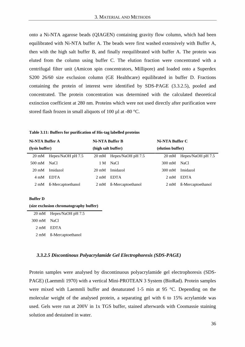
3. MATERIAL AND METHODS
36
onto a Ni-NTA agarose beads (QIAGEN) containing gravity flow column, which had been
equilibrated with Ni-NTA buffer A. The beads were first washed extensively with Buffer A,
then with the high salt buffer B, and finally reequilibrated with buffer A. The protein was
eluted from the column using buffer C. The elution fraction were concentrated with a
centrifugal filter unit (Amicon spin concentrators, Millipore) and loaded onto a Superdex
S200 26/60 size exclusion column (GE Healthcare) equilibrated in buffer D. Fractions
containing the protein of interest were identified by SDS-PAGE (3.3.2.5), pooled and
concentrated. The protein concentration was determined with the calculated theoretical
extinction coefficient at 280 nm. Proteins which were not used directly after purification were
stored flash frozen in small aliquots of 100 µl at -80 °C.
Table 3.11: Buffers for purification of His-tag labelled proteins
Ni-NTA Buffer A
(lysis buffer)
Ni-NTA Buffer B
(high salt buffer)
Ni-NTA Buffer C
(elution buffer)
20 mM Hepes/NaOH pH 7.5 20 mM Hepes/NaOH pH 7.5 20 mM Hepes/NaOH pH 7.5
500 mM NaCl 1 M NaCl 300 mM NaCl
20 mM Imidazol 20 mM Imidazol 300 mM Imidazol
4 mM EDTA 2 mM EDTA 2 mM EDTA
2 mM ß-Mercaptoethanol 2 mM ß-Mercaptoethanol 2 mM ß-Mercaptoethanol
Buffer D
(size exclusion chromatography buffer)
20 mM Hepes/NaOH pH 7.5
300 mM NaCl
2 mM EDTA
2 mM ß-Mercaptoethanol
3.3.2.5 Discontinous Polyacrylamide Gel Electrophoresis (SDS-PAGE)
Protein samples were analysed by discontinuous polyacrylamide gel electrophoresis (SDS-
PAGE) (Laemmli 1970) with a vertical Mini-PROTEAN 3 System (BioRad). Protein samples
were mixed with Laemmli buffer and denaturated 1-5 min at 95 °C. Depending on the
molecular weight of the analysed protein, a separating gel with 6 to 15% acrylamide was
used. Gels were run at 200V in 1x TGS buffer, stained afterwards with Coomassie staining
solution and destained in water.

3. MATERIAL AND METHODS
37
4x lower buffer: 3 M Tris (pH 8.5), 0.4% (w/v) SDS
4x upper buffer: 0.5 M Tris (pH 6.8), 0.4% (w/v) SDS
4x Laemmli buffer: 0.11 M Tris (pH 6.8), 16% (v/v), 4% (w/v) SDS,
5% (v/v) β-mercaptoethanol, 0.05% (w/v) bromophenol blue
10x TGS buffer: 0.5 M Tris (pH 8.3), 1.9 M glycin, 1% (w/v) SDS
Coomassie stain: 50% (v/v) ethanol, 7% (v/v) acetic acid, 0.2% (w/v),
Coomassie Brilliant Blue R250
3.3.2.6 Western blot analysis
Protein samples were first fractionated by SDS-PAGE according to Laemmli (Laemmli 1970)
with a Mini Protean III system (Biorad, München) (3.3.2.5). Gels were then blotted to a
Nitrocellulose membrane (Roth, Karlsruhe, Germany) using a Mini Trans-Blot Cell system
with a transfer buffer containing 50 mM TRIS/HCl pH 8.3, 0.19 M glycine, 0.1 (w/v) % SDS
and 20 % methanol. Three layers of Whatman paper, followed by the nitrocellulose
membrane, the SDS-PAGE gel and another three layers of Whatman paper, all presoaked in
blotting buffer, were assembled in the blotting machine. Transfer of proteins to the membrane
occurred at 100V for 60 min at 8 °C. The nitrocellulose membrane was blocked with
TBST+M buffer (50 mM Tris/HCl pH 7.5, 150 mM NaCl, 0.1% Tween 20, 4% milk powder)
for 45 min. The first antibody was diluted in TBST+M buffer and incubated overnight at 8 C.
Next the membrane was washed with TBST+M buffer for 3x 15 min and incubated with a
Horseradish Peroxidase (HRP) coupled secondary antibody in TBST+M buffer at RT for 2h.
Afterwards, the membrane was washed 3x 15 min in TBS-T buffer without milk powder. All
used antibodies and the respective dilutions are listed under 3.1.1.
The immunostained proteins were detected by addition of Pierce ECL Western Blotting
Substrate solution (Thermo Scientific, Bonn) to the blotting membrane and subsequent
exposure with a light sensitive Hyperfilm™ ECL™ (GE Healthcare). The films were
developed with a Kodak X-Omat M35 developing machine.

3. MATERIAL AND METHODS
38
3.3.2.7 Analytical size exclusion chromatography
To analyse the complex formation of different Mre11 and Nbs1 protein constructs from
S. pombe, 10 nmol Mre11 and 7.5 nmol Nbs1 proteins were mixed and dialysed against the
chromatography running buffer containing 20 mM Hepes pH 7.5, 100 mM KCl, 5% glycerol,
2mM ß-mercaptoethanol and either 1 mM MnCl2 or 2 mM EDTA. A volume of 350 µl protein
sample was loaded at a flow rate of 0.5 ml/min onto a Superdex S200 10/300 GL column (GE
Healthcare), pre-equilibrated with running buffer. Elution fractions were analyzed for Mre11
and Nbs1 by SDS-PAGE and Coomassie staining (3.3.2.5).
3.3.2.8 Limited Proteolysis
A stock solution of 1mg/ml proteinase K dissolved in H20 was diluted to 1:10, 1:100, 1:1000,
1:3000, 1.6000 and 1:10000 with 2 mM HCl (pH ≈ 3). For each sample 45 µl of a 1 mg/ml
protein solution in 20 mM Tris/HCl pH 7.5, 150 mM NaCl, 2 mM ß-Mercaptoethanol was
mixed with 5 µl of the corresponding dilution of proteinase K. The samples were incubated at
RT for 1 h and stopped by addition of 1 µl PMSF-saturated isopropanol and 16.6 µl of 4x
SDS-sample buffer. An appropriate amount of sample was analyzed by SDS-PAGE (3.3.2.5).
Protein bands of interest were cut out and sent to the Zentrallabor für Proteinanalytik (ZfP -
Prof. Dr. Axel Imhof, München) for MALDI peptide mass fingerprint analysis.
3.3.2.9 Nuclease activity assay
Mre11 nuclease activity was tested using a 6-FAM- 5` labelled 60mer poly(dT)
oligonucleotide (Table 3.2). For each reaction, 10 nM DNA was incubated with 5 µM of
Mre11 (residues 1-413) or Mre11 (residues 1-413) H134S proteins in 10 µl buffer containing
20 mM Hepes pH 7.5, 100 mM KCl,.5% glycerol, 5 mM MnCl2 and 2 mM Mercaptoethanol
at 37 °C for 2 h. The reaction was stopped by addition of 3 µl loading buffer (10mM Tris, pH
8.0 16.6% formamide, 16.6% glycerol, 5mM EDTA) and incubated at 95 °C for 5 min.
Reaction products were resolved on a denaturating 18% acrylamide gel in 1x TBE buffer
containing 8M Urea. Gels were imaged with a Typhoon 9400 fluorescence scanner (GE
Healthcare) using the green-exited (532nm) fluorescence mode.

3. MATERIAL AND METHODS
39
3.3.2.10 EMSA (electrophoretic mobility shift assay)
Binding of different S. pombe Mre11 and Nbs1 constructs to DNA was analyzed by
electrophoretic mobility shift assays (EMSAs). The binding of proteins to DNA can be
visualized by a mobility shift in native gel electrophoreses. Protein-DNA complexes posses
normally a lower charge/mass ratio than free DNA and therefore migrate slower in the gel.
The DNA is visualized by a covalently coupled fluorescence label. The samples contained the
DNA substrate at a concentration of 50 nM and the protein in different excess concentrations
(0/1.5/3/6/12 µM for Mre11 and 0/0.5/5/50 µM for Nbs1 constructs) and in a total volume of
10 μl. The assay buffer contained 20 mM Hepes/NaOH pH 7.5, 100 mM KCl, 10% Glycerol,
1 mM MnCl2 and 2 mM ß-Mercaptoethanol. The protein samples had been equilibrated by
dialysis against the assay buffer before use. After addition of protein to the DNA, the samples
were mixed and incubated for 30 min at RT. Afterwards they were analyzed with a 0.5%
(w/v) agarose gel in 1x TB buffer (90 mM Tris, 90 mM boric acid, pH 8 without adjustment).
The gel was run at 4 V/cm and 8 °C for 2 h and visualized with a Typhoon 9400 fluorescence
scanner (GE Healthcare).
3.3.3 Structural biology methods
3.3.3.1 Crystallization
All proteins were crystallized at 20 °C using the hanging drop vapour diffusion technique.
Initial crystals were obtained in commercial 96-well format sitting drop screens using the
Matrix Hydra II 96-channel microdispenser (Thermo Scientific) for dispensing of reservoir
solutions and protein drops. The initial screens were set up with a reservoir volume of 50 µl
and a dropsize of 0.5 µl reservoir + 0.5 µl protein solution. Initial crystals were optimized
manually in refinement screens by varying the composition of the reservoir solution as well as
the protein concentration. The refinement screens contained a reservoir volume of 500µl with
2 μl reservoir + 2 μl protein solution drops. The protein preparation buffers, protein
concentrations, screen compositions and cryo protectants for all crystallised proteins are listed
in Table 3.12. Cryocooling for data collection was achieved by soaking the crystals for 30 sec
in mother liquor solution containing the cryo protectant and flash freezing in liquid nitrogen.

3. MATERIAL AND METHODS
40
Table 3.12: Crystallization conditions for Nbs1mir
Mre11cd
complexes and Mre11cd
preparation buffer
protein
concen-
tration
crystallization solution cryo reagent
Nbs1mir
Mre11cd
SeMet
20 mM Hepes pH 7.5
200 mM NaCl
1 mM MnCl2
2 mM ß-Mercaptoethanol
10 mg/ml
400 mM ammonium
citrate pH 6.0
20% w/v PEG 3350
15 % D(-)
-2,3butanediol
Nbs1mir
Mre11cd
20 mM Hepes pH 7.5
200 mM NaCl
1 mM MnCl2
2 mM ß-Mercaptoethanol
9 mg/ml
100 mM Tri-Na-Citrate pH 5.0
200 mM ammonium sulfate
13% w/v PEG 4000
15 % D(-)
-2,3butanediol
Nbs1mir
Mre11cd
(+50 Mn2+
)
20 mM Hepes pH 7.5
200 mM NaCl
1 mM MnCl2
2 mM ß-Mercaptoethanol
8 mg/ml
400 mM ammonium
citrate pH 6.0
20% w/v PEG 3350
50 mM MnCl2
15 % D(-)
-2,3butanediol
Non-fused
Mre11cd
Nbs1mir
20 mM Hepes pH 8.0
200 mM NaCl
1 mM MnCl2
2 mM ß-Mercaptoethanol
8 mg/ml
200 mM ammonium
citrate pH 5.5,
14 % w/v PEG 3350
50 mM MnCl2
20% glycerol
Mre11cd
20 mM Hepes pH 7.5
300 mM NaCl
0.1 mM EDTA
1 mM MnCl2
2 mM ß-Mercaptoethanol
22 mg/ml
100 mM Hepes pH 8.1
9% w/v PEG 8000
200 mM NaCl.
12% D(-)
-2,3butanediol
3.3.3.2 Data collection, structure solution and model building
Diffraction data were collected at the PXI beamline at the SLS (Villigen, Switzerland) or at
beamline ID23-1/2 at the ESRF (Grenoble, France) as indicated in Table 4.1. Single-
wavelength anomalous dispersion (SAD) data to 2.8 Å were collected at the selenomethionine

3. MATERIAL AND METHODS
41
peak wavelength at the beamline ID23-2 on selenium-containing crystals. All data were
integrated and scaled with XDS (Kabsch 1993). The structure of the Nbs1mir
-Mre11cd
fusion
protein was determined by SAD phasing methods using SHARP and autoSHARP (Global
Phasing, Cambridge), which located 8 selenomethionine and 2 manganese sites within the
asymmetric unit. An initial model was built by alternate rounds of automatic model building
using the programs ARP/warp (Langer et al. 2008) and Buccaneer (Cowtan 2006). The
resulting model was used for molecular replacement with the program Phaser (McCoy 2007)
against the native dataset. The model was completed by manually model building in the
resulting electron density using the program COOT (Emsley and Cowtan 2004) and refined to
2.4 Å. Phases for the non-fused Mre11cd
-Nbs1mir
complex and apo-Mre11cd
were obtained by
molecular replacement with Phaser using the monomeric Mre11cd
model derived from the
Nbs1mir
-Mre11cd
fusion protein model. Prior to refinement, 5-10 % of the reflections were
randomly omitted for monitoring the free R-value. All models were refined by iterative cycles
of bulk solvent correction, overall anisotropic B factor refinement, positional, TLS group
refinement with Phenix (Zwart et al. 2008) and manual model building with COOT. Initial
NCS restraints were gradually removed in the final cycles of the refinement, to allow some
structural variations. Especially for refinement of flexible loop regions within the structures
the refinement program autoBUSTER (Global Phasing, Cambridge) was used during the last
steps of the refinement process. Data collection and model statistics are summarized in Table
4.1. All figures were prepared with PYMOL (DeLano Scientific).
3.3.3.3 Small angle x-ray scattering
Synchrotron radiation small angle X-ray scattering (SAXS) data were collected at the EMBL
X33 beamline at the DORIS storage ring (DESY, Hamburg, Germany) using a MAR345 two-
dimensional image plate detector. Scattering patterns from solutions of Mre11cd
at
concentrations of 5 mg/ml were measured in the corresponding preparation buffers. Various
programs of the ATSAS (Konarev et al. 2006) software package were used to process and
evaluate the SAXS data. PRIMUS was used for initial data analysis. The radius of gyration R
g was determined by fitting the measured scattering data with the Guinier equation (s×RG <
1.3) and the program GNOME was used to determine P(r)-functions. CRYSOL was utilized
to compute theoretical SAXS-curves from crystallographic coordinate files for data
comparison.

3. MATERIAL AND METHODS
42
3.3.4 Yeast specific methods
3.3.4.1 Yeast transformation
For transformation of Mre11 coding plasmids into the W303 ∆mre11 strain, 50 ml of yeast
culture were grown to an OD600 of 0.5 to 0.8 in YPD and pelleted by centrifugation at
3600 rpm (Rotanda 46R centrifuge) and room temperature (RT) for 3 min. After washing with
10 ml of H2O, the pellet was washed once with 500 µl of solution I (10 mM Tris-HCl, pH 7.5,
1 mM EDTA, 100 mM Li-acetate) and resuspended in 250 µl solution I. 3 µg DNA and 5 µl
of single stranded carrier DNA (DNA of salmon of herring testis, 2 mg/ml) were mixed with
50µl of cells in solution I and incubated with 300 µl solution II (10 mM Tris-HCl, pH 7.5;
1 mM EDTA; 100 mM Li-acetate; 40% w/v PEG 4000) on a turning wheel at RT for 30 min.
The samples were heat shocked at 42 °C for 10 min, followed by 3 min of incubation on ice.
1 ml of H2O was added, the sample centrifuged, the pellet resuspended in 50 µl H2O and
plated on a selective plate. To transform yeast cells grown on plate, one loop of
logarithmically grown cells was resuspended in 30 µl of 100 mM Li-acetate and vortexed.
The rest of the transformation was done according to the protocol described above.
3.3.4.2 Plate survival assays
One loop of freshly growing cells from a plate was resuspended in 1 ml H2O. Five 10 fold
dilutions were prepared and 6 µl of each dilution spotted onto the corresponding plates: SDC
(-Ura), SDC (-Ura) + 0.2/1/5 µg/ml Camptothecin (CPT), SDC (-Ura) + 0.005% methyl
methanesulfonate (MMS), SDC (-Ura) + 50 or 200 mM hydroxyurea (HU), YPD, YPD +
0.2/1/5 µg/ml CPT, YPD + 0.005 % MMS, YPD + 50 or 200 mM HU.
The compositions of SDC (-Ura) and YPD media are listed under 3.2.
3.3.4.3 Co-immunoprecipitation
The interaction of different S. cerevisiae Mre11 mutants with Rad50 and Xrs2 was analyzed
performing a modified co-immunoprecipitation protocol by (Strahl-Bolsinger et al. 1997):
W303 ∆mre11 cells containing a P527 ScMre11 plasmid, coding for the respective c-myc-
tagged Mre11 mutant protein (Table 3.6), were cultivated in SDC (-Ura) medium. 50 ml of

3. MATERIAL AND METHODS
43
medium were freshly inoculated with an overnight culture to a starting oD600 of 0.2 - 0.3 and
shaked at 30 °C until an OD600 of 0.7. Cells were pelleted by centrifugation at 3600 rpm for
3min (Rotanda 46R), washed twice with TBS (20 mM Tris-HC1 at pH 7.6, 200 mM NaC1),
and resuspended in 400 µ1 lysis buffer (50mM Hepes pH 7.5, 140mM NaCl, 10% Glycerin,
0.5% NP-40, 1mM PMSF, 2mM benzamidine hydrochloride, 2 µM Pepstatin, 0.5 µM
Leupeptin, 3.3 µM Chymostatin). Then, 400 µ1 glass beads were added and the cells lysed by
vortexing on an Eppendorf 5432 shaker for 40 min. The tubes were then punctured on the
bottom, and the lysate collected in a 15 ml falcon tube by centrifugation at 1000rpm for 1min.
The lysate was transferred into an 1.5 ml Eppendorf tube and clarified by centrifugation at
4 °C and 13000 rpm for 5 min and 15min. The supernatant was transferred into a fresh
Eppendorf tube, followed by addition of 5µg α-cMyc antibody (3.1.1) and 20 U of DNAse I
(New England Biolabs). The samples were incubated for 3h at 8 °C on a turning wheel to
allow formation of Mre11-cMyc-antibody complexes. Then, 50µl of Protein G Dynabeads
(Invitrogen), preequilibrated with lysis buffer, were added (beads in suspension) and the
incubation was continued for 1h. Then the beads were washed three times for 5 min with 1.4
ml lysisbuffer and subsequently resuspended in 60 µl 1.5x Laemmli buffer (3.3.2.5). The Co-
immunoprecipitates were analysed by SDS-PAGE (3.3.2.5) followed by western blot analysis
(3.3.2.6). For western blot analysis of IP inputs, whole cell extracts were prepared using TCA-
mediated protein precipitation as described before (Janke et al. 2010).
3.3.4.4 Indirect immunofluorescence
An exponentially grown yeast culture (10ml with OD600 of 0.5 – 1.0) was mixed with 1 ml of
37% HCOH to fix the cells and incubated on a turning wheel at 30 °C for 90 min. The cells
were pelleted by centrifugation at 3000 rpm and RT for 5 min (Rotanda 46R centrifuge),
washed twice with spheroblasting premix (1.2 M sorbitol, 0.1 M K-phosphate buffer, pH 7.4,
0.5 mM MgCl2), resuspended in 1 ml spheroblasting premix and stored at 4 °C up to 18h.
Afterwards, the cells were pelleted and resuspended in 200µl of spheroblasting premix,
containing 100 µg of 100T zymolyase and incubated in a 30 °C waterbath for 30 min.
Following spheroblasting, the cells were pelleted by centrifugation at 2000 rpm and RT (table
top centrifuge) for 4 min and resuspended in at least 10x the volume of the cells in
spheroblasting premix. Then, one drop of the suspension was pipetted on a fluorescence
microscopy slide, which had been precoated with polylysine. After 5 min of incubation the

3. MATERIAL AND METHODS
44
drops were pipetted off and the immobilized cells washed with 1 drop of blocking buffer (1%
BSA powder in PBS (phosphate buffered saline, 2.7 mM KCl, 7.9 mM Na2HPO4, 1.5 mM
KH2PO4, 137 mM NaCl). The cells were fixed by incubation of the slide with methanol in a
staining jar at -80 °C for 6 min followed by 30 sec incubation in -80 °C cold acetone. Next,
the slides were dried at RT and incubated with 100 µl blocking buffer for 10 min. The
solution was removed and the cells incubated with 20 µl of the first antibody solution
(antibody diluted in 1% BSA in PBS) at RT for 2 h. Then the slides were washed to remove
the antibody for 3x 1 min incubations with 10 µl blocking buffer additionally containing
0,1 % Triton-X at RT. Afterwards, 20 µl of the second antibody diluted in blocking buffer
was added to the cells followed by incubation in the dark for 1 h. Finally, the cells were
washed 3x with blocking buffer plus 0,1 % Triton-X. For staining of cell nuclei the slides
were put in a jar containing SSC buffer (300 mM NaCl; 30 mM Na-citrate, pH 7.0) and 5µl of
DAPI (10mg/ml) in the dark for 10 min. Afterwards the slides were washed with a 1:4
dilution of SSC buffer at RT for 5 min in the dark and dried. Finally the cells were covered
with 80% glycerol and sealed with the cover slide.
The stained cells were analyzed using a DMI 6000 B fluorescence microscope (Leica
Microsystems, Wetzlar). The signal of the Alexa Fluor 488 coupled antibody was detected at
an emission wavelength of 519 nm and the DAPI signal at an emission wavelength of 461 nm.
The excitation wavelength was at 495 nm for Alexa Fluor 488 and 358 nm for DAPI. The
signal intensity was 3 for Alexa Fluor 488 and 2 in the case of DAPI. A gain of 3.5 (Alexa
Fluor 488) or 3.3 (DAPI) was used for signal detection. Fluorescence signals were analyzed
with the LAS AF Lite software (Leica Microsystems, Wetzlar) and the pictures arranged
using ImageJ (NIH, Bethesda, USA).
3.3.5 Bioinformatical methods
3.3.5.1 Structure based sequence alignments
Protein sequences of Mre11 from eukaryotic organisms Schizosaccharomyces pombe
(SpMre11), Saccharomyces cerevisisae, Danio rerio and Homo sapiens were aligned with
ClustalW (Larkin et al. 2007). The archaeal Mre11 sequence from Pyrococcus furiosus
(PfMre11) was added after calculating a pairwise alignment of PfMre11 and SpMre11 with
the program FATCAT (Ye and Godzik 2004) using the pdb-coordinates of PfMre11 (Protein

3. MATERIAL AND METHODS
45
data base entry 1II7) and SpMre11cd
as input files. The PfuMre11/SpMre11 alignment was
further revised by comparison of the overlaid structures with Pymol (DeLano Scientific).

4. RESULTS
46
4. RESULTS
4.1 Cloning and expression of Mre11 and Nbs1 from S. pombe
The coding genes for full length Mre11 and Nbs1 from the fission yeast Schizosaccharomyces
pombe were amplified by PCR using a cDNA library and cloned into E. coli expression
vectors according to 3.3.1.1. Full length Mre11 (aa 1-649), could be purified but was prone to
aggregation and degradation over time. Full length Nbs1 (aa 1-613), also expressed in E. coli,
was mostly unsoluble and could be purified only in minor amounts (data not shown).
However, an Mre11-Nbs1 mutant complex consisting of Mre11 aa 1-478 and Nbs1 aa 474-
613 could be co-expressed and purified in mg amounts (3.3.2.4 and Table 3.5). Even though
this complex was extensively tested for crystallization, it did not yield crystals in any
condition. Furthermore, it was already degraded by proteolysis during the preparation. Thus,
to identify stable fragments of Mre11 and Nbs1, suitable for crystallization, a limited
proteolysis analysis was performed. For this purpose, the Mre11 aa 1-478 / Nbs1 aa 474-613
complex was incubated with different concentrations of Proteinase K. Nevertheless, no stable
Nbs1 fragment shorter than aa 474-613 could be identified. For Mre11, however, a stable
fragment of approximately 45 kDa was identified and further analyzed by a MALDI trypsin
fingerprint (3.3.2.8 and Figure 4.1). Here, peptides in the N-terminal region of Mre11 between
residues 1-412 were found, which matched also the observed molecular weight of the
fragment. Based on this observation a new construct of Mre11 was cloned which contained aa
1-413 (Table 3.5). The Mre11 construct aa 1-413 could be expressed and prepared in high
purity and with a yield of several mg protein. However, albeit remaining stable after
purification, it did not crystallize in any tested screen. A secondary structure analysis with
JPRED (Cole et al. 2008) predicted the first 17 residues of S. pombe Mre11 to be
unstructured. Therefore, a construct consisting of aa 15-413 was generated from which the
structure could later be determined (Table 3.5, Figure 4.2). The Mre11 fragment aa 15-413
contained, based on structure aligments with the archaeal Mre11 homologue from Pyrococcus
furiosus, both the nuclease and the DNA capping domain. It was therefore further named
Mre11cd
(cd = catalytical domain).
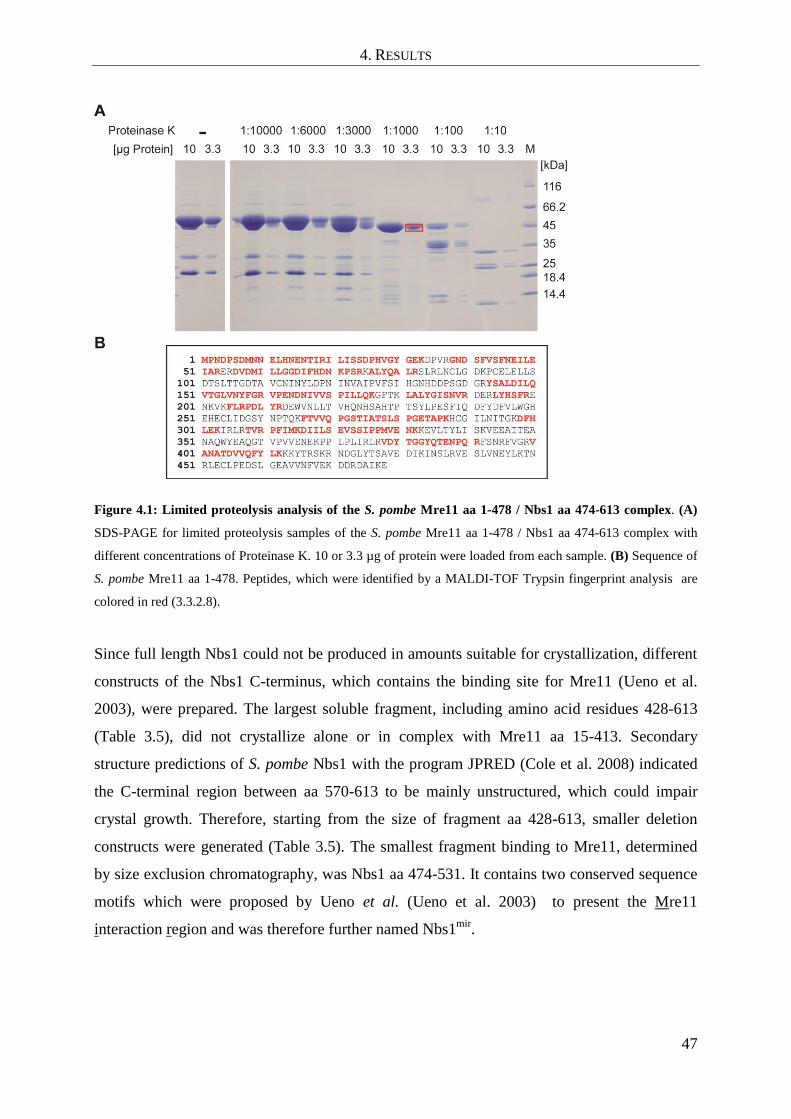
4. RESULTS
47
Figure 4.1: Limited proteolysis analysis of the S. pombe Mre11 aa 1-478 / Nbs1 aa 474-613 complex. (A)
SDS-PAGE for limited proteolysis samples of the S. pombe Mre11 aa 1-478 / Nbs1 aa 474-613 complex with
different concentrations of Proteinase K. 10 or 3.3 µg of protein were loaded from each sample. (B) Sequence of
S. pombe Mre11 aa 1-478. Peptides, which were identified by a MALDI-TOF Trypsin fingerprint analysis are
colored in red (3.3.2.8).
Since full length Nbs1 could not be produced in amounts suitable for crystallization, different
constructs of the Nbs1 C-terminus, which contains the binding site for Mre11 (Ueno et al.
2003), were prepared. The largest soluble fragment, including amino acid residues 428-613
(Table 3.5), did not crystallize alone or in complex with Mre11 aa 15-413. Secondary
structure predictions of S. pombe Nbs1 with the program JPRED (Cole et al. 2008) indicated
the C-terminal region between aa 570-613 to be mainly unstructured, which could impair
crystal growth. Therefore, starting from the size of fragment aa 428-613, smaller deletion
constructs were generated (Table 3.5). The smallest fragment binding to Mre11, determined
by size exclusion chromatography, was Nbs1 aa 474-531. It contains two conserved sequence
motifs which were proposed by Ueno et al. (Ueno et al. 2003) to present the Mre11
interaction region and was therefore further named Nbs1mir
.

4. RESULTS
48
Figure 4.2: Binding studies of Mre11 and Nbs1, construct design and protein preparations of Mre11/Nbs1
complexes for crystallization: (A) Analytical size exclusion chromatography with a Superdex S200 10/300GL
column showed that Mre11 aa 1-413 and Nbs1 474-531 form a stable complex with a 1:1 stoichiometry. (B-C)
Mre11cd
and Nbs1mir
fusion proteins used for crystallization. Nbs1mir
(aa 474-531) - Mre11cd
(aa 15-413) (B) was
directly crystallized, Mre11cd
(aa 7-413) - Nbs1mir
(aa 474-531) (C) was crystallized in the non-fused form after
proteolytic cleavage of the linker peptide with Tobacco etch virus protease at its recognition site in the linker
region between Mre11cd
and Nbs1mir
. Mre11 is shown with the phosphodiesterase in green and the DNA capping
domain in grey. The Mre11 interacting region of Nbs1 is depicted magenta. (D) SDS-PAGE of different protein
preparations (from left to right): Mre11 aa 15-413, Nbs1 (aa 474-531) - (Mre11 (aa 15-413) fusion protein, non-
fused Mre11 (aa 7-413) - Nbs1 (aa 474-531) complex.
Since none of the generated fragments initially crystallized with Mre11, a fusion protein
consisting of Nbs1mir
(aa 474-531) and Mre11cd
(aa 15-413) was generated to facilitate
crystallization (Figure 4.2 B and Table 3.5). The flexible linker, which covalently links the C-
terminus of Nbs1 with the N-terminus of Mre11 consisted of the amino acid sequence
GSAGSAGS. The construct could be crystallized and its structure solved (Table 3.12 and
4.3). To rule out, that the fusion influences the structure, also a reversed plus additionally

4. RESULTS
49
cleavable fusion protein was generated (Mre11cd
aa 7-413 fused to Nbsmir
aa 474-531). In this
construct the C-terminus of Mre11 aa 7-413 was linked via a TEV protease cleavage site to
the N-terminus of Nbs1 aa 474-531. After proteolytic cleavage of the linker peptide, the non-
fused complex of Mre11cd
and Nbsmir
could be crystallized and its structure determined (4.2.1
and Table 3.12).
4.2 Crystallization, structure solution and refinement
4.2.1 Nbs1mir
-Mre11cd
complex
Initial crystallization screenings with the covalently fused Nbs1mir
-Mre11cd
complex yielded
crystals in several different conditions (3.3.3.1). Larger three dimensional crystals grew in
conditions containing citrate salts and different PEGs. They could easily be reproduced in
refinement screenings with native and selenomethionine labeled proteins (Figure 4.3 A-C and
Table 3.12). Crystals of the selenomethionine labeled, covalently fused Nbs1mir
-Mre11cd
complex diffracted to a limiting resolution of 2.8 Å at the ESRF ID23-1 beamline and allowed
phase determination by SAD (3.3.3.2). Crystals of the native, covalently fused Nbs1mir
-
Mre11cd
complex, derived from a refinement screen containing ammoniumsulfate, diffracted
to a limiting resolution of 2.4 Å at the ESRF ID23-2 beamline (Figure 4.3 D and Table 4.1).
All measured crystals belonged to space group P212121. An initial model was built by
alternate rounds of automatic model building into the experimentally derived 2.8 Å electron
density derived from crystals of the selenomethionine labeled protein. The resulting model
was used for a molecular replacement against the native 2.4 Å dataset and completed by
manual model building in the resulting electron density (3.3.3.2). The geometry of all chains
was well within an acceptable range in the final model. The final R factors were 22.0% for
Rwork and 24.2% for Rfree. Crystallographic data and refinement statistics are summarized in
Table 4.1. An example of the refined electron density can be found in Figure 4.3 E.
As described under 4.6, the structure derived from these crystals contained only one Mn2+
ion
in the active site. An active site loop, responsible for complexing the second Mn2+
ion, was
instead involved in complexing a sulfate molecule. Therefore, additional refinement
screenings without ammonium sulfate but with high manganese chloride concentrations were
set up, which yielded crystals diffracting up to 2.5 Å at the SLS PX I beamline (Table 3.12

4. RESULTS
50
and Table 4.1). From these crystals a structure with two Mn2+
ions per active site could be
solved using molecular replacement (3.3.3.2).
The non-fused Mre11cd
(aa 7-413) / (Nbsmir
aa 474-531) complex, which was generated to
rule out that the protein fusion influences the structure, crystallized under conditions similar
to the Nbs1mir
(aa 474-531) / Mre11
cd (aa 15-413) fusion protein (Table 3.12 and Figure 4.3
F). A densitometric analysis with ImageJ (NIH, Bethesda, USA) of Nbs1 and Mre11 bands
from an SDS-PAGE gel of crystals confirmed, that both proteins were also in the non-fused
complex present in an exact 1:1 stoichiometry ( Figure 4.3 G).
Figure 4.3: Crystallization of covalently fused or non-fused S. pombe Nbs1mir-Mre11cd complexes and
diffraction data collection: (A) Initial crystals of the native, covalently fused Nbs1mir
-Mre11cd
complex
(condition: 0.1 M Sodium citrate pH 5.6, 0.2 M ammonium sulfate, 15% w/v PEG 4000). (B) Crystals of the
native, covalently fused Nbs1mir
-Mre11cd
complex from a refinement screen (condition: 0.5 M ammonium citrate
pH 5.5 and 20% w/v PEG 3350). (C) Crystals of the selenomethione labeled, covalently fused Nbs1mir
-Mre11cd
complex from a refinement screen (condition: 0.4 M ammonium citrate, pH 6.0, 20% w/v PEG 3350). (D)
Diffraction pattern of native crystal of covalently fused Nbs1mir
-Mre11cd
complex. (E) Final 2Fo-Fc electron
density map at 1.0 σ for the refined model of Nbs1mir
-Mre11cd
. Amino acid residues 178 to 184 of Mre11 chain
A are shown. Residues are presented as stick models with carbon atoms colored in green, nitrogen in blue, and
oxygen in red. (F) Crystals of the non-fused Mre11cd
-Nbs1mir
complex from a refinement screen containing
0.2 M Ammonium citrate, 14% w/v PEG 3350 and 50 mM MnCl2 (G) SDS-PAGE gel of non-fused Mre11cd
-
Nbs1mir
complex crystals stained with Coomassie blue. A densitometric analysis of the Nbs1 and Mre11 bands
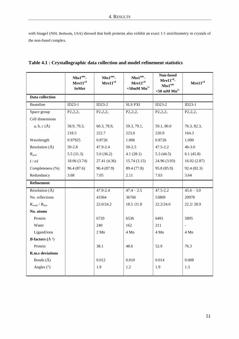
4. RESULTS
51
with ImageJ (NIH, Bethesda, USA) showed that both proteins also exhibit an exact 1:1 stoichiometry in crystals of
the non-fused complex.
Table 4.1 : Crystallographic data collection and model refinement statistics
Nbs1
mir-
Mre11cd
SeMet
Nbs1mir
-
Mre11cd
Nbs1mir
-
Mre11cd
+50mM Mn2+
Non-fused
Mre11cd
-
Nbs1mir
+50 mM Mn2+
Mre11cd
Data collection
Beamline ID23-1 ID23-2 SLS PXI ID23-2 ID23-1
Space group P212121 P212121 P212121 P212121 P212121
Cell dimensions
a, b, c (Å) 58.9, 79.3,
218.5
60.3, 78.9,
222.7
59.3, 79.1,
223.0
59.1, 80.0
220.9
76.3, 82.3,
164.3
Wavelength 0.97925 0.8726 1.006 0.8726 1.000
Resolution (Å) 50-2.8 47.9-2.4 50-2.5 47.5-2.2 46-3.0
Rsym 5.5 (31.3) 5.0 (36.2) 4.1 (28.1) 5.3 (44.5) 6.1 (45.8)
I / I 18.06 (3.74) 27.41 (4.36) 15.74 (3.15) 24.96 (3.93) 16.02 (2.87)
Completeness (%) 96.4 (87.6) 96.4 (87.9) 89.4 (77.8) 95.8 (85.9) 92.4 (82.3)
Redundancy 3.68 7.05 2.11 7.63 3.64
Refinement
Resolution (Å) 47.9-2.4 47.4 – 2.5 47.5-2.2 45.6 – 3.0
No. reflections 43364 36766 53869 20978
Rwork / Rfree 22.0/24.2 18.5 /21.8 22.2/24.0 22.2/ 28.9
No. atoms
Protein 6720 6536 6491 5895
Water 240 162 211 -
Ligand/ions 2 Mn 4 Mn 4 Mn 4 Mn
B-factors (Å ²)
Protein 38.1 48.6 52.9 76.3
R.m.s deviations
Bonds (Å) 0.012 0.010 0.014 0.008
Angles (°) 1.9 1.2 1.9 1.3

4. RESULTS
52
4.2.2 Apo-Mre11cd
Initial crystals of apo-Mre11cd
could be observed in a commercial crystallization screen after
4 weeks of incubation at 20 °C (Figure 4.4 A). However, they were too small to be suitable
for diffraction data collection. Subsequent refinement screenings yielded larger crystals from
which diffraction data with a limiting resolution of 3.0 Å was collected at the ESRF ID23-1
beamline (Figure 4.4 B and C and 3.3.3.2). The crystals belonged to the space group P212121.
The structure of apo-Mre11cd
was solved by molecular replacement methods using the earlier
solved Nbs1mir
-Mre11cd
structure as a replacement model (3.3.3.2 and 4.2.1). A high
percentage of structural elements in the apo-Mre11cd
structure exhibited a conformation
similar to Nbs1mir
-Mre11cd
. Nevertheless, especially the regions interacting with Nbs1
required extensive manual model building, due to large structural differences between the
Nbs1 bound and unbound states. After several rounds of manual model building and
refinement, the final R-factors were 28.9 for Rwork and 22.2 for Rfree (Table 4.1).
Figure 4.4: Crystallization and data collection of S. pombe Mre11cd
: (A) Initial crystals of apo-Mre11cd
(Condition: 0.2M sodium chloride, 0.1 M Hepes/NaOH pH 7.5, 12% w/v PEG 8000). (B) Crystals of apo-
Mre11cd
from a refinement screen (condition: 0.1 M Hepes/NaOH pH 8.1, 0.2 M sodium chloride, 9% w/v PEG
8000), which were used for data collection (see C). (C) Diffraction pattern of Mre11cd
crystals from the
refinement condition shown in (B).
4.3 Analysis of the apo-Mre11cd
structure
The structure of S. pombe apo-Mre11cd
exhibits a dimeric architecture. Two Mre11cd
protomers in the asymmetric unit assemble via a helix bundle interaction between the
phosphodiesterase domains. Each phosphodiesterase domain is flanked by an adjacent DNA
capping domain, thereby creating a broad U-shaped particle, which harbors the predicted
4. RESULTS
53
DNA binding cleft (seen in the archaeal P. furiosus Mre11-DNA structure) at its concave side
(Figure 4.5 A-B) (Williams et al. 2008).
Binding of Mn2+
to the phosphodiesterase motifs identified two apparently functional
nuclease sites at the bottom of the broad DNA groove. All Mn2+
coordinating residues
(Asp25, His27, Asp65, His68, Asn133, His134, His222, His 250, His 252) as well as the two
histidine residues (H68, H134), earlier reported to be crucial for exo- and endonuclease
activity, were present in an arrangement similar to prokaryotic Mre11 (Figure 4.5 C).
Furthermore, the crystallized Mre11cd
fragment was an active ssDNA endo/exo nuclease,
confirming that the crystallized Mre11cd
fragment contained all catalytic core elements (4.9).
Even though there is only a relatively low sequence homology between eukaryotic, archaeal
and eubacterial Mre11, some principal characteristics of the domain architecture are
conserved among structures from the three different domains of live (Figure 4.5D). Similar to
S. pombe Mre11, also the phosphodiesterase domains of archaeal Pyrococcus furiosus Mre11
and eubacterial Thermotoga maritima mediate the dimeric Mre11-Mre11 interaction (Hopfner
et al. 2001; Lammens et al. 2011). The capping domains are attached peripheral to the
phosphodisterase domain, resulting also in a U-shaped Mre11 dimer.
However, SpMre11 possesses also important differences to prokaryotic and archaeal Mre11
(Figure 4.5 D). Even though the dimer interfaces of Mre11 homologues from all three
domains of live consists of a four helix bundle, with two helices from each Mre11 protomer,
the assembly of these helices varies: SpMre11 displays a “parallel” helix arrangement, similar
to T. maritima (Tm) Mre11, while the helix bundle in P. furiosus (Pf) Mre11 dimers is
“tilted”. As a result, the observed SpMre11 dimer cannot bind DNA in the same way as
observed for PfMre11 (Williams et al. 2008). Furthermore, SpMre11 has a notable, large loop
that extends the dimer interface distal to the DNA binding cleft. It spreads from residue 91 to
123 and is strongly conserved between eukaryotic organisms while absent in archaea and
eubacteria (See also alignments below: Figure 4.7 D, Figure 4.8). Interestingly, the
N122SpMre11
residue within the loop is mutated in human A-TLD 3/4 (human: N117S). The
mutation was reported to impair Nbs1 binding while still permit interaction with Rad50
(Stewart et al. 1999). Therefore, it was speculated that this eukaryotic sequence insertion
might be a key interaction site for Nbs1 (Hopfner et al. 2001). Additional differences between
eu- and prokaryotic Mre11 exist also in the structural composition of the capping domain. It
contains an extended 40 Å long alpha helix element (helix αF) in S. pombe Mre11, which
spans from residue 332 to 358 αF, whereas the corresponding P. furiosus and T. maritima

4. RESULTS
54
helices are much shorter (21 and 19 Å respectively - see also sequence alignment in Figure
4.8).
Figure 4.5: Structure of S. pombe apo-Mre11cd
and comparison with homologous Mre11 structures from
P. furiosus and T. maritima: (A): Mre11cd
dimer (phosphodiesterase in green/cyan and DNA capping domains
in grey), shown as ribbon representation from side and top view. (B) Domain maps of full length S. pombe
Mre11 and the crystallized Mre11cd
construct (CTD stands for Mre11 C-terminal domain). (C) Overlay of Mre11
active sites from S. pombe Mre11cd
(protein model in green, Mn2+
ions shown as yellow spheres) and P. furiosus
Mre11 (PDB entry 1II7 - protein model in orange, Mn2+
ions shown as grey spheres). (D) Comparison of the
eukaryotic S. pombe Mre11cd
structure with archaeal P. furiosus Mre11 (PDB entry 1II7) and eubacterial T.

4. RESULTS
55
maritima Mre11 (PDB entry 2Q8U). The loop insertion in the nuclease domain of S. pombe (residues 91-123,
here colored in red) is specific for eukaryotic Mre11 and absent in archaea or bacteria. S. pombe Mre11 contains
furthermore an extended 40 Å long alpha helix element in the capping domain (colored in orange), which spans
from residue 332 to 358.
4.4 Analysis of the Nbs1mir
-Mre11cd
complex structure
4.4.1 The structure of Nbs1mir
-Mre11cd
- An overview
The Nbs1mir
-Mre11cd
complex structure reveals that both proteins form a complex with 2:2
stoichiometry. The two Nbs1mir
molecules bind to the Mre11 dimer at two distinct sites
(Figure 4.6 A-C). Each Nbs1mir
wraps around the outside of one Mre11 phosphodiesterase
domain in a highly extended conformation via an α-helix and a ß-strand (interaction region 1).
This first interaction region maps to the Nbs1mir
residues 477-498. Nbs1mir
becomes
disordered C-terminal from interaction region 1. However, a second stretch of residues from
one Nbs1 molecule, which contains a strongly conserved “KNFKxFxK motif (residues 518-
526), is ordered again and binds asymmetrically across the Mre11 dimer interface, opposite
from the active site cleft. This interaction region 2 is mediated by the eukaryote-specific
insertion loop (residues 91-123, see also 4.3), which is further named “latching” loop.
To rule out that the protein fusion between Nbs1mir
and Mre11cd
might influence the protein
structure, also a reversed plus additionally cleavable fusion protein (Mre11cd
7-413 fused to
Nbsmir
474-531) had been generated. The non-fused complex of Mre11cd
and Nbsmir
had been
crystallized and its structure determined to 2.2 Å resolution (4.2.1). Consistently, both
structures are highly similar, arguing against influences of fusion and construct design (Figure
4.6 D).

4. RESULTS
56
Figure 4.6: Structure of Nbs1mir-Mre11cd and domain map of Nbs1 (A) Structure of the two Nbs1mir
molecules
(magenta) bound to the Mre11cd
dimer (phosphodiesterase in green/cyan and DNA capping domains in grey),
shown as ribbon representation with highlighted secondary structures. Nbs1 binds with “interaction region 1”
around the outside of the phosphodiesterase domain. One of the two Nbs1 additionally binds with “interaction
region 2” to two latching loops at the Mre11 dimer interface. (B) Molecular surface representation of the Mre11
dimer with bound Nbs1 molecules highlights the asymmetric bridging of the Mre11 dimer by Nbs1 “interaction
region 2”. (C) Domains and motifs of S. pombe Nbs1. (D) A superposition of crystal structures of the Nbs1mir
-
Mre11cd
fusion protein complex and the non-fused Mre11cd
-Nbs1mir
complex (crystallized after proteolytic
cleavage) reveals highly similar structures that rule out structural artefacts of the fusion.
4.4.2 Analysis of protein interaction sites in the structure of Nbs1mir
-Mre11cd
Each of the two Nbs1mir
molecules in the structure binds via interaction region 1 to the outside
of one Mre11 phosphodiesterase domain. The interface residues D477Nbs1
-R486Nbs1
attach as
an -helix (A) to an Mre11 loop emerging from the metal coordinating active site motif IV
(Figure 4.7 A). This interaction would place the N-terminal FHA and BRCT module of Nbs1
(missing in this structure - Nbs1 domain map shown in Figure 4.6 C) near the entry/exit of
Mre11‟s DNA binding cleft, suitable for recruitment of repair and checkpoint factors to DSBs

4. RESULTS
57
(Lloyd et al. 2009; Williams et al. 2009). Following A, Nbs1 binds via an “S” shaped loop
structure (residues L487Nbs1
-G491Nbs1
) across the short Mre11 helix element αD, which
involves a hydrophobic interaction between the highly conserved residues F238Mre11
and
L490Nbs1
. Subsequent to the “S” loop, Nbs1 is attached as a thirdstrand (1Nbs1
S492Nbs1
-) to a
two-stranded -sheet of Mre11. All in all, the interaction region 1 is partially polar, but it
contains a few hydrophobic anchor points.
The two Nbs1mir
molecules become unstructured C-terminal from the interaction region 1.
Only one of the two chains is ordered again between residues 518-526 and binds as
interaction region 2 across both Mre11 latching loops. Thereby it breaks the pseudo-twofold
symmetry of the Mre11:Nbs1 complex (Figure 4.6 A-B). The contacts to Mre11 are mediated
by the highly conserved “KNFKxFxK” motif of Nbs1 via a hydrogen bonding and -stacking
network (Figure 4.7 B and sequence alignments Figure 4.7 C-D). In particular, residue
K519Nbs1
binds the backbone of G177Mre11
, N520Nbs1
binds the backbone of C94Mre11
and
stabilizes the conformation of KNFKxFxK, F521Nbs1
stacks with E97Mre11
, while K522Nbs1
and
K526Nbs1
bind to backbone carbonyls plus side chain oxygens of both N122Mre11
from the two
Mre11 protomers. Most notably, F524Nbs1
inserts in a -stacking interaction between two
peptide bonds and the two N122Mre11
carboxamide groups from each of the two Mre11
protomers and appears to “probe” the Mre11 dimer conformation. The importance of this
interaction is indicated by the extremely high conservation of the entire motif among the
otherwise poorly conserved Nbs1/Xrs2 orthologs (Figure 4.7 D).

4. RESULTS
58
Figure 4.7: Details of the Mre11-Nbs1 and Mre11 dimer interfaces: (A) Details of interaction region 1.
Mre11 interaction region 1 of Nbs1 (magenta) binds to the outside of Mre11‟s phosphodiesterase domain (cyan)
with two secondary structure elements (αA and ß1) in a partially polar, partially hydrophobic interface. Key
residues from both interaction partners are annotated. (B) Interaction region 2 of Nbs1 (magenta) contains the
highly conserved NFKxFxK motif and binds asymmetrically across the Mre11 dimer (cyan/green) via a network
of hydrogen bonds and π-stacking interactions (highlighted). (C) Sequence alignment of the Mre11 interaction
region of Nbs1 from Schizosaccharomyces pombe (Sp), Saccharomyces cerevisiae (Sc), Drosophila
melanogaster (Dm) and Homo sapiens (Hs). Conserved (grey) and highly conserved (black) residues are shaded.
The secondary structure is shown on top. Residues involved in direct interaction with the Mre11 dimer are
shown as green (one Mre11 protomer), blue (the other protomer) or green/blue (both protomers) spheres
(3.3.5.1). (D) Structure based sequence alignment of eukaryotic Mre11‟s (see C) with prokaryotic Mre11 from
Pyrococcus furiosus (Pf) around the eukaryotic Mre11 specific latching loop. Conserved (grey) or highly
conserved (black) residues are shaded. Secondary structure, nuclease motif III and A-TLD3/4 plus Mre11S
mutation sites are highlighted. Many highly conserved residues of the eukaryote-specific latching loop are
involved in Nbs1 (magenta spheres), or Mre11 dimer (blue spheres) contacts (3.3.5.1). (E) Details of the Mre11
dimer interface in the Nbs1mir-Mre11cd structure. The Mre11 monomers are colored in blue and green, while
Nbs1mir
is colored in pink. The dimer interface can be distinguished into two different regions, the hydrophobic

4. RESULTS
59
four helix and the latching loop interface. Both motifs are connected with each other via salt bridges between the
conserved residues R85Mre11
and D118Mre11
(colored in yellow).
Another remarkable feature of the Nbs1mir
-Mre11cd
structure is the Mre11 dimer interface
(Figure 4.7 E). It can be divided into two different sub-regions. The first part of the interface
is build up by the conserved four helix bundle, which is composed of helices αB and αC from
each monomer. The lower end of the helix bundle exposes some positively charged residues
(K71Mre11
, R74Mre11
) into the predicted concave DNA binding groove, whereas the main dimer
interactions are mediated by conserved hydrophobic interfaces between αB and αC. Here,
residues L77Mre11
, Y78Mre11
, L81Mre11
, Y143Mre11
and I148Mre11
are involved (Mre11 sequence
alignment - Figure 4.8). The second part of the Mre11 interface is mainly build up by the
Mre11 latching loops in the presence of Nbs1mir
and is absent in the apo-Mre11cd
structure
(4.3).
The Nbs1mir
engaged latching loops are connected to the basal four helix bundle interface via
two higly conserved salt bridges. Here, arginine residue R85Mre11
from helix αB of each
Mre11 monomer binds to its neighbouring residue D118Mre11
from the latching loop of the
other monomer. The latching loops interact with each other via a “kissing” loop conformation,
which involves hydrophobic packing of L89 and I121 as well as a network of side chain and
backbone hydrogen bonds via residues N120Mre11
, I121Mre11
and N122Mre11
.

4. RESULTS
60
Figure 4.8 Structure based sequence alignment and functional motifs of Mre11: Sequence alignment of the
nuclease modules of Mre11 species from Schizosaccharomyces pombe (Sp), Saccharomyces cerevisiae (Sc),
Danio rerio (Dr), Homo sapiens (Hs) and Pyrococcus furiosus (Pf). Conserved (grey) or highly conserved
(black) residues are shaded (3.3.5.1). The secondary structure is shown on top of the alignment.
Phoshopdiesterase motifs are annotated. Spheres represent residues implicated in Nbs1 interaction (magenta),
Mre11 dimer interaction (blue) and metal coordination (orange). Phosphodiesterase motifs and the DNA binding
site of S. cerevisiae (Sc) ans S. pombe (Sp) Mre11 are indicated. Mutation sites in ataxia telangiectasia like
disease (A-TLD, see main text), breast cancer (R202G) (Bartkova et al. 2008), S. cerevisiae Mre11S (Nairz and
Klein 1997) and Mre11(ts) (Chamankhah et al. 2000) are shown in yellow and are annotated.

4. RESULTS
61
4.5 Conformational impact of Nbs1mir
binding on the Mre11cd
dimer configuration
A comparison of the apo-Mre11cd
and Nbs1mir
-Mre11cd
structures reveals that Nbs1mir
binding
appears to substantially influence the Mre11cd
dimer and latching loop conformations: The
latching loops are mostly disordered and “swapped” between the two Mre11 protomers in the
apo-Mre11cd
structure. Nbs1mir
binding leads to a disorder to order transition and to
geometrically rearranged latching loops, which form a network of interactions with both, the
Mre11 phosphodiesterase domain “core” and Nbs1mir
(Figure 4.9 A-B). Furthermore, Nbs1
binding increases the Mre11 dimer interface to a large extent and promotes a dimeric “kissing
loop” conformation of the two Mre11 molecules by bridging both N122Mre11
residues via π-
stacking interactions.
Figure 4.9 C shows an overlay of monomers from apo-Mre11cd
and Nbs1mir
-Mre11cd
structures. Here, a more detailed view of the Nbs1 induced conformational rearrangements
within one Mre11 molecule can be seen: The latching loop, which orients towards the second
Mre11 molecule in apo-Mre11, is flipped in an almost 180° rotation towards the basal
phosphodiesterase core in the Nbs1 bound state. Interestingly, it is thereby directly connected
to the Mre11 dimer helix αC, which rotates by 15° towards a more tilted orientation in respect
to the Mre11 dimer axis. Also binding of the Mre11 interaction region 1 of Nbs1mir
leads to
conformational changes in Mre11, albeit not as pronounced as the changes seen for the
latching loops and Mre11 interacting region 2. A small eukaryotic loop insertion element,
consisting of residues 209-214, moves to cap the beta sheet interaction between ß8 of Mre11
and b1 of Nbs1mir
.
Remodeling of the latching loops and the hydrophobic Mre11 helix bundle also induces a
global change of the Mre11 dimer configuration: An alignment of Nbs1mir
-Mre11cd
to just one
apo-Mre11 protomer, reveals a 30° rotation shift in the Mre11 dimer angle of Nbs1mir
-
Mre11cd
towards a more compact conformation with a narrower DNA binding cleft (Figure
4.9 D). From this can be concluded, that the Mre11 dimer interface possesses an intrinsic
flexibility, which would allow the adoption of different conformations depending on the
functional state of Mre11.

4. RESULTS
62
Figure 4.9: Conformational impact of Nbs1 binding on the Mre11 dimer configuration: (A) A top view of
apo-Mre11cd
along the Mre11 dimer axis shows the latching loops indicated in green (molecule A) and blue
(molecule B). The latching loops are “swapped” between the molecules A and B. They appear highly flexible
and lack any secondary structure. (B) A top view of Nbs1mir
-Mre11cd
similar to (A) on the latching loop
conformation in presence of the bound Mre11 interaction region 2 of Nbs1. Nbs1 binding orders the latching
loops and promotes a dimeric “kissing loop” interaction between the latching loops by bridging both N122Mre11
via its phenylalanine residue F524Nbs1
. (C) Nbs1 binding causes several conformational rearrangements in
Mre11. An overlay of Mre11 monomers from apo-Mre11cd
and Nbs1mir
-Mre11cd
structures is shown. The
latching loop of apo-Mre11cd
reaches towards the neighboring Mre11cd
molecule. In Nbs1mir
-Mre11cd
it is flipped
by an almost 180° turn and interacts with both Nbs1 and the basal Mre11 phosphodiesterase core. Direct binding
of the latching loop induces a 15° rotation of helix αC towards a more tilted orientation in respect to the Mre11
dimer axis. A small loop element consisting of residues 209-214 gets positioned to bind cooperatively with ß8 to
the beta-strand b1 of Nbs1mir
. (D) An overlay of apo-Mre11cd
and Nbs1mir
-Mre11cd
by aligning of Nbs1mir
-
Mre11cd
to just one apo-Mre11 protomer reveals a distinct macromolecular change. The dimer angle of Nbs1mir
-
Mre11cd
is rotated by 30°, in comparison to apo-Mre11cd
, towards a more compact and “closed” conformation.

4. RESULTS
63
4.6 Comparison of Nbs1mir
-Mre11cd
structures with different metal coordinating
states
The structure of Nbs1mir
-Mre11cd
, derived from crystals grown without additional Mn2+
ions
in the crystallization condition contained only one Mn2+
ion in the active site. Therefore,
additional refinement screenings with high MnCl2 concentrations were performed. These
crystals yielded a structure with an active site containing two Mn2+
ions (4.2.1). By
comparison of both structures an interesting observation regarding a potential regulation
mechanism of the Mre11 catalytic site could be made. Apparently, each nuclease domain
contains both a metal binding site of high and low affinity (Figure 4.10). In the initially solved
Nbs1mir
-Mre11cd
structure, where only one Mn2+
ion was present in the active site, the
phosphodiesterase motif III (residues N133 and H134) and a C-terminally adjacent loop
element, spanning residues 135-142, were flipped into an inactive conformation. Sodium
citrate, which is included in the crystallization condition, might titrate out the Mn2+
ion with
the lower affinity. Addition of higher concentrations of Mn2+
resulted in binding of Mn2+
to
the lower affinity site. Coordinating of the second Mn2+
by N133 rearranges the entire loop,
yielding a structure with both Mn2+
ions present in a catalytically active arrangement similar
to the DNA bound P. furiosus Mre11cd
structure (Williams et al. 2008). A switchable
phosphodiesterase motif III loop might play a role in the allosteric communication between
DNA binding and active site structure, e.g. for the co-processing factor CtIP/Sae2 to stimulate
the Mre11 nuclease activity (Lengsfeld et al. 2007; Sartori et al. 2007).
Figure 4.10: Manganese coordination states of different S. pombe Nbs1mir
-Mre11cd
structures and a
comparison with the P. furiosus active site: Comparison of the manganese coordination site of S. pombe
Nbs1mir
-Mre11cd
in the presence of low (< 1 mM) (A) and high (50 mM) Mn2+
(B) with the coordination site of
DNA bound P. furiosus Mre11cd
(C) (PDB entry 3DSD) (Williams et al. 2008). Coordinating residues are shown

4. RESULTS
64
as green and color-coded sticks, the metals are shown as yellow spheres. One high affinity metal binding site (1)
is coordinated even at low manganese concentrations. Binding of a second manganese ion (2) at high
concentrations and its coordination by N133 flips a loop (orange) that might bind to the minor groove of a DNA
hairpin (brown cartoon model). The corresponding position of DNA (not present in the S. pombe structures) is
indicated as a grey model. These results suggest an allosteric interaction between DNA binding and active site
formation in eukaryotic Mre11.
4.7 SAXS analysis of Mre11cd
and comparison with Nbs1mir
-Mre11cd
The crystallized S. pombe Mre11cd
protein (4.3) was also analyzed by small angle x-ray
scattering (SAXS), to verify that the conformation seen in the crystal structure is also present
in solution. For this purpose, the experimentally derived angle scattering pair distributions
(Figure 4.11 A) and scattering intensity plots (Figure 4.11 B) of Mre11cd
were compared with
curves theoretically calculated from the crystal structures of apo-Mre11cd
and Nbs1mir
-
Mre11cd
.
Figure 4.11: Small angle X-ray scattering analysis of Mre11cd. Small angle scattering pair distribution (A) and scattering
intensities (B) as derived by the scattering experiment for S. pombe Mre11cd (SpMre11cd, colored in red) or calculated from

4. RESULTS
65
various structural models: Crystal structure of SpMre11cd dimer (green); crystal structure of the SpMre11 dimer in the Sp
Nbs1mir
-Mre11cd
complex (light blue, Nbs1mir coordinates omitted); model of the SpMre11cd dimer with a dimer angle
corresponding to the DNA bound P. furiosus Mre11cd dimer (PfMre11cd, dark blue).
Furthermore, a theoretical model of the DNA bound S. pombe Mre11cd
conformation was
generated by superposition of Mre11cd
protomers on the structure of DNA bound P. furiosus
Mre11 (3.3.3.3) (Williams et al. 2008). The P(r) function of apo-Mre11cd
measured with
SAXS is quite distinct from the model of the DNA bound state, which contains more short
vectors due its more compact dimeric shape. The apo-Mre11 P(r) function is rather in a range
between the crystal structures of apo-Mre11cd
and Nbs1mir
bound Mre11cd
. This suggests that
in solution, the S. pombe Mre11cd
dimer might be flexible and flip between the two
conformations observed in the apo-Mre11cd
and Nbs1mir
-Mre11cd
structures. Nbs1 binding
would then stabilize one of the two conformations.
4.8 Structural and biochemical characterization of the Mre11-Nbs1 interface and
disease causing Mre11 mutations
4.8.1 Analysis of A-TLD and NBS-like disease mutations
The structures of apo-Mre11cd
and Nbs1mir
-Mre11cd
allowed for the first time a structural
analysis of the molecular basis for several human Ataxia-telangiectasia like disease (A-TLD)
and Nijmegen breakage syndrome like (NBS-like) disease causing Mre11 mutations (Figure
4.12 A). Most remarkably, the human equivalent of N122SpMre11
- the residue that sandwiches
the Nbs1 phenylalanine in the Mre11 dimer interface - is mutated in A-TLD 3/4
(N117SHsMre11
) (Stewart et al. 1999) (Figure 4.12 B). W210CHsMre11
leads to A-TLD 7/8
(Fernet et al. 2005), and the equivalent S. pombe W215SpMre11
residue caps the three stranded
shared ß-sheet between Mre11 and Nbs1 (Figure 4.12 C). Thus, A-TLD 7/8 likely affects the
Nbs1 interaction at interaction region 1. The same structural region is mutated in yeast
mre11(ts), which also exhibits compromised MRX complex formation (Chamankhah et al.
2000). Most recently, a compound heterozygous mutation consisting of W243RHsMre11
(W248SpMre11
) and a deletion mutation Del(340-366) have been associated with A-TLD. In
Del(340-366) a part of the central long capping domain helix is deleted (Uchisaka et al. 2009)
(Figure 4.12 A). Such a severe truncation likely destabilizes the protein, explaining the

4. RESULTS
66
decreased Mre11 levels in these patients. The point mutation in the other allele maps to
W248SpMre11
, which forms the hydrophobic core of the structural region linking the nuclease
active site and contact site to ANbs1
(Figure 4.12 D). Consequently, W243hMre11
->R could
affect both nuclease activity as well as Nbs1 binding.
Until recently A-TLD was the only hereditary disease known to be caused by mutations in the
Mre11 gene. However, a new patient study reported the compound heterozygous mutation
D113GHsMre11
to be linked with a so far unreported NBS-like disease (NBSLD) (Matsumoto et
al. 2011). In the S. pombe Nbs1mir
-Mre11cd
structure, the homologous D118SpMre11
residue is
involved in Mre11 dimer formation by connecting the latching loop to the basal helical dimer
interface via a highly conserved salt bridge with R85SpMre11
from the neighboring Mre11
protomer (Figure 4.12 E). R85 furthermore hydrogen bonds to the C-terminal turn of helix αB
of the opposing Mre11 protomer. Since this bond is not seen in apo-Mre11, R85 is likely a
key residue in determining the Mre11 dimer angle in the presence of Nbs1.
Taken together, A-TLD and NBSLD mutations do not cluster to one particular site, but affect
the Nbs1 binding site at different interaction regions. Since Nbs1 binds as an extended peptide
at multiple sites, the structural results of this thesis could explain the observation that A-TLD
mutations in Mre11 retain residual Nbs1 binding activity (Stewart et al. 1999) and are
hypomorphic to a null mutation.
Figure 4.12: Structural basis for A-TLD and NBS-like disease causing mutations (A) A-TLD mutation sites
(orange sticks) are found throughout the Mre11 dimer (light/dark grey cartoon model). All point mutations are
located in places that are critical for the interaction with Nbs1 (magenta cartoon model). (B)-(D) Details of A-
TLD mutation sites. A-TLD associated residues (orange sticks) are found either in direct contact with Nbs1 (B)
or stabilize structural elements that interact with Nbs1 (C, D). (E) Details of NBS-like disease causing mutation
site D113G (homologous to D118 in S. pombe).

4. RESULTS
67
4.8.2 Structural and biochemical link between Nbs1 interaction and the nuclease active
site of Mre11
The structure of Nbs1mir
-Mre11cd
showed that Nbs1 binds to Mre11 as an elongated peptide
chain via multiple interacting residues. To characterize the impact of mutations in prominent
Mre11-Nbs1 interface residues and A-TLD causing mutations on the stability of the Mre11-
Nbs1 interaction, binding experiments using analytical size exclusion chromatography were
carried out (3.3.2.7). For this purpose, several mutants of S .pombe Mre11 aa 1-413 and Nbs1
aa 428-613 constructs were generated (Figure 4.13 A and Table 3.5). The mutations
F524ESpNb1
and the double mutants K522ESpNbs1
/ K526ESpNbs1
target key interactions in the
Mre11-Nbs1 interface 2 (Figure 4.7 B). Furthermore, three conserved residues of S. pombe
Mre11 were mutated to the corresponding human A-TLD causing mutations, namely
N122SSpMre11
(N117SHsMre11
/ A-TLD 3/4), W215CSpMre11
(W210CHsMre11
/ A-TLD 7/8) and
W248RSpMre11
(W243RHsMre11
) (Stewart et al. 1999; Fernet et al. 2005; Uchisaka et al. 2009).
Remarkably, all mutant proteins were still able to form stable Mre11-Nbs1 complexes in size
exclusion chromatography, but this ability depended strongly on the presence of Mn2+
in the
chromatography buffer. In fact, while Wt Nbs1 (aa 428-613) interacted both in the presence
and absence of Mn2+
, KNFKxFxK motif mutants F524ESpNbs1
and K522ESpNbs1
/ K526ESpNbs1
interacted in the presence but not in the absence of Mn2+
(Figure 4.13). Similarly, the A-TLD
like mutations N122SSpMre11
, W215CSpMre11
and W248RSpMre11
interacted with Wt Nbs1 (aa
428-613) in the presence but not absence of Mn2+
. Since a mutation in the phosphodiesterase
motif was found to disrupt MRX also in vivo (Bressan et al. 1998), it can be concluded that
1) Mn2+
binding to the active site stabilizes the Nbs1mir
interaction, likely by solidifying the
phosphodiesterase fold and dimer structure due to coordination of phosphodiesterase motifs
and 2) that A-TLD mutations as well as mutations in the Nbs1 part of the interface reduce but
not completely abolish the ability of Nbs1mir
to interact with Mre11. Taken together, these
structural and biochemical results suggest that the hypomorphic phenotype of A-TLD stems
from a functionally perturbed, but not completely abolished interaction of Nbs1mir
with the
phosphodiesterase dimer of Mre11. This is due to the interaction being mediated by an
extended stretch of multiple, independent interaction sites, rather than by a single, strong
interaction site.

4. RESULTS
68
Figure 4.13: Analytical size exclusion experiments with S. pombe mutant proteins. (A) Interaction studies of
Nbs1 (aa 428-613) with Mre11cd
(aa 1-413), analyzed by co-migration on gel filtration. Shown are SDS-PAGE
bands from gel filtration fractions for both Mre11cd
and Nbs1 (aa 428-613). Wild type (Wt) Nbs1 (aa 428-613)
co-migrates and hence interacts with the Mre11cd
dimer in both absence and presence of Mn2+
. Mutations in
Nbs1 (KNFKxFxK motif) and Mre11 (A-TLD sites) evidently reduce but not abolish affinity and lead to loss of
interaction in the absence but not in the presence of Mn2+
. The data also show that Mn2+
strengthens the
interaction of Mre11cd
with Nbs1, presumably by stabilizing the phosphodiesterase fold. (B) Interaction studies
with the Mre11 R85A mutation that disrupts the salt bridge between R85 and D118 in the Mre11 dimer interface.
The experimental approach is similar to Figure (A) but here a longer Mre11 construct was used (aa 1-421). Also
the Mre11 R85 mutant protein shows a reduced but not abolished affinity to Nbs1 with a loss of interaction in
the absence but not in the presence of Mn2+
.

4. RESULTS
69
In addition, the Mre11 mutant protein aa 1-421 R85ASpMre11
was tested for binding of Nbs1 aa
428-613 (Figure 4.13 B). The mutation disrupts the salt-bridge, which connects the latching
loops with the hydrophobic helix bundle in the dimer interface of the Nbs1mir
-Mre11cd
complex structure. The mutant protein was also binding to Nbs1 only the presence of Mn2+
ions but not in an EDTA containing buffer, indicating that the disruption of the dimer salt
bridge weakens the Mre11-Nbs1 interface 2.
4.9 Biochemical analysis of Mre11 and Nbs1 from S. pombe
So far, atomic resolution data regarding the binding mechanism of Mre11 to DNA substrates,
is only available from two archaeal P. furiosus Mre11-DNA complex structures (Williams et
al. 2008). Eukaryotic Mre11 mediates more diverse functions and is only partially sequence
conserved to P. furiosus. Thus, a crystal structure of a eukaryotic Mre11 homologue in
complex with DNA would be of great value for the functional characterization of the
eukaryotic MRN complex on a molecular level. To find a suitable DNA substrate for
crystallization, the DNA binding properties of different S. pombe Mre11 and Nbs1 constructs
were studied by electrophoretic mobility shift assays (EMSAs) (3.3.2.10). Deletion mutant
studies with the homologous S. cerevisiae protein had early indicated that eukaryotic Mre11
possesses two distinct DNA binding sites. A region containing the last 50 C-terminal amino
acids is especially important for meiotic repair but dispensible for mitotic repair functions. In
contrast, deletion of a basic region between residues aa 410 - 420 of ScMre11
(KKRSPVTRSKK) abolishes mitotic as well as meiotic repair in vivo and Mre11 nuclease
activity in vitro (Furuse et al. 1998; Usui et al. 1998). This region corresponds to the S. pombe
residues aa 411-421 (LKKKYTRSKRN). The crystallized Mre11 construct aa 15-413, which
was derived from a limited proteolysis approach, lacks part of this sequence element. The
Mre11 fragment aa 1-413 is still an active nuclease (Figure 4.14 A, 3.3.2.9) and degrades a
60mer poly(dT) ssDNA specifically, as seen by comparison with a control reaction where the
nuclease deficient mutant Mre11 H134S mutant was tested (Williams et al. 2008). However,
the nuclease deficient mutant construct Mre11 aa 1-413 H134S did not bind significantly to
any tested dsDNA or ssDNA substrate (Figure 4.14 B, here shown for a 5´-6FAM labeled
60mer poly(dT) ssDNA). In contrast, the Mre11 construct aa 15-421 H134S, which contains
the complete DNA binding motif, binds to both dsDNA and ssDNA. It is binding for example
to the 60mer poly(dT) ssDNA substrate with an affinity comparable to full length Mre11 (aa

4. RESULTS
70
1-649) for the tested concentrations. Therefore, also S. pombe Mre11 depends on the complete
basic DNA binding sequence element between residues 411-421 to bind efficiently to DNA in
vitro. However, neither the Mre11 aa 15-413 nor the aa 15-421 construct did crystallize in
complex with DNA so far.
Figure 4.14: Analysis of Mre11cd
nuclease acitivity and DNA binding properties of different S. pombe
Mre11 constructs. (A) Degradation of a 6-FAM 5`-labelled 60mer poly(dT) single-stranded oligo-nucleotide
(Table 3.2) in the presence of wild type SpMre11cd
(1-413) (Wt), but not in the absence of Mre11 (-) or presence
of a predicted nuclease deficient variant (His134->S), shows specific nuclease activity of Mre11cd
. Experiment
carried out by Carolin Möckel (AG Hopfner) (3.3.2.9). (B) Electrophoretic mobility shift assays (EMSAs) with
Mre11 aa 1-413 H134S, Mre11 15-421 H134S and Mre11 1-649 reveal that S. pombe Mre11 contains (similar to
S. cerevisiae) a DNA binding region at the C-terminal end of the cap domain. Residues 414-421 are crucial for
DNA binding to the tested 6-Fam labeled 60mer poly (T) ssDNA substrate. Mre11 15-421 binds to the DNA at
the tested concentrations with an affinity similar to the also tested Mre11 aa 1-649 construct.
It was reported before that human Nbs1 has a stimulatory role for nucleotide dependent
dsDNA binding of Mre11-Rad50 (Lee et al. 2003). In addition, the homologous S. cerevisiae
protein Xrs2 binds also as a single protein without Mre11-Rad50 to different DNA substrates
(Trujillo et al. 2003). The Mre11 interaction region of Nbs1, which was crystallized with
Mre11 (aa 474-531) contains a strongly conserved basic KNFKxFxK motif (4.4.2). In fact the
region between Nbs1 residues 518-531 includes 6 basic residues, which prompted the
question if it might be involved in DNA binding. To answer this question different Nbs1mir
constructs were analysed with EMSAs. The Nbs1 aa 474-531 peptide contains the complete
basic KNFKxFxK motif, whereas this region is lacking in Nbs1 aa 474-517. Indeed, Nbs1 aa
474-531 was interacting with both a 13mer blunt end dsDNA hairpin and a 60mer poly(dT)
ssDNA oligo (Table 3.2), whereas Nbs1 aa 474-517 exhibited no significant DNA interaction

4. RESULTS
71
(Figure 4.15). This raises the possibility that the KNFKxFxK motif, which mediates bridging
of the Mre11 dimer via interaction region 2 in the Mre11cd
Nbs1mir
structure, might be
involved in direct binding of DNA during the sensing and processing of DSBs by the MRN
complex. A model regarding this role of Nbs1 will be discussed later (5.6).
Figure 4.15: Analysis of DNA binding properties of different S. pombe Mre11 and Nbs1 constructs by
electrophoretic mobility shift assays (EMSAs): A DNA binding site within the crystallized Nbs1 construct
aa 474-531 maps to the region between amino acid residues 518-531, which contains the conserved KNFKxFxK
motif that bridges the Mre11 dimers via the Mre111 interactions region 2 (4.4).
4.10 Structure guided in vivo analysis of Mre11 from S. cerevisiae
Several functional motifs in Mre11 revealed by the crystal structure of S. pombe Nbs1mir
-
Mre11cd
were studied in vivo, using the budding yeast S. cerevisiae as a model organism. The
structure of Nbs1mir
-Mre11cd
raised different questions: How stable is the interaction of
Mre11 and Nbs1 (Xrs2 in S. cerevisiae) and how do point mutations in different regions of the
Mre11-Nbs1/Xrs2 interface affect the functionality of the complex? How important is the
dimeric configuration of Mre11 and which roles play the Nbs1/Xrs2 interacting latching loops
apart from mediating the interaction to Nbs1/Xrs2? In addition, also the active site adjacent
loop element which might play regulatory roles (4.6) and a potential phosphorylation motif of
Mre11 were targeted by mutational analysis in S. cerevisiae. For this purpose, Mre11 point
mutations were introduced into a pRS416 plasmid containing the coding sequence for a myc-

4. RESULTS
72
tagged version of Mre11 (D´Amours and Jackson 2001). The plasmid was then transformed
into a S. cerevisiae W303 ∆mre11 strain for in vivo studies.
Figure 4.16: Overview of S. cerevsiae Mre11 mutations studied in vivo: Mutated residues are highlighted
(orange) in the homologous S. pombe Nbs1mir
-Mre11cd
structure (Mre11 colored in grey and Nbs1 in pink). The
studies concentrated on several different functional regions in Mre11: The interaction regions 1 (P199, F229E)
and 2 (N113), latching loop salt bridge (R85, D109) in the Mre11 dimer interface, the dimer helix bundle (L72,
I139,T143), an active site adjacent loop element (residues 127-134) and a potential serine-aspartate
phosphorylation site (S91).
4.10.1 Mutational analysis of functional motifs in S. cerevisiae Mre11 by plate survival
assays
All generated S. cerevisiae mutations were tested for sensitivity different genotoxic agents by
plate survival assays (Figure 4.17 and 3.3.4.2). These included the Topoisomerase I poison
Camptothecin (CPT), the DNA alkylating chemical methyl methanesulfonate (MMS) and the
DNA replication inhibitor hydroxyurea (HU).
A sequence alignment of several eukaryotic Mre11 proteins had shown that S. cerevisiae
Mre11 contains a highly conserved serine-aspartate motif at position S91ScMre11
(Figure
4.7 D). Since the MRN(X) complex is phosphorylated by different kinases during the repair
of DSBs, it was plausible that this motif might represent a phosphorylation site for e.g. the
CK2 kinase (Kim 2005; Di Virgilio et al. 2009). However, a S91AScMre11
mutation did not

4. RESULTS
73
lead to any visible growth defect on CPT or HU plates, although a phosphorylation mimicking
S91EScMre11
mutation showed a mild sensitivity (Figure 4.17 A). The mutation maps to the
latching loops and is in close proximity to the Nbs1/Xrs2 interaction site observed in the S.
pombe structure. Since Nbs1/Xrs2 mediates the nuclear transport of Mre11-Rad50, the
observed defect might stem from a disrupted Mre11-Xrs2 interaction, which could exclude
Mre11-Rad50 from the nucleus. Indeed, an SV40 T large antigen nuclear localization
sequence (NLS) fused to Mre11 was shown to rescue the mitotic repair defect of a ∆xrs2
mutant, revealing that an Mre11-Rad50 complex is sufficient for mitotic repair when present
in the nucleus (Tsukamoto et al. 2005). To test the hypothesis, that the growth defect of
S91EScMre11
is caused by an exclusion from the nucleus, Mre11 constructs were generated,
which possess a SV40 T large antigen nuclear NLS at the C-terminus of Mre11. Clearly, the
NLS on S91EScMre11
rescued the growth defect of the mutant strain. Even though it cannot be
excluded that S91ScMre11
is indeed a phosphorylation site, the growth defect observed for the
S91EScMre11
mutation is rather caused by a weakened Mre11-Xrs2 interaction than by the
influence of the phosphorylation mimicking.
Next, a loop element, comprising residues 127-134, which connects the phosphodiesterase
motif III with the dimer mediating helix αC of Mre11 was studied (Figure 4.17 A).
Interestingly, this loop possessed two different conformations in the conserved S. pombe
structure, depending on the metal coordination state of the active site. Therefore, it might
regulate the nuclease function of Mre11 (4.6). Mutations of the aspartate residue D127ScMre11
,
which is strongly conserved among eukaryotic organisms, to either alanine or arginine had
different outcomes. Whereas the D127AScMre11
mutation exhibited only a very slight defect on
200 mM HU, the D127RScMre11
mutation displayed a significant growth defect on both CPT
and HU plates. The defect was even stronger with a D127RScMre11
/ D131RScMre11
double
mutant which also targets a second aspartate residue in this loop. Instead of an aspartate, the
homologous archaeal P. furiosus (Pf) Mre11 protein contains an arginine residue at the
position of D127ScMre11
, which mediates a major contact with the DNA backbone in the
PfMre11-DNA complex structure (Williams et al. 2008). This indicates that DNA binding of
eukaryotic Mre11 must be different from archaeal Mre11, at least regarding this motif.
However, it might also be that the loop is coordinated by Rad50 in a conformation which
allows DNA binding. Anyway, the sequence integrity of this loop is crucial for the repair
ability of S. cerevisiae Mre11 and exchanging the whole loop element in S. cerevisiae to the
sequence of the PfMre11 loop caused a strong growth defect similar to an mre11 deletion

4. RESULTS
74
(Figure 4.17 A: mre11∆ 127-133 replaced by RTQRG and mre11 ∆ 127-134 replaced by
RTQRGP).
A conserved hydrophobic four helix bundle mediates the lower part of the bipartitive Mre11
dimer interface in the crystal structure of S. pombe Nbs1mir
-Mre11cd
. The integrity of the
corresponding four helix bundle in S. cerevisiae was targeted by mutations which should
distort the interface (L72FScMre11
, I139FScMre11
, T143IScMre11
) or even disrupt it (L72RScMre11
).
From the first class of mutations only L72FScMre11
exhibited a mild growth defect on HU
plates, whereas I139FScMre11
and T143I ScMre11
showed a growth phenotype similar to wildtype
Mre11. This indicates that the introduction of bulky hydrophobic residues does not strongly
impair the functionality of Mre11 in mitotic repair. Instead, the helix bundle disrupting
mutation L72RScMre11
showed a growth defect similar to mre11∆, indicating that like S. pombe
Mre11 also S. cerevisiae Mre11 depends on a stable dimer interface in order to be DNA repair
proficient (Williams et al. 2008).
The region, which in S. pombe Mre11 mediates binding to the helix-ß-strand motif of Nbs1mir
via interaction region 1 (4.4), was probed in S. cerevisiae by mutation of two conserved
Mre11 residues (P199AScMre11
and F229EScMre11
). However, both mutations did not cause any
significant growth defects on CPT or HU plates. This observation fits with studies of Xrs2
deletion mutants which showed this region in Xrs2 (interaction region 1) to be dispensable for
resistance to MMS and only crucial for meiotic repair functions or telomere maintenance
(Tsukamoto et al. 2005).
The functional characterization of the salt bridge motif consisting of R76ScMre11
and
D109ScMre11
(R85 and D109 in SpMre11), which is linked to human NBSLD (Matsumoto et
al. 2011) was of special interest for this study. The S. pombe Mre11 dimer possesses an
intrinsic flexibility as seen by the different dimer angels in the apo-Mre11cd
and Nbs1mir
-
Mre11cd
structures. Such a flexible interface might be important for all Mre11 dimer
associated activities such as nuclease activity. The salt bridges between both Mre11 protomers
could allow dynamic rearrangements of the Mre11 dimer interface necessary for DNA
binding and signaling.
One approach to study the role of the R76-D109ScMre11
salt bridge was to exchange it to
hydrophobic residues. Since a hydrophobic interface might not dissociate as easily as a salt
bridge mediated interface, such mutations might “freeze” the Mre11 dimer interface in the
conformation seen in the Nbs1mir
-Mre11cd
crystal structure and thereby inactivate the MRX
complex (Figure 4.17 A). Indeed, all hydrophobic mutations tested in S. cerevisiae rendered

4. RESULTS
75
the yeast cells highly sensitive to CPT and HU (R76M, R76M D109F, R76M D109L, R76M
D109M, R76F D109M). However, immunofluorescence studies showed that these mutations
also lead to a delocalisation of Mre11 from the nucleus, strongly indicating that they affect the
interaction with Xrs2 (data not shown). Interestingly, an additional D109NScMre11
mutation in
the R76MScMre11
strain rescued partially the growth defect of R76MScMre11
. This indicates that
the charge of D109ScMre11
might have a negative effect on the complex stability if not
counteracted by a basic residue at position R76ScMre11
. If both residues are mutated to polar
instead of hydrophobic residues (R76QScMre11
/ D109NScMre11
), Mre11 exhibited no sensitivity
to HU and CPT, indicating that a polar nature of the Mre11 dimer interface is more important
than the charge provided by the R76-D109ScMre11
salt bridge.
The impact of an arginine side chain deletion on the functionality of S. cerevisiae Mre11 was
studied with an R76AScMre11
mutation. In addition, also an R76KScMre11
mutation was
generated, which preserves the positive charge of the arginine residue. Both the R76AScMre11
and the R76KScMre11
mutation were characterized in more detail to uncover the importance of
the conformational integrity of the latching loops for the general functionality of the MRX
complex. For comparison, the latching loop mutation N113SScMre11
, which is homologous to
the well characterised A-TLD3/4 mutation N117SHsMre11
(Stewart et al. 1999), was studied.
N117SHsMre11
was shown to reduce the binding of Mre11 to Nbs1, which can be explained by
the S. pombe Nbs1mir
-Mre11cd
structure, where this residue mediates a key interaction with
Nbs1 via interaction region 2. Dilution plate assays with selective minimal medium (SDC -
ura) revealed strong sensitivity to methyl methanesulfonate (MMS) and hydroxyurea (HU) for
both N113SScMre11
and mre11∆ (Figure 4.17 B) consistent with previous reports (D'Amours
and Jackson 2001; Lee et al. 2002). In addition, also a strong sensitivity towards the
Topoisomerase 1 inhibitor camptothecin was found. A comparable growth defect could be
observed for the R76AScMre11
mutant, indicating that the salt bridge, which connects both
Mre11 protomers in the Mre11 dimer, is critical for mitotic repair functions. The DNA
damage sensitivity caused by xrs2 can be rescued by adding a nuclear localization sequence
to Mre11 (Tsukamoto et al. 2005), and an NLS also rescues the repair defect of the
S91EScMre11
latching loop mutation (Figure 4.17 A). Therefore, it was tested whether an NLS
could also rescue the DNA damage sensitivity observed for R76AScMre11
and N113SScMre11
proteins.

4. RESULTS
76
Figure 4.17: Mutational analysis of functional motifs in S. cerevisiae Mre11 by plate survival assays with
camptothecin (CPT), methyl-methanesulfonate (MMS) and hydroxyurea (HU). (A) Plate survival assays
with YPD agar plates. The following motifs in Mre11 were targeted by mutations: Latching loop (S91A, S91A-
NLS, S91E, S91E-NLS), Mre11 dimer salt bridge (R76M, R76M D109N, D109N, R76F D109M, R76M D109L,
R76M D109M, R76M D109F), Mre11 dimer interface helix bundle (L72F, I139F, T143I, L72R), Mre11-
Xrs2/Nbs1 interaction region 1 (P199A, F229E), active site loop between phosphodiesterase motif III and Mre11
dimer interface helix αC (D127A, D127R, D127R D131R, mre11∆127-133 replaced by the P. furiosus RTQRG
loop sequence and mre11 ∆127-134 replaced by the P. furiosus RTQRGP loop sequence). (B) Plate survival
assays with SDC (-Ura) agar plates: The R76A and N113S mutations both exhibit growth defects similar to
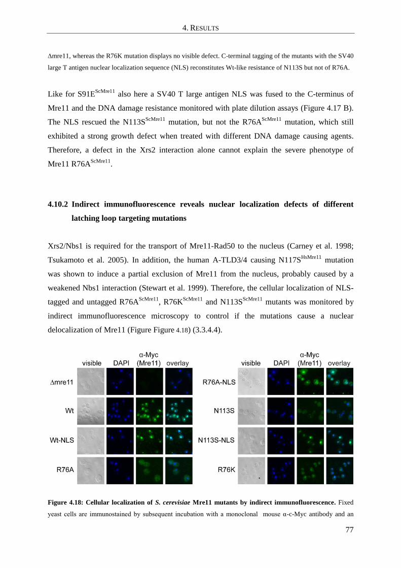
4. RESULTS
77
∆mre11, whereas the R76K mutation displays no visible defect. C-terminal tagging of the mutants with the SV40
large T antigen nuclear localization sequence (NLS) reconstitutes Wt-like resistance of N113S but not of R76A.
Like for S91EScMre11
also here a SV40 T large antigen NLS was fused to the C-terminus of
Mre11 and the DNA damage resistance monitored with plate dilution assays (Figure 4.17 B).
The NLS rescued the N113SScMre11
mutation, but not the R76AScMre11
mutation, which still
exhibited a strong growth defect when treated with different DNA damage causing agents.
Therefore, a defect in the Xrs2 interaction alone cannot explain the severe phenotype of
Mre11 R76AScMre11
.
4.10.2 Indirect immunofluorescence reveals nuclear localization defects of different
latching loop targeting mutations
Xrs2/Nbs1 is required for the transport of Mre11-Rad50 to the nucleus (Carney et al. 1998;
Tsukamoto et al. 2005). In addition, the human A-TLD3/4 causing N117SHsMre11
mutation
was shown to induce a partial exclusion of Mre11 from the nucleus, probably caused by a
weakened Nbs1 interaction (Stewart et al. 1999). Therefore, the cellular localization of NLS-
tagged and untagged R76AScMre11
, R76KScMre11
and N113SScMre11
mutants was monitored by
indirect immunofluorescence microscopy to control if the mutations cause a nuclear
delocalization of Mre11 (Figure Figure 4.18) (3.3.4.4).
Figure 4.18: Cellular localization of S. cerevisiae Mre11 mutants by indirect immunofluorescence. Fixed
yeast cells are immunostained by subsequent incubation with a monoclonal mouse α-c-Myc antibody and an

4. RESULTS
78
Alexa Fluor 488 conjugated goat α-mouse antibody. Nuclear localization is strongly decreased in the case of
R76AScMre11
and N113SScMre11
while partly reduced for R76KScMre11
. NLS-tagged R76K and N113S mutants
show a nuclear accumulation similar to Wt Mre11, while R76A-NLS accumulates at least in the majority of cells
comparable to Wt Mre11.
Here, it could be seen that the homologous N113SScMre11
mutation, as well as R76AScMre11
,
resulted in a strongly decreased nuclear accumulation of Mre11, consistent with the severe
phenotypes of these mutations. On the other hand, the repair competent mutant R76KScMre11
also displayed a partially decreased nuclear accumulation. Therefore, a decreased nuclear
accumulation of Mre11 does not necessarily lead to repair defects. The NLS relocated all
mutant proteins to the nucleus. However, the R76A-NLSScMre11
mutation possesses still a
severe repair defect (4.10.1). This raised the question to which degree nuclear mislocalisation
really accounts for the DNA damage sensitivity observed in R76AScMre11
and N113SScMre11
mutant strains.
4.10.3 Analysis of S. cerevisiae Mre11-Rad50-Xrs2 complex integrity and Mre11 dimer
interaction for different Mre11 latching loop targeting mutations
To test if the integrity of the MRX complex was affected by the latching loop targeting
mutations R76AScMre11
, R76KScMre11
and N113SScMre11
, co-immunoprecipitations (Co-IPs) of
the myc-tagged Mre11 strains were performed (Figure 4.19) (3.3.4.3). Here, protein levels of
Mre11, Rad50 and Xrs2 were relatively equal in cell lysates from Wt and mutant proteins,
ruling out misfolding or degradation as the main cause for the observed phenotype. Like its
human homologue, the Mre11 N113SScMre11
mutant was still able to form a stable complex
with Rad50 in Co-IPs (Stewart et al. 1999). Also for Mre11 R76KScMre11
a normal Rad50
interaction was observed, whereas this interaction was partly reduced but not abolished in Co-
IPs with the Mre11 R76AScMre11
mutant strain.
In contrast to Wt Mre11, however, Xrs2 was not detectable in immunoprecipitates from
R76AScMre11
, R76KScMre11
and N113SScMre11
mutant strains. This observation is in agreement
with the observed cellular mislocalisation of Mre11 R76AScMre11
and Mre11 N113SScMre11
(4.10.2). However, R76KScMre11
displayed no phenotype in plate survival assays with different
genotoxic agents and showed a reduced but significant nuclear accumulation (4.10.1 and

4. RESULTS
79
4.10.2). This means that R76K ScMre11
must possess a residing ability to bind Xrs2, which is
strong enough for a nuclear “piggy back” transport but too weak to be detectable in Co-IPs.
In conclusion, the phenotype of Mre11 N113SScMre11
can be explained as a mislocalisation
defect. The mutant is still competent in the Xrs2 independent mitotic repair functions of
Mre11-Rad50 and can be rescued by addition of a NLS to Mre11 (4.10.1). In contrast, the
R76AScMre11
mutation clearly impairs also the mitotic repair ability of Mre11-Rad50. Mre11
R76AScMre11
still binds to Rad50, even though at a partially decreased level. Therefore, the
dramatic sensitivity to genotoxic agents like HU or CPT can unlikely be explained by a fold
instability of Mre11 or loss of Rad50 binding, as it was reported for mutations with global
repair defects like Mre11-58 (Usui et al. 1998).
Figure 4.19: Analysis of Mre11-Rad50-Xrs2 complex integrity by co-immunoprecipitation for different S.
cerevisiae Mre11 mutations. Mre11-Rad50-Xrs2 complex formation defects caused by the R76A, R76K and
N113S mutations, which target the Mre11 latching loops, were detected by co-immunoprecipitation (3.3.4.3).
Cell extracts were prepared in buffer containing 150 mM NaCl and immunoprecipitated with an α-c-Myc
antibody against Mre11. Proteins were visualized by Western blotting with α-myc (Mre11), α-Rad50, and α-
Xrs2 antibodies, respectively. All tested latching loop targeting mutations caused a defect in Xrs2 binding,
underlining the crucial role of the latching loops for this function. Both R76 mutations showed reduced levels of
immunoprecipitated Mre11, indicating a reduced dimer stability. The ratio of co-immunoprecipitated Rad50 per
Mre11 was slightly reduced only in the case of Mre11 R76A.
Since R76ScMre11
maps to the dimer interface of Mre11, it was analyzed whether the
R76AScMre11
mutant has a defect in dimerization, which could explain its severe phenotype.
Williams et al. showed with mutational studies that the hydrophobic four helix bundle is
crucial for Mre11`s dimer integrity (Williams et al. 2008). The impact of the latching loop
mediated Mre11 dimer interaction, which is likely to be affected by the R76AScMre11
mutation,
however, is still unknown. To answer this question, a second plasmid, coding for a C-

4. RESULTS
80
terminally HA-tagged Mre11, was introduced into the myc-tagged mre11 shuffle strain to
create “mixed” dimer complexes. The myc-tagged Mre11 was then immunoprecipitated and
the dimer integrity analyzed using an antibody against HA-tagged Mre11 (Figure 4.20)
(3.3.4.3 and 3.3.2.6). Indeed, dimer formation was severely reduced in the R76AScMre11
strain
compared to Wt. This underlines that the Mre11 latching loops not only play a crucial role in
binding to Nbs1/Xrs2, but also in stabilizing Mre11 dimers in S. cerevisiae. In summary,
single point mutations in the Mre11-Xrs2 and the Mre11 dimer interface were identified,
which specifically disrupt these protein-protein interactions, thereby causing distinct
phenotypes.
Figure 4.20: Analysis of Mre11 dimer stability of S. cerevisiae Mre11 Wt and R76A by co-
immunoprecipitation. ScMre11 dimeric assembly is disrupted by the latching loop destabilizing R76A
mutation. Mixed Mre11 dimers, consisting of two monomers with different affinity tags were created by
introducing two plasmids, carrying the Mre11 Wt or R76A gene with a C-terminal 3xHA tag or 13xmyc tag,
respectively, into an S. cerevisiae W303 ∆mre11 strain. Dimer assembly was tested by co-immunoprecipitation
of myc-tagged Mre11 with a monoclonal α-c-Myc antibody and visualization of dimer assembly by western blot
analysis using antibodies against myc and HA-tagged Mre11.

5. DISCUSSION
81
5. DISCUSSION
All living organisms depend on surveillance mechanisms, which protect the stability of their
genomes from hazardous DNA damages. They therefore possess various repair pathways
which sense and repair DNA lesions and thereby maintain the genomic stability of the cell.
The efficient repair of DNA double strand breaks (DNA DSBs) is of special importance for
the cell, since unrepaired DSBs are highly cytotoxic and may induce chromosomal aberrations
and cancerogenic diseases. The Mre11-Rad50-Nbs1 (MRN) complex is a central component
of the DNA DSB repair machinery. The complex senses and binds as one of the first factors
to DSBs. It then works as an effector molecule, which recruits other repair proteins to the
break. MRN also actively processes DNA ends via its nuclease activity to facilitate repair of
the lesion. Furthermore it is involved in DNA damage signal transduction by recruiting and
activating the cell cycle checkpoint kinase ATM which then may promote cell cycle arrest or
apoptosis. The enzymatic core, consisting of the Mre11 nuclease dimer and two adjacent
Rad50 ABC-ATPases, is found in eukaryotes, archaea, bacteria and viruses. However, only
the eukaryotic complex contains with Nbs1 a third compound. Nbs1 possesses no enzymatic
activities. Instead it is a regulatory protein which mediates eukaryote specific functions of the
complex in DSB repair and signaling via interactions to Mre11 and in the human complex
also to Rad50 (Assenmacher and Hopfner 2004; Williams et al. 2010; Stracker and Petrini
2011). Nbs1 is crucial for the activation of ATM by the MRN complex in vivo (Berkovich et
al. 2007). Furthermore it was shown in vitro to influence the DNA binding specificity of
Mre11-Rad50 and to stimulate nucleolytic DNA hairpin processing by the complex (Paull and
Gellert 1999; Lee et al. 2003; Trujillo et al. 2003). However, it is only poorly understood on a
molecular level how Nbs1 carries out these functions and no atomic structures of eukaryotic
Mre11 or Rad50 in complex with Nbs1 are reported so far. Therefore, the aim of this work
was to analyze the structural and functional interplay between Nbs1 and Mre11 in detail. For
this purpose crystal structures of the catalytic core module of Mre11 alone and in complex
with the interaction region of Nbs1 were solved and biochemically characterized.
5.1 Preparation and crystallization of S. pombe apo-Mre11cd
and Nbs1mir
-Mre11cd
To gain insights into the structure of the eukaryotic Mre11-Rad50-Nbs1 complex, different
eukaryotic organisms where screened for soluble protein constructs. Here, the main focus was

5. DISCUSSION
82
to find constructs of Mre11 and Nbs1 suitable for crystallization. It turned out that especially
protein fragments from the fission yeast Schizosaccharomyces pombe could be expressed in a
soluble form in Escherichia coli in mg amounts which allowed extensive crystallization
screenings (4.1). Mre11 and Nbs1 from S. pombe are relatively compact in size compared to
their human protein homologs. S. pombe Mre11 has a size of 649 amino acid residues
compared to 708 residues for human Mre11. Likewise S. pombe Nbs1 comprises only 613
amino acids whereas its human counterpart is 754 residues long. However, important
structural elements like the Mre11 nuclease domain or the Mre11 interaction region of Nbs1
are conserved (see also domain maps in Figure 2.3). Albeit full length S. pombe Mre11 could
be purified recombinantly from E. coli, it was prone to proteolytic degradation over time and
did not crystallize. A secondary structure prediction with the program JPRED (Cole et al.
2008) showed that extensive areas of the C-terminal part of the protein lack secondary
structure elements, which explains the proteolytic degradation of the protein preparations.
Furthermore the C-terminus is involved in binding to Rad50 and might therefore be instable
when this binding partner is absent (Lammens et al. 2011). Since no soluble Rad50 construct
could be obtained, the aim was to crystallize the catalytic N-terminal part of Mre11. For this
purpose a limited proteolysis approach in combination with a secondary structure analysis
was applied. Based on this data a new construct containing residues 15-413 was generated
(4.1). Alignments with Mre11 from the archaeal organism P. furiosus indicated that it
included the complete nuclease core, which is composed of a phosphodiesterase domain and
an adjacent DNA capping domain (Hopfner et al. 2001). Therefore the construct was named
Mre11cd
(cd = catalytic domains). The protein was tested in crystallization screenings and
yielded crystals diffracting to a limiting resolution of 3.0 Å. The structure of Mre11cd
was
later solved by molecular replacement methods using the Nbs1mir
-Mre11cd
complex structure
(4.2.2).
Full length Nbs1 from S. pombe could not be expressed and purified in amounts suitable for
crystallization. Of special interest for this work was the C-terminal region of Nbs1, which
contains the binding sites for Mre11 and the checkpoint kinase ATM/Tel1. Indeed, a C-
terminal fragment containing residues 428-613 could be purified in mg amounts (4.1).
However it did not crystallize alone or in complex with Mre11cd
and was prone to proteolytic
degradation. The N-terminal part of S. pombe Nbs1, which contains the Forkhead and two
BRCT domains was already crystallized by Williams et al. (Williams et al. 2009). The authors
also characterized the C-terminal region, comprising residues 330-613, by proteolysis and
SAXS studies to be mainly unstructured. This likely explains why the construct 428-613 did

5. DISCUSSION
83
not crystallize. Therefore, different shorter deletion constructs of Nbs1 were purified and
tested for binding with Mre11cd
. By this approach, the core interaction motifs of Nbs1 for
Mre11 were narrowed down to the region between residues 474-531. This region contains two
conserved sequence elements which had been proposed before to be important for Mre11
binding (Ueno et al. 2003). The construct was further named Nbs1mir
(mir = Mre11 interaction
region) (4.1). Since Mre11cd
aa 15-413 and Nbs1 aa 474-531 did not crystallize initially, a
fusion protein was generated, which connected both proteins by an eight residues long linker,
to facilitate crystallization (Figure 4.2 B). The Nbs1mir
-Mre11cd
fusion protein could be
crystallized and native crystals diffracted to a limiting resolution of 2.4 Å (4.2.1). The
structure was solved by SAD using data from crystals of the selenomethionine labeled
complex, which diffracted to 2.8 Å resolution (4.2.1).
5.2 The eukaryotic Mre11 dimer resembles the principal domain architecture of
prokaryotic Mre11 but exhibits additional structural characteristics
The structure of Mre11cd
revealed a dimeric molecule with the principle domain architecture
similar to previously reported archaeal Mre11 from P. furiosus and bacterial Mre11 from
T. maritima (4.3) (Hopfner et al. 2001; Lammens et al. 2011). The dimeric contacts are
mediated by the two central phosphodiesterase domains via two helices from each protomer,
which form a hydrophobic four helix bundle. Each phosphodiesterase domain is flanked by a
DNA capping domain. Thereby also S. pombe Mre11cd
builds a U shaped particle with a
predicted DNA binding region at its concave site. Alignments showed high sequence
conservation between S. pombe and human Mre11 for the crystallized region with 47.9%
identical and 33.7% conserved residues. Therefore, the structural architecture of S. pombe
Mre11cd
is presumably also representative for higher eukaryotic organisms. Overlays of the
S.pombe Mre11cd
structure with the P. furiosus and T. maritima Mre11 structures showed that
all active site residues are identical (4.3). This indicates that the catalytic mechanism of
Mre11 is conserved between the three domains of live.
However, there are also important structural differences between the eukaryotic S. pombe
Mre11cd
structure and archaeal or bacterial Mre11 structures. Most prominently, S. pombe
Mre11 contains a conserved 33 amino acid long insertion loop element between residues 91-
123, which is specific for eukaryotic organisms. It maps to the top of the Mre11 dimer

5. DISCUSSION
84
mediating four helix bundle on the opposite site of the predicted DNA binding cleft and is
highly conserved among eukaryotes (4.3 and 4.4.2). The Nbs1mir
-Mre11cd
structure revealed
that this loop element, which is further named latching loop, is mediating a key interaction
with Nbs1 (4.4).
Also the capping domain of S. pombe Mre11 differs in its architecture from bacteria and
archaea. In particular, it contains an extended 40 Å long alpha helix (helix αF), between
residues 332 and 358, which is much shorter in archaea or bacteria (4.3). The function of this
helix is not understood at the moment. One possibility is that it might be important for the
interaction with Rad50, since the capping domain was observed to bind to Rad50 in the
bacterial T. maritima Mre11-Rad50 complex structure (Lammens et al. 2011). Interestingly,
the S. pombe Mre11cd
structure contains a conserved surface exposed tyrosine residue
Y338SpMre11
at one end of the helix near the region of the putative Rad50 binding site.
Mutation of the corresponding Y328ScMre11
residue in S. cerevisiae renders cells strongly
sensitive to the genotoxic agents MMS, HU and CPT without affecting the protein levels of
Mre11 (Lammens et al. 2011). This indicates that the extended eukaryotic capping domain
helix might indeed be involved in binding Rad50.
5.3 Nbs1 binds to the Mre11 dimer via multiple contacts and controls its dimeric
configuration
The structure of Nbs1mir
-Mre11cd
revealed that Mre11cd
and Nbs1mir
build a complex with 2:2
stoichiometry (4.4.1). The possibility of a structural artifact, caused by the fusion protein, was
ruled out by an additional construct of Mre11cd
and Nbs1mir
with a cleavable TEV protease
recognition site linker, which was cleaved during the purification procedure before the size
exclusion chromatography step. Consistently, the structure of the non-fused Mre11mir
-Nbs1cd
complex is highly similar to the fusion protein structure in all aspects of stoichiometry and
conformation (4.4.1).
A main characteristic of the Nbs1mir
-Mre11cd
structure is that Nbs1 interacts with Mre11 not
via a compact region but instead as an extended peptide at multiple sites. Each of the two
Nbs1mir
peptides binds to one Mre11cd
protomer. This so called interaction region 1 consists
of a long stretched α-helix and ß-strand motif between residues 477-498 of Nbs1 (4.4.2). C-
terminal to the interaction region 1, both Nbs1mir
chains are disordered. However, one of the
two peptides is ordered again between residues 518-526 to bind the Mre11 dimer at

5. DISCUSSION
85
interaction site 2 via a highly conserved KNFKxFxK motif. Interestingly, this interaction is
asymmetric and it breaks the two fold symmetry of the complex. The KNFKxFxK motif binds
across the central Mre11 dimer cleft, which is build up by the two latching loops. It thereby
sterically excludes binding of the second Nbs1 molecule to the latching loops. The
significance of the observed interaction is underlined by the high sequence conservation on
both sides of the interface. The KNFKxFxK motif is one of a few sequence elements of the
poorly conserved Nbs1 C-terminus which is conserved between all eukaryotic organisms
(4.4.2). The most interesting finding is that the corresponding human residue of N122SpMre11
is
mutated in A-TLD 3/4 (N117SHsMre11
) (Stewart et al. 1999). The two N122SpMre11
residues
coordinate the central phenylalanine residue F524ESpNbs1
from both sides via π-stacking.
Consistently, the N117SHsMre11
and N122
SpMre11 mutations were reported to reduce the affinity
of Mre11 to Nbs1 in human and S. pombe cell extracts, respectively (Lee et al. 2003; Porter-
Goff and Rhind 2009). The revealed interface between Mre11 and Nbs1 also corresponds well
to the interaction region of S. cerevisiae Xrs2 to Mre11 in two-hybrid studies (Desai-Mehta et
al. 2001). In addition point mutations in F521Nbs1
, K522Nbs1
and/or K526Nbs1
lead to
compromised or abolished Mre11 interaction and resemble xrs2 phenotypes in formation of
meiotic breaks in S. cerevisiae (Shima et al. 2005). Thus, the identified Mre11-Nbs1
interaction can be explained and is supported by a broad spectrum of in vitro and in vivo data.
By bridging both Mre11 molecules Nbs1 appears to “tether” the Mre11 dimer to determine its
dimeric configuration. A comparison of Nbs1mir
-Mre11cd
with apo-Mre11cd
showed
significant conformational differences between both structures (4.5). Binding of the
KNFKxFxK motif orders the latching loops, which are unstructured and “swapped” between
the Mre11 protomers in the apo-Mre11cd
structure. Besides, both structures also exhibit global
conformational differences. Superposition of apo-Mre11cd
and Nbs1mir
-Mre11cd
revealed a
difference in the Mre11 dimer angle of 30° towards a more compact conformation in the
Nbs1mir
-Mre11cd
structure. Therefore, the S. pombe Mre11 dimer possesses an intrinsic
flexibility, which allows the adoption of different dimer conformations. Furthermore, the
binding of Nbs1 coordinates the latching loops and thereby determines a distinct dimeric
conformation. A model, which links the Nbs1 dependent Mre11 dimer configuration to DNA
DSB signaling, is discussed in the last chapter of the discussion (5.6).

5. DISCUSSION
86
5.4 The extended structure of the Mre11-Nbs1 interface may explain the
hypomorphic character of A-TLD causing mutations
Several mutations in the human Mre11 gene were reported to cause the genetic disease
Ataxia-telangiectasia like disorder (A-TLD), which is characterized by genomic instability,
neurodegeneration and for one subtype also by mental retardation and lung carcinoma
(Stewart et al. 1999; Fernet et al. 2005; Uchisaka et al. 2009). In addition, a heterozygous
Mre11 mutation was recently reported to be linked to a Nijmegen-breakage syndrome (NBS)
like disease (2.6) (Matsumoto et al. 2011).
The structure of S. pombe Nbs1mir
-Mre11cd
now allows for the first time a structural analysis
of the mutations causing the diseases (4.8.1). The most prominent finding regards the A-TLD
subtype 3/4, which is caused by a mutation of an asparagine residue N117SHsMre11
(Stewart et
al. 1999). The corresponding S. pombe residue N122SpMre11
is mediating a key interaction with
Nbs1 residue F524 at interaction region 2. Therefore, a weakened Nbs1 interaction very likely
contributes to the repair and signaling defects seen in A-TLD 3/4. This is also in agreement to
Co-IP studies with the human protein, where the interaction was found to be strongly reduced
in cells derived from patients with ATLD 3/4 (Stewart et al. 1999). The mutation
W210CHsMre11
(W215CSpMre11
), which leads to A-TLD 7/8 (Fernet et al. 2005), on the other
hand appears to have an impact on the interaction region 1, since W215CSpMre11
caps the three
stranded shared ß-sheet between S. pombe Mre11cd
and Nbs1mir
. W215SpMre11
is not surface
exposed. Hence, the mutation may also destabilize the overall fold of the protein and thereby
impair its functionality. This is also the case for the W243RHsMre11
and Del(340-366)
mutations, which very likely destabilize the Mre11 protein fold (Uchisaka et al. 2009).
The impact of the NBS-like disease causing mutation D113GHsMre11
is more difficult to
evaluate, since it occurs as a heterozygous mutation (Matsumoto et al. 2011) (4.8.1). The
mutation in the second allele maps to a non-coding region and causes a splicing defect. This
results in a low level expression of Wt Mre11 from the second allele. However, it also means
that here a small population of Mre11 is fully functional and might be responsible for the
relatively high ATM activation levels in NBS-like disease compared to A-TLD. Therefore, it
cannot be clearly distinguished, to which degree the D113GHsMre11
mutation contributes to the
observed phenotype. The S. pombe Nbs1mir
-Mre11cd
crystal structure revealed that
D109SpMre11
, the corresponding residue of D113GHsMre11
, mediates an important Mre11 dimer
interface contact (4.4.2). It maps to the latching loop and is positioned by arginine residue

5. DISCUSSION
87
R85SpMre11
, which protrudes from the interface helix F of the neighboring Mre11 protomer. An
R76AScMre11
mutation of the corresponding arginine residue in S. cerevisiae caused a null
phenotype of Mre11 (4.10.1). Likewise, also the aspartate to glycine mutation of
D113GHsMre11
likely destabilizes the fold of the latching loops and disturbs the interaction to
Nbs1. Hence, it is suggested, that the D113GHsMre11
mutation renders the protein strongly
dysfunctional.
The influence of A-TLD causing mutations on the Mre11-Nbs1 interaction was also further
studied in vitro by binding assays using analytical size-exclusion chromatography and
recombinant, purified S. pombe Mre11 aa 1-413 and Nbs1 aa 428-613 (4.8.2). The binding
experiments showed that all of the tested mutations retain the ability to bind to Mre11.
However, the mutations weaken the Mre11-Nbs1 interaction. The Wt Mre11 protein fragment
interacted in the presence of Mn2+
as well as in EDTA containing buffer with Nbs1. However,
EDTA completely disrupted the Mre11-Nbs1 interaction of all A-TLD mutants. Coordination
of Mn2+
ions by the active site residues likely stabilizes the phosphodiesterase domain fold.
Since EDTA removes the Mn2+
ions from the active site, it weakens the Mre11 fold stability
and thereby also the Mre11-Nbs1 interaction. The additive effects of folding destabilization
by removal of Mn2+
from the active site plus the weakening influence of A-TLD causing
mutations on the Mre11-Nbs1 interface finally result in a disruption of the Mre11-Nbs1
complex in analytical size exclusion chromatography. The experiment suggests that A-TLD
causing mutations lead to a partially weakened but not abolished Mre11-Nbs1 interaction.
This can be explained by the structure of S. pombe Nbs1mir
-Mre11cd
, since Nbs1 interacts with
Mre11 as an extended peptide via multiple independent contacts. The single A-TLD point
mutations therefore presumably disrupt only a part of the interface. As a result, A-TLD
mutations render the complex partially dysfunctional, thereby causing the hypomorphic
disease phenotype.
5.5 The latching loops of S. cerevisiae Mre11 are crucial for the general functionality
of the Mre11-Rad50-Xrs2 complex
Different motifs found in the S. pombe Nbs1mir
-Mre11cd
structure where studied in vivo, using
the budding yeast S. cerevisiae as a model organism (4.10). Mre11 proteins from both
organisms share a high sequence conservation for the crystallized Mre11 nuclease core

5. DISCUSSION
88
(51.3% identical + 28.4 % similar residues). Therefore, the S. pombe structure can be used as
a guiding model for the mutational analysis of conserved functional residues in S. cerevisiae.
One interesting observation of the S. cerevisiae studies was the MMS and CPT sensitivity,
caused by mutations in the loop element, which connects the phosphodiesterase motif III of
the active site with the Mre11 dimer interface helix αC (4.10.1). A recently published
structure of the archaeal Methanococcus jannaschii Mre11-Rad50 complex bound to ATPγS
showed the corresponding archaeal loop to interact with Rad50 in the nucleotide bound state
of the complex (Lim et al. 2011). This could also be the function of the eukaryotic loop.
Exchange of the S. cerevisiae loop by the sequence from the archaeal organism P. furiosus led
to severe growth defects on MMS and CPT plates similar to mre11∆. This can likely be
explained by sequence and charge differences of both loops. It contains two arginine residues
in P. furiosus, but two aspartate residues in S. cerevisiae and S. pombe (Figure 4.8). One
theory would be that coordination of the eukaryotic loop by Rad50 in the nucleotide and DNA
bound state renders the active site accessible for DNA ends. Another indication for this theory
comes from the observation, that the active site of S. pombe Mre11 contains binding sites of
high and low Mn2+
affinity. When crystallized with low Mn2+
concentrations, only one Mn2+
is bound and the discussed loop element is flipped in a conformation where it could sterically
block binding of DNA to the active site (4.6). Rad50 could therefore be directly involved in
the allosteric regulation of the eukaryotic Mre11 nuclease function by ordering the active site
via coordination of the Mre11 active site loop element.
The main focus of the S. cerevisiae in vivo studies was on the mutational analysis of the
Mre11-Nbs1/Xrs2 interface and the functional role of the Mre11 latching loops (4.10.1). Here
mutations in S. cerevisiae Mre11 residues, which mediate binding to Nbs1 via interaction
region 1, exhibited no growth defects in the presence of MMS and CPT. This is in agreement
with the observation that the complete deletion of the corresponding interaction region 1
region in S. cerevisiae Xrs2 does not impair mitotic repair but it is instead crucial for the
meiotic function of the complex (Tsukamoto et al. 2005). However, analysis of interaction
region 1 mutations for spore viability by the group of Steve Jackson showed also no meiosis
defect (unpublished results - Laboratory of Steve Jackson, Cambridge). This might be
explained by the extended conformation of Nbs1 in the Nbs1mir
-Mre11cd
structure (4.4).
Single point mutations are probably not strong enough to disrupt the complete interaction
region 1 and therefore preserve the meiotic function of MRX.

5. DISCUSSION
89
The interaction region 2, observed between the S. pombe Mre11cd
latching loops and Nbs1mir
(4.4), appears to also be crucial for the interaction of S. cerevisisae Mre11 and Xrs2. The
mutations S91EScMre11
and N113SScMre11
, which both map to the latching loop of S. cerevisiae
Mre11, render the cells sensitive to genotoxic agents like HU and CPT (4.10.1). A control
experiment with the corresponding NLS-tagged Mre11 mutants showed a rescue of the
resistance against HU and CPT. Xrs2 contains the only NLS within the Mre11-Rad50-Xrs2
complex and is therefore crucial for the nuclear localization of the complex. However, when
Mre11 is tagged by an NLS, Xrs2 is not needed for the resistance to genotoxic agents
(Tsukamoto et al. 2005). Hence, the defects of S91EScMre11
and N113SScMre11
can be explained
by a disturbed Mre11-Xrs2 interaction which leads to a cellular delocalization of Mre11-
Rad50.
In contrast, the R76AScMre11
mutation targets not only the Xrs2 interface but also the overall
stability of the latching loops (4.10.1). R76ScMre11
, which protrudes from the dimer interface
helix αC, coordinates the latching loops via a salt bridge to the latching loop residue
D109ScMre11
(4.4.2). Remarkably, the corresponding human residue of D109ScMre11
is mutated
in human NBS-like disease (Matsumoto et al. 2011). Co-immunoprecipitation (Co-IP) studies
and indirect immunofluorescence analysis of the Mre11 R76AScMre11
mutant strain revealed an
abolished interaction of Mre11 and Xrs2 (4.10.2 and 4.10.3). However, the repair defect,
monitored by sensitivity to MMS; HU and CPT, could not be rescued by the addition of a
NLS to Mre11 R76A (4.10.1). The mutation therefore also impairs the function of Mre11
apart from weakening the interaction to Xrs2. Co-IP analysis of two Mre11 alleles with
different affinity tags revealed, that the R76AScMre11
mutation is strongly impairing the
stability of Mre11 dimers (4.10.3). This might explain the severe mitotic repair defect of the
NLS tagged R76AScMre11
mutant strain, since Mre11 is only functional in vivo as a dimeric
molecule (Williams et al. 2008).
In summary, single point mutations in the Mre11-Xrs2 or the Mre11 dimer interfaces were
identified, which specifically disrupt these protein-protein interactions, thereby causing
mitotic repair defects. The in vivo analysis of S. cerevisiae Mre11 (4.10) verified the
interactions between Mre11 and Nbs1 determined by the S. pombe Nbs1mir
-Mre11cd
crystal
structure (4.4). The latching loops are not only crucial for the interaction to Xrs2 in vivo. They
also are important for the stability of the Mre11 dimer (4.10.3). In this aspect the eukaryotic
Mre11 protein architecture strongly differs from bacterial and archaeal Mre11, where the

5. DISCUSSION
90
dimer interface is solely composed of the conserved hydrophobic four helix bundle (Hopfner
et al. 2001; Lammens et al. 2011).
5.6 A model for Mre11 dimer and Nbs1 mediated DSB signaling
The asymmetric interaction of Nbs1 with the Mre11 dimer suggests a functional link between
Mre11 dimer conformation and DSB signaling. It was already discussed how Nbs1
structurally orders the Mre11 dimer interface (5.3). Superposition of S. pombe (Sp)Mre11cd
with the SpNbs1mir
-Mre11cd
complex additionally revealed an unexpected flexibility in the
Mre11 dimer angle by approx. 30° (4.5). Such alterations in the Mre11 dimer angle could
indeed be linked to DSB recognition as a comparison of both structures with the crystal
structure of P. furiosus (Pf)Mre11cd
bound to DNA shows (Figure 5.1) (Williams et al. 2008) .
The DNA complex of PfMre11cd
revealed that DNA ends are bound across both Mre11
protomers and therefore need a particular Mre11 dimer orientation (Figure 5.1 A).
Figure 5.1: Structural variability in the Mre11 dimer and model for DNA double-strand break signaling.
(A) Overlay of S. pombe Mre11cd
(yellow) with the Nbs1mir
-Mre11cd
complex (green with magenta Nbs1) and
5. DISCUSSION
91
DNA bound dimer of P. furiosus Mre11cd
(blue with brown DNA) reveals that the Mre11 dimer pivot angle is
highly variable. All three structures are superimposed via one Mre11 protomer, respectively. DNA binding with
multiple interaction sites, as observed in the PfMre11-DNA complex, requires a substantial structural change in
the Mre11-Nbs1 complex that could be the basis of damage sensing and signaling, by modulating the interaction
of Nbs1 with the Mre11 dimer interface. (B) In apo-SpMre11cd
(top panel, shown as cartoon model with omitted
capping domains), two phosphodiesterase domain closely interact via two helix pairs (green and blue), one from
each protomer. In the absence of Nbs1, the latching loops are swapped between protomers and partially
disordered, allowing the dimer interface to be closed. Middle panel: Nbs1 (magenta) orders the latching loops
into a “kissing” conformation and changes the mre11 dimer pivot angle by slightly opening the interface. This
brings a DNA binding helix (orange) of one protomer into closer proximity to the DNA bound to the other
protomer (modeled after the PfMre11:DNA complex). Lower panel: full DNA binding, as seen in the DNA-
PfMre11 complex (protein data bank entry 3DSD), is achieved after further opening the dimer interface,
allowing the other protomer to assist in binding the minor groove (orange helix).
Superimposing SpNbs1mir
-Mre11cd
with PfMre11cd
-DNA reveals now another large pivot
motion of the Mre11 dimer by approx. 30° (Figure 5.1 A, B). In fact, to form the dsDNA
binding interactions of the PfMre11cd
:DNA complex, SpMre11cd
protomers must rotate by
approx. 30° from the conformation observed in the Nbs1 complex and even 60° from the
conformation seen in the absence of Nbs1.
The revealed conformational flexibility in the Mre11 dimer, its evident coupling to Nbs1
binding, and expected conformational changes upon DNA binding, suggest an intriguing but
simple, testable model for DSB signaling (Figure 5.2). Since most DNA binding residues are
conserved between archaeal and eukaryotic Mre11, it is plausible that DSB recognition will
induce an Mre11 dimer orientation as seen in the PfMre11cd
-DNA complex (Figure 5.1 A, B).
Such a conceivable conformational change will likely affect the latching loops by the
underlying rigid body movements of the phosphodiesterase domains and hence modulate the
interaction with the Nbs1 molecules at this region. This is plausible because the observed
differences between Nbs1 bound and free Mre11 suggest that latching loop structure, Mre11
dimer angle and Nbs1 interaction are coupled.
Moreover, the Hopfner group and others showed that bacterial and archaeal Mre11-Rad50
bind to DNA as an ATP dependent clamp, and DNA binding involves large conformational
rearrangements in both proteins (Lammens et al. 2011; Lim et al. 2011). Such a DNA and
ATP induced conformational switch of Mre11-Rad50 could be the driving force for a rotation
of Mre11 dimers and remodeling of the Mre11-Nbs1 interaction. Interestingly, mutational
studies showed that the activation of ATM depends on nucleotide binding by Rad50 (Lee and
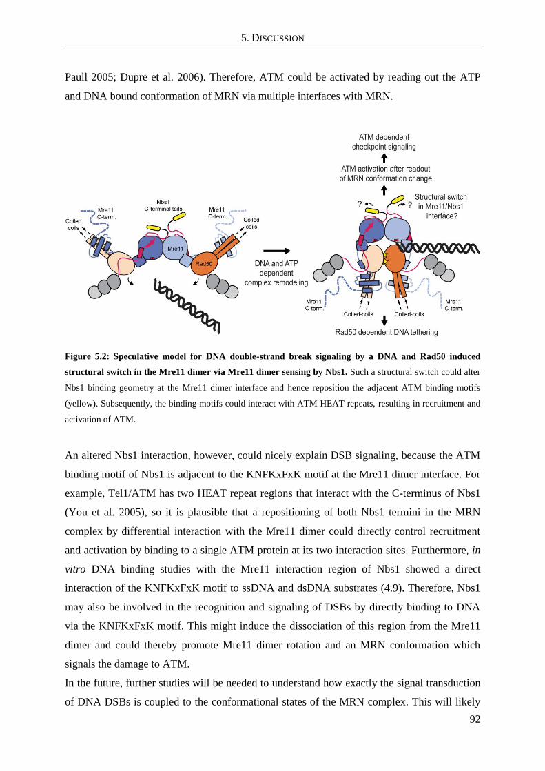
5. DISCUSSION
92
Paull 2005; Dupre et al. 2006). Therefore, ATM could be activated by reading out the ATP
and DNA bound conformation of MRN via multiple interfaces with MRN.
Figure 5.2: Speculative model for DNA double-strand break signaling by a DNA and Rad50 induced
structural switch in the Mre11 dimer via Mre11 dimer sensing by Nbs1. Such a structural switch could alter
Nbs1 binding geometry at the Mre11 dimer interface and hence reposition the adjacent ATM binding motifs
(yellow). Subsequently, the binding motifs could interact with ATM HEAT repeats, resulting in recruitment and
activation of ATM.
An altered Nbs1 interaction, however, could nicely explain DSB signaling, because the ATM
binding motif of Nbs1 is adjacent to the KNFKxFxK motif at the Mre11 dimer interface. For
example, Tel1/ATM has two HEAT repeat regions that interact with the C-terminus of Nbs1
(You et al. 2005), so it is plausible that a repositioning of both Nbs1 termini in the MRN
complex by differential interaction with the Mre11 dimer could directly control recruitment
and activation by binding to a single ATM protein at its two interaction sites. Furthermore, in
vitro DNA binding studies with the Mre11 interaction region of Nbs1 showed a direct
interaction of the KNFKxFxK motif to ssDNA and dsDNA substrates (4.9). Therefore, Nbs1
may also be involved in the recognition and signaling of DSBs by directly binding to DNA
via the KNFKxFxK motif. This might induce the dissociation of this region from the Mre11
dimer and could thereby promote Mre11 dimer rotation and an MRN conformation which
signals the damage to ATM.
In the future, further studies will be needed to understand how exactly the signal transduction
of DNA DSBs is coupled to the conformational states of the MRN complex. This will likely

5. DISCUSSION
93
require a hybrid techniques approach to combine atomic resolution structures of the
nucleotide and DNA bound Mre11-Rad50-Nbs1 complex and ATM/Tel1 with biochemical
and cell biology methods.

6. REFERENCES
I
6. REFERENCES
Ahnesorg, P., P. Smith and S. P. Jackson (2006). "XLF interacts with the XRCC4-DNA ligase
IV complex to promote DNA nonhomologous end-joining." Cell 124(2): 301-313.
Ajimura, M., S. H. Leem and H. Ogawa (1993). "Identification of new genes required for
meiotic recombination in Saccharomyces cerevisiae." Genetics 133(1): 51-66.
Al-Minawi, A. Z., Y. F. Lee, D. Hakansson, F. Johansson, C. Lundin, N. Saleh-Gohari, N.
Schultz, D. Jenssen, H. E. Bryant, M. Meuth, J. M. Hinz and T. Helleday (2009). "The
ERCC1/XPF endonuclease is required for completion of homologous recombination at
DNA replication forks stalled by inter-strand cross-links." Nucleic Acids Res 37(19):
6400-6413.
Alani, E., R. Padmore and N. Kleckner (1990). "Analysis of wild-type and rad50 mutants of
yeast suggests an intimate relationship between meiotic chromosome synapsis and
recombination." Cell 61(3): 419-436.
Almeida, K. H. and R. W. Sobol (2007). "A unified view of base excision repair: lesion-
dependent protein complexes regulated by post-translational modification." DNA
Repair (Amst) 6(6): 695-711.
Assenmacher, N. and K. P. Hopfner (2004). "MRE11/RAD50/NBS1: complex activities."
Chromosoma 113(4): 157-166.
Bartkova, J., J. Tommiska, L. Oplustilova, K. Aaltonen, A. Tamminen, T. Heikkinen, M.
Mistrik, K. Aittomaki, C. Blomqvist, P. Heikkila, J. Lukas, H. Nevanlinna and J.
Bartek (2008). "Aberrations of the MRE11-RAD50-NBS1 DNA damage sensor
complex in human breast cancer: MRE11 as a candidate familial cancer-predisposing
gene." Mol Oncol 2(4): 296-316.
Berkovich, E., R. J. Monnat, Jr. and M. B. Kastan (2007). "Roles of ATM and NBS1 in
chromatin structure modulation and DNA double-strand break repair." Nat Cell Biol
9(6): 683-690.
Bonetti, D., M. Martina, M. Clerici, G. Lucchini and M. P. Longhese (2009). "Multiple
pathways regulate 3' overhang generation at S. cerevisiae telomeres." Mol Cell 35(1):
70-81.
Borde, V. (2007). "The multiple roles of the Mre11 complex for meiotic recombination."
Chromosome Res 15(5): 551-563.
Borde, V. and J. Cobb (2009). "Double functions for the Mre11 complex during DNA double-
strand break repair and replication." Int J Biochem Cell Biol 41(6): 1249-1253.
Bosco, E. E., C. N. Mayhew, R. F. Hennigan, J. Sage, T. Jacks and E. S. Knudsen (2004).
"RB signaling prevents replication-dependent DNA double-strand breaks following
genotoxic insult." Nucleic Acids Res 32(1): 25-34.
Boulton, S. J. and S. P. Jackson (1998). "Components of the Ku-dependent non-homologous
end-joining pathway are involved in telomeric length maintenance and telomeric
silencing." EMBO J 17(6): 1819-1828.
Bressan, D. A., H. A. Olivares, B. E. Nelms and J. H. Petrini (1998). "Alteration of N-
terminal phosphoesterase signature motifs inactivates Saccharomyces cerevisiae
Mre11." Genetics 150(2): 591-600.
Bromberg, K. D., A. B. Burgin and N. Osheroff (2003). "A two-drug model for etoposide
action against human topoisomerase IIalpha." J Biol Chem 278(9): 7406-7412.
Bryson, K., L. J. McGuffin, R. L. Marsden, J. J. Ward, J. S. Sodhi and D. T. Jones (2005).
"Protein structure prediction servers at University College London." Nucleic Acids
Res 33(Web Server issue): W36-38.

6. REFERENCES
II
Buis, J., Y. Wu, Y. Deng, J. Leddon, G. Westfield, M. Eckersdorff, J. M. Sekiguchi, S. Chang
and D. O. Ferguson (2008). "Mre11 nuclease activity has essential roles in DNA repair
and genomic stability distinct from ATM activation." Cell 135(1): 85-96.
Bzymek, M., N. H. Thayer, S. D. Oh, N. Kleckner and N. Hunter (2010). "Double Holliday
junctions are intermediates of DNA break repair." Nature 464(7290): 937-941.
Cadet, J., M. Berger, T. Douki and J. L. Ravanat (1997). "Oxidative damage to DNA:
formation, measurement, and biological significance." Rev Physiol Biochem
Pharmacol 131: 1-87.
Carney, J. P., R. S. Maser, H. Olivares, E. M. Davis, M. Le Beau, J. R. Yates, 3rd, L. Hays,
W. F. Morgan and J. H. Petrini (1998). "The hMre11/hRad50 protein complex and
Nijmegen breakage syndrome: linkage of double-strand break repair to the cellular
DNA damage response." Cell 93(3): 477-486.
Chamankhah, M., T. Fontanie and W. Xiao (2000). "The Saccharomyces cerevisiae mre11(ts)
allele confers a separation of DNA repair and telomere maintenance functions."
Genetics 155(2): 569-576.
Chamankhah, M. and W. Xiao (1999). "Formation of the yeast Mre11-Rad50-Xrs2 complex
is correlated with DNA repair and telomere maintenance." Nucleic Acids Res 27(10):
2072-2079.
Chapman, J. R. and S. P. Jackson (2008). "Phospho-dependent interactions between NBS1
and MDC1 mediate chromatin retention of the MRN complex at sites of DNA
damage." EMBO Rep 9(8): 795-801.
Chen, L., K. Trujillo, W. Ramos, P. Sung and A. E. Tomkinson (2001). "Promotion of Dnl4-
catalyzed DNA end-joining by the Rad50/Mre11/Xrs2 and Hdf1/Hdf2 complexes."
Mol Cell 8(5): 1105-1115.
Ciccia, A. and S. J. Elledge (2010). "The DNA damage response: making it safe to play with
knives." Mol Cell 40(2): 179-204.
Clerici, M., D. Mantiero, I. Guerini, G. Lucchini and M. P. Longhese (2008). "The Yku70-
Yku80 complex contributes to regulate double-strand break processing and checkpoint
activation during the cell cycle." EMBO Rep 9(8): 810-818.
Coic, E., G. F. Richard and J. E. Haber (2006). "Saccharomyces cerevisiae donor preference
during mating-type switching is dependent on chromosome architecture and
organization." Genetics 173(3): 1197-1206.
Cole, C., J. D. Barber and G. J. Barton (2008). "The Jpred 3 secondary structure prediction
server." Nucleic Acids Res 36(Web Server issue): W197-201.
Connelly, J. C., E. S. de Leau and D. R. Leach (1999). "DNA cleavage and degradation by the
SbcCD protein complex from Escherichia coli." Nucleic Acids Res 27(4): 1039-1046.
Connelly, J. C., E. S. de Leau and D. R. Leach (2003). "Nucleolytic processing of a protein-
bound DNA end by the E. coli SbcCD (MR) complex." DNA Repair (Amst) 2(7):
795-807.
Costanzo, V., K. Robertson, M. Bibikova, E. Kim, D. Grieco, M. Gottesman, D. Carroll and J.
Gautier (2001). "Mre11 protein complex prevents double-strand break accumulation
during chromosomal DNA replication." Mol Cell 8(1): 137-147.
Cowtan, K. (2006). "The Buccaneer software for automated model building. 1. Tracing
protein chains." Acta Crystallogr D Biol Crystallogr 62(Pt 9): 1002-1011.
Critchlow, S. E. and S. P. Jackson (1998). "DNA end-joining: from yeast to man." Trends
Biochem Sci 23(10): 394-398.
D'Amours, D. and S. P. Jackson (2001). "The yeast Xrs2 complex functions in S phase
checkpoint regulation." Genes Dev 15(17): 2238-2249.

6. REFERENCES
III
de Jager, M., J. van Noort, D. C. van Gent, C. Dekker, R. Kanaar and C. Wyman (2001).
"Human Rad50/Mre11 is a flexible complex that can tether DNA ends." Mol Cell
8(5): 1129-1135.
Degrassi, F., M. Fiore and F. Palitti (2004). "Chromosomal aberrations and genomic
instability induced by topoisomerase-targeted antitumour drugs." Curr Med Chem
Anticancer Agents 4(4): 317-325.
Deriano, L., T. H. Stracker, A. Baker, J. H. Petrini and D. B. Roth (2009). "Roles for NBS1 in
alternative nonhomologous end-joining of V(D)J recombination intermediates." Mol
Cell 34(1): 13-25.
Deribe, Y. L., T. Pawson and I. Dikic (2010). "Post-translational modifications in signal
integration." Nat Struct Mol Biol 17(6): 666-672.
Desai-Mehta, A., K. M. Cerosaletti and P. Concannon (2001). "Distinct functional domains of
nibrin mediate Mre11 binding, focus formation, and nuclear localization." Mol Cell
Biol 21(6): 2184-2191.
Di Virgilio, M., C. Y. Ying and J. Gautier (2009). "PIKK-dependent phosphorylation of
Mre11 induces MRN complex inactivation by disassembly from chromatin." DNA
Repair (Amst) 8(11): 1311-1320.
Dianov, G. L. and J. L. Parsons (2007). "Co-ordination of DNA single strand break repair."
DNA Repair (Amst) 6(4): 454-460.
Dinkelmann, M., E. Spehalski, T. Stoneham, J. Buis, Y. Wu, J. M. Sekiguchi and D. O.
Ferguson (2009). "Multiple functions of MRN in end-joining pathways during isotype
class switching." Nat Struct Mol Biol 16(8): 808-813.
Dudley, D. D., J. Chaudhuri, C. H. Bassing and F. W. Alt (2005). "Mechanism and control of
V(D)J recombination versus class switch recombination: similarities and differences."
Adv Immunol 86: 43-112.
Dupre, A., L. Boyer-Chatenet and J. Gautier (2006). "Two-step activation of ATM by DNA
and the Mre11-Rad50-Nbs1 complex." Nat Struct Mol Biol 13(5): 451-457.
Emsley, P. and K. Cowtan (2004). "Coot: model-building tools for molecular graphics." Acta
Crystallogr D Biol Crystallogr 60(Pt 12 Pt 1): 2126-2132.
Errico, A. and V. Costanzo (2010). "Differences in the DNA replication of unicellular
eukaryotes and metazoans: known unknowns." EMBO Rep 11(4): 270-278.
Falck, J., J. Coates and S. P. Jackson (2005). "Conserved modes of recruitment of ATM, ATR
and DNA-PKcs to sites of DNA damage." Nature 434(7033): 605-611.
Falck, J., J. H. Petrini, B. R. Williams, J. Lukas and J. Bartek (2002). "The DNA damage-
dependent intra-S phase checkpoint is regulated by parallel pathways." Nat Genet
30(3): 290-294.
Fattah, F., E. H. Lee, N. Weisensel, Y. Wang, N. Lichter and E. A. Hendrickson (2010). "Ku
regulates the non-homologous end joining pathway choice of DNA double-strand
break repair in human somatic cells." PLoS Genet 6(2): e1000855.
Faure, V., S. Coulon, J. Hardy and V. Geli (2010). "Cdc13 and telomerase bind through
different mechanisms at the lagging- and leading-strand telomeres." Mol Cell 38(6):
842-852.
Fernet, M., M. Gribaa, M. A. Salih, M. Z. Seidahmed, J. Hall and M. Koenig (2005).
"Identification and functional consequences of a novel MRE11 mutation affecting 10
Saudi Arabian patients with the ataxia telangiectasia-like disorder." Hum Mol Genet
14(2): 307-318.
Frappart, P. O. and P. J. McKinnon (2006). "Ataxia-telangiectasia and related diseases."
Neuromolecular Med 8(4): 495-511.

6. REFERENCES
IV
Furuse, M., Y. Nagase, H. Tsubouchi, K. Murakami-Murofushi, T. Shibata and K. Ohta
(1998). "Distinct roles of two separable in vitro activities of yeast Mre11 in mitotic
and meiotic recombination." EMBO J 17(21): 6412-6425.
Game, J. C. and R. K. Mortimer (1974). "A genetic study of x-ray sensitive mutants in yeast."
Mutat Res 24(3): 281-292.
Goudsouzian, L. K., C. T. Tuzon and V. A. Zakian (2006). "S. cerevisiae Tel1p and Mre11p
are required for normal levels of Est1p and Est2p telomere association." Mol Cell
24(4): 603-610.
Haber, J. E. (1998). "The many interfaces of Mre11." Cell 95(5): 583-586.
Haber, J. E. (1998). "Mating-type gene switching in Saccharomyces cerevisiae." Annu Rev
Genet 32: 561-599.
Haber, J. E. (2008). "Alternative endings." Proc Natl Acad Sci U S A 105(2): 405-406.
Hari, F. J., C. Spycher, S. Jungmichel, L. Pavic and M. Stucki (2010). "A divalent
FHA/BRCT-binding mechanism couples the MRE11-RAD50-NBS1 complex to
damaged chromatin." EMBO Rep 11(5): 387-392.
Harper, J. W. and S. J. Elledge (2007). "The DNA damage response: ten years after." Mol
Cell 28(5): 739-745.
Hector, R. E., R. L. Shtofman, A. Ray, B. R. Chen, T. Nyun, K. L. Berkner and K. W. Runge
(2007). "Tel1p preferentially associates with short telomeres to stimulate their
elongation." Mol Cell 27(5): 851-858.
Helmink, B. A., A. L. Bredemeyer, B. S. Lee, C. Y. Huang, G. G. Sharma, L. M. Walker, J. J.
Bednarski, W. L. Lee, T. K. Pandita, C. H. Bassing and B. P. Sleckman (2009). "MRN
complex function in the repair of chromosomal Rag-mediated DNA double-strand
breaks." J Exp Med 206(3): 669-679.
Hendrickson, W. A., J. R. Horton and D. M. LeMaster (1990). "Selenomethionyl proteins
produced for analysis by multiwavelength anomalous diffraction (MAD): a vehicle for
direct determination of three-dimensional structure." EMBO J 9(5): 1665-1672.
Henner, W. D., L. O. Rodriguez, S. M. Hecht and W. A. Haseltine (1983). "gamma Ray
induced deoxyribonucleic acid strand breaks. 3' Glycolate termini." J Biol Chem
258(2): 711-713.
Herdendorf, T. J., D. W. Albrecht, S. J. Benkovic and S. W. Nelson (2011). "Biochemical
characterization of bacteriophage T4 Mre11-Rad50 complex." J Biol Chem 286(4):
2382-2392.
Heyer, W. D. (2004). "Recombination: Holliday junction resolution and crossover formation."
Curr Biol 14(2): R56-58.
Hirano, Y., K. Fukunaga and K. Sugimoto (2009). "Rif1 and rif2 inhibit localization of tel1 to
DNA ends." Mol Cell 33(3): 312-322.
Hoeijmakers, J. H. (2001). "Genome maintenance mechanisms for preventing cancer." Nature
411(6835): 366-374.
Hopfner, K. P., L. Craig, G. Moncalian, R. A. Zinkel, T. Usui, B. A. Owen, A. Karcher, B.
Henderson, J. L. Bodmer, C. T. McMurray, J. P. Carney, J. H. Petrini and J. A. Tainer
(2002). "The Rad50 zinc-hook is a structure joining Mre11 complexes in DNA
recombination and repair." Nature 418(6897): 562-566.
Hopfner, K. P., A. Karcher, L. Craig, T. T. Woo, J. P. Carney and J. A. Tainer (2001).
"Structural biochemistry and interaction architecture of the DNA double-strand break
repair Mre11 nuclease and Rad50-ATPase." Cell 105(4): 473-485.
Hopfner, K. P., A. Karcher, D. Shin, C. Fairley, J. A. Tainer and J. P. Carney (2000). "Mre11
and Rad50 from Pyrococcus furiosus: cloning and biochemical characterization reveal
an evolutionarily conserved multiprotein machine." J Bacteriol 182(21): 6036-6041.

6. REFERENCES
V
Huertas, P. (2010). "DNA resection in eukaryotes: deciding how to fix the break." Nat Struct
Mol Biol 17(1): 11-16.
Huertas, P., F. Cortes-Ledesma, A. A. Sartori, A. Aguilera and S. P. Jackson (2008). "CDK
targets Sae2 to control DNA-end resection and homologous recombination." Nature
455(7213): 689-692.
Huertas, P. and S. P. Jackson (2009). "Human CtIP mediates cell cycle control of DNA end
resection and double strand break repair." J Biol Chem 284(14): 9558-9565.
Hug, N. and J. Lingner (2006). "Telomere length homeostasis." Chromosoma 115(6): 413-
425.
Inagaki, A., S. Schoenmakers and W. M. Baarends (2010). "DNA double strand break repair,
chromosome synapsis and transcriptional silencing in meiosis." Epigenetics 5(4): 255-
266.
Ivanov, E. L., V. G. Korolev and F. Fabre (1992). "XRS2, a DNA repair gene of
Saccharomyces cerevisiae, is needed for meiotic recombination." Genetics 132(3):
651-664.
Ivanov, E. L., N. Sugawara, J. Fishman-Lobell and J. E. Haber (1996). "Genetic requirements
for the single-strand annealing pathway of double-strand break repair in
Saccharomyces cerevisiae." Genetics 142(3): 693-704.
Jacob, S., C. Miquel, A. Sarasin and F. Praz (2005). "Effects of camptothecin on double-
strand break repair by non-homologous end-joining in DNA mismatch repair-deficient
human colorectal cancer cell lines." Nucleic Acids Res 33(1): 106-113.
Janke, R., K. Herzberg, M. Rolfsmeier, J. Mar, V. I. Bashkirov, E. Haghnazari, G. Cantin, J.
R. Yates, 3rd and W. D. Heyer (2010). "A truncated DNA-damage-signaling response
is activated after DSB formation in the G1 phase of Saccharomyces cerevisiae."
Nucleic Acids Res 38(7): 2302-2313.
Jones, D. T. (1999). "Protein secondary structure prediction based on position-specific scoring
matrices." J Mol Biol 292(2): 195-202.
Kabsch, W. (1993). "Automatic processing of rotation diffraction data from crystals of
initially unknown symmetry and cell constants." Journal of Appl. Crystallogr. 26(6):
795-800.
Keelagher, R. E., V. E. Cotton, A. S. Goldman and R. H. Borts (2011). "Separable roles for
Exonuclease I in meiotic DNA double-strand break repair." DNA Repair (Amst)
10(2): 126-137.
Keeney, S., C. N. Giroux and N. Kleckner (1997). "Meiosis-specific DNA double-strand
breaks are catalyzed by Spo11, a member of a widely conserved protein family." Cell
88(3): 375-384.
Keeney, S. and N. Kleckner (1995). "Covalent protein-DNA complexes at the 5' strand
termini of meiosis-specific double-strand breaks in yeast." Proc Natl Acad Sci U S A
92(24): 11274-11278.
Kim, S. T. (2005). "Protein kinase CK2 interacts with Chk2 and phosphorylates Mre11 on
serine 649." Biochem Biophys Res Commun 331(1): 247-252.
Konarev, P. V., M. V. Petoukhov, V. V. Volkov and D. I. Svergun (2006). "ATSAS 2.1, a
program package for small-angle scattering data analysis." Journal of Applied
Crystallography 39(2): 277-286.
Kuzminov, A. (2001). "DNA replication meets genetic exchange: chromosomal damage and
its repair by homologous recombination." Proc Natl Acad Sci U S A 98(15): 8461-
8468.
Laemmli, U. K. (1970). "Cleavage of structural proteins during the assembly of the head of
bacteriophage T4." Nature 227(5259): 680-685.

6. REFERENCES
VI
Lam, A. F., B. O. Krogh and L. S. Symington (2008). "Unique and overlapping functions of
the Exo1, Mre11 and Pso2 nucleases in DNA repair." DNA Repair (Amst) 7(4): 655-
662.
Lammens, K., D. J. Bemeleit, C. Mockel, E. Clausing, A. Schele, S. Hartung, C. B. Schiller,
M. Lucas, C. Angermuller, J. Soding, K. Strasser and K. P. Hopfner (2011). "The
Mre11:Rad50 Structure Shows an ATP-Dependent Molecular Clamp in DNA Double-
Strand Break Repair." Cell 145(1): 54-66.
Langer, G., S. X. Cohen, V. S. Lamzin and A. Perrakis (2008). "Automated macromolecular
model building for X-ray crystallography using ARP/wARP version 7." Nat Protoc
3(7): 1171-1179.
Larkin, M. A., G. Blackshields, N. P. Brown, R. Chenna, P. A. McGettigan, H. McWilliam, F.
Valentin, I. M. Wallace, A. Wilm, R. Lopez, J. D. Thompson, T. J. Gibson and D. G.
Higgins (2007). "Clustal W and Clustal X version 2.0." Bioinformatics 23(21): 2947-
2948.
Lavin, M. F. (2008). "Ataxia-telangiectasia: from a rare disorder to a paradigm for cell
signalling and cancer." Nat Rev Mol Cell Biol 9(10): 759-769.
Lee, J. H., R. Ghirlando, V. Bhaskara, M. R. Hoffmeyer, J. Gu and T. T. Paull (2003).
"Regulation of Mre11/Rad50 by Nbs1: effects on nucleotide-dependent DNA binding
and association with ataxia-telangiectasia-like disorder mutant complexes." J Biol
Chem 278(46): 45171-45181.
Lee, J. H. and T. T. Paull (2005). "ATM activation by DNA double-strand breaks through the
Mre11-Rad50-Nbs1 complex." Science 308(5721): 551-554.
Lee, S. E., D. A. Bressan, J. H. Petrini and J. E. Haber (2002). "Complementation between N-
terminal Saccharomyces cerevisiae mre11 alleles in DNA repair and telomere length
maintenance." DNA Repair (Amst) 1(1): 27-40.
Lengsfeld, B. M., A. J. Rattray, V. Bhaskara, R. Ghirlando and T. T. Paull (2007). "Sae2 is an
endonuclease that processes hairpin DNA cooperatively with the Mre11/Rad50/Xrs2
complex." Mol Cell 28(4): 638-651.
Lewis, L. K., F. Storici, S. Van Komen, S. Calero, P. Sung and M. A. Resnick (2004). "Role
of the nuclease activity of Saccharomyces cerevisiae Mre11 in repair of DNA double-
strand breaks in mitotic cells." Genetics 166(4): 1701-1713.
Lieber, M. R. (2010). "The mechanism of double-strand DNA break repair by the
nonhomologous DNA end-joining pathway." Annu Rev Biochem 79: 181-211.
Lim, H. S., J. S. Kim, Y. B. Park, G. H. Gwon and Y. Cho (2011). "Crystal structure of the
Mre11-Rad50-ATP{gamma}S complex: understanding the interplay between Mre11
and Rad50." Genes Dev.
Limbo, O., C. Chahwan, Y. Yamada, R. A. de Bruin, C. Wittenberg and P. Russell (2007).
"Ctp1 is a cell-cycle-regulated protein that functions with Mre11 complex to control
double-strand break repair by homologous recombination." Mol Cell 28(1): 134-146.
Limoli, C. L., E. Giedzinski, W. M. Bonner and J. E. Cleaver (2002). "UV-induced
replication arrest in the xeroderma pigmentosum variant leads to DNA double-strand
breaks, gamma -H2AX formation, and Mre11 relocalization." Proc Natl Acad Sci U S
A 99(1): 233-238.
Lloyd, J., J. R. Chapman, J. A. Clapperton, L. F. Haire, E. Hartsuiker, J. Li, A. M. Carr, S. P.
Jackson and S. J. Smerdon (2009). "A supramodular FHA/BRCT-repeat architecture
mediates Nbs1 adaptor function in response to DNA damage." Cell 139(1): 100-111.
Longhese, M. P., D. Bonetti, N. Manfrini and M. Clerici (2010). "Mechanisms and regulation
of DNA end resection." EMBO J 29(17): 2864-2874.

6. REFERENCES
VII
Mahaney, B. L., K. Meek and S. P. Lees-Miller (2009). "Repair of ionizing radiation-induced
DNA double-strand breaks by non-homologous end-joining." Biochem J 417(3): 639-
650.
Mansour, W. Y., S. Schumacher, R. Rosskopf, T. Rhein, F. Schmidt-Petersen, F. Gatzemeier,
F. Haag, K. Borgmann, H. Willers and J. Dahm-Daphi (2008). "Hierarchy of
nonhomologous end-joining, single-strand annealing and gene conversion at site-
directed DNA double-strand breaks." Nucleic Acids Res 36(12): 4088-4098.
Matsumoto, Y., T. Miyamoto, H. Sakamoto, H. Izumi, Y. Nakazawa, T. Ogi, H. Tahara, S.
Oku, A. Hiramoto, T. Shiiki, Y. Fujisawa, H. Ohashi, Y. Sakemi and S. Matsuura
(2011). "Two unrelated patients with MRE11A mutations and Nijmegen breakage
syndrome-like severe microcephaly." DNA Repair (Amst) 10(3): 314-321.
McCoy, A. J. (2007). "Solving structures of protein complexes by molecular replacement with
Phaser." Acta Crystallogr D Biol Crystallogr 63(Pt 1): 32-41.
McKee, A. H. and N. Kleckner (1997). "A general method for identifying recessive diploid-
specific mutations in Saccharomyces cerevisiae, its application to the isolation of
mutants blocked at intermediate stages of meiotic prophase and characterization of a
new gene SAE2." Genetics 146(3): 797-816.
Meek, K., P. Douglas, X. Cui, Q. Ding and S. P. Lees-Miller (2007). "trans
Autophosphorylation at DNA-dependent protein kinase's two major
autophosphorylation site clusters facilitates end processing but not end joining." Mol
Cell Biol 27(10): 3881-3890.
Melander, F., S. Bekker-Jensen, J. Falck, J. Bartek, N. Mailand and J. Lukas (2008).
"Phosphorylation of SDT repeats in the MDC1 N terminus triggers retention of NBS1
at the DNA damage-modified chromatin." J Cell Biol 181(2): 213-226.
Mimitou, E. P. and L. S. Symington (2009). "DNA end resection: many nucleases make light
work." DNA Repair (Amst) 8(9): 983-995.
Mimitou, E. P. and L. S. Symington (2011). "DNA end resection--unraveling the tail." DNA
Repair (Amst) 10(3): 344-348.
Misteli, T. and E. Soutoglou (2009). "The emerging role of nuclear architecture in DNA
repair and genome maintenance." Nat Rev Mol Cell Biol 10(4): 243-254.
Moreau, S., J. R. Ferguson and L. S. Symington (1999). "The nuclease activity of Mre11 is
required for meiosis but not for mating type switching, end joining, or telomere
maintenance." Mol Cell Biol 19(1): 556-566.
Moreno-Herrero, F., M. de Jager, N. H. Dekker, R. Kanaar, C. Wyman and C. Dekker (2005).
"Mesoscale conformational changes in the DNA-repair complex Rad50/Mre11/Nbs1
upon binding DNA." Nature 437(7057): 440-443.
Nairz, K. and F. Klein (1997). "mre11S--a yeast mutation that blocks double-strand-break
processing and permits nonhomologous synapsis in meiosis." Genes Dev 11(17):
2272-2290.
Nakada, D., K. Matsumoto and K. Sugimoto (2003). "ATM-related Tel1 associates with
double-strand breaks through an Xrs2-dependent mechanism." Genes Dev 17(16):
1957-1962.
New, J. H., T. Sugiyama, E. Zaitseva and S. C. Kowalczykowski (1998). "Rad52 protein
stimulates DNA strand exchange by Rad51 and replication protein A." Nature
391(6665): 407-410.
Palmbos, P. L., J. M. Daley and T. E. Wilson (2005). "Mutations of the Yku80 C terminus
and Xrs2 FHA domain specifically block yeast nonhomologous end joining." Mol Cell
Biol 25(24): 10782-10790.
Pandita, T. K. and C. Richardson (2009). "Chromatin remodeling finds its place in the DNA
double-strand break response." Nucleic Acids Res 37(5): 1363-1377.

6. REFERENCES
VIII
Paull, T. T. and M. Gellert (1998). "The 3' to 5' exonuclease activity of Mre 11 facilitates
repair of DNA double-strand breaks." Mol Cell 1(7): 969-979.
Paull, T. T. and M. Gellert (1999). "Nbs1 potentiates ATP-driven DNA unwinding and
endonuclease cleavage by the Mre11/Rad50 complex." Genes Dev 13(10): 1276-1288.
Porter-Goff, M. E. and N. Rhind (2009). "The Role of MRN in the S-Phase DNA Damage
Checkpoint is Independent of its Ctp1-Dependent Roles in Double-Strand Break
Repair and Checkpoint Signaling." Mol Biol Cell.
Prinz, S., A. Amon and F. Klein (1997). "Isolation of COM1, a new gene required to
complete meiotic double-strand break-induced recombination in Saccharomyces
cerevisiae." Genetics 146(3): 781-795.
Raghavan, S. C., C. L. Hsieh and M. R. Lieber (2005). "Both V(D)J coding ends but neither
signal end can recombine at the bcl-2 major breakpoint region, and the rejoining is
ligase IV dependent." Mol Cell Biol 25(15): 6475-6484.
Rahal, E. A., L. A. Henricksen, Y. Li, R. S. Williams, J. A. Tainer and K. Dixon (2010).
"ATM regulates Mre11-dependent DNA end-degradation and microhomology-
mediated end joining." Cell Cycle 9(14): 2866-2877.
Rass, E., A. Grabarz, I. Plo, J. Gautier, P. Bertrand and B. S. Lopez (2009). "Role of Mre11 in
chromosomal nonhomologous end joining in mammalian cells." Nat Struct Mol Biol
16(8): 819-824.
Sambrook, J. and D. W. Russell (2001). Molecular Cloning: A Laboratory Manual. New
York, Cold Spring Harbor Laboratory Press.
San Filippo, J., P. Sung and H. Klein (2008). "Mechanism of eukaryotic homologous
recombination." Annu Rev Biochem 77: 229-257.
Sartori, A. A., C. Lukas, J. Coates, M. Mistrik, S. Fu, J. Bartek, R. Baer, J. Lukas and S. P.
Jackson (2007). "Human CtIP promotes DNA end resection." Nature 450(7169): 509-
514.
Sharples, G. J. and D. R. Leach (1995). "Structural and functional similarities between the
SbcCD proteins of Escherichia coli and the RAD50 and MRE11 (RAD32)
recombination and repair proteins of yeast." Mol Microbiol 17(6): 1215-1217.
Shim, E. Y., W. H. Chung, M. L. Nicolette, Y. Zhang, M. Davis, Z. Zhu, T. T. Paull, G. Ira
and S. E. Lee (2010). "Saccharomyces cerevisiae Mre11/Rad50/Xrs2 and Ku proteins
regulate association of Exo1 and Dna2 with DNA breaks." EMBO J 29(19): 3370-
3380.
Shima, H., M. Suzuki and M. Shinohara (2005). "Isolation and characterization of novel xrs2
mutations in Saccharomyces cerevisiae." Genetics 170(1): 71-85.
Shull, E. R., Y. Lee, H. Nakane, T. H. Stracker, J. Zhao, H. R. Russell, J. H. Petrini and P. J.
McKinnon (2009). "Differential DNA damage signaling accounts for distinct neural
apoptotic responses in ATLD and NBS." Genes Dev 23(2): 171-180.
Soding, J., A. Biegert and A. N. Lupas (2005). "The HHpred interactive server for protein
homology detection and structure prediction." Nucleic Acids Res 33(Web Server
issue): W244-248.
Soulas-Sprauel, P., P. Rivera-Munoz, L. Malivert, G. Le Guyader, V. Abramowski, P. Revy
and J. P. de Villartay (2007). "V(D)J and immunoglobulin class switch
recombinations: a paradigm to study the regulation of DNA end-joining." Oncogene
26(56): 7780-7791.
Spycher, C., E. S. Miller, K. Townsend, L. Pavic, N. A. Morrice, P. Janscak, G. S. Stewart
and M. Stucki (2008). "Constitutive phosphorylation of MDC1 physically links the
MRE11-RAD50-NBS1 complex to damaged chromatin." J Cell Biol 181(2): 227-240.
Stewart, G. S., R. S. Maser, T. Stankovic, D. A. Bressan, M. I. Kaplan, N. G. Jaspers, A.
Raams, P. J. Byrd, J. H. Petrini and A. M. Taylor (1999). "The DNA double-strand

6. REFERENCES
IX
break repair gene hMRE11 is mutated in individuals with an ataxia-telangiectasia-like
disorder." Cell 99(6): 577-587.
Stracker, T. H. and J. H. Petrini (2011). "The MRE11 complex: starting from the ends." Nat
Rev Mol Cell Biol 12(2): 90-103.
Strahl-Bolsinger, S., A. Hecht, K. Luo and M. Grunstein (1997). "SIR2 and SIR4 interactions
differ in core and extended telomeric heterochromatin in yeast." Genes Dev 11(1): 83-
93.
Sung, P. and H. Klein (2006). "Mechanism of homologous recombination: mediators and
helicases take on regulatory functions." Nat Rev Mol Cell Biol 7(10): 739-750.
Sutherland, B. M., P. V. Bennett, O. Sidorkina and J. Laval (2000). "Clustered DNA damages
induced in isolated DNA and in human cells by low doses of ionizing radiation." Proc
Natl Acad Sci U S A 97(1): 103-108.
Symington, L. S. (2002). "Role of RAD52 epistasis group genes in homologous
recombination and double-strand break repair." Microbiol Mol Biol Rev 66(4): 630-
670, table of contents.
Tonegawa, S. (1983). "Somatic generation of antibody diversity." Nature 302(5909): 575-581.
Trujillo, K. M., D. H. Roh, L. Chen, S. Van Komen, A. Tomkinson and P. Sung (2003).
"Yeast xrs2 binds DNA and helps target rad50 and mre11 to DNA ends." J Biol Chem
278(49): 48957-48964.
Trujillo, K. M. and P. Sung (2001). "DNA structure-specific nuclease activities in the
Saccharomyces cerevisiae Rad50*Mre11 complex." J Biol Chem 276(38): 35458-
35464.
Trujillo, K. M., S. S. Yuan, E. Y. Lee and P. Sung (1998). "Nuclease activities in a complex
of human recombination and DNA repair factors Rad50, Mre11, and p95." J Biol
Chem 273(34): 21447-21450.
Tsukamoto, Y., C. Mitsuoka, M. Terasawa, H. Ogawa and T. Ogawa (2005). "Xrs2p regulates
Mre11p translocation to the nucleus and plays a role in telomere elongation and
meiotic recombination." Mol Biol Cell 16(2): 597-608.
Uchisaka, N., N. Takahashi, M. Sato, A. Kikuchi, S. Mochizuki, K. Imai, S. Nonoyama, O.
Ohara, F. Watanabe, S. Mizutani, R. Hanada and T. Morio (2009). "Two brothers with
ataxia-telangiectasia-like disorder with lung adenocarcinoma." J Pediatr 155(3): 435-
438.
Ueno, M., T. Nakazaki, Y. Akamatsu, K. Watanabe, K. Tomita, H. D. Lindsay, H. Shinagawa
and H. Iwasaki (2003). "Molecular characterization of the Schizosaccharomyces
pombe nbs1+ gene involved in DNA repair and telomere maintenance." Mol Cell Biol
23(18): 6553-6563.
Usui, T., T. Ohta, H. Oshiumi, J. Tomizawa, H. Ogawa and T. Ogawa (1998). "Complex
formation and functional versatility of Mre11 of budding yeast in recombination." Cell
95(5): 705-716.
Uziel, T., Y. Lerenthal, L. Moyal, Y. Andegeko, L. Mittelman and Y. Shiloh (2003).
"Requirement of the MRN complex for ATM activation by DNA damage." EMBO J
22(20): 5612-5621.
van der Linden, E., H. Sanchez, E. Kinoshita, R. Kanaar and C. Wyman (2009). "RAD50 and
NBS1 form a stable complex functional in DNA binding and tethering." Nucleic Acids
Res 37(5): 1580-1588.
Varon, R., C. Vissinga, M. Platzer, K. M. Cerosaletti, K. H. Chrzanowska, K. Saar, G.
Beckmann, E. Seemanova, P. R. Cooper, N. J. Nowak, M. Stumm, C. M. Weemaes,
R. A. Gatti, R. K. Wilson, M. Digweed, A. Rosenthal, K. Sperling, P. Concannon and
A. Reis (1998). "Nibrin, a novel DNA double-strand break repair protein, is mutated
in Nijmegen breakage syndrome." Cell 93(3): 467-476.
6. REFERENCES
X
Waltes, R., R. Kalb, M. Gatei, A. W. Kijas, M. Stumm, A. Sobeck, B. Wieland, R. Varon, Y.
Lerenthal, M. F. Lavin, D. Schindler and T. Dork (2009). "Human RAD50 deficiency
in a Nijmegen breakage syndrome-like disorder." Am J Hum Genet 84(5): 605-616.
Weemaes, C. M., T. W. Hustinx, J. M. Scheres, P. J. van Munster, J. A. Bakkeren and R. D.
Taalman (1981). "A new chromosomal instability disorder: the Nijmegen breakage
syndrome." Acta Paediatr Scand 70(4): 557-564.
Weterings, E. and D. C. van Gent (2004). "The mechanism of non-homologous end-joining: a
synopsis of synapsis." DNA Repair (Amst) 3(11): 1425-1435.
Williams, G. J., S. P. Lees-Miller and J. A. Tainer (2010). "Mre11-Rad50-Nbs1
conformations and the control of sensing, signaling, and effector responses at DNA
double-strand breaks." DNA Repair (Amst) 9(12): 1299-1306.
Williams, G. J., R. S. Williams, J. S. Williams, G. Moncalian, A. S. Arvai, O. Limbo, G.
Guenther, S. Sildas, M. Hammel, P. Russell and J. A. Tainer (2011). "ABC ATPase
signature helices in Rad50 link nucleotide state to Mre11 interface for DNA repair."
Nat Struct Mol Biol 18(4): 423-431.
Williams, R. S., G. E. Dodson, O. Limbo, Y. Yamada, J. S. Williams, G. Guenther, S.
Classen, J. N. Glover, H. Iwasaki, P. Russell and J. A. Tainer (2009). "Nbs1 flexibly
tethers Ctp1 and Mre11-Rad50 to coordinate DNA double-strand break processing and
repair." Cell 139(1): 87-99.
Williams, R. S., G. Moncalian, J. S. Williams, Y. Yamada, O. Limbo, D. S. Shin, L. M.
Groocock, D. Cahill, C. Hitomi, G. Guenther, D. Moiani, J. P. Carney, P. Russell and
J. A. Tainer (2008). "Mre11 dimers coordinate DNA end bridging and nuclease
processing in double-strand-break repair." Cell 135(1): 97-109.
Xie, A., A. Kwok and R. Scully (2009). "Role of mammalian Mre11 in classical and
alternative nonhomologous end joining." Nat Struct Mol Biol 16(8): 814-818.
Xu, C., L. Wu, G. Cui, M. V. Botuyan, J. Chen and G. Mer (2008). "Structure of a second
BRCT domain identified in the nijmegen breakage syndrome protein Nbs1 and its
function in an MDC1-dependent localization of Nbs1 to DNA damage sites." J Mol
Biol 381(2): 361-372.
Yazdi, P. T., Y. Wang, S. Zhao, N. Patel, E. Y. Lee and J. Qin (2002). "SMC1 is a
downstream effector in the ATM/NBS1 branch of the human S-phase checkpoint."
Genes Dev 16(5): 571-582.
Ye, Y. and A. Godzik (2004). "FATCAT: a web server for flexible structure comparison and
structure similarity searching." Nucleic Acids Res 32(Web Server issue): W582-585.
You, Z., C. Chahwan, J. Bailis, T. Hunter and P. Russell (2005). "ATM activation and its
recruitment to damaged DNA require binding to the C terminus of Nbs1." Mol Cell
Biol 25(13): 5363-5379.
Zakharyevich, K., Y. Ma, S. Tang, P. Y. Hwang, S. Boiteux and N. Hunter (2010).
"Temporally and biochemically distinct activities of Exo1 during meiosis: double-
strand break resection and resolution of double Holliday junctions." Mol Cell 40(6):
1001-1015.
Zha, S., C. Boboila and F. W. Alt (2009). "Mre11: roles in DNA repair beyond homologous
recombination." Nat Struct Mol Biol 16(8): 798-800.
Zha, S., C. Guo, C. Boboila, V. Oksenych, H. L. Cheng, Y. Zhang, D. R. Wesemann, G.
Yuen, H. Patel, P. H. Goff, R. L. Dubois and F. W. Alt (2011). "ATM damage
response and XLF repair factor are functionally redundant in joining DNA breaks."
Nature 469(7329): 250-254.
Zhuang, J., G. Jiang, H. Willers and F. Xia (2009). "Exonuclease function of human Mre11
promotes deletional nonhomologous end joining." J Biol Chem 284(44): 30565-
30573.

6. REFERENCES
XI
Zierhut, C. and J. F. Diffley (2008). "Break dosage, cell cycle stage and DNA replication
influence DNA double strand break response." EMBO J 27(13): 1875-1885.
Zwart, P. H., P. V. Afonine, R. W. Grosse-Kunstleve, L. W. Hung, T. R. Ioerger, A. J.
McCoy, E. McKee, N. W. Moriarty, R. J. Read, J. C. Sacchettini, N. K. Sauter, L. C.
Storoni, T. C. Terwilliger and P. D. Adams (2008). "Automated structure solution with
the PHENIX suite." Methods Mol Biol 426: 419-435.

7. ABBREVATIONS
XII
7. ABBREVATIONS
6-FAM 6-carboxyfluorescein
Å Angstrom
aa amino acid (residue)
ABC ATP binding cassette
AFM Atomic force microscopy
APS ammonium persulfate
A-TLD Ataxia-telangiectasia like disorder
ATM Ataxia telangiectasia mutated
ATP adenosine triphosphate
bp base pair
BRCT breast cancer C-terminus
°C degree celsius
CK2 casein kinase 2
DAPI 4„,6-diamino-2-phenylindole
DNA deoxyribonucleic acid
DNA-PK DNA-dependent protein kinase
DrMre11 Mre11 from Danio rerio
DSB DNA double-strand break
DSBR DNA double-strand break repair
dsDNA double-stranded DNA
E. coli Escherichia coli
ECL enhanced chemoluminiscence
EDTA ethylenediaminetetraacetic acid
EMSA electrophoretic mobility shift assay
FHA forkhead associated
h hours
HEAT Huntingtin, elongation factor 3, the A subunit of protein
phosphatase 2A, TOR lipid kinase HR homologous recombination
HsMre11 Mre11 from Homo sapiens
IPTG Isopropyl-β-D-thiogalactopyranosid
kDa kilo Dalton
L litre
LB Luria-Bertani
M molar
min minute
MMS methyl methanesulfonate
Mn manganese
Mre11 meiotic recombination 11

7. ABBREVATIONS
XIII
Mre11cd
Mre11 catalytic domain
MRN Mre11-Rad50-Nbs1
MRX Mre11-Rad50-Xrs2 (S. cerevisiae complex)
n nano
Nbs Nijmegen breakage syndrome
Nbs1mir
Mre11 interaction region of Nbs1
NHEJ non-homologous end joining
NLS nuclear localisation sequence
NMR nuclear magnetic resonance spectroscopy
nt nucleotide
OD600 optical density at 600 nm
PAGE polyacrylamide gelelectrophoresis
PBS phosphate-buffered saline
PBS phosphate buffered saline
PCR polymerase chain reaction
PDB protein data bank
PEG polyethylene glycol
PfMre11 Mre11 from Pyrococcus furiosus
Pfu Pyrococcus furiosus
pH potential of hydrogen
PMSF phenylmethanesulfonylfluoride
ROS reactive oxygen species
rpm rotation per minute
RT room temperature
S. cerevisiae Saccharomyces cerevisiae
S. pombe Schizosaccharomyces pombe
SAD single-wavelength anomalous dispersion
SAXS small angle X-ray scattering
ScMre11 Mre11 from Saccharomyces cerevisiae
SDS sodium dodecyl-sulphate
sec second
SpMre11 Mre11 from Schizosaccharomyces pombe
TB Tris/Borate
TBE Tris/Borate/EDTA
TBS tris-buffered saline
TBST tris-buffered saline + Tween 20
TEMED N,N,N′,N′-tetramethylethylenediamine
TEV Tobacco Etch Virus

7. ABBREVATIONS
XIV
TmMre11 Mre11 from Thermotoga maritima
Tris tris(hydroxymethyl)aminomethane
UV ultraviolet
Wt Wild type
XRCC1 X-ray repair cross-complementing protein 1
YPD yeast extract, peptone, glucose containing medium
μ micro
8. CURRICULUM VITAE
XV
8. CURRICULUM VITAE
Personal Details
Name Christian Bernd Schiller
Date of birth November 28th
, 1980
Place of birth Kassel, Germany
Nationality German
Education
1987 - 1991 Grundschule in Großalmerode, Germany
1991 - 1993 Orientierungsstufe in Großalmerode, Germany
1993 - 1997 Gymnasium in Großalmerode, Germany
1997 - 2000 Gymnasium in Hessisch Lichtenau, Großalmerode
Civilian service
2000 - 2001 Jugendwaldheim Meißner, Hessisch Lichtenau, Germany
Academic Training
2001 - 2006 Studies in biology, University of Göttingen, Germany
2005 - 2006 Diploma thesis in the group of Prof. Dr. Ralf Ficner at the Centre for
Molecular Biosciences (GZMB), University of Göttingen
Title: “Preparation and crystallisation of ribosomal proteins with the
nuclear import factor Importin 7“
2006 Diploma in Biology
Since 12/2006 PhD thesis in the group of Prof. Dr. Karl-Peter Hopfner, Gene Center,
Department of Biochemistry, University of Munich, Germany

9. ACKNOWLEDGEMENTS
XVI
9. ACKNOWLEDGEMENTS
First of all I want to thank my supervisor Prof. Dr. Karl-Peter Hopfner for giving me the
chance to work on this fascinating project in the field of genome maintenance. I am especially
grateful for him being always approachable to discuss scientific problems and for all his very
helpful and creative ideas on the project.
I am also thankful to Prof. Dr. Dietmar Martin for being the second examiner of this thesis.
Many thanks go to all the members of the Hopfner group. I really enjoyed the friendly and
inspiring working atmosphere and also the leisure time outside the lab. Thank you, for all
your support with experiments and helpful scientific discussions. I want to acknowledge in
particular also Alexandra Schele, Brigitte Keßler, Manuela Moldt and Olga Fettscher for the
excellent organization of the laboratory.
My special thanks go to all the members of the “Fellowship of the Ring” who experienced
with me to work on this very versatile complex, which still keeps some secrets to uncover: To
Katja Lammens, Carolin Möckel, Alexandra Schele, Derk Bemeleit, Ania Rojowska, Maria
Lucas and Matthew Bennett. I am really thankful for all your fruitful ideas, inspiring
discussions and your important contributions to this project. It was a great experience to work
with you.
I am especially grateful to Katja Lammens for her supervision on this project and her help and
expertise in structural biology.
I would like to thank Ilaria Guerini and Prof. Steve Jackson for a very interesting and fruitful
collaboration, which allowed us to combine the structural biology of this project with in vivo
studies.
My special thanks go also to Britta Coordes for all her expertise and support with the budding
yeast experiments.
I would further like to also thank my former bachelor students Florian Schlauderer, Anna-
Kathrin Kopetzki and Irina Heckmann for their great enthusiasm and contributions to this
project.
I deeply want to thank you Britta for your patience, love and support. You mean the world to
me.
I am deeply grateful to my parents Gerhard and Siegrid for all their love and support, to my
brother Armin, my sister Annette and their families.