structural and biochemical investigation of pglf from ... · prokaryotic n-linked glycosylation...
TRANSCRIPT
Structural and Biochemical Investigation of PglF from Campylobacterjejuni Reveals a New Mechanism for a Member of the Short ChainDehydrogenase/Reductase SuperfamilyAlexander S. Riegert,§ James B. Thoden,§ Ian C. Schoenhofen,† David C. Watson,† N. Martin Young,†
Peter A. Tipton,‡ and Hazel M. Holden*,§
§Department of Biochemistry, University of Wisconsin, Madison, Wisconsin 53706, United States†National Research Council Canada, Human Health Therapeutics, Ottawa, Ontario K1A 0R6, Canada‡Department of Biochemistry, University of Missouri, Columbia, Missouri 65211, United States
ABSTRACT: Within recent years it has become apparent that proteinglycosylation is not limited to eukaryotes. Indeed, in Campylobacter jejuni, aGram-negative bacterium, more than 60 of its proteins are known to beglycosylated. One of the sugars found in such glycosylated proteins is 2,4-diacetamido-2,4,6-trideoxy-α-D-glucopyranose, hereafter referred to as QuiNAc4-NAc. The pathway for its biosynthesis, initiating with UDP-GlcNAc, requires threeenzymes referred to as PglF, PglE, and PlgD. The focus of this investigation is onPglF, an NAD+-dependent sugar 4,6-dehydratase known to belong to the shortchain dehydrogenase/reductase (SDR) superfamily. Specifically, PglF catalyzes thefirst step in the pathway, namely, the dehydration of UDP-GlcNAc to UDP-2-acetamido-2,6-dideoxy-α-D-xylo-hexos-4-ulose. Most members of the SDR super-family contain a characteristic signature sequence of YXXXK where the conserved tyrosine functions as a catalytic acid or a base.Strikingly, in PglF, this residue is a methionine. Here we describe a detailed structural and functional investigation of PglF fromC. jejuni. For this investigation five X-ray structures were determined to resolutions of 2.0 Å or better. In addition, kinetic analysesof the wild-type and site-directed variants were performed. On the basis of the data reported herein, a new catalytic mechanismfor a SDR superfamily member is proposed that does not require the typically conserved tyrosine residue.
Glycosylation has been well recognized as an important co-or post-translational protein modification in eukaryotes
for nearly 80 years. Indeed, the first report of such amodification was published in 1938.1 Originally thought to beeukaryotic specific, it is now known that such modifications alsooccur in prokaryotes.2,3 To date, the best understoodprokaryotic N-linked glycosylation pathway is that observedin Campylobacter jejuni, a Gram negative organism that is amajor culprit in human bacterial gastroenteritis worldwide.4
Indeed, as first reported in 1999, C. jejuni contains an N-linkedprotein glycosylation pathway, and more than 60 of its proteinsare now known to be N-glycosylated by a conservedheptasaccharide.5,6 The structure of the asparagine-linkedheptasaccharide unit, assembled using nucleotide-linked sugars,has been shown to be the following: GalNAc-α1,4-GalNAc-α1,4-[Glcβ1,3-]GalNAc-α1,4-GalNAc-α1,4-GalNAc-α1,3-di-NAcBac-β1 where diNAcBac refers to 2,4-diacetamido-2,4,6-trideoxy-α-D-glucopyranose (also known in the literature asQuiNAc4NAc or N,N′-diacetylbacillosamine).7 For this reportit will be referred to as QuiNAc4NAc.The pathway for QuiNAc4NAc production is outlined in
Scheme 1.5 Thus far, the structures and functions of both PglEand PglD have been well characterized.8−10 The focus of thisinvestigation is on the enzyme PglF, which catalyzes the firststep in the pathway, namely, the dehydration of UDP-GlcNAc
to UDP-2-acetamido-2,6-dideoxy-α-D-xylo-hexos-4-ulose. PglFis a NAD+-dependent membrane associated protein containing590 amino acid residues. On the basis of its amino acidsequence, it appears that PglF is composed of three domains:an N-terminal motif containing four transmembrane regions(Met 1−Arg 129), a linker section of unknown functiondefined by Met 130−Ile 247, and a C-terminal catalytic domainformed by Ser 248−Ile 590.11
The dehydration step catalyzed by PglF is common to manypathways for the biosynthesis of unusual di- and trideoxysugars.12 The enzymes involved in such 4,6-dehydrogenationevents are known to belong to the short-chain dehydrogenase/reductase (SDR) superfamily.13−17 Members of the family areNAD(H)- or NADP(H)-dependent and are involved in a widerange of biological processes including normal and metastaticgrowth, fertility, and hypertension, among others.18,19 Despitehaving limited amino acid sequence identities (15−30%) andvarying quaternary structures, members of the SDR superfamilyhave similar bilobal subunit architectures with their N-terminalregions dominated by a modified Rossmann fold. Twocharacteristic signature sequences are found in SDR superfamily
Received: September 13, 2017Revised: October 19, 2017Published: October 20, 2017
Article
pubs.acs.org/biochemistry
© 2017 American Chemical Society 6030 DOI: 10.1021/acs.biochem.7b00910Biochemistry 2017, 56, 6030−6040
Cite This: Biochemistry 2017, 56, 6030-6040
Dow
nloa
ded
via
UN
IV O
F W
ISC
ON
SIN
-MA
DIS
ON
on
Dec
embe
r 20
, 201
8 at
23:
54:2
0 (U
TC
).
See
http
s://p
ubs.
acs.
org/
shar
ingg
uide
lines
for
opt
ions
on
how
to le
gitim
atel
y sh
are
publ
ishe
d ar
ticle
s.

members: a YXXXK motif where the conserved tyrosine isthought to serve as a catalytic acid/base and a GXXX[AG]XGmotif involved in dinucleotide binding.During the past 17 years the sugar 4,6-dehydratases have
been the focus of numerous structural and biochemicalinvestigations.20−36 From these studies, and as outlined inScheme 2, the conserved tyrosine, in its tyrosinate form, isthought to function as the catalytic base that abstracts a protonfrom the C-4′ hydroxyl group of the sugar, while the hydride onC-4′ is transferred to a tightly bound NAD+. In the second step,
a conserved glutamate (or sometimes a lysine) abstracts aproton from C-5′ of the sugar, while a proton from a conservedaspartate is donated to the C-6′ hydroxyl moiety therebyleading to the elimination of a water molecule and theformation of a NDP-4-keto-glucose-5,6-ene intermediate.Transfer of the proton from the conserved glutamate back toC-5′ and transfer of the hydride from NADH to C-6′ yields thefinal product. Indeed, the mechanism highlighted in Scheme 2represents the current dogma regarding these intriguing 4,6-dehydratases. Remarkably, in PglF the conserved tyrosine isreplaced with a methionine residue, and yet the enzyme iscatalytically competent. This change from a tyrosine to amethionine residue was first reported for WbpM fromPseudomonas aeruginosa, which is a large membrane bound4,6-dehydratase that catalyzes the same reaction as PglF.37 Itsthree-dimensional structure is unknown, however. Thestructure of another protein with the tyrosine to methioninereplacement was recently reported for CapE from Staph-ylococcus aureus, an enzyme involved in capsular polysaccharidebiosynthesis.35,36
Herein we describe a detailed structural and functionalanalysis of PglF from C. jejuni. Notably, the closest structuralhomologue of PglF is FlaA1 from Helicobacter pylori, whichcontains the typical tyrosine residue.33 On the basis of the datapresented, a new catalytic mechanism is put forth for a memberof the short-chain dehydrogenase/reductase superfamily thatdoes not require a tyrosine residue to function as a catalyticacid or base.
Scheme 1. Pathway for the Biosynthesis of UDP-QuiNAc4NAc
Scheme 2. Accepted Mechanism for the Sugar 4,6-Dehydratases
Biochemistry Article
DOI: 10.1021/acs.biochem.7b00910Biochemistry 2017, 56, 6030−6040
6031

■ MATERIALS AND METHODS
Cloning, Expression, and Purification. A pglF genecloned from C. jejuni NCTC11168 served as the startingtemplate for PCR using Platinum Pfx DNA polymerase(Invitrogen). Primers were designed that incorporated NdeIand XhoI restriction sites. The PCR product was digested withNdeI and XhoI and ligated into pET31b. Upon alignment ofthe pglF gene sequence with those of other sugar 4,6-dehydratases, a truncation was incorporated after the trans-membrane region and linker domain to create a catalyticglobular domain (Ala 244 to the C-terminus). A purification tag
at the C-terminus was also included with the followingsequence: AAGFNRIPAAALEHHHHHH.The pET31 vector was utilized to transform Rosetta(DE3)
Escherichia coli cells (Novagen). The cells harboring thepET31b-pglF plasmid were cultured in lysogeny brothsupplemented with ampicillin (100 mg/L) and chlorampheni-col (50 mg/L). The cells were grown with shaking at 37 °C.When the optical density reached 0.8 at 600 nm, the flasks werecooled in an ice water bath, and the cultures were subsequentlyinduced with 1 mM isopropyl β-D-1-thiogalactopyranoside andtransferred to a refrigerated shaker at 16 °C where they wereallowed to express protein for 18 h after induction.
Table 1. X-ray Data Collection Statistics
wild-type enzyme withNAD(H) and UDP
D396N/K397A variant withNAD(H) and UDP-GlcNAc
T395S variant withNAD(H) and UDP
T395V variant withNAD(H) and UDP
M405Y variant withNAD(H) and UDP
resolution limits 50.0−2.00 50.0−1.80 50.0−1.60 50.0−1.60 50.0−1.60(2.10−2.00)b (1.90−1.80) (1.63−1.60) (1.66−1.60) (1.66−1.60)
space group P212121 P212121 P21 P212121 P212121Unit Cell
a (Å) 69.1 69.8 59.2 69.2 69.5b (Å) 107.9 108.5 69.2 108.3 108.3c (Å) 110.2 109.0 101.5 108.4 109.7
β (deg) 100.4number ofindependentreflections
54971 (7076) 74633 (10235) 104005 (5250) 106859 (10499) 103648 (9614)
completeness (%) 97.3 (93.4) 96.4 (89.3) 97.6 (99.7) 99.0 (98.4) 94.7 (88.4)redundancy 3.7 (2.1) 4.2 (2.0) 4.5 (4.0) 6.8 (5.1) 6.6 (3.8)avg I/avg σ(I) 13.2 (4.6) 7.7 (2.0) 45.7 (23.6) 58.1 (21.2) 58.3 (10.9)Rsym (%)a 9.2 (25.3) 9.5 (31.0) 6.3 (10.6) 6.7 (12.1) 6.6 (12.6)aRsym = (∑|I − I|/ ∑ I) × 100. bStatistics for the highest resolution bin.
Table 2. Refinement Statistics
wild-type enzyme withNAD(H) and UDP
D396N/K397A variant withNAD(H) and UDP-GlcNAc
T395S variant withNAD(H) and UDP
T395V variant withNAD(H) and UDP
M405Y variant withNAD(H) and UDP
resolution limits (Å) 50−2.0 50.0−1.8 50.0−1.6 50.0−1.6 50.0−1.6R-factora(overall)%/no.reflections
19.5/54971 19.4/74633 17.5/104005 16.3/106859 17.0/103648
R-factor (working)%/no.reflections
19.3/52185 19.2/70815 17.3/98833 16.2/96193 16.9/98441
R-factor (free)%/no. reflections 23.7/2728 23.2/3754 20.5/5150 18.5/5315 19.6/5186number of protein atoms 5400 5397 5326 5401 5371number of ligands (NAD(H) orUDP or UDP-GlcNAc)
4 4 4 4 4
number of ethylene glycols 1 3 4 5 5number of sodium ions 2 4 4 8 6number of water molecules 490 612 697 731 727average B valuesprotein atoms (Å2) 17.3 18.1 17.8 15.9 20.9ligands (Å2) 11.7 10.3 12.4 8.6 15.9solvent (Å2) 22.4 26.1 28.9 28.7 32.6weighted RMS deviationsfrom ideality
bond lengths (Å) 0.009 0.011 0.011 0.011 0.013bond angles (deg) 1.7 1.9 1.7 1.7 1.9general planes (deg) 0.007 0.008 0.008 0.008 0.008Ramachandran regions (%)b
most favored 99.0 98.2 99.6 99.7 99.4additionally allowed 1.0 1.6 0.4 0.3 0.6generously allowed 0.1aR-factor = (Σ|Fo − Fc|/Σ|Fo|) × 100 where F0 is the observed structure-factor amplitude and Fc. is the calculated structure-factor amplitude.bDistribution of the Ramachandran angles according to PROCHECK.42
Biochemistry Article
DOI: 10.1021/acs.biochem.7b00910Biochemistry 2017, 56, 6030−6040
6032
PglF with the C-terminal histidine tag was purified bynitrilotriacetic acid resin (Qiagen) according to the manufac-turer’s instructions. Following purification, the protein wasdialyzed against 10 mM Tris-HCl and 200 mM NaCl at pH 8.0and concentrated to 22 mg/mL based on the calculatedextinction coefficient of 1.94 (mg/mL)−1 cm−1.Site-Directed Mutagenesis. All site-directed mutant
variants of PglF were generated via the QuikChange methodof Stratagene. The following protein variants were expressedand purified as described for the wild-type enzyme: D396N/K397A, D396N, T395S, T395V, and M405Y.Protein Crystallization and X-ray Structural Analyses.
Crystallization conditions were initially surveyed by the hangingdrop method of vapor diffusion using a sparse matrix screendeveloped in the Holden laboratory. The first experiments wereconducted with the wild-type enzyme in complex with 10 mMUDP at room temperature. X-ray diffraction quality crystalsappeared as thin plates after 1−2 days and were grown bymixing 1:1 the protein sample at 22 mg/mL with a precipitantsolution composed of 22% monomethyl ether poly(ethyleneglycol) 5000, 2% 2-methyl-2,4-pentanediol, 10 mM UDP, and100 mM MES (pH 6.0). Prior to X-ray data collection, thecrystals were transferred to solutions composed of 28%monomethyl ether poly(ethylene glycol) 5000, 2% 2-methyl-2,4-pentanediol, 300 mM NaCl, 100 mM MES (pH 6.0), 10mM UDP, and 15% ethylene glycol. The T395S, T395V, andM405Y mutant variants in the presence of UDP werecrystallized under similar conditions to those of the wild-typeenzyme.X-ray diffraction quality crystals of the PglF D396N/K397A
variant in the presence of 10 mM UDP-GlcNAc were alsogrown from hanging drop by mixing 1:1 the protein sample at20 mg/mL with a precipitant solution composed of 13%monomethyl ether poly(ethylene glycol) 5000 and 100 mMMES (pH 6.0). Prior to X-ray data collection, the crystals weretransferred to solutions composed of 24% monomethyl etherpoly(ethylene glycol) 5000, 300 mM NaCl, 10 mM UDP-GlcNAc, 100 mM MES (pH 6.0), and 15% ethylene glycol.X-ray data sets for the PglF wild-type and D396N/K397A
mutant variant crystals were collected at 100 K with a BrukerAXS Platinum 135 CCD detector controlled by the Proteumsoftware suite (Bruker AXS Inc.). The X-ray source was Cu Kαradiation from a Rigaku RU200 X-ray generator equipped withMontel optics and operated at 50 kV and 90 mA. These X-raydata were processed with SAINT version 7.06A (Bruker AXSInc.) and internally scaled with SADABS version 2005/1(Bruker AXS Inc.). Relevant X-ray data collection statistics arelisted in Table 1.The structure of wild-type PglF was determined via
molecular replacement with the software package PHASER38
and using as a search model the partially refined PglF X-raycoordinates from the laboratory of Dr. Allan Matte. Iterativecycles of model building with COOT39 and refinement withREFMAC40 reduced the Rwork and Rfree to 19.3% and 23.7%,respectively, from 50 to 2.0 Å resolution. The structure of thePglF D396N/K397A/UDP-GlcNAc complex was refined in asimilar manner. Iterative cycles of model building with COOTand refinement with REFMAC reduced the Rwork and Rfree to19.2% and 23.2%, respectively, from 50 to 1.80 Å resolution.Relevant refinement statistics are listed in Table 2. Note thatnoncrystallographic symmetry restraints were never utilizedduring the model refinements.
X-ray data sets from the T395S, T395V, and M405Y mutantprotein crystals were collected at the Structural Biology Centerbeamline 19-BM at a wavelength of 0.9794 Å (AdvancedPhoton Source). The X-ray data were processed and scaledwith HKL3000.41 The structures were solved via molecularreplacement with the software package PHASER using thewild-type enzyme coordinates as the search model. Relevantrefinement statistics are listed in Table 2.
Kinetic Analyses. The kinetic constants for the wild-typePglF and all mutant variants were determined via adiscontinuous assay using an AKTA HPLC system. Thereaction rates were determined by coupling PglF with theaminotransferase (PglE) which immediately follows it in thebiosynthetic pathway (Scheme 1) and calculating the amountof aminated product (UDP-4-amino-4,6-dideoxy-N-acetyl-α-D-glucosamine) formed on the basis of the peak area on theHPLC trace as measured at 262 nm. The area was correlated toconcentration via a calibration curve created with standardsamples that had been treated in the same manner as thereaction aliquots. The PglE required for the assay was preparedas previously described.8
Specifically, the kinetic parameters for the wild-type enzymewith UDP-GlcNAc as a substrate were determined using 2.5mL reaction mixtures containing 50 mM HEPPS (pH 8.0), 1mM pyridoxal 5′-phosphate, 50 mM sodium glutamate, 1 mg/mL PglE, 0.088 μM PglF, and substrate concentrations rangingfrom 0.1 to 10 mM UDP-GlcNAc. Eight 250 μL aliquots weretaken at time points over 3.5 min, and the reactions werequenched by the addition of 6 μL of 6 M HCl. Subsequently,200 μL of carbon tetrachloride was added, the samples werevigorously mixed and spun at 14000 × g for 2 min, and 100 μLof the aqueous phase was taken for analysis via HPLC. Thesamples were diluted with 2 mL of water and loaded onto a 1mL ResQ column. The products were quantified after elutionwith a 10-column volume gradient from 0 to 725 mMammonium acetate (pH 6.0).Kinetic parameters for the M405Y variant with UDP-GlcNAc
were determined in a similar manner using 2.5 mL reactionmixtures containing 50 mM HEPPS (pH 8.0), 1 mM pyridoxal5′-phosphate, 50 mM sodium glutamate, 1 mg/mL PglE, 1.3μM PglF, and substrate concentrations ranging from 0.05 to 4mM UDP-GlcNAc. Eight 200 μL aliquots were taken at timepoints over 10 min, and the reactions were quenched by theaddition of 6 μL of 6 M HCl.Kinetic parameters for the T395S variant with UDP-GlcNAc
were also determined in a similar manner using 2.5 mL reactionmixtures containing 50 mM HEPPS (pH 8.0), 1 mM pyridoxal5′-phosphate, 50 mM sodium glutamate, 1 mg/mL PglE, 0.10μM PglF, and substrate concentrations ranging from 0.1 to 20mM UDP-GlcNAc. Eight 200 μL aliquots were taken at timepoints over 10 min, and the reactions were quenched by theaddition of 6 μL of 6 M HCl.Using the above assay conditions, no enzymatic activity could
be detected for the D396N and T395V variants. Relevantsteady state kinetic parameters are listed in Table 3.
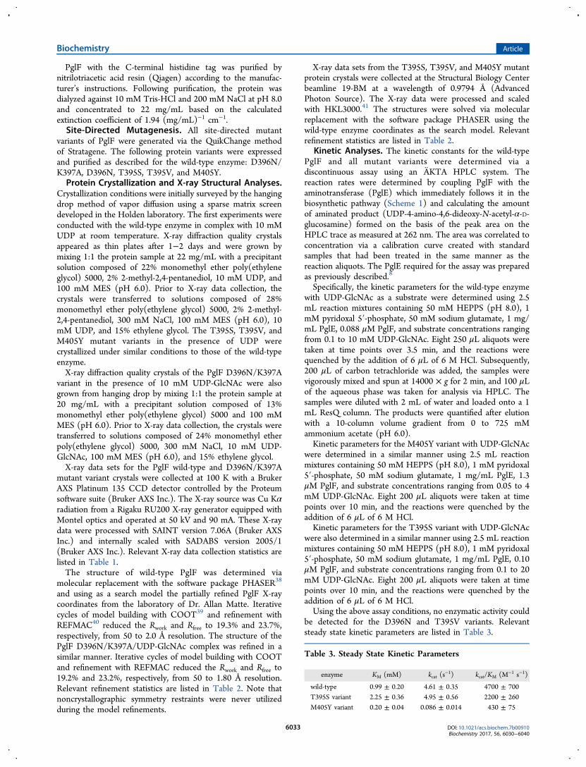
Table 3. Steady State Kinetic Parameters
enzyme KM (mM) kcat (s−1) kcat/KM (M−1 s−1)
wild-type 0.99 ± 0.20 4.61 ± 0.35 4700 ± 700T395S variant 2.25 ± 0.36 4.95 ± 0.56 2200 ± 260M405Y variant 0.20 ± 0.04 0.086 ± 0.014 430 ± 75
Biochemistry Article
DOI: 10.1021/acs.biochem.7b00910Biochemistry 2017, 56, 6030−6040
6033
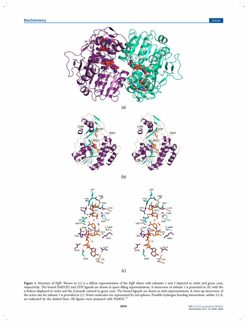
Figure 1. Structure of PglF. Shown in (a) is a ribbon representation of the PglF dimer with subunits 1 and 2 depicted in violet and green cyan,respectively. The bound NAD(H) and UDP ligands are drawn in space filling representations. A stereoview of subunit 1 is presented in (b) with theα-helices displayed in violet and the β-strands colored in green cyan. The bound ligands are drawn in stick representations. A close-up stereoview ofthe active site for subunit 1 is provided in (c). Water molecules are represented by red spheres. Possible hydrogen bonding interactions, within 3.2 Å,are indicated by the dashed lines. All figures were prepared with PyMOL.46
Biochemistry Article
DOI: 10.1021/acs.biochem.7b00910Biochemistry 2017, 56, 6030−6040
6034

Note that the concentration of PglE utilized in the assaysensured that the aminotransferase reaction was not rate-limiting.Stopped-flow kinetic experiments were performed with a
KinTek SF-2001 instrument using absorbance detection withthe monochromator set at 340 nm. The mixing chamber wasmaintained at 25 °C with a circulating water bath. Identicalsolutions were prepared in H2O and in D2O. One syringecontained 108 μM PglF and the other contained 20 mM UDP-GlcNAc; both solutions were buffered with 50 mM HEPES,pH(D) 8.0, and 150 mM NaCl.
■ RESULTS AND DISCUSSIONOverall Architecture of PglF. PglF is a membrane bound
protein. In order to simplify the structural analysis, varioustruncation mutant variants were constructed in the linker
region. In most cases, catalytic activity was retained, and thevariants expressed well in E. coli. Importantly, only thosemutant proteins that were truncated near the catalytic domainyielded preliminary crystals. After many trials, the final proteinconstruct utilized in this investigation extended from Ser 248 toLys 587. Also, the cofactor is referred to as NAD(H)throughout this paper in light of the fact that its exact oxidationstate is unknown.The first structure determined in this investigation was that
of the PglF/NAD(H)/UDP ternary complex. The crystalsutilized belonged to the space group P212121 with two subunitsin the asymmetric unit. These two subunits were related by180° rotation suggesting a dimeric quaternary structure. Themodel was refined at 2.0 Å resolution to an overall R-factor of19.5%. A ribbon representation of the dimer is presented inFigure 1a. It has overall dimensions of ∼58 Å × 81 Å × 63 Å
Figure 2. Structure of the PglF/(D396N/K397A)/NAD(H)/UDP-GlcNAc complex. The electron densities corresponding to the bound ligands areshown in (a). The electron density map was calculated with (Fo − Fc) coefficients and contoured at 3σ. The ligands were not included in the X-raycoordinate file used to calculate the omit map, and thus there is no model bias. A close-up view of the active site is depicted in (b). Water moleculesare displayed as red spheres, and possible hydrogen bonding interactions are indicated by the dashed lines. As indicated by the green line between C-4′ of the sugar and C-4 of the nicotinamide ring, the substrate is ideally positioned in the active site for hydride transfer.
Biochemistry Article
DOI: 10.1021/acs.biochem.7b00910Biochemistry 2017, 56, 6030−6040
6035

and a total buried surface area of 2400 Å2. The quaternarystructure of PglF, as observed in the asymmetric unit, isdecidedly different from that for other dimeric sugar 4,6-dehydratases. Typically, in these enzymes the interface isformed by a four α-helical bundle with each subunit providingtwo α-helical regions.43 Additionally, the adenosine aminogroups of the bound NAD(H) cofactors are separated by ∼22Å. In PglF, the two α-helical regions required for the formationof the four α-helical bundle are located nearly on the oppositesides of the subunit−subunit interface. As a consequence, theadenosine amino groups of the bound NAD(H) cofactors inPglF are separated by only ∼11 Å. One caveat, however, is thatthe transmembrane linker was removed from PglF, and thus itis possible that the packing arrangement seen in the crystallinelattice is an artifact. Nevertheless, the total buried surface areaas well as the nature of the subunit/subunit interface issuggestive of a dimeric quaternary structure. Furthermore, gelfiltation experiments on the protein utilized in this investigationwere indicative of a dimer (unpublished data).The α-carbons for the two subunits of the PglF dimer
superimpose with a root-mean-square deviation of 0.1 Å. Giventhe close structural correspondence, the following discussionrefers only to subunit 1 in the X-ray coordinate file unlessotherwise indicated. Shown in Figure 1b is a ribbonrepresentation of the subunit which displays a bilobal typearchitecture. The polypeptide chain initiates with a short α-helix followed by random coil. The N-terminal domainresponsible for binding NAD(H) is dominated by an eight-stranded parallel β-sheet flanked on each side by three α-helices. The C-terminal domain that houses the substratebinding site is characterized primarily by two α-helices and twoβ-sheets containing two β-strands each.A close-up stereoview of the active site is displayed in Figure
1c. Both the UDP and NAD(H) ligands are anchored into theactive site via numerous hydrogen bonding interactions.Specifically, the uracil ring of UDP lies within 3.2 Å of thecarbonyl oxygen and the backbone amide nitrogen of Thr 456and Thr 458, respectively. The UDP ribose adopts the C2′-endopucker with the C-2 hydroxyl group hydrogen bonding to theside chains of Thr 458 and Glu 526. The side chains of Lys 397,Asn 433, and Arg 464 interact with a β-phosphoryl oxygen ofthe UDP ligand whereas the backbone amide nitrogen of Val441 hydrogen bonds to an α-phosphoryl oxygen. Five watermolecules within 3.2 Å surround the UDP ligand. As expectedfor members of the short chain dehydrogenase/reductasesuperfamily, the nicotinamide moiety of the NAD(H) adoptsthe syn conformation thereby positioning the B-side of the ringtoward the UDP ligand. The side chain of Ser 437 and theamide nitrogen of Val 434 hydrogen bond to the carboxamidegroup of the nicotinamide ring. Those side chains that play keyroles in anchoring the NAD(H) cofactor into the active sitecleft include Asp 303, Asn 308, Lys 355, Asn 370, Lys 409, andSer 438. Additionally, the backbone amide nitrogens of Thr282, Ile 283, and Ile 330 and six ordered water molecules aid incofactor positioning. Both Asp 396 and Lys 397 are conservedamong some sugar 4,6-dehydratases, but strikingly Met 405resides in the region normally occupied by a tyrosine residue,which is thought to serve as a catalytic base in these enzymes.Active Site Geometry of PglF. From previous reports on
a similar sugar 4,6-dehydratase, it can be speculated that bothAsp 396 and Lys 397 in PglF play critical roles in catalysis.33
Thus, in order to prepare a ternary complex of the enzyme withNAD(H) and its substrate, UDP-GlcNAc, a double site-
directed mutant protein, D396N/K397A, was subsequentlyprepared. Crystals of this mutant variant with bound NAD(H)and UDP-GlcNAc also belonged to the space group P212121with a dimer in the asymmetric unit. The model was refined at1.8 Å resolution to an overall R-factor of 19.4%. The root-mean-square deviation between the α-carbons for the wild-typeenzyme and the mutant variant superimpose with a root-mean-square deviation of 0.3 Å, thus indicating little structural changeupon UDP-sugar binding, at least in the crystalline state. Shownin Figure 2a is the observed electron density corresponding tothe bound ligands in subunit 1. As can be seen, the electrondensity is unambiguous for both the UDP-sugar and theNAD(H) cofactor. The pyranosyl group of the UDP-sugaradopts the 4C1 conformation. A stereoview of the PglF ternarycomplex active site is presented in Figure 2b. Those side chainsimportant for anchoring the pyranosyl group to the proteininclude Lys 355 and Thr 395. In addition, nine waters surroundthe bound substrate. Importantly, the sugar C-4′ lies within 3.9Å of the nicotinamide C-4 carbon, and it is also in the properorientation for hydride transfer.
Kinetic Analyses of PglF. Given that the typical tyrosineresidue in the sugar 4,6-dehydratases is not found in PglF, thequestion then arises as to the identity of the catalytic baserequired to initiate the first half of the reaction outlined inScheme 2. It is particularly noteworthy that in the wild-typestructure, the side chain of Thr 395 is positioned at 3.5 Å froma carboxylate oxygen of Asp 396, and in the ternary complex itis located at 2.8 Å from the sugar C-4′ hydroxyl group. It canthus be envisioned that the side chain of Thr 395 mightfunction as a proton shuttle by linking the sugar C-4′ hydroxylgroup to the side chain of Asp 396, which would ultimatelyserve as the catalytic base.To more fully address the roles of the individual amino acids
in the activity of PglF, the following site-directed mutantproteins were constructed: T395S, T395V, D396N, andM405Y. Under the assay conditions utilized, no measurableactivity was detected for the D396N mutant variant. Becausethe crystals of the D396N mutant variant were not of highquality, the structure of it will not be reported here.Mutation at Thr 395 showed an interesting pattern. When
changed to a serine, the enzyme retained catalytic activity albeitat a reduced level (Table 3). The major cause for the reducedcatalytic efficiency was the increased KM for the UDP-sugarsubstrate: 0.99 ± 0.20 mM for the wild-type enzyme versus2.25 ± 0.36 mM for the T395S variant. Within experimentalerror, the kcat values for the two proteins were the same,however (Table 3). Given that Thr 395 is important inpositioning the pyranosyl group into the active site, it is notsurprising that the KM would be affected. The loss of the methylgroup in substituting a serine for a threonine most likely opensthe active site enough to raise the KM for the UDP-sugarsubstrate. The three-dimensional structure of the T395Smutant variant was subsequently determined to 1.6 Åresolution, and the model was refined to an overall R-factorof 17.5%. The wild-type protein and T395S mutant variantstructures are remarkably similar such that their α-carbonssuperimpose with a root-mean-square deviation of 0.1 Å.Unlike that observed for the T395S variant, mutation of Thr395 to a valine residue resulted in a completely inactive protein.To ensure no major structural changes occurred upon thismutation, the structure of the T395V mutant protein wassubsequently determined to 1.6 Å resolution (overall R-factorof 16.3%). As expected, the structures of the wild-type enzyme
Biochemistry Article
DOI: 10.1021/acs.biochem.7b00910Biochemistry 2017, 56, 6030−6040
6036
and T395V protein variant are nearly identical such that theirα-carbons superimpose with a root-mean-square deviation of0.2 Å. The above results clearly indicate the importance of ahydroxyl group at position 395 for the PglF reactionmechanism.We sought additional evidence for a proton-shuttle
mechanism from a proton inventory experiment. Hydridetransfer to NAD+ is concomitant with proton abstraction fromthe hydroxyl group at C-4′; if this proton removal is mediatedby Thr 395, which is itself deprotonated by Asp 396, theformation of NADH would occur with two protons in flight inthe transition state. However, the transient increase inabsorbance at 340 nm observed in multiturnover experiments
conducted in either protio or deuterio solvent, with enzymesaturated with UDP-GlcNAc, was negligible, equivalent to nomore than 0.3% of the enzyme active sites, frustrating ourattempt to perform the proton inventory experiment. Theabsence of NADH accumulation indicates that its formation israte-limiting in the catalytic cycle.Given that the typically conserved tyrosine residue in the
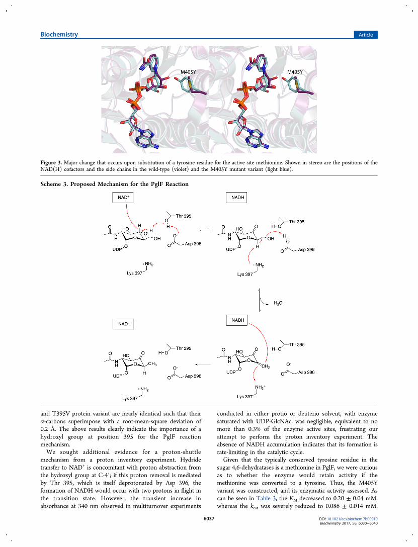
sugar 4,6-dehydratases is a methionine in PglF, we were curiousas to whether the enzyme would retain activity if themethionine was converted to a tyrosine. Thus, the M405Yvariant was constructed, and its enzymatic activity assessed. Ascan be seen in Table 3, the KM decreased to 0.20 ± 0.04 mM,whereas the kcat was severely reduced to 0.086 ± 0.014 mM.
Figure 3. Major change that occurs upon substitution of a tyrosine residue for the active site methionine. Shown in stereo are the positions of theNAD(H) cofactors and the side chains in the wild-type (violet) and the M405Y mutant variant (light blue).
Scheme 3. Proposed Mechanism for the PglF Reaction
Biochemistry Article
DOI: 10.1021/acs.biochem.7b00910Biochemistry 2017, 56, 6030−6040
6037
The structure of the M405Y, determined at 1.6 Å resolution(overall R-factor of 17.0%), reveals the largest three-dimen-sional perturbation among the site-directed mutant proteinsthat were studied in this investigation. Whereas the α-carbonsfor the wild-type enzyme and the M405Y mutant variantsuperimpose with a root-mean-square deviation of 0.2 Å, thesubstitution of a tyrosine for a methionine effectively positionsthe ribose of the NAD(H) more into the active site pocket asshown in Figure 3.Catalytic Mechanism of PglF. On the basis of the
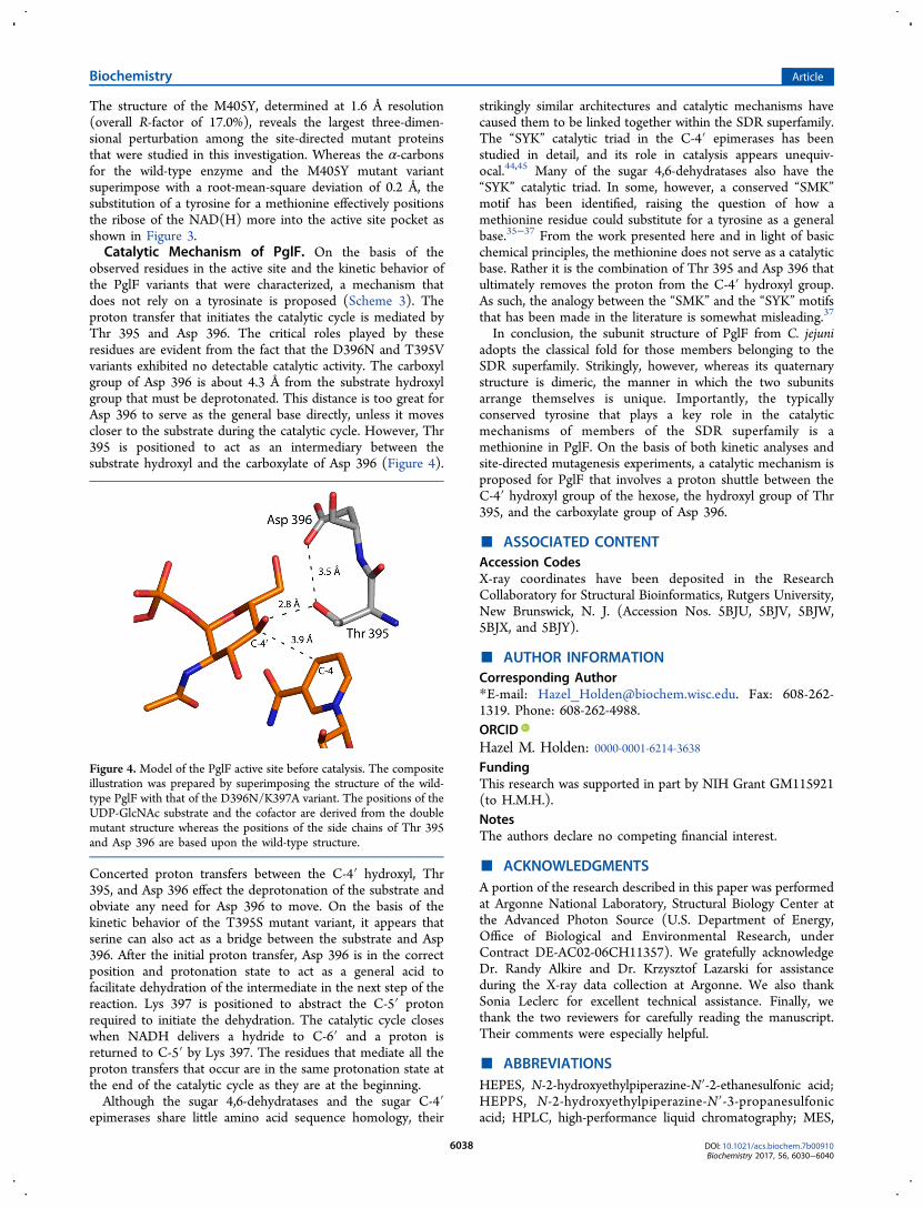
observed residues in the active site and the kinetic behavior ofthe PglF variants that were characterized, a mechanism thatdoes not rely on a tyrosinate is proposed (Scheme 3). Theproton transfer that initiates the catalytic cycle is mediated byThr 395 and Asp 396. The critical roles played by theseresidues are evident from the fact that the D396N and T395Vvariants exhibited no detectable catalytic activity. The carboxylgroup of Asp 396 is about 4.3 Å from the substrate hydroxylgroup that must be deprotonated. This distance is too great forAsp 396 to serve as the general base directly, unless it movescloser to the substrate during the catalytic cycle. However, Thr395 is positioned to act as an intermediary between thesubstrate hydroxyl and the carboxylate of Asp 396 (Figure 4).
Concerted proton transfers between the C-4′ hydroxyl, Thr395, and Asp 396 effect the deprotonation of the substrate andobviate any need for Asp 396 to move. On the basis of thekinetic behavior of the T395S mutant variant, it appears thatserine can also act as a bridge between the substrate and Asp396. After the initial proton transfer, Asp 396 is in the correctposition and protonation state to act as a general acid tofacilitate dehydration of the intermediate in the next step of thereaction. Lys 397 is positioned to abstract the C-5′ protonrequired to initiate the dehydration. The catalytic cycle closeswhen NADH delivers a hydride to C-6′ and a proton isreturned to C-5′ by Lys 397. The residues that mediate all theproton transfers that occur are in the same protonation state atthe end of the catalytic cycle as they are at the beginning.Although the sugar 4,6-dehydratases and the sugar C-4′
epimerases share little amino acid sequence homology, their
strikingly similar architectures and catalytic mechanisms havecaused them to be linked together within the SDR superfamily.The “SYK” catalytic triad in the C-4′ epimerases has beenstudied in detail, and its role in catalysis appears unequiv-ocal.44,45 Many of the sugar 4,6-dehydratases also have the“SYK” catalytic triad. In some, however, a conserved “SMK”motif has been identified, raising the question of how amethionine residue could substitute for a tyrosine as a generalbase.35−37 From the work presented here and in light of basicchemical principles, the methionine does not serve as a catalyticbase. Rather it is the combination of Thr 395 and Asp 396 thatultimately removes the proton from the C-4′ hydroxyl group.As such, the analogy between the “SMK” and the “SYK” motifsthat has been made in the literature is somewhat misleading.37
In conclusion, the subunit structure of PglF from C. jejuniadopts the classical fold for those members belonging to theSDR superfamily. Strikingly, however, whereas its quaternarystructure is dimeric, the manner in which the two subunitsarrange themselves is unique. Importantly, the typicallyconserved tyrosine that plays a key role in the catalyticmechanisms of members of the SDR superfamily is amethionine in PglF. On the basis of both kinetic analyses andsite-directed mutagenesis experiments, a catalytic mechanism isproposed for PglF that involves a proton shuttle between theC-4′ hydroxyl group of the hexose, the hydroxyl group of Thr395, and the carboxylate group of Asp 396.
■ ASSOCIATED CONTENTAccession CodesX-ray coordinates have been deposited in the ResearchCollaboratory for Structural Bioinformatics, Rutgers University,New Brunswick, N. J. (Accession Nos. 5BJU, 5BJV, 5BJW,5BJX, and 5BJY).
■ AUTHOR INFORMATIONCorresponding Author*E-mail: [email protected]. Fax: 608-262-1319. Phone: 608-262-4988.ORCIDHazel M. Holden: 0000-0001-6214-3638FundingThis research was supported in part by NIH Grant GM115921(to H.M.H.).NotesThe authors declare no competing financial interest.
■ ACKNOWLEDGMENTSA portion of the research described in this paper was performedat Argonne National Laboratory, Structural Biology Center atthe Advanced Photon Source (U.S. Department of Energy,Office of Biological and Environmental Research, underContract DE-AC02-06CH11357). We gratefully acknowledgeDr. Randy Alkire and Dr. Krzysztof Lazarski for assistanceduring the X-ray data collection at Argonne. We also thankSonia Leclerc for excellent technical assistance. Finally, wethank the two reviewers for carefully reading the manuscript.Their comments were especially helpful.
■ ABBREVIATIONSHEPES, N-2-hydroxyethylpiperazine-N′-2-ethanesulfonic acid;HEPPS, N-2-hydroxyethylpiperazine-N′-3-propanesulfonicacid; HPLC, high-performance liquid chromatography; MES,
Figure 4. Model of the PglF active site before catalysis. The compositeillustration was prepared by superimposing the structure of the wild-type PglF with that of the D396N/K397A variant. The positions of theUDP-GlcNAc substrate and the cofactor are derived from the doublemutant structure whereas the positions of the side chains of Thr 395and Asp 396 are based upon the wild-type structure.
Biochemistry Article
DOI: 10.1021/acs.biochem.7b00910Biochemistry 2017, 56, 6030−6040
6038
2-(N-morpholino)ethanesulfonic acid; NAD+, nicotinamideadenine dinucleotide (oxidized); NADH, nicotinamide adeninedinucleotide (reduced); NADP+, nicotinamide adenine dinu-cleotide phosphate (oxidized); PCR, polymerase chainreaction; Tris, tris-(hydroxymethyl)aminomethane; UDP,uridine diphosphate; UDP-GlcNAc, uridine diphosphate N-acetylglucosamine
■ REFERENCES(1) Neuberger, A. (1938) Carbohydrates in protein: Thecarbohydrate component of crystalline egg albumin. Biochem. J. 32,1435−1451.(2) Nothaft, H., and Szymanski, C. M. (2010) Protein glycosylationin bacteria: sweeter than ever. Nat. Rev. Microbiol. 8, 765−778.(3) Nothaft, H., and Szymanski, C. M. (2013) Bacterial protein N-glycosylation: new perspectives and applications. J. Biol. Chem. 288,6912−6920.(4) Galanis, E. (2007) Campylobacter and bacterial gastroenteritis.CMAJ. 177, 570−571.(5) Szymanski, C. M., Yao, R., Ewing, C. P., Trust, T. J., and Guerry,P. (1999) Evidence for a system of general protein glycosylation inCampylobacter jejuni. Mol. Microbiol. 32, 1022−1030.(6) Scott, N. E., Parker, B. L., Connolly, A. M., Paulech, J., Edwards,A. V., Crossett, B., Falconer, L., Kolarich, D., Djordjevic, S. P., Hojrup,P., Packer, N. H., Larsen, M. R., and Cordwell, S. J. (2011)Simultaneous glycan-peptide characterization using hydrophilicinteraction chromatography and parallel fragmentation by CID, higherenergy collisional dissociation, and electron transfer dissociation MSapplied to the N-linked glycoproteome of Campylobacter jejuni. Mol.Cell. Proteomics 10, M000031-MCP000201.(7) Young, N. M., Brisson, J. R., Kelly, J., Watson, D. C., Tessier, L.,Lanthier, P. H., Jarrell, H. C., Cadotte, N., St, St. Michael, F., Aberg, E.,and Szymanski, C. M. (2002) Structure of the N-linked glycan presenton multiple glycoproteins in the Gram-negative bacterium, Campylo-bacter jejuni. J. Biol. Chem. 277, 42530−42539.(8) Riegert, A. S., Young, N. M., Watson, D. C., Thoden, J. B., andHolden, H. M. (2015) Structure of the external aldimine form of PglE,an aminotransferase required for N,N′-diacetylbacillosamine biosyn-thesis. Protein Sci. 24, 1609−1616.(9) Rangarajan, E. S., Ruane, K. M., Sulea, T., Watson, D. C., Proteau,A., Leclerc, S., Cygler, M., Matte, A., and Young, N. M. (2008)Structure and active site residues of PglD, an N-acetyltransferase fromthe bacillosamine synthetic pathway required for N-glycan synthesis inCampylobacter jejuni. Biochemistry 47, 1827−1836.(10) Olivier, N. B., and Imperiali, B. (2008) Crystal structure andcatalytic mechanism of PglD from Campylobacter jejuni. J. Biol. Chem.283, 27937−27946.(11) Schoenhofen, I. C., McNally, D. J., Vinogradov, E., Whitfield, D.,Young, N. M., Dick, S., Wakarchuk, W. W., Brisson, J. R., and Logan, S.M. (2006) Functional characterization of dehydratase/aminotransfer-ase pairs from Helicobacter and Campylobacter: enzymes distinguishingthe pseudaminic acid and bacillosamine biosynthetic pathways. J. Biol.Chem. 281, 723−732.(12) He, X., and Liu, H. W. (2002) Mechanisms of enzymatic C-Obond cleavages in deoxyhexose biosynthesis. Curr. Opin. Chem. Biol. 6,590−597.(13) Kallberg, Y., Oppermann, U., Jornvall, H., and Persson, B.(2002) Short-chain dehydrogenases/reductases (SDRs). Eur. J.Biochem. 269, 4409−4417.(14) Kallberg, Y., Oppermann, U., Jornvall, H., and Persson, B.(2002) Short-chain dehydrogenase/reductase (SDR) relationships: alarge family with eight clusters common to human, animal, and plantgenomes. Protein Sci. 11, 636−641.(15) Oppermann, U., Filling, C., Hult, M., Shafqat, N., Wu, X., Lindh,M., Shafqat, J., Nordling, E., Kallberg, Y., Persson, B., and Jornvall, H.(2003) Short-chain dehydrogenases/reductases (SDR): the 2002update. Chem.-Biol. Interact. 143−144, 247−253.
(16) Kavanagh, K. L., Jornvall, H., Persson, B., and Oppermann, U.(2008) Medium- and short-chain dehydrogenase/reductase gene andprotein families: the SDR superfamily: functional and structuraldiversity within a family of metabolic and regulatory enzymes. Cell.Mol. Life Sci. 65, 3895−3906.(17) Kallberg, Y., Oppermann, U., and Persson, B. (2010)Classification of the short-chain dehydrogenase/reductase superfamilyusing hidden Markov models. FEBS J. 277, 2375−2386.(18) Duax, W. L., Ghosh, D., and Pletnev, V. (2000) Steroiddehydrogenase structures, mechanism of action, and disease. Vitam.Horm. 58, 121−148.(19) Duax, W. L., Pletnev, V., Addlagatta, A., Bruenn, J., and Weeks,C. M. (2003) Rational proteomics I. Fingerprint identification andcofactor specificity in the short-chain oxidoreductase (SCOR) enzymefamily. Proteins: Struct., Funct., Genet. 53, 931−943.(20) Somoza, J. R., Menon, S., Schmidt, H., Joseph-McCarthy, D.,Dessen, A., Stahl, M. L., Somers, W. S., and Sullivan, F. X. (2000)Structural and kinetic analysis of Escherichia coli GDP-mannose 4,6dehydratase provides insights into the enzyme’s catalytic mechanismand regulation by GDP-fucose. Structure 8, 123−135.(21) Allard, S. T., Giraud, M. F., Whitfield, C., Graninger, M.,Messner, P., and Naismith, J. H. (2001) The crystal structure ofdTDP-D-glucose 4,6-dehydratase (RmlB) from Salmonella entericaserovar Typhimurium, the second enzyme in the dTDP-L-rhamnosepathway. J. Mol. Biol. 307, 283−295.(22) Gross, J. W., Hegeman, A. D., Gerratana, B., and Frey, P. A.(2001) Dehydration is catalyzed by glutamate-136 and aspartic acid-135 active site residues in Escherichia coli dTDP-glucose 4,6-dehydratase. Biochemistry 40, 12497−12504.(23) Hegeman, A. D., Gross, J. W., and Frey, P. A. (2001) Probingcatalysis by Escherichia coli dTDP-glucose-4,6-dehydratase: identifica-tion and preliminary characterization of functional amino acid residuesat the active site. Biochemistry 40, 6598−6610.(24) Allard, S. T., Beis, K., Giraud, M. F., Hegeman, A. D., Gross, J.W., Wilmouth, R. C., Whitfield, C., Graninger, M., Messner, P., Allen,A. G., Maskell, D. J., and Naismith, J. H. (2002) Toward a structuralunderstanding of the dehydratase mechanism. Structure (Oxford, U. K.)10, 81−92.(25) Hegeman, A. D., Gross, J. W., and Frey, P. A. (2002) Concertedand stepwise dehydration mechanisms observed in wild-type andmutated Escherichia coli dTDP-glucose 4,6-dehydratase. Biochemistry41, 2797−2804.(26) Mulichak, A. M., Bonin, C. P., Reiter, W. D., and Garavito, R. M.(2002) Structure of the MUR1 GDP-mannose 4,6-dehydratase fromArabidopsis thaliana: implications for ligand binding and specificity.Biochemistry 41, 15578−15589.(27) Beis, K., Allard, S. T., Hegeman, A. D., Murshudov, G., Philp, D.,and Naismith, J. H. (2003) The structure of NADH in the enzymedTDP-D-glucose dehydratase (RmlB). J. Am. Chem. Soc. 125, 11872−11878.(28) Allard, S. T., Cleland, W. W., and Holden, H. M. (2004) Highresolution X-ray structure of dTDP-glucose 4,6-dehydratase fromStreptomyces venezuelae. J. Biol. Chem. 279, 2211−2220.(29) Webb, N. A., Mulichak, A. M., Lam, J. S., Rocchetta, H. L., andGaravito, R. M. (2004) Crystal structure of a tetrameric GDP-D-mannose 4,6-dehydratase from a bacterial GDP-D-rhamnose bio-synthetic pathway. Protein Sci. 13, 529−539.(30) Vogan, E. M., Bellamacina, C., He, X., Liu, H. W., Ringe, D., andPetsko, G. A. (2004) Crystal structure at 1.8 A resolution of CDP-D-glucose 4,6-dehydratase from Yersinia pseudotuberculosis. Biochemistry43, 3057−3067.(31) Koropatkin, N. M., and Holden, H. M. (2005) Structure ofCDP-D-glucose 4,6-dehydratase from Salmonella typhi complexed withCDP-D-xylose. Acta Crystallogr., Sect. D: Biol. Crystallogr. 61, 365−373.(32) Rosano, C., Zuccotti, S., Sturla, L., Fruscione, F., Tonetti, M.,and Bolognesi, M. (2006) Quaternary assembly and crystal structure ofGDP-D-mannose 4,6 dehydratase from Paramecium bursaria Chlorellavirus. Biochem. Biophys. Res. Commun. 339, 191−195.
Biochemistry Article
DOI: 10.1021/acs.biochem.7b00910Biochemistry 2017, 56, 6030−6040
6039

(33) Ishiyama, N., Creuzenet, C., Miller, W. L., Demendi, M.,Anderson, E. M., Harauz, G., Lam, J. S., and Berghuis, A. M. (2006)Structural studies of FlaA1 from Helicobacter pylori reveal themechanism for inverting 4,6-dehydratase activity. J. Biol. Chem. 281,24489−24495.(34) Morrison, J. P., Schoenhofen, I. C., and Tanner, M. E. (2008)Mechanistic studies on PseB of pseudaminic acid biosynthesis: a UDP-N-acetylglucosamine 5-inverting 4,6-dehydratase. Bioorg. Chem. 36,312−320.(35) Miyafusa, T., Caaveiro, J. M., Tanaka, Y., and Tsumoto, K.(2013) Dynamic elements govern the catalytic activity of CapE, acapsular polysaccharide-synthesizing enzyme from Staphylococcusaureus. FEBS Lett. 587, 3824−3830.(36) Miyafusa, T., Caaveiro, J. M., Tanaka, Y., Tanner, M. E., andTsumoto, K. (2013) Crystal structure of the capsular polysaccharidesynthesizing protein CapE of Staphylococcus aureus. Biosci. Rep. 33,463−474.(37) Creuzenet, C., Urbanic, R. V., and Lam, J. S. (2002) Structure-function studies of two novel UDP-GlcNAc C6 dehydratases/C4reductases. Variation from the SYK dogma. J. Biol. Chem. 277, 26769−26778.(38) McCoy, A. J., Grosse-Kunstleve, R. W., Adams, P. D., Winn, M.D., Storoni, L. C., and Read, R. J. (2007) Phaser crystallographicsoftware. J. Appl. Crystallogr. 40, 658−674.(39) Emsley, P., and Cowtan, K. (2004) Coot: model-building toolsfor molecular graphics. Acta Crystallogr., Sect. D: Biol. Crystallogr. 60,2126−2132.(40) Murshudov, G. N., Vagin, A. A., and Dodson, E. J. (1997)Refinement of macromolecular structures by the maximum-likelihoodmethod. Acta Crystallogr., Sect. D: Biol. Crystallogr. 53, 240−255.(41) Otwinowski, Z., and Minor, W. (1997) Processing of X-raydiffraction data collected in oscillation mode. Methods Enzymol. 276,307−326.(42) Laskowski, R. A., Moss, D. S., and Thornton, J. M. (1993) Main-chain bond lengths and bond angles in protein structures. J. Mol. Biol.231, 1049−1067.(43) Allard, S. T., Giraud, M. F., and Naismith, J. H. (2001)Epimerases: structure, function and mechanism. Cell. Mol. Life Sci. 58,1650−1665.(44) Liu, Y., Thoden, J. B., Kim, J., Berger, E., Gulick, A. M., Ruzicka,F. J., Holden, H. M., and Frey, P. A. (1997) Mechanistic roles oftyrosine 149 and serine 124 in UDP-galactose 4-epimerase fromEscherichia coli. Biochemistry 36, 10675−10684.(45) Thoden, J. B., Wohlers, T. M., Fridovich-Keil, J. L., and Holden,H. M. (2000) Crystallographic evidence for Tyr 157 functioning as theactive site base in human UDP-galactose 4-epimerase. Biochemistry 39,5691−5701.(46) DeLano, W. L. (2002) The PyMOL Molecular GraphicsSystem. DeLano Scientific, San Carlos, CA, USA, The PyMOLMolecular Graphics System, DeLano Scientific, San Carlos, CA, USA.
Biochemistry Article
DOI: 10.1021/acs.biochem.7b00910Biochemistry 2017, 56, 6030−6040
6040