stress testing in valvular disease - american society of...
TRANSCRIPT

10/10/2017
1
Muhamed Sarić MD, PhD, MPADirector of Noninvasive Cardiology | Echo LabAssociate Professor of Medicine
Stress Testingin Valvular Disease
2017 ASE Florida | Orlando, FL
October 10, 2017 | 2:40 – 2:50 PM | 10 min | Grand Harbor Ballroom South
Disclosures
Speakers Bureau (Philips, Medtronic)Advisory Board (Siemens)

10/10/2017
2
Case #1: Stress in Aortic Regurgitation
Stages of Valvular Heart Disease2014 ACC/AHA Valvular Guidelines
A
B
C
D
Nonsevere
Severe
Asymptomatic
Symptomatic
Valvular disease is typically NOT symptomatic unless SEVERE!
Stress Testing

10/10/2017
3
48-year-old man with • Severe aortic regurgitation
• Bicuspid aortic valve and aortic root dilatation
• Reports occasional exertional dyspnea which he attributes to recent weigh gain
Case Presentation
***
Referred for exercise stress echo
Resting TTE

10/10/2017
4
Resting TTE
Vena Contracta
Resting TTE

10/10/2017
5
Resting MRI
Regurgitant volume = 71 mL | Regurgitant Fraction 51%
Exercise Stress Ech0: A4C
REST STRESS
STRESSSTRESS
10/10/2017
6
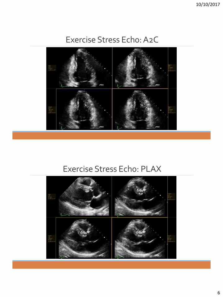
Exercise Stress Ech0: A2C
Exercise Stress Ech0: PLAX

10/10/2017
7
Exercise Stress Ech0: SAX
Question
With respect to ischemia, is this a positive or a negative stress echo?

10/10/2017
8
Ischemia Conclusion
• Resting LVEF was 65 %.
• Stress echo images were obtained within 60 to 90 seconds post-stress. • At time of stress imaging, the maximum heart rate was 125 bpm. • At peak stress LVEF was 75 %. • The left ventricular cavity size became smaller with stress.
• There were no stress-induced wall motion abnormalities.
• Stress echo is negative for ischemia.
Question
Should have I provided you with stress echo data specific to aortic regurgitation?

10/10/2017
9
Stress Testing in Aortic Regurgitation2014 ACC/AHA Valvular Guidelines
• No formal recommendation for stress testing in aortic regurgitation.
• There is merely a descriptive paragraph in the guidelines.
Exercise Data
• Exercised for 12 minutes and 0 seconds on Bruce protocol which represents 114 % of expected exercise duration of approximately 10.5 min adjusted for age and gender.
• Reached stage 4 and achieved 12.4 mets.
• Patient's functional capacity was above average for age.• Developed no symptoms during the test.

10/10/2017
10
Conclusion
• Despite severe aortic regurgitation, patient has excellent exercise capacity and no exercise-induced symptoms.
• Continue watchful waiting; no need for aortic valve replacement referral at this time.
Case #2: Stress Testing in Aortic Stenosis
10/10/2017
11
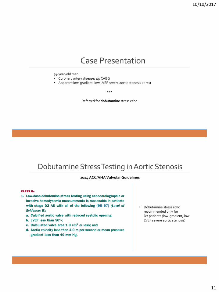
74-year-old man • Coronary artery disease; s/p CABG• Apparent low-gradient, low LVEF severe aortic stenosis at rest
Case Presentation
***
Referred for dobutamine stress echo
Dobutamine Stress Testing in Aortic Stenosis2014 ACC/AHA Valvular Guidelines
• Dobutamine stress echo recommended only for D2 patients (low-gradient, low LVEF severe aortic stenosis)

10/10/2017
12
Exercise Stress Ech0: A2C
LVOT diameter 2.2 cm (area 3.8 cm2)
LVOT Size

10/10/2017
13
LVOT VTI
RestSV = 30 mL
Dobutamine 20μg/kg/minSV = 46 mL
Question
With respect to stroke volume change, was this a diagnostic test?

10/10/2017
14
Answer
YesDiagnostic test implies an increase in SV of at least 20%.
Aortic Valve Spectral Doppler
RestMean gradient = 10 mm Hg
AVA = 0.89 cm2
Dobutamine 20μg/kg/minMean gradient 17 mm Hg
AVA = 0.89 cm2

10/10/2017
15
Question
Is this a true AS or pseudo AS?
Answer
This patient has true severe low-gradient aortic stenosisin the setting of low LVEF.
• With peak dobutamine dose of 20 ug/kg/min, there was >20% increase in LVOT stroke volume, an increase in the mean AV gradient from 10 mm Hg to 17 mm Hg and with no change in AVA.
• These findings are consistent with true severe low-gradient aortic stenosis in the setting of severe LV systolic dysfunction and contractile reserve.

10/10/2017
16
Complete Calculations
ACC/AHA recommendations for exercise stress testing in aortic stenosis.
Bonus Slides

10/10/2017
17
Exercise Stress Testing in Aortic Stenosis2014 ACC/AHA Valvular Guidelines
• Recommendations are only for high-gradient severe AS
• Test for• Symptoms• Exercise duration• BP response• Ventricular arrhythmia
• Don’t use exercise stress testing in symptomatic patients with high-gradient AS
Exercise Stress Testing in Aortic Stenosis
Source: saric.us/echonomy

10/10/2017
18
Case #3:Stress Testing in Mitral Stenosis
40-year-old woman• Grew up in the former Soviet Union• History of rheumatic heart disease• Complains of exertional dyspnea• Resting TTE shows
• Modestly elevated mitral gradients and • No pulmonary hypertension.
Case Presentation
***
Referred for exercise stress echo

10/10/2017
19
Stress Testing in Mitral Stenosis2014 ACC/AHA Valvular Guidelines
• Either exercise or dobutamine stress testing is acceptable.
• Assess for stress-related• Symptoms• Worsening mean mitral
gradient• Worsening PA systolic
pressure
Resting Echo: Rheumatic Mitral Stenosis

10/10/2017
20
Resting Echo: Rheumatic Mitral Stenosis
Exercise Stress Echo Data41
REST PEAK STRESS
Mean Mitral
Gradient
Tricuspid Regurgitation
Gradient
ΔP = 8 mm Hg ΔP = 19 mm Hg
RV-RA = 18 mm Hg RV-RA = 56 mm Hg

10/10/2017
21
Question
Is this a diagnostic stress echo with respect to mitral stenosis?
Answer
Yes, this stress echo is positive with respect to mitral stenosis?

10/10/2017
22
Question
Now that we know that the stress echo is positive with respect to mitral stenosis, what should we do next?
Referred for Percutaneous Mitral Balloon Valvuloplasty
Inoue balloon is reinforced with a nylon micromesh. Its shape changes in 3 stages, depending on the extent
of inflation.

10/10/2017
23
Stress Testing in Mitral Stenosis
In the absence of contraindications, PMBV is recommended in following instances:
• Symptomatic patients with moderate or severe mitral stenosis.
• In asymptomatic patients with moderate or severe mitral stenosis, PMBV is indicated when there is pulmonary artery systolic pressure is > 50 mm Hg at rest or > 60 mm Hg with exercise, or when there is new onset atrial fibrillation.
• PMBV may also be considered in symptomatic patient with mild mitral stenosis (valve area >1.5 cm2) when pulmonary artery systolic pressure greater > 60 mm Hg, pulmonary artery wedge pressure > 25 mm Hg, or mean mitral valve gradient > 15 mm Hg during exercise.
Indications for Mitral Valvuloplasty(2006 ACC/AHA Valvular Guidelines)
Dobutamine stress echo is an acceptable alternative to exercise stress testing in mitral stenosis.
15/60 mm Hg Rule
New York University Langone Medical Center
Thank You!

10/10/2017
24
Stress in Mitral Regurgitation
Exercise Stress Testing in Primary Mitral Regurgitation
2014 ACC/AHA Valvular Guidelines
• Assess for worsening symptoms, MR and PASP with exercise
• Assess for symptoms and exercise capacity

10/10/2017
25
Exercise Stress Testing in Functional Mitral Regurgitation
2014 ACC/AHA Valvular Guidelines
• Stress echo is done primarily to test for ischemia and need for revascularization.
• No specific recommendations regarding symptom, MR or PASP response to exercise.