streptococcus pyogenes glycoprotein-binding strepadhesin ... · streptococcus pyogenes is a human...
TRANSCRIPT
INFECTION AND IMMUNITY, Feb. 2003, p. 784–793 Vol. 71, No. 20019-9567/03/$08.00�0 DOI: 10.1128/IAI.71.2.784–793.2003Copyright © 2003, American Society for Microbiology. All Rights Reserved.
Streptococcus pyogenes Glycoprotein-Binding Strepadhesin Activity IsMediated by a Surface-Associated Carbohydrate-Degrading
Enzyme, PullulanaseJukka Hytonen,* Sauli Haataja, and Jukka Finne
Department of Medical Biochemistry and Molecular Biology, University of Turku, FIN-20520 Turku, Finland
Received 20 August 2002/Returned for modification 22 October 2002/Accepted 13 November 2002
The interactions between pathogenic bacteria and the host need to be resolved at the molecular level in orderto develop novel antiadhesive drugs and vaccines. We have previously identified strepadhesin, a novel glyco-protein-binding activity in Streptococcus pyogenes binding to thyroglobulin, submaxillar mucin, fetuin, andasialofetuin. The activity is known to be regulated by Mga, a regulator of streptococcal virulence factors, andis carried by the surface-associated streptococcal cysteine protease, SpeB. In the present study, we focused onthe high strepadhesin activity in an S. pyogenes strain (NZ131rgg) lacking SpeB expression. By extractingsurface proteins from the bacteria, a new strepadhesin protein was identified, and mass spectrometric analysisand database search identified it as a putative pullulanase. The gene was cloned, and the recombinantpullulanase (PulA) exhibited pullulanase and starch hydrolyzing activity, as well as strepadhesin activity.Sequencing of the pulA gene revealed an open reading frame with 3,498 bp encoding a protein of 1,165 aminoacids with a predicted molecular mass of 129 kDa. PulA exhibited properties typical for a gram-positive surfaceprotein with a putative signal sequence and LPKTGE cell wall anchoring motif and contained the four highlyconserved regions common to pullulanases. Mutant bacteria deficient in PulA expression showed diminishedstrepadhesin activity on bacterial dot blot assay and reduced adherence to thyroglobulin immobilized onmicrotiter plates. Thus, S. pyogenes strepadhesin activity is carried by a surface-bound pullulanase, whichcombines glycoprotein-binding and carbohydrate-degrading activities in the same molecule.
In most infectious diseases, the initial event is the adherenceof pathogenic organisms to the host. The process is in manycases mediated by the interaction between lectins on the sur-face of the infectious organism and carbohydrates on the hosttissues. The interaction can be blocked by soluble carbohy-drates, or analogous structures, recognized by the bacteriallectins or by antibodies binding to the lectins. This approach toprevent and treat infectious diseases, called antiadhesion ther-apy (5, 29, 30, 52, 59, 60), is a highly promising approach in thefight against pathogens in the era of increasing antibiotic re-sistance. In order to develop novel antiadhesion drugs, how-ever, we need to first characterize the interactions betweenbacterial adhesins and host receptors in molecular detail.
Streptococcus pyogenes is a human pathogen that causes avariety of diseases, such as pharyngitis, impetigo, and erysipe-las, and also life-threatening infections, necrotizing fasciitis,and toxic shock-like syndrome. The number of S. pyogenesinfections, especially the severe ones, has been increasing sincethe beginning of the last decade (6, 12), and there are severalreports concerning the appearance of S. pyogenes strains resis-tant to antibiotics (37, 51). This emphasizes the need for newpreventive methods and therapies against S. pyogenes infec-tions.
Carbohydrate binding has been described in some strepto-coccal species. In a recent report, Ryan et al. demonstratedthat group A streptococcal M protein mediates binding of the
bacterium to sialic acid residues of bovine mucin (49). Otheradhesion mechanisms involving carbohydrate receptors in-clude the interaction between Streptococcus pneumoniae and avariety of oligosaccharide structures (1, 2, 15, 32). Amongwell-characterized interactions is the binding of the meningitis-associated Streptococcus suis to Gal�1-4Gal and to sialic acid-containing oligosaccharides (20, 21, 35). Streptococcus gordoniiexpresses the SspB adhesin, which mediates binding to sialicacid-containing salivary glycoproteins (16, 17). Antiadhesiontherapy with carbohydrates has proven successful in strepto-coccal infections. Experiments in an animal model of pneumo-coccal pneumonia have shown that oligosaccharides can inter-fere with the establishment and progression of the infection(25).
In previous reports we have studied the mechanism of thebinding of S. pyogenes to glycoproteins in order to better un-derstand the molecular interactions between the bacteriumand the host. We have characterized a streptococcal “strepad-hesin” activity that mediates binding of S. pyogenes to glycop-roteins and is present in the majority of the strains studied (23,24). The activity is controlled by the Mga regulator and is insome strains mediated mainly by the surface-associated viru-lence factor SpeB. In the present study, we have identified anew protein carrying strepadhesin activity. It was found to be abacterial surface-associated streptococcal pullulanase, whichthus represents a molecule containing both glycoprotein-bind-ing and carbohydrate-degrading activity in the same molecule.
MATERIALS AND METHODS
Bacterial strains and culture conditions. Streptococcus pyogenes clinical isolateA8173 (type M2) was provided by K. Kunnas, National Public Health Institute,
* Corresponding author. Mailing address: Department of MedicalBiochemistry and Molecular Biology, University of Turku, Kiinamyl-lynkatu 10, FIN-20520 Turku, Finland. Phone: 358-2-333-7254. Fax:358-2-333-7229. E-mail: [email protected].
784
on June 12, 2020 by guesthttp://iai.asm
.org/D
ownloaded from
Kuopio, Finland. S. pyogenes NZ131 wild-type and the speB and rgg mutantstrains were kind gifts from M. Chaussee, National Institutes of Health, Hamil-ton, Mont. (9, 10). Streptococcal strains were grown on Todd-Hewitt (Difco)plates or media (THY) supplemented with 0.5% yeast extract (Biokar Diagnos-tics). Escherichia coli was grown in Luria broth. All bacteria were stored at �70°Cin growth medium containing 15% glycerol. When appropriate, antibiotics wereadded to the culture media: for S. pyogenes, erythromycin at 3 �g/ml and kana-mycin at 500 �g/ml; for Escherichia coli, kanamycin at 30 �g/ml and chloram-phenicol at 34 �g/ml.
Materials. Bovine thyroglobulin, fetuin, asialofetuin, and submaxillary mucin,horse myoglobin, streptavidin-horseradish peroxide (HRP), the cysteine pro-tease inhibitor trans-epoxysuccinyl-L-leucylamido-(4-guanidino)butane (E64),and human placenta laminin were purchased from Sigma. Sequencing-grademodified trypsin was from Promega. NHC-LC biotin was obtained from Pierce,and the biotinylated derivatives were prepared according to the instructions ofthe manufacturer. Nitrocellulose membrane (0.45 �m [pore size]) was fromSchleicher & Schuell. The enhanced chemiluminescence (ECL) Western blottingkit and Hyperfilm were obtained from Amersham. Tran35S-label was from ICNBiomedicals, and Microbeta plates were from Wallac. Bacillus pullulanase wasfrom Fluka, and SpeB antiserum was from Fitzgerald. Mature SpeB was purifiedfrom S. pyogenes T19 growth medium and was generously provided by D.Gerlach, Friedrich-Schiller-University, Jena, Germany.
Manipulation of DNA. Plasmid DNA was prepared from E. coli with theQiaprep kit (Qiagen). Restriction endonucleases and polymerases were usedaccording to the recommendations of the manufacturers. Chromosomal DNAfrom S. pyogenes was prepared as described previously (8). S. pyogenes wastransformed by electroporation by the method of Simon and Ferretti (54).Sequencing of DNA was performed by using an automated ABI Prism DNAsequencer, and sequences were assembled by using MacDNAsis software.
Binding assays. Bacteria grown overnight on THY media were collected bycentrifugation, washed twice with phosphate-buffered saline (PBS; 10 mM sodi-um-phosphate buffer, 0.15 M NaCl [pH 7.4]) and serially diluted (to an opticaldensity at 600 nm [OD600] of 4.0 to 0.125). Aliquots of 1 �l were pipetted ontoa gridded nitrocellulose membrane and allowed to dry, and nonspecific bindingsites were saturated with 3% bovine serum albumin (BSA)–0.1% Tween 20 inPBS at room temperature for 1 h. The biotinylated ligand was added to themembrane in 1% BSA–0.1% Tween 20 in PBS to a final concentration of 1 �g/mland then incubated at room temperature for 30 min. The membrane was washedthree times with PBS, and 0.1 �g of streptavidin-HRP was added/ml to themembrane in 1% BSA–0.1% Tween 20 in PBS, followed by incubation at roomtemperature for 30 min. The membrane was washed as described above, and thebinding of the ligand was detected by ECL autoradiography. The same protocolwas used to detect the binding of glycoproteins to Western blots.
Preparation and analysis of sodium dodecyl sulfate (SDS) lysates of thebacteria. Bacteria grown and collected as described above were suspended inSDS-PAGE sample buffer, boiled at 95°C for 10 min, and centrifuged. The lysatewas analyzed by SDS-PAGE and Western hybridization, and the presence ofSpeB on the blots was detected with above protocol by using SpeB specificantiserum (1:1,000) and HRP-conjugated anti-rabbit goat antiserum (1:10,000).
Preparation of NZ131rgg surface extracts and identification of the glycopro-tein-binding protein by mass spectrometry. NZ131rgg bacteria were grown in 50ml of THY with no agitation overnight at 37°C, collected by centrifugation,washed in PBS, and resuspended in 0.5 ml of the same buffer. The bacterialsuspension was digested with SpeB at a concentration of 10 �g/ml for 30 min at37°C, and the digestion was terminated by the addition of the SpeB inhibitor E64to a final concentration of 1 �M. The bacteria were pelleted, the supernatant wasrecovered, and 20 �l of the supernatant was analyzed by SDS-PAGE on a 7.5%polyacrylamide gel and stained with silver. An identical gel was run in parallel,transferred to nitrocellulose membrane, and probed with labeled thyroglobulinas described above.
For identification of the thyroglobulin-binding protein, the protein was cut outfrom the silver-stained gels and in-gel digested (40, 48, 53). The protein wasreduced and alkylated before digestion with trypsin overnight at 37°C. Theresulting peptides were extracted by adding 30 to 40 �l of 5% formic acid to thedigestion mixture and incubating them at 37°C for 30 min, followed by directdesalting. Desalting was performed with �-Tips (34) containing Oligo R3-mate-rial (PerSeptive Biosystems).
Peptide mass fingerprinting was performed with a Voyager DE PRO matrix-assisted laser desorption ionization–time of flight instrument in the positive ionreflector mode by using �-cyano-4-hydroxycinnamic acid as the matrix. The massspectra were internally calibrated with autoproteolytic trypsin fragments of842.50 and 2,211.10 Da. Database searches were performed by using the program
MS-Fit (http://prospector.ucsf.edu), and the search criteria were 0.05 Da massaccuracy and a minimum of five matching peptides.
Amplification of the pullulanase gene. For the PCR amplification of thepullulanase gene (pulA) primers were designed on the basis of the published S.pyogenes M1 genomic sequence (18). Underlined sequences correspond to vectorcompatible overhangs. Primers 1 (5�-GACGACGACAAGATGAAAAAGAAAGTCAACCAAG-3�) and 2 (5�-GAGGAGAAGCCCGGTCTAATCTTTTTGGCGCTTCAT-3�) were designed to amplify the whole gene. Primers 3 (5�-GACGACGACAAGATGGCTAAAACCATTGTTGGACTA-3�) and 4 (5�-GAGGAGAAGCCCGGTTTACTTTTGGTTTGTTTTAGCTCT-3�) were designed toamplify the sequence between nucleotides 136 and 3396 of the pullulanase gene.This sequence lacks the parts of the gene corresponding to the putative N-terminal signal peptide and the C-terminal membrane-spanning region and cellwall-anchoring motif. PCR was performed by using Vent polymerase and stan-dard conditions in a Perkin-Elmer thermal cycler.
Expression and purification of recombinant pullulanase. The pullulanasegene was amplified from S. pyogenes NZ131 genomic DNA by using primers 3and 4. The PCR fragment was treated with T4 DNA polymerase to create thevector-specific compatible overhangs and annealed to pET-30 Ek/LIC expressionvector (Novagen). The plasmid was transformed into NovaBlue Singles compe-tent cells (Novagen) and plated on Luria-Bertani (LB) agar plates containing theappropriate antibiotic. Recombinant plasmid was purified from one clone andtransformed to the E. coli BL21(DE3)pLysS strain used in the expression of therecombinant protein. Cells were grown in LB medium at 37°C to an OD600 of 0.8,IPTG (isopropyl-�-D-thiogalactopyranoside) was added to a final concentrationof 1 mM, and the cells were grown for an additional 4 h at room temperature.After centrifugation at 3,000 � g for 10 min, the supernatant was discarded andthe cell pellet was stored at �70°C. The cells were thawed on ice, and resus-pended in 50 mM sodium phosphate buffer (pH 8.0), protease inhibitor cocktail(Boehringer), and Benzonase (Novagen) were added, followed by incubation onice for 30 min. The cell lysate was separated by centrifugation, and the recom-binant pullulanase recovered by affinity purification by using Ni-NTA matrix(Qiagen). The purified protein was stored at 4°C.
Mutagenesis of pulA in NZ131rgg. For mutagenesis of pulA in NZ131rgg, aninternal NsiI-BamHI fragment (nucleotides 297 to 1476) of the pulA gene wascloned into a PstI-BamHI-digested suicide vector pSF151 that does not replicatein streptococci (57). The plasmid was electroporated into E. coli K-12 GM2163(New England Biolabs), and transformants were selected for kanamycin resis-tance. One clone was isolated and used for preparing the plasmid pulA-pSF151for transformation into NZ131rgg. Then, 10 �g of the purified plasmid waselectroporated into competent bacteria as described previously (54). Recombi-nants were selected on THY plates containing 500 �g of kanamycin and 3 �g oferythromycin/ml. The integration of the suicide plasmid in pulA gene was con-firmed by PCR with primers specific to pSF151 (5�-TTAGCTCACTCATTAGGCAC-3�) and to 5� upstream region of pulA gene (5�-GGACACGGTATTAGAGACCAAG-3�). One mutant (NZ131rgg-pulA) was chosen for further analysis.
Binding of radiolabeled streptococci to glycoproteins on microtiter plates.Bacteria were grown in 5 ml of THY broth containing 0.15 mCi of Tran35S-labelat 37°C overnight. The bacteria were concentrated by centrifugation (3,000 � gfor 10 min at 4°C) to 1 ml of PBS, and the unbound label was removed bycentrifuging the bacteria as described above through 5 ml of 6% BSA in PBS.Microtiter plate wells were coated with 100 �l of the ligand at a concentration of10 �g/ml at 37°C for 2 h. The wells were rinsed with PBS and saturated with 2%BSA–0.1% Tween 20 in PBS for 1 h at room temperature. The wells wereoverlaid with 100 �l of the bacteria in 0.1% BSA–0.05% Tween 20 in PBS at aconcentration that gave an OD600 of 0.3 corresponding to ca. 0.5 � 108 CFU/ml(determined by plating), incubated for 2 h at 37°C with gentle agitation, andwashed three times for 10 min each time with 200 �l of PBS per well. Binding ofthe bacteria to wells coated with 2% BSA was used as control. Experiments werecarried out in eight parallel wells. The bound bacteria were detected by liquidscintillation counter. The results are given as CFU, which were obtained by firstdetermining the counts per minute (cpm)/CFU ratios for each strain, and thendividing the cpm by these ratios.
Enzymatic activity assays. Pullulanase and amylase activities of the recombi-nant pullulanase were determined by the dinitrosalisylic acid method (38). Theactivities were measured at 37°C in a reaction mixture containing 10 mg of thepolysaccharide/ml and a suitable aliquot of pullulanase in either 25 mM sodiumcitrate buffer (pH 5), PBS (pH 7), or 50 mM glycine-NaOH buffer (pH 9). Thereducing sugars formed were quantified, after the addition of dinitrosalisylic acidreagent and boiling the sample, by measuring to absorbance of the sample at 550nm versus sample blanks. Commercially available Bacillus pullulanase was usedas a control enzyme.
VOL. 71, 2003 GLYCOPROTEIN BINDING AND S. PYOGENES PULLULANASE 785
on June 12, 2020 by guesthttp://iai.asm
.org/D
ownloaded from

RESULTS
Strepadhesin activity of SpeB� S. pyogenes strains. In ourprevious report we found that the strepadhesin activity is car-ried by streptococcal virulence factor SpeB (23). S. pyogenesstrains that do not express SpeB (NZ131speB and NZ131rgg)were analyzed for their glycoprotein-binding activity comparedto the wild-type strains NZ131 and A8173. SDS lysates of thetwo wild-type strains (A8173 and NZ131) clearly expressed thezymogen form of SpeB, whereas the mutant strains(NZ131speB and NZ131rgg) showed no expression (Fig. 1A).Bacterial dot blot analysis for strepadhesin activity with labeledthyroglobulin demonstrated that both wild-type strains boundit, but the NZ131 wild-type strain bound considerably less than
the A8173 wild-type strain (Fig. 1B). The NZ131speB strainwith an insertional mutation in the speB gene, expressed asexpected weaker thyroglobulin-binding activity compared tothe wild-type strain NZ131. Unexpectedly, the NZ131rgg strainwith a mutation in the regulatory rgg gene and with no SpeBproduction bound thyroglobulin with high activity. The thyro-globulin-binding activity of NZ131rgg was similar to that ofA8173 in that it was cross-inhibited by fetuin, asialofetuin, andsubmaxillary mucin and inactivated by trypsin digestion, andthus it represented strepadhesin activity (24) (data not shown).The results suggested that there may be in addition to SpeBanother molecule with strepadhesin activity in S. pyogenes.
Preparation of NZ131rgg surface extracts and identificationof the glycoprotein-binding protein by mass spectrometry anddatabase search. Streptococcal cysteine protease is known tocleave proteins from the bacterial surface (4). In order toextract the new molecule containing strepadhesin activity,NZ131rgg bacteria were digested with SpeB under conditionsthat allowed partial digestion. By analyzing the NZ131rgg ex-tract by SDS-PAGE and Western blotting we were able toidentify a protein fragment that carried thyroglobulin-bindingactivity (Fig. 2A). This fragment was not present in the extractof the strain NZ131speB, which suggested that the strepadhe-sin activity carrying fragment was upregulated in the NZ131rggstrain. Complete SpeB digestion abolished the thyroglobulin-binding activity of the treated NZ131rgg bacteria (Fig. 2B). Theapparent size of the binding fragment in the NZ131rgg extractwas ca. 100 kDa. This fragment was subjected to mass spec-trometric analysis, and the obtained peptide mass fingerprintwas used to search databases. The result of the search sug-gested that the protein carrying strepadhesin activity is a pu-tative streptococcal pullulanase.
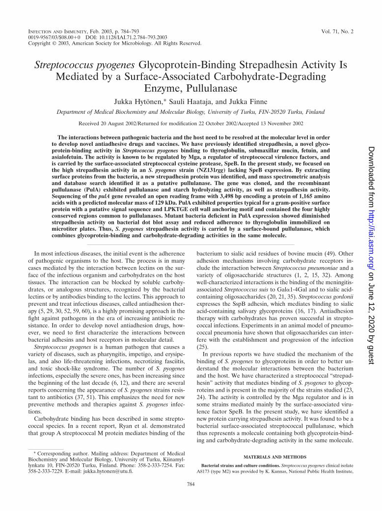
Expression and purification of recombinant putative pullu-lanase. In order to confirm the finding that the putative strep-tococcal pullulanase carries strepadhesin activity, the pullula-nase was recombinantly expressed in E. coli. A PCR fragmentlacking the 5� end of the gene corresponding to the putativeN-terminal signal peptide, as well as the 3� end correspondingto the C-terminal cell wall-anchoring motif and membrane-spanning region, was generated and cloned into the E. coliexpression vector pET30 Ek/LIC. The recombinant putativepullulanase was expressed in E. coli BL21(DE3)pLysS as afusion protein with N-terminal His and S tags and purified byaffinity chromatography on Ni-NTA resin. The purified proteinhad a mobility corresponding to the expected molecular massof ca. 125 kDa of the recombinant protein (Fig. 3, left panel).
Strepadhesin activity of the recombinant putative pullula-nase. The recombinant putative pullulanase was subjected toSDS-PAGE and blotted to a nitrocellulose membrane. Themembrane was probed with biotin-labeled glycoproteins thy-roglobulin, submaxillary mucin, fetuin, and asialofetuin to de-termine whether the recombinant protein exhibited strepadhe-sin activity. The results demonstrated that the recombinantprotein bound these glycoproteins, and thus indicated that thepullulanase carried strepadhesin activity (Fig. 3, right panel).
Generation of a pullulanase-negative double mutant fromthe NZ131rgg strain. In order to further investigate the con-tribution of streptococcal pullulanase to the strepadhesin ac-tivity, an isogenic pulA mutant strain was generated from theNZ131rgg strain. An internal 1,180-bp NsiI-BamHI fragment
FIG. 1. (A) Production of SpeB by A8173 and NZ131 wild-typebacteria and the NZ131speB and NZ131rgg SpeB-defective mutants.Bacteria from overnight cultures were lysed in SDS-PAGE samplebuffer, and the samples subjected to SDS-PAGE and transferred to anitrocellulose membrane, and then the membrane was probed withSpeB antiserum. (B) Strepadhesin activity of wild-type and SpeB-defective mutant streptococci. A membrane containing 1-�l aliquots ofserially diluted (OD600 � 4.0 to 0.125) suspensions of A8173 andNZ131 wild-type bacteria and the SpeB-defective NZ131speB andNZ131rgg mutants was incubated with biotinylated thyroglobulin, andthe binding was detected with streptavidin-HRP and by ECL autora-diography.
786 HYTONEN ET AL. INFECT. IMMUN.
on June 12, 2020 by guesthttp://iai.asm
.org/D
ownloaded from

of the pulA gene was cloned into pSF151, a suicide vectorincapable of replicating in streptococci, resulting in plasmidpulA-pSF151. The plasmid was electroporated in NZ131rgg,and a mutant, designated NZ131rgg-pulA, was chosen for fur-ther experiments (Fig. 4A). Genomic DNA was extracted fromthe bacteria, and PCR amplification was performed to verifythe integration of the plasmid into the pulA gene in NZ131rgg-pulA genome (Fig. 4B). Primers specific for pSF151 plasmidand 5� upstream region of pulA gene were used in the exper-iment. A fragment of the expected size (ca. 1.6 kb) could beamplified from NZ131rgg-pulA but not from NZ131rgg, a find-ing which indicated integration of the suicide plasmid into thepulA gene in NZ131rgg-pulA genome.
The strepadhesin activity of NZ131rgg-pulA bacteria wasstudied on nitrocellulose membrane (Fig. 4C). The binding of
thyroglobulin, submaxillary mucin, fetuin, and asialofetuin tothe double-mutant bacteria was clearly reduced compared tothe parental strain NZ131rgg. This demonstrated that thestreptococcal putative pullulanase is a carrier of strepadhesinactivity.
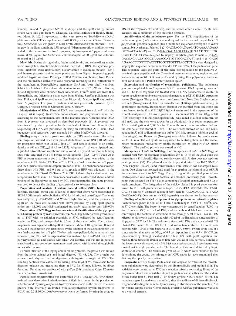
Binding of radiolabeled streptococci to glycoproteins on mi-crotiter plates. From the results of the bacterial dot blot assaysabove, it is clear that immobilized NZ131 wild-type and mutantstrains exhibit differential binding to liquid-phase glycopro-teins. In order to study whether the bacteria are able to bind toligands immobilized on a solid surface, we examined the ad-hesion of radiolabeled NZ131 wild-type and mutant strepto-cocci to thyroglobulin on microtiter plate wells. The number ofNZ131rgg bacteria adhered to wells coated with thyroglobulinwas markedly larger than the number of NZ131 wild-type bac-
FIG. 2. (A) Binding of thyroglobulin to SpeB extracts of the rgg mutant strain NZ131rgg. Bacteria were digested with 10 �g of SpeB/ml at 37°Cfor 30 min, and the digestion was terminated by adding the SpeB inhibitor E64 to a concentration of 1 �M. The extract was subjected toSDS-PAGE on a 7% gel and then stained with silver. A parallel sample transferred to nitrocellulose membrane was probed with biotinylatedthyroglobulin to identify strepadhesin activity. An extract of strain NZ131speB is shown as a control. The molecular weights (MW) in thousandsare indicated on the left. (B) Effect of SpeB treatment on the strepadhesin activity of NZ131rgg. Bacteria were either kept on ice (0°C), incubatedin PBS at 37°C, or treated with SpeB at a concentration of 100 �g/ml at 37°C for 4 h to totally abolish the strepadhesin activity. The binding ofthyroglobulin to 1-�l aliquots of bacterial suspension (OD600 � 4.0) on nitrocellulose membrane was detected as described for Fig. 1B.
VOL. 71, 2003 GLYCOPROTEIN BINDING AND S. PYOGENES PULLULANASE 787
on June 12, 2020 by guesthttp://iai.asm
.org/D
ownloaded from
teria (Fig. 5). Moreover, the NZ131rgg-pulA strain defective inputative pullulanase adhered less than did NZ131rgg. Thebinding of the NZ131rgg-pulA bacteria was, however, higherthan the binding of the wild-type bacteria, which suggests thatthere may be additional molecules involved in the binding ofthe glycoproteins.
In addition, we investigated the binding activities of thebacteria to human laminin and found that the adhesion of thergg and rgg-pulA mutants was three to four times that of thewild-type strain. However, there was no difference in the bindingbetween the mutants, which suggested that the laminin-bindingactivity is independent of the putative streptococcal pullulanase.
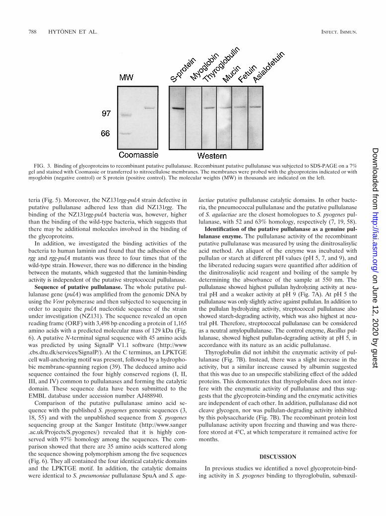
Sequence of putative pullulanase. The whole putative pul-lulanase gene (pulA) was amplified from the genomic DNA byusing the Vent polymerase and then subjected to sequencing inorder to acquire the pulA nucleotide sequence of the strainunder investigation (NZ131). The sequence revealed an openreading frame (ORF) with 3,498 bp encoding a protein of 1,165amino acids with a predicted molecular mass of 129 kDa (Fig.6). A putative N-terminal signal sequence with 45 amino acidswas predicted by using SignalP V1.1 software (http://www.cbs.dtu.dk/services/SignalP/). At the C terminus, an LPKTGEcell wall-anchoring motif was present, followed by a hydropho-bic membrane-spanning region (39). The deduced amino acidsequence contained the four highly conserved regions (I, II,III, and IV) common to pullulanases and forming the catalyticdomain. These sequence data have been submitted to theEMBL database under accession number AJ488940.
Comparison of the putative pullulanase amino acid se-quence with the published S. pyogenes genomic sequences (3,18, 55) and with the unpublished sequence from S. pyogenessequencing group at the Sanger Institute (http://www.sanger.ac.uk/Projects/S�pyogenes/) revealed that it is highly con-served with 97% homology among the sequences. The com-parison showed that there are 35 amino acids scattered alongthe sequence showing polymorphism among the five sequences(Fig. 6). They all contained the four identical catalytic domainsand the LPKTGE motif. In addition, the catalytic domainswere identical to S. pneumoniae pullulanase SpuA and S. aga-
lactiae putative pullulanase catalytic domains. In other bacte-ria, the pneumococcal pullulanase and the putative pullulanaseof S. agalactiae are the closest homologues to S. pyogenes pul-lulanase, with 52 and 63% homology, respectively (7, 19, 58).
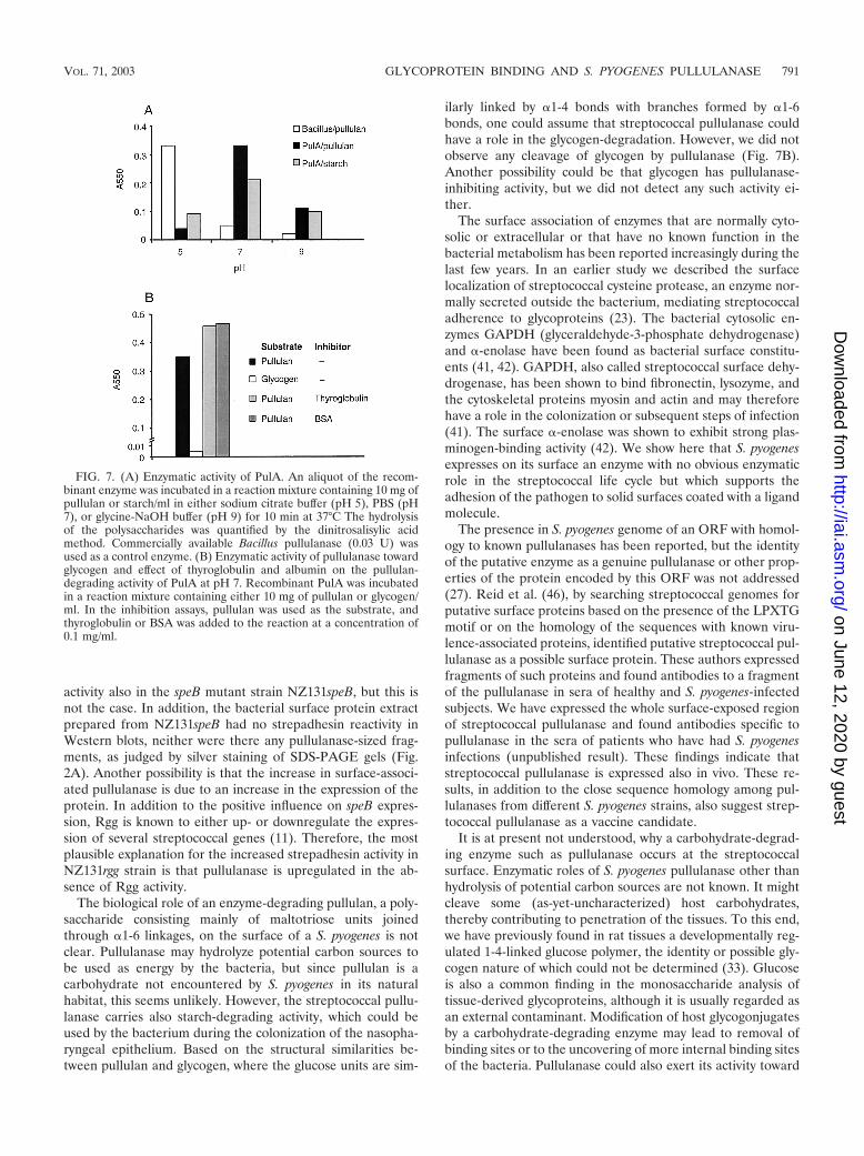
Identification of the putative pullulanase as a genuine pul-lulanase enzyme. The pullulanase activity of the recombinantputative pullulanase was measured by using the dinitrosalisylicacid method. An aliquot of the enzyme was incubated withpullulan or starch at different pH values (pH 5, 7, and 9), andthe liberated reducing sugars were quantified after addition ofthe dinitrosalisylic acid reagent and boiling of the sample bydetermining the absorbance of the sample at 550 nm. Thepullulanase showed highest pullulan hydrolyzing activity at neu-tral pH and a weaker activity at pH 9 (Fig. 7A). At pH 5 thepullulanase was only slightly active against pullulan. In addition tothe pullulan hydrolyzing activity, streptococcal pullulanase alsoshowed starch-degrading activity, which was also highest at neu-tral pH. Therefore, streptococcal pullulanase can be consideredas a neutral amylopullulanase. The control enzyme, Bacillus pul-lulanase, showed highest pullulan-degrading activity at pH 5, inaccordance with its nature as an acidic pullulanase.
Thyroglobulin did not inhibit the enzymatic activity of pul-lulanase (Fig. 7B). Instead, there was a slight increase in theactivity, but a similar increase caused by albumin suggestedthat this was due to an unspecific stabilizing effect of the addedproteins. This demonstrates that thyroglobulin does not inter-fere with the enzymatic activity of pullulanase and thus sug-gests that the glycoprotein-binding and the enzymatic activitiesare independent of each other. In addition, pullulanase did notcleave glycogen, nor was pullulan-degrading activity inhibitedby this polysaccharide (Fig. 7B). The recombinant protein lostpullulanase activity upon freezing and thawing and was there-fore stored at 4°C, at which temperature it remained active formonths.
DISCUSSION
In previous studies we identified a novel glycoprotein-bind-ing activity in S. pyogenes binding to thyroglobulin, submaxil-
FIG. 3. Binding of glycoproteins to recombinant putative pullulanase. Recombinant putative pullulanase was subjected to SDS-PAGE on a 7%gel and stained with Coomassie or transferred to nitrocellulose membranes. The membranes were probed with the glycoproteins indicated or withmyoglobin (negative control) or S protein (positive control). The molecular weights (MW) in thousands are indicated on the left.
788 HYTONEN ET AL. INFECT. IMMUN.
on June 12, 2020 by guesthttp://iai.asm
.org/D
ownloaded from

lary mucin, fetuin, and asialofetuin (23, 24). The activity, calledstrepadhesin activity, was found to be present in the majority ofstrains studied representing different M types and various in-fection foci. The strepadhesin activity of the M2 strain A8173was demonstrated to be regulated by the virulence factor reg-ulator Mga. In subsequent experiments, we were able to iden-tify a molecule carrying strepadhesin activity to be a bacterial-surface-associated streptococcal cysteine protease, SpeB. We
describe here the identification of another strepadhesin activ-ity-carrying molecule, streptococcal pullulanase.
The occurrence of more than one molecule displaying sim-ilar biological activity is not unusual. As an example of this,there are several adhesins described in various S. pyogenesstrains mediating adhesion to fibronectin. These includeamong others protein F (SfbI) (22, 56), protein F2 (26), fi-bronectin-binding protein FBP54 (14), SfbII/serum opacity
FIG. 4. (A) Strategy for mutagenesis of the streptococcal putative pullulanase gene, pulA. An internal pulA fragment comprising the nucleo-tides 297 to 1476 was cloned into the suicide plasmid pSF151, generating plasmid pulA-pSF151. The plasmid was electroporated into NZ131rgg,and the transformants were screened for homologous recombination by plating the bacteria on THY-kanamycin-erythromycin agar plates.(B) PCR analysis of the pulA region of the genomic DNA from NZ131rgg and NZ131rgg-pulA. Primers a and b (see Materials and Methods) wereused to amplify a 1.6-kb fragment from the NZ131rgg-pulA genome to verify the integration of pulA-pSF151 into the correct location. (C) Bindingof thyroglobulin, submaxillary mucin, fetuin, and asialofetuin to dilution series of NZ131rgg and NZ131 rgg-pulA mutant strains.
VOL. 71, 2003 GLYCOPROTEIN BINDING AND S. PYOGENES PULLULANASE 789
on June 12, 2020 by guesthttp://iai.asm
.org/D
ownloaded from
factor (31, 45), 28-kDa fibronectin-binding protein (13), andPFBP (47). Therefore, it is not unexpected that S. pyogenesstrepadhesin activity is mediated by more than just one mole-cule, which may vary among strains. The receptor structure forstrepadhesin in host tissues and the binding specificity of theactivity in terms of the molecular details is yet unknown. Theapparently broad binding specificity toward different glycopro-teins is, however, not unusual among bacterial adhesins. Forexample, antigen I and II adhesins of oral streptococci mediatebinding to a wide variety of molecules, such as salivary agglu-tinin, collagen, laminin, and fibronectin, and also to othermicroorganisms of the oral cavity without any apparent spec-ificity toward a single molecular determinant (28, 36, 43, 44,50).
The increased amount of streptococcal pullulanase on thesurface of the Rgg-deficient strain NZ131rgg and the increasedstrepadhesin activity could be due to at least two cellular mech-anisms. An obvious explanation would be that in this strain thelack of secretion of SpeB, which is known to cleave proteinsfrom the bacterial surface, results in decreased shedding ofpullulanase and thereby in increased strepadhesin activity.However, in this case one would expect increased strepadhesin
FIG. 5. Binding of radiolabeled streptococci to glycoproteins onmicrotiter plates. The plates were coated with 10 �g of thyroglobulin,human laminin, or BSA/ml, and the adhesion of radiolabeled NZ131wild-type and the rgg and rgg-pulA deficient mutant streptococci wasdetected with a liquid scintillation counter (see Materials and Meth-ods). The results are given as mean values � the standard deviation ofeight parallel wells.
FIG. 6. Deduced amino acid sequence of PulA. The putative N-terminal signal sequence with 45 amino acids and the C-terminal cellwall-anchoring motif LPKTGE are underlined. The four conserved regions common to pullulanases are underlined and are indicated by numeralsI, II, III, and IV. The amino acids polymorphic between the NZ131, SF370 (18), MGAS8232 (55), MGAS315 (3), and Manfredo strains(http://www.sanger.ac.uk/Projects/S�pyogenes/) are indicated in boldface.
790 HYTONEN ET AL. INFECT. IMMUN.
on June 12, 2020 by guesthttp://iai.asm
.org/D
ownloaded from
activity also in the speB mutant strain NZ131speB, but this isnot the case. In addition, the bacterial surface protein extractprepared from NZ131speB had no strepadhesin reactivity inWestern blots, neither were there any pullulanase-sized frag-ments, as judged by silver staining of SDS-PAGE gels (Fig.2A). Another possibility is that the increase in surface-associ-ated pullulanase is due to an increase in the expression of theprotein. In addition to the positive influence on speB expres-sion, Rgg is known to either up- or downregulate the expres-sion of several streptococcal genes (11). Therefore, the mostplausible explanation for the increased strepadhesin activity inNZ131rgg strain is that pullulanase is upregulated in the ab-sence of Rgg activity.
The biological role of an enzyme-degrading pullulan, a poly-saccharide consisting mainly of maltotriose units joinedthrough �1-6 linkages, on the surface of a S. pyogenes is notclear. Pullulanase may hydrolyze potential carbon sources tobe used as energy by the bacteria, but since pullulan is acarbohydrate not encountered by S. pyogenes in its naturalhabitat, this seems unlikely. However, the streptococcal pullu-lanase carries also starch-degrading activity, which could beused by the bacterium during the colonization of the nasopha-ryngeal epithelium. Based on the structural similarities be-tween pullulan and glycogen, where the glucose units are sim-
ilarly linked by �1-4 bonds with branches formed by �1-6bonds, one could assume that streptococcal pullulanase couldhave a role in the glycogen-degradation. However, we did notobserve any cleavage of glycogen by pullulanase (Fig. 7B).Another possibility could be that glycogen has pullulanase-inhibiting activity, but we did not detect any such activity ei-ther.
The surface association of enzymes that are normally cyto-solic or extracellular or that have no known function in thebacterial metabolism has been reported increasingly during thelast few years. In an earlier study we described the surfacelocalization of streptococcal cysteine protease, an enzyme nor-mally secreted outside the bacterium, mediating streptococcaladherence to glycoproteins (23). The bacterial cytosolic en-zymes GAPDH (glyceraldehyde-3-phosphate dehydrogenase)and �-enolase have been found as bacterial surface constitu-ents (41, 42). GAPDH, also called streptococcal surface dehy-drogenase, has been shown to bind fibronectin, lysozyme, andthe cytoskeletal proteins myosin and actin and may thereforehave a role in the colonization or subsequent steps of infection(41). The surface �-enolase was shown to exhibit strong plas-minogen-binding activity (42). We show here that S. pyogenesexpresses on its surface an enzyme with no obvious enzymaticrole in the streptococcal life cycle but which supports theadhesion of the pathogen to solid surfaces coated with a ligandmolecule.
The presence in S. pyogenes genome of an ORF with homol-ogy to known pullulanases has been reported, but the identityof the putative enzyme as a genuine pullulanase or other prop-erties of the protein encoded by this ORF was not addressed(27). Reid et al. (46), by searching streptococcal genomes forputative surface proteins based on the presence of the LPXTGmotif or on the homology of the sequences with known viru-lence-associated proteins, identified putative streptococcal pul-lulanase as a possible surface protein. These authors expressedfragments of such proteins and found antibodies to a fragmentof the pullulanase in sera of healthy and S. pyogenes-infectedsubjects. We have expressed the whole surface-exposed regionof streptococcal pullulanase and found antibodies specific topullulanase in the sera of patients who have had S. pyogenesinfections (unpublished result). These findings indicate thatstreptococcal pullulanase is expressed also in vivo. These re-sults, in addition to the close sequence homology among pul-lulanases from different S. pyogenes strains, also suggest strep-tococcal pullulanase as a vaccine candidate.
It is at present not understood, why a carbohydrate-degrad-ing enzyme such as pullulanase occurs at the streptococcalsurface. Enzymatic roles of S. pyogenes pullulanase other thanhydrolysis of potential carbon sources are not known. It mightcleave some (as-yet-uncharacterized) host carbohydrates,thereby contributing to penetration of the tissues. To this end,we have previously found in rat tissues a developmentally reg-ulated 1-4-linked glucose polymer, the identity or possible gly-cogen nature of which could not be determined (33). Glucoseis also a common finding in the monosaccharide analysis oftissue-derived glycoproteins, although it is usually regarded asan external contaminant. Modification of host glycogonjugatesby a carbohydrate-degrading enzyme may lead to removal ofbinding sites or to the uncovering of more internal binding sitesof the bacteria. Pullulanase could also exert its activity toward
FIG. 7. (A) Enzymatic activity of PulA. An aliquot of the recom-binant enzyme was incubated in a reaction mixture containing 10 mg ofpullulan or starch/ml in either sodium citrate buffer (pH 5), PBS (pH7), or glycine-NaOH buffer (pH 9) for 10 min at 37°C The hydrolysisof the polysaccharides was quantified by the dinitrosalisylic acidmethod. Commercially available Bacillus pullulanase (0.03 U) wasused as a control enzyme. (B) Enzymatic activity of pullulanase towardglycogen and effect of thyroglobulin and albumin on the pullulan-degrading activity of PulA at pH 7. Recombinant PulA was incubatedin a reaction mixture containing either 10 mg of pullulan or glycogen/ml. In the inhibition assays, pullulan was used as the substrate, andthyroglobulin or BSA was added to the reaction at a concentration of0.1 mg/ml.
VOL. 71, 2003 GLYCOPROTEIN BINDING AND S. PYOGENES PULLULANASE 791
on June 12, 2020 by guesthttp://iai.asm
.org/D
ownloaded from

carbohydrates on the bacterial surface of itself or of otherbacteria.
In summary, we described here the identification of strep-tococcal pullulanase, a novel cell surface-associated moleculethat carries strepadhesin activity in addition to polysacchari-dase activity. Although several carbohydrate-degrading rolescan be envisioned for pullulanase, the enzymatic function ofthe protein remains obscure in terms of in vivo substrate spec-ificity. Therefore, from the viewpoint of the evolution of strep-tococcal virulence, it is possible that S. pyogenes has acquiredthe gene coding for pullulanase not because of the enzymaticactivity but because of the advantage gained through the stre-padhesin activity it encodes.
ACKNOWLEDGMENTS
This work was supported by grants from the Sigrid Juselius Foun-dation and the Academy of Finland.
We thank Dieter Gerlach for the SpeB enzyme, Kyllikki Kunnas,and Michael Chaussee for providing the bacterial strains and TerttuJompero for technical assistance.
REFERENCES
1. Andersson, B., J. Dahmen, T. Frejd, H. Leffler, G. Magnusson, G. Noori, andC. Svanborg-Eden. 1983. Identification of an active disaccharide unit of aglycoconjugate receptor for pneumococci attaching to human pharyngealepithelial cells. J. Exp. Med. 158:559–570.
2. Barthelson, R., A. Mobasseri, D. Zopf, and P. Simon. 1998. Adherence ofStreptococcus pneumoniae to respiratory epithelial cells is inhibited by sialy-lated oligosaccharides. Infect. Immun. 66:1439–1444.
3. Beres, S. B., G. L. Sylva, K. D. Barbian, B. Lei, J. S. Hoff, N. D. Mammarella,M. Y. Liu, J. C. Smoot, S. F. Porcella, L. D. Parkins, D. S. Campbell, T. M.Smith, J. K. McCormick, D. Y. Leung, P. M. Schlievert, and J. M. Musser.2002. Genome sequence of a serotype M3 strain of group A streptococcus:phage-encoded toxins, the high-virulence phenotype, and clone emergence.Proc. Natl. Acad. Sci. USA 99:10078–10083.
4. Berge, A., and L. Bjorck. 1995. Streptococcal cysteine proteinase releasesbiologically active fragments of streptococcal surface proteins. J. Biol. Chem.270:9862–9867.
5. Beuth, J., B. Stoffel, and G. Pulverer. 1996. Inhibition of bacterial adhesionand infections by lectin blocking. Adv. Exp. Med. Biol. 408:51–56.
6. Bochicchio, G. V., M. Joshi, M. Joshi, S. Henry, and T. Scalea. 2001. GroupA streptococcus (GAS) soft-tissue infections: a lethal organism on the rise.Am. Surg. 67:1089–1092.
7. Bongaerts, R. J., H. P. Heinz, U. Hadding, and G. Zysk. 2000. Antigenicity,expression, and molecular characterization of surface-located pullulanase ofStreptococcus pneumoniae. Infect. Immun. 68:7141–7143.
8. Caparon, M. G., and J. R. Scott. 1991. Genetic manipulation of pathogenicstreptococci. Methods Enzymol. 204:556–586.
9. Chaussee, M. S., D. Ajdic, and J. J. Ferretti. 1999. The rgg gene of Strepto-coccus pyogenes NZ131 positively influences extracellular SPE B production.Infect. Immun. 67:1715–1722.
10. Chaussee, M. S., D. Gerlach, C. E. Yu, and J. J. Ferretti. 1993. Inactivationof the streptococcal erythrogenic toxin B gene (speB) in Streptococcus pyo-genes. Infect. Immun. 61:3719–3723.
11. Chaussee, M. S., G. L. Sylva, D. E. Sturdevant, L. M. Smoot, M. R. Graham,R. O. Watson, and J. M. Musser. 2002. Rgg influences the expression ofmultiple regulatory loci to coregulate virulence factor expression in Strepto-coccus pyogenes. Infect. Immun. 70:762–770.
12. Chelsom, J., A. Halstensen, T. Haga, and E. A. Hoiby. 1994. Necrotisingfasciitis due to group A streptococci in western Norway: incidence andclinical features. Lancet 344:1111–1115.
13. Courtney, H. S., D. L. Hasty, J. B. Dale, and T. P. Poirier. 1992. A 28-kilodalton fibronectin-binding protein of group A streptococci. Curr. Micro-biol. 25:245–250.
14. Courtney, H. S., Y. Li, J. B. Dale, and D. L. Hasty. 1994. Cloning, sequencing,and expression of a fibronectin/fibrinogen-binding protein from group Astreptococci. Infect. Immun. 62:3937–3946.
15. Cundell, D. R., and E. I. Tuomanen. 1994. Receptor specificity of adherenceof Streptococcus pneumoniae to human type-II pneumocytes and vascularendothelial cells in vitro. Microb. Pathog. 17:361–374.
16. Demuth, D. R., C. A. Davis, A. M. Corner, R. J. Lamont, P. S. Leboy, and D.Malamud. 1988. Cloning and expression of a Streptococcus sanguis surfaceantigen that interacts with a human salivary agglutinin. Infect. Immun. 56:2484–2490.
17. Demuth, D. R., E. E. Golub, and D. Malamud. 1990. Streptococcal-host
interactions. Structural and functional analysis of a Streptococcus sanguisreceptor for a human salivary glycoprotein. J. Biol. Chem. 265:7120–7126.
18. Ferretti, J. J., W. M. McShan, D. Ajdic, D. J. Savic, G. Savic, K. Lyon, C.Primeaux, S. Sezate, A. N. Suvorov, S. Kenton, H. S. Lai, S. P. Lin, Y. Qian,H. G. Jia, F. Z. Najar, Q. Ren, H. Zhu, L. Song, J. White, X. Yuan, S. W.Clifton, B. A. Roe, and R. McLaughlin. 2001. Complete genome sequence ofan M1 strain of Streptococcus pyogenes. Proc. Natl. Acad. Sci. USA 98:4658–4663.
19. Glaser, P., C. Rusniok, C. Buchrieser, F. Chevalier, L. Frangeul, T. Msadek,M. Zouine, E. Couve, L. Lalioui, C. Poyart, P. Trieu-Cuot, and F. Kunst.2002. Genome sequence of Streptococcus agalactiae, a pathogen causinginvasive neonatal disease. Mol. Microbiol. 45:1499–1513.
20. Haataja, S., K. Tikkanen, J. Liukkonen, C. Francois-Gerard, and J. Finne.1993. Characterization of a novel bacterial adhesion specificity of Strepto-coccus suis recognizing blood group P receptor oligosaccharides. J. Biol.Chem. 268:4311–4317.
21. Haataja, S., K. Tikkanen, U. Nilsson, G. Magnusson, K. A. Karlsson, and J.Finne. 1994. Oligosaccharide-receptor interaction of the Gal�1–4Gal bind-ing adhesin of Streptococcus suis. Combining site architecture and charac-terization of two variant adhesin specificities. J. Biol. Chem. 269:27466–27472.
22. Hanski, E., and M. Caparon. 1992. Protein F, a fibronectin-binding protein,is an adhesin of the group A streptococcus Streptococcus pyogenes. Proc.Natl. Acad. Sci. USA 89:6172–6176.
23. Hytonen, J., S. Haataja, D. Gerlach, A. Podbielski, and J. Finne. 2001. TheSpeB virulence factor of Streptococcus pyogenes, a multifunctional secretedand cell surface molecule with strepadhesin, laminin-binding and cysteineprotease activity. Mol. Microbiol. 39:512–519.
24. Hytonen, J., S. Haataja, P. Isomaki, and J. Finne. 2000. Identification of anovel glycoprotein-binding activity in Streptococcus pyogenes regulated by themga gene. Microbiology 146:31–39.
25. Idanpaan-Heikkila, I., P. M. Simon, D. Zopf, T. Vullo, P. Cahill, K. Sokol,and E. Tuomanen. 1997. Oligosaccharides interfere with the establishmentand progression of experimental pneumococcal pneumonia. J. Infect. Dis.176:704–712.
26. Jaffe, J., S. Natanson-Yaron, M. G. Caparon, and E. Hanski. 1996. ProteinF2, a novel fibronectin-binding protein from Streptococcus pyogenes, pos-sesses two binding domains. Mol. Microbiol. 21:373–384.
27. Janulczyk, R., and M. Rasmussen. 2001. Improved pattern for genome-based screening identifies novel cell wall-attached proteins in gram-positivebacteria. Infect. Immun. 69:4019–4026.
28. Jenkinson, H. F., and D. R. Demuth. 1997. Structure, function and immu-nogenicity of streptococcal antigen I/II polypeptides. Mol. Microbiol. 23:183–190.
29. Karlsson, K. A. 1995. Microbial recognition of target-cell glycoconjugates.Curr. Opin. Struct. Biol. 5:622–635.
30. Karlsson, K. A. 1998. Meaning and therapeutic potential of microbial rec-ognition of host glycoconjugates. Mol. Microbiol. 29:1–11.
31. Kreikemeyer, B., S. R. Talay, and G. S. Chhatwal. 1995. Characterization ofa novel fibronectin-binding surface protein in group A streptococci. Mol.Microbiol. 17:137–145.
32. Krivan, H. C., D. D. Roberts, and V. Ginsburg. 1988. Many pulmonarypathogenic bacteria bind specifically to the carbohydrate sequence GalNAc�1–4Gal found in some glycolipids. Proc. Natl. Acad. Sci. USA 85:6157–6161.
33. Krusius, T., J. Finne, J. Karkkainen, and J. Jarnefelt. 1974. Neutral andacidic glycopeptides in adult and developing rat brain. Biochim. Biophys.Acta 365:80–92.
34. Kussmann, M., E. Nordhoff, H. Rahbek-Nielsen, S. Haebel, M. Rossel-Larssen, L. Jakobsen, J. Gobom, E. Mirgorodskaya, A. Kroll-Kristensen, L.Palm, and P. Roepstroff. 1997. Matrix-assisted laser desorption/ionizationmass spectrometry sample preparation techniques designed for various pep-tide and protein analytes. J. Mass Spectrom. 32:593–601.
35. Liukkonen, J., S. Haataja, K. Tikkanen, S. Kelm, and J. Finne. 1992. Iden-tification of N-acetylneuraminyl �233 poly-N-acetyllactosamine glycans asthe receptors of sialic acid-binding Streptococcus suis strains. J. Biol. Chem.267:21105–21111.
36. Love, R. M., M. D. McMillan, and H. F. Jenkinson. 1997. Invasion ofdentinal tubules by oral streptococci is associated with collagen recognitionmediated by the antigen I/II family of polypeptides. Infect. Immun. 65:5157–5164.
37. Martin, J. M., M. Green, K. A. Barbadora, and E. R. Wald. 2002. Erythro-mycin-resistant group A streptococci in schoolchildren in Pittsburgh.N. Engl. J. Med. 346:1200–1206.
38. Miller, G. L., R. Blum, W. E. Glennon, and A. L. Burton. 1960. Measurementof carboxymethylcellulase activity. Anal. Biochem. 1:127–132.
39. Navarre, W. W., and O. Schneewind. 1994. Proteolytic cleavage and cell wallanchoring at the LPXTG motif of surface proteins in gram-positive bacteria.Mol. Microbiol. 14:115–121.
40. Nyman, T. A., S. Matikainen, T. Sareneva, I. Julkunen, and N. Kalkkinen.2000. Proteome analysis reveals ubiquitin-conjugating enzymes to be a newfamily of interferon-alpha-regulated genes. Eur. J. Biochem. 267:4011–4019.
792 HYTONEN ET AL. INFECT. IMMUN.
on June 12, 2020 by guesthttp://iai.asm
.org/D
ownloaded from
41. Pancholi, V., and V. A. Fischetti. 1992. A major surface protein on group Astreptococci is a glyceraldehyde-3-phosphate-dehydrogenase with multiplebinding activity. J. Exp. Med. 176:415–426.
42. Pancholi, V., and V. A. Fischetti. 1998. �-enolase, a novel strong plasmin(o-gen) binding protein on the surface of pathogenic streptococci. J. Biol.Chem. 273:14503–14515.
43. Petersen, F. C., S. Assev, H. C. van der Mei, H. J. Busscher, and A. A. Scheie.2002. Functional variation of the antigen I/II surface protein in Streptococcusmutans and Streptococcus intermedius. Infect. Immun. 70:249–256.
44. Petersen, F. C., S. Pasco, J. Ogier, J. P. Klein, S. Assev, and A. A. Scheie.2001. Expression and functional properties of the Streptococcus intermediussurface protein antigen I/II. Infect. Immun. 69:4647–4653.
45. Rakonjac, J. V., J. C. Robbins, and V. A. Fischetti. 1995. DNA sequence ofthe serum opacity factor of group A streptococci: identification of a fibronec-tin-binding repeat domain. Infect. Immun. 63:622–631.
46. Reid, S. D., N. M. Green, J. K. Buss, B. Lei, and J. M. Musser. 2001.Multilocus analysis of extracellular putative virulence proteins made bygroup A streptococcus: population genetics, human serologic response, andgene transcription. Proc. Natl. Acad. Sci. USA 98:7552–7557.
47. Rocha, C. L., and V. A. Fischetti. 1999. Identification and characterization ofa novel fibronectin-binding protein on the surface of group A streptococci.Infect. Immun. 67:2720–2728.
48. Rosenfeld, J., J. Capdevielle, J. C. Guillemot, and P. Ferrara. 1992. In-geldigestion of proteins for internal sequence analysis after one- or two-dimen-sional gel electrophoresis. Anal. Biochem. 203:173–179.
49. Ryan, P. A., V. Pancholi, and V. A. Fischetti. 2001. Group A streptococcibind to mucin and human pharyngeal cells through sialic acid-containingreceptors. Infect. Immun. 69:7402–7412.
50. Sciotti, M. A., I. Yamodo, J. P. Klein, and J. A. Ogier. 1997. The N-terminalhalf part of the oral streptococcal antigen I/IIf contains two distinct bindingdomains. FEMS Microbiol. Lett. 153:439–445.
51. Seppala, H., T. Klaukka, J. Vuopio-Varkila, A. Muotiala, H. Helenius, K.Lager, P. Huovinen, et al. 1997. The effect of changes in the consumption ofmacrolide antibiotics on erythromycin resistance in group A streptococci inFinland. N. Engl. J. Med. 337:441–446.
52. Sharon, N., and I. Ofek. 2000. Safe as mother’s milk: carbohydrates as futureanti-adhesion drugs for bacterial diseases. Glycoconj. J. 17:659–664.
53. Shevchenko, A., M. Wilm, O. Vorm, and M. Mann. 1996. Mass spectrometricsequencing of proteins silver-stained polyacrylamide gels. Anal. Chem. 68:850–858.
54. Simon, D., and J. J. Ferretti. 1991. Electrotransformation of Streptococcuspyogenes with plasmid and linear DNA. FEMS Microbiol. Lett. 66:219–224.
55. Smoot, J. C., K. D. Barbian, J. J. Van Gompel, L. M. Smoot, M. S. Chaussee,G. L. Sylva, D. E. Sturdevant, S. M. Ricklefs, S. F. Porcella, L. D. Parkins,S. B. Beres, D. S. Campbell, T. M. Smith, Q. Zhang, V. Kapur, J. A. Daly,L. G. Veasy, and J. M. Musser. 2002. Genome sequence and comparativemicroarray analysis of serotype M18 group A streptococcus strains associ-ated with acute rheumatic fever outbreaks. Proc. Natl. Acad. Sci. USA99:4668–4673.
56. Talay, S. R., P. Valentin-Weigand, K. N. Timmis, and G. S. Chhatwal. 1994.Domain structure and conserved epitopes of Sfb protein, the fibronectin-binding adhesin of Streptococcus pyogenes. Mol. Microbiol. 13:531–539.
57. Tao, L., D. J. LeBlanc, and J. J. Ferretti. 1992. Novel streptococcal-integra-tion shuttle vectors for gene cloning and inactivation. Gene 120:105–110.
58. Tettelin, H., V. Masignani, M. J. Cieslewicz, J. A. Eisen, S. Peterson, M. R.Wessels, I. T. Paulsen, K. E. Nelson, I. Margarit, T. D. Read, L. C. Madoff,A. M. Wolf, M. J. Beanan, L. M. Brinkac, S. C. Daugherty, R. T. DeBoy, A. S.Durkin, J. F. Kolonay, R. Madupu, M. R. Lewis, D. Radune, N. B. Fedorova,D. Scanlan, H. Khouri, S. Mulligan, H. A. Carty, R. T. Cline, S. E. Van Aken,J. Gill, M. Scarselli, M. Mora, E. T. Iacobini, C. Brettoni, G. Galli, M.Mariani, F. Vegni, D. Maione, D. Rinaudo, R. Rappuoli, J. L. Telford, D. L.Kasper, G. Grandi, and C. M. Fraser. 2002. Complete genome sequence andcomparative genomic analysis of an emerging human pathogen, serotype VStreptococcus agalactiae. Proc. Natl. Acad. Sci. USA 99:12391–12396.
59. Zopf, D., and S. Roth. 1996. Oligosaccharide anti-infective agents. Lancet347:1017–1021.
60. Zopf, D., P. Simon, R. Barthelson, D. Cundell, I. Idanpaan-Heikkila, and E.Tuomanen. 1996. Development of anti-adhesion carbohydrate drugs for clin-ical use. Adv. Exp. Med. Biol. 408:35–38.
Editor: A. D. O’Brien
VOL. 71, 2003 GLYCOPROTEIN BINDING AND S. PYOGENES PULLULANASE 793
on June 12, 2020 by guesthttp://iai.asm
.org/D
ownloaded from