strategies to enhance drug absorption via nasal and
TRANSCRIPT
pharmaceutics
Review
Strategies to Enhance Drug Absorption via Nasal andPulmonary Routes
Maliheh Ghadiri * , Paul M. Young and Daniela Traini
Respiratory Technology, Woolcock Institute of Medical Research and Discipline of Pharmacology,Faculty of Medicine and Health, The University of Sydney, Camperdown, NSW 2006, Australia;[email protected] (P.M.Y.); [email protected] (D.T.)* Correspondence: [email protected]; Tel.: +61(2)911-40366
Received: 16 January 2019; Accepted: 5 March 2019; Published: 11 March 2019�����������������
Abstract: New therapeutic agents such as proteins, peptides, and nucleic acid-based agents are beingdeveloped every year, making it vital to find a non-invasive route such as nasal or pulmonary for theiradministration. However, a major concern for some of these newly developed therapeutic agents istheir poor absorption. Therefore, absorption enhancers have been investigated to address this majoradministration problem. This paper describes the basic concepts of transmucosal administrationof drugs, and in particular the use of the pulmonary or nasal routes for administration of drugswith poor absorption. Strategies for the exploitation of absorption enhancers for the improvementof pulmonary or nasal administration are discussed, including use of surfactants, cyclodextrins,protease inhibitors, and tight junction modulators, as well as application of carriers such as liposomesand nanoparticles.
Keywords: nasal; pulmonary; drug administration; absorption enhancers; nanoparticle; and liposome
1. Background
Absorption enhancers are functional excipients included in formulations to improve theabsorption of drugs across biological barriers. They have been investigated for many years, particularlyto enhance the efficacy of peptides, proteins, and other pharmacologically active compounds that havepoor barrier permeability [1]. The ideal absorption enhancer should be one that protects biologicalagents against enzymatic degradation and causes a rapid opening of the relevant barrier whileenhancing absorption transiently.
As a portal for non-invasive delivery, nasal and pulmonary administration has several advantagesover traditional oral medication or injection. Nasal and pulmonary delivery are non-invasive routesof administration that target the delivered dose directly to the site of drug action [2,3]. Moreover,drug delivery to the respiratory area can also be used for systemic delivery of peptides and proteins dueto the large surface area for drug absorption. Pulmonary and nasal administration bypasses first-passmetabolism that is observed in oral administration and the lung and nasal cavity have a low drugmetabolizing environment [4]. Despite all these advantages, there is a significant challenge to enhancethe absorption of the active agent via these routes. In nasal and pulmonary administration, absorptionenhancers have been investigated over the last two decades, to increase the rate of absorption bytargeting different mechanisms [5,6]. These mechanisms are either to improve the permeation ofmaterials across the epithelial barrier via intracellular or paracellular mechanisms (Figure 1) or toenhance stability and mucus solubility of the drugs regionally. However, to date, no safe absorptionenhancer for pulmonary administration of drugs has translated into commercial products. Their usehas generated safety concerns due to potential irreversible alteration of the epithelial cell membrane,which could potentially make the lung susceptible to the entry of exogenous allergens. While there are
Pharmaceutics 2019, 11, 113; doi:10.3390/pharmaceutics11030113 www.mdpi.com/journal/pharmaceutics
Pharmaceutics 2019, 11, 113 2 of 20
no absorption enhancers for pulmonary drug administration on the market, quite a few appear to beat the threshold of becoming products for nasal administration.
This review critically assesses advances in the field of absorption enhancers for nasal andpulmonary drug administration. The various agents used to increase the absorption of poorlypermeable drugs, their mechanisms of action, safety, and effectiveness are also presented.
Pharmaceutics 2019, 11, x FOR PEER REVIEW 2 of 20
of the epithelial cell membrane, which could potentially make the lung susceptible to the entry of exogenous allergens. While there are no absorption enhancers for pulmonary drug administration on the market, quite a few appear to be at the threshold of becoming products for nasal administration.
This review critically assesses advances in the field of absorption enhancers for nasal and pulmonary drug administration. The various agents used to increase the absorption of poorly permeable drugs, their mechanisms of action, safety, and effectiveness are also presented.
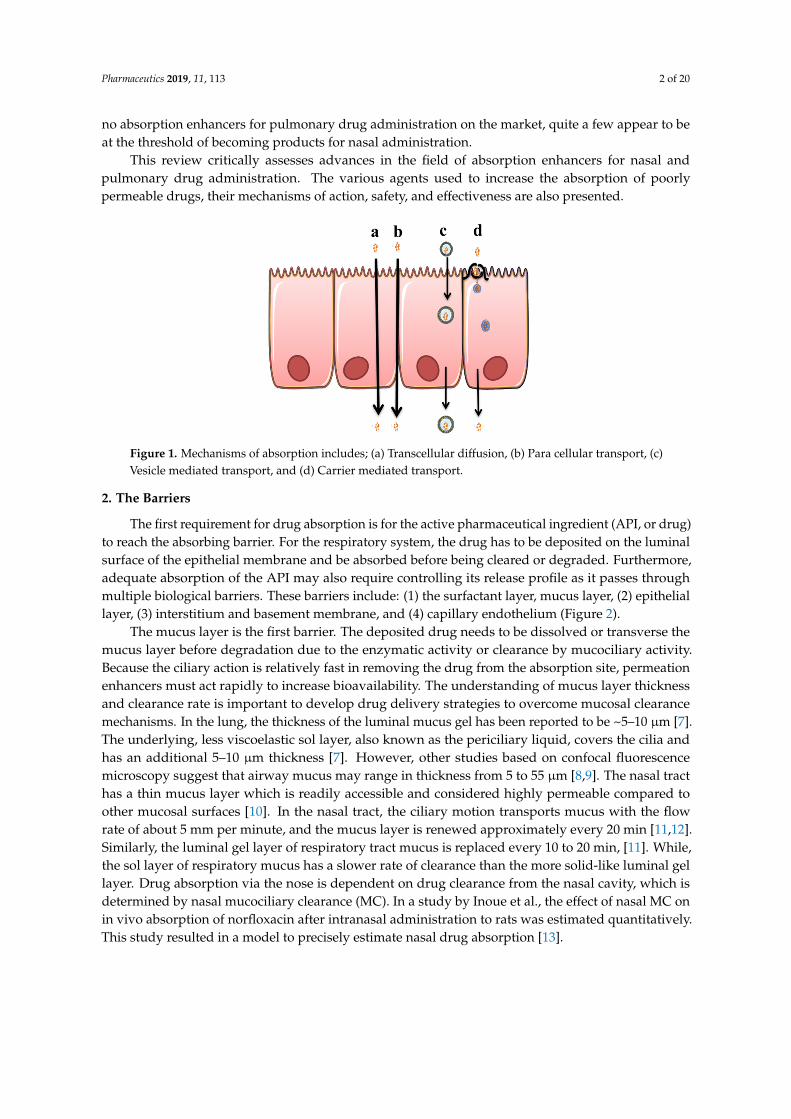
Figure 1. Mechanisms of absorption includes; (a) Transcellular diffusion, (b) Para cellular transport, (c) Vesicle mediated transport, and (d) Carrier mediated transport.
2. The Barriers
The first requirement for drug absorption is for the active pharmaceutical ingredient (API, or drug) to reach the absorbing barrier. For the respiratory system, the drug has to be deposited on the luminal surface of the epithelial membrane and be absorbed before being cleared or degraded. Furthermore, adequate absorption of the API may also require controlling its release profile as it passes through multiple biological barriers. These barriers include: (1) the surfactant layer, mucus layer, (2) epithelial layer, (3) interstitium and basement membrane, and (4) capillary endothelium (Figure 2).
The mucus layer is the first barrier. The deposited drug needs to be dissolved or transverse the mucus layer before degradation due to the enzymatic activity or clearance by mucociliary activity. Because the ciliary action is relatively fast in removing the drug from the absorption site, permeation enhancers must act rapidly to increase bioavailability. The understanding of mucus layer thickness and clearance rate is important to develop drug delivery strategies to overcome mucosal clearance mechanisms. In the lung, the thickness of the luminal mucus gel has been reported to be ~5–10 μm [7]. The underlying, less viscoelastic sol layer, also known as the periciliary liquid, covers the cilia and has an additional 5–10 μm thickness [7]. However, other studies based on confocal fluorescence microscopy suggest that airway mucus may range in thickness from 5 to 55 μm [8,9]. The nasal tract has a thin mucus layer which is readily accessible and considered highly permeable compared to other mucosal surfaces [10]. In the nasal tract, the ciliary motion transports mucus with the flow rate of about 5 mm per minute, and the mucus layer is renewed approximately every 20 min [11,12]. Similarly, the luminal gel layer of respiratory tract mucus is replaced every 10 to 20 min, [11]. While, the sol layer of respiratory mucus has a slower rate of clearance than the more solid-like luminal gel layer. Drug absorption via the nose is dependent on drug clearance from the nasal cavity, which is determined by nasal mucociliary clearance (MC). In a study by Inoue et al., the effect of nasal MC on in vivo absorption of norfloxacin after intranasal administration to rats was estimated quantitatively. This study resulted in a model to precisely estimate nasal drug absorption [13].
Figure 1. Mechanisms of absorption includes; (a) Transcellular diffusion, (b) Para cellular transport, (c)Vesicle mediated transport, and (d) Carrier mediated transport.
2. The Barriers
The first requirement for drug absorption is for the active pharmaceutical ingredient (API, or drug)to reach the absorbing barrier. For the respiratory system, the drug has to be deposited on the luminalsurface of the epithelial membrane and be absorbed before being cleared or degraded. Furthermore,adequate absorption of the API may also require controlling its release profile as it passes throughmultiple biological barriers. These barriers include: (1) the surfactant layer, mucus layer, (2) epitheliallayer, (3) interstitium and basement membrane, and (4) capillary endothelium (Figure 2).
The mucus layer is the first barrier. The deposited drug needs to be dissolved or transverse themucus layer before degradation due to the enzymatic activity or clearance by mucociliary activity.Because the ciliary action is relatively fast in removing the drug from the absorption site, permeationenhancers must act rapidly to increase bioavailability. The understanding of mucus layer thicknessand clearance rate is important to develop drug delivery strategies to overcome mucosal clearancemechanisms. In the lung, the thickness of the luminal mucus gel has been reported to be ~5–10 µm [7].The underlying, less viscoelastic sol layer, also known as the periciliary liquid, covers the cilia andhas an additional 5–10 µm thickness [7]. However, other studies based on confocal fluorescencemicroscopy suggest that airway mucus may range in thickness from 5 to 55 µm [8,9]. The nasal tracthas a thin mucus layer which is readily accessible and considered highly permeable compared toother mucosal surfaces [10]. In the nasal tract, the ciliary motion transports mucus with the flowrate of about 5 mm per minute, and the mucus layer is renewed approximately every 20 min [11,12].Similarly, the luminal gel layer of respiratory tract mucus is replaced every 10 to 20 min, [11]. While,the sol layer of respiratory mucus has a slower rate of clearance than the more solid-like luminal gellayer. Drug absorption via the nose is dependent on drug clearance from the nasal cavity, which isdetermined by nasal mucociliary clearance (MC). In a study by Inoue et al., the effect of nasal MC onin vivo absorption of norfloxacin after intranasal administration to rats was estimated quantitatively.This study resulted in a model to precisely estimate nasal drug absorption [13].
Pharmaceutics 2019, 11, 113 3 of 20
Pharmaceutics 2019, 11, x FOR PEER REVIEW 3 of 20
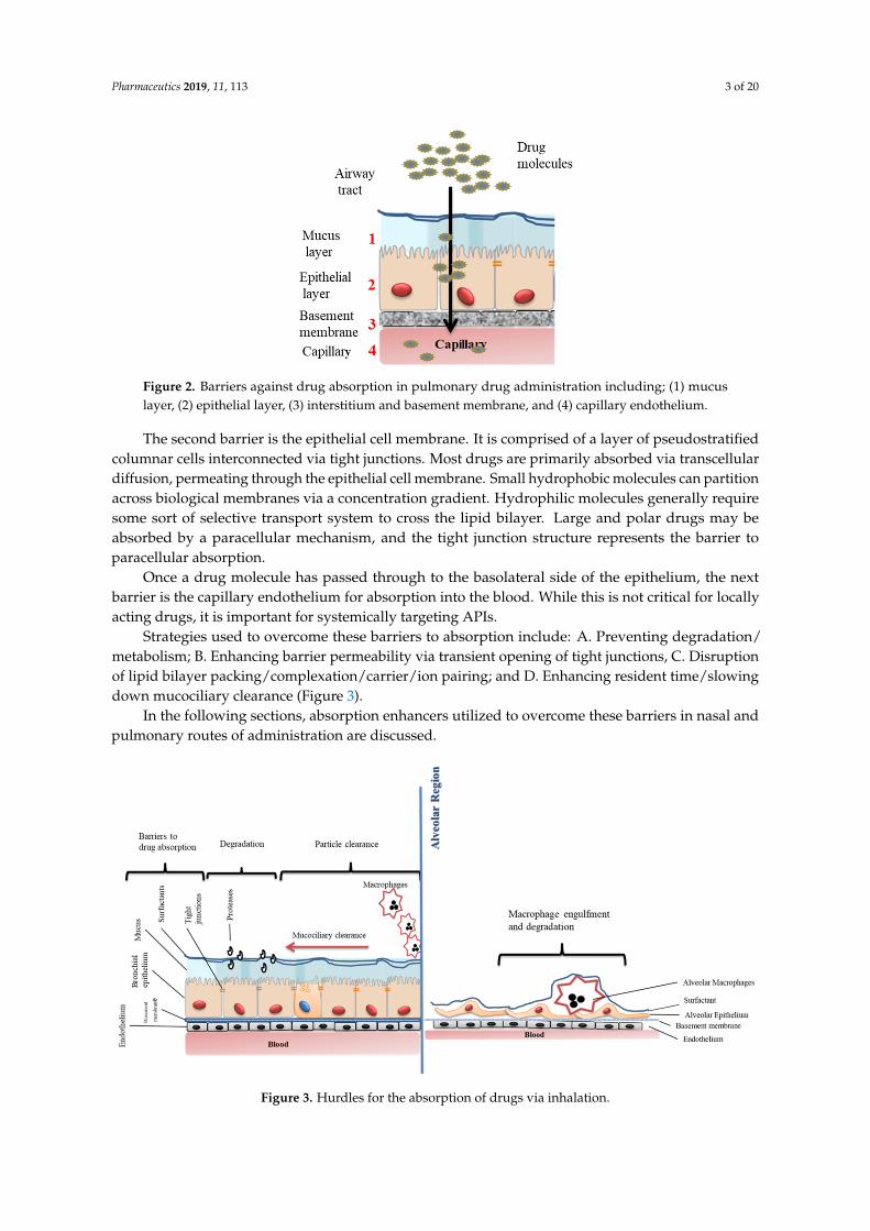
Figure 2. Barriers against drug absorption in pulmonary drug administration including; (1) mucus layer, (2) epithelial layer, (3) interstitium and basement membrane, and (4) capillary endothelium.
The second barrier is the epithelial cell membrane. It is comprised of a layer of pseudostratified columnar cells interconnected via tight junctions. Most drugs are primarily absorbed via transcellular diffusion, permeating through the epithelial cell membrane. Small hydrophobic molecules can partition across biological membranes via a concentration gradient. Hydrophilic molecules generally require some sort of selective transport system to cross the lipid bilayer. Large and polar drugs may be absorbed by a paracellular mechanism, and the tight junction structure represents the barrier to paracellular absorption.
Once a drug molecule has passed through to the basolateral side of the epithelium, the next barrier is the capillary endothelium for absorption into the blood. While this is not critical for locally acting drugs, it is important for systemically targeting APIs.
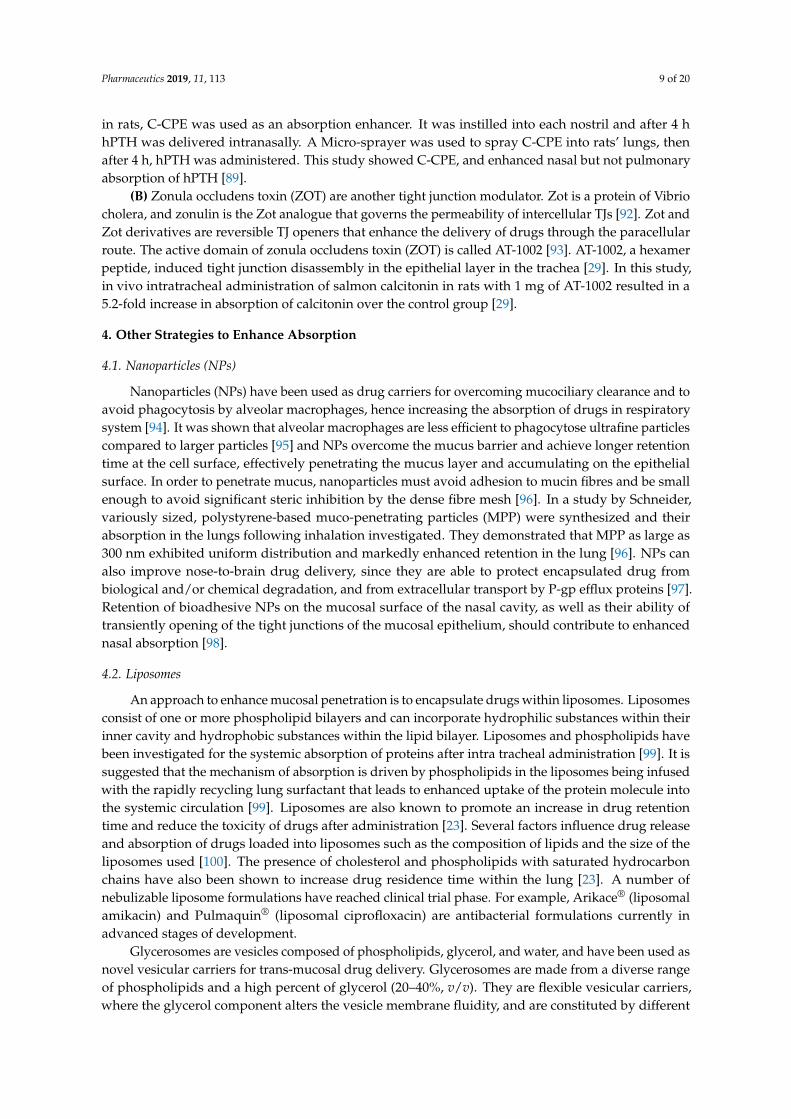
Strategies used to overcome these barriers to absorption include: A. Preventing degradation/metabolism; B. Enhancing barrier permeability via transient opening of tight junctions, C. Disruption of lipid bilayer packing/complexation/carrier/ion pairing; and D. Enhancing resident time/slowing down mucociliary clearance (Figure 3).
In the following sections, absorption enhancers utilized to overcome these barriers in nasal and pulmonary routes of administration are discussed.
Figure 3. Hurdles for the absorption of drugs via inhalation.
Figure 2. Barriers against drug absorption in pulmonary drug administration including; (1) mucuslayer, (2) epithelial layer, (3) interstitium and basement membrane, and (4) capillary endothelium.
The second barrier is the epithelial cell membrane. It is comprised of a layer of pseudostratifiedcolumnar cells interconnected via tight junctions. Most drugs are primarily absorbed via transcellulardiffusion, permeating through the epithelial cell membrane. Small hydrophobic molecules can partitionacross biological membranes via a concentration gradient. Hydrophilic molecules generally requiresome sort of selective transport system to cross the lipid bilayer. Large and polar drugs may beabsorbed by a paracellular mechanism, and the tight junction structure represents the barrier toparacellular absorption.
Once a drug molecule has passed through to the basolateral side of the epithelium, the nextbarrier is the capillary endothelium for absorption into the blood. While this is not critical for locallyacting drugs, it is important for systemically targeting APIs.
Strategies used to overcome these barriers to absorption include: A. Preventing degradation/metabolism; B. Enhancing barrier permeability via transient opening of tight junctions, C. Disruptionof lipid bilayer packing/complexation/carrier/ion pairing; and D. Enhancing resident time/slowingdown mucociliary clearance (Figure 3).
In the following sections, absorption enhancers utilized to overcome these barriers in nasal andpulmonary routes of administration are discussed.
Pharmaceutics 2019, 11, x FOR PEER REVIEW 3 of 20
Figure 2. Barriers against drug absorption in pulmonary drug administration including; (1) mucus layer, (2) epithelial layer, (3) interstitium and basement membrane, and (4) capillary endothelium.
The second barrier is the epithelial cell membrane. It is comprised of a layer of pseudostratified columnar cells interconnected via tight junctions. Most drugs are primarily absorbed via transcellular diffusion, permeating through the epithelial cell membrane. Small hydrophobic molecules can partition across biological membranes via a concentration gradient. Hydrophilic molecules generally require some sort of selective transport system to cross the lipid bilayer. Large and polar drugs may be absorbed by a paracellular mechanism, and the tight junction structure represents the barrier to paracellular absorption.
Once a drug molecule has passed through to the basolateral side of the epithelium, the next barrier is the capillary endothelium for absorption into the blood. While this is not critical for locally acting drugs, it is important for systemically targeting APIs.
Strategies used to overcome these barriers to absorption include: A. Preventing degradation/metabolism; B. Enhancing barrier permeability via transient opening of tight junctions, C. Disruption of lipid bilayer packing/complexation/carrier/ion pairing; and D. Enhancing resident time/slowing down mucociliary clearance (Figure 3).
In the following sections, absorption enhancers utilized to overcome these barriers in nasal and pulmonary routes of administration are discussed.
Figure 3. Hurdles for the absorption of drugs via inhalation.
Figure 3. Hurdles for the absorption of drugs via inhalation.

Pharmaceutics 2019, 11, 113 4 of 20
3. Absorption Enhancers Investigated for Nasal and Pulmonary Drug Administration
The most common permeation enhancers investigated for nasal and pulmonary drugadministration and their classification are listed in Table 1. Most of these agents have been divided intoone of the following major classifications: (I) surfactants, (II) cyclodextrins, (III) protease inhibitors,(IV) cationic polymers, and (V) tight junction modulators. Each of these classifications are discussed ingreater detail below.
Table 1. Common absorption permeation enhancers- application for nasal and pulmonary administration.
Class Enhancers Examples References
Surfactants Bile saltsSodium taurocholate [14,15]
Sodium deoxycholate sodium [16]
Glycodeoxycholat [17,18]
Surfactants Fatty acids andderivatives
Palmitic acid [19]
Palmitoleic acid
Stearic acid
Oleyl alcohol
Oleic acid
Capric acid [20,21]
DHA, EPA [20]
Surfactants Phospholipids Dipalmitoyl phophatidyl choline, soybeanlecithin, phosphatidylcholine [22,23]
Cationic polymers Polymers Chitosan and their derivatives [24–26]
Enzyme inhibitors Human neutrophil elastase inhibitor (ER143)
Cyclodextrins Beta-Cyclodextrin [27]
Tight junctionmodulators
Claudinemodulator Clostridium perfringens enterotoxin [28]
ZO modulator Zonula occludens toxin (ZOT) [29]
3.1. Surfactants
Surface active agents, or surfactants, are amphiphilic molecules possessing both lipophilic andhydrophilic residues. Surfactants have various applications in pulmonary drug administration, due totheir high interfacial activity; one of which is as an absorption enhancer [30]. Surfactants can enhanceabsorption with more than one mechanism; these include perturbing the cell membrane by leaching ofmembrane proteins, opening of tight junctions, or preventing enzymatic degradation of the drugs [31].These surfactants are mainly used and studied in oral drug administration; however, there have beena few studies looking at their application in nasal and pulmonary drug delivery. Surfactants usedas absorption enhancers can be classified as; (A) phospholipids [32], (B) bile salts, (such as sodiumtaurocholate etc.) [33], (C) non-ionic surfactants [31], (D) salt of fatty acids [20], and (E) alkyl glycosides(e.g., tetradecylmaltoside, N-lauryl-b-d-maltopyranoside etc.) [34].
(a) Phospholipids: Natural pulmonary surfactant is a complex mixture of phospholipids (90%)and proteins (10%). The main function of this surfactant is to reduce the surface tensionat the alveolar air–liquid interface of the lungs to avoid alveolar collapse [35]. It has alsobeen shown that phospholipids can enhance the absorption of active agents in the lung [36].A review by Wauthoz extensively discussed their role in pulmonary drug administration [36].For example, Dipalmitoylphosphatidylcholine (DPPC) is a main component of lung surfactant,representing 40% by weight [37]. DPPC has been used as a lung absorption enhancer inseveral studies [23,38,39]; for instance, it was used to optimize the absorption of parathyroid

Pharmaceutics 2019, 11, 113 5 of 20
hormone 1-34 (PTH) from the lungs into the bloodstream [38]. Other lung surfactants,including phosphatidyl cholines (35%), phosphatidylglycerol (10%), phosphatidylinositol (2%),phosphatidylethanolamine (3%), sphingomyelin (2.5%), and neutral lipid (3%) have also beenused as absorption enhancers. These natural pulmonary surfactants (PS) and their artificialsubstitute phospholipid hexadecanol tyloxapol (PHT) have been tested as absorption enhancersfor promoting recombinant human insulin (Rh-ins) absorption in vivo from the lung in a diabeticrat model [40]. In another study, the same phospholipids were tested in vitro on Calu-3 ALI(air-liquid culture) model [41] to further investigate their absorption potential. This in vitro studydemonstrated an enhanced permeation of Rh-ins and fluorescein isothiocyanate-labelled dextran(FD-4) (4000 Dalton molecular weight) through the cell layer. Hence, PS demonstrated a greaterabsorption enhancing effect than that of PHT. However, they could not identify the underlyingmechanism of enhanced absorption. It was suggested that PS and PHT may interact directly withthe tight junctions and increase the absorption via the paracellular pathway.
(b) Bile salts; one of the primary roles of bile salts and their derivatives in drug delivery istheir ability to enhance absorption [42]. For pulmonary drug delivery applications, salts ofcholate, deoxycholate, glycocholate, glycodeoxycholate, taurocholate, and taurodeoxycholate [43]have been tested as absorption enhancers. Sodium taurocholate is one of the most-usedbile salts to increase bioavailability of proteins, especially insulin [33] via the pulmonaryroute. The ranking of enhancement by bile salts for insulin has been reported to be sodiumdeoxycholate > sodium cholate > sodium glycocholate > sodium glycodeoxycholate (GDCA) >sodium taurodeoxycholate [33]. Even though bile salts and derivatives have shown potentialas absorption enhancers, their toxicity on the epithelial surface is a main challenge in clinicalapplications. In a recent study the effect of inhaled bile salts on lung surfactant function asabsorption enhancers was investigated in two in vitro models and then correlated to in vivo lungeffects [44]. This study demonstrated that bile salts in vitro disrupted surfactant function andin vivo induced pulmonary irritation. Therefore, even though the bile salts did not affect thebarrier integrity or viability of human airway epithelial cells at the tested doses, they have showntoxicity to some extent.
(c) Fatty acids; Fatty acids, polyunsaturated fatty acids (PUFA), and their salts have also beeninvestigated as absorption enhancers via the nasal and pulmonary route [21]. They have showna tight junction modulatory effect to some extent, and enhanced permeation of drugs throughthe epithelial cell barrier. Although the exact mechanism is still unknown, previous studies havesuggested that they may alter the membrane’s permeability, increasing fluidity of the membraneor through Ca2+ dependent tight junction mechanisms [45]. Their potential as an absorptionenhancer has been demonstrated in both in vitro [20] and in vivo studies [16]. For example,the effects of arachidonic acid as an absorption enhancer combined with amino acid Taurineenhanced absorption of fluorescein isothiocyanate 4000 (FD-4) via the pulmonary route [46].Among the fatty acids, medium chain fatty acids such as capric acid and lauric acid havebeen studied extensively as absorption enhancers due to their safety and effectiveness [45,47].The suggested mechanism for capric acid (sodium caprate) is most likely by activation ofphospholipase-C and increase of intracellular calcium levels, resulting in contraction of actinmicrofilaments and dilation of the tight junctions [45].
(d) Non-ionic surfactants—Non-ionic surfactants, consisting of a hydrophilic head group and ahydrophobic tail, carry no charge and are relatively non-toxic [48]. Poloxamer 188, a non-ionicsurfactant, has been widely studied as intranasal drug delivery system [49]. in vitro and in vivostudies demonstrated that poloxamer 188 played a key role in promoting intranasal absorption ofboth isosorbide dinitrate [49] and sumatriptan succinate [50] in rats. Incorporation of poloxamer188 was reported to be able to influence the elasticity of nano-cubic vehicles for intranasaldelivery [51]. Other non-ionic surfactants such as cremophor EL, laurate sucrose ester (SE), andsucrose cocoate have also shown absorption enhancement properties via nasal administration. SE

Pharmaceutics 2019, 11, 113 6 of 20
has shown an efficient absorption-enhancing effect of poorly permeable drugs [31], furthermore,intranasal administration of an insulin formulation containing 0.5% sucrose cocoate showeda rapid and significant increase in plasma insulin level, with a concomitant decrease in bloodglucose level [52]. Alkylglycosides (AGs) are a type of non-ionic surfactant class with groups suchas maltose, sucrose or monosaccharides (e.g., glucose) attached to alkyl chains of variable length.Tetradecylmaltoside and N-lauryl-b-d-maltopyranoside are the most commonly used AGs. Theyhave shown effective nasal absorption enhancement properties at extremely low concentrations.Pillion and his colleagues showed that AGs could be used effectively to enhance nasal absorptionof insulin, calcitonin, and glucagon [53]. They synthesized a series of new glycosides withextended alkyl side-chains (C13–16) linked to maltose or sucrose and tested their efficacy as apenetration enhancer for delivery of nasal insulin in anesthetized rats [54]. Of the AGs tested,tetradecyl maltoside (TDM), a 14-carbon alkyl chain attached to a maltose ring, has been shownto be the most efficacious in enhancing nasal insulin absorption. The effects of TDM on therespiratory epithelium were shown to be reversible, with the epithelium reversing to its normalphysiological barrier function 120 min after exposure to these agents. The molecular mechanisminvolved in these absorption-enhancing effects in vivo is unclear. It has been suggested that AGshave a direct effect on the epithelium layer, probably via the para cellular pathway [55]. AlthoughAGs have shown absorption enhancing effect, they exhibit significant toxicity towards airwayepithelial cells (Calu-3 cells), probably from a membrane-damaging effect [56].
(e) Bio-surfactants—Biosurfactants are surface-active substances synthesised by living cells such asbacteria, fungi, and yeast. Bio-surfactants are generally non-toxic, environmentally benign,and biodegradable. Biosurfactants have been investigated as drug absorption enhancerspreviously [57]. One of the most well characterized classes of biosurfactants are rhamnolipids.The effect of rhamnolipids on the epithelial permeability of FD-4 and FD-10 across Caco-2 andCalu-3 monolayers has been reported [58]. It was shown that rhamnolipids increased permeationof FD-4 and FD-10 across both cell lines at a safe concentration with a dose-dependent effect.
(f) Animal derived surfactants—The animal-derived surfactant poractant alfa (Curosurf®) wasused to deliver polymyxin E and gentamicin to the lung in a neonatal rabbit model [59]. In thisstudy, polymyxin E was mixed with poractant alfa and administered to a near-term rabbit.This mixture increased bactericidal effect of the antibiotics against Pseudomonas aeruginosain vivo. This may be due to more efficient spreading mediated by interactions between drugsand surfactant.
3.2. Enzyme Inhibitors
Airway surface liquid and mucus contains a high number of enzymes including proteases, andnucleases that may degrade active agents before they are absorbed [60]. Among these enzymes,serine proteases and aminopeptidases constitute the majority of degrading enzymes present in thelung. Given the high number of enzymes in the lung, these may metabolize respiratory drugs beforethey reach the absorbing membrane. Peptide, proteins, and nucleic acid-based drugs are especiallyvulnerable to metabolism. Therefore, a strategy to protect drugs against enzymatic degradation withinthe lung and nasal cavity may be necessary in some cases. Some of the protease inhibitors studiedover the last decade in pulmonary/nasal drug administration as absorption enhancers are: nafamostatmesilate [61], aprotinin [62], bacitracin [63], soybean trypsin inhibitor [63], phosphoramidon [15],leupeptin [64], and bestatin [65]. In one study, aprotinin, bacitracin, and soybean trypsin were usedas protease inhibitors in combination with other absorption enhancers (sodium glycocholate, linoleicacid-surfactant mixed micelles, and N-lauryl-β-d-maltopyranoside) and tested on rat lung for theabsorption of insulin and Asu (1,7)) eel-calcitonin (ECT) [62,63]. During the aforementioned study, ratsreceived insulin with protease inhibitor via intra tracheal administration. The absorption of insulinfrom the lung was evaluated by their hypoglycemic and hypocalcemic response when used with theseadditives [62,63]. In the presence of protease inhibitors, the plasma concentration of glucose reached a

Pharmaceutics 2019, 11, 113 7 of 20
minimum of 24.0–66.7% of baseline within 90 min of solution administration. It was also found thatbacitracin (20 mM) was more effective at enhancing the pulmonary absorption of insulin than aprotininand soybean trypsin inhibitor. In the same study, the effects of these protease inhibitors on the stabilityof insulin in rat lung homogenate were investigated, to elucidate the enhancing mechanisms of theseprotease inhibitors. All protease inhibitors were effective in reducing insulin degradation and thesefindings suggest that combination of absorption enhancers and protease inhibitors would be a usefulapproach for improving the pulmonary absorption of biologically active drugs. Studies have shownthat when nafamostat mesilate, which strongly inhibits a variety of proteases, such as trypsin, plasmin,and kallikaren, was co-administered with insulin in the lung, the relative bioavailability of insulin wasapproximately twice that obtained when the peptide was administered alone [66].
During chronic lung inflammation and infection proteases such as neutrophil elastase (NE),a neutrophil-specific serine protease against P. aeruginosa, will be released into the lung lumen tofight pathogens involved in lung infections [67]. Excessive accumulation of NE in pulmonary fluidsand tissues of patients with chronic lung infection is thought to reduce the absorption of inhaleddrugs. Therefore, neutrophil elastase inhibitors (NEIs) have shown potential as absorption enhancersby protecting drug moieties from degradation by neutrophil elastase [68]. The potential use of NEIs,such as peptide chloromethyl ketones or reversible peptide aldehydes, tripeptide ketones, modifiedNE-specific β-lactams, or peptide boronic acids have been largely replaced by the development ofEPI-HNE-4, a rapid acting and potent NE inhibitor [69] which can potentially be nebulized to CFpatients [70]. However, clear clinical efficacy of this NE inhibitor remains to be demonstrated.
3.3. Cationic Polymers as Absorption Enhancers
Polymeric systems with positive charges or modified with cationic entities, incorporated ontheir backbone and/or side chains, are considered cationic polymers [71]. Cationic polymers havethe potential to enhance absorption of macromolecules [72]. Cationated gelatins [72], cationatedpullulans, poly-L-arginine, polyethylenamine (PEI), chitosan, and its derivatives are types of cationicpolymers. Cationic polymers interact with the mucosal barriers and enhance the absorption ofwater-soluble macromolecules via tight junctions modification. For example, in the case of insulinwith negative charges in neutral solutions, interaction between the cationic polymers and insulin isimportant to promote effective insulin absorption. An appropriate interaction can help insulin toaccess the cell surface; however, strong interaction can inhibit insulin absorption. PEI is a cationicand highly water-soluble polymer that has shown potential as a carrier for nasal drug administration.It was also demonstrated that the degree of positive charge was linearly correlated with absorptionenhancing effect of PEI, suggesting that positive charge of PEI might be related to its absorptionmechanisms for enhancing pulmonary absorption of insulin in rats [73]. Spermined dextran (SD), acationic polymer, has been studied as absorption enhancer for pulmonary application of peptidedrugs [74]. Its enhancing effects on the absorption of insulin and permeation of FD-4 throughCalu-3 cells increased with an increase in the molecular weight of SD, over the range 10–70 kDa.The mechanism of action of SD is not fully understood yet, but it is hypothesized that the moleculemay interact directly with the luminal surface of the mucus barriers via an ion-ion interaction, inducingthe opening of tight junctions, resulting in intercellular permeation of water soluble drugs [74].Chitosan and its derivatives are excellent examples of cationic polyelectrolytes. They have beenextensively used to develop mucoadhesive polymers [25,75] and have favorable characteristics such asbiocompatibility, biodegradation, and low toxicity. These aforementioned characteristics make themsuitable as a pharmaceutical excipient [76]. Chitosan interacts electrostatically with the negativelycharged mucin chains thereby demonstrating mucoadhesive properties. This mucoadhesion prolongsthe residence time of the drug and thus enhances API absorption [77]. However, Chitosan derivativesare poorly soluble in water at physiological pH, limiting their application. Chitosan exhibits excellentmucoadhesive properties when dissolved in neutral or alkaline medium so to overcome solubilityissues, different derivatives have been synthesized [78]. Chitosan oligomers [79], for example, have

Pharmaceutics 2019, 11, 113 8 of 20
relatively high solubility in water compared to conventional chitosan and have been tested for theirabsorption enhancing potential via lung [25]. For example, the pulmonary absorption of interferon-αhas been shown to be effective when using chitosan oligomers [25]. Of these chitosan oligomers, 0.5%w/v chitosan hexamer appeared to be more effective in enhancing the pulmonary absorption of IFNthan other oligomers at the same concentration, and the AUC value of IFN with chitosan hexamerincreased 2.6-fold when compared to control. In a recent study, O-palmitoyl chitosan, synthesized fromchitosan and palmitoyl chloride, demonstrated improved mucoadhesive and absorption enhancingproperties [80]. In addition, bioadhesive properties of chitosan may be useful in enhancing drugabsorption following inhalation [81].
Sperminated pullulans (SP) have been shown to enhance pulmonary absorption of insulin in rats,with their enhancing effects correlated to the amino group content and their molecular weight [82].SP acted as an enhancer for insulin absorption when a 0.1% w/v solution was applied with insulinsimultaneously in vivo. Ikada et al. studied the use of negatively and positively charged gelatinmicrospheres for pulmonary administration of salmon calcitonin in rats [83]. The pharmacologicaleffect after administration of salmon calcitonin in positively charged gelatin microspheres wassignificantly higher than that in negatively charged gelatin microspheres. Additionally, administrationof salmon calcitonin in positively charged gelatin microspheres with smaller particle sizes led to ahigher pharmacological effect. The pharmacological effect after pulmonary administration of salmoncalcitonin in positively charged gelatin microspheres with particle sizes of 3.4 and 11.2 µm wasapproximately 50% [83].
Polyamines have also been tested for their absorption enhancing properties [84]. The polyaminesspermidine and spermine are commonly found in all mammalian cells [84]. They are essential forthe maintenance of cell growth in many tissues. Specifically, for the lungs, He et al. showed thatpolyamines, particularly spermine and spermidine, can effectively improve the pulmonary absorptionof insulin and other water soluble macromolecules without any membrane damage of the lung tissuesin rats [84]. It was suggested that the absorption-enhancing mechanism of spermine partly includesopening of the epithelial tight junctions. Sperminated dextrans have also been studied as absorptionenhancers with different average molecular weights (MWs; 10, 40, and 70 kDa) and numbers of aminogroups, prepared as cationized polymers [74]. Sperminated dextrans increased pulmonary absorptionof insulin in rats and also permeation of FD-4 through Calu-3 cell monolayers in vitro [74].
3.4. Tight Junction Modulators
The intercellular spaces between adjacent epithelial cells are sealed by tight junctions (TJs).Modulation of TJs is an effective strategy for drug absorption via the paracellular pathway.The paracellular transport is not suitable for the transport of large macromolecules and is generallyrestricted to the compounds of molecular radii less than 11 Å. Hydrophilic drugs with low molecularweight, peptides, and proteins often have poor bioavailability. However, it has been shown that somepeptide drugs, such as octreotide, desmopressin, and thyrotropin-releasing hormone are absorbed bythis route in which tight junctions play a fundamental role [85]. Tight junction modulators effective onTJ proteins such as Claudin and ZO are extremely potent in opening these tight junctions, 400 foldstronger than other agents [86]. Until now they have mainly been tested on intestinal and dermal [87]tight junction barriers [88,89] and on the blood–brain barrier [90]. Of these modulators, two have beentested for pulmonary drug administration.
(A) Clostridium perfringens enterotoxin (CPE)—The C-terminal fragment of CPE (C-CPE) isknown to modulate the barrier function of claudin [28]. Claudin is one of the key structural andfunctional components of the TJ-seal (70). Therefore, it has been suggested that claudin may bea potential target for paracellular API delivery. C-CPE is a potent absorption-enhancer and thisenhancing activity is greater than clinically used enhancers. The main problem with tight junctionmodulators is their toxicity [91]; therefore, many variants of CPE have been synthesized to decreasetoxicity [86]. In a study on nasal and pulmonary absorption of human parathyroid hormone hPTH
Pharmaceutics 2019, 11, 113 9 of 20
in rats, C-CPE was used as an absorption enhancer. It was instilled into each nostril and after 4 hhPTH was delivered intranasally. A Micro-sprayer was used to spray C-CPE into rats’ lungs, thenafter 4 h, hPTH was administered. This study showed C-CPE, and enhanced nasal but not pulmonaryabsorption of hPTH [89].
(B) Zonula occludens toxin (ZOT) are another tight junction modulator. Zot is a protein of Vibriocholera, and zonulin is the Zot analogue that governs the permeability of intercellular TJs [92]. Zot andZot derivatives are reversible TJ openers that enhance the delivery of drugs through the paracellularroute. The active domain of zonula occludens toxin (ZOT) is called AT-1002 [93]. AT-1002, a hexamerpeptide, induced tight junction disassembly in the epithelial layer in the trachea [29]. In this study,in vivo intratracheal administration of salmon calcitonin in rats with 1 mg of AT-1002 resulted in a5.2-fold increase in absorption of calcitonin over the control group [29].
4. Other Strategies to Enhance Absorption
4.1. Nanoparticles (NPs)
Nanoparticles (NPs) have been used as drug carriers for overcoming mucociliary clearance and toavoid phagocytosis by alveolar macrophages, hence increasing the absorption of drugs in respiratorysystem [94]. It was shown that alveolar macrophages are less efficient to phagocytose ultrafine particlescompared to larger particles [95] and NPs overcome the mucus barrier and achieve longer retentiontime at the cell surface, effectively penetrating the mucus layer and accumulating on the epithelialsurface. In order to penetrate mucus, nanoparticles must avoid adhesion to mucin fibres and be smallenough to avoid significant steric inhibition by the dense fibre mesh [96]. In a study by Schneider,variously sized, polystyrene-based muco-penetrating particles (MPP) were synthesized and theirabsorption in the lungs following inhalation investigated. They demonstrated that MPP as large as300 nm exhibited uniform distribution and markedly enhanced retention in the lung [96]. NPs canalso improve nose-to-brain drug delivery, since they are able to protect encapsulated drug frombiological and/or chemical degradation, and from extracellular transport by P-gp efflux proteins [97].Retention of bioadhesive NPs on the mucosal surface of the nasal cavity, as well as their ability oftransiently opening of the tight junctions of the mucosal epithelium, should contribute to enhancednasal absorption [98].
4.2. Liposomes
An approach to enhance mucosal penetration is to encapsulate drugs within liposomes. Liposomesconsist of one or more phospholipid bilayers and can incorporate hydrophilic substances within theirinner cavity and hydrophobic substances within the lipid bilayer. Liposomes and phospholipids havebeen investigated for the systemic absorption of proteins after intra tracheal administration [99]. It issuggested that the mechanism of absorption is driven by phospholipids in the liposomes being infusedwith the rapidly recycling lung surfactant that leads to enhanced uptake of the protein molecule intothe systemic circulation [99]. Liposomes are also known to promote an increase in drug retentiontime and reduce the toxicity of drugs after administration [23]. Several factors influence drug releaseand absorption of drugs loaded into liposomes such as the composition of lipids and the size of theliposomes used [100]. The presence of cholesterol and phospholipids with saturated hydrocarbonchains have also been shown to increase drug residence time within the lung [23]. A number ofnebulizable liposome formulations have reached clinical trial phase. For example, Arikace® (liposomalamikacin) and Pulmaquin® (liposomal ciprofloxacin) are antibacterial formulations currently inadvanced stages of development.
Glycerosomes are vesicles composed of phospholipids, glycerol, and water, and have been used asnovel vesicular carriers for trans-mucosal drug delivery. Glycerosomes are made from a diverse rangeof phospholipids and a high percent of glycerol (20–40%, v/v). They are flexible vesicular carriers,where the glycerol component alters the vesicle membrane fluidity, and are constituted by different
Pharmaceutics 2019, 11, 113 10 of 20
substances such as cholesterol that enhance the lipidic bilayer stability. They also may contain basic oracidic lipid molecules which adjust the electrical charge of vesicular surfaces and decrease liposomeaggregation. Glycerosomes can be prepared by the same common techniques that are used for thepreparation of conventional liposomes.
As an example, curcumin was incorporated in glycerosomes for delivery into the lungs. In thisstudy, curcumin was loaded into glycerosomes which were then combined with two polymers,sodium hyaluronate and trimethyl chitosan, to form polymer-glycerosomes [101]. The study showedthat nebulized curcumin vesicles were able to protect in vitro A549 cells stressed with hydrogenperoxide, restoring healthy conditions, not only by directly scavenging free radicals but also byindirectly inhibiting the production of cytokine IL6 and IL8. Also, in vivo results in rats showed thehigh capacity of these glycerosomes to favor the curcumin accumulation in the lungs, confirmingtheir potential use as a pulmonary drug delivery system. In another study, Rifampicin loadedglycerosomes, vesicles composed of phospholipids, glycerol, and water, were combined with trimethylchitosan chloride (TMC) to prepare TMC-glycerosomes or with sodium hyaluronate (HY) to obtainHY-glycerosomes [102]. These new hybrid nanovesicles were tested as carriers for pulmonary deliveryof rifampicin. Rifampicin nanoincorporation in vesicles reduced the in vitro drug toxicity on A549 cells,as well as increasing its efficacy against Staphylococcus aureus. Finally, the in vivo biodistribution andaccumulation, evaluated after intra-tracheal administration to rats, confirmed the improvement ofrifampicin accumulation in lung.
4.3. Dendrimers
Dendrimers possess high water solubility, and as highly efficient absorption promoters, theycan easily penetrate through barriers. In addition, they can be used as carriers for different routesof drug administration [103,104]. Dendrimers have been studied intensively as a drug carrier indelivery systems [103,104]. As a novel class of artificial macromolecules, polyamidoamine (PAMAM)dendrimers have shown excellent performance in drug delivery systems due to their unique physicaland chemical properties [105]. PAMAM dendrimers with generation 0 to generation 3 (G0–G3) andconcentrations (0.1–1.0%) were tested in terms of their capacity to enhance pulmonary absorption ofmacromolecules [105]. The results showed that treatment with a 0.1% G3 PAMAM dendrimer couldincrease the secretion of organic cation transporters (OCTs), OCT1, OCT2, and OCT3, which might berelated to the absorption-enhancing mechanisms of the pulmonary absorption of the macromolecule.
4.4. Exosomes
Exosomes are a subgroup of 30–100 nm size extracellular vesicles (EVs) secreted by cells intothe extracellular environment. EVs have the distinct advantage that their membranes are structurallysimilar to the cell membrane. This means that EVs lipid composition, fluidity, protein membranes, andother fusogenic proteins are similar to what is found in cell membranes. Because of this unique property,cellular uptake of EVs surpasses that of more traditional carriers, such as liposomes or nanoparticles.Exosomes can be used as a biological nano-platform for enhanced drug delivery [106]. Their advantagesinclude their small size for penetration into deep tissues, slightly negative zeta potential for longcirculation, and deformable cytoskeleton, as well as their similarity to cell membranes [107]. In addition,some exosomes also exhibit an increased capacity to escape degradation or clearance by the immunesystem [108]. Exosomes have been developed as drug delivery vehicles for a variety of drugs [109,110].Tumor-derived exosomes are of great significance for guiding the targeted therapy of lung cancer, andexosomes themselves can be a target for treatment. For example, GW4869, a neutral sphingomyelaseinhibitor (regulates ceramide biosynthesis, promotes exosomes inward budding), tested in mice,demonstrated inhibition of exosomes production with reduced metastasis in lung cancer [111].In another study, exosomes derived from curcumin-treated cells alleviated oxidative stress, tightjunctions (ZO-1, claudin, occludin), adherent junction (VE-cadherin) proteins, and endothelial celllayer permeability [112]. A nano-formulation consisting of exosomes loaded with paclitaxel (PTX),
Pharmaceutics 2019, 11, 113 11 of 20
a commonly used chemotherapeutic agent, developed by Batrakova et al. [113], showed efficacy inthe treatment of multi-drug resistant cancer cells. Incorporation of PTX into exosomes increasedcytotoxicity more than 50 times in drug resistant MDCKMDR1 (Pgp+) cells and showed a potentanticancer effect in the murine Lewis lung carcinoma pulmonary metastases model. It was shown thatexosomes loaded with PTX may alter drug intracellular trafficking and bypass the drug efflux system.The potential of intranasally administered exosomes as delivery vehicles for the treatment of neuroinflammatory diseases has also been investigated. After intranasal administration of exosomes loadedwith potent antioxidant, considerable amounts of catalase were detected in a Parkinson’s disease (PD)mouse brain model. It provided significant neuroprotective effects in in vitro and in vivo models ofPD [114]. Low molecular antioxidant [115], anticancer agents, doxorubicin (Dox) [116], and a modeldrug rhodamine [117], have also been loaded into exosomes or exosome-like vesicles for nasal drugdelivery to enhance absorption and efficacy.
4.5. Cell Penetrating Peptides (CPPs)
Improving the translocation of drugs across the plasma membrane will significantly enhancetheir absorption. Therefore, using cell penetrating peptides (CPPs) to enhance drug penetration hasthe potential to significantly reduce the quantity of drug to be administered, thus reducing possibleside effects. Cell-penetrating peptides are short peptides that enable cellular intake/uptake of variousmolecular moieties with poor permeability across epithelial and endothelial barriers [118]. Severaltypes of cargoes, for example, proteins, nucleic acid based macromolecules such as siRNA, plasmidDNA, and small drug molecules, can be transported by CPPs to overcome the natural cellular biologicalbarriers [119]. The mechanism of action of CPPs is still a matter of some debate. Some research suggestsCPPs could pass through the plasma membrane via an energy-independent pathway, with othersclaiming the formation of micro-micelles at the membrane [120], or direct translocation through thelipid bilayer [121]. Multiple studies have consolidated the high efficiency of CPP-mediated drugdelivery in vitro. For instance, CPPs have been used successfully to deliver macromolecules [122],oligonucleotides [119,123], and peptides [124] across different cellular barriers. Specifically, CPPs havebeen recently investigated in nasal and pulmonary drug administration [125–127], where they havebeen used as a conjugate to liposomes and nanoparticles to enhance absorption of model DNA [128].Co-administration of CPPs could improve nose-to-brain drug transport. In Kamei’s study, it wasdemonstrated that, insulin was transported into the brain when co-administered with amphipathicCPP, and eventually insulin reached the deeper regions of the brain such as the hippocampus in bothmice and rat [126]. The immunohistological examination of the hippocampus demonstrated enhancednose-to-brain delivery of insulin had a partial neuroprotective effect but unexpectedly increasedamyloid β plaque deposition. Therefore, CPPs seem to hold great promise as delivery agents forbiomacromolecules. However, CPP-mediated delivery is apparently not tissue- or cell-type specific, sofor specific targeting purposes additional agents need to be included in the drug delivery system.
4.6. Surface Modification
To enhance the absorption of poorly permeable drugs, another strategy is to modify the inhaledparticle surface with agents that enhance their absorption. For example, by coating particles with lipids,the disturbance of the particles on the cellular layer can be reduced [129]. Moreover, the lipid-coatedparticles can be readily enfolded by the surfactant layer to form vesicular structures that can fusewith the cell membrane. Polymer coating of nanoparticles and liposomes is another strategy toenhance the mucosal penetration of particles via nasal/pulmonary administration [130]. For example,spray dried polymer coated liposomes composed of soy phosphatidylcholine and phospholipiddimyristoyl phosphatidylglycerol coated with alginate, chitosan, or trimethyl chitosan, increasedpenetration of liposomes through the nasal mucosa over uncoated liposomes when delivered as adry powder [130]. Surface-modified liposomes for pulmonary administration of peptides were alsoinvestigated. For example, chitosan oligosaccharide (oligoCS) and polyvinyl alcohol (PVA-R) with a
Pharmaceutics 2019, 11, 113 12 of 20
hydrophobic anchor were used as surface modifiers [131]. The effect of surface modified liposomeson potential toxicity via inhalation was evaluated in vitro and in vivo. In vitro studies on alveolarepithelial cells (A549 cells) demonstrated that PVA-R modification reduced interaction with this cell line,whereas oligoCS modification electrostatically enhanced cellular interaction. The therapeutic efficacy ofa peptide (elcatonin) after pulmonary administration to rats was significantly enhanced and prolongedfor 48 h after separate administration with oligoCS- or PVA-R-modified liposomes. Furthermore,oligoCS-modified liposomes increased residency of liposomes in the lung and had a tight junctionopening effect. On the other hand, PVA-R-modified liposomes induced long-term retention of elcatoninin the lung fluid, leading to sustained absorption. Lactoferrin, a natural iron binding protein whosereceptor is highly expressed in both respiratory epithelial cells and neurons, was utilized to facilitate thenasal delivery of nucleic based therapeutic agents. For example, a Lactoferrin-modified PEG-co-PCLNPs was recently developed to enhance brain delivery of a neuroprotective peptide—NAPVSIPQfollowing intranasal administration [132]. In another study, rotigotine, dopamine agonist for thetreatment of PD, was loaded in lactoferrin (Lf) modified poly(ethylene glycol)–poly(lactic-co-glycolicacid) (PEG-PLGA) nanoparticles. Following intranasal administration, brain delivery of rotigotine wasmore effective with Lf-NPs than with NPs alone. The brain distribution of rotigotine was heterogeneous,with a higher concentration in the striatum, the primary region affected in PD. This strongly suggestedthat Lf-NPs enable the targeted delivery of rotigotine for the treatment of PD.
4.7. Cyclodextrins (CDs)
Cyclodextrins (CDs) are a distinct family of chemical reagents that contain six, seven, or eightmonosaccharide units in a cyclized ring with a central cavity that can accommodate other agents [133].Hydrophobic drug molecules or hydrophobic parts of drugs are introduced into the CD apolar cavities,thereby presenting the potential to modify the properties of the inhaled drug. For example, Shimpi et al.showed that CDs have an effect on absorption of hydrophilic macromolecules through the pulmonaryand nasal routes [27]. They have demonstrated many benefits in pulmonary drug administration, suchas improvements in aqueous solubility, systemic absorption and bioavailability of drugs. It has alsobeen shown that modified CDs, such as methylated β-cyclodextrin (M-β-CD), dimethyl-β-cyclodextrin(DM-β-CD), and hydroxypropyl-beta-cyclodextrin (HP-β-CD) as its derivatives notably enhanceintranasal absorption of drugs [134]. β-cyclodextrins (β-CD) have been broadly studied as an intranasalabsorption enhancer [49]. The effect of CDs on the respiratory cell layer permeability was investigatedin vitro and shown to be concentration-dependent and variable according to the type of CDs, type ofchemical modification, and degree of substitution [135]. It has been shown that CDs, in general, do notcause a decrease in cell viability at concentrations ≤1 mM, whereas differences between various CDswere observed at concentrations ≥2 mM [136,137]. HP-β-CD and natural-CDs are safest in terms ofcytotoxicity, while M-β-CD were the least safe for pulmonary administration [138]. Particularly, it hasbeen shown that CDs are more effective in animal studies compared to in vitro studies as absorptionenhancers [134].
The mechanism of absorption enhancement with CDs is still unclear; however, there are reportsthat M-β-CD increase transcellular, as well as paracellular, movement of peptide drugs [135]. CDs mayalso have a direct disruptive effect on the alveolar epithelial membrane as evidenced by the extractionof membrane lipids and proteins [139]. CDs derivatives can also stimulate transmucosal absorptionof peptide drugs. Importantly, M-β-CD, such as DM-β-CD, strongly enhance transmucosal insulinabsorption, whereas unmodified-cyclodextrin has little effect on insulin absorption [139]. It is suggestedthat both DM-β-CD and tetradecyl-beta-maltoside cyclodextrin (TDM-CD) enhance absorption ofinsulin by different mechanisms. It has been demonstrated that CDs enhance transmucosal absorptionof insulin by formation of an inclusion complex, with insulin or by direct action on the membrane [139].The latter may involve removal of membrane proteins, complexation with different membranecomponents, or inhibition of proteolytic enzyme activity. However, it has been claimed that TDM-CDmay act by opening cell–cell tight junctions. There is a commercial product of CDs available in
Pharmaceutics 2019, 11, 113 13 of 20
the market called Kleptose® HPB (hydroxypropylβ-cyclodextrin) which has been suggested as anattractive excipient for nasal and pulmonary drug administration due to its potential in solubilizingdrugs, enhancing absorption of drugs, and low toxicity profile.
5. Products in Development
Currently, there are few products in development that employ an absorption-enhancingtechnology for nasal and pulmonary formulations, as seen in Table 2.
Table 2. Nasal and Inhalation products in development that employ penetration enhancers.
Technology DevelopmentStage Biological Products Company
AbsorptionEnhancer Used in
the Technology
CyclopentaDecalactone
MarketedPhase 2
Testosterone (Testim)Nocturia
CPEX PharmaceuticalsSerenity Surfactant
ChiSysTM
PecSysTM
Phase 2Phase 2Phase 3
Intranasal ApomorphineIntranasal DiazepamIntranasal fentanyl citrate(NasalFent)
Archimedes PharmaLtd.
Chitosan baseddelivery
IntravailTMPhase 1Phase 2Phase 1
Proteins (IFN-β, EPO) andpeptides (PTH, GLP-1),SumatriptanNaltrexone, Nalmefene
Neurelis, Inc. (AegisTherapeutics Inc.)Opiant Pharmaceuticals
Cationicpolymers-Alkylsaccharide
GelSite®
GelVac™ nasal drypowder
Phase 1 Vaccines Carrington Labs(Delsite Biotech)
Cationicpolymers-Polysaccharide
µco™ Phase IIPhase 1
Granisetron-zolmitriptanPeptides: (insulin, PTH,FSH, GHRP)Nasal epinephrineformulation
SNBL, Ltd.G2B Pharma Inc.
Polymer-Microcrystallinecellulose (Powder)
6. Conclusions
Several technologies for enhancing the absorption of poorly permeable therapeutic biomoleculeshave progressed from early studies. They have demonstrated permeation enhancement in isolatedbarrier model, and a number of absorption-enhancing technologies, especially for nasal applications,are now in clinical trials. These absorption enhancers increase systemic absorption of biomoleculesas indicated by improved bioavailability or bioactivity, and appear to represent possible alternativesto existing products, which have sub-optimal bioavailability. Still, one of the challenges towardsclinical application of these agents is their lack of safety. Understanding the mechanism of absorptionenhancement may be very useful toward reducing their side effects and improving their efficacybefore these could be considered as future platforms for inhalation formulation of drugs withpoor permeability.
Funding: This research received no external funding.
Conflicts of Interest: The authors declare no conflict of interest.
References
1. Helmstadter, A. Endermatic, epidermatic, enepidermatic-the early history of penetration enhancers. Int. J.Pharm. 2011, 416, 12–15. [CrossRef] [PubMed]
2. Thwala, L.N.; Preat, V.; Csaba, N.S. Emerging delivery platforms for mucosal administration ofbiopharmaceuticals: A critical update on nasal, pulmonary and oral routes. Expert Opin. Drug Deliv.2017, 14, 23–36. [CrossRef] [PubMed]
Pharmaceutics 2019, 11, 113 14 of 20
3. Turker, S.; Onur, E.; Ozer, Y. Nasal route and drug delivery systems. Pharm. World Sci. 2004, 26, 137–142.[CrossRef] [PubMed]
4. Agu, R.U.; Ugwoke, M.I.; Armand, M.; Kinget, R.; Verbeke, N. The lung as a route for systemic delivery oftherapeutic proteins and peptides. Respir. Res. 2001, 2, 198–209. [PubMed]
5. Fan, Y.; Chen, M.; Zhang, J.Q.; Maincent, P.; Xia, X.F.; Wu, W. Updated progress of nanocarrier-basedintranasal drug delivery systems for treatment of brain diseases. Crit. Rev. Ther. Drug 2018, 35, 433–467.[CrossRef] [PubMed]
6. Zhu, Z.T.; Pan, J.H.; Wu, X.H.; Jiang, Z.T. [screen absorption enhancer for intranasal administrationpreparations of paeoniflorin based on nasal perfusion method in rats]. Zhongguo Zhong yao za zhi = Zhongguozhongyao zazhi = China J. Chin. Mater. Med. 2017, 42, 493–497.
7. Sleigh, M.A.; Blake, J.R.; Liron, N. The propulsion of mucus by cilia. Am. Rev. Respir. Dis. 1988, 137, 726–741.[CrossRef] [PubMed]
8. Clunes, M.T.; Boucher, R.C. Cystic fibrosis: The mechanisms of pathogenesis of an inherited lung disorder.Drug Discov. Today Dis. Mech. 2007, 4, 63–72. [CrossRef] [PubMed]
9. Verkman, A.S.; Song, Y.; Thiagarajah, J.R. Role of airway surface liquid and submucosal glands in cysticfibrosis lung disease. Am. J. Physiol. Cell Physiol. 2003, 284, C2–C15. [CrossRef] [PubMed]
10. Rosen, H.; Abribat, T. The rise and rise of drug delivery. Nat. Rev. Drug Discov. 2005, 4, 381–385. [CrossRef][PubMed]
11. Ali, M.S.; Pearson, J.P. Upper airway mucin gene expression: A review. Laryngoscope 2007, 117, 932–938.[CrossRef] [PubMed]
12. Mainardes, R.M.; Urban, M.C.; Cinto, P.O.; Chaud, M.V.; Evangelista, R.C.; Gremiao, M.P. Liposomesand micro/nanoparticles as colloidal carriers for nasal drug delivery. Curr. Drug Deliv. 2006, 3, 275–285.[CrossRef] [PubMed]
13. Inoue, D.; Tanaka, A.; Kimura, S.; Kiriyama, A.; Katsumi, H.; Yamamoto, A.; Ogawara, K.I.; Kimura, T.;Higaki, K.; Yutani, R.; et al. The relationship between in vivo nasal drug clearance and in vitro nasalmucociliary clearance: Application to the prediction of nasal drug absorption. Eur. J. Pharm. Sci. 2018, 117,21–26. [CrossRef] [PubMed]
14. Karasulu, E.; Yavasoglu, A.; Evrensanal, Z.; Uvanikgil, Y.; Karasulu, H.Y. Permeation studies and histologicalexamination of sheep nasal mucosa following administration of different nasal formulations with or withoutabsorption enhancers. Drug Deliv. 2008, 15, 219–225. [CrossRef] [PubMed]
15. Kobayashi, S.; Kondo, S.; Juni, K. Study on pulmonary delivery of salmon-calcitonin in rats—Effects ofprotease inhibitors and absorption enhancers. Pharm. Res.-Dordr. 1994, 11, 1239–1243. [CrossRef]
16. Chavanpatil, M.D.; Vavia, P.R. The influence of absorption enhancers on nasal absorption of acyclovir. Eur. J.Pharm. Biopharm. 2004, 57, 483–487. [CrossRef] [PubMed]
17. Kim, I.W.; Yoo, H.; Song, I.S.; Chung, Y.B.; Moon, D.C.; Chung, S.J.; Shim, C.K. Effect of excipients on thestability and transport of recombinant human epidermal growth factor (rhegf) across caco-2 cell monolayers.Arch. Pharm. Res. 2003, 26, 330–337. [CrossRef] [PubMed]
18. Ikeda, K.; Murata, K.; Kobayashi, M.; Noda, K. Enhancement of bioavailability of dopamine via nasal routein beagle dogs. Chem. Pharm. Bull. 1992, 40, 2155–2158. [CrossRef] [PubMed]
19. Amancha, K.P.; Hussain, A. Effect of protease inhibitors on pulmonary bioavailability of therapeutic proteinsand peptides in the rat. Eur. J. Pharm. Sci. 2015, 68, 1–10. [CrossRef] [PubMed]
20. Ghadiri, M.; Canney, F.; Pacciana, C.; Colombo, G.; Young, P.M.; Traini, D. The use of fatty acids as absorptionenhancer for pulmonary drug delivery. Int. J. Pharm. 2018, 541, 93–100. [CrossRef]
21. Ghadiri, M.; Mamlouk, M.; Spicer, P.; Jarolimek, W.; Grau, G.E.R.; Young, P.M.; Traini, D. Effect ofpolyunsaturated fatty acids (pufas) on airway epithelial cells’ tight junction. Pulm. Pharmacol. Ther. 2016, 40,30–38. [CrossRef] [PubMed]
22. Li, F.; Yang, X.L.; Yang, Y.A.; Li, P.; Yang, Z.L.; Zhang, C.F. Phospholipid complex as an approach forbioavailability enhancement of echinacoside. Drug Dev. Ind. Pharm. 2015, 41, 1777–1784. [CrossRef][PubMed]
23. Chono, S.; Fukuchi, R.; Seki, T.; Morimoto, K. Aerosolized liposomes with dipalmitoyl phosphatidylcholineenhance pulmonary insulin delivery. J. Control. Release 2009, 137, 104–109. [CrossRef] [PubMed]

Pharmaceutics 2019, 11, 113 15 of 20
24. Benediktsdottir, B.E.; Gudjonsson, T.; Baldursson, O.; Masson, M. N-alkylation of highly quaternizedchitosan derivatives affects the paracellular permeation enhancement in bronchial epithelia in vitro. Eur. J.Pharm. Biopharm. 2014, 86, 55–63. [CrossRef] [PubMed]
25. Yamada, K.; Odomi, M.; Okada, N.; Fujita, T.; Yamamoto, A. Chitosan oligomers as potential and safeabsorption enhancers for improving the pulmonary absorption of interferon-alpha in rats. J. Pharm. Sci.-US2005, 94, 2432–2440. [CrossRef] [PubMed]
26. Yamamoto, A.; Yamada, K.; Muramatsu, H.; Nishinaka, A.; Okumura, S.; Okada, N.; Fujita, T.; Muranishi, S.Control of pulmonary absorption of water-soluble compounds by various viscous vehicles. Int. J. Pharm.2004, 282, 141–149. [CrossRef] [PubMed]
27. Shimpi, S.; Chauhan, B.; Shimpi, P. Cyclodextrins: Application in Different Routes of Drug Administration.Acta Pharm. 2005, 55, 139–156. [PubMed]
28. Suzuki, H.; Kondoh, M.; Li, X.; Takahashi, A.; Matsuhisa, K.; Matsushita, K.; Kakamu, Y.; Yamane, S.;Kodaka, M.; Isoda, K.; et al. A toxicological evaluation of a claudin modulator, the c-terminal fragment ofclostridium perfringens enterotoxin, in mice. Pharmazie 2011, 66, 543–546. [PubMed]
29. Gopalakrishnan, S.; Pandey, N.; Tamiz, A.P.; Vere, J.; Carrasco, R.; Somerville, R.; Tripathi, A.; Ginski, M.;Paterson, B.M.; Alkan, S.S. Mechanism of action of zot-derived peptide at-1002, a tight junction regulatorand absorption enhancer. Int. J. Pharm. 2009, 365, 121–130. [CrossRef] [PubMed]
30. Shubber, S.; Vllasaliu, D.; Rauch, C.; Jordan, F.; Illum, L.; Stolnik, S. Mechanism of mucosal permeabilityenhancement of criticalsorb (r) (solutol (r) hs15) investigated in vitro in cell cultures. Pharm. Res.-Dordr 2015,32, 516–527. [CrossRef] [PubMed]
31. Li, Y.; Li, J.; Zhang, X.; Ding, J.; Mao, S. Non-ionic surfactants as novel intranasal absorption enhancers:In vitro and in vivo characterization. Drug Deliv. 2016, 23, 2272–2279. [PubMed]
32. Brockman, J.M.; Wang, Z.D.; Notter, R.H.; Dluhy, R.A. Effect of hydrophobic surfactant proteins sp-b andsp-c on binary phospholipid monolayers ii. Infrared external reflectance-absorption spectroscopy. Biophys. J.2003, 84, 326–340. [CrossRef]
33. Johansson, F.; Hjertberg, E.; Eirefelt, S.; Tronde, A.; Bengtsson, U.H. Mechanisms for absorption enhancementof inhaled insulin by sodium taurocholate. Eur. J. Pharm. Sci. 2002, 17, 63–71. [CrossRef]
34. Chen-Quay, S.C.; Eiting, K.T.; Li, A.W.A.; Lamharzi, N.; Quay, S.C. Identification of tight junction modulatinglipids. J. Pharm. Sci.-US 2009, 98, 606–619. [CrossRef] [PubMed]
35. Griese, M. Pulmonary surfactant in health and human lung diseases: State of the art. Eur. Respir. J. 1999, 13,1455–1476. [CrossRef] [PubMed]
36. Wauthoz, N.; Amighi, K. Phospholipids in pulmonary drug delivery. Eur. J. Lipid Sci. Technol. 2014, 116,1114–1128. [CrossRef]
37. Veldhuizen, R.; Nag, K.; Orgeig, S.; Possmayer, F. The role of lipids in pulmonary surfactant. BBA-Mol. BasisDis. 1998, 1408, 90–108. [CrossRef]
38. Hoyer, H.; Perera, G.; Bernkop-Schnurch, A. Noninvasive delivery systems for peptides and proteins inosteoporosis therapy: A retroperspective. Drug Dev. Ind. Pharm. 2010, 36, 31–44. [CrossRef] [PubMed]
39. Ventura, C.A.; Fresta, M.; Paolino, D.; Pedotti, S.; Corsaro, A.; Puglisi, G. Biomembrane model interaction andpercutaneous absorption of papaverine through rat skin: Effects of cyclodextrins as penetration enhancers.J. Drug Target 2001, 9, 379–393. [CrossRef] [PubMed]
40. Zheng, J.H.; Zhang, G.; Lu, Y.; Fang, F.; He, J.K.; Li, N.; Talbi, A.; Zhang, Y.; Tang, Y.; Zhu, J.B.; et al. Effect ofpulmonary surfactant and phospholipid hexadecanol tyloxapol on recombinant human-insulin absorptionfrom intratracheally administered dry powders in diabetic rats. Chem. Pharm. Bull. 2010, 58, 1612–1616.[CrossRef] [PubMed]
41. Zheng, J.H.; Zheng, Y.; Chen, J.Y.; Fang, F.; He, J.K.; Li, N.; Tang, Y.; Zhu, J.B.; Chen, X.J. Enhanced pulmonaryabsorption of recombinant human insulin by pulmonary surfactant and phospholipid hexadecanol tyloxapolthrough calu-3 monolayers. Pharmazie 2012, 67, 448–451. [PubMed]
42. Stojancevic, M.; Pavlovic, N.; Golocorbin-Kon, S.; Mikov, M. Application of bile acids in drug formulationand delivery. Front. Life Sci. 2013, 7, 112–122. [CrossRef]
43. Moghimipour, E.; Ameri, A.; Handali, S. Absorption-enhancing effects of bile salts. Molecules 2015, 20,14451–14473. [CrossRef] [PubMed]
Pharmaceutics 2019, 11, 113 16 of 20
44. Sørli, J.B.; Balogh Sivars, K.; Da Silva, E.; Hougaard, K.S.; Koponen, I.K.; Zuo, Y.Y.; Weydahl, I.E.K.;Åberg, P.M.; Fransson, R. Bile salt enhancers for inhalation: Correlation between in vitro and in vivo lungeffects. Int. J. Pharm. 2018, 550, 114–122. [CrossRef] [PubMed]
45. Del Vecchio, G.; Tscheik, C.; Tenz, K.; Helms, H.C.; Winkler, L.; Blasig, R.; Blasig, I.E. Sodium capratetransiently opens claudin-5-containing barriers at tight junctions of epithelial and endothelial cells.Mol. Pharm. 2012, 9, 2523–2533. [CrossRef] [PubMed]
46. Miyake, M.; Minami, T.; Yamazaki, H.; Emoto, C.; Mukai, T.; Toguchi, H. Arachidonic acid with taurineenhances pulmonary absorption of macromolecules without any serious histopathological damages. Eur. J.Pharm. Biopharm. 2017, 114, 22–28. [CrossRef] [PubMed]
47. Coyne, C.B.; Ribeiro, C.M.P.; Boucher, R.C.; Johnson, L.G. Acute mechanism of medium chain fattyacid-induced enhancement of airway epithelial permeability. J. Pharmacol. Exp. Ther. 2003, 305, 440–450.[CrossRef] [PubMed]
48. Ujhelyi, Z.; Fenyvesi, F.; Varadi, J.; Feher, P.; Kiss, T.; Veszelka, S.; Deli, M.; Vecsernyes, M.; Bacskay, I.Evaluation of cytotoxicity of surfactants used in self-micro emulsifying drug delivery systems and theireffects on paracellular transport in caco-2 cell monolayer. Eur. J. Pharm. Sci. 2012, 47, 564–573. [CrossRef][PubMed]
49. Na, L.; Mao, S.; Wang, J.; Sun, W. Comparison of different absorption enhancers on the intranasal absorptionof isosorbide dinitrate in rats. Int. J. Pharm. 2010, 397, 59–66. [CrossRef] [PubMed]
50. Kreuter, J. Mechanism of polymeric nanoparticle-based drug transport across the blood-brain barrier (bbb).J. Microencapsul. 2013, 30, 49–54. [CrossRef] [PubMed]
51. Salama, H.A.; Mahmoud, A.A.; Kamel, A.O.; Abdel Hady, M.; Awad, G.A.S. Phospholipid based colloidalpoloxamer–nanocubic vesicles for brain targeting via the nasal route. Colloids Surf. B Biointerfaces 2012, 100,146–154. [CrossRef] [PubMed]
52. Ahsan, F.; Arnold, J.J.; Meezan, E.; Pillion, D.J. Sucrose cocoate, a component of cosmetic preparations,enhances nasal and ocular peptide absorption. Int. J. Pharm. 2003, 251, 195–203. [CrossRef]
53. Maggio, E.T.; Pillion, D.J. High efficiency intranasal drug delivery using intravail (r) alkylsaccharideabsorption enhancers. Drug Deliv. Transl. Res. 2013, 3, 16–25. [CrossRef] [PubMed]
54. Pillion, D.J.; Ahsan, F.; Arnold, J.J.; Balusubramanian, B.M.; Piraner, O.; Meezan, E. Synthetic long-chainalkyl maltosides and alkyl sucrose esters as enhancers of nasal insulin absorption. J. Pharm. Sci.-US 2002, 91,1456–1462. [CrossRef] [PubMed]
55. Ahsan, F.; Arnold, J.; Meezan, E.; Pillion, D.J. Enhanced bioavailability of calcitonin formulated withalkylglycosides following nasal and ocular administration in rats. Pharm. Res.-Dordr. 2001, 18, 1742–1746.[CrossRef]
56. Vllasaliu, D.; Shubber, S.; Fowler, R.; Garnett, M.; Alexander, C.; Stolnik, S. Epithelial toxicity of alkylglycosidesurfactants. J. Pharm. Sci.-US 2013, 102, 114–125. [CrossRef] [PubMed]
57. Wallace, C.J.; Medina, S.H.; ElSayed, M.E.H. Effect of rhamnolipids on permeability across caco-2 cellmonolayers. Pharm. Res.-Dordr. 2014, 31, 887–894. [CrossRef] [PubMed]
58. Perinelli, D.R.; Vllasaliu, D.; Bonacucina, G.; Come, B.; Pucciarelli, S.; Ricciutelli, M.; Cespi, M.; Itri, R.;Spinozzi, F.; Palmieri, G.F.; et al. Rhamnolipids as epithelial permeability enhancers for macromoleculartherapeutics. Eur. J. Pharm. Biopharm. 2017, 119, 419–425. [CrossRef] [PubMed]
59. Basabe-Burgos, O.; Zebialowicz, J.; Stichtenoth, G.; Curstedt, T.; Bergman, P.; Johansson, J.; Rising, A. Naturalderived surfactant preparation as a carrier of polymyxin e for treatment of pseudomonas aeruginosa pneumoniain a near-term rabbit model. J. Aerosol. Med. Pulm. Drug Deliv. 2018. [CrossRef] [PubMed]
60. Candiano, G.; Bruschi, M.; Pedemonte, N.; Musante, L.; Ravazzolo, R.; Liberatori, S.; Bini, L.; Galietta, L.J.;Zegarra-Moran, O. Proteomic analysis of the airway surface liquid: Modulation by proinflammatorycytokines. Am. J. Physiol. Lung Cell. Mol. Physiol. 2007, 292, L185–L198. [CrossRef] [PubMed]
61. Morita, T.; Yamamoto, A.; Takakura, Y.; Hashida, M.; Sezaki, H. Improvement of the pulmonary absorptionof (asu(1,7))-eel calcitonin by various protease inhibitors in rats. Pharm. Res.-Dordr. 1994, 11, 909–913.[CrossRef]
62. Yamamoto, A.; Umemori, S.; Muranishi, S. Absorption enhancement of intrapulmonary administered insulinby various absorption enhancers and protease inhibitors in rats. J. Pharm. Pharmacol. 1994, 46, 14–18.[CrossRef] [PubMed]

Pharmaceutics 2019, 11, 113 17 of 20
63. Yamamoto, A.; Fujita, T.; Muranishi, S. Pulmonary absorption enhancement of peptides by absorptionenhancers and protease inhibitors. J. Control. Release 1996, 41, 57–67. [CrossRef]
64. Yamahara, H.; Morimoto, K.; Lee, V.H.L.; Kim, K.J. Effects of protease inhibitors on vasopressin transportacross rat alveolar epithelial-cell monolayers. Pharm. Res.-Dordr. 1994, 11, 1617–1622. [CrossRef]
65. Machida, M.; Hayashi, M.; Awazu, S. The effects of absorption enhancers on the pulmonary absorption ofrecombinant human granulocyte colony-stimulating factor (rhg-csf) in rats. Biol. Pharm. Bull. 2000, 23, 84–86.[CrossRef] [PubMed]
66. Okumura, K.; Iwakawa, S.; Yoshida, T.; Seki, T.; Komada, F. Intratracheal delivery of insulin absorption fromsolution and aerosol by rat lung. Int. J. Pharm. 1992, 88, 63–73. [CrossRef]
67. Hirche, T.O.; Benabid, R.; Deslee, G.; Gangloff, S.; Achilefu, S.; Guenounou, M.; Lebargy, F.; Hancock, R.E.;Belaaouaj, A. Neutrophil elastase mediates innate host protection against pseudomonas aeruginosa.J. Immunol. 2008, 181, 4945–4954. [CrossRef] [PubMed]
68. Gibbons, A.; McElvaney, N.G.; Cryan, S.A. A dry powder formulation of liposome-encapsulated recombinantsecretory leukocyte protease inhibitor (rslpi) for inhalation: Preparation and characterisation. AAPSPharmSciTech 2010, 11, 1411–1421. [CrossRef] [PubMed]
69. Delacourt, C.; Herigault, S.; Delclaux, C.; Poncin, A.; Levame, M.; Harf, A.; Saudubray, F.; Lafuma, C.Protection against acute lung injury by intravenous or intratracheal pretreatment with epi-hne-4, a newpotent neutrophil elastase inhibitor. Am. J. Respir Cell Mol. Biol. 2002, 26, 290–297. [CrossRef] [PubMed]
70. Grimbert, D.; Vecellio, L.; Delepine, P.; Attucci, S.; Boissinot, E.; Poncin, A.; Gauthier, F.; Valat, C.;Saudubray, F.; Antonioz, P.; et al. Characteristics of epi-hne4 aerosol: A new elastase inhibitor for treatmentof cystic fibrosis. J. Aerosol. Med. 2003, 16, 121–129. [CrossRef] [PubMed]
71. Schulz, J.D.; Gauthier, M.A.; Leroux, J.C. Improving oral drug bioavailability with polycations? Eur. J. Pharm.Biopharm. 2015, 97, 427–437. [CrossRef] [PubMed]
72. Seki, T. Enhancement of insulin absorption through mucosal membranes using cationic polymers. YakugakuZasshi 2010, 130, 1115–1121. [CrossRef] [PubMed]
73. Zhang, H.; Huang, X.; Sun, Y.; Lu, G.; Wang, K.; Wang, Z.; Xing, J.; Gao, Y. Improvement of pulmonaryabsorption of poorly absorbable macromolecules by hydroxypropyl-β-cyclodextrin grafted polyethylenimine(hp-β-cd-pei) in rats. Int. J. Pharm. 2015, 489, 294–303. [CrossRef] [PubMed]
74. Morimoto, K.; Fukushi, N.; Chono, S.; Seki, T.; Tabata, Y. Spermined dextran, a cationized polymer, asabsorption enhancer for pulmonary application of peptide drugs. Pharmazie 2008, 63, 180–184. [PubMed]
75. Zhang, J.; Zhu, X.; Jin, Y.; Shan, W.; Huang, Y. Mechanism study of cellular uptake and tight junctionopening mediated by goblet cell-specific trimethyl chitosan nanoparticles. Mol. Pharm. 2014, 11, 1520–1532.[CrossRef] [PubMed]
76. Yamada, K. Control of pulmonary absorption of drugs by various pharmaceutical excipients. Yakugaku Zasshi2007, 127, 631–641. [CrossRef] [PubMed]
77. Roy, S.; Pal, K.; Anis, A.; Pramanik, K.; Prabhakar, B. Polymers in mucoadhesive drug-delivery systems:A brief note. Des. Monomers Polym. 2009, 12, 483–495. [CrossRef]
78. Botelho da Silva, S.; Krolicka, M.; van den Broek, L.A.M.; Frissen, A.E.; Boeriu, C.G. Water-soluble chitosanderivatives and ph-responsive hydrogels by selective c-6 oxidation mediated by tempo-laccase redox system.Carbohydr. Polym. 2018, 186, 299–309. [CrossRef] [PubMed]
79. Jeon, Y.J.; Shahidi, F.; Kim, S.K. Preparation of chitin and chitosan oligomers amd their applications inphysiological functional foods. Food Rev. Int. 2000, 16, 159–176. [CrossRef]
80. Zariwala, M.G.; Bendre, H.; Markiv, A.; Farnaud, S.; Renshaw, D.; Taylor, K.M.; Somavarapu, S.Hydrophobically modified chitosan nanoliposomes for intestinal drug delivery. Int. J. Nanomed. 2018,13, 5837–5848. [CrossRef] [PubMed]
81. Sonia, T.A.; Rekha, M.R.; Sharma, C.P. Bioadhesive hydrophobic chitosan microparticles for oral deliveryof insulin: In vitro characterization and in vivo uptake studies. J. Appl. Polym. Sci. 2011, 119, 2902–2910.[CrossRef]
82. Seki, T.; Fukushi, N.; Maru, H.; Kimura, S.; Chono, S.; Egawa, Y.; Morimoto, K.; Ueda, H.; Morimoto, Y.Effects of sperminated pullulans on the pulmonary absorption of insulin. Yakugaku Zasshi 2011, 131, 307–314.[CrossRef] [PubMed]

Pharmaceutics 2019, 11, 113 18 of 20
83. Morimoto, K.; Katsumata, H.; Yabuta, T.; Iwanaga, K.; Kakemi, M.; Tabata, Y.; Ikada, Y. Gelatin microspheresas a pulmonary delivery system: Evaluation of salmon calcitonin absorption. J. Pharm. Pharmacol. 2000, 52,611–617. [CrossRef] [PubMed]
84. He, L.; Gao, Y.; Lin, Y.; Katsumi, H. Improvement of pulmonary absorption of insulin and other water-solublecompounds by polyamines in rats. J. Control. Release 2007, 122, 94–101. [CrossRef] [PubMed]
85. Pauletti, G.M.; Okumu, F.W.; Borchardt, R.T. Effect of size and charge on the passive diffusion of peptidesacross caco-2 cell monolayers via the paracellular pathway. Pharm. Res.-Dordr. 1997, 14, 164–168. [CrossRef]
86. Smedley, J.G.; Saputo, J.; Parker, J.C.; Fernandez-Miyakawa, M.E.; Robertson, S.L.; McClane, B.A.; Uzal, F.A.Noncytotoxic clostridium perfringens enterotoxin (cpe) variants localize cpe intestinal binding and demonstratea relationship between cpe-induced cytotoxicity and enterotoxicity. Infect. Immun. 2008, 76, 3793–3800.[CrossRef] [PubMed]
87. Nakajima, M.; Nagase, S.; Iida, M.; Takeda, S.; Yamashita, M.; Watari, A.; Shirasago, Y.; Fukasawa, M.;Takeda, H.; Sawasaki, T.; et al. Claudin-1 binder enhances epidermal permeability in a human keratinocytemodel. J. Pharmacol. Exp. Ther. 2015, 354, 440–447. [CrossRef] [PubMed]
88. Saaber, D.; Wollenhaupt, S.; Baumann, K.; Reichl, S. Recent progress in tight junction modulation forimproving bioavailability. Expert Opin. Drug Dis. 2014, 9, 367–381. [CrossRef] [PubMed]
89. Uchida, H.; Kondoh, M.; Hanada, T.; Takahashi, A.; Hamakubo, T.; Yagi, K. A claudin-4 modulator enhancesthe mucosal absorption of a biologically active peptide. Biochem. Pharmacol. 2010, 79, 1437–1444. [CrossRef][PubMed]
90. Helms, H.C.; Waagepetersen, H.S.; Nielsen, C.U.; Brodin, B. Paracellular tightness and claudin-5 expressionis increased in the bcec/astrocyte blood-brain barrier model by increasing media buffer capacity duringgrowth. AAPS J. 2010, 12, 759–770. [CrossRef] [PubMed]
91. Deli, M.A. Potential use of tight junction modulators to reversibly open membranous barriers and improvedrug delivery. BBA-Biomembranes 2009, 1788, 892–910. [CrossRef] [PubMed]
92. Fasano, A.; Not, T.; Wang, W.L.; Uzzau, S.; Berti, I.; Tommasini, A.; Goldblum, S.E. Zonulin, a newlydiscovered modulator of intestinal permeability, and its expression in coeliac disease. Lancet 2000, 355,1518–1519. [CrossRef]
93. Li, M.; Oliver, E.; Kitchens, K.M.; Vere, J.; Alkan, S.S.; Tamiz, A.P. Structure-activity relationship studies ofpermeability modulating peptide at-1002. Bioorg. Med. Chem. Lett. 2008, 18, 4584–4586. [CrossRef] [PubMed]
94. Porter, A.E.; Muller, K.; Skepper, J.; Midgley, P.; Welland, M. Uptake of c60 by human monocyte macrophages,its localization and implications for toxicity: Studied by high resolution electron microscopy and electrontomography. Acta Biomater. 2006, 2, 409–419. [CrossRef] [PubMed]
95. Kreyling, W.G.; Semmler-Behnke, M.; Moller, W. Ultrafine particle-lung interactions: Does size matter? J.Aerosol. Med. 2006, 19, 74–83. [CrossRef] [PubMed]
96. Schneider, C.S.; Xu, Q.; Boylan, N.J.; Chisholm, J.; Tang, B.C.; Schuster, B.S.; Henning, A.; Ensign, L.M.;Lee, E.; Adstamongkonkul, P.; et al. Nanoparticles that do not adhere to mucus provide uniform andlong-lasting drug delivery to airways following inhalation. Sci. Adv. 2017, 3, e1601556. [CrossRef] [PubMed]
97. Mistry, A.; Stolnik, S.; Illum, L. Nanoparticles for direct nose-to-brain delivery of drugs. Int. J. Pharm. 2009,379, 146–157. [CrossRef] [PubMed]
98. Kulkarni, A.D.; Vanjari, Y.H.; Sancheti, K.H.; Belgamwar, V.S.; Surana, S.J.; Pardeshi, C.V. Nanotechnology-mediated nose to brain drug delivery for parkinson’s disease: A mini review. J. Drug Target 2015, 23, 775–788.[CrossRef] [PubMed]
99. Hussain, A.; Arnold, J.J.; Khan, M.A.; Ahsan, F. Absorption enhancers in pulmonary protein delivery.J. Control. Release 2004, 94, 15–24. [CrossRef] [PubMed]
100. Cupri, S.; Graziano, A.C.E.; Cardile, V.; Skwarczynski, M.; Toth, I.; Pignatello, R. A study on the encapsulationof an occludin lipophilic derivative in liposomal carriers. J. Liposome Res. 2015, 25, 287–293. [CrossRef][PubMed]
101. Manca, M.L.; Peris, J.E.; Melis, V.; Valenti, D.; Cardia, M.C.; Lattuada, D.; Escribano-Ferrer, E.;Fadda, A.M.; Manconi, M. Nanoincorporation of curcumin in polymer-glycerosomes and evaluation of theirin vitro–in vivo suitability as pulmonary delivery systems. Rsc. Adv. 2015, 5, 105149–105159. [CrossRef]
102. Melis, V.; Manca, M.L.; Bullita, E.; Tamburini, E.; Castangia, I.; Cardia, M.C.; Valenti, D.; Fadda, A.M.;Peris, J.E.; Manconi, M. Inhalable polymer-glycerosomes as safe and effective carriers for rifampicin deliveryto the lungs. Colloid Surf. B 2016, 143, 301–308. [CrossRef] [PubMed]

Pharmaceutics 2019, 11, 113 19 of 20
103. Bai, S.; Thomas, C.; Ahsan, F. Dendrimers as a carrier for pulmonary delivery of enoxaparin, a low-molecularweight heparin. J. Pharm. Sci.-US 2007, 96, 2090–2106. [CrossRef] [PubMed]
104. Inapagolla, R.; Guru, B.R.; Kurtoglu, Y.E.; Gao, X.; Lieh-Lai, M.; Bassett, D.J.P.; Kannan, R.M. In vivo efficacyof dendrimer–methylprednisolone conjugate formulation for the treatment of lung inflammation. Int. J.Pharm. 2010, 399, 140–147. [CrossRef] [PubMed]
105. Lu, J.; Li, N.; Gao, Y.; Guo, Y.; Liu, H.; Chen, X.; Zhu, C.; Dong, Z.; Yamamoto, A. The Effect ofAbsorption-Enhancement and the Mechanism of the PAMAM Dendrimer on Poorly Absorbable Drugs.Molecules 2018, 23, 2001. [CrossRef] [PubMed]
106. Luan, X.; Sansanaphongpricha, K.; Myers, I.; Chen, H.; Yuan, H.; Sun, D. Engineering exosomes as refinedbiological nanoplatforms for drug delivery. Acta Pharmacol. Sin. 2017, 38, 754. [CrossRef] [PubMed]
107. Vader, P.; Mol, E.A.; Pasterkamp, G.; Schiffelers, R.M. Extracellular vesicles for drug delivery. Adv. DrugDeliv. Rev. 2016, 106, 148–156. [CrossRef] [PubMed]
108. Hood, J.L. Post isolation modification of exosomes for nanomedicine applications. Nanomedicine (London,England) 2016, 11, 1745–1756. [CrossRef] [PubMed]
109. Batrakova, E.V.; Kim, M.S. Using exosomes, naturally-equipped nanocarriers, for drug delivery. J. Control.Release 2015, 219, 396–405. [CrossRef] [PubMed]
110. Harding, C.V.; Heuser, J.E.; Stahl, P.D. Exosomes: Looking back three decades and into the future. J. Cell Biol.2013, 200, 367–371. [CrossRef] [PubMed]
111. Fabbri, M.; Paone, A.; Calore, F.; Galli, R.; Gaudio, E.; Santhanam, R.; Lovat, F.; Fadda, P.; Mao, C.; Nuovo, G.J.;et al. Micrornas bind to toll-like receptors to induce prometastatic inflammatory response. Proc. Natl. Acad.Sci. USA 2012, 109, E2110–E2116. [CrossRef] [PubMed]
112. Kalani, A.; Kamat, P.K.; Chaturvedi, P.; Tyagi, S.C.; Tyagi, N. Curcumin-primed exosomes mitigate endothelialcell dysfunction during hyperhomocysteinemia. Life Sci. 2014, 107, 1–7. [CrossRef] [PubMed]
113. Batrakova, E.V.; Kim, M.S.; Haney, M.J.; Zhao, Y.; Deygen, I.; Klyachko, N.L.; Kabanov, A.V. Development ofexosome-encapsulated paclitaxel to treat cancer metastases. In Vitro Cell Dev. 2017, 53, S9.
114. Haney, M.J.; Klyachko, N.L.; Zhao, Y.; Gupta, R.; Plotnikova, E.G.; He, Z.; Patel, T.; Piroyan, A.; Sokolsky, M.;Kabanov, A.V.; et al. Exosomes as drug delivery vehicles for parkinson’s disease therapy. J. Control. Release2015, 207, 18–30. [CrossRef] [PubMed]
115. Zhuang, X.; Xiang, X.; Grizzle, W.; Sun, D.; Zhang, S.; Axtell, R.C.; Ju, S.; Mu, J.; Zhang, L.; Steinman, L.; et al.Treatment of brain inflammatory diseases by delivering exosome encapsulated anti-inflammatory drugsfrom the nasal region to the brain. Mol. Ther. 2011, 19, 1769–1779. [CrossRef] [PubMed]
116. Tian, Y.; Li, S.; Song, J.; Ji, T.; Zhu, M.; Anderson, G.J.; Wei, J.; Nie, G. A doxorubicin delivery platform usingengineered natural membrane vesicle exosomes for targeted tumor therapy. Biomaterials 2014, 35, 2383–2390.[CrossRef] [PubMed]
117. Yang, T.; Martin, P.; Fogarty, B.; Brown, A.; Schurman, K.; Phipps, R.; Yin, V.P.; Lockman, P.; Bai, S. Exosomedelivered anticancer drugs across the blood-brain barrier for brain cancer therapy in danio rerio. Pharm. Res.2015, 32, 2003–2014. [CrossRef] [PubMed]
118. Foged, C.; Nielsen, H.M. Cell-penetrating peptides for drug delivery across membrane barriers. Expert Opin.Drug Deliv. 2008, 5, 105–117. [CrossRef] [PubMed]
119. McClorey, G.; Banerjee, S. Cell-penetrating peptides to enhance delivery of oligonucleotide-basedtherapeutics. Biomedicines 2018, 6, 51. [CrossRef] [PubMed]
120. Derossi, D.; Calvet, S.; Trembleau, A.; Brunissen, A.; Chassaing, G.; Prochiantz, A. Cell internalization of thethird helix of the antennapedia homeodomain is receptor-independent. J. Biol. Chem. 1996, 271, 18188–18193.[CrossRef] [PubMed]
121. Thorén, P.E.G.; Persson, D.; Esbjörner, E.K.; Goksör, M.; Lincoln, P.; Nordén, B. Membrane binding andtranslocation of cell-penetrating peptides. Biochemistry-US 2004, 43, 3471–3489. [CrossRef] [PubMed]
122. Tan, J.; Cheong, H.; Park, Y.S.; Kim, H.; Zhang, M.; Moon, C.; Huang, Y.Z. Cell-penetrating peptide-mediatedtopical delivery of biomacromolecular drugs. Curr. Pharm. Biotechnol. 2014, 15, 231–239. [CrossRef][PubMed]
123. Kato, T.; Yamashita, H.; Misawa, T.; Nishida, K.; Kurihara, M.; Tanaka, M.; Demizu, Y.; Oba, M. PlasmidDNA delivery by arginine-rich cell-penetrating peptides containing unnatural amino acids. Bioorg. Med.Chem. 2016, 24, 2681–2687. [CrossRef] [PubMed]

Pharmaceutics 2019, 11, 113 20 of 20
124. Eudes, F.; Shim, Y.S. Cell-penetrating peptides trojan horse for DNA and protein delivery in plant. In VitroCell Dev. 2010, 46, S112–S113.
125. Dos Reis, L.G.; Svolos, M.; Hartwig, B.; Windhab, N.; Young, P.M.; Traini, D. Inhaled gene delivery:A formulation and delivery approach. Expert Opin. Drug Deliv. 2017, 14, 319–330. [CrossRef] [PubMed]
126. Kamei, N. Nose-to-brain delivery of peptide drugs enhanced by coadministration of cell-penetrating peptides:Therapeutic potential for dementia. Yakugaku Zasshi 2017, 137, 1247–1253. [CrossRef] [PubMed]
127. Kamei, N.; Tanaka, M.; Choi, H.; Okada, N.; Ikeda, T.; Itokazu, R.; Takeda-Morishita, M. Effect of an enhancednose-to-brain delivery of insulin on mild and progressive memory loss in the senescence-accelerated mouse.Mol. Pharm. 2017, 14, 916–927. [CrossRef] [PubMed]
128. Zhang, X.; Lin, C.; Lu, A.; Lin, G.; Chen, H.; Liu, Q.; Yang, Z.; Zhang, H. Liposomes equipped with cellpenetrating peptide br2 enhances chemotherapeutic effects of cantharidin against hepatocellular carcinoma.Drug Deliv. 2017, 24, 986–998. [CrossRef] [PubMed]
129. Xu, Y.; Li, S.; Luo, Z.; Ren, H.; Zhang, X.; Huang, F.; Zuo, Y.Y.; Yue, T. Role of lipid coating in the transport ofnanodroplets across the pulmonary surfactant layer revealed by molecular dynamics simulations. Langmuir2018, 34, 9054–9063. [CrossRef] [PubMed]
130. Chen, K.H.; Di Sabatino, M.; Albertini, B.; Passerini, N.; Kett, V.L. The effect of polymer coatings onphysicochemical properties of spray-dried liposomes for nasal delivery of bsa. Eur. J. Pharm. Sci. 2013, 50,312–322. [CrossRef] [PubMed]
131. Murata, M.; Nakano, K.; Tahara, K.; Tozuka, Y.; Takeuchi, H. Pulmonary delivery of elcatonin usingsurface-modified liposomes to improve systemic absorption: Polyvinyl alcohol with a hydrophobic anchorand chitosan oligosaccharide as effective surface modifiers. Eur. J. Pharm. Biopharm. 2012, 80, 340–346.[CrossRef] [PubMed]
132. Liu, Z.Y.; Jiang, M.Y.; Kang, T.; Miao, D.Y.; Gu, G.Z.; Song, Q.X.; Yao, L.; Hu, Q.Y.; Tu, Y.F.; Pang, Z.Q.;et al. Lactoferrin-modified peg-co-pcl nanoparticles for enhanced brain delivery of nap peptide followingintranasal administration. Biomaterials 2013, 34, 3870–3881. [CrossRef] [PubMed]
133. Marttin, E.; Verhoef, J.C.; Merkus, F.W.H.M. Efficacy, safety and mechanism of cyclodextrins as absorptionenhancers in nasal delivery of peptide and protein drugs. J. Drug Target 1998, 6, 17–36. [CrossRef] [PubMed]
134. Schipper, N.G.M.; Verhoef, J.; Romeijn, S.G.; Merkus, F.W.H.M. Absorption enhancers in nasal insulindelivery and their influence on nasal ciliary functioning. J. Control. Release 1992, 21, 173–185. [CrossRef]
135. Salem, L.B.; Bosquillon, C.; Dailey, L.A.; Delattre, L.; Martin, G.P.; Evrard, B.; Forbes, B. Sparing methylationof beta-cyclodextrin mitigates cytotoxicity and permeability induction in respiratory epithelial cell layersin vitro. J. Control. Release 2009, 136, 110–116. [CrossRef] [PubMed]
136. Kiss, T.; Fenyvesi, F.; Bacskay, I.; Varadi, J.; Fenyvesi, E.; Ivanyi, R.; Szente, L.; Tosaki, A.; Vecsernyes, M.Evaluation of the cytotoxicity of beta-cyclodextrin derivatives: Evidence for the role of cholesterol extraction.Eur. J. Pharm. Sci. 2010, 40, 376–380. [CrossRef] [PubMed]
137. Roka, E.; Ujhelyi, Z.; Deli, M.; Bocsik, A.; Fenyvesi, E.; Szente, L.; Fenyvesi, F.; Vecsernyes, M.; Varadi, J.;Feher, P.; et al. Evaluation of the cytotoxicity of alpha-cyclodextrin derivatives on the caco-2 cell line andhuman erythrocytes. Molecules 2015, 20, 20269–20285. [CrossRef] [PubMed]
138. Matilainen, L.; Toropainen, T.; Vihola, H.; Hirvonen, J.; Jarvinen, T.; Jarho, P.; Jarvinen, K. In vitro toxicityand permeation of cyclodextrins in calu-3 cells. J. Control. Release 2008, 126, 10–16. [CrossRef] [PubMed]
139. Hussain, A.; Yang, T.; Zaghloul, A.-A.; Ahsan, F. Pulmonary absorption of insulin mediated bytetradecyl-β-maltoside and dimethyl-β-cyclodextrin. Pharm. Res. 2003, 20, 1551–1557. [CrossRef] [PubMed]
© 2019 by the authors. Licensee MDPI, Basel, Switzerland. This article is an open accessarticle distributed under the terms and conditions of the Creative Commons Attribution(CC BY) license (http://creativecommons.org/licenses/by/4.0/).