strain stiffening in collagen i networks - columbia university
TRANSCRIPT

Strain Stiffening in Collagen I NetworksStephanie Motte, Laura J. KaufmanDepartment of Chemistry, Columbia University, New York, NY 10027
Received 3 May 2012; revised 24 July 2012; accepted 25 July 2012
Published online 31 July 2012 in Wiley Online Library (wileyonlinelibrary.com). DOI 10.1002/bip.22133
This article was originally published online as an accepted
preprint. The ‘‘Published Online’’ date corresponds to the
preprint version. You can request a copy of the preprint by
emailing the Biopolymers editorial office at biopolymers@wiley.
com
INTRODUCTION
Intracellular and extracellular biopolymer gels, from the
highly dynamic actin cytoskeleton to more static extrac-
ellular networks such as basement membrane, are ubiq-
uitous in animals. These gels provide cells and tissues
with high sensitivity to local deformations that allows for
cell shape change on the intracellular level and matrix
remodeling on the extracellular level. On the other hand, bio-
polymer networks also provide significant structure and
strength to cells and tissues. One property of biopolymer gels
that contributes to cell and tissue integrity is their tendency
to strain stiffen at low-to-intermediate strains.1–6 The strain-
stiffening response reinforces these materials in the presence
of forces exerted from both internal and external sources.
Understanding the origin of strain stiffening in biopoly-
mer networks is of great importance from both fundamental
and applied perspectives. From a biomedical engineering
point of view, enhanced understanding of strain stiffening
can aid development of tissue replacements that allow for cell
remodeling while providing resistance to failure at moderate
strains. Materials that are relatively soft but stiffen under
strain satisfy these requirements and best recapitulate the
behavior of native tissues. From a fundamental point of view,
understanding strain stiffening and its ubiquity in biological
polymers and general absence in synthetic ones is also of in-
terest. Although it was originally assumed that the complex
nonlinear mechanical behaviors of tissue were integrally
related to the organizational hierarchy commonly found in
tissue, recent experiments investigating a variety of purified
intracellular and extracellular biopolymer gels in vitro have
shown that such behavior may emerge generically and not
require particular microscopic and/or macroscopic organiza-
tion.2 Although the generic nature of strain stiffening of bio-
polymer networks is now accepted, the origin of the behavior
is not agreed upon. Indeed, it is not yet clear whether the
strain hardening behavior seen in these materials emerges
from stretching out individual filaments in a manner that
limits their thermal fluctuations (entropic strain stiffening)
and/or from inhomogeneous, nonaffine deformations of sets
of filaments (one source of enthalpic strain stiffening).2,4,6–12
Strain Stiffening in Collagen I Networks
Additional Supporting Information may be found in the online version of this
article.Correspondence to: Laura J. Kaufman; e-mail: [email protected]
ABSTRACT:
Biopolymer gels exhibit strain stiffening that is generally
not seen in synthetic gels. Here, we investigate the strain-
stiffening behavior in collagen I gels that demonstrate
elasticity derived from a variety of sources including
crosslinking through telopeptides, bundling through low-
temperature gelation, and exogenous crosslinking with
genipin. In all cases, it is found that these gels exhibit
strain stiffening; in general, onset of strain stiffening
occurs earlier, yield strain is lower, and degree of strain
stiffening is smaller in higher concentration gels and in
those displaying thick fibril bundles. Recovery after
exposure to high strains is substantial and similar in all
gels, suggesting that much of the stiffening comes from
reversible network deformations. A key finding of this
study is that collagen I gels of identical storage and loss
moduli may display different nonlinear responses and
different capacities to recover from high strain. # 2012
Wiley Periodicals, Inc. Biopolymers 99: 35–46, 2013.
Keywords: collagen; confocal microscopy; gel; rheology;
semi-flexible polymer
Contract grant sponsor: Beckman Young Investigator Award
VVC 2012 Wiley Periodicals, Inc.
Biopolymers Volume 99 / Number 1 35

Indeed, differences in particular biopolymer filament micro-
scopic structure and stiffness as well as overall network archi-
tecture may influence the type of deformations that contrib-
ute most substantially to the strain-stiffening behavior.13
Among semi-flexible polymers, those composed of filaments
with relatively low stiffness and short persistence lengths are
most likely to undergo the deformations driven by entropy
as described by Storm et al.2 This is especially true in dense,
isotropic networks. In networks composed of stiffer filaments
and/or in the presence of sparse, anisotropic networks,
details of the network topology, and nonaffine network rear-
rangements, as described by Onck et al.,7 may dominate the
strain hardening behavior.13
Even for a given biopolymer, preparation conditions of
the gel may subtly affect microscopic organization within the
individual filamentous structures and/or macroscopic orga-
nization of the network, which in turn may affect origin and
details of the strain-stiffening behavior. In this study, we
investigate whether this is the case for the strain stiffening in
collagen I gels. Collagen I forms fibrils with long persistence
lengths and, as such, one would expect collagen I network
elasticity to be inconsistent with that derived from entropic
origins13; however, mechanical behavior of collagen I has
been shown to be similar to that of less stiff semi-flexible bio-
polymers and consistent with at least some predictions of
elasticity derived from entropic origins.14,15 Aspects of the
nonlinear mechanical response of collagen I networks have
been investigated with uni- and bi-axial testing as well as
with simple and oscillatory shear.4,6,16–22 Here, imaging dur-
ing extension and shear, as well as the studies of realistic
model networks, has suggested that the strain stiffening in
collagen I gels is consistent with enthalpically driven nonaf-
fine deformations and rearrangements within the gel4,6,7,16,20
though at least one study also points to the importance of
the extension of individual collagen fibrils.16
In this study, we investigate the strain-stiffening behavior
of collagen I gels with different microscopic and/or macro-
scopic organizations. In particular, linear and nonlinear
rheological responses to oscillatory shear are measured in
acid-solubilized (AS) collagen prepared at physiological con-
ditions and AS collagen in which fibril bundling has been
induced via low-temperature gelation. In addition, AS colla-
gen stiffened with an exogenous crosslinker and pepsin-
solubilized (PS) collagen is also investigated. The collagen
gels investigated exhibit different types of intra and interfi-
brillar interactions: PS collagen lacks the telopeptides pres-
ent in vivo, whereas AS collagen retains these telopeptides.
In vivo, telopeptides allow for crosslinking via the enzyme
lysyl oxidase. In vitro, a key difference between AS and PS
collagen preparations is that high-molecular-weight oligom-
ers crosslinked through telopeptides are present in AS colla-
gen solutions. As such, reconstituted AS collagen fibrils
likely possess intrafibrillar crosslinks absent in reconstituted
PS fibrils.23,24 AS collagen prepared at low temperature
develops thick fibril bundles lacking in both PS and AS net-
works formed at physiological conditions. Finally, AS colla-
gen stiffened with the exogenous crosslinking agent genipin
develops additional crosslinks beyond any already present in
the AS preparation. Although AS and PS collagen networks
have been compared previously in terms of structure and
certain mechanical properties,22,25 detailed comparison of
their nonlinear rheological response has not been under-
taken. Investigating the strain-stiffening behavior of this set
of collagen networks with interactions ranging from mini-
mal (only entanglements, no crosslinks) to substantial
(intrafibrillar cross-linking in addition to interfibrillar bun-
dling and/or cross-linking) will reveal how these factors
contribute to strain stiffening in collagen networks and help
clarify the relative importance of entropic and enthalpic
contributions to strain stiffening in collagen I gels.
MATERIALS AND METHODS
MaterialsAS type I collagen extracted from rat tail tendon was obtained from
BD Biosciences (San Jose, CA). The solution was delivered at �10
mg/mL in acid. PS type I collagen extracted from bovine hide
(Nutragen) was obtained from Advanced BioMatrix (San Diego,
CA). The solution was delivered at �6 mg/mL in acid. 103 Dulbec-
co’s Modified Eagle’s Medium (DMEM) solution, sterile NaOH
(1N), and genipin were purchased from Sigma Aldrich (St. Louis,
MO). Gibco 4-(2-hydroxyethyl)-1-piperazineethanesulfonic acid
(HEPES) buffer (1M) was obtained from Invitrogen (Carlsbad, CA).
Preparation of Collagen GelsCollagen gel solutions from 0.5 to 4.0 mg/mL were prepared by
diluting the high-concentration collagen stock solutions. Appropri-
ate amounts of high-concentration collagen, depending on the final
concentration desired, were mixed with 10% (v/v) 103 DMEM,
2.5% (v/v) HEPES buffer, and distilled, deionized water to reach the
final desired volume. The solution was prepared at 48C to prevent
self-assembly of collagen monomers. NaOH was added to neutralize
the solution just before the temperature was raised to effect collagen
fibrillogenesis. Final pH of the solutions was 7.4 and final ionic
strength of the solutions was I 5 0.13.
For imaging, collagen solutions were gelled by neutralizing them
as described above and immediately placing them in an incubator at
37 or 248C for at least 1 h. For rheological experiments, collagen
solutions were prepared in situ: they were neutralized as described
above and immediately placed on the rheometer. To avoid potential
perturbation of the system during gelation, solutions were
allowed to gel on the rheometer for 30 min prior to rheological
36 Motte and Kaufman
Biopolymers

measurements. Fixed frequency oscillatory shear measurements on
gels constructed in this manner displayed identical storage and loss
moduli to networks gelled for longer times in situ. Except for low-
collagen content PS gels, these gels also displayed storage and loss
moduli identical to those gelled during rheological time sweeps in
which oscillations were imposed during gelation.
Genipin-stiffened gels were prepared either by adding genipin
solution atop a formed collagen network as described previously26,27
or by adding genipin to the collagen solution before gelation. The
second approach allowed genipin-stiffened gels to be prepared in
situ on the rheometer in the same manner as the other collagen gels
investigated in this study. For genipin-stiffened gels investigated
with microscopy, genipin dissolved in DMSO at 4 or 10 mM was ei-
ther added atop the collagen gel or to the collagen solution before
neutralization. When added to the solution, gels were then formed
as described above for gels prepared for imaging. Genipin-stiffened
gels were prepared for rheological studies solely through adding
genipin to the collagen solution. Control experiments on collagen
gels with the same amount of DMSO without genipin appeared
structurally and rheologically the same as collagen gels prepared in
the usual manner (data not shown).
Confocal MicroscopyConfocal reflectance microscopy (CRM) images were recorded with
an inverted confocal laser scanning microscope (Olympus Fluoview
300) equipped with a 603, NA 5 1.2 water objective. An Ar1 laser
at 488 nm was used to illuminate the sample, and the reflected light
was detected with photomultiplier tube detectors.
RheologyRheological experiments were conducted on an AR-2000 rheometer
with built-in temperature and gap calibration (TA Instruments). A
18 acrylic cone geometry (diameter 60 mm, 26-lm truncation gap)
with a solvent trap was used. Fixed strain experiments (time sweeps
and frequency sweeps) were conducted in oscillatory mode at a
fixed frequency of 1.0 Hz with controlled strain amplitude (c) of
0.01. In all cases, 1.0 mL of collagen solution was neutralized and
applied to the measuring stage at 48C. After applying the sample,
the solvent trap was added, and the tool was heated to the desired
temperature (37 or 248C). The measurement began 30 min after the
tool reached that temperature.
For strain-stiffening measurements, both individual and consec-
utive strain sweeps were performed. For single-strain sweep meas-
urements, the elastic and loss moduli were measured by applying
strains from c 5 0.001–2.0 (or less if the material broke at c\ 2.0)
using a logarithmic ramp at x 5 1.0 Hz. For consecutive measure-
ments, strain was ramped from 0.001 to strains above the critical
strain but below the yield strain of each type of gel. The onset of
strain stiffening, critical strain (cc), was characterized as the strain at
which the stress, r exceeds G00c by more than 10%, where G0
0 is the
storage modulus in the small-strain linear regime. The yield strain
(cy) was characterized as the maximal strain before network weak-
ening or breakage was seen, as characterized by a (usually sharp)
decrease in storage modulus. Maximum strain imposed in the con-
secutive strain measurements varied from c 5 0.2 for high-concen-
tration AS gels formed at 248C to c 5 1.0 for low-concentration AS
gels formed at 378C. Elastic and loss moduli were obtained using
standard TA Instruments software. Although characterizing the
nonlinear response of materials in this manner can overestimate
critical strain and underestimate maximal stiffening, this approach
has been used previously for studying collagen gels and is suitable
for comparison of nonlinear response across a set of collagen
gels.4,6,19,28 Sample Lissajous curves in the linear and nonlinear re-
gime are provided in the Supporting Information.
All measurements were repeated at least three times except for
the sets of consecutive strain measurements, which were repeated at
least two times.
RESULTS
Gel Structure
We prepared gels of AS collagen at 378C, AS collagen at
248C, and PS collagen at 378C at 0.5, 1.0, 2.0, and 4.0 mg/
mL. AS collagen at 4.0 mg/mL gelled at 378C and treated
with genipin is also prepared. Figure 1 shows representative
CRM images of 0.5 and 4.0 mg/mL PS gels prepared at 378Cand AS gels at those concentrations prepared at both temper-
atures of 37 and 248C. As we and others have noted previ-
ously, collagen fibril morphology as revealed by CRM in pure
AS collagen gels is distinct from that in pure PS collagen
gels.22,29 This can be appreciated more fully in lower collagen
content gels in which fibrils are well separated. In the 0.5 mg/
mL images shown in Figure 1, it is apparent that the PS gel is
more homogeneous than the AS gel. Visual inspection also
suggests that the PS gel has fewer fibrils than the AS gel and
that the fibrils are longer and straighter than those in the AS
gel. To further investigate this, wavelength-dependent turbid-
ity measurements were performed, which revealed that the
fibrils in the AS and PS gels are of nearly the same diameter,
�100 nm (details are provided in the Supporting Informa-
tion). For preparations of a particular concentration, given
fibrils of the same diameter, gels with fewer fibrils will have
longer fibrils than those with more numerous fibrils, consist-
ent with the images of AS and PS collagen shown in Figure 1.
At 4.0 mg/mL, both the AS and the PS gels are filled with a
dense, homogeneous network of fibrils, though it still
appears that the individual PS fibrils are longer on average
than are those in the AS gels.
AS collagen gels at 0.5, 1.0, 2.0, and 4.0 mg/mL are also
prepared at 248C. As we have noted previously, AS collagen
induced to self-assemble at temperatures below 378C formed
fibril bundles, or fibers.15 This is shown in Figures 1c and 1f,
where thick fibers composed of individual fibrils (straighter
and longer than those apparent in the AS gels prepared at
378C) are evident. Unlike AS collagen, PS collagen does not
form thick fibers at low temperature though fibrils that are
somewhat longer and thicker than those produced at 378Cdo form (data not shown).30
Collagen Strain Stiffening 37
Biopolymers

AS collagen gels formed at 378C and stiffened with 4 and
10 mM genipin are also investigated with CRM. As described
in MATERIALS AND METHODS these gels are prepared in
two ways: placing genipin solution atop formed collagen net-
works and placing genipin within collagen solutions prior to
gelation. CRM reveals no differences in these gels (data not
shown). Further, CRM images of AS gels with and without
genipin added appear very similar (Figure 1e and inset).
Some subtle differences are apparent, with the genipin-stiff-
ened gel images displaying somewhat lower signal:noise than
the images of untreated AS collagen.
Linear Rheological Response
AS collagen gelled at 37 and 248C and PS collagen gelled at
378C have been characterized by their equilibrium storage
(G00) and loss (G@0) moduli in the small strain regime. This
is done using oscillatory shear measurements at x 5 1.0 Hz
and c 5 0.01 following 30 min of gelation on the rheometer.
Elastic and loss moduli are also quantified from the strain-
sweep measurements, averaging measured moduli at strains
in the linear regime, from c 5 0.003 to 0.03. Both methods
yield the same results to within error (data not shown).
Equilibrium storage and loss moduli in gels of 0.5–4.0
mg/mL AS collagen as well as 2.0 mg/mL PS collagen gelled
at 378C obtained from the linear portion of the strain-sweep
measurements are shown in Figure 2. The AS collagen gel
storage moduli over this concentration range are found to
scale with concentration as G0 � c2.2, similar to that meas-
ured previously in AS collagen (G0 � c2.1).15 AS collagen gels
formed at 248C display qualitatively similar structure to AS
FIGURE 1 (a–c) 0.5 mg/mL and (d–f) 4.0 mg/mL (left) PS collagen prepared at 378C, (middle) AS
collagen prepared at 378C, and (right) AS collagen prepared at 248C. Inset in (e) is from a 4.0 mg/mL
AS collagen gel formed at 378C with 10 mM genipin in the solution during gelation. Scale bar is 20 lm
and is the same in all images.
FIGURE 2 Plateau storage modulus, G00, (solid symbols) and loss
modulus, G@0, (open symbols) for AS collagen gelled at 378C (black
squares), PS collagen gelled at 378C (blue squares), and AS collagen
gelled at 248C (red diamonds) obtained from the small strain re-
gime of strain-sweep measurements. Averages and standard devia-
tions are obtained from three to four samples.
38 Motte and Kaufman
Biopolymers
collagen gels formed at 228C but have somewhat lower stor-
age moduli.15 The power law scaling of the storage moduli
with concentration is consistent with that measured at 228Cpreviously. Here, G0 � c3.0 compared with G0 � c2.8 measured
in an earlier study.15 Power law fits to the data are shown in
the Supporting Information.
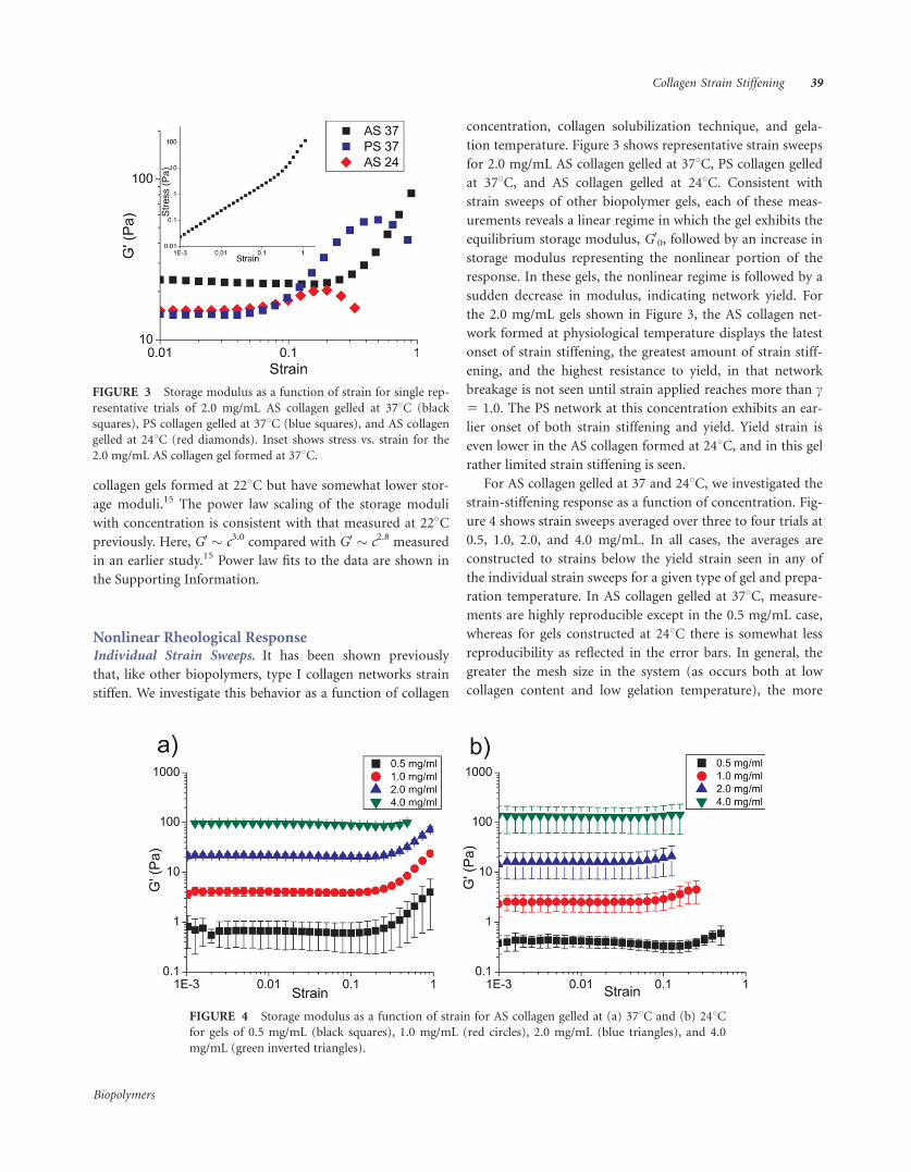
Nonlinear Rheological ResponseIndividual Strain Sweeps. It has been shown previously
that, like other biopolymers, type I collagen networks strain
stiffen. We investigate this behavior as a function of collagen
concentration, collagen solubilization technique, and gela-
tion temperature. Figure 3 shows representative strain sweeps
for 2.0 mg/mL AS collagen gelled at 378C, PS collagen gelled
at 378C, and AS collagen gelled at 248C. Consistent with
strain sweeps of other biopolymer gels, each of these meas-
urements reveals a linear regime in which the gel exhibits the
equilibrium storage modulus, G00, followed by an increase in
storage modulus representing the nonlinear portion of the
response. In these gels, the nonlinear regime is followed by a
sudden decrease in modulus, indicating network yield. For
the 2.0 mg/mL gels shown in Figure 3, the AS collagen net-
work formed at physiological temperature displays the latest
onset of strain stiffening, the greatest amount of strain stiff-
ening, and the highest resistance to yield, in that network
breakage is not seen until strain applied reaches more than c5 1.0. The PS network at this concentration exhibits an ear-
lier onset of both strain stiffening and yield. Yield strain is
even lower in the AS collagen formed at 248C, and in this gel
rather limited strain stiffening is seen.
For AS collagen gelled at 37 and 248C, we investigated the
strain-stiffening response as a function of concentration. Fig-
ure 4 shows strain sweeps averaged over three to four trials at
0.5, 1.0, 2.0, and 4.0 mg/mL. In all cases, the averages are
constructed to strains below the yield strain seen in any of
the individual strain sweeps for a given type of gel and prepa-
ration temperature. In AS collagen gelled at 378C, measure-
ments are highly reproducible except in the 0.5 mg/mL case,
whereas for gels constructed at 248C there is somewhat less
reproducibility as reflected in the error bars. In general, the
greater the mesh size in the system (as occurs both at low
collagen content and low gelation temperature), the more
FIGURE 3 Storage modulus as a function of strain for single rep-
resentative trials of 2.0 mg/mL AS collagen gelled at 378C (black
squares), PS collagen gelled at 378C (blue squares), and AS collagen
gelled at 248C (red diamonds). Inset shows stress vs. strain for the
2.0 mg/mL AS collagen gel formed at 378C.
FIGURE 4 Storage modulus as a function of strain for AS collagen gelled at (a) 378C and (b) 248Cfor gels of 0.5 mg/mL (black squares), 1.0 mg/mL (red circles), 2.0 mg/mL (blue triangles), and 4.0
mg/mL (green inverted triangles).
Collagen Strain Stiffening 39
Biopolymers

varied is network structure as well as linear and nonlinear
rheological behavior. In gels constructed at both 37 and
248C, the largest degree of strain stiffening relative to initial
modulus is seen at the lowest concentration and decreases as
concentration increases. At 4.0 mg/mL, there is a slight strain
weakening of the system before strain stiffening occurs.
Strain weakening has been noted previously in 4.0 mg/mL
AS collagen gelled at 378C, though in that case no eventual
stiffening was seen even at strains up to c 5 1.0.19 Strain
weakening followed by stiffening was recently seen by Kur-
niawan et al. in 3.5 mg/mL AS collagen formed at 378C.28
The decrease in degree of strain stiffening with increasing
collagen content is owing, at least in part, to the fact that
yield occurs at higher strains at the lower concentrations;
thus, at these concentrations the strain sweeps continue to
higher strains, allowing for enhanced stiffening. For a given
concentration, a gel formed at 378C tends to show greater
strain stiffening before yield than the same collagen content
gel formed at 248C.
The nonlinear rheological response of AS collagen gels as
well as that of 2.0 mg/mL PS collagen formed at 378C is
shown in Figure 5. Here, critical strain (cc), yield strain (cy),
and maximal modulus before yield relative to plateau modu-
lus (G0max/G0
0) are shown as a function of gel type, gelation
temperature, and collagen content. Critical strain increases as
a function of collagen content for AS collagen gelled at 378Cbut not for AS collagen gelled at 248C. In all cases except 0.5
mg/mL AS collagen, the gels formed at 378C display a higher
critical strain than those formed at 248C. The 2.0 mg/mL PS
collagen gelled at 378C exhibits a critical strain lower than
that of AS collagen at the same collagen content and gelation
temperature and very similar to that of 2.0 mg/mL AS colla-
gen gelled at 248C. Critical strain values are highly reproduci-
ble for 378C gels of 1.0 mg/mL and higher and somewhat less
reproducible for the other gels, which are less homogeneous
and display more diverse mechanical properties. Yield strain
shows somewhat different behavior than critical strain. For
AS collagen gelled at 37 and 248C, there is a decrease in yield
strain with increasing collagen content. As for critical strain,
PS collagen gelled at 378C exhibits yield strain behavior more
similar to AS collagen gelled at 248C than that gelled at 378C.
In part owing to differences in yield strain, there are also
differences in the maximum modulus achieved in the gels
relative to their plateau storage moduli (Figure 5c). The value
of G0max/G0
0 is greater in gels constructed at 378C than in the
gels constructed at lower temperature. For AS collagen gelled
at 248C, G0max/G0
0 is small and quite consistent across colla-
gen concentration. In these gels, the network yields when
strain stiffening of less than a factor of two has occurred. The
gels formed at 378C achieve more stiffening before yield,
with increase in storage modulus up to a factor of 10. Among
the gels constructed at physiological temperature, the least
stiffening is seen in the highest concentration gel, which dis-
plays an increase in modulus of a factor of approximately 2,
similar to that in the AS gel formed at lower temperature.
Collagen stiffened through use of the natural product gen-
ipin was also investigated rheologically. Genipin is a non-
zero-length crosslinker of collagen that has been shown to be
suitable for use in vivo.26,31,32 Figure 6 shows a strain sweep
of a representative 4.0 mg/mL AS collagen network gelled at
378C as well as a 4.0 mg/mL AS collagen gel formed with 10
mM genipin in the solution. Time between the beginning of
gelation and the measured strain sweeps is 30 min in both
cases. In the genipin-treated gel, additional stiffening contin-
ues to occur for many hours after gelation is complete; for
consistency with the other gels and to avoid difficulties of gel
drying, we performed rheological studies following 30 min
of gelation rather than upon ultimate stiffening. Though the
FIGURE 5 (a) Critical strain, (b) yield strain, and (c) storage modulus at yield strain normalized by
plateau storage modulus as a function of collagen concentration. In (a) averages and standard deviations
are over three to four runs. In all cases, black squares represent AS collagen gelled at 378C, the blue
square represents PS collagen gelled at 378C, and red diamonds represent AS collagen gelled at 248C.
40 Motte and Kaufman
Biopolymers

gel is stiffening over time, this stiffening is sufficiently slow
so that it does not significantly affect individual strain sweep
measurements, as can be appreciated from the fact that the
storage modulus does exhibit a plateau in the linear regime
of the strain sweep. The genipin-treated network exhibits a
plateau storage modulus that is greater than that of the
untreated gel by a factor of approximately 3, consistent with
the treatments of collagen at this concentration via the more
typical post-gelation approach.27 Differences are apparent
not only in the plateau modulus but also in the strain-stiffen-
ing behavior of the genipin-treated network. In the stiffened
network, cc 5 0.16, lower than that seen in any of the AS gels
formed at 378C and much lower than the cc 5 0.54 measured
in the untreated AS gel at this concentration. At c 5 0.5,
where the untreated gel has not even reached critical strain,
the genipin-stiffened gel has demonstrated an increase in
modulus of more than a factor of 4. Additionally, the gel
does not undergo the intermediate strain weakening seen in
the 4.0 mg/mL unstiffened collagen gel that precedes the
strain stiffening. The gel also remains intact at strains well
beyond the yield strain of the untreated 4.0 mg/mL AS gel.
Consecutive Strain Sweeps. In addition to single-trial strain
sweeps, consecutive strain sweeps were performed to investi-
gate network ability to recover after subjection to strains suf-
ficient to alter storage modulus. In these measurements,
strain was increased to c 5 1.0 for 0.5–2.0 mg/mL AS gels
and to c 5 0.5 for 4.0 mg/mL AS gels constructed at 378C.
These strains are three to four times the critical strain and
�80% of the yield strain for 0.5–2.0 mg/mL gels and are
close to both critical and yield strain for the 4.0 mg/mL gel,
where the values of critical and yield strain are relatively
small and similar to each other. For gels constructed at 248C,
the consecutive strain sweeps extend to c 5 0.5, 0.2, 0.2, and
0.15 for gels of 0.5, 1.0, 2.0, and 4.0 mg/mL, respectively. In
all cases, this is a factor of approximately 2 above critical
strain and just below yield strain except for the 4.0 mg/mL
gel where variability in yield strain requires only low levels of
strain to be applied to avoid breakage.
Figure 7 shows an example of five consecutive strain
sweeps for 1.0 mg/mL AS collagen gels formed at 37 and
248C. In all measurements of this type, the initial strain
sweep is consistent with single-strain-sweep measurements
for gels of given type, concentration, and preparation tem-
perature. For subsequent strain sweeps, behavior is very simi-
lar in AS gels formed at both temperatures. In the examples
shown in Figure 7, the second strain sweeps look similar to
the first except the values of G0 at all strains are lower. Critical
strain occurs at nearly the same point, and ultimate storage
FIGURE 6 Strain-sweep measurement of 4.0 mg/mL AS collagen
gelled at 378C (black squares) and 4.0 mg/mL AS collagen gelled at
378C stiffened with 10 mM genipin (red circles).
FIGURE 7 Five consecutive strain sweeps for 1.0 mg/mL AS collagen gelled at (a) 378C and (b)
248C.
Collagen Strain Stiffening 41
Biopolymers

modulus relative to plateau modulus is very similar in the
first and second sweeps. The third through fifth strain sweeps
show little additional change. The behavior of collagen gels
subjected to consecutive strain sweeps is shown in Figure 8.
In all AS gels investigated, decrease in storage modulus in the
linear regime between the first and the second strain sweeps
is similar and rather insensitive to collagen concentration or
gelation temperature (Figure 8a). Similarly, critical strain
value changes little with additional strain sweeps—all AS col-
lagen gels start to strain harden at approximately the same
strain independent of how many times the strain ramp has
been applied so long as yield strain has not been reached
(Figure 8b). Finally, modulus at particular strains in the non-
linear regime relative to plateau modulus does not vary with
subsequent strain sweeps and is similar in all AS gels tested
(Figure 8c). PS collagen was only investigated at 2.0 mg/mL.
It shows similar response to consecutive strain sweeps as AS
collagen but with a steeper drop in storage modulus between
the first and the subsequent sweeps. Consecutive strain
sweeps of genipin-stiffened collagen were not analyzed as the
continued stiffening occurring during the experiment com-
plicates interpretation of the data.
DISCUSSIONThe study presented here investigates structure, linear rheo-
logical behavior, and nonlinear rheological behavior of colla-
gen gels of different solubilization techniques, concentra-
tions, and preparation temperatures. These gels exhibit a
range of interactions: AS gels prepared at 378C have intrafi-
brillar crosslinks and interfibrillar entanglements, AS gels
formed at 248C have intrafibrillar crosslinks, interfibrillar
entanglements, and interfibrillar bundling, PS gels formed at
378C have only interfibrillar entanglements, and AS gels
treated with genipin are expected to have native intrafibrillar
crosslinking as well as additional crosslinking owing to geni-
pin activity. Although the degree of each type of crosslinking
has not been quantified, measurements on these systems can
suggest the relative importance of intra and interfibrillar
crosslinks on gel structure and mechanical properties,
including on strain-stiffening behavior.
CRM reveals that collagen fibril and network structure
vary as a function of collagen solubilization technique and
preparation conditions (Figure 1). Consistent with other
studies, we find PS collagen forms fibrils that are longer,
straighter, and more homogeneous than those of AS collagen
at the same concentration and gelation temperature.22,29 We
also find that AS collagen, unlike PS collagen, forms thick
fiber bundles at low gelation temperature. We hypothesize
that fibril bundling occurs in AS but not PS collagen formed
at low temperature because in AS collagen residual telopepti-
des are present along the outside of AS collagen fibrils, allow-
ing for interfibrillar associations; fibril interactions of this
type occur preferentially at low temperatures, where gelation
is slow, allowing for both larger nucleation centers and
increased chance of fibril encounters in advance of arrest of
the network.15,30
Although fibril microstructure is not investigated in this
study beyond that which is revealed via visual inspection of
CRM images, collagen microstructure may vary somewhat as
a function of solubilization technique, species and tissue
source, and gelation conditions.22,23,29,33 PS collagen is
FIGURE 8 (a) Plateau modulus of the second strain sweep relative to the first (G00(2)/G0
0), (b) criti-
cal strain of the fifth strain sweep relative to the first (cc(5)/cc), and (c) relative value of modulus in the
strain-stiffening region (at c 5 0.1 for PS collagen and AS collagen gelled at 248C and c 5 1.0 for AS
collagen gelled at 378C) of the fifth strain relative to the first strain (G0c(5)/G0
0(5))/(G0c/G
00), all as a
function of collagen concentration. In all cases, black squares represent AS collagen gelled at 378C, the
blue square represents PS collagen gelled at 378C, and the red diamonds represent AS collagen gelled at
248C.
42 Motte and Kaufman
Biopolymers

thought to be less well organized than AS collagen, though it
does generally retain the 67 nm banding seen in vivo.34,35
Additionally, collagen’s microstructure is dependent on gela-
tion temperature, and AS collagen at 378C is expected to
have somewhat less regular banding and thinner individual
fibrils than gels constructed at lower temperature.15,36,37
Genipin-stiffened AS collagen has been shown to have a sig-
nificant proportion of fibrils with non-native banding. Addi-
tionally, nonfibrillar aggregates have been seen in these prep-
arations.26 Both factors may contribute to the lower signal:-
noise seen in these gels relative to those formed in the
absence of genipin (Figure 1e and inset).
Just as the collagen network structure measured in this
study is largely consistent with previous measurements, the
linear and nonlinear rheology of collagen described here is
consistent with previous findings. First, the measured values
of plateau storage and loss moduli are similar to those ascer-
tained via earlier shear and microrheological measurements
in both AS and PS collagens.4,21,22,30,33,38 In our measure-
ments on AS collagen, we find as G0 � c2.2, similar to that
measured previously by us in AS and PS collagens (G0 �c2.1).15,30 AS collagen gels formed at 248C display qualita-
tively similar structure to AS collagen gels formed at 228C,
but they have somewhat lower storage moduli.15 This differ-
ence may be due to the higher temperature of gelation, which
results in less fibril bundling than in the gels constructed at
the lower temperature. Despite small differences in moduli
values, the power law scaling of the storage moduli with con-
centration is consistent with that measured at 228C previ-
ously. Here, G0 � c3.0 compared to G0 � c2.8 measured previ-
ously.15 Similar studies of PS collagen formed at 378C have
yielded G0 � c2.1 and G0 � c2.7.6,30
The general strain-stiffening behavior measured here is
similar to that seen in other studies of biopolymer gels.1–
4,6,19,21,22,28 Although our findings are largely in accordance
with the previous measurements on collagen I gels, some dif-
ferences are seen. In large amplitude oscillatory shear meas-
urements similar to those performed here, AS collagen was
found to strain stiffen and exhibit critical strain at cc 5
0.20–0.30 for concentrations up to 2 mg/mL but to strain
weaken at 4.0 mg/mL.19 For AS collagen gels constructed at
378C, we find somewhat higher critical strains (cc 5 0.24–
0.54), and we find strain stiffening even in the highest con-
centration gel investigated. This is consistent with the recent
study of Kurniawan et al. on 3.5 mg/mL AS collagen, a study
which also reveals very similar Lissajous curves to those we
measure (Supporting Information Figure 1) and similar
response to repeated application of strain.28 Another study
found critical strain in PS collagen to be in the range of cc 5
0.05–0.15 for gels of 0.5–5.0 mg/mL.4 These measurements
are in accordance with our finding of cc 5 0.10 for PS colla-
gen of 2.0 mg/mL.
Yield strain has also been investigated previously: In con-
tinuous shear measurements, AS collagen was found to have
a gap-dependent yield strain that increased with decreasing
gap size.21 In that study, regardless of gap, yield strain
decreased with increasing collagen content from 1.0 to 3.0
mg/mL.21 For the smallest gap measured (50 lm), cy � 1.20
at 1.0 mg/mL and cy � 0.45 at 3.0 mg/mL. This is consistent
with our measurements of cy � 1.60 at 1.0 mg/mL and cy 5
0.60 at 4.0 mg/mL for an even smaller gap, though direct
comparison is complicated by the fact that that study used a
parallel plate tool while we employed a cone and plate. AS
and PS collagen gel yield has also been measured using uni-
axial testing, which revealed no clear difference between AS
and PS collagen and no clear trend with concentration from
0.5 to 2.0 mg/mL.22 In that study, across collagen types and
concentrations, yield strains were in the range of cy 5 0.40–
0.60, somewhat higher than those found in oscillatory rheol-
ogy done with the largest gaps (300 lm).21,22 Finally, strain-
stiffening behavior in collagen gels treated with genipin,
which has not been measured previously, displays some simi-
larities to collagen stiffened with other crosslinkers. In PS
collagen stiffened with glutaraldehyde, critical strain meas-
ured with two-point stretching was found to be cc 5 0.10.4
This critical strain was generally lower than that measured
for collagen without glutaraldehyde as determined via either
two-point stretching or oscillatory rheology.4 The finding of
earlier onset of strain stiffening for glutaraldehyde-treated
collagen relative to native collagen is consistent with our
finding on genipin-treated AS collagen.
Comparison of strain-stiffening behavior across the differ-
ent types of collagen gels investigated here allows us to specu-
late on the relative importance of fibrillar structure, network
structure, and crosslinking in setting nonlinear rheological
behavior. Trends found in this study include that AS collagen
gels formed at 378C display increasing critical strain with
increasing concentration, with critical strains in the range of
cc 5 0.24–0.54 for collagen of 0.5–4.0 mg/mL. Significantly
lower critical strains are found in gels of the same type and
collagen content formed at low temperature, where fibril
bundling and large network pore size exist. Yield strain is
also much higher in the AS gels formed at 378C than at 248C,
as is the ultimate modulus attained in the 378C gels relative
to the 248C gels of the same concentration. Additionally, we
find that PS gels act more similarly to AS gels constructed at
248C than to those constructed at 378C in terms of their crit-
ical and yield strain.
The key difference between AS collagen gels of a given
concentration formed at 37 and 248C is network
Collagen Strain Stiffening 43
Biopolymers

organization, with AS gels formed at 248C exhibiting fibril
bundling that leads to a network of thick struts and large
pores, as opposed to a network of thinner struts and smaller
pores as is seen for AS gels of the same collagen content
formed at 378C. The key difference between AS and PS colla-
gen gels formed at the same temperature is less clear: both
types of networks are composed of thin struts and relatively
small pores but some network structural differences are evi-
dent (Figure 1); moreover, intrafibrillar interactions are
expected to differ between the two preparations with AS col-
lagen fibrils bearing some intrafibrillar crosslinking. We note
that the AS gels formed at 378C, the AS gels formed at 248C,
and the PS gels of a given collagen content have very similar
linear rheological response (Figure 2) but distinct nonlinear
mechanical responses. We suggest that differences in nonlin-
ear rheological response between AS gels formed at 37 and
248C are caused by differences in network organization,
whereas differences in response between AS and PS gels may
be owing to the differences in intrafibrillar interactions or
network organization.
The clear differences in critical and yield strain for AS col-
lagen gels of a particular concentration constructed at 37 and
248C are consistent with the nonlinear mechanical response
of these systems being very sensitive to network architecture.
For gels of similar linear elastic storage and loss modulus,
those gels with thin unbundled fibrils and relatively small
pore size exhibit higher critical strains, higher yield strains,
and a greater degree of strain stiffening than do those gels
with bundled fibers and larger pore size. Lower critical strain
in gels with large pore size and relatively few total fibrils or
fibers (i.e., gels of low concentration and/or low gelation
temperature) is consistent with strain stiffening owing to
nonaffine deformations: such deformations are more likely
in open, inhomogeneous systems, where few fibrils and
entanglement points are present. The more fibrils and entan-
glement points, the more resistant the gel is to the substantial
deformations that lead to this type of enthalpic strain stiffen-
ing. The fact that PS collagen, with much more similar net-
work organization to AS collagen of the same concentration,
also exhibits low critical strain suggests this property can also
emerge from a second mechanism associated with lack of
telopeptides. Although telopeptides are primarily thought to
lead to intrafibrillar crosslinks, we have suggested that they
may also reinforce entanglement points; this is consistent
with the presence of bundles in low-temperature AS collagen
gels but not low-temperature PS collagen gels.29 If this is the
case, PS collagen of the same network organization as AS col-
lagen would be more likely to rearrange under shear and thus
would be expected to strain stiffen at a lower strain than does
AS collagen. The fact that strain stiffening sets in earlier in
the high-concentration genipin-stiffened AS gel relative to
the untreated one is not immediately consistent with the
view of strain stiffening being dominated by athermal nonaf-
fine deformations. Indeed, one would expect gels reinforced
with crosslinks at entanglement points to be more resistant
to the types of nonaffine deformations that we suspect lead
to strain stiffening in most AS collagen gels.20,39 Genipin,
however, effects many changes in the collagen network and
may even limit the proportion of collagen molecules present
in fibrils, complicating interpretation of the results.26
The fact that we find a decrease in yield strain with
increasing collagen concentration as occurs in AS gels con-
structed at both temperatures 37 and 248C is not intuitive, as
it may be expected that with fewer fibrils failure of any given
fibril or interfibrillar entanglement or crosslink would be
more likely to disrupt the spanning structure, breaking the
gel. However, measurement of decreased yield strain with
increasing concentration is in accordance with the other
measurement of yield strain as a function of collagen concen-
tration as well as similar measurements in actin gels.21,39
Indeed, Gardel et al. showed that for entropic elasticity, it is
expected that yield strain will decrease weakly with number
of fibrils for a fixed concentration of crosslinks.39 Although
some features of the elasticity of collagen networks have been
shown to be consistent with entropic elasticity,15 the rela-
tively stiff fibrils of collagen make elasticity emerging solely
from thermal, entropic sources unlikely in these networks.
Similarly, comparison to actin results is complicated by the
fact that in collagen networks crosslink density varies with
collagen content. Although the origin of the decreasing yield
strain with increasing concentration may differ for collagen
and actin, the observation is similar.
The response of the gels to consecutive strain sweeps also
sheds light on the origins of strain stiffening in these gels. So
long as yield strain is not reached, changes to AS collagen
gels’ mechanical properties induced by high strains are gener-
ally found to be reversible. Although there is some drop in
storage modulus after the initial strain sweep, no further
changes are seen with subsequent sweeps, and critical strain
and modulus at a given strain relative to that in the initial
strain sweep remain constant for gels of all concentrations
and gelation temperatures (Figure 8). These results are con-
sistent with strain-stiffening behavior being largely set by re-
versible nonaffine deformations of the network. The initial
drop in modulus after the first strain sweep may be owing to
irreversible deformations of individual fibrils. This effect is
greatest in PS collagen, where the modulus decreases to
approximately half of the original modulus after the first
strain sweep, suggesting that the telopeptides in the AS colla-
gen that allow for intrafibrillar crosslinking limit irreversible
44 Motte and Kaufman
Biopolymers

changes to individual fibrils. The fact that subsequent strain
sweeps in AS and PS collagen are consistent with the previ-
ous strain sweeps in terms of onset and degree of strain stiff-
ening suggests the majority of the strain hardening is owing
to reversible network reorganization. The suggestion of re-
versible deformations is inconsistent with the direct micros-
copy performed during two-point stretching of PS collagen
gels performed by Vader et al.4 In this study, permanent
alignment was seen after strains in the nonlinear regime were
imposed on untreated PS gels through reversible alignment
was seen in glutaraldehyde-treated samples.4 On the other
hand, Tower et al. found that prealigned untreated PS colla-
gen matrices do exhibit reversible alignment in both cross
and parallel-loaded uniaxial tests.16 Differences in the details
of the sample configuration as well as the particular stresses
applied may influence the reversibility of network deforma-
tions in these two studies as well as in the study presented
here.
The picture beginning to emerge is that of initial strain
stiffening occurring from small individual fibril stretching
deformations followed by more substantial stiffening emerg-
ing from nonaffine (potentially reversible) fiber rearrange-
ments. This is consistent with a recent finding tracking bire-
fringence and storage modulus of fibrin gels under strain,
which showed that initial strain stiffening proceeds align-
ment.5 Although both entropic and enthalpic contributions
likely play a role in collagen strain stiffening, their relative
importance likely depends sensitively on the degree of orga-
nization and strength of the individual fibrils as well as the
interfibrillar interactions and initial network organization.
CONCLUSIONSCollagen gels of different solubilization technique and gela-
tion temperature were prepared: these gels display similar
storage moduli in the linear regime but exhibit different me-
chanical responses in the nonlinear regime. Those gels formed
from monomers with telopeptides exhibit higher critical and
yield strains. Similarly, gels prepared at physiological tempera-
ture, where the network is organized as a relatively dense set
of thin fibrils, display higher critical and yield strains than the
gels constructed at low temperature, where fibril bundles
dominate the network architecture. These results suggest that
the difference in strain-stiffening behavior is owing largely to
differences in network architecture. We speculate that initial
strain stiffening at low strains emerges from stretching defor-
mations of individual fibrils and that these deformations are
most prominent in gels with thin fibrils, no crosslinking, and/
or poor individual fibril organization. The remaining strain
stiffening is likely due to nonaffine deformations of the
network including reversible fibril alignment. The fact that
collagen I gels with very similar linear storage and loss moduli
may have significantly different nonlinear mechanical
responses in terms of critical strain, yield strain, and degree of
strain stiffening is an important consideration for applica-
tions requiring recapitulation of native tissue response to
both cell-driven and external strains.
L. J. K. thanks Hector Acaron and Jieling Zhu for assistance in tur-
bidity studies. L. J. K. dedicates this paper to the memory of S.M.
REFERENCES1. Janmey, P. A.; Euteneuer, U.; Traub, P.; Schliwa, M. J Cell Biol
1991, 113, 155–160.
2. Storm, C.; Pastore, J. J.; MacKintosh, F. C.; Lubensky, T. C.; Jan-
mey, P. A. Nature 2005, 435, 191–194.
3. Janmey, P. A.; McCormick, M. E.; Rammensee, S.; Leight, J. L.;
Georges, P. C.; Mackintosh, F. C. Nat Mater 2007, 6, 48–51.
4. Vader, D.; Kabla, A.; Weitz, D.; Mahadevan, L. PLOS One 2009,
4, 5902.
5. Kang, H.; Wen, Q.; Janmey, P. A.; Tang, J. X.; Conti, E.; MacKin-
tosh, F. C. J Phys Chem B 2009, 113, 3799–3805.
6. Stein, A. M.; Vader, D. A.; Weitz, D. A.; Sander, L. M. Complex-
ity 2011, 16, 22–28.
7. Onck, P. R.; Koeman, T.; van Dillen, T.; van der Giessen, E. Phys
Rev Lett 2005, 95, 178102.
8. Heussinger, C.; Frey, E. Phys Rev Lett 2006, 97, 105501.
9. Heussinger, C.; Frey, E. Phys Rev Lett 2006, 96, 017802.
10. Heussinger, C.; Schaefer, B.; Frey, E. Phys Rev E 2007, 76,
031906.
11. Wyart, M.; Liang, H.; Kabla, A.; Mahadevan, L. Phys Rev Lett
2008, 101, 215501.
12. Broedersz, C. P.; Sheinman, M.; MacKintosh, F. C. Phys Rev
Lett 2012, 108, 078102.
13. Chen, D. T. N.; Wen, Q.; Janmey, P. A.; Crocker, J. C.; Yodh, A.
G. Ann Rev Cond Matter Phys 2010, 301–322.
14. Mackintosh, F. C.; Kas, J.; Janmey, P. A. Phys Rev Lett 1995, 75,
4425–4428.
15. Yang, Y. L.; Leone, L. M.; Kaufman, L. J. Biophys J 2009, 97,
2051–2060.
16. Tower, T. T.; Neidert, M. R.; Tranquillo, R. T. Ann Biomed Eng
2002, 30, 1221–1233.
17. Roeder, B. A.; Kokini, K.; Sturgis, J. E.; Robinson, J. P.; Voytik-
Harbin, S. L. J Biomech Eng Trans ASME 2002, 124, 214–222.
18. Roeder, B. A.; Kokini, K.; Voytik-Harbin, S. L. J Biomech Eng
Trans ASME 2009, 131, 031004.
19. Piechocka, I. K.; van Oosten, A. S. G.; Breuls, R. G. M.; Koen-
derink, G. H. Biomacromolecules 2011, 12, 2797–2805.
20. Arevalo, R. C.; Urbach, J. S.; Blair, D. L. Chaos 2011, 21, 041102.
21. Arevalo, R. C.; Urbach, J. S.; Blair, D. L. Biophys J 2010, 99,
L65–L67.
22. Kreger, S. T.; Bell, B. J.; Bailey, J.; Stites, E.; Kuske, J.; Waisner,
B.; Voytik-Harbin, S. L. Biopolymers 2010, 93, 690–707.
23. Kuznetsova, N.; Leikin, S. J Biol Chem 1999, 274, 36083–36088.
24. Bailey, J. L.; Critser, P. J.; Whittington, C.; Kuske, J. L.; Yoder, M.
C.; Voytik-Harbin, S. L. Biopolymers 2010, 95, 77–93.
Collagen Strain Stiffening 45
Biopolymers

25. Zeugolis, D. I.; Paul, R. G.; Attenburrow, G. J Biomed Mater Res
A 2008, 86A, 892–904.
26. Hwang, Y.-J.; Larsen, J.; Krasieva, T. B.; Lyubovitsky, J. G. ACS
Appl Mater Interfaces 2011, 3, 2579–2584.
27. Sundararaghavan, H. G.; Monteiro, G. A.; Lapin, N. A.; Chabal,
Y. J.; Miksan, J. R.; Shreiber, D. I. J Biomed Mater Res A 2008,
87A, 308–320.
28. Kurniawan, N. A.; Wong, L. H.; Rajagopalan, R. Biomacromole-
cules, 13, 691–698.
29. Yang, Y. L.; Sun, C.; Wilhelm, M. E.; Fox, L. J.; Zhu, J. L.; Kauf-
man, L. J. Biomaterials 2011, 32, 7932–7940.
30. Yang, Y. L.; Kaufman, L. J. Biophys J 2009, 96, 1566–1585.
31. Huang, L. L. H.; Sung, H. W.; Tsai, C. C.; Huang, D. M. J
Biomed Mater Res 1998, 42, 568–576.
32. Sung, H. W.; Liang, I. L.; Chen, C. N.; Huang, R. N.; Liang, H.
F. J Biomed Mater Res 2001, 55, 538–546.
33. Raub, C. B.; Suresh, V.; Krasieva, T.; Lyubovitsky, J.; Mih, J. D.;
Putnam, A. J.; Tromberg, B. J.; George, S. C. Biophys J 2007, 92,
2212–2222.
34. Comper, W. D.; Veis, A. Biopolymers 1977, 16, 2113–2131.
35. Walton, R. S.; Brand, D. D.; Czernuszka, J. T. J Mater Sci Mater
Med 2010, 21, 451–461.
36. Wood, G. C.; Keech, M. K. Biochem J 1960, 75, 588–598.
37. Leikina, E.; Mertts, M. V.; Kuznetsova, N.; Leikin, S. Proc Natl
Acad Sci USA 2002, 99, 1314–1318.
38. Velegol, D.; Lanni, F. Biophys J 2001, 81, 1786–1792.
39. Gardel, M. L.; Shin, J. H.; MacKintosh, F. C.; Mahadevan, L.;
Matsudaira, P.; Weitz, D. A. Science 2004, 304, 1301–1305.
Reviewing Editor: Eric J. Toone
46 Motte and Kaufman
Biopolymers