stodieck et al. (2014) brief dark exposure restored ocular dominance plasticity in aging mice and...
TRANSCRIPT
Experimental Gerontology 60 (2014) 1–11
Contents lists available at ScienceDirect
Experimental Gerontology
j ourna l homepage: www.e lsev ie r .com/ locate /expgero
Brief dark exposure restored ocular dominance plasticity in aging miceand after a cortical stroke
Sophia Katharina Stodieck a,b, Franziska Greifzu b,⁎, Bianka Goetze b, Karl-Friedrich Schmidt b, Siegrid Löwel b,c
a Göttingen Graduate School for Neurosciences, Biophysics, and Molecular Biosciences, Justus-von-Liebig-Weg 11, D-37077 Göttingen, Germanyb Bernstein Fokus Neurotechnologie (BFNT) and Johann-Friedrich-Blumenbach Institut für Zoologie und Anthropologie, Universität Göttingen, Von-Siebold-Str. 6, D-37075 Göttingen, Germanyc Sensory Collaborative Research Center 889, University of Göttingen, D-37075 Göttingen, Germany
Abbreviations:DE,darkexposure;EE,enrichedenvironmoculardeprivation;OD,oculardominance;ODI,oculardomPNN,perineuronalnet; PT, photothrombosis; PV,parvalbumcortex; SC, standard cage; V1, primary visual cortex.⁎ Corresponding author.
E-mail addresses: [email protected] (S.K. Stodieck),[email protected] (F. Greifzu),[email protected] (B. Goetze), kfs(K.-F. Schmidt), [email protected] (S. Löwel).
http://dx.doi.org/10.1016/j.exger.2014.09.0070531-5565/© 2014 The Authors. Published by Elsevier Inc(http://creativecommons.org/licenses/by-nc-nd/3.0/).
a b s t r a c t
a r t i c l e i n f oArticle history:Received 28 May 2014Received in revised form 31 July 2014Accepted 11 September 2014Available online 16 September 2014
Section Editor: Christian Humpel
Keywords:Visual cortexDark exposureOcular dominance plasticityMonocular deprivationOptical imagingPhotothrombosisIntracortical inhibitionAging
In the primary visual cortex (V1), monocular deprivation (MD) induces a shift in the ocular dominance (OD) ofbinocular neurons towards the open eye (Wiesel and Hubel, 1963; Gordon and Stryker, 1996). In V1 of C57Bl/6Jmice, this OD-plasticity is maximal in juveniles, declines in adults and is absent beyond postnatal day (PD) 110(Lehmann and Löwel, 2008) if mice are raised in standard cages. Since it was recently shown that brief dark ex-posure (DE) restored OD-plasticity in young adult rats (PD70-100) (He et al., 2006), we wondered whether DEwould restore OD-plasticity also in adult and old mice and after a cortical stroke. To this end, we raised mice instandard cages until adulthood and transferred them to a darkroom for 10–14 days. Using intrinsic signal opticalimaging we demonstrate that short-term DE can restore OD-plasticity after MD in both adult (PD138) and oldmice (PD535), and that OD-shifts were mediated by an increase of open eye responses in V1. Interestingly, re-stored OD-plasticity after DE was accompanied by a reduction of both parvalbumin expressing cells andperineuronal nets and was prevented by increasing intracortical inhibition with diazepam. DE also maintainedOD-plasticity in adult mice (PD150) after a stroke in the primary somatosensory cortex. In contrast, short-termDE did not affect basic visual parameters asmeasured by optomotry. In conclusion, short-termDEwas able to re-store OD-plasticity in both adult and aging mice and even preserved plasticity after a cortical stroke, most likelymediated by reducing intracortical inhibition.
© 2014 The Authors. Published by Elsevier Inc. This is an open access article under the CC BY-NC-ND license(http://creativecommons.org/licenses/by-nc-nd/3.0/).
1. Introduction
Rodent V1 is dominated by input from the contralateral eye (Dräger,1975, 1978) but MD can induce a shift in the OD of binocular neurons to-wards the open eye (Wiesel and Hubel, 1963; Gordon and Stryker, 1996;Cang et al., 2005a). In V1 of C57Bl/6Jmice, this OD-plasticity ismaximal at4 weeks of age, declines in 2–3 month old animals and is absent beyondPD110 (Espinosa and Stryker, 2012; Levelt and Hübener, 2012), evenafter longer deprivation times (Greifzu et al., 2012), if animals are raisedin standard cages (SC). In critical period mice (PD19-32), 4 days of MDare sufficient to induce OD-shifts towards the open eye (Gordon andStryker, 1996). This “juvenile” OD-shift is predominantly mediated by adecrease in the cortical responses to visual stimulation of the deprived
ent;LR, lightreared;MD,mon-inance index;PD,postnatal day;in; S1, primary somatosensory
. This is an open access article under
eye. In contrast, in 2–3 month old SC-raised mice, significant OD-shiftsneed 7 days of MD and are primarily mediated by increased open eye re-sponses (“adult” OD-plasticity) (Sawtell et al., 2003; Hofer et al., 2006;Heimel et al., 2007; Sato and Stryker, 2008). It has been shown that thebalance between excitation and inhibition is important for regulatingplasticity in V1: during postnatal development, the maturation ofGABAergic (γ-aminobutyric acid) circuitry opens the critical period forOD-plasticity (Huang et al., 1999, 2010; Rozas et al., 2001) and raisingmice in an enriched environment (EE) both preserves a juvenile inhibito-ry tone and juvenile-like OD-plasticity into adulthood (Greifzu et al.,2014). While dark-rearing has been extensively studied (Hooks andChen, 2007; Berardi et al., 2000), the effects of short periods of dark expo-sure (DE) during adulthood are less well understood. Recently, it wasshown that 10 days of DE in PD70-100 SC-raised rats reactivated OD-plasticity in V1, reduced the level of GABAA receptors relative to AMPA(α-amino-3-hydroxy-5-methyl-4-isoxazole-propionic acid) receptorsand caused a return to the juvenile form of NMDA (N-methyl-D-aspar-tate) receptors (He et al., 2006). We therefore wondered whethershort-term DE would restore OD-plasticity in adult and old mice alreadybeyond their sensitive phase for OD-plasticity and would also pre-serve OD-plasticity after a cortical stroke. Testing not only youngadult but also old animals is rather important since there are
the CC BY-NC-ND license

2 S.K. Stodieck et al. / Experimental Gerontology 60 (2014) 1–11
numerous age-dependent changes of cortical circuitry such as reducedfunctional selectivity and signal-to-noise ratio.
Thus a possible therapeutic strategy that increases cortical plasticityin young adult animals must not necessarily be effective during old age.We therefore tested the effect of brief DE on both adult and old mice.Using optical imaging of intrinsic signals, we show that 14 days of DEreactivated OD-plasticity in 4–6 month old mice after 4 and 7 days ofMD. The restored OD-shift was mediated by a significant increase ofopen eye responses in V1 and prevented by diazepam injections. In addi-tion, we observed a significant reduction of the number of parvalbuminexpressing (PV+) cells and perineuronal nets (PNNs) in the visual cortexof DE-mice compared to light-reared (LR) controls. Fourteen days of DEwere also sufficient to restore OD-plasticity in old mice (PD535) after7 days of MD. Finally, brief DE in adulthood (PD150) also maintainedOD-plasticity after a stroke in the primary somatosensory cortex (S1).Short-term DE during adulthood did neither impair basic visual acuitynor the experience-dependent increase of visual acuity after MD asmeasured by optomotry (Prusky et al., 2004). Taken together, our datastrongly suggest short-term DE as a highly efficient therapeutic interven-tion to restore plasticity in both adult and oldmice and even after a corti-cal lesion, and that the DE-effect is most likely mediated via reducedintracortical inhibition.
2. Materials and methods
2.1. Animal treatment
All experimental procedures were approved by the local govern-ment (Niedersächsisches Landesamt für Verbraucherschutz undLebensmittelsicherheit, registration number 33.9-42502-04-10/0326).Bothmale and female C57BL/6Jmice from themouse colony of the centralanimal facility of the University Medical Center, Göttingen (groups I–III,V–VII, see next paragraph) and 10 C57BL/6JRccmice from the Harlan lab-oratories (Netherlands; group IV) were raised in SCs on a 12 h light/darkcycle (LR), with food and water available ad libitum.
2.2. Dark exposure
For short-term visual deprivation (dark exposure = DE) animalswere housed in SCs placed into a scantainer (Scanbur Technology,Denmark) in a completely light-tight darkroom for 10 or 14 days.
We investigated a total of 7 different experimental groups: I) adultPD138 mice (age range PD125-157; average: PD138) received either4 days or II) 7 days of MD after 14 days of DE; III) old PD535 mice(PD517-564) received 7 days of MD after 14 days of DE; IV) diazepamor saline-treated adult mice (PD151-157; average: PD154) received7 days of MD after 14 days of DE as well as daily injections ofdiazepam/saline during the MD period; V) Chronically imaged adultmice: imaging was performed before (PD116-123; average: PD118)and after 14 days of DE and 7 days of MD (PD141-150; average:PD145); VI) young adult mice (PD57-80; average: PD72) received4 days of MD after 10 days of DE; VII) Adult mice (PD135-165; average:PD150) received a photothrombotic (PT) lesion in S1 (sham-surgery forcontrols) and7 days ofMD after 14days of DE. Experimental groups alsocontained equally treated light-reared (LR) controlmice (groups I, II, VI)and control animals without MD (groups I, II, III, VI, VII).
2.3. Optomotry
To test whether short-term DE modified basic visual abilities, thespatial frequency threshold ("visual acuity") and contrast sensitivity ofthe optomotor reflex of animals from all experimental groups (exceptgroups IV and V) were determined using the virtual-reality optomotorsystem (Prusky et al., 2004). Briefly, freely moving animals were ex-posed to moving vertical sine wave gratings of various spatial frequen-cies and contrasts and will reflexively track the gratings by head
movements as long as they can see them. Spatial frequency at full con-trast and contrast at six different spatial frequencies [0.031, 0.064, 0.092,0.103, 0.192, 0.272 cycles/degree (cyc/deg)] were varied by the experi-menter until the threshold of tracking was determined.
2.4. Monocular deprivation (MD)
According to already published protocols, we always sutured theright eye of our mice (Gordon and Stryker, 1996; Cang et al., 2005a;Lehmann and Löwel, 2008) to trigger visual plasticity. DE-mice weretransported (immediately after DE) in a light-tight box to the surgicalsuite and MD-surgery started directly to minimize light exposure afterDE. After box-anesthesia with 2% isofluorane in a 1:1 mixture of nitrousoxide (N2O) and oxygen (O2), anesthesia was maintained with 1.5%isoflurane, lid margins were trimmed and an antibiotic gel (gentamicingel) was applied. The eye was closed with two mattress sutures. AfterMD, the mice were returned to their standard home cages and stayedin normal 12 h light/dark conditions. Animals were checked daily tomake sure that the eye remained closed.
2.5. Surgical preparations for optical imaging (Kalatsky and Stryker, 2003;Cang et al., 2005a)
Mice were initially box-anesthetized with 2% halothane in a 1:1mixture of O2:N2O and received atropine (Franz Köhler, 0.3 mg, sub-cutaneously (s.c.)), dexamethasone (Ratiopharm, 0.2 mg, s.c.), andchlorprothixene (Sigma, 0.2 mg, intramuscularly). Mice were placed ina stereotaxic frame and anesthesia was maintained with 0.6–0.8% halo-thane in a 1:1mixture of O2:N2O applied through a tube attached to thenose. Body temperaturewasmaintained at 37 °C and the heart ratewasmonitored throughout the experiment. Lidocaine (2% xylocaine jelly)was applied locally to all incisions. The skin above the skull was incisedto expose V1 of the left hemisphere and agarose (2.5% in 0.9% NaCl) anda glass cover-slip were placed over the exposed area. Imaging was per-formed through the skull. Mouse visual cortical responses were record-ed using the imagingmethod developed by Kalatsky and Stryker (2003)and optimized for the assessment of OD-plasticity (Cang et al., 2005a).In this method, a temporally periodic stimulus is continuously present-ed to the animal and the cortical responses at the stimulus frequency areextracted by Fourier analysis. Optical images of intrinsic cortical signalswere obtained using a CCD (charge-coupled device) camera (Dalsa)controlled by custom software. The surface vascular pattern and intrin-sic signal imageswere visualizedwith illuminationwavelengths set by agreen (550 ± 10 nm) or red (610 ± 10 nm) interference filter, respec-tively. After acquisition of a surface image, the camera was focused600 μm below the cortical surface. An additional red filter wasinterposed between the brain and the CCD camera. Frames were ac-quired at a rate of 30 Hz, temporally binned to 7.5 Hz and stored as512 × 512 pixel images after spatial binning of the camera image.Drifting horizontal bars (2°wide) were presented to the animal at a dis-tance of 25 cm on a high refresh-rate monitor. The distance between2 bars was 80° and they were presented at a temporal frequency of0.125 Hz. The visual stimulus was restricted to the binocular visualfield of the left V1 (−5° to+15° azimuth) and animalswere stimulatedthrough either the left or the right eye in alternation to assess the OD ofthe left hemisphere. To assess map quality the stimulus monitor wasplaced in the right visual field of the animal at a distance of 25 cmwith its left edge approximately aligned to the animal to optimallystimulate the right eye (contralateral to the recorded hemisphere). Thedrifting bars (elevation or azimuth) were shown across the full screen.
2.6. Data analysis
Visual cortical maps were calculated from the acquired frames byFourier analysis to extract the signal at the stimulation frequencyusing custom software (Kalatsky and Stryker, 2003). While the phase
3S.K. Stodieck et al. / Experimental Gerontology 60 (2014) 1–11
component of the signal is used for the calculation of retinotopy, theamplitude component represents the intensity of neuronal activation(expressed as fractional change in reflectance × 10−4) and was usedto calculate OD (for details, see Cang et al. (2005a)). Briefly, we calculat-ed theOD-score of each pixel in the binocularly activated region in V1 as(C− I)/(C+ I), with C and I representing the raw responsemagnitudesof each pixel to visual stimulation of the contralateral and ipsilateral eye,respectively. We then calculated an OD-index (ODI) as the averageof the OD-scores of all responsive pixels. This ODI ranged from −1to +1, with negative values representing ipsilateral and positive valuesrepresenting contralateral dominance. We obtained at least three ODIsper animal; experiments with fewer than three ODIs were discardedfrom further analyses. All activity and phase maps of one animal wereaveraged for further quantification and data display. The quality of theretinotopic maps was assessed by the calculation introduced by Canget al. (2005b). Briefly, the 20,000 pixels that had the greatest responsemagnitude were selected. For each of these pixels, the difference be-tween its position and the mean position of its surrounding 24 pixelswas calculated. For maps of high quality, the position differences arequite small because of smooth progression. The standard deviation ofthe position difference was then used as an index of the quality ofretinotopic maps with a small standard deviation indicating high mapquality and high values indicating low map quality.
2.7. Diazepam/saline administration
To increase GABAergic inhibitionwe applied diazepam (Rotexmedica,intraperitoneally, i.p.), an allosteric GABAA receptor modulator, in oneadditional experimental group. Adult DE-mice (PD151-157; average:PD154) received either diazepam (0.25 μg/μl diluted in 0.9% NaCl; 1 mgper kgmouse) or as control saline (0.9%NaCl) 7 h prior toMDand contin-ued for 6 days during MDwith one injection per day. The applied dosageof diazepam allowed the normal activity and exploratory behavior of theanimals.
2.8. Induction of a photothrombotic stroke
A PT-lesion was induced in the left S1 neighboring V1 by using theRose Bengal technique introduced by Watson et al. (1985). Briefly,mice (PD150) were initially box-anesthetized with 2% isoflurane, andanesthesia was maintained with 1% isoflurane in 1:1 O2:N2O. Theanimals were placed in a stereotaxic frame and body temperature wasmaintained at 37 °C. The skin above the skull was incised and an opticfiber bundle (aperture: 1.0mm)mounted on a cold light source (SchottKL 1500)was positioned 2mm lateral to themidline and 1mmposteriorto the bregma. Next, 100 μl Rose Bengal (Sigma-Aldrich; 10 mg/ml in 0.9%NaCl)was injected into the tail vein. Afterwaiting for 5min, the illumina-tionperiod of 15minwith the cold light sourcewas performed. All controlanimals were treated identically but the light sourcewas not switched on(sham-treatment). Finally, the skin was sutured and the animals recov-ered in their home cages. For the DE-group, mice were transferred tothe darkroom for 14 days immediately after waking up from anesthesia.Directly after DE, the right eye was deprived in half of the animals (seeabove) and the mice were transferred to their home cage in a normallylit room for the following 7 days. Finally, we imaged V1-activity in bothdeprived and non-deprived animals.
2.9. Perfusion and tissue processing
After optical imaging, all mice were deeply anesthetized with anoverdose of 0.3 ml 30% chloral hydrate (i.p.) and were perfusedtranscardially with 1% heparin in 0.9% NaCl for 2 min followed by 4%paraformaldehyde (PFA, pH 7.4) for 3 min. The brain was removedand postfixed in 4% PFA (pH 7.4) for one day and then transferred to acryoprotectant solution (10% sucrose, 20% glycerol). The brains were
frozen in methylbutane and stored at −80 °C. Coronal 40 μm thickbrain sections were cut on a sledge microtome.
2.10. Immunofluorescence
The number of wisteria floribunda agglutinin (WFA)-positive PNNsand of PV+-cells was examined in coronal brain sections of the visual cor-tex (2.5 mm to 3.5 mm posterior to bregma) of both DE- and LR-mice.Free floating sections were incubated for 30 min in a blocking solution(10% donkey serum, 0.3% Triton X-100 in PB, pH 7.4) at room tempera-ture. Brain sections were incubated overnight at 4 °C with mouse anti-PV (Immunological Science, 1:500) and biotin-conjugated lectin WFA(Sigma, 1:1000) in 0.1 M PB including 0.3% Triton X-100. Antibodieswere revealed with Cy2-conjugated donkey anti-mouse (Biotium,1:200) and Cy3-conjugated streptavidin (Jackson ImmunoResearch,1:1000) in 0.1 M PB including 0.3% Triton X-100 (2 h incubation atroom temperature). Sectionswere thenmountedwith Fluoromount con-taining 4′,6-diamidino-2-phenylindole (DAPI) to stain and visualize cellnuclei. Four 40 μm thick visual cortex sections (approximately every300 μm) per mouse were analyzed. The visual cortex was localizedusing the Paxinosmouse brain atlas (Paxinos and Franklin, 2001). Stainedsections were examined with a fluorescence microscope (Axioskop, CarlZeiss) and images of the visual cortex of each hemisphere were acquiredusing a 20× objective (AxioVision 40 4.8.2.0., Zeiss). Individual imageshad a size of 335.64 μm × 448.38 μm and were analyzed using ImageJ(Rasband). Background brightness was subtracted from the brightnessmeasurement to reduce noise. All cells specifically stained for PV orWFAwere counted in layers II–IV of the visual cortex of both hemispheresusing the particle analysis cell counter function. In every group, sections of3 animals containing the visual cortex of both hemispheres were ana-lyzed. Each hemisphere was analyzed by taking 3 images (335.64 μm× 448.38 μm) of the visual cortex.
2.11. Lesion analysis
To determine size and location of the cortical PT-lesions, coronalbrain sections were Nissl-stained and every third section was analyzedunder the microscope (Axioskop, Carl Zeiss). Quantitative parameterswere measured using AxioVision (40 4.8.2.0., Zeiss).
2.12. Statistical analyses
All intra- and intergroup comparisonswere analyzed by a two-tailedStudent t-test (with Bonferroni correction). The intergroup comparisonof the enhancement of visual acuity was analyzed by ANOVA withrepeated measurements and Bonferroni correction. The levels of signif-icancewere set as *Pb 0.05; **P b 0.01; ***P b 0.001. Data are represent-ed as means ± SEM.
3. Results
3.1. Short-term DE restored OD-plasticity in adult mice (PD138)
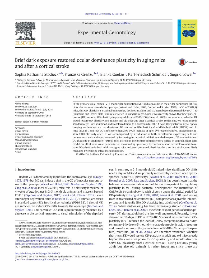
Since OD-plasticity is absent in SC-mice beyond PD110 (Lehmannand Löwel, 2008), we tested whether short-term DE can rescue OD-plasticity in animals of this age group. To this end, we raised mice inSCs to an average age of PD118 (age range PD104-136), exposed themto complete darkness (DE) for 14 days and subsequently imagedV1-activities after either 4 or 7 days of MD or without MD for controls.In both LR- and DE-mice without MD, V1-activity was dominated byinput from the contralateral eye: the activity spot induced by contralat-eral eye stimulation was darker than the spot induced by ipsilateral eyestimulation, the 2-dimensional OD-map showedwarm colors indicatingcontralateral dominance and the histogram of all OD-indices peakedat positive values (Fig. 1A,C). After 7 days of MD, the OD shifted towardsthe open eye only in the adult DE-mice (Fig. 1D; 2A)while V1-activation
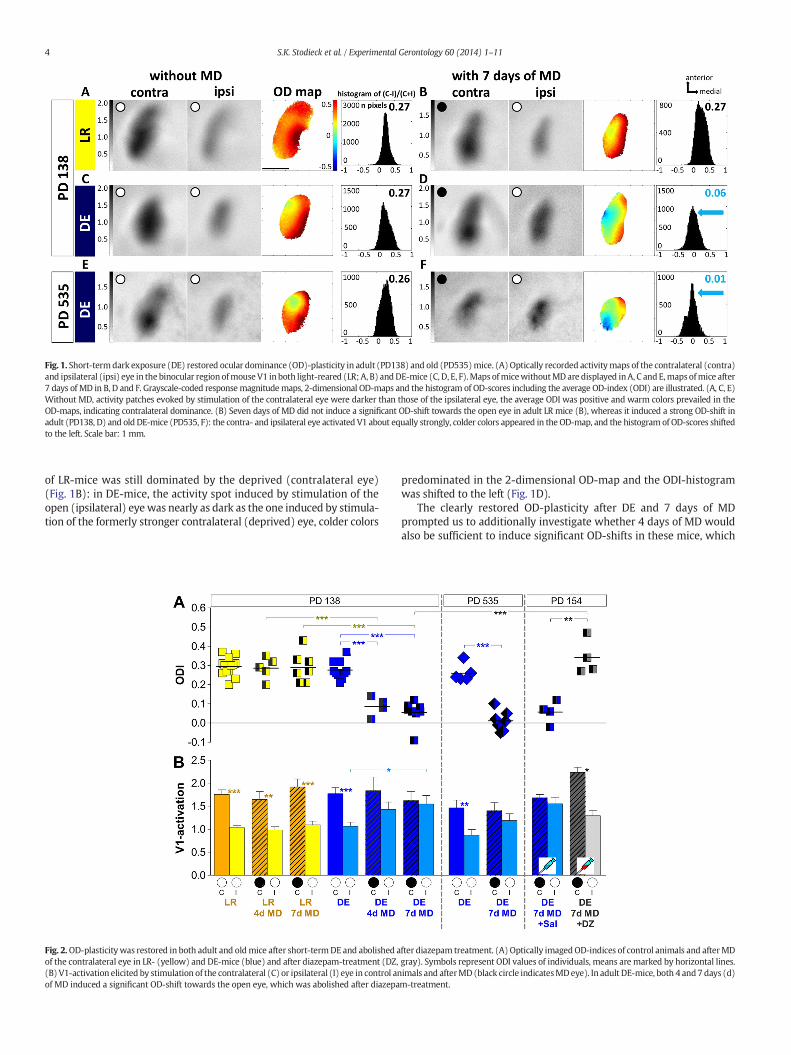
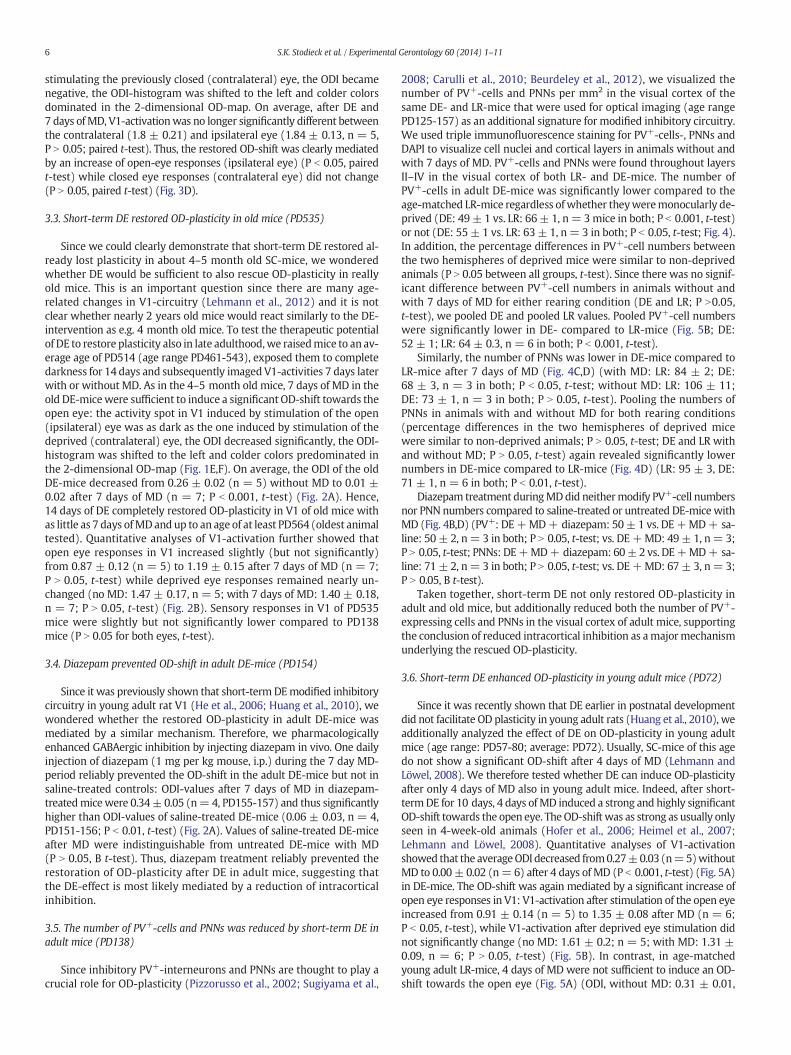
Fig. 1. Short-termdark exposure (DE) restored ocular dominance (OD)-plasticity in adult (PD138) and old (PD535)mice. (A)Optically recorded activitymaps of the contralateral (contra)and ipsilateral (ipsi) eye in the binocular region ofmouse V1 inboth light-reared (LR; A, B) andDE-mice (C, D, E, F).Maps ofmicewithoutMDare displayed inA, C and E,mapsofmice after7 days of MD in B, D and F. Grayscale-coded responsemagnitudemaps, 2-dimensional OD-maps and the histogram of OD-scores including the average OD-index (ODI) are illustrated. (A, C, E)Without MD, activity patches evoked by stimulation of the contralateral eye were darker than those of the ipsilateral eye, the average ODI was positive and warm colors prevailed in theOD-maps, indicating contralateral dominance. (B) Seven days of MD did not induce a significant OD-shift towards the open eye in adult LR mice (B), whereas it induced a strong OD-shift inadult (PD138, D) and old DE-mice (PD535, F): the contra- and ipsilateral eye activated V1 about equally strongly, colder colors appeared in the OD-map, and the histogram of OD-scores shiftedto the left. Scale bar: 1 mm.
4 S.K. Stodieck et al. / Experimental Gerontology 60 (2014) 1–11
of LR-mice was still dominated by the deprived (contralateral eye)(Fig. 1B): in DE-mice, the activity spot induced by stimulation of theopen (ipsilateral) eye was nearly as dark as the one induced by stimula-tion of the formerly stronger contralateral (deprived) eye, colder colors
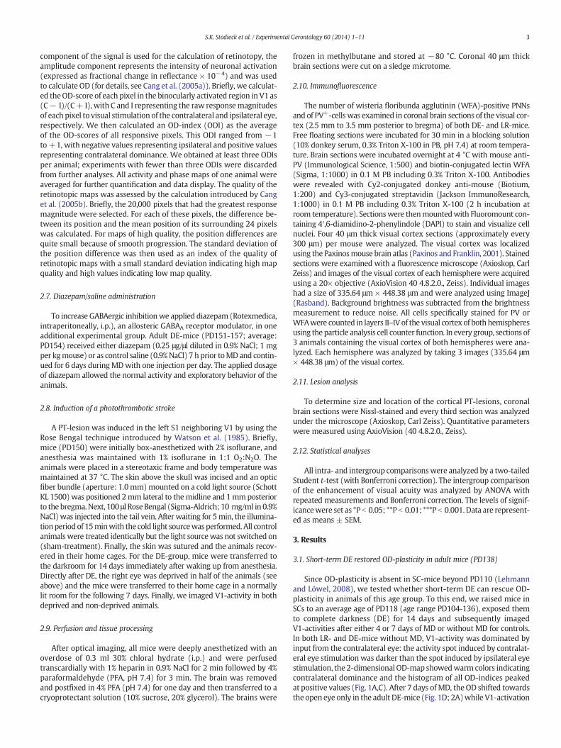
Fig. 2.OD-plasticity was restored in both adult and oldmice after short-termDE and abolished aof the contralateral eye in LR- (yellow) and DE-mice (blue) and after diazepam-treatment (DZ,(B) V1-activation elicited by stimulation of the contralateral (C) or ipsilateral (I) eye in control anof MD induced a significant OD-shift towards the open eye, which was abolished after diazepa
predominated in the 2-dimensional OD-map and the ODI-histogramwas shifted to the left (Fig. 1D).
The clearly restored OD-plasticity after DE and 7 days of MDprompted us to additionally investigate whether 4 days of MD wouldalso be sufficient to induce significant OD-shifts in these mice, which
fter diazepam treatment. (A) Optically imagedOD-indices of control animals and afterMDgray). Symbols represent ODI values of individuals, means are marked by horizontal lines.imals and afterMD(black circle indicatesMDeye). In adult DE-mice, both 4 and7 days (d)m-treatment.
5S.K. Stodieck et al. / Experimental Gerontology 60 (2014) 1–11
has been shown for 4-week-old SC-mice. This was indeed the case.Quantification of V1-activation showed that in DE-mice, the averageODI decreased highly significantly from 0.28 ± 0.02 (n = 10) withoutMD to 0.09 ± 0.03 after 4 days of MD (n = 4; P b 0.001, Bonferroni-adjusted (B) t-test) and to 0.06 ± 0.02 after 7 days of MD (n = 8;P b 0.001, t-test) (Fig. 2A). In contrast, in adult LR-mice of the sameage (PD138), OD-plasticity was absent, consistent with previous publi-cations (Lehmann and Löwel, 2008; Sato and Stryker, 2008). Neither 4nor 7 days of MD induced significant OD-shifts in LR-mice (Figs. 1A,B;2A) and ODIs did not differ between groups (LR: no MD: 0.29 ± 0.01,n = 13; with 4 days of MD: 0.29 ± 0.03, n = 5; with 7 days of MD:0.29 ± 0.03, n = 7; comparison between all groups: P N 0.05, B t-test;comparison between LR- and DE-mice without MD: P N 0.05, t-test)(Fig. 2A). Thus, 14 days of DE were sufficient to restore OD-plasticityof adult mice (up to an age of at least PD157) after only 4 days of MD.Quantitative analyses of V1-activation further showed that theOD-shift of adult DE-mice was mediated by a significant increase inopen (ipsilateral) eye responses in V1 while deprived eye responsesremained unchanged.While V1-activities induced by visual stimulationof the open eye increased already slightly after 4 days of MD, 7 days ofMDwere necessary to induce significant changes of open eye responsesin V1: V1-activation through the ipsilateral (open) eye increased from1.07 ± 0.09 (n = 10) to 1.43 ± 0.16 (n = 4; P N 0.05, t-test) after4 days of MD and to 1.55 ± 0.18 after 7 days of MD (n = 8; P b 0.05,t-test) (Fig. 2B). In contrast, deprived eye activities in V1 were notchanged after MD (no MD: 1.77 ± 0.13, n = 10; with 4 days of MD:1.83 ± 0.30, n = 4; P N 0.05, t-test; with 7 days of MD: 1.62 ± 0.20,n = 8; P N 0.05, t-test). In LR-mice, V1-responses of both eyes werenot significantly different between the control group without MD andmice after either 4 or 7 days of MD (contra: no MD: 1.76 ± 0.10, n =13; with 4 days of MD: 1.65 ± 0.17, n = 5; with 7 days of MD:1.91 ± 0.17, n = 7; P N 0.05 for all comparisons, B t-test; ipsi: no MD:1.03 ± 0.05, n = 13, with 4 days of MD: 0.98 ± 0.07, n = 5; with7 days of MD: 1.1 ± 0.08, n = 7; P N 0.05 for all comparisons; B t-test,Fig. 2B).
Fig. 3. Chronic imaging in adult mice revealed that the DE-induced restoration of OD-plasticityiments: Mice were raised in standard cages to an age of≥PD116 and then V1-activity inducedafter imaging 1, mice were dark-exposed for 14 days, the right eye was closed and a second imactivity maps after stimulation of the contralateral (contra) and ipsilateral (ipsi) eye in the bindisplay as in Fig. 1. Before DE and MD, V1 was dominated by input from the contralateral e(C) OD-indices of mice before (yellow) and after MD (blue) (n = 5). (D) V1-activation afterafter MD: after DE and MD, open eye responses increased significantly while deprived eye resp
3.2. Chronic optical imaging of V1-activity changes in DE-mice
To unambiguously determine the mechanism underlying therestored OD-plasticity in adultmice after DE, we additionally performedchronic optical imaging experiments in another group of adult micebefore and after DE and MD. To this end, we raised mice beyondPD116, imaged V1-activities after visual stimulation of the ipsi- andthe contralateral eye (first imaging) and then transferred the animalsto a dark room for 14 days. Immediately after DE, we deprived the con-tralateral eye of vision (MD) and transferred the animals back to a nor-mally lit room (12 h light/dark cycle) for the following 7 days. Finally,V1-activation of all mice was imaged again after 14 days of DE and7 days of MD (three weeks after the first imaging session–second imag-ing) (Fig. 3A). Our chronic imaging experiments clearly confirmed thatbrief DE can rescue OD-plasticity in V1 of SC-mice that were already be-yond their sensitive phase for OD-plasticity. In particular, the chronicdata showed i) that 14 days of DE restored OD-plasticity after 7 daysof MD in adult SC-mice (PD145) (Fig. 3B,C) and ii) that the restoredOD-shifts were mediated by an increase of open eye responses in V1while deprived eye responses were not significantly different beforeand after DE and MD (Fig. 3D). In the illustrated representative example(first imaging: before DE and MD), visual stimulation of the contralateraleye activated V1 more strongly than stimulation of the ipsilateral eye(Fig. 3B), warm colors prevailed in the 2-dimensional OD-map and thehistogramof the OD indiceswas centered to the right of zero. The averageODI of all chronic mice before DE and MD was 0.28 ± 0.02 (n = 5)(Fig. 3C), and V1-activation was significantly higher after visual stimula-tion of the contralateral eye (1.92 ± 0.29) compared to the ipsilateraleye stimulation (1.14 ± 0.19, n = 5, P b 0.001; paired t-test) (Fig. 3B,D). After 14 days of DE, 7 days of MD induced a strong and highly signif-icant OD-shift towards the open eye in all adult DE-mice (PD141-150):the average ODI decreased to −0.04 ± 0.04 (n = 5; P b 0.001, pairedt-test) (Fig. 3C). Actually, the OD-shifts of the chronically imaged adultmice were as strong as usually seen in 4-week-old LR-animals after4 days of MD (Hofer et al., 2006; Heimel et al., 2007; Lehmann andLöwel, 2008), and the visually driven activity in V1 could even becomedominated by input from the formerly weaker, ipsilateral eye (Fig. 3B):in the illustrated case, the activity spot in V1 induced by stimulation ofthe open (ipsilateral) eye was slightly darker than the one induced by
wasmediated by an increase of open eye responses in V1. (A) Time-line of imaging exper-by visual stimulation of the ipsi- and contralateral eye was visualized (imaging 1). Directlyaging was performed 7 days later in the same animals (imaging 2). (B) Optically imagedocular region of V1 before (upper row) and after DE and 7 days of MD (lower row). Dataye. After DE and 7 days of MD, there was a significant OD-shift towards the open eye.visual stimulation of the deprived (contralateral) and open (ipsilateral) eye before andonses did not change. Scale bar: 1 mm.
6 S.K. Stodieck et al. / Experimental Gerontology 60 (2014) 1–11
stimulating the previously closed (contralateral) eye, the ODI becamenegative, the ODI-histogram was shifted to the left and colder colorsdominated in the 2-dimensional OD-map. On average, after DE and7 days ofMD, V1-activationwas no longer significantly different betweenthe contralateral (1.8 ± 0.21) and ipsilateral eye (1.84 ± 0.13, n = 5,P N 0.05; paired t-test). Thus, the restored OD-shift was clearly mediatedby an increase of open-eye responses (ipsilateral eye) (P b 0.05, pairedt-test) while closed eye responses (contralateral eye) did not change(P N 0.05, paired t-test) (Fig. 3D).
3.3. Short-term DE restored OD-plasticity in old mice (PD535)
Since we could clearly demonstrate that short-term DE restored al-ready lost plasticity in about 4–5 month old SC-mice, we wonderedwhether DE would be sufficient to also rescue OD-plasticity in reallyold mice. This is an important question since there are many age-related changes in V1-circuitry (Lehmann et al., 2012) and it is notclear whether nearly 2 years old mice would react similarly to the DE-intervention as e.g. 4 month old mice. To test the therapeutic potentialof DE to restore plasticity also in late adulthood,we raisedmice to an av-erage age of PD514 (age range PD461-543), exposed them to completedarkness for 14 days and subsequently imaged V1-activities 7 days laterwith or without MD. As in the 4–5 month old mice, 7 days of MD in theold DE-micewere sufficient to induce a significant OD-shift towards theopen eye: the activity spot in V1 induced by stimulation of the open(ipsilateral) eye was as dark as the one induced by stimulation of thedeprived (contralateral) eye, the ODI decreased significantly, the ODI-histogram was shifted to the left and colder colors predominated inthe 2-dimensional OD-map (Fig. 1E,F). On average, the ODI of the oldDE-mice decreased from 0.26 ± 0.02 (n = 5) without MD to 0.01 ±0.02 after 7 days of MD (n = 7; P b 0.001, t-test) (Fig. 2A). Hence,14 days of DE completely restored OD-plasticity in V1 of old mice withas little as 7 days ofMDand up to an age of at least PD564 (oldest animaltested). Quantitative analyses of V1-activation further showed thatopen eye responses in V1 increased slightly (but not significantly)from 0.87 ± 0.12 (n = 5) to 1.19 ± 0.15 after 7 days of MD (n = 7;P N 0.05, t-test) while deprived eye responses remained nearly un-changed (no MD: 1.47 ± 0.17, n = 5; with 7 days of MD: 1.40 ± 0.18,n = 7; P N 0.05, t-test) (Fig. 2B). Sensory responses in V1 of PD535mice were slightly but not significantly lower compared to PD138mice (P N 0.05 for both eyes, t-test).
3.4. Diazepam prevented OD-shift in adult DE-mice (PD154)
Since it was previously shown that short-term DEmodified inhibitorycircuitry in young adult rat V1 (He et al., 2006; Huang et al., 2010), wewondered whether the restored OD-plasticity in adult DE-mice wasmediated by a similar mechanism. Therefore, we pharmacologicallyenhanced GABAergic inhibition by injecting diazepam in vivo. One dailyinjection of diazepam (1 mg per kg mouse, i.p.) during the 7 day MD-period reliably prevented the OD-shift in the adult DE-mice but not insaline-treated controls: ODI-values after 7 days of MD in diazepam-treatedmicewere 0.34± 0.05 (n=4, PD155-157) and thus significantlyhigher than ODI-values of saline-treated DE-mice (0.06 ± 0.03, n = 4,PD151-156; P b 0.01, t-test) (Fig. 2A). Values of saline-treated DE-miceafter MD were indistinguishable from untreated DE-mice with MD(P N 0.05, B t-test). Thus, diazepam treatment reliably prevented therestoration of OD-plasticity after DE in adult mice, suggesting thatthe DE-effect is most likely mediated by a reduction of intracorticalinhibition.
3.5. The number of PV+-cells and PNNs was reduced by short-term DE inadult mice (PD138)
Since inhibitory PV+-interneurons and PNNs are thought to play acrucial role for OD-plasticity (Pizzorusso et al., 2002; Sugiyama et al.,
2008; Carulli et al., 2010; Beurdeley et al., 2012), we visualized thenumber of PV+-cells and PNNs per mm2 in the visual cortex of thesame DE- and LR-mice that were used for optical imaging (age rangePD125-157) as an additional signature for modified inhibitory circuitry.We used triple immunofluorescence staining for PV+-cells-, PNNs andDAPI to visualize cell nuclei and cortical layers in animals without andwith 7 days of MD. PV+-cells and PNNs were found throughout layersII–IV in the visual cortex of both LR- and DE-mice. The number ofPV+-cells in adult DE-mice was significantly lower compared to theage-matched LR-mice regardless of whether theyweremonocularly de-prived (DE: 49 ± 1 vs. LR: 66± 1, n= 3mice in both; P b 0.001, t-test)or not (DE: 55± 1 vs. LR: 63± 1, n= 3 in both; P b 0.05, t-test; Fig. 4).In addition, the percentage differences in PV+-cell numbers betweenthe two hemispheres of deprived mice were similar to non-deprivedanimals (P N 0.05 between all groups, t-test). Since there was no signif-icant difference between PV+-cell numbers in animals without andwith 7 days of MD for either rearing condition (DE and LR; P N0.05,t-test), we pooled DE and pooled LR values. Pooled PV+-cell numberswere significantly lower in DE- compared to LR-mice (Fig. 5B; DE:52 ± 1; LR: 64 ± 0.3, n = 6 in both; P b 0.001, t-test).
Similarly, the number of PNNs was lower in DE-mice compared toLR-mice after 7 days of MD (Fig. 4C,D) (with MD: LR: 84 ± 2; DE:68 ± 3, n = 3 in both; P b 0.05, t-test; without MD: LR: 106 ± 11;DE: 73 ± 1, n = 3 in both; P N 0.05, t-test). Pooling the numbers ofPNNs in animals with and without MD for both rearing conditions(percentage differences in the two hemispheres of deprived micewere similar to non-deprived animals; P N 0.05, t-test; DE and LR withand without MD; P N 0.05, t-test) again revealed significantly lowernumbers in DE-mice compared to LR-mice (Fig. 4D) (LR: 95 ± 3, DE:71 ± 1, n = 6 in both; P b 0.01, t-test).
Diazepam treatment duringMDdid neithermodify PV+-cell numbersnor PNN numbers compared to saline-treated or untreated DE-mice withMD (Fig. 4B,D) (PV+: DE+MD+ diazepam: 50± 1 vs. DE+MD+ sa-line: 50 ± 2, n= 3 in both; P N 0.05, t-test; vs. DE+MD: 49± 1, n= 3;P N 0.05, t-test; PNNs: DE+MD+ diazepam: 60±2 vs. DE+MD+ sa-line: 71 ± 2, n= 3 in both; P N 0.05, t-test; vs. DE+MD: 67± 3, n= 3;P N 0.05, B t-test).
Taken together, short-term DE not only restored OD-plasticity inadult and old mice, but additionally reduced both the number of PV+-expressing cells and PNNs in the visual cortex of adult mice, supportingthe conclusion of reduced intracortical inhibition as amajormechanismunderlying the rescued OD-plasticity.
3.6. Short-term DE enhanced OD-plasticity in young adult mice (PD72)
Since it was recently shown that DE earlier in postnatal developmentdid not facilitate OD plasticity in young adult rats (Huang et al., 2010), weadditionally analyzed the effect of DE on OD-plasticity in young adultmice (age range: PD57-80; average: PD72). Usually, SC-mice of this agedo not show a significant OD-shift after 4 days of MD (Lehmann andLöwel, 2008). We therefore tested whether DE can induce OD-plasticityafter only 4 days of MD also in young adult mice. Indeed, after short-termDE for 10 days, 4 days ofMD induced a strong and highly significantOD-shift towards the open eye. TheOD-shift was as strong as usually onlyseen in 4-week-old animals (Hofer et al., 2006; Heimel et al., 2007;Lehmann and Löwel, 2008). Quantitative analyses of V1-activationshowed that the averageODI decreased from0.27±0.03 (n=5)withoutMD to 0.00± 0.02 (n= 6) after 4 days of MD (P b 0.001, t-test) (Fig. 5A)in DE-mice. The OD-shift was again mediated by a significant increase ofopen eye responses in V1: V1-activation after stimulation of the open eyeincreased from 0.91 ± 0.14 (n = 5) to 1.35 ± 0.08 after MD (n = 6;P b 0.05, t-test), while V1-activation after deprived eye stimulation didnot significantly change (no MD: 1.61 ± 0.2; n = 5; with MD: 1.31 ±0.09, n = 6; P N 0.05, t-test) (Fig. 5B). In contrast, in age-matchedyoung adult LR-mice, 4 days of MD were not sufficient to induce an OD-shift towards the open eye (Fig. 5A) (ODI, without MD: 0.31 ± 0.01,
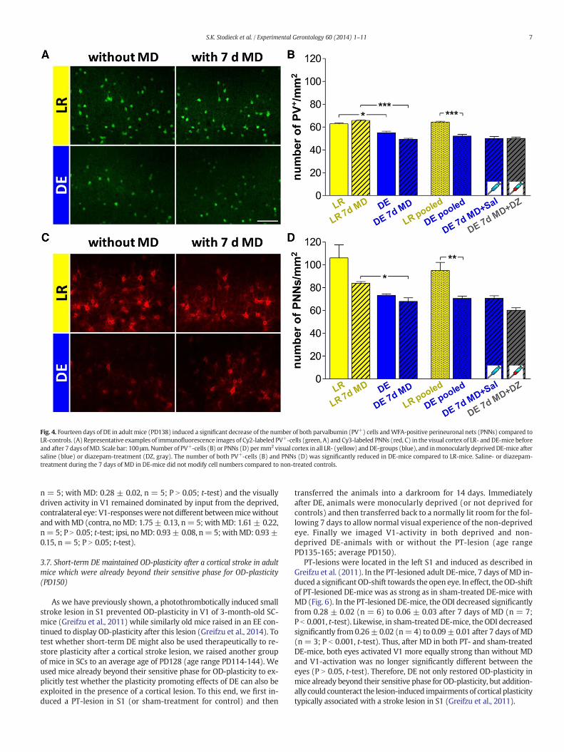
Fig. 4. Fourteen days of DE in adult mice (PD138) induced a significant decrease of the number of both parvalbumin (PV+) cells andWFA-positive perineuronal nets (PNNs) compared toLR-controls. (A) Representative examples of immunofluorescence images of Cy2-labeled PV+-cells (green, A) and Cy3-labeled PNNs (red, C) in the visual cortex of LR- and DE-mice beforeand after 7 days ofMD. Scale bar: 100 μm.Number of PV+-cells (B) or PNNs (D) permm2 visual cortex in all LR- (yellow) andDE-groups (blue), and inmonocularly deprivedDE-mice aftersaline (blue) or diazepam-treatment (DZ, gray). The number of both PV+-cells (B) and PNNs (D) was significantly reduced in DE-mice compared to LR-mice. Saline- or diazepam-treatment during the 7 days of MD in DE-mice did not modify cell numbers compared to non-treated controls.
7S.K. Stodieck et al. / Experimental Gerontology 60 (2014) 1–11
n = 5; with MD: 0.28 ± 0.02, n = 5; P N 0.05; t-test) and the visuallydriven activity in V1 remained dominated by input from the deprived,contralateral eye: V1-responseswere not different betweenmicewithoutandwithMD (contra, noMD: 1.75± 0.13, n= 5; withMD: 1.61± 0.22,n= 5; P N 0.05; t-test; ipsi, noMD: 0.93± 0.08, n=5; withMD: 0.93±0.15, n = 5; P N 0.05; t-test).
3.7. Short-term DE maintained OD-plasticity after a cortical stroke in adultmice which were already beyond their sensitive phase for OD-plasticity(PD150)
As we have previously shown, a photothrombotically induced smallstroke lesion in S1 prevented OD-plasticity in V1 of 3-month-old SC-mice (Greifzu et al., 2011) while similarly old mice raised in an EE con-tinued to display OD-plasticity after this lesion (Greifzu et al., 2014). Totest whether short-term DE might also be used therapeutically to re-store plasticity after a cortical stroke lesion, we raised another groupof mice in SCs to an average age of PD128 (age range PD114-144). Weused mice already beyond their sensitive phase for OD-plasticity to ex-plicitly test whether the plasticity promoting effects of DE can also beexploited in the presence of a cortical lesion. To this end, we first in-duced a PT-lesion in S1 (or sham-treatment for control) and then
transferred the animals into a darkroom for 14 days. Immediatelyafter DE, animals were monocularly deprived (or not deprived forcontrols) and then transferred back to a normally lit room for the fol-lowing 7 days to allow normal visual experience of the non-deprivedeye. Finally we imaged V1-activity in both deprived and non-deprived DE-animals with or without the PT-lesion (age rangePD135-165; average PD150).
PT-lesions were located in the left S1 and induced as described inGreifzu et al. (2011). In the PT-lesioned adult DE-mice, 7 days of MD in-duced a significant OD-shift towards the open eye. In effect, theOD-shiftof PT-lesioned DE-mice was as strong as in sham-treated DE-mice withMD (Fig. 6). In the PT-lesioned DE-mice, the ODI decreased significantlyfrom 0.28 ± 0.02 (n = 6) to 0.06 ± 0.03 after 7 days of MD (n = 7;P b 0.001, t-test). Likewise, in sham-treated DE-mice, the ODI decreasedsignificantly from 0.26± 0.02 (n= 4) to 0.09± 0.01 after 7 days of MD(n= 3; P b 0.001, t-test). Thus, after MD in both PT- and sham-treatedDE-mice, both eyes activated V1 more equally strong than without MDand V1-activation was no longer significantly different between theeyes (P N 0.05, t-test). Therefore, DE not only restored OD-plasticity inmice already beyond their sensitive phase for OD-plasticity, but addition-ally could counteract the lesion-induced impairments of cortical plasticitytypically associated with a stroke lesion in S1 (Greifzu et al., 2011).

Fig. 5. Short-term DE restored OD-plasticity after 4 days of MD in young adult (PD72)mice. (A) Optically imagedOD-indices of control animals and afterMDof the contralateraleye in LR- (yellow) and DE-mice (blue). Data display as in Fig. 2. (B) V1-activation elicitedby stimulation of the contralateral (C) or ipsilateral (I) eye in control animals and afterMD.In DE- but not LR-mice, 4 days of MD were sufficient to induce a significant OD-shift to-wards the open eye, mediated by an increase of open eye responses in V1.
Fig. 6. Short-termDE rescuedOD-plasticity in adultmice even after a stroke in the primarysomatosensory cortex. Data are displayed as in Fig. 2. Optically imaged OD-indices of DEadult (PD150) sham-treated mice (white) or mice after a photothrombotically inducedstroke (PT, gray) are illustrated. In spite of the PT-lesion, OD-plasticity was maintainedin adult DE-mice.
8 S.K. Stodieck et al. / Experimental Gerontology 60 (2014) 1–11
3.8. Visual cortical maps and basic visual performance were similar inDE- and LR-mice
3.8.1. V1-activationTo analyze a possible influence of DE on stimulus-driven activity in V1
we visualized elevation maps after visual stimulation with moving hori-zontal bars and azimuth maps after stimulation with moving verticalbars in PD72andPD138mice. Despite the reduced intracortical inhibition,the magnitude of sensory-driven activity in V1 was indistinguishablebetween DE- and LR-mice for both, elevation and azimuth maps at bothages (P N 0.05, B t-test for all comparisons; elevation: PD138 DE/LR:2.2 ± 0.1/2.5 ± 0.2, n = 9/12; azimuth: PD138 DE/LR: 1.8 ± 0.1/1.9 ±0.2 n = 9/12).
3.8.2. Retinotopic mapsQuantification of retinotopic maps showed that short-term DE did
also not affect map quality. Retinotopic map scatter was indistinguish-able between DE- and LR-mice for both azimuth and elevation mapsat both ages (P N 0.05 for all comparisons, B t-test; elevation: PD138DE/LR 2.7 ± 1.2/1.7 ± 0.3, n = 9/12; azimuth: PD138 DE/LR 11.9 ±1.8/13.5 ± 1.5, n = 9/12).
3.8.3. Visual acuity and contrast sensitivityWe used a virtual-reality optomotor setup (Prusky et al., 2004) to
test whether short-term DE affected basic visual parameters comparedto light-reared controls. In particular, we determined i) the highest spa-tial frequency (“visual acuity”) gratings and ii) lowest contrast (contrastsensitivity) that elicited an optomotor response in animals of all exper-imental groups and iii) the experience-dependent increase of open eyeacuity/contrast sensitivity afterMDwith daily training in the optomotorsetup (Prusky et al., 2006). DE did not have any effect on baseline visualacuity (PD72: DE/LR: 0.39 ± 0.001/0.39 ± 0.002 cyc/deg, n = 5/5;F2,11 = 0.71, P N 0.05, B ANOVA; PD138: DE/LR: 0.38 ± 0.000/0.38 ±0.001 cyc/deg, n= 10/13; F2,27= 0.30, P N 0.05, B ANOVA). As reportedpreviously (Lehmann et al., 2012), age had an effect on basic visual acu-ity of the tested mice: while values were not significantly different be-tween PD72 and PD138 DE-mice (F2,16 = 1.02, P N 0.05, B ANOVA),visual acuity was significantly reduced in the old DE-mice (PD535:0.35 ± 0.006 cyc/deg, n = 5; F2,19 = 67.41, P b 0.001, B ANOVA).
3.8.4. Experience-enabled increase in visual acuity and contrast sensitivityafter MD
After MD and daily training in the optomotor setup, both the spatialfrequency threshold and the contrast sensitivity threshold of theoptomotor reflex of the open eye increased similarly in age-matchedDE- and LR-mice (P N 0.05, t-test for all comparisons). In DE PD138mice, visual acuity of the open eye increased by 21 ± 1% from 0.39 ±0.001 cyc/deg to 0.47 ± 0.001 cyc/deg after 4 days of MD (n = 4;P b 0.001, t-test) and by 27 ± 2% to 0.49 ± 0.006 cyc/deg on day 7(n = 8; P b 0.001, t-test); this enhancement was not different fromLR-mice (increase by 21 ± 0.2%: from 0.39 ± 0.001 cyc/deg to0.47 ± 0.001 cyc/deg after 4 days of MD (n = 5, F2,25 = 0.375;P N 0.05, B ANOVA) and by 27 ± 2% to 0.49 ± 0.07 cyc/deg after7 days of MD (n= 7; P b 0.001, t-test)). Likewise, thresholds increasedsimilarly in PD72 DE- and LR-mice (F2,12 = 56.79, P N 0.05, B ANOVA).
In line with our previous observations (Lehmann et al., 2012), theexperience-dependent increase of the spatial frequency threshold ofthe optomotor reflex of the open eye afterMDwas significantly reducedin old mice. In PD535 DE-mice, visual acuity values of the open eye in-creased by only 12 ± 1% from 0.35 ± 0.006 cyc/deg on day 0 to0.39 ± 0.004 cyc/deg on day 7 (n = 7; P b 0.001, t-test). This increasewas significantly different from values in PD138 DE-mice in which acu-ity increased by 27 ± 2% (F2,19 = 37.85, P b 0.001, B ANOVA).
9S.K. Stodieck et al. / Experimental Gerontology 60 (2014) 1–11
3.8.5. Contrast sensitivityShort-term DE did neither have an effect on baseline contrast sensi-
tivity nor on the experience-dependent increase of contrast thresholdsof the open eye after MD. Values were similar in both DE- and LR-mice for all measured frequencies (baseline contrast sensitivity:P N 0.05, B t-test, in all groups; MD-induced increase: at least P b 0.01,t-test in all groups).
3.9. Adult DE-mice had normal visual capabilities after a stroke (PD150)
In PT-lesioned adult DE-mice, visual acuity values of the open eyeincreased significantly from 0.38 ± 0.001 cyc/deg on day 0 to0.48 ± 0.003 cyc/deg after 7 days of MD (n = 7; P b 0.001, t-test),corresponding to an increase of 24 ± 1% on baseline. This increasewas as high as in sham-treated non-lesioned DE-mice with 7 daysof MD (25 ± 1%; P N 0.05, t-test) in which visual acuity values ofthe open eye increased from 0.39 ± 0.000 cyc/deg on day 0 to0.49 ± 0.001 cyc/deg after 7 days of MD (n = 3; P b 0.001, t-test).Since it was previously shown that after PT and late MD (14 daysafter inducing the lesion), the increase in visual acuity of the openeye was indistinguishable from sham-treated animals (Greifzuet al., 2011), our results of normal visual capabilities and normal en-hancement of the spatial frequency threshold of the optomotor reflexafter PT are perfectly in line with the previous data. Contrast sensitivityvalues were also not significantly different between PT- (n = 6) andsham-treated mice without MD (n = 4, P N 0.05, t-test) and increasedsimilarly in both groups after 7 days of MD.
4. Discussion
Our results demonstrate that brief DE restored OD-plasticity in V1 ofadult and aging SC-mice until at least PD564. The rescued OD-shift inDE-mice was most likely mediated by reduced intracortical inhibitionsince application of diazepam during the MD-period completelyabolished the rescue of plasticity, and the numbers of both PV+-cellsand PNNs were reduced in DE-mice compared to LR-mice. Finally, DEpreserved OD-plasticity in aging mice even after a cortical stroke in S1.In contrast, visual cortical maps and basic visual performance weresimilar in DE- and LR-mice, and DE also did not influence experience-dependent sensory learning after MD.
These findings are consistent with studies showing that DE in youngadult rats (bPD100) promoted OD-plasticity most likely by reducingintracortical inhibition (He et al., 2006) and reactivated synaptic plastic-ity in adult V1 (Montey and Quinlan, 2011). Extending these observa-tions, we show here that DE restored OD-plasticity in much olderanimals and even after a cortical stroke. To understand themechanismsunderlying the restored OD-plasticity in agingmice after DE, we applieddiazepam during theMD period. Diazepam has previously been used toincrease GABAA-receptor mediated inhibition in mouse V1 (Henschet al., 1998; Fagiolini et al., 2004). In V1 of SC-mice, the inhibitory toneincreases during postnatal development and GABA-levels exceeding acertain limit may preclude plasticity in adult animals (Espinosa andStryker, 2012; Levelt and Hübener, 2012). Application of diazepamwith a dosage that reliably blocked OD-shifts in 3-month-old SC-mice(Greifzu et al., 2014) in fact blocked OD-shifts in V1 of our DE-mice,indicating that DE exerts its effectmost likely by reducing intracortical in-hibition. This interpretation is supported by the data of He et al. (2006)showing that restored OD-plasticity was accompanied by reduced num-bers of GABAA-receptors relative to AMPA-receptors and also by the reap-pearance of the juvenile form of NMDA receptors in V1.
To further analyze signatures of altered inhibitory circuits in thevisual cortex of aging DE-mice, we quantified PV+-interneurons thatmight play a crucial role in OD-plasticity (Espinosa and Stryker, 2012).It was hypothesized that the maturation of PV+-cells opens the criticalperiod for OD-plasticity (Hensch, 2005) which is then closed by the for-mation of PNNs around these cells (Beurdeley et al., 2012). Interestingly,
short-term DEwas accompanied by reduced numbers of both PV+-cellsand PNNs in ourDE-mice compared to LR-controls. Reduced numbers ofPV+-cells most likely indicate a reduction of PV-expression rather thana disappearance of the cells. Whether reduced PV-expression correlateswith reduced firing-rates of PV+-cells is not yet clear; nevertheless, itmay indicate reduced intracortical inhibition which in turn couldpromote OD-plasticity. Indeed, pharmacological reduction of PV+-cellfiring rates was recently shown to extend the critical period for OD-plasticity, while enhanced inhibition was shown to block it (Kuhlmanet al., 2013). Active inhibition of PV+-cells is also involved in the initialphases of reinforced associative learning in S1 (Froemke et al., 2007;Letzkus et al., 2011) and reduced numbers of GAD+-cells accompaniedthe restoration of OD-plasticity by short-term EE-housing (Sale et al.,2007; Baroncelli et al., 2010; Scali et al., 2012).
The prominent role of reduced intracortical inhibition for promotingOD-plasticity after DE does not rule out additional mechanisms. Enzy-matic degradation of extracellular matrix components which arrangeinto PNNs at the end of the critical period restored OD-plasticity inadult rats (Pizzorusso et al., 2002). PNNs preferentially enwrap PV+-cells (Ye and Miao, 2013) and PNN formation may limit structural plas-ticity by forming a physical barrier creating a non-permissive state forplasticity (Moon et al., 2001; Sugiyama et al., 2009). Since short-termDE reduced the number of PNNs compared to LR-controls, the reducedexpression of PNNs most likely has contributed to the increased OD-plasticity after DE. A reduced PNN-density was recently also reportedafter short-term EE-housing in rat V1 which increased plasticity (Saleet al., 2007; Scali et al., 2012). Likewise, orthodenticle homeobox 2homeoprotein binding to PNNs might regulate plasticity: a transientloss of Otx2 lead to a reduction of both PV+ and PNN expression inadult mouse V1 and reopened plasticity of visual acuity after MD(Beurdeley et al., 2012). Furthermore, neuromodulatory desensiti-zation and an increase in structural factors that inhibit neurite re-modeling have also been implicated in closing the sensitive phasefor OD-plasticity (reviewed in Bavelier et al., 2010; Espinosa andStryker, 2012; Levelt and Hübener, 2012).
Surprisingly, DE also rescued OD-plasticity in adult mice (PD150)after a cortical stroke. In particular, V1 of lesioned adult DE-mice reactedto an MD like sham-treated adult DE-animals, which was previouslyonly observed in juvenile SC-mice and in adult EE-mice (Greifzu et al.,2014). Thus, V1 of adult DE-mice reacted to the stroke lesion like4-week-old SC-raised mice. This cannot be due to a delay between PTand MD in the present study (2 weeks of DE in between) since wehave previously shown that OD-plasticity remained absent in PT-lesioned mice even if MD was induced with a delay of two weeks afterthe PT (Greifzu et al., 2011). Since both juvenile SC- and adult EE-micecontinue to show OD-plasticity after a S1-stroke and have a juvenileGABA/AMPA ratio, our new observations thus indicate that a V1 witha reduced inhibitory tone has a reduced susceptibility for stroke-induced impairments of cortical plasticity. In addition, short-termDE is highly effective: just 2 weeks of DE were sufficient to rescueOD-plasticity after MD, even in the presence of a cortical stroke lesion,and in aging animals already very far beyond their sensitive phase forOD-plasticity.
While it was recently shown that DE earlier in postnatal develop-ment (in PD35 rats), did not facilitate OD-plasticity (Huang et al.,2010), short-term DE was able to enhance OD-plasticity in our PD72mice: at this age, 4 days of MD are not sufficient to induce a significantOD-shift in LR-mice (this study and Lehmann and Löwel, 2008). In con-trast, a brief dark exposure (10 days) rescued OD-plasticity after 4 daysof MD. Thus, DE promotes OD-plasticity in mice of any age betweenPD57 and at least PD564. Whether DE would facilitate OD-plasticity ateven younger ages was not tested, since even LR-mice may showOD-plasticity after 4 days of MD. In addition, increasing OD-plasticityat an age where it is still present but reduced may depend on differentmechanisms than restoring plasticity in aging mice already beyondtheir sensitive phase for plasticity.

10 S.K. Stodieck et al. / Experimental Gerontology 60 (2014) 1–11
Interestingly, the restored OD-shifts of our adult and aging DE-micewere alwaysmediated by an increase of open eye responses after both 4and 7 days ofMD,without accompanying reductions of deprived eye re-sponses in V1.While this is consistent with literature findings reportingincreases in open eye responses in V1 after 5–7 days of MD in adult SC-raised mice (“adult” plasticity; Sawtell et al., 2003; Pham et al., 2004;Tagawa et al., 2005; Hofer et al., 2006; Mrsic-Flogel et al., 2007; Satoand Stryker, 2008) brief dark exposure in P70-P100 rats has recently re-vealed both a rapid depression of the V1-responses to stimulation of thedeprived eye after 3 days ofMDand a simultaneous potentiation of non-deprived eye responses (He et al., 2006). Decreases of deprived eye re-sponses have previously been observed in 4-week-old SC-raised miceafter 4 days of MD (Frenkel and Bear, 2004; Hofer et al., 2006; Satoand Stryker, 2008), and are generally taken as a signature of “juvenile”OD-plasticity. Interestingly, adult mice raised in an EE also displayed re-ductions of deprived eye responses after 7 days of MD, in addition tohaving a juvenile GABA/AMPA ratio (Greifzu et al., 2014). Taken togeth-er, our new data indicate that short-term DE in adult and agingmice re-stored OD-plasticity of the adult form.
It has recently been shown that the elimination of inhibitory synap-ses on distal apical dendrites of layer 2/3 pyramidal neurons is a majorcomponent of adult OD-plasticity and resulted in increased responsive-ness of V1 to stimulation of the non-deprived eye (vanVersendaal et al.,2012). This raises the question whether short-term DE might promotestructural changes in inhibitory boutons on dendritic spines and thuscontributes to the disinhibition of inputs serving the non-deprivedeye.While inhibition is reduced in V1 after DE, additional changes of ex-citatory circuitry like the reported reduction in the ratio of NR2A toNR2B subunits in dark reared or DE-rats that can be reversed rapidlyupon re-exposure to light (Quinlan et al., 1999; Bear, 2003; Guo et al.,2012) could contribute to the observed increases in open eye responsesafter MD in the present study. In fact, a low level of inhibition relativeto excitation may be required to allow V1 to respond rapidly to manip-ulations in visual input (He et al., 2006) and establish a milieu that ismore permissive to Hebbian types of plasticity that are normally quitelimited in the adult compared with developmental critical periods(Fagiolini and Hensch, 2000; Rozas et al., 2001; Chen and Nedivi,2013). According to the sliding threshold hypothesis, short-term DEcould thus lower the “modification threshold” to enable visual experi-ence to drive an increase of the non-deprived (ipsilateral) eye responsesin V1 (Bear, 1995; Kirkwood et al., 1996).
Surprisingly, reduced intracortical inhibition did not lead to an over-all enhancement of visual responses in the DE-mice. We have recentlyreported a similar observation in mice raised in an enriched environ-ment that also had reduced intracortical inhibition (Greifzu et al.,2014). Our interpretation of both findings is that homeostatic mecha-nisms must have compensatorily downregulated the excitatory drive.
The slight reduction of sensory activation in V1 of PD535 micecompared to PD138 mice is consistent with a previous study of ourgroup (Lehmann et al., 2012) that showed a significantly reduced V1-activation in 23-month-old mice compared to 7-month-old mice. Thus,a significant dampening of visual responses is indeed expected to occurin mouse V1, but only at a later time during aging.
4.1. Conclusion
Taken together, our data clearly show that brief DE not only helpedto enhance and restore OD-plasticity in adult and aging mice, and thuspresumably throughout life, but additionally also protected V1 fromlesion-induced impairments of OD-plasticity. Thus, DE offers a promis-ing, non-pharmacological and highly effective tool for restoring andpreserving plasticity of adult and especially aging neuronal circuits.
Conflict of interest
The authors have read the disclosure of potential conflicts of interestand have nothing to declare.
Acknowledgments
Thisworkwas supported by the FederalMinistry of Education and Re-search, Germany, grant numbers 01GQ0921 (S.S., F.G.) and 01GQ0810(S.L., B.G., K.S.) and by grants of the Deutsche Forschungsgemeinschaftthrough the Collaborative Research Center 889 “Cellular Mechanisms ofSensory Processing” to S.L. (Project B5).We thankM. Schink for the excel-lent animal care.
References
Baroncelli, L., Braschi, C., Spolidoro, M., Begenisic, T., Sale, A., Maffei, L., 2010. Nurturingbrain plasticity: impact of environmental enrichment. Cell Death Differ. 17,1092–1103.
Bavelier, D., Levi, D.M., Li, R.W., Dan, Y., Hensch, T.K., 2010. Removing brakes on adultbrain plasticity: from molecular to behavioral interventions. J. Neurosci. 30,14964–14971.
Bear, M.F., 1995. Mechanism for a sliding synaptic modification threshold. Neuron 15, 1–4.Bear, M.F., 2003. Bidirectional synaptic plasticity: from theory to reality. Philos. Trans. R.
Soc. Lond. B Biol. Sci. 358, 649–655.Berardi, N., Pizzorusso, T., Maffei, L., 2000. Critical periods during sensory development.
Curr. Opin. Neurobiol. 10, 138–145.Beurdeley, M., Spatazza, J., Lee, H.H., Sugiyama, S., Bernard, C., Di Nardo, A.A., Hensch, T.K.,
Prochiantz, A., 2012. Otx2 binding to perineuronal nets persistently regulates plastic-ity in the mature visual cortex. J. Neurosci. 32, 9429–9437.
Cang, J., Kalatsky, V.A., Löwel, S., Stryker, M.P., 2005a. Optical imaging of the intrinsic sig-nal as a measure of cortical plasticity in the mouse. Vis. Neurosci. 22, 685–691.
Cang, J., Renteria, R.C., Kaneko, M., Liu, X., Copenhagen, D.R., Stryker, M.P., 2005b. Devel-opment of precise maps in visual cortex requires patterned spontaneous activity inthe retina. Neuron 48, 797–809.
Carulli, D., Pizzorusso, T., Kwok, J.C., Putignano, E., Poli, A., Forostyak, S., Andrews, M.R.,Deepa, S.S., Glant, T.T., Fawcett, J.W., 2010. Animals lacking link protein have attenu-ated perineuronal nets and persistent plasticity. Brain 133, 2331–2347.
Chen, J.L., Nedivi, E., 2013. Highly specific structural plasticity of inhibitory circuits in theadult neocortex. Neuroscientist 19, 384–393.
Dräger, U.C., 1975. Receptive fields of single cells and topography in mouse visual cortex.J. Comp. Neurol. 160, 269–290.
Dräger, U.C., 1978. Observations on monocular deprivation in mice. J. Neurophysiol. 41,28–42.
Espinosa, J.S., Stryker, M.P., 2012. Development and plasticity of the primary visual cortex.Neuron 75, 230–249.
Fagiolini, M., Hensch, T.K., 2000. Inhibitory threshold for critical-period activation in pri-mary visual cortex. Nature 404, 183–186.
Fagiolini, M., Fritschy, J.M., Low, K., Mohler, H., Rudolph, U., Hensch, T.K., 2004. SpecificGABAA circuits for visual cortical plasticity. Science 303, 1681–1683.
Frenkel, M.Y., Bear, M.F., 2004. How monocular deprivation shifts ocular dominance invisual cortex of young mice. Neuron 44, 917–923.
Froemke, R.C., Merzenich, M.M., Schreiner, C.E., 2007. A synapticmemory trace for corticalreceptive field plasticity. Nature 450, 425–429.
Gordon, J.A., Stryker, M.P., 1996. Experience-dependent plasticity of binocular responsesin the primary visual cortex of the mouse. J. Neurosci. 16, 3274–3286.
Greifzu, F., Schmidt, S., Schmidt, K.F., Kreikemeier, K., Witte, O.W., Löwel, S., 2011. Globalimpairment and therapeutic restoration of visual plasticity mechanisms after a localizedcortical stroke. Proc. Natl. Acad. Sci. U. S. A. 108, 15450–15455.
Greifzu, F., Wolf, F., Löwel, S., 2012. Network influences on cortical plasticity. e-Neuroforum2/12, 41–48.
Greifzu, F., Pielecka-Fortuna, J., Kalogeraki, E., Krempler, K., Favaro, P.D., Schlüter, O.M.,Löwel, S., 2014. Environmental enrichment extends ocular dominance plasticityinto adulthood and protects from stroke-induced impairments of plasticity. Proc.Natl. Acad. Sci. U. S. A. 111, 1150–1155.
Guo, Y., Huang, S., de Pasquale, R., McGehrin, K., Lee, H.K., Zhao, K., Kirkwood, A., 2012.Dark exposure extends the integration window for spike-timing-dependent plastici-ty. J. Neurosci. 32, 15027–15035.
He, H.Y., Hodos, W., Quinlan, E.M., 2006. Visual deprivation reactivates rapid ocular dom-inance plasticity in adult visual cortex. J. Neurosci. 26, 2951–2955.
Heimel, J.A., Hartman, R.J., Hermans, J.M., Levelt, C.N., 2007. Screening mouse vision withintrinsic signal optical imaging. Eur. J. Neurosci. 25, 795–804.
Hensch, T.K., 2005. Critical periodmechanisms in developing visual cortex. Curr. Top. Dev.Biol. 69, 215–237.
Hensch, T.K., Fagiolini, M., Mataga, N., Stryker, M.P., Baekkeskov, S., Kash, S.F., 1998. LocalGABA circuit control of experience-dependent plasticity in developing visual cortex.Science 282, 1504–1508.
Hofer, S.B., Mrsic-Flogel, T.D., Bonhoeffer, T., Hübener, M., 2006. Prior experienceenhances plasticity in adult visual cortex. Nat. Neurosci. 9, 127–132.
Hooks, B.M., Chen, C., 2007. Critical periods in the visual system: changing views for amodel of experience-dependent plasticity. Neuron 56, 312–326.
11S.K. Stodieck et al. / Experimental Gerontology 60 (2014) 1–11
Huang, Z.J., Kirkwood, A., Pizzorusso, T., Porciatti, V., Morales, B., Bear, M.F., Maffei, L.,Tonegawa, S., 1999. BDNF regulates the maturation of inhibition and the critical periodof plasticity in mouse visual cortex. Cell 98, 739–755.
Huang, S., Gu, Y., Quinlan, E.M., Kirkwood, A., 2010. A refractory period for rejuvenatingGABAergic synaptic transmission and ocular dominance plasticity with dark expo-sure. J. Neurosci. 30, 16636–16642.
Kalatsky, V.A., Stryker, M.P., 2003. New paradigm for optical imaging: temporally encodedmaps of intrinsic signal. Neuron 38, 529–545.
Kirkwood, A., Rioult, M.C., Bear, M.F., 1996. Experience-dependent modification of synap-tic plasticity in visual cortex. Nature 381, 526–528.
Kuhlman, S.J., Olivas, N.D., Tring, E., Ikrar, T., Xu, X., Trachtenberg, J.T., 2013. A disinhibitorymicrocircuit initiates critical-period plasticity in the visual cortex. Nature 501,543–546.
Lehmann, K., Löwel, S., 2008. Age-dependent ocular dominance plasticity in adult mice.PLoS One 3, e3120.
Lehmann, K., Schmidt, K.F., Löwel, S., 2012. Vision and visual plasticity in ageing mice.Restor. Neurol. Neurosci. 30, 161–178.
Letzkus, J.J., Wolff, S.B., Meyer, E.M., Tovote, P., Courtin, J., Herry, C., Luthi, A., 2011. Adisinhibitory microcircuit for associative fear learning in the auditory cortex. Nature480, 331–335.
Levelt, C.N., Hübener, M., 2012. Critical-period plasticity in the visual cortex. Annu. Rev.Neurosci. 35, 309–330.
Montey, K.L., Quinlan, E.M., 2011. Recovery from chronic monocular deprivation follow-ing reactivation of thalamocortical plasticity by dark exposure. Nat. Commun. 2, 317.
Moon, L.D., Asher, R.A., Rhodes, K.E., Fawcett, J.W., 2001. Regeneration of CNS axons backto their target following treatment of adult rat brain with chondroitinase ABC. Nat.Neurosci. 4, 465–466.
Mrsic-Flogel, T.D., Hofer, S.B., Ohki, K., Reid, R.C., Bonhoeffer, T., Hübener, M., 2007.Homeostatic regulation of eye-specific responses in visual cortex during ocular dom-inance plasticity. Neuron 54, 961–972.
Paxinos, G., Franklin, K.B.J., 2001. TheMouse Brain in Stereotaxic Coordinates, 2nd edition.Academic Press, San Diego.
Pham, T.A., Graham, S.J., Suzuki, S., Barco, A., Kandel, E.R., Gordon, B., Lickey, M.E., 2004. Asemi-persistent adult ocular dominance plasticity in visual cortex is stabilized by ac-tivated CREB. Learn. Mem. 11, 738–747.
Pizzorusso, T., Medini, P., Berardi, N., Chierzi, S., Fawcett, J.W., Maffei, L., 2002. Reactivationof ocular dominance plasticity in the adult visual cortex. Science 298, 1248–1251.
Prusky, G.T., Alam, N.M., Beekman, S., Douglas, R.M., 2004. Rapid quantification of adultand developing mouse spatial vision using a virtual optomotor system. Invest.Ophthalmol. Vis. Sci. 45, 4611–4616.
Prusky, G.T., Alam, N.M., Douglas, R.M., 2006. Enhancement of vision by monocular dep-rivation in adult mice. J. Neurosci. 26, 11554–11561.
Quinlan, E.M., Philpot, B.D., Huganir, R.L., Bear, M.F., 1999. Rapid, experience-dependentexpression of synaptic NMDA receptors in visual cortex in vivo. Nat. Neurosci. 2,352–357.
Rasband,W.S., 1997–2014. ImageJ. U. S. National Institutes of Health, Bethesda, Maryland,USA (http://imagej.nih.gov/ij/).
Rozas, C., Frank, H., Heynen, A.J., Morales, B., Bear, M.F., Kirkwood, A., 2001. Developmen-tal inhibitory gate controls the relay of activity to the superficial layers of the visualcortex. J. Neurosci. 21, 6791–6801.
Sale, A., Maya Vetencourt, J.F., Medini, P., Cenni, M.C., Baroncelli, L., De Pasquale, R., Maffei,L., 2007. Environmental enrichment in adulthood promotes amblyopia recoverythrough a reduction of intracortical inhibition. Nat. Neurosci. 10, 679–681.
Sato, M., Stryker, M.P., 2008. Distinctive features of adult ocular dominance plasticity. J.Neurosci. 28, 10278–10286.
Sawtell, N.B., Frenkel, M.Y., Philpot, B.D., Nakazawa, K., Tonegawa, S., Bear, M.F., 2003.NMDA receptor-dependent ocular dominance plasticity in adult visual cortex. Neu-ron 38, 977–985.
Scali, M., Baroncelli, L., Cenni, M.C., Sale, A., Maffei, L., 2012. A rich environmental experi-ence reactivates visual cortex plasticity in aged rats. Exp. Gerontol. 47, 337–341.
Sugiyama, S., Di Nardo, A.A., Aizawa, S., Matsuo, I., Volovitch, M., Prochiantz, A., Hensch, T.K.,2008. Experience-dependent transfer of Otx2 homeoprotein into the visual cortex acti-vates postnatal plasticity. Cell 134, 508–520.
Sugiyama, S., Prochiantz, A., Hensch, T.K., 2009. From brain formation to plasticity:insights on Otx2 homeoprotein. Dev. Growth Differ. 51, 369–377.
Tagawa, Y., Kanold, P.O., Majdan,M., Shatz, C.J., 2005.Multiple periods of functional oculardominance plasticity in mouse visual cortex. Nat. Neurosci. 8, 380–388.
van Versendaal, D., Rajendran, R., Saiepour, M.H., Klooster, J., Smit-Rigter, L., Sommeijer, J.P.,De Zeeuw, C.I., Hofer, S.B., Heimel, J.A., Levelt, C.N., 2012. Elimination of inhibitory syn-apses is a major component of adult ocular dominance plasticity. Neuron 74, 374–383.
Watson, B.D., Dietrich, W.D., Busto, R., Wachtel, M.S., Ginsberg, M.D., 1985. Induction ofreproducible brain infarction by photochemically initiated thrombosis. Ann. Neurol.17, 497–504.
Wiesel, T.N., Hubel, D.H., 1963. Effects of visual deprivation on morphology and physiol-ogy of cells in the cats lateral geniculate body. J. Neurophysiol. 26, 978–993.
Ye, Q., Miao, Q.L., 2013. Experience-dependent development of perineuronal nets andchondroitin sulfate proteoglycan receptors in mouse visual cortex. Matrix Biol. 32,352–363.