steroid and g protein binding characteristics of the
TRANSCRIPT

Steroid and G Protein Binding Characteristics of theSeatrout and Human Progestin Membrane Receptor �Subtypes and Their Evolutionary Origins
Peter Thomas, Y. Pang, J. Dong, P. Groenen, J. Kelder, J. de Vlieg, Y. Zhu, and C. Tubbs
University of Texas Marine Science Institute (P.T., Y.P., J.D., C.T.), Port Aransas, Texas 78373; Centre for Molecular andBiomolecular Informatics (P.G., J.K., J.d.V.), Nijmegen Centre for Molecular Life Sciences, Radbout University Nijmegen,6500 HC Nijmegen, The Netherlands; and Department of Biology (Y.Z.), East Carolina University, Greenville, NorthCarolina 27858
A novel progestin receptor (mPR) with seven-transmembranedomains was recently discovered in spotted seatrout and ho-mologous genes were identified in other vertebrates. We showthat cDNAs for the mPR � subtypes from spotted seatrout(st-mPR�) and humans (hu-mPR�) encode progestin recep-tors that display many functional characteristics of G protein-coupled receptors. Flow cytometry and immunocytochemicalstaining of whole MDA-MB-231 cells stably transfected withthe mPR�s using antibodies directed against their N-terminalregions show the receptors are localized on the plasma mem-brane and suggest the N-terminal domain is extracellular.Both recombinant st-mPR� and hu-mPR� display high affin-ity (Kd 4.2–7.8 nM), limited capacity (Bmax 0.03–0.32 nM), anddisplaceable membrane binding specific for progestins. Pro-gestins activate a pertussis toxin-sensitive inhibitory G pro-tein (Gi) to down-regulate membrane-bound adenylyl cyclaseactivity in both st-mPR�- and hu-mPR�-transfected cells. Co-
immunoprecipitation experiments demonstrate the receptorsare directly coupled to the Gi protein. Similar to G protein-coupled receptors, dissociation of the receptor/G protein com-plex results in a decrease in ligand binding to the mPR�s andmutation of the C-terminal, and third intracellular loop ofst-mPR� causes loss of ligand-dependent G protein activation.Phylogenetic analysis indicates the mPRs are members of aprogesterone and adipoQ receptor (PAQR) subfamily that isonly present in chordates, whereas other PAQRs also occurin invertebrates and plants. Progesterone and adipoQ recep-tors are related to the hemolysin3 family and have origins inthe Eubacteria. Thus, mPRs arose from Eubacteria indepen-dently from members of the GPCR superfamily, which arosefrom Archeabacteria, suggesting convergent evolution of sev-en-transmembrane hormone receptors coupled to G proteins.(Endocrinology 148: 705–718, 2007)
ALTHOUGH THE IMPORTANCE of rapid (i.e. nonclas-sical) steroid actions initiated at the cell surface
through binding to steroid membrane receptors has becomemore widely accepted within the past few years, details of theinitial steroid-mediated events, including the identities of thesteroid membrane receptors and their mechanisms of action,remain unclear and are surrounded by controversy (1–3).There is clear evidence that a variety of receptor proteins areinvolved in initiating these nonclassical steroid actions indifferent cell models, including nuclear steroid receptors ornuclear steroid receptor-like forms (1, 2, 4), receptors forother ligands that also bind steroids (2, 5), and unidentifiedreceptors with different characteristics from those of anyknown receptors (2, 6). Recently, a novel cDNA was discov-ered in spotted seatrout ovaries that has several character-istics of the progestin membrane receptor (mPR) mediatingprogestin induction of oocyte maturation in this species by
a nongenomic mechanism (7). The seatrout cDNA (st-mPR�)encodes a 40 kDa protein, which has seven transmembranedomains, and receptor activation alters pertussis toxin-sen-sitive adenylyl cyclase activity, both of which suggest st-mPR� is a G protein-coupled receptor (GPCR) or GPCR-likeprotein (7). More than 20 closely related genes have beencloned from other vertebrate species, including three mPRsubtypes in humans, named �, �, and �, which show highlevels of expression in human reproductive, brain, and kid-ney tissues, respectively (8). The identification of a new classof putative steroid receptors, unrelated to nuclear steroidreceptors, but instead related to GPCRs, provides a plausibleexplanation of how steroids can initiate rapid hormonal re-sponses in target cells by activating receptors on the cellsurface. There has been broad recognition of the potentialsignificance of these findings (1, 9, 10) and also an extensiveresearch effort to determine the distribution, hormonal reg-ulation, and biological roles of the mPRs in various verte-brate models (11–16). However, critical information is stilllacking on several key features of mPRs essential for clearlyestablishing this proposed alternative model of steroid actionand for understanding its likely evolutionary origins.
The st-mPR� protein has been localized to the plasma mem-brane of seatrout oocytes (7), but progestin binding and acti-vation of signal transduction pathways in the plasma mem-branes of cells transfected with the st-mPR� and human mPRsremain to be demonstrated. To date, progestin binding has only
First Published Online November 9, 2006Abbreviations: GPCR, G protein-coupled receptor; HLY3, hemolysin
3; hu-mPR�, human membrane progestin receptor �; MMD, monocyteto macrophage differentiation protein; mPR, membrane progestin re-ceptor; nPR, nuclear progestin receptor; st-mPR�, spotted seatrout mem-brane progestin receptor �; PAQR, progesterone and adipoQ receptors;RBA, relative binding affinity.Endocrinology is published monthly by The Endocrine Society (http://www.endo-society.org), the foremost professional society serving theendocrine community.
0013-7227/07/$15.00/0 Endocrinology 148(2):705–718Printed in U.S.A. Copyright © 2007 by The Endocrine Society
doi: 10.1210/en.2006-0974
705
at East Carolina University Health Sciences Library on February 2, 2007 endo.endojournals.orgDownloaded from

been shown to the soluble recombinant mPR proteins producedin a bacterial expression system (7, 8). Moreover, the bindingcharacteristics of st-mPR� in the plasma membrane and itsphysiological role are still uncertain, because the principal te-leost progestin hormones that induce oocyte maturation do notshow any binding affinity for this soluble recombinant protein(7). Practically no information is currently available for thehuman counterpart, hu-mPR�, including whether its recombi-nant protein is expressed in plasma membranes and bindsprogestins specifically, whether it transduces signals in targetcells by activating G proteins, and its orientation in the plasmamembrane. Several phylogenetic analyses have grouped themPRs with adiponectin receptors as members of a progesteroneand adipoQ receptor (PAQR) family (17–19), which have anopposite orientation in the plasma membrane to GPCRs with anintracellular N terminal (20), and it has been proposed that allPAQRs, including mPRs, do not activate G proteins (18). Inaddition, an intracellular location, rather than a plasma mem-brane one of mammalian mPR�s, has been observed in certaineukaryotic expression systems (15, 16). Although our previousstructural and functional analyses suggest st-mPR� may be aGPCR, clear evidence that the receptor activates a G protein isdirectly coupled to it and has the characteristics of a GPCR islacking. Finally, the phylogenetic relationship of the mPRs toGPCRs is unknown. Therefore, in the present study, we inves-tigated the localization of the recombinant seatrout and humanmPR� proteins, steroid binding, and activation of second mes-sengers in the plasma membranes of st-mPR� and hu-mPR�-transfected human breast cancer cells (MDA-MB-231 cells),which do not express the nuclear progestin receptor (21). Ac-tivation of G proteins, their identities, as well as direct recep-tor/G protein coupling were also examined after progestintreatment. A preliminary investigation of the functional do-mains of the st-mPR� protein for G protein coupling and theirsimilarity to GPCRs was conducted with st-mPR� mutants witha truncated C-terminal and modified intracellular loop threedomains. Finally, phylogenetic analyses of mPRs and otherPAQRs were performed to reveal their likely origins. Collec-tively, the results show that the mPR�s are membrane-boundspecific progestin receptors that activate G proteins and func-tion as GPCRs but have a different ancestral origin to membersof the GPCR superfamily.
Materials and MethodsChemicals
The steroids progesterone, 17,20�,21-trihydroxy-4-pregnen-3-one(20�-S), 17,20�-dihydroxy-4-pregnen-3-one, 17-hydroxyprogesterone,20�-hydroxyprogesterone, cortisol, estradiol-17�, testosterone, 11-de-oxycorticosterone were purchased from Steraloids (Newport, RI). Thesynthetic progestin R5020 was purchased from Amersham (Piscataway,NJ). The synthetic antiprogestin RU486 was purchased from Sigma-Aldrich (St. Louis, MO). The other synthetic and natural progestins wereobtained from Organon (Oss, The Netherlands). [2,4,6,7-3H]-11-deoxy-cortisol, activity 50 Ci/mmol was obtained from American RadiolabeledChemicals (ARC, St. Louis, MO) and [2,4,6,7-3H]-progesterone ([3H-P4]),approximately 102 Ci/mmol, was purchased from Amersham. 20�-hydroxysteroid dehydrogenase and all other chemicals, buffers, andmedia were purchased from Sigma-Aldrich unless otherwise noted.
Culture of MDA-MB-231 cells stably expressing mPR�s
MDA-MB-231 cells stably transfected with the st-mPR�, obtained asdescribed previously (7), or hu-mPR� (described below) were cultured
in DMEM/Ham’s F-12 medium supplemented with 10% charcoal-stripped fetal bovine serum (FBS) and 100 �g/ml of gentamicin. Char-coal-stripped FBS was prepared by incubating FBS with 0.5% activatedcharcoal and 0.05% dextran T-70 for 30 min at 55 C. The charcoal particlesthen were removed by centrifugation at 4 C for 20 min at 4500 � g. Thestripped serum was sterile filtered and stored in aliquots at �80 C untiluse. The transfected cells were selectively maintained with 500 �g/mlgeneticin with changes of medium every 1–2 days. The cells reached 80%confluence after 3 days in culture. One day before the experiments, freshmedium without phenol red and supplemented with 5% charcoal-stripped FBS was added to the cell cultures. A 150-mm culture dish witha monolayer culture typically contained approximately 2 � 108 cells andyielded approximately 0.6 mg of cell membrane protein. Cells weresubsequently collected with a cell scraper and washed twice beforeexperimentation.
Expression of hu-mPR� and st-mPR� mutants in MDA-MB-231 cells
The procedures described previously for PCR amplification of themPR� cDNA, its insertion into an expression vector, and transfection inhuman MDA-MB-231 breast carcinoma cells (American Type CultureCollection, Manassas, VA) (7) were followed with few modifications forstable expression of hu-mPR� and transient transfection of st-mPR�mutants (see supplemental Fig. 1, A and B, published on The EndocrineSociety’s Journals Online web site at http://endo.endojournals.org). Thecoding regions of hu-mPR� and st-mPR� mutants were amplified byPCR from full-length cDNA plasmid clones (2 min denaturation at 94 C;5 PCR cycles with denaturation at 94 C for 1 min, annealing at 50 C for1 min, and polymerization at 72 C for 2 min followed by 25 cycles underthe same conditions except annealing, which was conducted at 55 C) andthe PCR products were purified by electrophoresis using a low-meltingagarose and a QIAquick Gel Extraction Kit as described previously (8)before ligation into a PBK-CMV expression vector (Stratagene, La Jolla,CA). The correct insertion was confirmed by DNA sequencing. Cellswere transfected with hu-mPR�, st-mPR�, st-mPR� mutant cDNAs, orvectors containing reversed mPR� inserts using Lipofectamine (LifeTechnologies, Inc., Gaithersburg, MD) following the manufacturer’ssuggestions. Transient transfection experiments with st-mPR� mutantswere conducted with cells grown to confluence for 2–3 d in mediacontaining 10% charcoal-stripped FBS, whereas experiments with stablytransfected hu-mPR� cells were conducted after continued selectionwith geneticin (500 �g/ml) for several (8–10) weeks.
Confirmation of correct expression of mPR� mRNAs intransfected cells
RT-PCR was performed periodically during the course of the func-tional studies as described previously followed by sequencing to con-firm continued successful expression of the entire coding regions ofst-mPR� and hu-mPR� in stably transfected cells (reverse transcriptasereactions without the addition of reverse transcriptase were used ascontrols to verify lack of genomic DNA contamination). Two overlap-ping DNA fragments from RT-PCR of hu-mPR� and st-mPR� obtainedfrom the transfected cell lines [primers: hu-mPR� 1, sense 5�-GCT CCCTGC CCA GGC CCA CA-3� (before start codon), antisense: 5�-GCC AGCAGA AAG AAG ACC AC-3�; 2, sense: 5�-TCT TTG TGG AGA CCG TGGAC-3�, antisense 5�-TCC CTA CCA GAT GCC ATC CC-3� (after stopcodon); st-mPR� 1, sense: 5�-TAC CGT CTA CAA GTT TGC C-3� (beforestart codon), antisense: 5�-GTG AGC AGC AGC CAA AGC AAG-3�; 2,sense 5�-CGC CAT AGA GAA AGA GTG G-3� antisense, 5�-AGT CACTGT CAC AAA CTT CAT T-3� (after stop codon)] were cloned into apGEM vector with a TA cloning system (Promega, Madison, WI). Theplasmids containing the mPR�s were subsequently sequenced with SP6and T7 primers from both ends. The results confirmed that the trans-fected mPR�s were correctly transcribed.
Membrane preparation and solubilization
Plasma membrane fractions of transfected MDA-MB-231 cells wereobtained following procedures described previously (7, 22) with fewmodifications. The cell suspension was washed once with assay bufferand then sonicated for 15 sec followed by a 1000 � g centrifugation for
706 Endocrinology, February 2007, 148(2):705–718 Thomas et al. • mPR Characteristics
at East Carolina University Health Sciences Library on February 2, 2007 endo.endojournals.orgDownloaded from

7 min to remove any nuclear and heavy mitochondrial material (nuclearfraction). The resulting supernatant was centrifuged at 20,000 � g for 20min to obtain the plasma membrane fraction. The remaining supernatantwas centrifuged at 100,000 � g for 60 min to obtain the microsomal andthe cytosolic fractions. The plasma membrane was further purified forsome studies by centrifuging the membrane pellet one to three timeswith a sucrose pad (1.2 m sucrose) at 6500 � g for 45 min (7).
mPR� binding assays
The general procedures for measuring binding of radioactive steroidligand to plasma membranes (22, 23) was used to measure binding of[2,4,6,7-3H]-17,20�,21-trihydroxy-4-pregnen-3-one ([3H]-20�-S,41.9 Ci/mmol) to the recombinant st-mPR� and [2,4,6,7-3H]-progesterone ([3H]-P4,102.1 Ci/mmol) to the recombinant hu-mPR� in the presence orabsence of steroid competitors. [3H]-20�-S was converted from [3H]-11deoxycortisol as described in (24). One set of tubes contained radio-labeled progestin alone (total binding); another set also contained non-radiolabeled progestin at concentrations 60- to 100-fold greater than theKd of the receptor (450–750 nm) to measure nonspecific binding (NSB).For competition assays, a third set of tubes contained the radiolabeledprogestins and 4–6 different concentrations of the steroid (range 1–10�m) competitors (dissolved in 1–5 �l ethanol, which does not affectligand binding in the receptor assays). After a 30-min incubation at 4 Cwith the membrane fractions, the reaction was stopped by filtration(Whatman GF/B filters, presoaked in assay buffer). The filters werewashed twice with 25 ml assay buffer and bound radioactivity wasmeasured by scintillation counting. The displacement of the radiola-beled steroid binding by the steroid competitors was expressed as apercentage of the maximum specific binding of the steroid for its re-ceptor. Progestin binding to membranes pretreated with nonradiola-beled GTP�S (25–50 �m) or activated and inactive pertussis toxin (0.5�g/ml) was performed as described previously (25). The same filtrationassay protocol was used to measure specific [3H]-P4 binding to micro-somal and nuclear fractions of MDA cells transfected with hu-mPR�(�65% of the proteins in these subcellular fractions are retained on theglass-fiber filters), whereas dextran-coated charcoal was used to sepa-rate bound from free [3H]-P4 in a soluble radioreceptor assay for cyto-solic fractions as described previously (7).
Western blot analysis and immunocytochemistry
Polyclonal antibodies generated by a commercial vendor (Sigma-Genosys, Woodlands, TX) in rabbits against six injections of synthetic15-mer peptides derived from the N-terminal domains of st-mPR�(YRQPDQSWRYYFLTL) and hu-mPR� (TVDRAEVPPLFWKPC) and a11-mer peptide from C-terminal domain of hu-mPR� (RPIYEPLHTHW)conjugated to keyhole limpet hemocyanin were used for immunode-tection of the mPR�s (see supplemental Fig. 1, A and B). Plasma mem-brane fractions were resuspended and solubilized in gel loading buffercontaining sodium dodecyl sulfate (SDS) for electrophoresis. Ten mi-crograms of solubilized plasma membrane proteins were resolved in12% SDS-polyacrylamide gel electrophoresis gels and transferred to anitrocellulose membrane for Western blot analysis. The membraneswere incubated for 1 h with blocking solution (5% nonfat milk in TBSTbuffer: 50 mm Tris/100 mm NaCl/0.1% Tween 20, pH 7.4) before in-cubation with the mPR� antibodies overnight at 4 C. The specificity ofthe immunoreactions was evaluated by blocking them with the peptideantigens (2–3 ng/�l mPR� antiserum diluted 1:20 in PBS buffer andpreincubated at room temperature with the antibodies for 1.5–2 h). Thenext day, the membranes were washed with TBST buffer and incubatedfor 1 h at room temperature with horseradish peroxidase conjugated togoat antirabbit antibody (Cell Signaling, Beverly, MA). The blots werewashed three times for 15 min with TBST buffer and treated withenhanced chemiluminescence (Pierce) and exposed to x-ray film.
Transfected cells were grown on coverslips for immunocytochemicalanalysis. The cells were rinsed twice with PBS and fixed with 2% para-formaldehyde and 0.25% glutaraldehyde in PBS for 15 min at 4 C. Cellswere permeabilized by adding 0.5% Triton X-100 to the fixative. The cellswere then rinsed briefly with PBS, incubated with 13 mm NaBH4 in PBSfor 10 min at 4 C to reduce autofluorescence, followed by three 5-minwashes in PBS. The cells were subsequently blocked in 2% BSA in PBSfor 1.5 h at 4 C followed by three 5-min washes in PBS. The cells were
then incubated with the st-mPR� or hu-mPR� primary antibodies (di-lution 1:1000) described previously in 2% BSA overnight at 4 C followedby three 5-min washes in PBS. Antisera were preabsorbed with peptide(0.02 mg peptide/1 ml antibody) overnight at 4 C for peptide blockcontrols. The cells were then incubated with AlexaFluor 488 goat anti-rabbit secondary antibody (Molecular Probes, Carlsbad, CA; dilution1:2000) followed by three 5-min washes in PBS. The coverslips werewet-mounted to slides using 80% glycerol in PBS and the presence offluorescent-labeled mPR� proteins in the cells visualized using a NikonC1 confocal microscope.
Adenylyl cyclase activity in transfected MDA-MB-231 cells
The production of cAMP by isolated plasma membranes over 30 minat 25 C was measured as an estimation of adenylyl cyclase activity inresponse to treatment with progestins (20–100 nm) in the presence orabsence of activated (30 min incubation with 50 mm DTT) or heat-inactivated (15 min incubation at 100 C) pertussis toxin (0.5 �g/ml).cAMP concentrations in the membranes were measured by enzymeimmunoassay using a kit following the manufacturer’s instructions (Bio-medical Technologies Inc., Stoughton, MA).
[35S]GTP�-S binding to cell membranes
Activation of G proteins was determined by measuring an increasein specific binding of [35S]GTP�-S to plasma membranes as describedpreviously (6, 23). Membranes (�10 �g protein) were incubated for 30min at 25 C in the presence of 20 nm progestins with 10 �m GDP and0.5 nm [35S]GTP�-S (�12,000 cpm, 1 Ci/mol; Amersham) in the absence(total binding) or presence of 100 �m GTP�-S (nonspecific binding).Bound [35S]GTP�-S was separated from free by filtering the incubationmixture through Whatman GF/B glass fiber filters followed by severalwashes.
Immunoprecipitation of [35S]GTP�-S-labeled G protein�-subunits
Immunoprecipitation of the G protein �-subunits was performed asdescribed previously (23, 26). Plasma membranes (�20 �g protein) of thetransfected cells were incubated for 30 min at 25 C in 250 �l buffercontaining 4 nm [35S]GTP�-S, 10 �m GDP, and protease inhibitors with1 �m progestin and stopped by the addition of ice-cold buffer containing100 �m GDP and 100 �m unlabeled GTP�-S. The samples were subse-quently centrifuged and the pellet resuspended in immunoprecipitationbuffer containing Triton X-100, SDS, and protease inhibitors. Specificantisera to the �-subunits of G proteins (Gi, Go, and Gs, 1:300; Santa CruzBiotechnology, Santa Cruz, CA) were added to the mixture and incu-bated at 4 C with gentle shaking for 6 h. Protein A-Sepharose beads wereadded and after an overnight incubation the immunoprecipitates werecollected by centrifugation, washed, boiled in SDS, and the radioactivityin the immunoprecipitated [35S]GTP�-S-labeled G protein �-subunitscounted.
Coimmunoprecipitation of G protein �i-subunit withmPR�s
Transfected cells were treated with 100 nm progestin (20�-S for re-combinant st-mPR� and progesterone for hu-mPR�) for 10 min or un-treated (controls) followed by two washes with PBS at 4 C. Triethanol-amine buffer (50 mm triethanolamine, 25 mm KCl, 5 mm MgCl2, 0.25 msucrose, 0.1% protease inhibitor cocktail; Sigma-Aldrich; pH 7.5) wasadded and the cells were frozen at �80 C until analyzed. Plasma mem-branes were prepared as described previously and resuspended in im-munoprecipitation buffer (0.1 mm EDTA, 1% Triton X-100 in Ca2�- andMg2�-free PBS, pH 7.5) to a final volume of 300 �l (2 mg/ml membraneprotein). The membrane suspension was incubated overnight at 4 C with1:100 of goat anti-Gi and -Go antibody and control goat IgG (Santa CruzBiotechnology, Inc.). Plasma membranes were then incubated for anadditional 2 h at 4 C with 20 �l protein-A agarose beads (Santa CruzBiotechnology, Inc.) added in the immunoprecipitation buffer. Beadswere washed twice with 1 ml immunoprecipitation buffer, and immu-noprecipitates were eluted by boiling for 10 min in SDS sample buffer.Samples were run on a 10% Tris-glycine SDS-polyacrylamide gel, and
Thomas et al. • mPR Characteristics Endocrinology, February 2007, 148(2):705–718 707
at East Carolina University Health Sciences Library on February 2, 2007 endo.endojournals.orgDownloaded from

proteins were transferred to nitrocellulose membranes. Membraneswere blocked with 5% nonfat milk in a buffer of 50 mm Tris, 100 mmNaCl, and 0.1% Tween 20 (pH 7.4) for 1 h, and then incubated at 4 Covernight with the st-mPR� or hu-mPR� antibodies (1:2500). The mem-brane was then washed three times with Tris-buffered saline and thenincubated for 1 h at room temperature with horseradish peroxidaseconjugated goat antirabbit (hu-mPR�) or mouse (st-mPR�) antibodies(Cell Signaling), and visualized by treatment with enhanced chemilu-minescence substrate (SuperSignal kit; Pierce, Rockford, IL).
Flow cytometry
Cells were carefully removed from the culture plates with a scraperand washed several times in PBS followed by low speed centrifugationto remove any cellular debris and damaged cells. Before conducting flowcytometry, the integrity of the cell membranes and their impermeabilitywas confirmed by incubating them with clathrin antibody. Cells wereeither pretreated with 90% methanol for 15 min on ice and then blockedin the 1% BSA in PBS (permeabilized group) or directly incubated inblocking solution (nonpermeabilized group). N-terminal and C-terminalhu-mPR� antibodies (�1:1000) were added to the cell suspension inblocking solution and incubated at room temperature for 1 h followedby two washes in PBS. Alexa Fluor 488 goat antirabbit IgG antibody(Alexa 488; Molecular Probes) in blocking solution was added to the cellsuspension and incubated for 30 min at room temperature in the darkfollowed by two washes with blocking solution. The cells were resus-pended in 500 �l PBS and analyzed within 24 h on a flow cytometer(Becton and Dickinson FACSCallbur). Data were analyzed withCellQuest Pro software (BD Biosciences, San Jose, CA).
Phylogenetic analyses
The majority of the sequences used to construct the phylogeny of thePAQR/hemolysin 3 (HLY3) family was derived from the SMART da-tabase (27) corresponding to the Pfam model HLY3 (291 sequences)including all species. The Hidden Markow Model was used to screen foradditional PAQR protein sequences in the complete eukaryotic genomesequences using HMMSearch (version 2.3.2c; part of HMMer package byS. Eddy, Janelia Farm Research Campus, Howard Hughes Medical In-stitute Laboratory). These complete proteomes were obtained from theEnsembl database (www.ensembl.org). The Ciona intestinalis proteinsequences were obtained from the Joint Genome institute (http://genome.jgi-psf.org/Cioin2/Cioin2.home.html). The total set of proteinsequences (397) was then aligned using ClustalW1.83 (28). The align-ment was manually curated to remove redundancy (fragments, dupli-cates, chimaeras). All the remaining sequences (281) were aligned againand used for construction of a phylogeny using the Neighbor-Joiningmethod and subsequently visualized using Treeview 1.6 (29).
ResultsSteroid binding characteristics of recombinant seatrout andhuman mPR�s
Expression of st-mPR� and hu-mPR� mRNAs in MDA-MB-231 cells and proteins in the plasma membranes wasconfirmed after stable transfection of their cDNAs (Figs. 1Aand 2A). Weak endogenous expression of hu-mPR� mRNAwas also detected in untransfected MDA-MB-231 cells after35 cycles of RT-PCR (Fig. 2A). Plasma membranes of st-mPR�-transfected cells showed a 6.5-fold increase in specific20�-S binding compared with untransfected cells in single-point receptor binding assays (P � 0.001, Fig. 1B). Saturationanalysis and Scatchard plotting indicated the presence of ahigh-affinity (Kd 7.58 � 0.93 nm, n 3), limited-capacity(Bmax 0.026 nm), saturable, single, specific binding site for20�-S (Fig. 1C). Dissociation of [3H]-20�-S from the receptorwas demonstrated in the presence of excess unlabeled steroid(Fig. 1D). Moreover, the rates of dissociation and associationwere rapid and reached 50% binding within 5 and 2 min,
respectively (Fig. 1D). Competitive binding assays revealedthat the major seatrout progestin hormone, 20�-S, and thetetrapod hormone, progesterone, bound with high affinity tothe receptor with IC50 values of 47.5 nm and 185 nm, respec-tively (Fig. 1, E and F). Receptor binding was specific forprogestins. Testosterone had a 28-fold lower affinity than20�-S (IC50: 1374 nm), whereas 200-fold higher concentra-tions of estradiol-17� and cortisol (10 �m) than the IC50 of20�-S did not cause any displacement of [3H]-20�-S from thereceptor. Interestingly, the synthetic progestin R5020 andantiprogestin RU486, which have relatively high bindingaffinities for the mammalian nuclear progesterone receptor,failed to bind to the recombinant seatrout mPR� at concen-trations up to 10 �m (Fig. 1F).
The steroid binding characteristics of the recombinant hu-man receptor (hu-mPR�) protein are similar to those of st-mPR�. Transfection of the MDA-MB-231 cells with hu-mPR�resulted in a 2.5-fold increase in specific progesterone bind-ing (Fig. 2B, P � 0.05). A high-affinity (Kd 4.17 nm), limited-capacity (Bmax 0.32 nm), single, specific progesterone bind-ing site was detected by saturation analysis and Scatchardplots (Fig. 2C). Progesterone ([3H]-P4) was readily displacedfrom the receptor, and ligand association and dissociationwere rapid, reaching 50% binding in approximately 5 min,which is typical of steroid membrane receptors (Fig. 2D).Binding to the hu-mPR� protein was specific primarily forprogestins; several androgens displayed moderate affinityfor the receptor, whereas no binding of estradiol-17� andcortisol to the receptor was detected at concentrations up to1 �m (Fig. 2E). Progesterone showed the highest affinity forthe receptor among the more than 30 steroidal compoundstested with an IC50 of 87.3 nm. The relative binding affinities(RBA) of the progestins, norprogesterone and pregna-4,9(11)-diene-3,20-dione, were 51.8 and 50.9% that of pro-gesterone. The RBAs of the remaining progestins and 11-deoxycorticosteroids tested were less than 50% (Table 1). Thetwo teleost progestin hormones, 17,20�,21-trihydroxy-4-pregnen-3-one (20�-S) and 17,20�-dihydroxy-4-pregnen-3-one, had low affinities with RBAs less than 1%. Similar to theresults with st-mPR�, RU486 and R5020 were poor compet-itors for hu-mPR�. R5020 had low binding affinity for thereceptor with an IC50 of 2 �m and a RBA of 4.1%, whereasRU486 caused less than 50% displacement of P4 from thereceptor at the highest concentration tested, 10 �m (Fig. 2F).Interestingly, testosterone also bound to hu-mPR� (IC50 390nm) with an RBA 22.4% that of progesterone, somewhathigher than its affinity for the st-mPR�. Several other an-drogens, dihydrotestosterone, and a methyl ether testoster-one derivative were also moderately effective competitors ofprogesterone binding (Table 1).
Activation of G proteins and second messengers
Treatment of plasma membranes of st-mPR�-transfectedcells, but not untransfected cells, with 100 nm 20�-S causeda decrease in adenylyl cyclase activity (Fig. 3A). The pro-gestin-induced decrease in adenylyl cyclase activity wasblocked by prior treatment with pertussis toxin, a specificinhibitor of activation of inhibitory G proteins, Gi/o (Fig. 3A).Progesterone (20 nm) caused a similar decrease in adenylyl
708 Endocrinology, February 2007, 148(2):705–718 Thomas et al. • mPR Characteristics
at East Carolina University Health Sciences Library on February 2, 2007 endo.endojournals.orgDownloaded from

cyclase activity in membranes of cells transfected with hu-mPR�, whereas cortisol, which showed no binding affinityfor hu-mPR�, was ineffective (Fig. 3B). These progestin-in-duced decreases in adenylyl cyclase activities in the st-mPR�- and hu-mPR�-transfected cells were dose-dependent(see supplemental Fig. 1, C and D). Possible activation of Gproteins was investigated using a radiolabeled nonhydro-lyzable form of GTP, [35S]GTP�-S. Treatment of st-mPR�-and hu-mPR�-transfected cell membranes, but not untrans-fected ones, with progestins (st-mPR�: 20 nm 20�-S; hu-mPR�: 20 nm progesterone) in the presence of [35S]GTP�-Scaused significant increases in specific GTP�-S binding to thecell membranes, indicating ligand-dependent activation of G
proteins (Fig. 3, C and D). G protein activation of hu-mPR�was specific for progestin ligands, because cortisol andR5020, which do not bind to the receptor, were ineffective(Fig. 3D), and was dose-dependent (see supplemental Fig.1D). The identities of the G protein(s) activated on progestinbinding to st-mPR� and hu-mPR� were determined by im-munoprecipitation of the activated G protein �-subunitsbound to [35S]GTP�-S with specific G protein �-subunit an-tibodies. A polyclonal rabbit antibody directed against the�-subunit of inhibitory G proteins (Gi) precipitated approx-imately 90% of the total radioactive GTP�-S activatedthrough st-mPR� (Fig. 3E) and approximately 75% of thetotal GTP�-S activated via hu-mPR� (Fig. 3G), whereas little
FIG. 1. Progestin binding to plasma membranes of MDA-MB-231 cells stably transfected with st-mPR�. A, Detection of st-mPR� protein intransfected (Tr-231) cell membranes by Western blot analysis and st-mPR� mRNA in cells by RT-PCR [(�)RT]. 231, Untransfected cells; OV,seatrout ovarian membranes; M, molecular weight protein standards; (-)RT, lacking reverse transcriptase; peptide blocked, blocked with peptideantigen. B, Specific [3H]-20�-S binding to transfected cell membranes in a single point assay (see key in A) (n 6,*, P � 0.05, Student’s t test).C, Representative saturation curve and Scatchard plot of specific [3H]-20�-S binding to plasma membranes of transfected cells. D, Time courseof association (Assoc) and dissociation (Dissoc) of [3H]-20�-S binding. E and F, Competition curves for steroid (E) and progestin (F) bindingexpressed as a percentage of maximum specific 20�-S binding. E2, Estradiol-17�; T, testosterone; P4, progesterone; cort, cortisol; 17,20�-P,17,20�-dihydroxy-4-pregnen-3-one; RU486, mifepristone; R5020, promegestone.
Thomas et al. • mPR Characteristics Endocrinology, February 2007, 148(2):705–718 709
at East Carolina University Health Sciences Library on February 2, 2007 endo.endojournals.orgDownloaded from

radioactivity was precipitated with a specific � Gs antibodyand control rabbit serum (Fig. 3, E and F).
G protein coupling to seatrout and human mPR�s
Coimmunoprecipitation studies, in which the receptor/Gprotein complex was first precipitated with antibodies to theG protein � Gi- and Go-subunits, and subsequently probedwith the mPR� antibodies by Western blot analysis, showeddirect coupling of st-mPR� (Fig. 4A) and hu-mPR� (Fig. 4B)to an inhibitory (Gi) G protein. No coupling of the mPR�s tothe � Go-subunit could be detected. As predicted, prior treat-ment with progestins resulted in decreased amounts of themPR� proteins detected on the Western blots (Fig. 4, A andB). Uncoupling of the G proteins caused decreases in the
binding affinities of the mPR�s for their progestin ligands(Fig. 4, C–F). Pretreatment of the plasma membranes withactive pertussis toxin (0.5 �g/ml), but not with the inactiveform, caused a marked decrease in specific progestin bindingto both st-mPR�-transfected (Fig. 4C) and hu-mPR�-trans-fected cells (Fig. 4D). Similarly, prior incubation of trans-fected plasma membranes with 25 �m and 50 �m GTP�-Scaused a decrease in specific progestin binding, whereas thistreatment did not affect the minor amounts of steroid bind-ing to untransfected cell membranes (Fig. 4, E and F).
Localization, orientation, and mutational analysis
Confocal microscopy of nonpermeabilized mPR�-trans-fected MDA-MB-231 cells after immunocytochemical stain-
FIG. 2. Progestin binding to plasma membranes of MDA-MB-231 cells stably transfected with hu-mPR� (see Fig. 1 for experimental detailsand key). A, Detection of hu-mPR� protein and mRNA. B, Specific [3H]-progesterone (P4) binding in a single point assay. C, Representativesaturation curve and Scatchard plot of specific [3H]-P4 binding (n 5). D, Time course of association and dissociation of [3H]-P4 binding. Eand F, Competition curves for steroid (E) and progestin (F) binding expressed as a percentage of maximum specific P4 binding. Norp,Norprogesterone; Nandr, nandrolone; Ethist, ethisterone; Nore, norethisterone; Norg, norgestrel.
710 Endocrinology, February 2007, 148(2):705–718 Thomas et al. • mPR Characteristics
at East Carolina University Health Sciences Library on February 2, 2007 endo.endojournals.orgDownloaded from
ing with antibodies directed against the mPR� N-terminaldomains showed both st-mPR� and hu-mPR� are expressedin the cell membrane and suggest the N terminal is extra-cellular (Fig. 5, Aa and Ba). The specificity was verified bydemonstrating that the immunocytochemical reactions wereblocked after preincubating the antibodies with their peptideantigens (Fig. 5, Ab and Bb). Neither of the antibodiesshowed significant immunoreactivity with untransfectedcells (Fig. 5, Ac and Bc). No immunoreactivity was alsoobserved on nonpermeabilized hu-mPR�-transfected cellswith the antibody directed against the C-terminal domain ofhu-mPR� (Fig. 5Bd). The efficacy of the permeabilization andnonpermeabilization procedures was confirmed using anantibody to clathrin, which is present intracellularly (data notshown). The orientation of hu-mPR� in the cell membrane,with the N terminal extracellularly, was independently ver-ified by flow cytometry of transfected cells, which had notbeen treated with fixatives using the N-terminal and C-terminal hu-mPR� antibodies. Incubation with clathrin an-tibody confirmed that the harvesting and washing proceduredid not damage the cell membranes of the transfected cells
making them permeable to the antibody (see supplementalFig. 1, G and H). Incubation of nonpermeabilized transfectedcells, but not untransfected ones, with the N-terminal anti-body resulted in a marked increase in immunoflorescence(Fig. 5C), whereas no increase in fluorescence was observedafter incubation of nonpermeabilized transfected cells withthe C-terminal antibody (Fig. 5D). Specific [3H]-P4 bindingwas localized by radioreceptor assay in the plasma mem-brane fractions of cells transfected with hu-mPR� (Fig. 5E).Negligible [3H]-P4 binding was detected in the microsomaland nuclear fractions in the filtration assay or cytoplasmicfractions in the soluble radioreceptor assay. Western blottingof the subcellular fractions with an integrin �3 antibodyconfirmed lack of cell membrane contamination in theseother subcellular fractions (see supplemental Fig. 1D). Sim-ilarly, lack of significant contamination of plasma membrane,nuclear, and cytoplasmic fractions with microsomal proteinswas confirmed with a spectrophometric reduced NAD phos-phate enzyme assay (results not shown). Several potentialfunctional domains of st-mPR� involved in G protein cou-pling were investigated by mutational analysis (see supple-mental Fig. 1, A and B). Truncation of the C-terminal (aminoacids RQRVRASLHEKGE deletion, amino acid no. 340–352)resulted in a significant decrease in G protein activation inresponse to progestin treatment, whereas C-terminal trun-cation combined with substitution of three amino acids in thethird intracellular loop (IL3, KCD changed to VAV, aminoacid no. 273–275) completely blocked activation of G proteinsafter progestin treatment, suggesting that both these intra-cellular domains near the C-terminal end of st-mPR� areimportant for G protein coupling (Fig. 5F). Uncoupling of Gproteins to the C-terminal st-mPR� mutant was also accom-panied with a predicted decrease in ligand binding affinityfor the receptor (Fig. 5G).
Phylogenetic analysis
The construction of an unrooted phylogenetic tree (seesupplemental Fig. 2) clearly shows the clustering of the se-quences into two major groups. The largest group can befurther subdivided into two subgroups, one of which rep-resents the large group of bacterial HLY3 proteins. Themonocyte to macrophage differentiation protein (MMD) 2and MMD members of the different species tightly clustertogether with the HLY3 proteins. A simpler representation ofthis unrooted tree excluding the prokaryotes is shown in Fig.6. Here the major eukaryotic species that were used in thephylogenetic analysis are schematically depicted in a phy-logram. The table to the right of the phylogram shows thepresence of the individual members of the PAQR familyidentified in humans, adiponectin receptors 1 and 2 (ADR1and 2), MMD and MMD2, mPR�, � and � and PAQR3, 4, 6,and 9). Each dot represents a gene encoding a protein. Doubledots indicate gene duplications, resulting in two genes withhigh similarity. The table summarizes the findings from thephylogenetic tree in the supplement and shows the uniquerepresentation of mPR�, mPR�, mPR� (PAQR 7, 8, and 5)and PAQR6 in the Chordata. The dense clustering of PAQR6,mPR�, mPR�, and mPR� in the dendrogram in supplemental
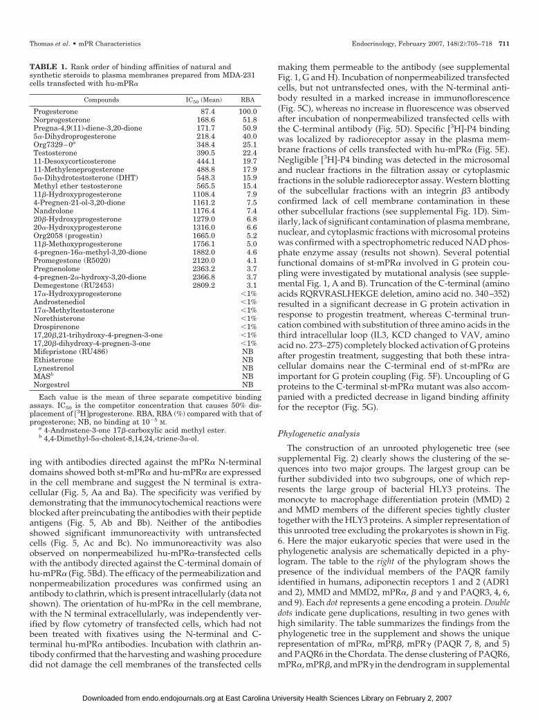
TABLE 1. Rank order of binding affinities of natural andsynthetic steroids to plasma membranes prepared from MDA-231cells transfected with hu-mPR�
Compounds IC50 (Mean) RBA
Progesterone 87.4 100.0Norprogesterone 168.6 51.8Pregna-4,9(11)-diene-3,20-dione 171.7 50.95�-Dihydroprogesterone 218.4 40.0Org7329–0a 348.4 25.1Testosterone 390.5 22.411-Desoxycorticosterone 444.1 19.711-Methyleneprogesterone 488.8 17.95�-Dihydrotestosterone (DHT) 548.3 15.9Methyl ether testosterone 565.5 15.411�-Hydroxyprogesterone 1108.4 7.94-Pregnen-21-ol-3,20-dione 1161.2 7.5Nandrolone 1176.4 7.420�-Hydroxyprogesterone 1279.0 6.820�-Hydroxyprogesterone 1316.0 6.6Org2058 (progestin) 1665.0 5.211�-Methoxyprogesterone 1756.1 5.04-pregnen-16�-methyl-3,20-dione 1882.0 4.6Promegestone (R5020) 2120.0 4.1Pregnenolone 2363.2 3.74-pregnen-2�-hydroxy-3,20-dione 2366.8 3.7Demegestone (RU2453) 2809.2 3.117�-Hydroxyprogesterone �1%Androstenediol �1%17�-Methyltestosterone �1%Norethisterone �1%Drospirenone �1%17,20�,21-trihydroxy-4-pregnen-3-one �1%17,20�-dihydroxy-4-pregnen-3-one �1%Mifepristone (RU486) NBEthisterone NBLynestrenol NBMASb NBNorgestrel NB
Each value is the mean of three separate competitive bindingassays. IC50 is the competitor concentration that causes 50% dis-placement of 3H�progesterone. RBA, RBA (%) compared with that ofprogesterone; NB, no binding at 10�5 M.
a 4-Androstene-3-one 17�-carboxylic acid methyl ester.b 4,4-Dimethyl-5�-cholest-8,14,24,-triene-3�-ol.
Thomas et al. • mPR Characteristics Endocrinology, February 2007, 148(2):705–718 711
at East Carolina University Health Sciences Library on February 2, 2007 endo.endojournals.orgDownloaded from

Fig. 3 further indicates the close structural relationshipamong members of this subfamily. In contrast, the adiponec-tin receptors (ADR1 and ADR2, PAQR 1 and 2) and MMDand MMD2 (PAQR11) together with PAQR 3 are foundthroughout the eukaryotes (Fig. 6).
Figure 7 schematically depicts the proposed parallel con-vergent evolution of GPCRs and PAQR/HLY3 related pro-teins. Originating from Archaebacteria, bacteriorhodopsinevolved into the rhodopsin like GPCRs in eukaryotes, whichin turn gave rise to the other GPCR classes, the Glutamate,Adhesion, Frizzled, and Secretin families as classified byFredriksson et al. (30). In contrast, HLY3 family members arefound exclusively in the Eubacteria, none have been identifiedin the Archaebacteria. Proteins of various species in the PAQR10 and 11 members of the PAQR family tightly cluster to-gether with the HLY3 proteins. The sequence comparisons
show the PAQR family in eukaryotes shares many structuralfeatures with the prokaryotic HLY3 family, suggesting acommon bacterial origin. Thus, the PAQR family appears tohave arisen from the Eubacteria in contrast to members of theGPCR superfamily, which arose from the Archaebacteria.
Discussion
The progestin binding results demonstrate that mPR� cD-NAs from two distantly related vertebrate species, spottedseatrout and humans, encode membrane-bound progestinbinding moieties with all the characteristics of functionalsteroid membrane receptors. Transfection of MDA-MB-231cells with these cDNAs resulted in expression of the st-mPR�and hu-mPR� recombinant proteins in the cell membranesand severalfold increases in specific binding of the fish and
FIG. 3. Activation of G proteins and second messengers in membranes of cells transfected with st-mPR� and hu-mPR�. A, Effects of 15-mintreatment with 100 nM 20�-S on cAMP production by membranes of st-mPR�-transfected cells (Tr-231) with or without 30-min pretreatmentwith activated (aPTX) or heat-inactivated pertussis toxin (iPTX, 0.5 �g/ml). 231-untransfected cells. *, P � 0.05, n 4. B, Effects of 10-mintreatment with 20 nM progesterone (P4), 20 nM cortisol (Cort), or vehicle (Veh) on cAMP production by membranes from cells transfected withhu-mPR�. C, Effects of treatment with 20 nM 20�-S or 20 nM R5020 on specific [35S]GTP�-S binding to membranes of cells transfected withst-mPR�. *, P � 0.05 compared with vehicle treatment (Veh), n 4. D, Effects of treatment with 20 nM progesterone, 20 nM cortisol (Cort),or 20 nM R5020 on specific [35S]GTP�-S binding to membranes of cells transfected with hu-mPR�. *, P � 0.05 compared with vehicle treatment(Veh). E, Immunoprecipitation of [35S]GTP�-S bound to G protein �-subunits activated on 1 �M 20�-S treatment of st-mPR�-transfected cellmembranes with specific G�s (anti-Gs) and G�i (anti-Gi) G protein antibodies or control rabbit serum (rabbit ser). *, P � 0.05 (n 4) comparedwith vehicle control. F, Immunoprecipitation of [35S]GTP�-S bound to G protein �-subunits activated upon 1 �M progesterone treatment ofhu-mPR�-transfected cell membranes with specific antibodies described in E. *, P � 0.05 (n 4) compared with vehicle control.
712 Endocrinology, February 2007, 148(2):705–718 Thomas et al. • mPR Characteristics
at East Carolina University Health Sciences Library on February 2, 2007 endo.endojournals.orgDownloaded from

mammalian progestin hormones, 20�-S and progesterone.Saturation analysis and Scatchard plotting indicated thatboth mPR� proteins have high-affinity, limited-capacity, sin-gle binding sites specific for progestin hormones that arecharacteristic of steroid hormone receptors and distinguishthem from other steroid-binding proteins. The binding af-finity of 20�-S to the recombinant st-mPR� (Kd 7.58 nm) issimilar to its binding affinity to seatrout ovarian and testic-ular membranes, which express wild-type st-mPR� (Refs. 22and 31; Kd: ovary 5.0 nm, testis 18.0 nm), whereas the wild-type nuclear progestin receptor in seatrout displays a 4-foldhigher progestin binding affinity (Ref. 32; mean cytosolicbinding: Kd 1.9 nm). Similarly, the binding affinity of pro-gesterone to the human nuclear progesterone receptor-B(Ref. 33; Kd 0.8 nm) is 5-fold higher than that to recombinanthu-mPR� (Kd 4.2 nm). Information on whether these appar-ent differences in binding affinities result in differential ac-tivation of the two progestin receptor systems, the mPRs onlybeing continuously activated where progestin levels are highnear their sites of synthesis and intermittently activated at
other target tissues when plasma progestin levels are ele-vated, may provide clues of the physiological roles of themPRs during the reproductive cycle.
The high binding specificity of the recombinant st-mPR� produced in the mammalian expression system for20�-S is consistent with its identity as the major progestinhormone regulating maturation of oocytes and motility ofsperm in this species and with previous receptor bindingresults with seatrout ovarian and sperm membranes (22,31). The lack of 20�-S binding to the soluble recombinantst-mPR� protein produced in Escherichia coli observed inour earlier study is probably due to protein modificationdeficiencies in this prokaryotic expression system (7). Ingeneral, however, the steroid binding characteristics ofrecombinant st-mPR� and hu-mPR� produced in MDA-MB-231 cell membranes are remarkably similar to those ofthe recombinant soluble receptors produced in the pro-karyotic system (8). The more than 100-fold lower bindingaffinity of 20�-S and the other teleost progestin, 17,20�-P,for hu-mPR� (RBAs �1%) is noteworthy and warrants
FIG. 4. Coupling of seatrout and human mPR� proteins to G proteins. A and B, Coimmunoprecipitation of st-mPR� (A) and hu-mPR� (B) coupledto G protein �-subunits with specific G protein antibodies followed by immunodetection of the mPR�s by Western blot analysis. Pretreated for10 min with 100 nM 20�-S or progesterone. C and D, Effects of 30-min pretreatment with 0.5 �g/ml activated pertussis toxin (aPTX) or inactivatedpertussis toxin (iPTX) on specific [3H]-20�-S binding to membranes of st-mPR�-transfected cells (C) or on specific [3H]-progesterone bindingto membranes of hu-mPR�-transfected cells (D). *, P � 0.05, n 4. E and F, Effects of 30-min pretreatment with 25 �M GTP�S on specific[3H]-20�-S binding to membranes of st-mPR�-transfected cells (E) or pretreatment with 25 or 50 �M GTP�S on specific [3H]-P4 binding tomembranes of hu-mPR� transfected cells (F). *, P � 0.05, n 4.
Thomas et al. • mPR Characteristics Endocrinology, February 2007, 148(2):705–718 713
at East Carolina University Health Sciences Library on February 2, 2007 endo.endojournals.orgDownloaded from

investigation, because all the other steroids tested havesimilar affinities for the two mPR�s. For example, thenuclear receptor agonist R5020 and antagonist RU486 have
very low affinities for both mPR�s, whereas testosterone,which shows low affinities for nuclear PRs, has RBAs of10% and 22% for st-mPR� and hu-mPR�, respectively. The
FIG. 5. Localization, orientation, and functional domains of the mPR�s. A and B, Immunocytochemical localization of st-mPR� (Aa) andhu-mPR� (Ba) on external surface of plasma membranes of transfected cells with specific N-terminal directed antibodies. Ab and Bb, Blockedwith peptide antigens; Ac and Bc, lack of immunoreaction with nontransfected cells; Bd, lack of immunoreaction with nonpermeabilized cellusing C-terminal antibody. C and D, Flow cytometry of cells transfected with hu-mPR� using N-terminal (C) and C-terminal (D) antibodies.C, 231, Background fluorescence on nontransfected cells in the presence mPR� N-terminal antibody; Tr-231 Non-perm., immunodetection ofthe hu-mPR� N-terminal on the surface of nonpermeabilized transfected cells by flow cytometry using the N-terminal antibody. D, Tr-231Non-perm., fluorescence of nonpermeabilized transfected cells using the C-terminal antibody; Tr-231 Perm., immunodetection of the hu-mPR�C-terminal in permeabilized transfected cells by flow cytometry using a C-terminal directed antibody. E, Specific binding of [3H]-P4 to subcellularfractions of cells transfected with hu-mPR�. m, Plasma membranes; m-sp, plasma membranes further purified with a sucrose pad; ms,microsomes; nu, nuclear fraction; cyt, cytosolic fraction; #, dextran-coated charcoal used to separate bound from free. F and G, Mutationalanalysis of st-mPR� functional domains. F, Effects of C-terminal truncation (Mu1) and amino acid substitution in the third intracellular loop(Mu3) on specific [35S]GTP�S binding to cell membranes in response to 20�-S treatment. G, Effects of C-terminal truncation (mutant 1) on specific[3H]-20�-S binding to cell membranes.
714 Endocrinology, February 2007, 148(2):705–718 Thomas et al. • mPR Characteristics
at East Carolina University Health Sciences Library on February 2, 2007 endo.endojournals.orgDownloaded from

finding that the steroid specificities of the recombinantseatrout and human mPR�s differ markedly from those oftheir nuclear progestin receptor counterparts was antici-pated because there is no homology in any regions of themPRs to the ligand binding domain of the nuclear pro-gesterone receptor. Competitive binding assays with morethan 30 steroidal compounds revealed different effects ofsubstituting various functional groups on the progester-one nucleus on their binding affinities to hu-mPR� com-pared with previously published results with human nPR(Refs. 33 and 34 and Table 1). For example, removal ofcarbon 19 (substitution of a methyl group for hydrogen:norprogesterone, also see R5020 and Organon 2058) de-
creases binding affinity to mPR� (Table 1) but modestlyincreases it for the nPR (33, 34). However, removal of theside chain and addition of a hydroxyl at C17 (i.e. testos-terone, dihydrotestosterone, nandrolone), which practi-cally eliminated binding to the human nPR, only reducedbinding with mPR� to 22–7% that of P4 (33, 34). Finally,the hu-mPR� also does not recognize C19 steroids (an-drogens) with a substitution of a hydrogen on C17 with anethinyl group (i.e. ethisterone, norethisterone, and nor-gestrel), whereas these steroids have relatively high af-finities for the nPR (33, 35). These marked differences inbinding affinities suggest selective mPR� modulators, inaddition to nPR ones such as R5020, can be developed to
FIG. 6. Schematic representation of the presence of PAQR family members 1–11 among the eukaryotes. The phylogram on the left depicts theevolutionary relationships between the different species used in this study. The table on the right shows the presence of a particular PAQRfamily member by a dot. A double dot indicates a duplication event. *, Missing data, incomplete genome sequence; ¶, gene duplication; ¥, possibleredundancy; low-quality sequence shows divergence in phylogenetic tree. The results show that the mPR�, mPR�, and mPR� genes firstappeared in the Euteleostomi.
Thomas et al. • mPR Characteristics Endocrinology, February 2007, 148(2):705–718 715
at East Carolina University Health Sciences Library on February 2, 2007 endo.endojournals.orgDownloaded from

independently explore progestin actions in target tissuesmediated by each of these two receptor systems.
Identification of the ligand binding pocket by site-directedmutagenesis will be required for definitive proof that themPRs directly bind progestins and are not merely compo-nents of a multiple-protein receptor complex. However, allthe results obtained to date with the mPRs are consistent withour previous suggestion that they are true steroid membranereceptors (7). The finding that prokaryotic as well as eukary-otic expression systems produce recombinant st-mPR�s andhu-mPR�s with the binding characteristics of steroid mem-brane receptors suggests that the ability to bind progestins isan intrinsic property of these proteins and is not dependenton their association with other proteins present only in eu-karyotic cells (7, 8). The demonstration that transfection ofMDA cells with the various mPRs confers progestin bindingspecificity to the cell membrane preparations also suggests adirect role for these proteins in ligand binding. Membranesfrom cells transfected with st-mPR� display a high bindingaffinity for 20�-S, the principal progestin in this species,whereas those from hu-mPR�-transfected cells have low af-finity for this steroid but show highest affinity for proges-terone, the major progestin hormone in mammals. In addi-tion, MDA cells transfected with zebrafish mPR� show thehighest binding affinity for 17,20�-P, the likely progestin
hormone in this species (36). The characteristics of G proteinactivation and ligand binding to the mPRs on G proteinuncoupling are also typical of seven-transmembrane hor-mone receptors. For example, dissociation of G proteins fromGPCRs results in a decrease in ligand binding affinity of thereceptors directly coupled to them. The finding that disso-ciation of the mPR�-G protein complex by pretreatment withGTP�-S and pertussis toxin causes a decline in progestinbinding is further evidence that the progestin binding site islocated on the mPR� protein.
There is now evidence that representatives of all three mPRsubtypes described in the original papers, �, �, and � (7, 8),corresponding to PAQR types 7, 8, and 5 (17), display high-affinity, specific and limited-capacity binding to progestinscharacteristic of steroid membrane receptors. Sequence andphylogenetic analyses reveal that another PAQR type, PAQR 6,is closely related to the mPRs (supplemental Fig. 2) and there-fore is a candidate as an additional member of this novel groupof membrane progestin receptors. A notable feature of thisprogestin receptor PAQR subfamily (PAQR 5–9) is that repre-sentatives have only been identified in vertebrates, includingteleosts and tetrapods, suggesting that they arose at the base ofthe vertebrate lineage. PAQR 9 could also possibly belong tothis subgroup of progestin receptors because it is primarilyfound in the Chordata (Fig. 6) and is clustered with the mPRs(supplemental Fig. 3). In contrast, all other members of PAQRfamily, including the adiponectin (PAQR 1, 2) and MMD re-ceptors (PAQR 10, 11), have both invertebrate and vertebraterepresentatives, indicating they have a more ancient evolution-ary origin (Fig. 6). The high similarity of PAQR 10 and 11 withthe HLY3 proteins in bacteria suggests that this is the arche-typical PAQR in eukaryotes, although the phylogenetic anal-ysis suggests that the ADR2 and PAQR 3 genes appeared firstin prokaryotes. The present results with seatrout and humanmPR�s indicate that, at least for the PAQR 7 subtype, theirability to bind progestins arose early in vertebrate evolution,before the divergence of the teleost and tetrapod lineages, atleast 200 million years ago (37). Members of this progestinPAQR subfamily have not been identified in the urochordateCiona genome, which appear to lack many of the key steroi-dogenic enzymes (steroid dehydrogenases and cytochromeP450s) and enzymes that metabolize steroids (38, 39). The par-allels between the evolution of the progestin membrane recep-tor subfamily, PAQR 5–8, and that of nuclear steroid receptorsare striking. Duplication of nuclear steroid receptors from anancestral estrogen receptor-like gene in vertebrates also oc-curred early in vertebrate evolution, coincident with the adventof the jawed vertebrates, to give rise to nuclear progesteronereceptors and multiple estrogen receptors and later to give riseto androgen, glucocorticoid, and mineralocorticoid receptors(39–41). Thus, phylogenetic analyses indicate that functionalmPRs probably arose around the same time as nuclear proges-tin receptors (nPRs), coincident with the appearance of criticalsteroidogenic enzymes and the production of significant quan-tities of progestins and other steroids in early vertebrates. Theappearance of both mPRs and nPRs in early vertebrates likelyreflects an intimate and complimentary relationship betweenthe two receptor systems, both of which may be required for acoordinated or varied response of target cells in the reproduc-tive system to progestin hormones. The first evidence of such
FIG. 7. Schematic depiction of the parallel convergent evolution ofthe PAQR family and GPCR superfamily. The bacteriorhodopsins arestrictly found in Archaebacteria and constitute the origin of the GPCRsuperfamily, whereas the HLY3 genes are strictly found in Eubac-teria.
716 Endocrinology, February 2007, 148(2):705–718 Thomas et al. • mPR Characteristics
at East Carolina University Health Sciences Library on February 2, 2007 endo.endojournals.orgDownloaded from
an intimate relationship between these two progestin receptorsignaling pathways has recently been obtained in human myo-metrial cells, where progesterone was shown to regulate trans-activation of nPR and coactivator function through mPR�- andmPR�-mediated pathways (26). Complex interactions betweenthese two types of progesterone receptors are expected in somebreast cancer cell lines (e.g., MCF-7), which show progesteroneactivation of nonclassical pathways through both mPRs andnPRs in addition to classical genomic responses through thenPR (4, 42). Information on the differing roles of mPRs and nPRsin nonclassical signaling in these cells may provide insights intowhy alternative signaling pathways mediated by two differentclasses of progesterone receptors evolved in vertebrates. Struc-tural analyses of the nuclear steroid receptor ligand bindingdomains in a wide variety of chordates indicate that the an-cestral receptor initially recognized the estrogens produced invertebrate gonads associated with reproductive activity. It hasbeen suggested that nPRs evolved subsequently to respond toprogesterone, a steroid intermediate in estrogen production, bya process called ligand exploitation (40). A similar analyticalapproach may provide valuable insights into the evolution ofmPRs from the ancestral PAQR once information on the struc-tures of the ligand binding domains of mPRs and other mem-bers of the PAQR family becomes available.
The results of the present study also demonstrate that boththe seatrout and human recombinant mPR� proteins activateinhibitory G proteins (Gi), are coupled to them and sharemany other critical features of GPCRs. Clear evidence wasobtained that mPR�s, like GPCRs, are localized and functionas seven-transmembrane receptors in the plasma membranesof cells. However, the mPR�s have also been identified inother cellular compartments in fish oocytes and in certaineukaryotic expression systems (7, 15, 16). Moreover, themPR�s are similar to GPCRs in that they likely can occur asdimers as well a monomers (43), because an 80-kDa band isalso frequently observed on Western blots, the intensity ofwhich varies with the membrane solubilization conditionsused (unpublished observation). G protein activation, de-tected with GPCRs by an increase in specific [35S]GTP�-Sbinding to the cell membranes (44), was also demonstratedafter progestin treatment of mPR�-transfected cells. Thedemonstration that progestins activate the inhibitory G pro-tein, Gi, and down-regulate pertussis toxin-sensitive adeny-lyl cyclase activity in the plasma membranes of MDA-MB-231 cells transfected with receptors indicates the mPR�sfunction as GPCRs. The activation of an inhibitory G proteinby the mPR�s is also consistent with our recent findings onthe signaling pathways activated by 20�-S during meioticmaturation of seatrout oocytes (25) as well as those activatedby progesterone through the wild-type hu-mPR� in humanmyometrial cells (26). A unique characteristic of GPCRs isthat treatment with nonhydrolyzable forms of guanine nu-cleotides such as GTP�-S, and for Gi/o-protein coupled re-ceptors with pertussis toxin, cause uncoupling of G proteinsfrom the receptors (45, 46), resulting in a decrease in theirligand binding affinities (47, 48). These treatments causedsimilar decreases in ligand binding to st-mPR� and hu-mPR�. Thus, these results further support the conclusions ofthe coimmunoprecipitation studies that the mPR�s are di-rectly coupled to G proteins and are not acting through
intermediaries. The results of the mutational analyses indi-cate that two functional domains of GPCRs critical for Gprotein activation/coupling, the C-terminal domain and thethird intracellular loop (49), are also important for activationof G proteins through st-mPR�. The demonstration that theC-terminal of st-mPR� is involved in activation of G proteinssuggests it is intracellular in agreement with the model weproposed earlier for the orientation of the receptor in theplasma membrane (8). This orientation is also consistent withthe results of the flow cytometry studies with cells trans-fected with hu-mPR� and probed with N-terminal and C-terminal antibodies. Therefore, several lines of evidence sug-gest the mPR�s have the same orientation in the plasmamembrane as GPCRs.
Although the mPR�s function by activating G proteins andhave many characteristics of GPCRs, extensive phylogeneticanalysis reveals that they are unrelated to members of the GPCRsuperfamily, in agreement with our previous conclusions (50),which were subsequently confirmed by Tang et al. (18). ThemPRs belong to the large eukaryotic family of PAQRs, whichin turn is related to the prokaryotic HLY3 family, suggestingthey have a common bacterial ancestor. Members of the HLY3family have only been identified in Eubacteria, consisting ofGram-positive and -negative bacteria, and are not present inArchaebacteria (18, 50). The results clearly indicate that the eu-karyotic PAQR family descended from ancestral HLY3-likeproteins in Eubacteria and therefore their bacterial origin differsfrom that of the GPCR superfamily, which descended fromBacteriorhodopsin, which is only found in the Archaebacteria (51,52). This would explain the structural similarity but the lowdegree of sequence similarity between GPCRs and PAQRs.Activation of G proteins by PAQRs has only been demonstratedto date for mPR� and mPR� (PAQR 7, 8) and predicted to beabsent in adiponectin receptors (20), suggesting that this re-ceptor function was acquired early in vertebrate evolution.However, additional studies on G protein activation with otherPAQRs will be required to confirm this hypothesis. In conclu-sion, the mPR�s are coupled to G proteins, activate them onligand binding, and have many characteristics of GPCRs. How-ever, the mPR�s and members of the GPCR superfamily areunrelated, indicating that hormone signaling through seven-transmembrane receptors coupled to G proteins arose inde-pendently more than once during vertebrate evolution, for longthought to be a unique characteristic of the GPCR superfamily.
Acknowledgments
Received July 20, 2006. Accepted October 26, 2006.Address all correspondence and requests for reprints to: Peter
Thomas, University of Texas Marine Science Institute, 750 Channel ViewDrive, Port Aransas, Texas 78373. E-mail: [email protected].
This work was supported by Organon and National Institutes ofHealth Grants ESO4214 and ESO12961.
Author Disclosure Summary: All of the authors have nothing todisclose.
References
1. Norman AW, Mizwicki MT, Norman DP 2004 Steroid-hormone rapid actions,membrane receptors and a conformational ensemble model. Nat Rev DrugDiscov 3:27–41
2. Watson CS 2003 Preface. In: Watson CS, ed. The identities of membrane steroidreceptors. Boston: Kluwer Academic Publishers; xi–xiv
Thomas et al. • mPR Characteristics Endocrinology, February 2007, 148(2):705–718 717
at East Carolina University Health Sciences Library on February 2, 2007 endo.endojournals.orgDownloaded from

3. Bramley T 2003 Non-genomic progesterone receptors in the mammalian ova-ry: some unresolved issues. Reproduction 125:3–15
4. Boonyaratnakornkit V, Scott MP, Ribbon V, Sherman L, Anderson SM,Maller JL, Miller WT, Edwards DP 2001 Progesterone receptor contains pro-line-rich motif that directly interacts with SH3 domains and activates c-Srcfamily tyrosine kinase. Mol Cell 8:269–280
5. Evans SJ, Searcy BT, Moore FL 2000 A subset of kappa opioid ligands bindto the membrane glucocorticoid receptor in amphibian brain. Endocrinology141:2294–2300
6. Liu D, Dillon JS 2002 Dehydroepiandrosterone activates endothelial cell ni-tric-oxide synthase by a specific plasma membrane receptor coupled toG�(i2,3). J Biol Chem 277:21379–21388
7. Zhu Y, Rice CD, Pang Y, Pace M, Thomas P 2003 Cloning, expression, andcharacterization of a membrane progestin receptor and evidence it is an in-termediary in meiotic maturation of fish oocytes. Proc Natl Acad Sci USA100:2231–2236
8. Zhu Y, Bond J, Thomas P 2003 Identification, classification, and partial char-acterization of genes in humans and other vertebrates homologous to a fishmembrane progestin receptor. Proc Natl Acad Sci USA 100:2237–2242
9. Hammes SR 2003 The further redefining of steroid-mediated signaling. ProcNatl Acad Sci USA 100:2168–2170
10. Maller JL 2003 Fishing at the cell surface. Science 300:594–59511. Kazeto Y, Goto-Kazeto R, Thomas P, Trant JM 2005 Molecular characteriza-
tion of three forms of putative membrane-bound progestin receptors and theirtissue-distribution in channel catfish, Ictalurus punctatus. J Mol Endocrinol34:781–791
12. Thomas P, Tubbs C, Detweiler D, Das S, Ford L, Breckenridge-Miller D 2005Binding characteristics, hormonal regulation and identity of the sperm mem-brane progestin receptor in Atlantic croaker. Steroids 70:427–433
13. Cai Z, Stucco C 2005 Expression and regulation of progestin membrane re-ceptors in the rat corpus luteum. Endocrinology 146:5522–5532
14. Tokumoto M, Nagahama Y, Thomas P, Tokumoto T 2006 Cloning and iden-tification of a membrane progestin receptor in goldfish ovaries and evidenceit is an intermediary in oocyte meiotic maturation. Gen Comp Endocrinol145:101–108
15. Krietsch T, Fernandes MS, Kero J, Losel R, Heyens M, Lam EW-F, Huhta-niemi I, Brosens JJ, Gellersen B 2006 Human homologs of the putative Gprotein-coupled membrane progestin receptors (mPR�,�,�) localize to theendoplasmic reticulum and are not activated by progesterone. Mol Endocrinol20:3146–3164
16. Ashley RL, Clay CM, Farmerie TA, Niswender GD, Nett TM 2006 Cloning andcharacterization of an intracellular seven transmembrane receptor for progester-one that mediates calcium mobilization. Endocrinology 147:4151–4159
17. Lyons TJ, Villa NY, Regalla LM, Kupchak BR, Vagstad A, Elde DJ 2004Metalloregulation of yeast membrane receptor homologs. Proc Natl Acad SciUSA 101:5506–5511
18. Tang YT, Hu T, Arterburn M, Boyle B, Bright JM, Emtage PC, Funk WD 2005PAQR proteins: novel membrane receptor family defined by an ancient7-transmembrane pass motif. J Mol Evol 61:372–380
19. Fernandes MS, Pierron V, Michalovich D, Astle S, Thorton S, Peltoketo H,Lam EW, Gellersen B, Huhtaniemi I, Allen J, Brosens JJ 2005 Regulatedexpression of putative membrane progestin receptor homologues in humanendometrium and gestational tissues. J Endocrinol 187:89–101
20. Yamauchi T, Kamon J, Ito Y, Tsuchida A, Yokomizo T, Kita S, Sugiyama T,Miyagishi M, Hara K, Tsunoda M, Murakami K, Ohteki T, Uchida S,Takekawa S, Waki H, Tsuno NH, Shibata Y, Terauchi Y, Froguel P, Tobe K,Koyasu S, Taira K, Kitamura T, Nagai R, Kadowaki T 2003 Cloning ofadiponectin receptors that mediate antidiabetic metabolic affects. Nature 423:762–769
21. Horwitz K, Zava D, Thilangar A, Jensen E, McGuire W 1978 Steroid receptoranalyses of nine human breast cancer cell lines. Cancer Research 38: 2434–2437
22. Patino R, Thomas P 1990 Characterization of membrane receptor activity for17�,20�,21-trihydroxy-4-pregnen-3-one in ovaries of spotted seatrout (Cynoscionnebulosus). Gen Comp Endocrinol 78:204–217
23. Thomas P, Pang Y, Filardo EJ, Dong J 2005 Identity of an estrogen membranereceptor coupled to a G-protein in human breast cancer cells. Endocrinology146:624–632
24. Scott AP, Sheldrick EL, Flint APF 1982 Measurement of 17�,20�-dihydroxy-4-pregnen-3-one in plasma of trout (Salmo Gairdneri Richardson): seasonalchanges and response to salmon pituitary extract. Gen Comp Endocrinol46:444–451
25. Pace MC, Thomas P 2005 Activation of a pertussis toxin sensitive, inhibitoryG-protein is necessary for steroid-mediated oocyte maturation in spotted seat-rout. Dev Biol 285:70–79
26. Karteris E, Zervou S, Pang Y, Dong J, Hillhouse EW, Randeva HS, ThomasP 2006. Progesterone signaling in human myometrium through two novel
membrane G protein coupled receptors: potential role in functional proges-terone withdrawal at term. Mol Endocrinol 20:1519–1534
27. Schultz J, Milpetz F, Bork P, Ponting CP 1998 SMART, a simple modulararchitecture research tool: identification of signaling domains. Proc Natl AcadSci USA 95:5857–5864
28. Thompson JD, Higgins DG, Gibson J 1994 CLUSTALW: Improving thesensitivity of progressive multiple sequence alignments through sequenceweighting, position-specific gap penalties and weight matrix choice. NucleicAcids Res 22:4673–4680
29. Page RD 1996 TreeView: an application to display phylogenetic trees onpersonal computers. Comput Appl Biosci 12:357–358
30. Fredriksson R, Lagerstrom MC, Lundin L-G, Schioth HC 2003 The G-protein-coupled receptors in the human genome form five main families. Phylogeneticanalysis, paralogon groups, and fingerprints. Mol Pharmacol 63:1256–1272
31. Thomas P, Breckenridge-Miller D, Detweiler C 1997 Binding characteristicsand regulation of the 17,20�,21-trihydroxy-4-pregnen-3-one (20�-S) receptoron testicular and sperm plasma membranes of spotted seatrout (Cynoscionnebulosus). Fish Physiol Biochem 17:109–116
32. Pinter J, Thomas P 1995 Characterization of a progestogen receptor in theovary of the spotted seatrout, Cynoscion nebulosus. Biol Reprod 52:667–675
33. Dijkema R, Shoonen WGEJ, Teuwen R, van der Struik E, de Ries RJH, vander Kar BAT, Olijve W 1998 Human progesterone receptor A and B isoformsin CHO cells. I stable transfection of receptor and receptor-responsive reportergenes: transcription modulation by (anti)progestagens J Steroid BiochemMolec Biol 64:147–156
34. Smith HE, Smith RG, Toft DO, Neegaard JR, Burrows EP, O’Malley BW 1974Binding of steroids to progesterone receptor proteins in chick oviduct andhuman uterus. J Biol Chem 249:5924–5932
35. Sitruk-Ware R 2004 New progestogens: a review of their effects on perim-enopausal and postmenopausal women. Drugs Aging 21:865–883
36. Hanna R, Pang Y, Thomas P, Zhu Y 2006 Cell surface expression, progestinbinding and rapid nongenomic signaling of zebrafish membrane progestinreceptors � and � in transfected cells. J Endocrinol 190:247–260
37. Colbert EH, Morales M 1991 Evolution of the Vertebrates: A History of theBack-Boned Animals Through Time. New York: Wiley-Liss. 4th edition
38. Dehal P, Satou Y, Campbell RK, Chapman J, Degnan B, De Tomaso A,Davidson B, Di Gregorio A, Gelpke M, Goodstein DM, Harafuji N, HastingsKEM, Ho I, Hotta K, Huang W, Kawashima T, Lemaire P, Martinez D,Meinertzhagen IA, Necula S, Nonaka M, Putnam N, Rash S, Saiga H, SatakeM, et al. 2002 The draft genome of Ciona intestinalis: insights into chordate andvertebrate origins. Science 298:2157–2167
39. Baker ME 2004 Co-evolution of steroidogenic and steroid-inactivating en-zymes and adrenal and sex steroid receptors. Mol Cell Endocrinol 215:55–62
40. Thornton JW 2001 Evolution of vertebrate steroid receptors from an ancestralestrogen receptor by ligand exploitation and serial genome expansions. ProcNatl Acad. Sci USA 98:5671–5676
41. Laudet V 1997 Evolution of the nuclear receptor superfamily: early diversi-fication from an ancestral orphan receptor. J Mol Endocrinol 19:207–226
42. Dressing G, Thomas P, Membrane progesterone receptor (mPR) character-ization and function in the breast cancer cell lines MCF7 and SK BR 3 andnormal and malignant breast tissue. Program of the 88th Annual Meeting ofThe Endocrine Society, Boston, MA, 2006 (Abstract P2-392)
43. Rios CD, Jordan BA, Gomes I, Devi LA 2001 G-protein-coupled receptordimerization: modulation of receptor function. Pharmacol Ther 92:71–87
44. Harrison C, Traynor JR 2003 The [S35]GTP�S binding assay: approaches andapplications in pharmacology. Life Sci 74:489–508
45. Chidiac P 1998 Rethinking receptor-G protein-effector interactions. BiochemPharmacol 55:549–556
46. West RE, Moss J, Vaughn M, Lui T, Lui TY 1985 Pertussis toxin catalyzedADP-ribosylation of transducin. Cysteine 347 is the ADP-ribose acceptor site.J Biol Chem 260:14428–14430
47. Orchinik M, Murray TF, Franklin PH, Moore FL 1992 Guanyl nucleotidesmodulate binding to steroid receptors in neuronal membranes. Proc Natl AcadSci USA 89:3830–3834
48. Tucker AL, LiGuo JIA, Holeton D, Taylor AJ, Linden J 2000 Dominance ofGs in doubly Gs/Gi-coupled chimaeric A1A/sA adenosine receptors in HEK-293 cells. Biochem J 352:203–210
49. Ulloa-Aguirre A, Stanislaus D, Janovick JA, Conn PM 1999 Structure-activityrelationships of G-protein coupled receptors. Arch Med Res 30:420–435
50. Heijst A van, Groenen PMA, Kelder J 2005 New pathways for steroid sig-naling: exploring the new family of membrane bound steroid receptors. Ste-roids 70:482–483
51. Graul RC, Sadee W 2001 Evolutionary relationships among G protein-coupledreceptors using a clustered database approach. AAPS Pharmsci 3:1–23
52. Josefsson LG 1999 Evidence for kinship between diverse G-protein coupledreceptors. Gene 239:333–340
Endocrinology is published monthly by The Endocrine Society (http://www.endo-society.org), the foremost professional society serving theendocrine community.
718 Endocrinology, February 2007, 148(2):705–718 Thomas et al. • mPR Characteristics
at East Carolina University Health Sciences Library on February 2, 2007 endo.endojournals.orgDownloaded from

Supplementary Figure 1. (A,B) Schematic representation of the regions of st-mPRα (A) and hu-
mPRα (B) used for antibody production and mutational analyses. (C, D) Adenylyl cyclase
activity in plasma membranes prepared from st-mPRα-transfected cells after treatment with 0, 5, 20 and 100 nM of 20β-S for 15 min (C); and in hu-mPRα-transfected cells after treatment with the same concentrations of progesterone for 15 min. (D) *: P<0.05 compared to 0 nM treatment control. N=3. (E) G-protein activation on the hu-mPRα-transfected cell membrane by progesterone. [35S]GTPγS binding were significantly increased after the membrane samples were treated with 5 and 20 nM of progesterone. *: P<0.05, N=3. (F) Western blot of subcellular fractions of MDA cells with human integrinβ3 antibody (Cell Signaling). M, marker; mem, plasma membrane; cyt, cytosol; ms, microsomes. (G, H) Immunocytochemical staining of MDA cells with clathrin antibody and Alexa 488 fluorescence antibody. Cells were scraped from the culture dish and washed twice, then either permeabilized with 90% methanol (G) or with PBS for control (H). Then the cells were blocked with 1% BSA and then incubated with primary and secondary antibodies. The cells were examined under a fluorescence microscope.
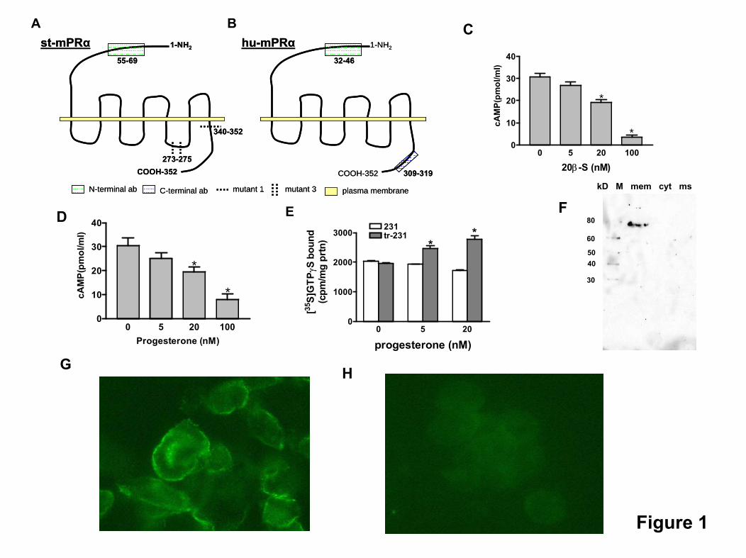
340-352
1-NH2
COOH-352273-275
st-mPRα55-69
1-NH2hu-mPRα
COOH-352
32-46
309-319
mutant 3mutant 1N-terminal ab C-terminal ab plasma membrane
340-352
1-NH2
COOH-352273-275
st-mPRα55-69
1-NH2hu-mPRα
COOH-352
32-46
309-319
mutant 3mutant 1N-terminal ab C-terminal ab plasma membrane
A B
0 5 20 1000
10
20
30
40
20β-S (nM)
cAM
P(pm
ol/m
l)
*
*
0 5 20 1000
10
20
30
40
Progesterone (nM)
cAM
P(pm
ol/m
l)
*
*
0 5 200
1000
2000
3000231tr-231
**
progesterone (nM)
[35S]
GTP
γS b
ound
(cpm
/mg
prtn
)
kD M mem cyt ms
30
4050
60
80
C
D E F
G H
Figure 1

Supplementary Figure 2. The total unrooted phylogenetics tree from all sequences, from a diverse set of species that revealed similarity to the PAQR/HLY3 signature. The results from this tree have been used to design figures 6 and 7 in the text. All sub-clusters have been annotated with the current PAQR family gene name nomenclature. The yellow shaded boxes indicate the genes that have undergone duplication in teleost fish and amphibians.
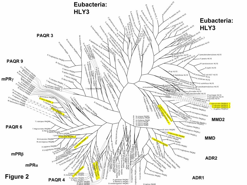
D.rerio PAQR5_1
D.rerio PAQR5_2
D.rerio PAQR2
T.nigroviridis PAQR2T.rubripes PAQR2
G.gallus PAQR2B.taurus PAQR2S.scrofa PAQR2P.troglodytes PAQR2
H.sapiens PAQR2
R.norvegicusPAQR2
M.musculus PAQR2
X.tropicalis
PAQ
R2
X.laevis
PAQ
R2
T.nigroviridis PAQR2
D.rerio
PAQ
R1
T.nigroviridis PAQR1
T.rubripes PAQR1
G.gallus PAQR1
B.taurus PAQR1R.norvegicus PAQR1M.musculus PAQR1
S.s
crofa
PAQ
R1
troglo
dyt
esPA
QR1
H.s
apie
ns
PAQ
R1
X.trop
icalis
PAQR1
X.lae
visPA
QR1
D.melanogaster PAQR1A.Mellifera PAQR1A.gambiae PAQR1
C.b
riggsae
PAQ
R1_2
C.E
legan
sPA
QR1
C.b
riggsae
PAQ
R1_1
C.eleg
ans
PAQ
R1_1
A.th
aliana
PAQ
RA.th
aliana
PAQ
RA.th
aliana
PAQ
RO.sativa PAQR
O.sativa PAQR
A.thaliana PAQR
N.crassa PAQR1
S.p
ombe
PAQ
R2
D.h
ansen
iiPA
QR2
Y.lip
olyt ica
PAQ
RE.g
ossypii
PAQ
R2
S.cerevisiae
PAQ
R2
K.lactis
PAQ
R4
C.g
labrata
PAQ
R4
E.g
ossypiiPA
QR
K.lactis
PAQ
R
C.g
labrata
PAQ
RS.cerevisiae
PAQ
RP.tetrau
reliaPA
QR
P.Tro
glo
dytes
PAQ
R4
B.T
auru
sPA
QR4
H.s
apie
ns
PAQ
R4
R.n
orve
gic
us
PAQ
R4
M.m
usc
ulu
sPA
QR4
X.laevis PAQR4D.Rerio PAQR4
T.rubripes PAQR4D.Rerio PAQR4
T.rubripes PAQR4
A.gambiae PAQR4D.melanogaster PAQR4
D.R
erioPA
QR7
T.Nigrovirid
isPA
QR7
O.latip
esPA
QR7
T.ru
brip
esPA
QR7_2
T.ru
brip
esPA
QR7_1
O.latip
esPA
QR7
C.N
ebulosu
sPA
QR7
T.Nigrovidiris
PAQ
R7
T.ru
bripes
PAQR7
I.pu
ncta
tus
PAQR7
C.au
ratu
sPA
QR7
D.re
rioPA
QR7
B.taurus PAQR8
B.ta
urus
PAQR7
S.scrofa PAQR7
H.sapiens PAQR7
P.troglodytes PAQR7
R.norvegicus PAQR7
M.musculus PAQR7
T.ru
bripe
s PAQR8_
1
X.lae
visPA
QR8
G.gall
usPA
QR8
G.g
allu
sPA
QR7
S.scrofa PAQR8
P.troglodytes PAQR8H.sapiens PAQR8
R.norvegicus PAQR8
M.musculus PAQR8T.rubripes PAQR8_2
I.punctatus PAQR8D.rerio pAQR8
B.Taurus PAQR6
X.laevis PAQR6D.Rerio PAQR6
T.Nigroviridis PAQR6
T.rubripes PAQR6
M.fascicularis PAQR6
P.Troglodytes PAQR6
H.sapiens PAQR6
R.norvegicus PAQR6
M.musculus PAQR6
G.gallus PAQR5
X.laevis PAQR
T.rubripes PAQR5
I.punctatus PAQR5
B.Taurus PAQR5
P.Troglodytes PAQR5
P.pygmaeus PAQR5
R.norvegicus PAQR5
M.musculus PAQR5
R.norvegicus PAQR9
M.musculus PAQR9
B.Taurus PAQR9
P.troglodytes PAQR9
H.sapiens PAQR9
T.NigroviridisPAQ
R9
T.RubripesPAQ
R9
C.Elegans PAQR9
C.briggsaePAQR9
D.hansenii PAQR3
T.Nigroviridis PAQR3_1
T.rubripes PAQR3_2
T.Nigroviridis PAQR3_2
T.rubripes PAQR3_1
D.Rerio PAQR3
X.laevis PAQR3
G.gallus PAQR3
X.laevis PAQR3
B.Taurus PAQR3
P.Troglodytes PAQR3
H.sapiensPAQ
R3R.norvegicus
PAQR3
M.m
uscu
lus
PAQ
R3
D.m
elanog
asterPA
QR3
A.M
elliferaPA
QR3
A.g
ambiae
PAQ
R3
C.B
rigssae
PAQ
R3
C.eleg
ans
PAQ
R3
N.crassa
PAQR3
Y.lipolytica PAQR3
K.lactis
PAQR3
S.cerevisiae
PAQR3_1
C.glabrata
PAQ
R3
S.cerevisiae
PAQ
R3_1
E.cuniculi PAQ
R3
P.falciparumPAQ
R3
T.Nigroviridis
PAQR3
y.phytoplasmaHLY3
F.nucleatumHLY3
S.pneumoniae
HLY3
S.pyogenes HLY3
S.agalactiae HLY3
S.thermophilus
HLY3S.m
utansHLY3
S.epidermidis HLY3
S.aureusHLY3
R.balticaHLY3
D.Vulgaris HLY3
T.thermophilus
HLY3
D.radioduransHLY3
C.DiphtheriaeHLY3
C.amm
oniagenesHLY3
C.glutamicum
HLY3
C.efficiens
HLY3
N.farcinicaHLY3
M.aviu
mH
LY3
M.tubercu
losisH
LY3
P.acnesHLY3
L.xyli HLY
3S.averm
itilisH
LY3
S.averm
itilisH
LY3S.coelicolor
HLY
3
B.lo
ngum
HLY
3_2
B.lo
ngum
HLY
3_1
D.p
sychrop
hilaH
LY3
B.frag
ilis
HLY
3
B.t
het
aiota
om
icro
nH
LY3
I.loihiensisPAQ
R1
D.discoideum
PAQ
R1
O.iheyensis
HLY3
B.clausii HLY3
B.halodurans
HLY3
B.cereus
HLY3_2
B.lic
henifo
rmis
HLY3
B.subti
lisHLY
3
L.mon
ocytog
enes
HLY3
L.innocua HLY3
E.fa
ecali
s HLY3
L.joh
nson
ii HLY
3
L.lactis
HLY3
L.plan
tarum
HLY3
T.denticola HLY3
T.pallidum
HLY3
C.perfringens HLY3
C.tetani HLY3
C.aceto
butylicu
mHLY3
O.ihey
ensis
HLY3
C.aceto
butyl
icum
HLY3
C.perfr
ingens HLY
3
B.thurin
giensis
HLY3
B.cereu
s HLY3_2
B.anthracis HLY3
B.garinii HLY3
B.burgdorferi HLY3
A.sp. HLY3
S.oneidensis HLY3
A.gambiae HLY3Y.pseudotuberculosis HLY3
Y.pestis HLY3
P.carotovorum HLY3
S.typhi HLY3
E.coli K57 HLY3P.profundum HLY3V.cholerae HLY3
V.vulnificus HLY3
V.parahaemolyticus HLY3
P.sp HLY3
X.fastidiosa HLY3
X.campestris HLY3
X.axonopodis HLY3
B.bacteriovorus HLY3N.meningitidis HLY3B.bacteriovorus HLY3C.violaceum HLY3B.mallei HLY3
B.pseudomallei HLY3P.syringae HLY3
P.putida HLY3P.aeruginosa HLY3
B.melitensis HLY3R.palustris HLY3
B.japonicum HLY3M.loti HLY3
A.tumefaciens HLY3S.meliloti HLY3
A.brasilense HLY3C.vibrioides HLY3X.campestris HLY3
X.axonopodis HLY3B.taurus PAQR10
T.nigroviridis PAQR10_1T.rubripes PAQR10_1T.rubripes PAQR10_2
G.gallus PAQR10
P.troglodytes PAQR10H.sapiens PAQR10R.norvegicus PAQR10M.musculus PAQR10
T.rubripes PAQR11
D.rerio PAQR11
G.gallus PAQR11X.tropicalis PAQR11
P.troglodytes PAQR11H.sapiens PAQR11R.norvegicus PAQR11M.musculus PAQR11
T.nigroviridisPAQ
R10_3
T.nigroviridisPAQ
R10_2
D.m
elanogasterPAQ
R
A.m
elliferaPA
QR10
A.gam
biaePA
QR10
C.briggsae
PAQ
R10
C.elegans
PAQ
R10
S.p
ombe
PAQ
R3
E.g
ossypii
PAQ
R3
H.sapiens PAQR5
O.sativa PAQRO.sativa PAQR
A.thaliana PAQR
12
34
56
654321
Figure 2
Eubacteria: HLY3
MMD2
MMD
ADR2
ADR1PAQR 4
mPRα
mPRβ
PAQR 6
PAQR 9
mPRγ
PAQR 3
Eubacteria: HLY3

Supplementary Figure 3. A unrooted phylogenetic tree classifying 4 mammals, showing the high similarity and clustering of mPRα, mPRβ, mPRγ, PAQR6 and PAQR9. These data suggest a possible functional similarity (steroid binding) of PAQR6 and 9.

R.norvegicus PAQR4
M.musculus PAQR4H.sapiens PAQR4
P.Troglodytes PAQR4
P.Troglodytes mPRα
H.sapiens mPRα
R.norvegicus mPRα
M.Musculus mPRαP.Troglodytes mPRβ
H.sapiens mPRβR.norvegicus mPRβ
M.musculus mPRβP.Troglodytes PAQR6
H.sapiens PAQR6R.norvegicus PAQR6
M.musculus PAQR6P.Troglodytes mPRγP.pygmaeus mPRγ
H.sapiens mPRγ
R.norvegicus mPRγ
M.musculus mPRγ
P.Troglodytes PAQR9
H.sapiens PAQR9
R.norvegicus PAQR9
M.musculus PAQR9
P.Troglodytes MMD
H.Sapiens MMD
R.norvegicus MMD
M.musculus MMD
P.Troglodytes MMD2H.sapiens MMD2
R.norvegicus MMD2M.musculus MMD2
P.Troglodytes ADR2H.sapiens PAQR3R.norvegicus PAQR3
M.musculus PAQR3 P.Troglodytes PAQR3
H.sapiens ADR2
R.norvegicus ADR2
M.musculus ADR2
R.norvegicus ADR1
M.musculus ADR1
P.Troglodytes ADR1
H.sapiens ADR1
Figure 3