stem cell therapy in acute myocardial infarction · citation: reejhsinghani r, shih hhj, lotfi as...
TRANSCRIPT

Research Article Open Access
Clinical & Experimental
CardiologyReejhsinghani et al., J Clin Exp Cardiolog 2012, S:11
http://dx.doi.org/10.4172/2155-9880.S11-004
ISSN:2155-9880 JCEC, an open access journalCardiac Stem CellsJ Clin Exp Cardiolog
*Corresponding author: Amir S Lotfi, MD, Department of Cardiology, Baystate Medical Center, Springfield, Massachusetts 01199, USA, E-mail: [email protected]
Received September 27, 2012; Accepted November 02, 2012; Published November 05, 2012
Citation: Reejhsinghani R, Shih HHJ, Lotfi AS (2012) Stem Cell Therapy in Acute Myocardial Infarction. J Clin Exp Cardiolog S11:004. doi:10.4172/2155-9880.S11-004
Copyright: © 2012 Reejhsinghani R, et al. This is an open-access article distributed under the terms of the Creative Commons Attribution License, which permits unrestricted use, distribution, and reproduction in any medium, provided the original author and source are credited.
AbstractOver the last several years, much headway has been made in the arena of coronary artery disease, specifically
in the rapid diagnosis and revascularization therapy of acute myocardial infarction. Despite these advances however, given the natural history of acute myocardial infarction (AMI), a significant proportion of patients are left with significant morbidities by consequences of impaired left ventricular function. Stem cell therapy, which was initially introduced as a novel approach to regenerate injured cardiac myocytes, has widely been gaining popularity as a feasible strategy for repairing injured myocardial muscle tissue.
Over a decade of basic and clinical research has gone into determining the effectiveness of targeted progenitor stem cell delivery in the improvement of myocardial function and cardiac physiology. Our paper is a general review of the stem cells therapy in patients that have had acute myocardial infarction. Although, much of the data thus far has been suggestive of the potential benefit of this approach in human models, a quest for a definitive answer is still underway. In the treatment of acute myocardial infarction, targeted stem cell therapy, at the very least, is a union of cellular biology and clinical cardiology, which albeit nascent in its development, has laid the framework for a clear direction into the future of cardiovascular medicine.
Stem Cell Therapy in Acute Myocardial InfarctionRisheen Reejhsinghani, Henry Han-Jen Shih and Amir S Lotfi*
Department of Cardiology, Baystate Medical Center, USA
Introduction“Ideal cardiovascular health” is defined by the absence of clinically
manifest cardiovascular disease and the simultaneous presence of optimal levels of all 7 health behaviors (lean body mass, avoidance of smoking, participation in physical activity, and healthy dietary intake) and health factors (untreated total cholesterol <200 mg/dL, untreated blood pressure <120/<80 mm Hg, and fasting blood glucose <100 mg/dL) [1].
On the basis of unpublished data from the Atherosclerosis Risk in Communities (ARIC) and Cardiovascular Health Study (CHS) of National Heart, Lung, and Blood Institute (NHLBI), this year 785000 Americans will have a new coronary attack and 470000 will have a recurrent attack. The estimated annual incidence of MI is 610000 new attacks and 325000 recurrent attacks [1]. The American Heart Association (AHA) recently created a new set of ‘Impact Goals’ for the current decade. The aim, by 2020, is to improve the cardiovascular health of all Americans by 20%, while reducing deaths from cardiovascular disease and stroke by 20% [2].
The early biochemical consequence of myocardial ischemia is the cessation of aerobic metabolism within seconds, leading to inadequate production of high-energy phosphates and accumulation of potentially noxious metabolites [3]. Because of the exquisite dependence of myocardial function on oxygen, severe ischemia induces loss of contractility within 60 seconds. Nevertheless, these early changes are potentially reversible. As demonstrated both experimentally and in clinical studies, only severe ischemia lasting 20 to 30 minutes or longer leads to irreversible necrosis of cardiac myocytes [3].
The current standards of practice for treatment of AMI are percutaneous coronary intervention (PCI), thrombolytic therapy if PCI facility is not available when appropriate [4]. The aim of any medical or surgical therapy however, is to establish prompt revascularization and limit the degree of myocardial injury. Yet the success of any of these therapies is dependent on one key factor, the time to presentation. Given this lacuna in the treatment of AMI, the need to investigate for therapies that add incremental benefit to the current established treatment options is an ongoing endeavor. It was promising to note
that the early effects achieved with bone marrow cell (BMC) therapy is comparable to what is achieved by established therapies including acute PCI, angiotensin-converting-enzyme inhibition, or β-blocker therapy [5].
Given the uncertainty of myocardial salvage, dictated by the degree of necrosis from the sentinel event, it soon became clear that in order to change the long term outcomes of AMI, we needed to search for a therapy that takes the time to presentation out of the equation. A possible solution to that dilemma appeared in the form of targeted stem cell therapy.
Evidence of resident cardiac stem cells (SC) began to formulate in studies of human patients who had died within 4-12 days after MI [6]. This concept further gained strength looking at patients who had undergone gender-mismatched heart transplants, in which hearts from female donor were implanted into male recipients, and at the time of death (ranging from 4 to 552 days after transplant), all donor hearts displayed Y-chromosome-positivity, suggesting that SC came from some location outside the heart [6].
Efficacy data suggests its potential to improve left ventricular (LV) function recovery beyond current state of the art therapy, but results are mixed, modest at best and do not supports true cardiomyogenesis. Hence, due to its complexity, costs and remaining uncertainties, it is still too early to implement progenitor cell therapy in its current form as a standard treatment strategy for ischemic heart disease [7].

Citation: Reejhsinghani R, Shih HHJ, Lotfi AS (2012) Stem Cell Therapy in Acute Myocardial Infarction. J Clin Exp Cardiolog S11:004. doi:10.4172/2155-9880.S11-004
Page 2 of 6
ISSN:2155-9880 JCEC, an open access journalCardiac Stem CellsJ Clin Exp Cardiolog
Types of Stem CellsSeveral different populations of cells have been studied for their
use in post MI patients. The most common types are enlisted in table 1.
Most clinical studies have used unfractionated bone marrow cells as the delivery product, postulating that stem and progenitor cells within this population are the biologically relevant therapeutic agents [8].
However, it is the embryonic stem cells and induced pluripotent stem cells which are the only cell types that currently have the potential to generate bona fide cardiomyocytes on a scale that may potentially replace the cell numbers lost in AMI [7].
Stem cells may be classified as autologous or allogenic, depending on their origin. Allogenic cells provide a promising option for stem cell therapy, given that they are free from the immunogenic complications and also the risks of malignancy. However, autologous cells have fairly limited differentiation potential, when compared with allogenic stem cells. SC may also be subdivided into adult cells and fetal and embryonic cells depending on the source they were derived from.
Embryonic stem cells
Embryonic stem cells (ESC) are derived from the blastocyst (inner cell mass) of human embryos prior to implantation. They have the capability to differentiate into any cell from the three germ lines, one of which is cardiac myocytes. However, their inherent totipotency also predisposes to tumor formation including teratomas, which have been observed in animal models [9]. Additionally, there is controversy surrounding the ethical issues of ESC use [10].
Human umbilical cord cells
Cord blood contains a large number of non-hematopoietic stem cells which rarely express human leukocyte antigen (HLA) class II antigens and appear to be immunologically naïve, thus reducing the risk of rejection. In animal models of AMI injection of human umbilical cord blood cell (hUCBC) is associated with significant reductions in infarct size, particularly when given by the intramyocardial route [11].
Fetal cardiomyocytes
These are multipotent cells which already have a similar phenotype to cardiac cells. However, there is use is fraught with similar concerns about ethical appropriateness, as is with ESC. Furthermore, these cells have immunogenicity, necessitating the use of immune suppression post transplant.
Induced pluripotent stem cells
Induced pluripotent stem cells (iPSC) can be generated from fully differentiated somatic cells by genetic reprogramming, thereby resulting in a fully undifferentiated ESC-like phenotype, capable of extensive in vitro proliferation and differentiation towards cells from all three germ layers [12]. These cells are obtained from the patient’s own skin fibroblasts, thereby alleviating any immunological discord.
Resident cardiac stem cells
These cells have the potential to differentiate into different lineages like vascular smooth muscle and myocardial cells. These cells have been studied in animal models of myocardial infarction with beneficial outcomes in terms of reducing infarct size and improving LV function [13]. However, it is unclear how feasible it is to produce these cells in numbers that are adequate for post MI cardiac repair.
Adipose and skeletal muscle derived stem cells
Adipose derived stem cells (ASC) are easy to harvest via liposuction and contain, in addition to mesenchymal elements, hematopoietic and endothelial cell lines. Preclinical studies have shown that ASC are associated with improvement in ejection fraction in animal models of myocardial infarction and neoangiogenesis via paracrine factors [14].
Skeletal muscle cells are harvested via muscle biopsy from the index patient, thereby mitigating any immunogenicity issues. Preclinical animal studies have demonstrated the ability for skeletal myoblasts to engraft, form myotubules and enhance cardiac function after transplantation into infarcted myocardium [15]. Unfortunately, it appears that the grafts do not couple electrically with adjacent cardiomyocytes [16] and these islands of transplanted cells with different electrophysiological properties, may serve as an arrhythmogenic substrate [17].
Bone marrow derived stem cells
Bone marrow stem cell has been the most widely studied, after a landmark trial published by Orlic et al. [18] which demonstrated that bone marrow cells regenerate infarcted myocardium in mouse models [18]. The transplanted cells appeared to undergo transdifferentiation into cardiomyocytes with newly formed myocardium occupying a significant proportion of the infarcted area with significant improvement in the left ventricular ejection fraction (LVEF) just 9 days after cell transplantation. Furthermore, bone marrow cells (BMC) transplanted into rats following left anterior descending (LAD) artery ligation were shown to improve cardiac function and induce angiogenesis, with formation of new capillaries positive for human markers [19].
Experimental data suggested that stem cells from the mononuclear fraction of BMC undergo transdifferentiation, a process by which they differentiate into other cell lineages, including cardiomyocytes. However, the currently used timing, dosing and modes of administration of bone marrow stem cell (BMSC) is not well founded on clinical evidence, but is mainly based on theoretical assumptions, observations in small animal models, post hoc analysis from small clinical trials and practical feasibility in the clinical setting [9].
Methods of DeliveryThe importance of cell delivery lies in the fact that cell retention,
defined as how much of the delivered dose reaches the area of interest, is in large part determined by how the cells are administered. Table 1: Types of Stem Cells.
TYPES OF STEM CELLSEmbryonic Stem Cells
Human Umbilical Cord Cells
Fetal Cardiomyocytes
Induced Pluripotent Stem Cells
Resident Cardiac Stem Cells
Adipose and Skeletal Muscle Derived Stem Cells
Bone Marrow Derived Stem Cells
Citation: Reejhsinghani R, Shih HHJ, Lotfi AS (2012) Stem Cell Therapy in Acute Myocardial Infarction. J Clin Exp Cardiolog S11:004. doi:10.4172/2155-9880.S11-004
Page 3 of 6
ISSN:2155-9880 JCEC, an open access journalCardiac Stem CellsJ Clin Exp Cardiolog
Progenitor cells can be delivered either via the transvascular approach, peripherally or via retrograde administration through the coronary sinus or by direct intramyocardial injection [7].
Intracoronary infusion
This technique is reminiscent of that used in coronary angioplasty. Stem cells are infused under pressure via a balloon catheter while simultaneously occluding antegrade coronary blood flow. A preclinical study in dogs raised concerns regarding the possibility of mircoinfarcts caused by intracoronary injection of mesenchymal stromal cells [20] and there is likely to be a safety threshold with regards to the size and dose of cells delivered using the intracoronary route. The advantage of intracoronary delivery is that cells are directly injected into areas of good blood supply, rich in nutrients and oxygen, which is essential for cell survival. Myocardial ischemia is a major stimulus for incorporation of circulating progenitor cells, and potently up-regulates the chemo-attractants for neoangiogenesis [21].
Intravenous (peripheral) infusion
This technique is applicable only in patients with AMI as it is reliant on physiological homing signals, which are not present in chronic heart failure. Another significant limitation is that only a few cells appear to reach the affected area due to trapping of the cells in the microvasculature of the lungs, liver and lymphoid tissues [22].
Granulocyte colony stimulating factor (G-CSF) is one of the options that have been studied as a method to stimulate and mobilize bone marrow stem cells, but the data published so far has been equivocal on any clear cut benefit of G-CSF.
Intramyocardial injection
Quite the opposite of peripheral infusion, intramyocardial injection of stem cells is a technique that is more effective in chronic ischemic cardiomyopathy. In contrast to the AMI setting, these patients are unlikely to release signals from chronically infarcted myocardium to induce stem cell homing and therefore it may be more effective to use intramyocardial injection to deliver the cells to the target area [14]. Stem cells delivered by this method use either a transepicardial or transendocardial approach of injection.
Transplantation of engineered tissue
The post-infarct myocardium has drastic changes in tissue microenvironment from the original hypoxic insult, reperfusion injury, and the inflammatory process. Injection of stem cell directly to the dynamic cardiac tissue usually results in significant cell loss either via mechanical damage from the injection or lack of survivability in the harsh post-infarct environment [23,24]. Tissue engineering explores the concept of delivering stem cells on a protected structure to the post-infarct myocardium [25,26]. It has potential for allowing efficient delivery of adequate stem cells by ensuring the transplant stem cell stay on the engineered structure. Additionally, cell signaling factors on the structures can provide a pro-survival microenvironment and pro-differentiation environment for the stem cell. To date, several methods of engineered tissue transplantation has been explored, including collagen patch or extracellular matrix patch, biological sutures, and fibrin micro threads [27-31]. The experimental results generally favors delivering stem cell via engineered tissue versus direct injection in quantity of retaining transplanted stem cells and precision of delivery into the post-infarct-myocardium. Future studies are needed to evaluate the cardiac functional end-point, differentiability of transplanted engineered tissue, and long term survival of the transplanted tissue.
Mechanism of ActionIn order to produce a therapeutic response, stem cells need to home
to the injured myocardium, adhere to and transmigrate through the endothelium, invade the interstitium and finally engraft the damaged myocardium [7]. In addition to the characteristics of the stem cells themselves, the property of the infarcting myocardial tissue, as it passes from a peri-ischemic phase, right until remodeling occurs, plays a tremendous role in the stem cell biophysiology.
MI results in loss of functional myocardium due to hypoxic necrosis, inflammatory change and cardiomyocyte apoptosis. Besides progressing through different stages of inflammation and healing, the dynamic microenvironment in the infarcted tissue also express cardiac cytokines that promotes stem cell migration and homing [32]. Transplanting stem cells to the post-infarct myocardium augment the cytokine effect to attract endogenous stem cells.
Other paracrines secreted by stem cells thought to have therapeutic effect by promoting angiogenesis, proliferation of endogenous vascular cells, loosening of fibrotic extracellular matrix, inhibition of cardiomyocyte apoptosis, and regulation of inflammatory response. Together the paracrine signals from stem cells expedite wound healing and promote the endogenous myocardial regeneration process [33].
In addition to the paracrine signaling effects, stem cells carry the potential to differentiate into functional myocardium [34]. The supplementation of exogenous stem cell post-MI can engraft into the existing myocardium as functional cardiomyocyte that beats in synchrony with the existing myocardium. Stem cells can also differentiate into smooth muscle cells and vascular endothelial cells. Together, the delivery and differentiation of stem cells replenishes the lost cardiomyocytes from MI and provides increased vascularity in the post-injury zone to prevent further ischemic tissue damage [35].
Clinical Trials Using Stem Cells in Acute MIAlthough, multiple experimental animal models and clinical trials
of cell-based cardiac therapy have delivered promising results, the mechanisms of their effect are unclear [36]. Clinical trials in humans were aimed at determining whether stem cell therapy could translate into a feasible therapeutic modality.
The majority of large clinical trials used bone marrow stem cells alone, but some trials did use two populations. The TOPCARE AMI [37], REGENT [38] and HEBE [39] trials were ones that used a second cell line in addition to BMCs. Most commonly stem cells were delivered via intracoronary injection and some of the parameters that were assessed for significance were, increase in left ventricular ejection fraction, decrease in end systolic and end diastolic volumes and reduction in infarct size. These parameters were measured by LV angiography, MRI, SPECT or 2D echocardiography.
The first clinical trial performed in humans was published in 2002 by Strauer et al. [40] in Germany. It was a non-randomized trial that studied 10 patients in each arm with one group receiving intracoronary BMCs in addition to the standard therapy for AMI, in the form of PCI. After 3 months of follow-up, left ventriculography showed a significant decrease in the infarct region within the cell therapy group (from 30 ± 13 to 12 ± 7%, P=0.005) and was also significantly smaller compared with the standard therapy group (P=0.04) [40]. They also demonstrated a significant improvement in stroke volume index, left ventricular end-systolic volume, contractility and myocardial perfusion of the infarct region, within the study group. Their group concluded that the marked
Citation: Reejhsinghani R, Shih HHJ, Lotfi AS (2012) Stem Cell Therapy in Acute Myocardial Infarction. J Clin Exp Cardiolog S11:004. doi:10.4172/2155-9880.S11-004
Page 4 of 6
ISSN:2155-9880 JCEC, an open access journalCardiac Stem CellsJ Clin Exp Cardiolog
therapeutic effect noted in their study could be attributed to BMC-associated myocardial regeneration and neovascularization [41].
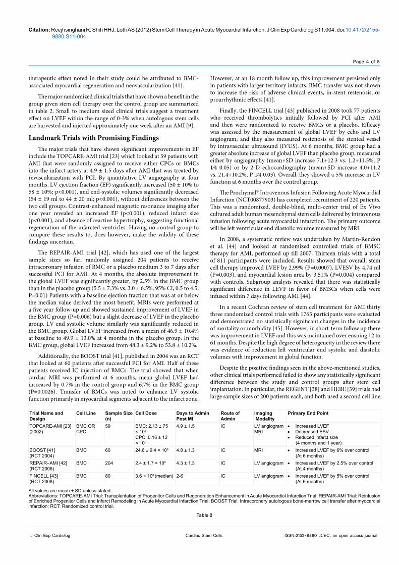
The major randomized clinical trials that have shown a benefit in the group given stem cell therapy over the control group are summarized in table 2. Small to medium sized clinical trials suggest a treatment effect on LVEF within the range of 0-3% when autologous stem cells are harvested and injected approximately one week after an AMI [9].
Landmark Trials with Promising FindingsThe major trials that have shown significant improvements in EF
include the TOPCARE-AMI trial [23] which looked at 59 patients with AMI that were randomly assigned to receive either CPCs or BMCs into the infarct artery at 4.9 ± 1.5 days after AMI that was treated by revascularization with PCI. By quantitative LV angiography at four months, LV ejection fraction (EF) significantly increased (50 ± 10% to 58 ± 10%; p<0.001), and end-systolic volumes significantly decreased (54 ± 19 ml to 44 ± 20 ml; p<0.001), without differences between the two cell groups. Contrast-enhanced magnetic resonance imaging after one year revealed an increased EF (p<0.001), reduced infarct size (p<0.001), and absence of reactive hypertrophy, suggesting functional regeneration of the infarcted ventricles. Having no control group to compare these results to, does however, make the validity of these findings uncertain.
The REPAIR-AMI trial [42], which has used one of the largest sample sizes so far, randomly assigned 204 patients to receive intracoronary infusion of BMC or a placebo medium 3 to 7 days after successful PCI for AMI. At 4 months, the absolute improvement in the global LVEF was significantly greater, by 2.5% in the BMC group than in the placebo group (5.5 ± 7.3% vs. 3.0 ± 6.5%; 95% CI, 0.5 to 4.5; P=0.01) Patients with a baseline ejection fraction that was at or below the median value derived the most benefit. MRIs were performed at a five year follow-up and showed sustained improvement of LVEF in the BMC group (P=0.006) but a slight decrease of LVEF in the placebo group. LV end systolic volume similarly was significantly reduced in the BMC group. Global LVEF increased from a mean of 46.9 ± 10.4% at baseline to 49.9 ± 13.0% at 4 months in the placebo group. In the BMC group, global LVEF increased from 48.3 ± 9.2% to 53.8 ± 10.2%.
Additionally, the BOOST trial [41], published in 2004 was an RCT that looked at 60 patients after successful PCI for AMI. Half of these patients received IC injection of BMCs. The trial showed that when cardiac MRI was performed at 6 months, mean global LVEF had increased by 0.7% in the control group and 6.7% in the BMC group (P=0.0026). Transfer of BMCs was noted to enhance LV systolic function primarily in myocardial segments adjacent to the infarct zone.
However, at an 18 month follow up, this improvement persisted only in patients with larger territory infarcts. BMC transfer was not shown to increase the risk of adverse clinical events, in-stent restenosis, or proarrhythmic effects [41].
Finally, the FINCELL trial [43] published in 2008 took 77 patients who received thrombolytics initially followed by PCI after AMI and then were randomized to receive BMCs or a placebo. Efficacy was assessed by the measurement of global LVEF by echo and LV angiogram, and they also measured restenosis of the stented vessel by intravascular ultrasound (IVUS). At 6 months, BMC group had a greater absolute increase of global LVEF than placebo group, measured either by angiography (mean+SD increase 7.1+12.3 vs. 1.2+11.5%, P 1⁄4 0.05) or by 2-D echocardiography (mean+SD increase 4.0+11.2 vs. 21.4+10.2%, P 1⁄4 0.03). Overall, they showed a 5% increase in LV function at 6 months over the control group.
The Prochymal® Intravenous Infusion Following Acute Myocardial Infarction (NCT00877903) has completed recruitment of 220 patients. This was a randomized, double-blind, multi-center trial of Ex Vivo cultured adult human mesenchymal stem cells delivered by intravenous infusion following acute myocardial infarction. The primary outcome will be left ventricular end diastolic volume measured by MRI.
In 2008, a systematic review was undertaken by Martin-Rendon et al. [44] and looked at randomized controlled trials of BMSC therapy for AMI, performed up till 2007. Thirteen trials with a total of 811 participants were included. Results showed that overall, stem cell therapy improved LVEF by 2.99% (P=0.0007), LVESV by 4.74 ml (P=0.003), and myocardial lesion area by 3.51% (P=0.004) compared with controls. Subgroup analysis revealed that there was statistically significant difference in LEVF in favor of BMSCs when cells were infused within 7 days following AMI [44].
In a recent Cochran review of stem cell treatment for AMI thirty three randomized control trials with 1765 participants were evaluated and demonstrated no statistically significant changes in the incidence of mortality or morbidity [45]. However, in short-term follow up there was improvement in LVEF and this was maintained over ensuing 12 to 61 months. Despite the high degree of heterogeneity in the review there was evidence of reduction left ventricular end systolic and diastolic volumes with improvement in global function.
Despite the positive findings seen in the above-mentioned studies, other clinical trials performed failed to show any statistically significant difference between the study and control groups after stem cell implantation. In particular, the REGENT [38] and HEBE [39] trials had large sample sizes of 200 patients each, and both used a second cell line
Trial Name and Design
Cell Line Sample Size(n)
Cell Dose Days to Admin Post MI
Route of Admin
Imaging Modality
Primary End Point
TOPCARE-AMI [23](2002)
BMC OR CPC
59 BMC: 2.13 ± 75 × 108
CPC: 0.16 ± 12 × 108
4.9 ± 1.5 IC LV angiogramMRI
• Increased LVEF• Decreased ESV• Reduced infarct size (4 months and 1 year)
BOOST [41](RCT 2004)
BMC 60 24.6 ± 9.4 × 10⁸ 4.8 ± 1.3 IC MRI • Increased LVEF by 6% over control (At 6 months)
REPAIR–AMI [42](RCT 2006)
BMC 204 2.4 ± 1.7 × 10⁸ 4.3 ± 1.3 IC LV angiogram • Increased LVEF by 2.5% over control (At 4 months)
FINCELL [43](RCT 2008)
BMC 80 3.6 × 10⁸ (median) 2-6 IC LV angiogram • Increased LVEF by 5% over control (At 6 months)
All values are mean ± SD unless statedAbbreviations: TOPCARE-AMI Trial: Transplantation of Progenitor Cells and Regeneration Enhancement in Acute Myocardial Infarction Trial; REPAIR-AMI Trial: Reinfusion of Enriched Progenitor Cells and Infarct Remodeling in Acute Myocardial Infarction Trial; BOOST Trial: Intracoronary autologous bone-marrow cell transfer after myocardial infarction; RCT: Randomized control trial.
Table 2

Citation: Reejhsinghani R, Shih HHJ, Lotfi AS (2012) Stem Cell Therapy in Acute Myocardial Infarction. J Clin Exp Cardiolog S11:004. doi:10.4172/2155-9880.S11-004
Page 5 of 6
ISSN:2155-9880 JCEC, an open access journalCardiac Stem CellsJ Clin Exp Cardiolog
in addition to BMCs. However, when follow up MRIs were performed on the patients in both these studies; there was no significant increase in LVEF as compared to the control groups.
The recently published CADUCEUS [46] Trial primarily assessed safety after intracoronary injection of cardiosphere-derived cells (CDC) in patients after AMI. The primary endpoint of this trial was death at 6 months, arrhythmias, recurrent MI after cell infusion, occurrence of cardiac tumors or of a major adverse cardiac event. By 6 months, none of these end points were noted in either group. “Four patients (24%) in the CDC group had serious adverse events compared with one control (13%; P=1·00). MRI analysis of patients treated with CDCs showed reductions in scar mass (P=0·001), increases in viable heart mass (P=0·01) and regional contractility (P=0·02), and regional systolic wall thickening (P=0·015).” However, at 6 months, changes in end-diastolic volume, end-systolic volume, and LVEF did not differ between the control and study group.
Some of the landmark studies with equivocal findings are summarized in table 3. There are multiple factors that may have played into such equivocal findings, including the diversity of patient selection, method and dose of stem cells delivery and the imaging used to assess left ventricular function and myocardial perfusion and infarct size.
The Onward JourneyAs a theoretical construct, the concept of using biological
engineering techniques to counteract the destructive effects of myocardial infarction is extremely appealing. Furthermore, the use of stem cells appeared to be the answer that was long sought after, when even timely interventions like primary PCI and thrombolytic could not salvage sufficient myocardium in the face of an acute MI.
While some of the clinical trials have shown promising results with respect to recovery of EF and reduction in infarct size, the overall results in several studies have proved essentially equivocal. Similarly, positive animal studies have not been replicated in humans. This leaves much uncharted territory and going forward many questions still need to be addressed. Which cell type and mode delivery works in which clinical
setting (acute, sub acute, or chronic), mode of delivery, concentration, and using clinical outcomes rather than surrogates?
Clearly a path has been fashioned for the initiation of further and bolder studies which hopefully will deal with some of these unanswered questions and reveal if the benefits shown in animal models and hinted at in human studies can translate into an accepted standard of practice and improve clinical outcome.
References
1. Roger VL, Go AS, Lloyd-Jones DM, Adams RJ, Berry JD, et al. (2011) Heart disease and stroke statistics--2011 update: a report from the American Heart Association. Circulation 123: e18-e209.
2. Lloyd-Jones DM, Hong Y, Labarthe D, Mozaffarian D, Appel LJ, et al. (2010) Defining and setting national goals for cardiovascular health promotion and disease reduction: the American Heart Association’s strategic Impact Goal through 2020 and beyond. Circulation 121: 586-613.
3. Robbins SL, Kumar V, Cotran RS (2010) Robbins and Cotran pathologic basis of disease. (8thedn), Philadelphia: Saunders/Elsevier.
4. Kushner FG, Hand M, Smith SC Jr, King SB 3rd, Anderson JL, et al. (2009) 2009 Focused Updates: ACC/AHA Guidelines for the Management of Patients With ST-Elevation Myocardial Infarction (updating the 2004 Guideline and 2007 Focused Update) and ACC/AHA/SCAI Guidelines on Percutaneous Coronary Intervention (updating the 2005 Guideline and 2007 Focused Update): a report of the American College of Cardiology Foundation/American Heart Association Task Force on Practice Guidelines. Circulation 120: 2271-2306.
5. Reffelmann T, Konemann S, Kloner RA (2009) Promise of blood- and bone marrow-derived stem cell transplantation for functional cardiac repair: putting it in perspective with existing therapy. J Am Coll Cardiol 53: 305-308.
6. Dauwe DF, Janssens SP (2011) Stem cell therapy for the treatment of myocardial infarction. Curr Pharm Des 17: 3328-3340.
7. Mummery CL, Davis RP, Krieger JE (2010) Challenges in using stem cells for cardiac repair. Sci Transl Med 2: 27ps17.
8. Boilson BA, Gulati R (2010) Stem cell therapy for the heart: a perspective. Transl Res 155: 3-5.
9. Beitnes JO, Lunde K, Brinchmann JE, Aakhus S (2011) Stem cells for cardiac repair in acute myocardial infarction. Expert Rev Cardiovasc Ther 9: 1015-1025.
All values are mean ± SD unless statedAbbreviations: ASTAMI Trial: Autologous Stem cell Transplantation in Acute Myocardial Infarction Trial; REGENT Trial: Myocardial Regeneration by Intracoronary Infusion of Selected Population of Stem Cells in Acute Myocardial Infarction Trial; HEBE Trial: Intracoronary infusion of autologous mononuclear bone marrow cells or peripheral mononuclear blood cells after primary PCI Trial; BONAMI Trial: Bone Marrow in Acute Myocardial Infarction Trial; CADUCEUS Trial: Intracoronary cardiosphere-derived cells for heart regeneration after myocardial infarction trial; MRI: Magnetic resonance imaging, SPECT: Single Photon Emission Computed Tomography; CPC: Cardiac progenitor cell; LVESV: Left ventricular end systolic volume; PBC: Peripheral blood cell; RNA: Radionuclide angiography, CDC: Cardiosphere derived cell.
Table 3
Trial Name and Design
Cell Line Sample Size(n)
Cell Dose Days to Admin Post MI
Route of Admin
ImagingModality
Primary End Point
LEUVEN-AMI [47](RCT 2006)
BMC 67 3.0 ± 1.3 × 10⁸ <24 hours IC MRI • No increase in EF over control (At 4 months) • Possible decrease in infarct
sizeASTAMI [48](RCT 2006)
BMC 100 0.68 × 10⁸ (median) 6 ± 1 IC SPECT • No increase in EF over control (At 6 months)
REGENT [38](RCT 2009)
BMC orCD34/CXCR4
200 BMC: 1.78 × 10⁸ (median)CD34/CXCR4: 0.019 × 10⁸
(median)
3-12 IC MRI • No increase in EF over control
HEBE [39](RCT 2010)
BMC or mPBC
200 BMC: 3 ± 1.6 × 10⁸
mPBC: 2.9 ± 1.4 × 1083-8 IC MRI • No increase in EF over control
(At 4 months) BONAMI [49] (RCT 2009)
BMC 101 0.98 ± 0.09 × 10⁸ 9.3 ± 1.7 IC SPECTRNAMRIECHO
• No increase in EF over control• No decrease in infarct size (At 3 months)
CADUCEUS [46](RCT 2012)
CDC 31 12.5-25 × 106 1.5–3 mths IC MRI • No increase in EF over control (At 6 months)
Citation: Reejhsinghani R, Shih HHJ, Lotfi AS (2012) Stem Cell Therapy in Acute Myocardial Infarction. J Clin Exp Cardiolog S11:004. doi:10.4172/2155-9880.S11-004
Page 6 of 6
ISSN:2155-9880 JCEC, an open access journalCardiac Stem CellsJ Clin Exp Cardiolog
10. de Wert G, Mummery C (2003) Human embryonic stem cells: research, ethics and policy. Hum Reprod 18: 672-682.
11. Henning RJ, Burgos JD, Vasko M, Alvarado F, Sanberg CD, et al. (2007) Human cord blood cells and myocardial infarction: effect of dose and route of administration on infarct size. Cell Transplant 16: 907-917.
12. Takahashi K, Yamanaka S (2006) Induction of pluripotent stem cells from mouse embryonic and adult fibroblast cultures by defined factors. Cell 126: 663-676.
13. Oh H, Bradfute SB, Gallardo TD, Nakamura T, Gaussin V, et al. (2003) Cardiac progenitor cells from adult myocardium: homing, differentiation, and fusion after infarction. Proc Natl Acad Sci USA 100:12313-12318.
14. Mozid AM, Arnous S, Sammut EC, Mathur A (2011) Stem cell therapy for heart diseases. Br Med Bull 98: 143-159.
15. Taylor DA, Atkins BZ, Hungspreugs P, Jones TR, Reedy MC, et al. (1998) Regenerating functional myocardium: improved performance after skeletal myoblast transplantation. Nat Med 4: 929-933.
16. Leon MB, Piazza N, Nikolsky E, Blackstone EH, Cutlip DE, et al. (2011) Standardized endpoint definitions for Transcatheter Aortic Valve Implantation clinical trials: a consensus report from the Valve Academic Research Consortium. J Am Coll Cardiol 57: 253-269.
17. Fernandes S, Amirault JC, Lande G, Nguyen JM, Forest V, et al. (2006) Autologous myoblast transplantation after myocardial infarction increases the inducibility of ventricular arrhythmias. Cardiovasc Res 69: 348-358.
18. Orlic D, Kajstura J, Chimenti S, Jakoniuk I, Anderson SM, et al. (2001) Bone marrow cells regenerate infarcted myocardium. Nature 410: 701-705.
19. Kocher AA, Schuster MD, Szabolcs MJ, Takuma S, Burkhoff D, et al. (2001) Neovascularization of ischemic myocardium by human bone-marrow-derived angioblasts prevents cardiomyocyte apoptosis, reduces remodeling and improves cardiac function. Nat Med 7: 430-436.
20. Vulliet PR, Greeley M, Halloran SM, MacDonald KA, Kittleson MD (2004) Intra-coronary arterial injection of mesenchymal stromal cells and microinfarction in dogs. Lancet 363: 783-784.
21. Arnous S, Mozid A, Martin J, Mathur A (2012) Bone marrow mononuclear cells and acute myocardial infarction. Stem Cell Res Ther 3: 2.
22. Gao J, Dennis JE, Muzic RF, Lundberg M, Caplan AI (2001) The dynamic in vivo distribution of bone marrow-derived mesenchymal stem cells after infusion. Cells Tissues Organs 169: 12-20.
23. Assmus B, Schächinger V, Teupe C, Britten M, Lehmann R, et al. (2002) Transplantation of Progenitor Cells and Regeneration Enhancement in Acute Myocardial Infarction (TOPCARE-AMI). Circulation 106: 3009-3017.
24. Barbash IM, Chouraqui P, Baron J, Feinberg MS, Etzion S, et al. (2003) Systemic delivery of bone marrow-derived mesenchymal stem cells to the infarcted myocardium: feasibility, cell migration, and body distribution. Circulation 108: 863-868.
25. Gu Y, Yu J, Lum LG, Lee RJ (2007) Tissue engineering and stem cell therapy for myocardial repair. Front Biosci 12: 5157-5165.
26. Christman KL, Lee RJ (2006) Biomaterials for the treatment of myocardial infarction. J Am Coll Cardiol 48: 907-913.
27. Cortes-Morichetti M, Frati G, Schussler O, Duong Van Huyen JP, Lauret E, et al. (2007) Association between a cell-seeded collagen matrix and cellular cardiomyoplasty for myocardial support and regeneration. Tissue Eng 13: 2681-2687.
28. Proulx MK, Carey SP, Ditroia LM, Jones CM, Fakharzadeh M, et al. (2011) Fibrin microthreads support mesenchymal stem cell growth while maintaining differentiation potential. J Biomed Mater Res A 96: 301-312.
29. Guyette JP, Fakharzadeh M, Burford EJ, Tao ZW, Pins GD, et al. (2012) A novel suture-based method for efficient transplantation of stem cells. J Biomed Mater Res A .
30. Potapova IA, Doronin SV, Kelly DJ, Rosen AB, Schuldt AJ, et al. (2008) Enhanced recovery of mechanical function in the canine heart by seeding an extracellular matrix patch with mesenchymal stem cells committed to a cardiac lineage. Am J Physiol Heart Circ Physiol 295: H2257-2263.
31. Simpson D, Liu H, Fan TH, Nerem R, Dudley SC Jr (2007) A tissue engineering approach to progenitor cell delivery results in significant cell engraftment and improved myocardial remodeling. Stem Cells 25: 2350-2357.
32. Askari AT, Unzek S, Popovic ZB, Goldman CK, Forudi F, et al. (2003) Effect of stromal-cell-derived factor 1 on stem-cell homing and tissue regeneration in ischaemic cardiomyopathy. Lancet 362: 697-703.
33. Williams AR, Hare JM (2011) Mesenchymal stem cells: biology, pathophysiology, translational findings, and therapeutic implications for cardiac disease. Circ Res 109: 923-940.
34. Hsieh PC, Segers VF, Davis ME, MacGillivray C, Gannon J, et al. (2007) Evidence from a genetic fate-mapping study that stem cells refresh adult mammalian cardiomyocytes after injury. Nat Med 13: 970-974.
35. Hosoda T, Kajstura J, Leri A, Anversa P (2010) Mechanisms of myocardial regeneration. Circ J 74: 13-17.
36. George JC (2010) Stem cell therapy in acute myocardial infarction: a review of clinical trials. Transl Res 155: 10-19.
37. Schächinger V, Assmus B, Britten MB, Honold J, Lehmann R, et al. (2004) Transplantation of progenitor cells and regeneration enhancement in acute myocardial infarction: final one-year results of the TOPCARE-AMI Trial. J Am Coll Cardiol 44: 1690-1699.
38. Tendera M, Wojakowski W, Ruzyllo W, Chojnowska L, Kepka C, et al. (2009) Intracoronary infusion of bone marrow-derived selected CD34+CXCR4+ cells and non-selected mononuclear cells in patients with acute STEMI and reduced left ventricular ejection fraction: results of randomized, multicentre Myocardial Regeneration by Intracoronary Infusion of Selected Population of Stem Cells in Acute Myocardial Infarction (REGENT) Trial. Eur Heart J 30: 1313-1321.
39. Hirsch A, Nijveldt R, van der Vleuten PA, Tijssen JG, van der Giessen WJ, et al. (2011) Intracoronary infusion of mononuclear cells from bone marrow or peripheral blood compared with standard therapy in patients after acute myocardial infarction treated by primary percutaneous coronary intervention: results of the randomized controlled HEBE trial. Eur Heart J 32: 1736-1747.
40. Strauer BE, Brehm M, Zeus T, Köstering M, Hernandez A, et al. (2002) Repair of infarcted myocardium by autologous intracoronary mononuclear bone marrow cell transplantation in humans. Circulation 106: 1913-1918.
41. Wollert KC, Meyer GP, Lotz J, Ringes-Lichtenberg S, Lippolt P, et al. (2004) Intracoronary autologous bone-marrow cell transfer after myocardial infarction: the BOOST randomised controlled clinical trial. Lancet 364: 141-148.
42. Schächinger V, Erbs S, Elsässer A, Haberbosch W, Hambrecht R, et al. (2006) Intracoronary bone marrow-derived progenitor cells in acute myocardial infarction. N Engl J Med 355: 1210-1221.
43. Huikuri HV, Kervinen K, Niemelä M, Ylitalo K, Säily M, et al. (2008) Effects of intracoronary injection of mononuclear bone marrow cells on left ventricular function, arrhythmia risk profile, and restenosis after thrombolytic therapy of acute myocardial infarction. Eur Heart J 29: 2723-2732.
44. Martin-Rendon E, Brunskill SJ, Hyde CJ, Stanworth SJ, Mathur A, et al. (2008) Autologous bone marrow stem cells to treat acute myocardial infarction: a systematic review. Eur Heart J 29: 1807-1818.
45. Clifford DM, Fisher SA, Brunskill SJ, Doree C, Mathur A, et al. (2012) Stem cell treatment for acute myocardial infarction. Cochrane Database Syst Rev 2: CD006536.
46. Makkar RR, Smith RR, Cheng K, Malliaras K, Thomson LE, et al. (2012) Intracoronary cardiosphere-derived cells for heart regeneration after myocardial infarction (CADUCEUS): a prospective, randomised phase 1 trial. Lancet 379: 895-904.
47. Janssens S, Dubois C, Bogaert J, Theunissen K, Deroose C, et al. (2006) Autologous bone marrow-derived stem-cell transfer in patients with ST-segment elevation myocardial infarction: double-blind, randomised controlled trial. Lancet 367: 113-121.
48. Lunde K, Solheim S, Aakhus S, Arnesen H, Abdelnoor M, et al. (2006) Intracoronary injection of mononuclear bone marrow cells in acute myocardial infarction. N Engl J Med 355: 1199-1209.
49. Roncalli J, Mouquet F, Piot C, Trochu JN, Le Corvoisier P, et al. (2011) Intracoronary autologous mononucleated bone marrow cell infusion for acute myocardial infarction: results of the randomized multicenter BONAMI trial. Eur Heart J 32: 1748-1757.