stem cell microrna qpcr array manual - system … · greener™ qpcr supermix for abi prism®...
TRANSCRIPT

Stem Cell MicroRNA qPCR Array with QuantiMir™
Cat. # RA620A-1
User Manual
(ver. 1-070508)
Store kit at -20°C on receipt
A limited-use label license covers this product. By use of this product, you accept the terms and conditions outlined in the Licensing and Warranty Statement contained in this user manual.

Stem Cell MicroRNA qPCR Array with QuantiMir™ Cat. # RA620A-1
Contents I. Introduction and Background
A. Overview 2 B. Importance of MicroRNAs and Other Small RNAs 2 C. Overview of the Stem Cell MicroRNA Collection 4 D. List of Components 5 E. Additional Required Materials 5
II. Protocol A. QuantiMir™ RT Reaction Setup 6 B. Real-time qPCR Reaction Setup 7 C. How the MicroRNA-Specific Primers Are Designed 9 D. Stem Cell MicroRNA Array Arrangement 10
III. Quality Control and Sample Data A. Stem Cell qPCR Array Primer Validation Tests 13 B. Sensitivity Tests 14 C. Specificity Tests 15 D. Sample Data 16
IV. Troubleshooting 18
V. References A. General References 19 B. MicroRNA and Stem Cell References 20 C. MicroRNA and Cancer References 21
VI. Appendix
A. Related Products 23 B. Technical Support 24
VII. Licensing and Warranty Statement 25
888-266-5066 (Toll Free) 650-968-2200 (outside US) Page 1

System Biosciences (SBI) User Manual
I. Introduction and Background A. Overview
This manual provides details and information necessary to use the QuantiMir™ RT Kit to tag and convert small non-coding RNAs into detectable and quantifiable cDNAs. The system allows for the ability to quantitate fold differences of 95 separate microRNAs between 2 separate experimental RNA samples. The array plate also includes the U6 transcript as a normalization signal. All 95 microRNAs chosen for the array have published implications with regard to potential roles in cancer, cell development and apoptosis. To ensure optimal results, please read the entire manual before using the reagents and material supplied with this kit.
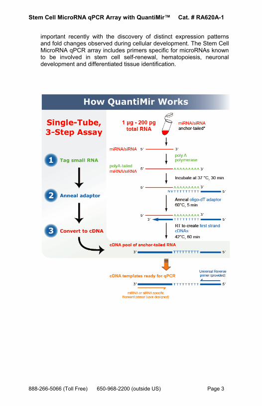
B. Importance of MicroRNAs and Other Small Non-Coding RNAs The field of non-coding RNAs has gained increasing attention in recent years, particularly due to the discovery of small interfering RNAs (siRNAs) and micro RNAs (miRNA). These RNAs are short (typically 19-24 nucleotides) single stranded moieties that regulate the expression of target genes by interacting with complementary sites within the target mRNAs and either repressing translation or eliciting target mRNA degradation. miRNAs and siRNAs are conserved groups of non-coding RNAs with very important regulatory roles. Mature miRNAs and siRNAs are excised from stem-loop precursors, which are themselves transcribed as part of longer primary transcripts. These primary miRNAs appear to be first processed by the RNase Drosha in the nucleus, after which the precursor miRNAs are exported to the cytoplasm where the RNase Dicer further processes them. These enzymes are also involved in the generation of mature small inhibitory RNAs (siRNA) from exogenously transferred double stranded siRNA precursors. The current, standard method for detecting and quantifying novel miRNA and siRNA molecules involves Northern blotting with hybridization. Detecting and quantitating known miRNAs can be done using pre-designed reverse priming and reverse transcription followed by primer sets built for the specific miRNA for Real-time PCR analysis. These sets require many steps and can take several hours to complete and trouble-shoot. The QuantiMir™ RT kit provides all the reagents necessary to anchor-tail and convert small, non-coding RNAs into cDNA starting from total RNA samples. Once the user performs the reactions on their RNA samples, the cDNAs are ready to use for either End-point PCR experiments or to perform Real-time qPCR analysis. MicroRNA expression signatures have become more clinically
Page 2 ver. 1-070508 www.systembio.com
Stem Cell MicroRNA qPCR Array with QuantiMir™ Cat. # RA620A-1
important recently with the discovery of distinct expression patterns and fold changes observed during cellular development. The Stem Cell MicroRNA qPCR array includes primers specific for microRNAs known to be involved in stem cell self-renewal, hematopoiesis, neuronal development and differentiated tissue identification.
888-266-5066 (Toll Free) 650-968-2200 (outside US) Page 3
System Biosciences (SBI) User Manual
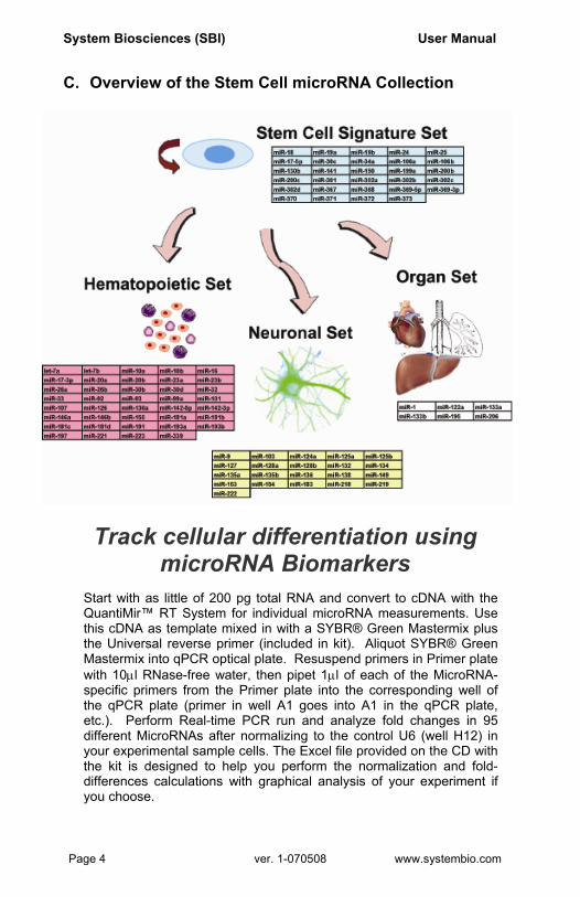
C. Overview of the Stem Cell microRNA Collection
Track cellular differentiation using microRNA Biomarkers
Start with as little of 200 pg total RNA and convert to cDNA with the QuantiMir™ RT System for individual microRNA measurements. Use this cDNA as template mixed in with a SYBR® Green Mastermix plus the Universal reverse primer (included in kit). Aliquot SYBR® Green Mastermix into qPCR optical plate. Resuspend primers in Primer plate with 10µl RNase-free water, then pipet 1µl of each of the MicroRNA-specific primers from the Primer plate into the corresponding well of the qPCR plate (primer in well A1 goes into A1 in the qPCR plate, etc.). Perform Real-time PCR run and analyze fold changes in 95 different MicroRNAs after normalizing to the control U6 (well H12) in your experimental sample cells. The Excel file provided on the CD with the kit is designed to help you perform the normalization and fold-differences calculations with graphical analysis of your experiment if you choose.
Page 4 ver. 1-070508 www.systembio.com

Stem Cell MicroRNA qPCR Array with QuantiMir™ Cat. # RA620A-1
D. List of Components
Each MicroRNA Stem Cell qPCR Array Kit contains the following components with enough material to perform 20 QuantiMir cDNA synthesis reactions and enough Primers in the Primer Array plate to perform 10 qPCR plates as outlined in this manual:
40 µl 5X PolyA Polymerase Buffer 10 µl Poly A Reaction 10 µl PolyA Polymerase (enough for 20 reactions) 20 µl 25 mM MnCl2 30 µl 5 mM ATP 10 µl Oligo dT Adaptor 20 µl RT Reaction 80 µl 5X Reverse Transcriptase Buffer (enough for 20 reactions) 20 µl Reverse Transcriptase 30 µl 0.1 M Dithiothreitol (DTT) 40 µl dNTP Mix
600 µl 3’ Universal Reverse PCR Primer End-point or qPCR Assay (enough for 1,200 reactions)
Array Primers, dried down in Primer plate (100 µmoles); resuspend in 10µl RNase-free Water
1.2 ml RNase-free Water
The kit is shipped on blue ice and should be stored at -20°C upon arrival. Properly stored kits are stable for 1 year from the date received. The oligonucleotides for the specific MicroRNAs are dried-down in the wells of the optical qPCR plates. Resuspend in 10µl RNase-free water.
E. Additional Required Materials
• Real-time qPCR Instrument • Instrument-specific optical qPCR plates • Thermocycler (with heated lid) • Thermocycler PCR tubes or plates for end-point reactions • PCR Mastermix, including Taq polymerase for PCR • 3.0-3.5% Agarose Gel in Tris-Borate EDTA (TBE) or Tris-Acetate
EDTA (TAE) Buffer • DNA Size Ladder with markers from 50 to 2,000 bp (Bio-Rad
AmpliSize™ DNA Ladder; Cat. # 170-8200) • Nuclease-free water for qPCR reactions
888-266-5066 (Toll Free) 650-968-2200 (outside US) Page 5

System Biosciences (SBI) User Manual
IMPORTANT: • Recommended 2X SYBR Green qPCR Mastermixes:
SBI has tested and recommends SYBR Green Master mix from three vendors: Power SYBR Master Mix® (Cat. #s 4368577, 4367650, 4367659, 4368706, 4368702, 4368708, 4367660) from Applied Biosystems; SYBR GreenER™ qPCR SuperMix for ABI PRISM® instrument from Invitrogen (Cat. #s 11760-100, 11760-500, and 11760-02K); and RT2 Real-Time™ SYBR Green / ROX PCR (Cat. #s PA-012 and PA-112) from SuperArray.
Page 6 ver. 1-070508 www.systembio.com

Stem Cell MicroRNA qPCR Array with QuantiMir™ Cat. # RA620A-1
II. Protocol A. QuantiMir™ RT Reaction Setup
(for 1 RNA sample to be assayed on 1 qPCR plate)
It is important to start with total RNA that includes the small RNA fraction. RNA input can be as low as 10 ng/µl. For optimum signals, perform the following.
Dilute your RNA to ~100 ng/µl
In a thin-walled PCR tube or
PCR-compatible plate well, combine:
Total RNA (~500ng) 5X PolyA Buffer 25mM MnCl2 5mM ATP PolyA Polymerase
Total in tube
Incub min. at 37°C
Add ligo dT Adaptor
Heat at 60°C Let c m temp for 2 min.
Add 5X RT Buffer dNTP mix 0.1M DTT RNase-free H2O Reverse Transcriptase Total in tube
IncubHeat
STEP 1: PolyA Tail
STEP 2: Anneal Anchor dT Adaptor
STEP 3: Synthesize cDNAs
Start: Done!
* The QuantiMir™sensitive applicawith ethanol preremove proteinsthis is not neces
888-266-5066 (Toll Free) 650-96
5 µl 2 µl 1 µl 1.5 µl 0.5 µl 10 µl
ate for 30
: 0.5 µl O
for 5 min.ool to roo
: 4 µl 2 µl 1.5 µl 1.5 µl
1 µl 20.5 µl
+ + +
ate for 60 min. at 42°C for 10 min. at 95°C
cDNAs can be stored at -20°C. For more tions, a single phenol:chloroform extraction cipitation can be performed on the cDNA to , unutilized dNTPs, and primers. Typically, sary.8-2200 (outside US) Page 7
System Biosciences (SBI) User Manual
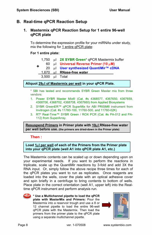
B. Real-time qPCR Reaction Setup 1. Mastermix qPCR Reaction Setup for 1 entire 96-well
qPCR plate To determine the expression profile for your miRNAs under study, mix the following for 1 entire qPCR plate:
For 1 entire plate:
1,750 µl 2X SYBR Green* qPCR Mastermix buffer 60 µl Universal Reverse Primer (10 µM) 20 µl User synthesized QuantiMir™ cDNA
* Sven
Aliqu
1.
2.
3.
Resuper w
Th
Loadinto y
The Mayour etriplicatRNA inthe qPloadedand spPlace ptime qP
*pM1qpu
Page 8
+
1,670 µl RNase-free water 3,500 µl TotalBI has tested and recommends SYBR Green Master mix from three dors:
ot 29µl of Mastermix per well in your qPCR Plate.
Power SYBR Master Mix® (Cat. #s 4368577, 4367650, 4367659, 4368706, 4368702, 4368708, 4367660) from Applied Biosystems SYBR GreenER™ qPCR SuperMix for ABI PRISM® instrument from Invitrogen (Cat. #s 11760-100, 11760-500, and 11760-02K) RT² Real-Time™ SYBR Green / ROX PCR (Cat. #s PA-012 and PA-112) from SuperArray.
spend Primers in Primer plate with 10µl RNase-free water ell before use. (the primers are dried-down in the Primer plate)
en :
1µl per well of each of the Primers from the Primer plate our qPCR plate (well A1 into qPCR plate A1, etc.)
stermix contents can be scaled up or down depending upon on xperimental needs. If you want to perform the reactions in e, scale up the QuantiMir reactions by 3-fold and add 3X the put. Or, simply follow the above recipe three times for each of CR plates you want to run as replicates. Once reagents are into the wells, cover the plate with an optical adhesive cover in briefly in a centrifuge to bring contents to bottom of wells. late in the correct orientation (well A1, upper left) into the Real-CR instrument and perform analysis run.
Use a Multichannel pipette to load the qPCR late with MasterMix and Primers: Pour the astermix into a reservoir trough and use a 8 or 2 channel pipette to load the entire 96-well PCR plate with the Mastermix. Then load the rimers from the primer plate to the qPCR plate sing a separate multichannel pipette.
ver. 1-070508 www.systembio.com

Stem Cell MicroRNA qPCR Array with QuantiMir™ Cat. # RA620A-1
2. Real-time qPCR Instrument Parameters Follow the guidelines as detailed for your specific Real-time instrumentation. The following parameters tested by SBI were performed on an Applied Biosystems 7300 Real-time PCR System but can also apply to an ABI 7500 or an ABI 7900 96-well system. The details of the thermal cycling conditions used in testing at SBI are below. A screenshot from SBI’s ABI7300 Real-time instrument setup is shown below also. Default conditions are used throughout. Create a detector:
Instrument Setup:
888-266-5
qPCR cycling and data accumulation conditions: 1. 50°C 2 min. 2. 95°C 10 min. 3. 95°C 15 sec. 4. 60°C 1 min. (40 cycles of steps 3 and 4), data read at 60°C 15 sec. Step (gold rectangle)
An additional recommendation is to include a melt analysis after the qPCR run to assess the Tm of the PCR amplicon to verify the specificity of the amplification reaction. Refer to the User Manual for your specific instrument to conduct the melt analysis and the data analyses of the amplification plots and Cycle Threshold (Ct) calculations. In general, Cycle thresholds should be set within the exponential phase of the amplification plots with software automatic baseline settings.
066 (Toll Free) 650-968-2200 (outside US) Page 9
System Biosciences (SBI) User Manual
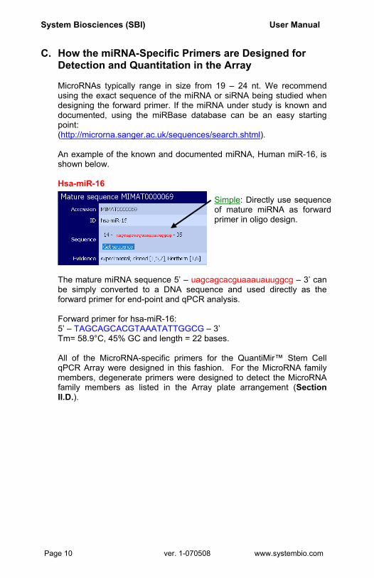
C. How the miRNA-Specific Primers are Designed for Detection and Quantitation in the Array MicroRNAs typically range in size from 19 – 24 nt. We recommend using the exact sequence of the miRNA or siRNA being studied when designing the forward primer. If the miRNA under study is known and documented, using the miRBase database can be an easy starting point: (http://microrna.sanger.ac.uk/sequences/search.shtml). An example of the known and documented miRNA, Human miR-16, is shown below. Hsa-miR-16
The mature miRNA sequence 5’ – uagcagcacguaaauauuggcg – 3’ can be simply converted to a DNA sequence and used directly as the forward primer for end-point and qPCR analysis.
Forward primer for hsa-miR-16: 5’ – TAGCAGCACGTAAATATTGGCG – 3’ Tm= 58.9°C, 45% GC and length = 22 bases. All of the MicroRNA-specific primers for the QuantiMir™ Stem Cell qPCR Array were designed in this fashion. For the MicroRNA family members, degenerate primers were designed to detect the MicroRNA family members as listed in the Array plate arrangement (Section II.D.).
Simple: Directly use sequence of mature miRNA as forward primer in oligo design.
Page 10 ver. 1-070508 www.systembio.com
Stem Cell MicroRNA qPCR Array with QuantiMir™ Cat. # RA620A-1
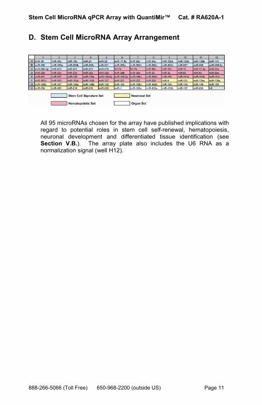
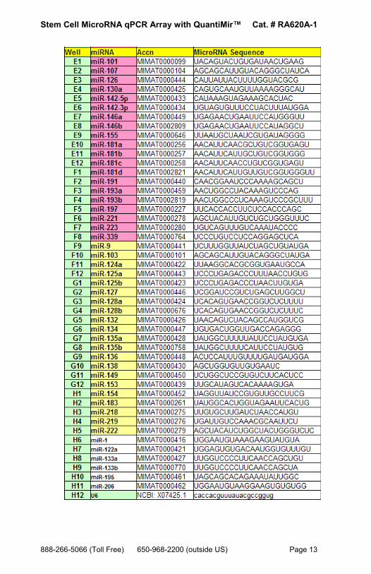
D. Stem Cell MicroRNA Array Arrangement
All 95 microRNAs chosen for the array have published implications with regard to potential roles in stem cell self-renewal, hematopoiesis, neuronal development and differentiated tissue identification (see Section V.B.). The array plate also includes the U6 RNA as a normalization signal (well H12).
888-266-5066 (Toll Free) 650-968-2200 (outside US) Page 11

System Biosciences (SBI) User Manual
Page 12 ver. 1-070508 www.systembio.com
Stem Cell MicroRNA qPCR Array with QuantiMir™ Cat. # RA620A-1
888-266-5066 (Toll Free) 650-968-2200 (outside US) Page 13

System Biosciences (SBI) User Manual
III. Quality Control and Sample Data
A. Stem Cell qPCR Array Primer Validation Tests
1. Real-time qPCR Validation
The Stem Cell qPCR Array plate was tested using Human cord blood CD34+ stem cell RNA samples converted to cDNA using the QuantiMir RT Kit. The resulting cDNA was tested using 0.2 µl per well. Shown at left is the resulting Real-time amplification plot for the entire plate. The Cts ranged from 19.47 to 36.30, reflecting approximately a 5-log fold expression detection range. The experiment was performed as detailed in Section II.E. Quantitative signals were observed for all wells in the array.
Page 14 ver. 1-070508 www.systembio.com

Stem Cell MicroRNA qPCR Array with QuantiMir™ Cat. # RA620A-1
B. Sensitivity Tests
The QuantiMir™ cDNAs were synthesized using decreasing amounts of total starting RNA input from a pool of Human Brain, Heart, Kidney, Placenta, and Testes RNAs. Real-time quantitative qPCR assays were performed with Forward primers specific for Human miR-16 and Human miR-24 (For procedure, see Section II.D.1, Protocol: Real-time qPCR in the QuantiMir™ RT Kit User Manual).
Fig. 2. Real-time qPCR data for Human miR-16 and Human miR-24. Real-time qPCR amplification plots are shown in the upper inset. Cycle threshold (Ct) values were determined using the software automatic baseline and Ct settings. The Bar graph depicts the relative %Signal per RNA input amount for the microRNA. The graph below shows the linear regression analysis with a R2 value of 0.971 for miR-16 and 0.993 for miR-24. Both microRNAs are readily detectable down to 200 pg of total starting RNA input.
888-266-5066 (Toll Free) 650-968-2200 (outside US) Page 15
System Biosciences (SBI) User Manual
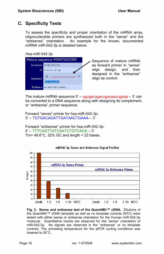
C. Specificity Tests To assess the specificity and proper orientation of the miRNA array, oligonucleotide primers are synthesized both in the “sense” and the “antisense” orientation. An example for the known, documented miRNA miR-542-3p is detailed below. Hsa-miR-542-3p
The mature miRNA sequence 5’ – ugugacagauugauaacugaaa – 3’ can be converted to a DNA sequence along with designing its complement, or “antisense” primer sequence.
Forward “sense” primer for hsa-miR-542-3p: 5’ – TGTGACAGATTGATAACTGAAA – 3’ Forward “antisense” primer for hsa-miR-542-3p: 5’ – TTTCAGTTATCAATCTGTCACA – 3’ Tm= 49.6°C, 32% GC and length = 22 bases.
Sequence of mature miRNA as forward primer in “sense” oligo design, and then designed in the “antisense” oligo as control.
Fig. 3. Sense and antisense test of the QuantiMir™ cDNA. Dilutions of the QuantiMir™ cDNA template as well as no template controls (NTC) were tested with either sense or antisense orientation for the Human miR-542-3p molecule. Quantitative results are observed for the “sense” orientation of miR-542-3p. No signals are observed in the “antisense” or no template controls. The annealing temperature for the qPCR cycling conditions was lowered to 50°C.
Page 16 ver. 1-070508 www.systembio.com

Stem Cell MicroRNA qPCR Array with QuantiMir™ Cat. # RA620A-1
D. Sample Data
1. Tissue Expression Pattern Determinations using the QuantiMir™ Kit on Normal Human Tissues The QuantiMir™ cDNA sets were synthesized from 18 separate normal Human tissues and tested with 2 primers specific for 2 known miRNA molecules: miR-1 (heart and skeletal muscle-specific) and miR-122a (abundant in liver). The amplification plots and corresponding expression bar graphs are shown in Figure 4, panels a and b.
These two known miRNAs, miR-1 and mir-122a, have very specific tissue expression patterns. Real-time qPCR data confirmed that miR-1 is restricted to skeletal muscle and heart. The sensitivity of the assays also reveals very low but detectable signals in additional tissues. miR-122a is known to be highly abundant in liver.
a. b. Fig. 4. Real-time qPCR data using primers specific for Human miR-1 (Panel a.) and for miR-122a (Panel b.). The amplification plots are shown on the left with the resulting expression profile bar graphs based on Ct values is shown on the right. The default qPCR cycling conditions were used with an annealing temperature of 60°C in Step 2 of Stage 3.
888-266-5066 (Toll Free) 650-968-2200 (outside US) Page 17
System Biosciences (SBI) User Manual
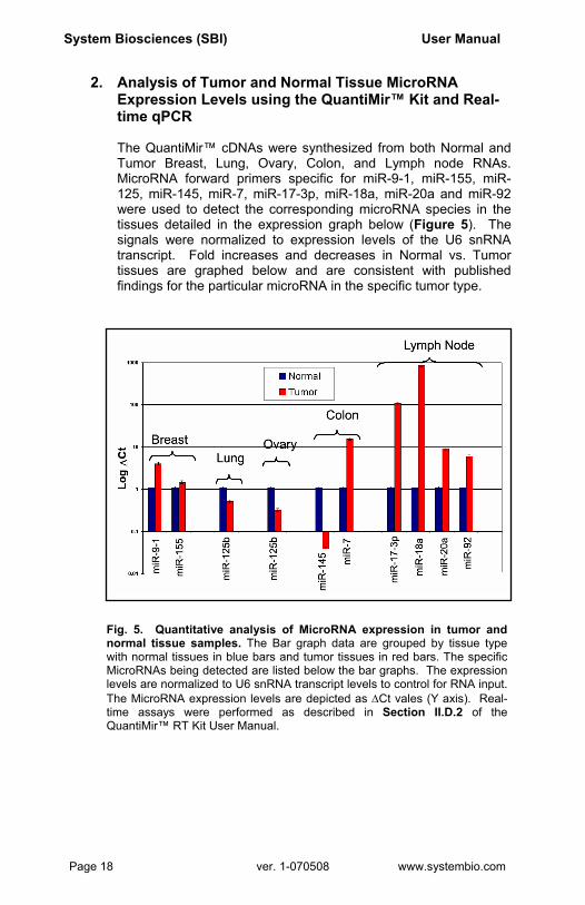
2. Analysis of Tumor and Normal Tissue MicroRNA Expression Levels using the QuantiMir™ Kit and Real-time qPCR
The QuantiMir™ cDNAs were synthesized from both Normal and Tumor Breast, Lung, Ovary, Colon, and Lymph node RNAs. MicroRNA forward primers specific for miR-9-1, miR-155, miR-125, miR-145, miR-7, miR-17-3p, miR-18a, miR-20a and miR-92 were used to detect the corresponding microRNA species in the tissues detailed in the expression graph below (Figure 5). The signals were normalized to expression levels of the U6 snRNA transcript. Fold increases and decreases in Normal vs. Tumor tissues are graphed below and are consistent with published findings for the particular microRNA in the specific tumor type.
Fig. 5. Quantitative analysis of MicroRNA expression in tumor and normal tissue samples. The Bar graph data are grouped by tissue type with normal tissues in blue bars and tumor tissues in red bars. The specific MicroRNAs being detected are listed below the bar graphs. The expression levels are normalized to U6 snRNA transcript levels to control for RNA input. The MicroRNA expression levels are depicted as ∆Ct vales (Y axis). Real-time assays were performed as described in Section II.D.2 of the QuantiMir™ RT Kit User Manual.
Page 18 ver. 1-070508 www.systembio.com

Stem Cell MicroRNA qPCR Array with QuantiMir™ Cat. # RA620A-1
IV. Troubleshooting
Problem Possible Solution
Too much background in qPCR signals
Use much less cDNA in the SYBR Green Mastermix.
No qPCR signals 1. Did you select SYBR Green as the Detector’s Reporter Dye?
2. Did the U6 control work? 3. Use more cDNA in Mastermix. 4. Check Mastermix contents and try
a subset with U6 as a positive control.
5. Also try lowering the Annealing Temperature to 50ºC.
How do I select the Threshold level for Ct analysis?
Typically, place the threshold setting in the upper third of the exponential phase of the amplification curve. Also, see the User Manual for your specific instrument or contact their technical support team for guidance.
888-266-5066 (Toll Free) 650-968-2200 (outside US) Page 19
System Biosciences (SBI) User Manual
V. References A. General References
1. Sonthelmer, E. J., Carthew, R. W. 2005. Silence from within: Endogenous siRNAs and miRNAs. Cell 122:9-12.
2. Zamore, P.D., Haley, B. 2005. Ribo-gnome: The big world of small RNAs. Science 309: 1519-1524.
3. Bartel, D. 2004. MicroRNAs: Genomics, Biogenesis, Mechanism, and Function. Cell 116: 281-297.
4. Kim, Narry V. 2005.Small RNAs: Classification, Biogenesis, and Function. Mol. Cells. 19:1-15.
5. Valencia-Sanchez, MA., Liu, J., Hannon, GJ., Parker, R., 2006. Control of translation and mRNA degradation by miRNAs and siRNAs. Genes Dev 20: 515-525.
6. Lewis B.P, Burge C.B, Bartel, D.P. 2005. Conserved seed pairing, often flanked by adenosines, indicates that thousands of human genes are microRNA targets. Cell 120: 15-20.
7. Xie X., Lu J., Kulbokas, E.J., Goulub, T.R., Mooth, V., Lindblad-Toh, K., Lander, E.S. and Kellis, M. Systematic discovery of regulatory motifs in human promoters and 3’ UTRs by comparison of several mammals. Nature.434:338-45.
8. Lagos-Quintana, M., Rauhut, R., Lendeckel, W., Tuschl, T. 2001. Identification of Novel Coding for Small Expresses RNAs. Science 294: 853-858.
9. Basyuk, E., Suavet, F., Doglio, A., Bordonne, R., Bertrand, E. 2003. Human let-7 stem-loop precursors harbor features of RNase III cleavage products. Nucleic Acids Res 31: 6593-6597.
10. Chomczynski P., and Mackey, K. One-hour downward capillary blotting of RNA at neutral pH. 1994, Anal. Biochem. 221, 303-305.
11. Shi, R., Chiang, V.L., 2005. Facile means for quantifying microRNA expression by real-time PCR. BioTechniques. 39:519-525.
12. Ding, Y., Chan, C.Y., and Lawrence, C.E. (2005) RNA secondary structure prediction by centroids in a Boltzmann weighted ensemble. RNA 11, 1157-1166.
13. Griffiths-Jones,S., Grocock, R.J., Van Dongen, S., Bateman, A., Enright, A.J. 2006. miRBase: microRNA sequences, targets and gene nomenclature. Nucleic Acids Research 34: D140-D144.
14. Shingara, J., Keiger, K., Shelton, J., Laosinchai-Wolf, W., Powers, P., Conrad, R., Brown, D., Labourier, E. 2005. An optimized isolation and labeling platform for accurate microRNA expression profiling. RNA 11:1461-1470.
15. He, L., Thomson, J.M., Hemann, M.T., Hernando-Monge, E., Mu, D., Goodson, S., Powers, S., Cordon-Cardo, C., Lowe, S.W., Hannon, G.J.,
Page 20 ver. 1-070508 www.systembio.com
Stem Cell MicroRNA qPCR Array with QuantiMir™ Cat. # RA620A-1
Hammond, S.M. 2005. A microRNA polycistron as a potential human oncogene. Nature 435: 828-833.
16. Lai, E.C., Wiel, C., Rubin, G.M. 2004. Complementary miRNA pairs suggest a regulatory role for miRNA:miRNA duplexes. RNA 10:171-175.
17. Ambros, V., Bartel, B., Bartel, D.P., Burge, C.B., Carrington, J.C., Chen, X., Dreyfuss, G., Eddy, S.R., Griffiths-Jones, S., Marshall, M., Matzke, M., Ruvkun, G., Tuschl, T. 2003. A uniform system for microRNA annotation. RNA 9:277-279.
18. Obernosterer, G., Leuschner, P.J.F., Alenius, M., Martinez, J. 2006. Post-transcriptional regulation of microRNA expression. RNA 12:1-7.
19. Dostie, J., Mourelatos, Z., Yang, M., Sharma, A., Dreyfuss, G. 2003. Numerous microRNPs in neuronal cells containing novel microRNAs. RNA 9: 180-186.
B. MicroRNA and Stem Cell References
DGCR8 is essential for microRNA biogenesis and silencing of embryonic stem cell self-renewal. Nat Genet. 2007 Mar;39(3):380-5. Wang Y, Medvid R, Melton C, Jaenisch R, Blelloch R. Dicer-deficient mouse embryonic stem cells are defective in differentiation and centromeric silencing. Genes Dev. 2005 Feb 15;19(4):489-501. Kanellopoulou C, Muljo SA, Kung AL, Ganesan S, Drapkin R, Jenuwein T, Livingston DM, Rajewsky K. MicroRNA expression profiling during human cord blood-derived CD34 cell erythropoiesis. Exp Hematol. 2007 Apr;35(4):551-64. Choong ML, Yang HH, McNiece I. MicroRNA expression profiling of single whole embryonic stem cells. Nucleic Acids Res. 2006 Jan 24;34(2):e9. Tang F, Hajkova P, Barton SC, Lao K, Surani MA. Stem cells: from epigenetics to microRNAs. Neuron. 2005 May 5;46(3):363-7. Review. Cheng LC, Tavazoie M, Doetsch F. MicroRNA: a new player in stem cells. J Cell Physiol. 2006 Nov;209(2):266-9. Review. Zhang B, Pan X, Anderson TA. Human embryonic stem cells express a unique set of microRNAs. Dev Biol. 2004 Jun 15;270(2):488-98. Suh MR, Lee Y, Kim JY, Kim SK, Moon SH, Lee JY, Cha KY, Chung HM, Yoon HS, Moon SY, Kim VN, Kim KS.
888-266-5066 (Toll Free) 650-968-2200 (outside US) Page 21
System Biosciences (SBI) User Manual
C. MicroRNA and Cancer References
Breast Cancer MicroRNA gene expression deregulation in human breast cancer. Cancer Res. 2005 Aug 15;65(16):7065-70. Iorio MV, Ferracin M, Liu CG, Veronese A, Spizzo R, Sabbioni S, Magri E, Pedriali M, Fabbri M, Campiglio M, Menard S, Palazzo JP, Rosenberg A, Musiani P, Volinia S, Nenci I, Calin GA, Querzoli P, Negrini M, Croce CM.
MicroRNA expression profiles classify human cancers. Nature. 2005 Jun 9;435(7043):745-6. Lu J, Getz G, Miska EA, Alvarez-Saavedra E, Lamb J, Peck D, Sweet-Cordero A, Ebert BL, Mak RH, Ferrando AA, Downing JR, Jacks T, Horvitz HR, Golub TR. B-cell Leukemia MicroRNA profiling reveals distinct signatures in B cell chronic lymphocytic leukemias. Proc Natl Acad Sci U S A. 2004 Aug 10;101(32):11755-60. Epub 2004 Jul 29. Calin GA, Liu CG, Sevignani C, Ferracin M, Felli N, Dumitru CD, Shimizu M, Cimmino A, Zupo S, Dono M, Dell'Aquila ML, Alder H, Rassenti L, Kipps TJ, Bullrich F, Negrini M, Croce CM.
A MicroRNA signature associated with prognosis and progression in chronic lymphocytic leukemia. N Engl J Med. 2005 Oct 27;353(17):1793-801. Calin GA, Ferracin M, Cimmino A, Di Leva G, Shimizu M, Wojcik SE, Iorio MV, Visone R, Sever NI, Fabbri M, Iuliano R, Palumbo T, Pichiorri F, Roldo C, Garzon R, Sevignani C, Rassenti L, Alder H, Volinia S, Liu CG, Kipps TJ, Negrini M, Croce CM. Lung Cancer Unique microRNA molecular profiles in lung cancer diagnosis and prognosis. Cancer Cell. 2006 Mar;9(3):189-98. Yanaihara N, Caplen N, Bowman E, Seike M, Kumamoto K, Yi M, Stephens RM, Okamoto A, Yokota J, Tanaka T, Calin GA, Liu CG, Croce CM, Harris CC.
A polycistronic microRNA cluster, miR-17-92, is overexpressed in human lung cancers and enhances cell proliferation. Cancer Res. 2005 Nov 1;65(21):9628-32. Hayashita Y, Osada H, Tatematsu Y, Yamada H, Yanagisawa K, Tomida S, Yatabe Y, Kawahara K, Sekido Y, Takahashi T.
MicroRNA and lung cancer. N Engl J Med. 2005 Jun 9;352(23):2446-8. Eder M, Scherr M. Pancreatic Cancer Expression profiling identifies microRNA signature in pancreatic cancer. Int J Cancer. 2006 Dec 5; [Epub ahead of print] Lee EJ, Gusev Y, Jiang J, Nuovo GJ, Lerner MR, Frankel WL, Morgan DL, Postier RG, Brackett DJ, Schmittgen TD.
Page 22 ver. 1-070508 www.systembio.com
Stem Cell MicroRNA qPCR Array with QuantiMir™ Cat. # RA620A-1
Solid Tumors: A microRNA expression signature of human solid tumors defines cancer gene targets. Proc Natl Acad Sci U S A. 2006 Feb 14;103(7):2257-61. Epub 2006 Feb 3. Volinia S, Calin GA, Liu CG, Ambs S, Cimmino A, Petrocca F, Visone R, Iorio M, Roldo C, Ferracin M, Prueitt RL, Yanaihara N, Lanza G, Scarpa A, Vecchione A, Negrini M, Harris CC, Croce CM. MicroRNA and Cancer Reviews/Other Publications Cancer genomics: small RNAs with big impacts. Nature. 2005 Jun 9;435(7043):745-6. Meltzer, PS. MicroRNAs in Gene Regulation: When the Smallest Governs It All. J Biomed Biotechnol. 2006;2006(4):69616. Ouellet DL, Perron MP, Gobeil LA, Plante P, Provost P. MicroRNAs as oncogenes. Curr Opin Genet Dev. 2006 Feb;16(1):4-9. Epub 2005 Dec 19. Hammond SM. Oncomirs - microRNAs with a role in cancer. Nat Rev Cancer. 2006 Apr;6(4):259-69. Esquela-Kerscher A, Slack FJ. MicroRNAs as oncogenes and tumor suppressors. Dev Biol. 2006 Aug 16; [Epub ahead of print] Zhang B, Pan X, Cobb GP, Anderson TA. MicroRNAs and the hallmarks of cancer. Oncogene. 2006 Oct 9;25(46):6170-5. Dalmay T, Edwards DR MicroRNAs exhibit high frequency genomic alterations in human cancer. Proc Natl Acad Sci U S A. 2006 Jun 13;103(24):9136-41. Epub 2006 Jun 5. Zhang L, Huang J, Yang N, Greshock J, Megraw MS, Giannakakis A, Liang S, Naylor TL, Barchetti A, Ward MR, Yao G, Medina A, O'brien-Jenkins A, Katsaros D, Hatzigeorgiou A, Gimotty PA, Weber BL, Coukos G.
888-266-5066 (Toll Free) 650-968-2200 (outside US) Page 23
System Biosciences (SBI) User Manual
VI. Appendix A. Related Products
• Cancer MicroRNA qPCR Array with QuantiMir (Cat. # RA610A-
1) The Cancer MicroRNA qPCR Array enables the simultaneous quantitation of 95 MicroRNAs known to be involved in Cancer. The technology utilizes the QuantiMir RT Kit which tags and converts all small RNAs into cDNA, ready to use as template for real-time qPCR. A universal reverse adaptor primer and positive control primers for Human U6 snRNA are inculded in the kit.
• QuantiMir™ RT Kit (Cat. # RA420A-1) Complete reagent kit for anchor-tagging small RNAs and converting them to quantifiable cDNA. Kit contains enough reagents for 20 RT reactions and can generate hundreds of qPCR templates. A universal reverse adaptor primer and positive control primers for Human U6 snRNA and Human miR-16 are also included with the kit.
• miRANDA™ qPCR-Ready miRNA Tissue Expression Array Kit
(Cat. # RA600A-1) 18 Human individual normal tissue miRNA cDNAs arrayed into a qPCR optical plate (4 complete sets of the 18 tissues, 72 individual reactions). A universal reverse adaptor primer and a positive control forward primer (U6 snRNA) are also included with the kit.
• miRANDA™ Universal miRNA cDNA template
(Cat. # RA601A-1) Pool of all 18 Human miRNA cDNAs (enough for 20 50 µl-reactions), a universal reverse adaptor primer, and a positive control forward primer (U6 snRNA)
• MicroRNA Discovery™ Kit (Cat. # RA410A-1)
Rapid identification of new MicroRNAs and MicroRNA-like molecules. Amplification and cloning can be initiated in a single day (3 steps, 1 day.) The alternative method takes approximately 1 week (9 steps.)
• Pre-Made MicroRNA-Enriched cDNAs (Cat. # RA500A-1 –
RA509A-1) Tissue-specific amplified cDNA generated by SBI using the MicroRNA Discovery™ Kit can be used for cloning microRNA.
• Global MicroRNA Amplification Kit (Cat. # RA400A-1)
Simple amplification kit allows cDNA amplification for qRT-PCR and microarray studies from as little as 50 ng of starting total RNA.
Page 24 ver. 1-070508 www.systembio.com

Stem Cell MicroRNA qPCR Array with QuantiMir™ Cat. # RA620A-1
• Full Spectrum™ Complete Transcriptome RNA Amplification
Kit (Cat. # RA101A-1) The Full Spectrum RNA Amplification Kit provides an inexpensive method to amplify reverse transcribed RNA in a sequence independent, unbiased, and uniform manner with better representation of 5’ end of mRNA sequences. This approach maintains the relative levels of each transcript in the starting mRNA samples—even when using starting amounts of RNA as low as 5 ng or when using heavily degraded RNA.
• Full Spectrum™ MultiStart Primers for T7 IVT
(Cat. # RA300A-2) Extract more data from your RNA than currently available primers in nearly all commercially-available T7 IVT kits using Full Spectrum™ technology. Just replace the existing T7 primer with the Full Spectrum™ primers. Compatible with Affymetrix GeneChip® hybridization.
B. Technical Support
For more information about SBI products and to download manuals in PDF format, please visit our web site: http://www.systembio.com For additional information or technical assistance, please call or email us at:
System Biosciences (SBI) 1616 North Shoreline Blvd. Mountain View, CA 94043 Phone: (650) 968-2200 (888) 266-5066 (Toll Free) Fax: (650) 968-2277 E-mail: General Information: [email protected] Technical Support: [email protected] Ordering Information: [email protected]
888-266-5066 (Toll Free) 650-968-2200 (outside US) Page 25

System Biosciences (SBI) User Manual
VII. Licensing and Warranty Statement Limited Use License Use of the Stem Cell MicroRNA qPCR Array Kit wth QuantiMir™ (i.e., the “Product”) is subject to the following terms and conditions. If the terms and conditions are not acceptable, return all components of the Product to System Biosciences (SBI) within 7 calendar days. Purchase and use of any part of the Product constitutes acceptance of the above terms. Purchase of the product does not grant any rights or license for use other than those explicitly listed in this Licensing and Warranty Statement. Use of the Product for any use other than described expressly herein may be covered by patents or subject to rights other than those mentioned. SBI disclaims any and all responsibility for injury or damage which may be caused by the failure of the buyer or any other person to use the Product in accordance with the terms and conditions outlined herein. SBI has pending patent applications related to the Product. For information concerning licenses for commercial use, contact SBI. Limited Warranty SBI warrants that the Product meets the specifications described in the accompanying Product Analysis Certificate. If it is proven to the satisfaction of SBI that the Product fails to meet these specifications, SBI will replace the Product or provide the purchaser with a refund. This limited warranty shall not extend to anyone other than the original purchaser of the Product. Notice of nonconforming products must be made to SBI within 30 days of receipt of the Product. SBI’s liability is expressly limited to replacement of Product or a refund limited to the actual purchase price. SBI’s liability does not extend to any damages arising from use or improper use of the Product, or losses associated with the use of additional materials or reagents. This limited warranty is the sole and exclusive warranty. SBI does not provide any other warranties of any kind, expressed or implied, including the merchantability or fitness of the Product for a particular purpose. SBI is committed to providing our customers with high-quality products. If you should have any questions or concerns about any SBI products, please contact us at (888) 266-5066. © 2007 System Biosciences (SBI).
Page 26 ver. 1-070508 www.systembio.com