stem and root anatomy of mnesithea selloana (hack.) de koning...
TRANSCRIPT

FYTON ISSN 0031 9457 (2018) 87: 231-235
Stem and root anatomy of Mnesithea selloana (Hack.) de Koning & SosefAnatomía de tallos y raíces de Mnesithea selloana (Hack.) de Koning & Sosef
Moya ME1,2, AA Galussi1,2, N Sattler2, MS Fontana2, YG Gillij1,2
Resumen. Se planteó como objetivo estudiar la anatomía de ta-llos y raíces de Mnesithea selloana. Se utilizó material fresco reali-zándose transcortes, a mano alzada, de tallos reproductivos y raíces adventicias, coloreados con safranina, montados en gelatina glice-rinada. Las observaciones se realizaron con un microscopio óptico Olympus CH2 y fotografiados. Los tallos presentaron sección circu-lar hacia el extremo distal de la sinflorescencia y semicircular hacia la zona proximal presentando dos costillas. Se observó una epidermis uniestratificada, continua, sin pelos y con cutícula lisa y gruesa. Las células parenquimáticas cercanas a la epidermis presentaron paredes ligeramente engrosadas aumentando de tamaño hacia la médula con paredes delgadas no lignificadas. Haces vasculares conformando una atactostela, con 3-4 anillos concéntricos, externamente haces peque-ños incluidos dentro de un anillo continuo de esclerénquima y hacia el centro haces de mayor tamaño sin el mencionado anillo. Médula hueca. Las raíces presentaron rizodermis con pelos unicelulares, exo-dermis con paredes marcadamente engrosadas y parénquima aeren-quimatoso con espacios aeríferos bien diferenciados, células endodér-micas con paredes tangenciales internas marcadamente engrosadas. Cilindro vascular presentando periciclo uniseriado, con estructura poliarca, presentando entre 13 y 16 polos xilemáticos y parénquima medular.
Palabras clave: Mnesithea selloana; Anatomía; Tallo; Raíces; Poácea.
Abstract. The objective of this study was to analyze the anatomy of stems and roots of Mnesithea selloana. Free hand cross sections were made from fresh material of reproductive stems and adventi-tious roots; they were colored with safranin and mounted on glycerin jelly. Observations were made using an Olympus CH2 light micro-scope and photographed. In cross section, the stems were circular towards the distal end of the synflorescence and semicircular to-ward the proximal end, and have two ribs. A uniseriate, continuous epidermis presenting a smooth and thick cuticle and no hairs was observed. Parenchyma cells increased in size towards the pith; they have slightly thickened walls close to the epidermis and thin, non-lignified walls near the pith. Vascular bundles formed an atactostele, with 3-4 concentric rings; externally, small bundles enclosed within a continuous ring of sclerenchyma, and toward the center larger bundles without the ring were observed. The pith was hollow. The roots presented rhizodermis with unicellular hairs, exodermis with markedly thickened walls, aerenchymatous parenchyma with well differentiated aeriferous spaces, and endodermal cells with markedly thickened inner tangential walls. The vascular cylinder presented a uniseriate pericycle, with polyarch structure exhibiting between 13 and 16 xylematic poles, and pithy parenchyma.
Keywords: Mnesithea selloana; Anatomy; Stem; Roots; Poaceae.
1 PID UNER 2132. 2 Cátedra Botánica Morfológica, Facultad de Ciencias Agropecuarias Universidad Nacional de Entre Ríos. Ruta Prov. 11 Km 10,5 Oro Verde Dpto. Paraná, Entre Ríos. Address correspondence to: M.E. Moya e-mail: [email protected] ; [email protected] Received 30.I.2018. Accepted 25.V.2018.

232
FYTON ISSN 0031 9457 (2018) 87: 231-235
Moya ME et al., FYTON 87 (2018)
INTRODUCTIONThe family Poaceae comprises 651 genera and about 10000
species worldwide (Clayton & Renvoize, 1986). The genus Mnesithea has about 32 species distributed worldwide (Veld-kamp et al., 1986). Four species are present in Argentina, of which two occur in Entre Ríos province: Mnesithea selloana (Hack.) de Koning & Sosef and M. balansae (Hack.) de Kon-ing & Sosef (Burkart, 1969; Anton & Zuloaga, 2012).
Mnesithea selloana (commonly known as “cola de lagarto” or “teyú ruguay”), a species distributed in Argentina, Brazil, Par-aguay, and Uruguay, is a forage grass occurring in natural fields in Entre Ríos. It is a tufted grass exhibiting profuse tillering and a spring-summer-autumn cycle (megathermal). Although it has been cited for different sites in the region, it is currently restricted to reduced areas.
Watson & Dallwitz (1992) provided a brief description of the genus Mnesithea Kunth and the species M. laevis and M. mollicoma, which are distributed in India, Indochina and Ma-laysia. Those descriptions included aspects about morphology, anatomy, physiology, photochemistry, cytology, taxonomy and pathogens. In Argentina, several works have addressed Mne-sithea selloana, such as the description of its synflorescence and characteristics of its spikelets and diaspores, reproduc-tive aspects related to species dispersal, caryopsis germination with and without floral structures, and leaf anatomy (Burkart, 1969; Anton, 1975; Quarín, 1979; Vegetti, 1997; Galussi et al., 2012a; Galussi et al., 2012b; Moya et al., 2012; Moya et al., 2015). The aim of this work was to describe the stems and roots in cross sections in order to complete the anatomical studies of M. selloana.
MATERIALS AND METHODSThe material was obtained from individuals collected and
identified from a natural area (31° 49’ 18.62” S; 60° 32’ 57.33” W) as part of the research project of M. selloana (PID UNER n°2132) . They were transplanted and cultivated in the Experi-mental Field (31° 49’ 59.51” S; 60° 31’21.84” W) of the Facul-tad de Ciencias Agropecuarias UNER (Galussi et al., 2012a). Stems that had synflorescences in their tips and adventitious roots were removed from the plants. Free hand cross sections were made at different positions of each stem and root. Sec-tions were cleared by immersion in 50% sodium hypochlorite for 2 minutes and then washed with abundant distilled water. They were stained with 1% safranin for 3 minutes with the aim of visualizing sclerosed walls and finally were mounded between slides with glycerin jelly (D’Ambrogio de Argüeso, 1986). Observations were made under a light microscope Olympus CH2 equipped with a MotiCAM 2000Motic Im-ages Plus 2.0ML camera, which was connected to a computer. Numerous micrographs of each of the cross sections were re-corded for further analysis.
RESULTSStem. The stem exhibited circular section in the most dis-
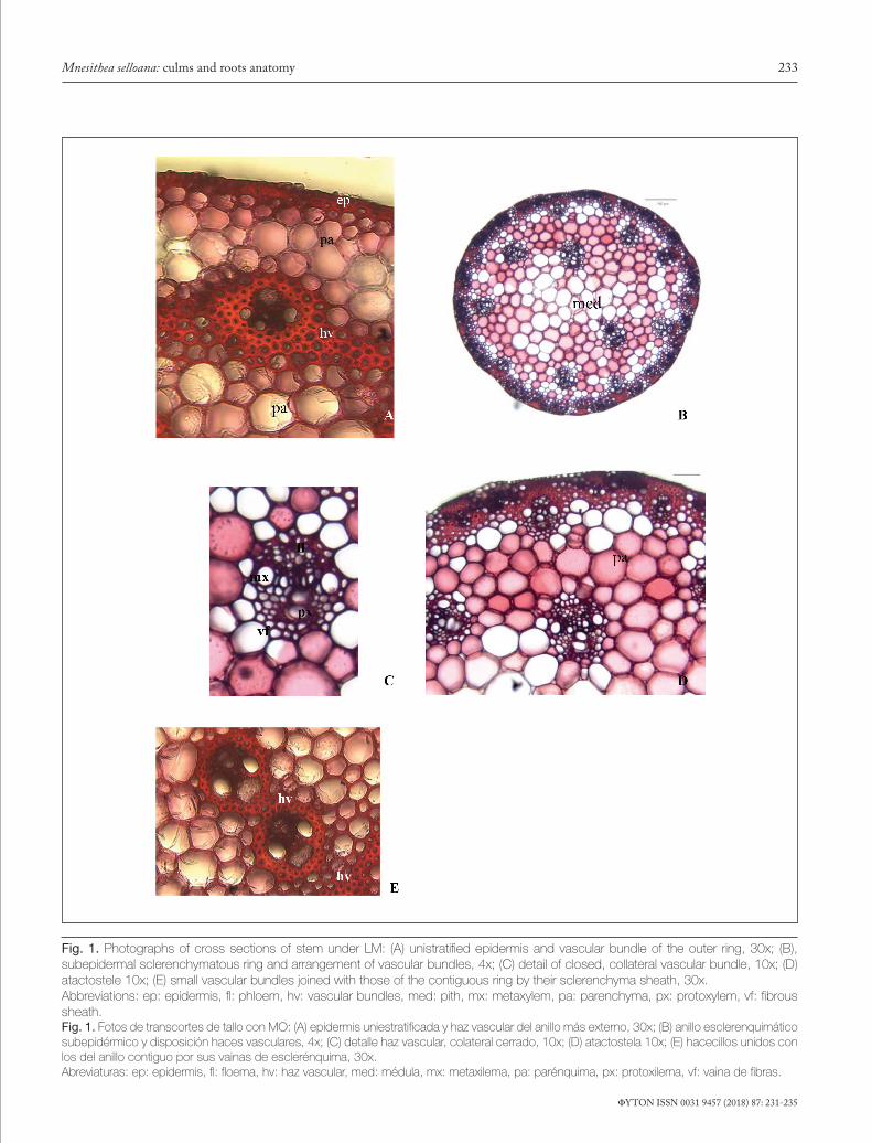
tal internodes to the synflorescence, becoming semicircular towards the proximal part. It exhibits two marked ribs at the ends of the semicircle. In cross section, three clearly differ-entiated tissue systems were observed: epidermal, parenchy-matous and vascular. The epidermisis was unistratified and continuous, with sclerosed epidermal cells, without hairs and with a smooth and thick cuticle (Fig.1 A).
A subepidermal sclerenchyma ring was observed, rang-ing between 3-6 layers of very thick walled cells (Fig. 1B). The cells of the subepidermal parenchyma exhibited slightly thickened walls; these cells increased their size towards the center of the section, exhibiting non-lignified thin walls form-ing the pith (Fig. 1B) or forming a pith cavity when cells were disintegrated.
The vascular system was composed of several typical closed collateral bundles (Fig.1C), which were dispersed throughout all the parenchyma, forming an atactostele, arranged in 3-4 concentric rings, with the outer one composed of smaller bun-dles included in a continuous sclerenchyma ring (Fig.1D).The remaining cycles of vascular bundles were located in the inner parenchyma (transition parenchyma) and each one exhibited a sclerified cell sheath. The latter small bundles occasionally joined with those of the contiguous ring through their scle-renchyma sheaths (Fig. 1E). The solid stems have a paren-chyma pith with cells of circular to polygonal outline, whereas in the hollow stems, a big pith lacuna was observed.
Root. In cross section, a one-layered rhizodermis with one-layered hairs is observed (Fig. 2A). One-layered exoder-mis with markedly thickened walls (Fig. 2B); below the exo-dermis is the aeriferous parenchyma instead of a cortical pa-renchyma, forming an aerechymatous parenchyma with well differentiated aeriferous spaces, and with circular to polygonal outlined cells. The spatial arrangement of the aerenchyma, i.e. arrangement of lacunae and spaces, resembled the shape of a bicycle wheel (Fig. 2C). The endodermis exhibited cells with markedly thickened internal tangential cells (Fig. 2D). A uniseriate pericycle was observed in the vascular cylinder; polyarch vascular bundles exhibited between 13 and 16 xylem poles (Fig. 2E) and a medullary parenchyma (Fig. 2F).
CONCLUSIONSThe study of M. selloana revealed an anatomical structure
of the reproductive stem characterized by a circular section in the basal part, and a semicircular section near the synflores-cence. The vascular bundles were distributed in several rings; and hollow and pithy stems were observed. These anatomical characteristics are present in most of Poaceae plants, and agree with the descriptions of Jensen & Salisbury (1988). The closed
233
FYTON ISSN 0031 9457 (2018) 87: 231-235
Mnesithea selloana: culms and roots anatomy
Fig. 1. Photographs of cross sections of stem under LM: (A) unistratified epidermis and vascular bundle of the outer ring, 30x; (B), subepidermal sclerenchymatous ring and arrangement of vascular bundles, 4x; (C) detail of closed, collateral vascular bundle, 10x; (D) atactostele 10x; (E) small vascular bundles joined with those of the contiguous ring by their sclerenchyma sheath, 30x. Abbreviations: ep: epidermis, fl: phloem, hv: vascular bundles, med: pith, mx: metaxylem, pa: parenchyma, px: protoxylem, vf: fibrous sheath.Fig. 1. Fotos de transcortes de tallo con MO: (A) epidermis uniestratificada y haz vascular del anillo más externo, 30x; (B) anillo esclerenquimático subepidérmico y disposición haces vasculares, 4x; (C) detalle haz vascular, colateral cerrado, 10x; (D) atactostela 10x; (E) hacecillos unidos con los del anillo contiguo por sus vainas de esclerénquima, 30x. Abreviaturas: ep: epidermis, fl: floema, hv: haz vascular, med: médula, mx: metaxilema, pa: parénquima, px: protoxilema, vf: vaina de fibras.

234
FYTON ISSN 0031 9457 (2018) 87: 231-235
Moya ME et al., FYTON 87 (2018)
Fig. 2. Photographs of cross sections of roots under LM. (A) rhizodermis with hairs,10x; (B) exodermis with markedly thickened walls, 20x; (C) aeriferous parenchyma, 20x; (D) endodermal cells with markedly thickened internal tangential walls, 20x; (E) vascular cylinder with uniseriated pericycle,20x; (F) pithy parenchyma, 20x. Abbreviations: en: endodermis, ex: exodermis, p: hair, pa: aeriferous parenchyma, pm: pithy parenchyma , pe: pericycle; px: xylematic poles.Fig. 1. Fotos de transcortes de raíz con MO: (A) rizodermis con pelos,10x; (B) exodermis con paredes marcadamente engrosadas, 20x; (C) parénquima aerífero, 20x; (D) células endodérmicas con paredes tangenciales internas marcadamente engrosadas, 20x; (E) cilindro vascular presentando periciclo uniseriado, 20x; (F) parénquima medular.Abreviaturas: en: endodermis, ex: exodermis, p: pelo, pa: parénquima aerífero, pm: parénquima medular, pe: periciclo, px: polos xilematicos.

235
FYTON ISSN 0031 9457 (2018) 87: 231-235
Mnesithea selloana: culms and roots anatomy
collateral vascular bundles, which were regularly distributed in the stem due to its sinuous longitudinal trajectory, formed an atactostele. This variant of the eustele, typical of monocotyle-dons, agrees with the findings reported by Esau (1987).
The root cortex of grasses from relatively dry habitats exhibits aerenchyma. According to its mode of origin, the aerenchyma may be schizogenous and lysigenous ( Jackson & Armstrong, 1999; Seago et al., 2005). The former, the one of interest in this work, is formed due to selective death of cells of the root cortex, leaving air spaces (Kawai et al., 1998). This type of aerenchyma is found in several cultivated species, such as barley (Arikado & Adachi, 1955), wheat (Trought & Drew, 1980), and rice ( Jus-tin & Armstrong, 1991). The aerenchyma of the roots of M. selloana resembles that of a bicycle wheel, which is consistent with one of the four anatomical typologies mentioned by Justin & Armstrong (1987) and Seago et al. (2005) for grasses.
REFERENCESAnton, A.M. (1975). Notas críticas sobre gramíneas de argentina.
Kurtziana 8: 61-69.Anton, A.R. & F.O. Zuloaga (2012). Flora Argentina. Plantas vas-
culares de la República Argentina, disponible en http://www.floraargentina.edu.ar/ (accessed August 12, 2014). Copyright © Instituto Darwinion. 1997-2014 www.darwin.edu.ar
Arikado, H & Y. Adachi (1955). Anatomical and ecological respons-es of barley and some forage crops to the flooding treatment. Bul-letin of the Faculty of Agriculture – Mie University 11: 1-27.
Burkart, A. (1969). Flora Ilustrada de Entre Ríos. Colección Cientí-fica del INTA. Tomo VI. Parte II. Gramíneas.
Clayton, W. & S. Renvoize (1986). Genera Graminum: Grasses of the world. Kew Bulletin, Additional series 13: 1-389.
D’Ambrogio de Argüeso, A. (1986). Manual de técnicas en His-tología vegetal. Editorial Hemisferio Sur S. A. Buenos Aires, Argentina. 83 p.
Esau, K. (1987). Anatomía de las plantas con semilla. Editorial Hemisferio Sur S.A. 512 p.
Galussi, A.A, M.E. Moya, L.R. Zimmermann, F.G. Marchese & Y.G. Gillij, F. (2012a). Mnesithea selloana: morfología de la sin-florescencia, madurez y calidad de las cariopsis. FYTON Interna-tional Journal of Experimental Botany 81: 255-260.
Galussi, A.A., M.E. Moya, L.R. Zimmermann, Y.G. Gillij, F.G. Marchese & F. Torres (2012b). Características reproductivas de Mnesithea selloana (Hack.) de Koning & Sosef “cola de lagarto, teyú ruguay”. Análisis de Semillas Tomo 6 N°22 69-72.
Jensen, W.A. & F.B. Salisbury (1988). Botánica. México. McGraw Hill, 254 p.
Justin, S.H.F.W & W. Armstrong (1987). The anatomical character-istics of roots and plant responses to soil flooding. New Phytologist 106: 465-495.
Justin, S.H.F.W & W. Armstrong (1991). Evidence for the involve-ment of ethylene in aerenchyma formation in adventitious roots of rice. New Phytologist 118: 49-62.
Kawai, M., P.K, Samarajeewa., R.A, Barrero., M, Nishiguchi & H. Uchimiya. (1998). Cellular dissection of the degradative pattern of cortical cell death during aerenchyma formation of rice roots. Planta 204: 277–287.
Moya, M.E., A.A. Galussi, F. Torres y M.F. Prand. (2012). Incidencia de las estructuras de la espiguilla en la germinación de Mnesithea selloana (Hack.) de Koning & Sosef “cola de lagarto, teyú ruguay”. Análisis de Semillas Tomo 6 N°22 66-68.
Moya, M.E., A.A., Galussi, M.J. Gómez y F.G. Marchese. (2015). “Contribución al conocimiento de la anatomía foliar de Mne-sithea selloana (Hack.) de Koning & Sosef (Andropogoneae-Panicoideae-Poaceae)”. FYTON International Journal of Experi-mental Botany 84: 176-183.
Seago, J.L. Jr., L.C. Marsh, K.J. Stevens, A. Soukup, O. Votrubová & D. Enstone (2005). A re-examination of the root cortex in wetland flowering plants with respect to aerenchyma. Annals of Botany 96: 565-579.
Trought, M.C.T & M.C. Drew (1980). The development of wa-terlogging damage in wheat seedlings (Triticum aestivum L.). i. shoot and root growth in relation to changes in the concentra-tions of dissolved gases and solutes in the soil solution. Plant and Soil 54: 77-94.
Quarin, C.L. (1979). Los Generos Rhytachne y Coelorhachis (Gra-mineae) en Argentina. Kurtziana 12-13: 7-35.
Veldkamp, J.F., R. de Koning & M.S.M. Sosef (1986). Generic de-limitation of Rottboellia and related genera (Gramineae). Blumea 31: 281-307.
Vegetti, A.C. (1997). Sobre la estructura de la inflorescencia en espe-cies de Rottboelliinae (Poaceae – Andropogoneae). Candollea 52: 475-495.
Watson, L. & M.J. Dallwitz (1992). The grass genera of the world: descriptions, illustrations, identification, and information re-trieval; including synonyms, morphology, anatomy, physiology, phytochemistry, cytology, classification, pathogens, world and lo-cal distribution, and references. Versión: 23rd July 2012. http://delta-intkey.com.