static-curis.ku.dk · venom insulins of cone snails diversify rapidly and track prey taxa helena...
TRANSCRIPT

u n i ve r s i t y o f co pe n h ag e n
Københavns Universitet
Venom insulins of cone snails diversify rapidly and track prey taxa
Safavi-Hemami, Helena; Lu, Aiping; Li, Qing; Fedosov, Alexander E.; Biggs, Jason; Corneli,Patrice Showers; Seger, Jon; Yandell, Mark; Olivera, Baldomero M.Published in:Molecular Biology and Evolution
DOI:10.1093/molbev/msw174
Publication date:2016
Document VersionPublisher's PDF, also known as Version of record
Citation for published version (APA):Safavi-Hemami, H., Lu, A., Li, Q., Fedosov, A. E., Biggs, J., Corneli, P. S., ... Olivera, B. M. (2016). Venominsulins of cone snails diversify rapidly and track prey taxa. Molecular Biology and Evolution, 33(11), 2924-2934.https://doi.org/10.1093/molbev/msw174
Download date: 10. aug.. 2019
Venom Insulins of Cone Snails Diversify Rapidly and TrackPrey Taxa
Helena Safavi-Hemami,*,1,2 Aiping Lu,3 Qing Li,4 Alexander E. Fedosov,5 Jason Biggs,6
Patrice Showers Corneli,1 Jon Seger,1 Mark Yandell,4,7 and Baldomero M. Olivera1
1Department of Biology, University of Utah, Salt Lake City, UT2Department of Biology, University of Copenhagen, Copenhagen, Denmark3School of Life Sciences and Technology, Institute of Protein Research, Tongji University, Shanghai, China4Eccles Institute of Human Genetics, University of Utah, Salt Lake City, UT5A.N. Severtzov Institute of Ecology and Evolution, Russian Academy of Science, Leninsky Prospect, Moscow, Russia6University of Guam Marine Laboratory, Agana, Guam7USTAR Center for Genetic Discovery, University of Utah, Salt Lake City, UT
*Corresponding author: E-mail: [email protected].
Associate editor: Sergei Kosakovsky
Abstract
A specialized insulin was recently found in the venom of a fish-hunting cone snail, Conus geographus. Here we show thatmany worm-hunting and snail-hunting cones also express venom insulins, and that this novel gene family has diversifiedexplosively. Cone snails express a highly conserved insulin in their nerve ring; presumably this conventional signalinginsulin is finely tuned to the Conus insulin receptor, which also evolves very slowly. By contrast, the venom insulinsdiverge rapidly, apparently in response to biotic interactions with prey and also possibly the cones’ own predators andcompetitors. Thus, the inwardly directed signaling insulins appear to experience predominantly purifying sele\ction totarget an internal receptor that seldom changes, while the outwardly directed venom insulins frequently experiencedirectional selection to target heterospecific insulin receptors in a changing mix of prey, predators and competitors. Preyinsulin receptors may often be constrained in ways that prevent their evolutionary escape from targeted venom insulins,if amino-acid substitutions that result in escape also degrade the receptor’s signaling functions.
Key words: venom, insulin gene family, diversification.
IntroductionThe fish-hunting geographer cone snail, Conus geographus,was recently shown to use a derived venom insulin, Con-Ins G1, in prey capture (Safavi-Hemami et al. 2015). The snailsappear to release this “weaponized” insulin into the water toinduce hypoglycemic shock in the fish on which they prey.Remarkably, Con-Ins G1 is more similar to fish insulins than tomolluscan insulins expressed in endocrine cells, and it acti-vates the fish insulin receptor, causing rapid depletion ofblood glucose and impaired swimming behavior (Safavi-Hemami et al. 2015). Con-Ins G1 is the smallest functionalinsulin reported to date, and like other venom peptides incone snails (but unlike any other characterized insulins), itcarries several unusual post-translational modifications (c-carboxylated glutamate residues and hydoxylated prolines).These unique features have probably evolved to maximizebiological activity in the prey (Safavi-Hemami et al. 2015).
Insulins and related peptides (insulin-like peptides, insulin-like growth factors and relaxins) form a large superfamily ofhormones that occurs throughout the animals (Shabanpoor
et al. 2009). In mammals, insulin is produced and released bythe endocrine b-cells of the pancreas where its primary role isthe regulation of glucose homeostasis. Insulin consists of an Aand B chain, cross-linked by two disulfide bonds and a thirddisulfide bridge within the A chain (Adams et al. 1969). Invertebrates, the structure and physiological role of insulinremains highly conserved (Ebberink et al. 1989; Blumenthal2010). By contrast, invertebrate insulins are more variable andcan serve more diverse functions, including regulation of hae-molymph glucose levels, neuronal signaling, memory, repro-duction and growth (Ebberink et al. 1989; Smit et al. 1998). Inmollusks, insulins are produced in endocrine cells associatedwith the gastrointestinal tract and neuroendocrine cells of thecentral nervous system (Ebberink et al. 1989; Smit et al. 1998;Floyd et al. 1999).
Our recent finding that a fish-like insulin is used as aweapon for prey capture by the fish-hunting C. geographusprovided the first example of insulin in a venom, and appar-ently the first example of insulin being used for a nefariouspurpose outside of humans (Safavi-Hemami et al. 2015). The
Article
� The Author 2016. Published by Oxford University Press on behalf of the Society for Molecular Biology and Evolution.This is an Open Access article distributed under the terms of the Creative Commons Attribution Non-Commercial License(http://creativecommons.org/licenses/by-nc/4.0/), which permits non-commercial re-use, distribution, and reproduction in anymedium, provided the original work is properly cited. For commercial re-use, please contact [email protected] Open Access2924 Mol. Biol. Evol. 33(11):2924–2934 doi:10.1093/molbev/msw174 Advance Access publication August 14, 2016
at Danm
arks NaturO
G on January 5, 2017
http://mbe.oxfordjournals.org/
Dow
nloaded from
ancestors of fish-hunting cone snails preyed on worms, as domany extant cone lineages. A few lineages have evolved tofeed on molluscs (Olivera et al. 2015). Here, we show thatmany but not all cone snail species express venom insulins (insome cases more than one), and that these unusual insulinsevolve rapidly and episodically in a pattern that suggests ad-aptation to the physiologies of diverse prey species. Althoughhighly diversified in their structures and amino-acid se-quences, the venom insulins share a conserved signal se-quence that differs from the one required to secrete thecone’s endocrine signaling insulin. All Conus species appearto express a signaling insulin in their nerve rings, and theamino-acid sequences of these conventional signaling insulinsare highly conserved.
These findings suggest that early in the evolution of conesnails, a duplicated insulin gene was recruited for expressionin the venom gland. As cones radiated and diversified eco-logically, their venom insulins diverged in sequence and inexpression levels, sometimes proliferating to form small fam-ilies of venom insulins, but sometimes being lost.
Results
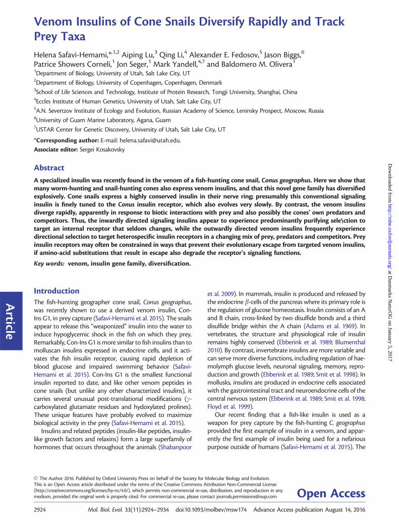
Venom Insulins are Widely Expressed in Diverse ConeSnail LineagesTranscriptome analysis of venom glands from several cone-snail species representing major branches of the phylogenyled to the identification of a diverse set of insulins that sharesequence similarity to the previously described C. geographusvenom insulin (Safavi-Hemami et al. 2015). The inferredamino-acid sequences all include a conserved N-terminal sig-nal sequence, while the region encoding proinsulin (the A andB chains and the C peptide) shows considerable variation(fig. 1). The average pairwise sequence identity is 84% forthe signal peptides, followed by 43%, 60% and 57% for theB chains, C-peptides and A chains, respectively (fig. 1). Suchclose juxtaposition of conserved and hypervariable regions isalso seen regularly in cone snail venom toxins (conotoxins)(Duda and Palumbi 1999; Olivera 2006). Several cone snailspecies were found to express more than one venom insulin,indicating that the gene family has expanded in some but notall cone-snail lineages. The largest number of distinct venominsulin mRNAs in a single species was seven (found inC. geographus). In all, we found 32 distinct sequences in thevenom glands of 21 species.
Of the 21 species examined, six including the fish-hunting cone snail C. bullatus did not express the venominsulin class at detectable levels in their venom glands(table 1). However, by mining the C. bullatus venom glandtranscriptome we identified an insulin-like sequence thatshared little sequence identity with members of thevenom-insulin class, although it shared significant similar-ity to the insulin-like peptide precursor II from the marinegastropod Aplysia californica (32% sequence identity, 91%coverage, e-value: 2e�14). A similar sequence was also re-trieved from the venom gland transcriptome of theworm-hunter C. virgo (96% sequence identity to theC. bullatus sequence). Members of the venom-insulin class
generally do not show interspecific conservation as highas that seen for the C. virgo and C. bullatus insulin-likesequences (see supplementary fig. S1, SupplementaryMaterial online, for sequence alignments). Furthermore,with between one and five reads per kilobase of transcriptper million mapped reads (rpkm), expression levels wereclose to the lower limit of detection by RNAseq for venomgland transcripts, while reads for the venom insulin classwere as abundant as 66,172 rpkm (table 2). This findingimplied that there are at least two kinds of insulins in conesnails, one forming a major component of venom (thevenom-insulin class), and the other expressed at low levelsin endocrine or neuroendocrine cells (the signaling-insulin class).
Tissue-Specific Expression of Insulins in ConusIn other mollusks, signaling insulins are typically expressed inthe nervous system (ganglia and nerve ring) and the epitheliallining of the digestive tract where they play roles in the reg-ulation of haemolymph glucose levels, memory and learning(Ebberink et al. 1989; Smit et al. 1998). To establish whetherthe putative venom-insulin class was venom-gland specificand the signaling-insulin class was predominantly expressedin neuroendocrine cells, we carried out reverse transcriptionPCR (RT-PCR) and quantitative real-time PCR (qPCR) onvenom glands and circumoesophageal nerve rings (contain-ing ganglia) isolated from three cone-snail species belongingto diverse lineages (C. geographus, C. striatus and C. bandanus)(Puillandre et al. 2014). Venom insulins were amplified fromthe venom glands of C. geographus and C. bandanus but notC. striatus (supplementary fig. S2, Supplementary Materialonline). The lack of venom insulin expression in C. striatusis consistent with transcriptome data (see table 1). Venominsulins were absent from all three nervous tissues tested. Incontrast, the putative signaling insulin class was expressedonly in nerve rings and could not be detected in venomglands (supplementary fig. S2, Supplementary Materialonline). Identification of the signaling-insulin class in thevenom-gland transcriptomes of C. bullatus and C. virgo(described earlier) is therefore most plausibly explained astissue contamination from nerve cells surrounding thevenom gland, consistent with very low read counts for thesetwo sequences.
To further confirm tissue-specific expression of the venomand signaling insulin classes, we analyzed the transcriptomesof five tissue types isolated from C. geographus (venom gland,venom bulb, foot, oesophagus and nerve ring). Illumina readswere mapped to known C. geographus insulin sequences andnormalized to the total number of reads obtained for eachtissue (reads per million total reads [rpmr]). RNAseq analysisconfirmed that the venom insulin class was highly expressedin the venom gland but absent from all other tissues testedwith the exception of the low level expression observed in thevenom bulb, a muscular organ located at the distal end of thevenom gland (Safavi-Hemami et al. 2010) (fig. 2). Conversely,the signaling class was exclusively expressed in the oesopha-gus and nervous tissue, consistent with a role in insulin sig-naling (Ebberink et al. 1989; Smit et al. 1998).
Venom Insulins of Cone Snails Diversify Rapidly and Track Prey Taxa . doi:10.1093/molbev/msw174 MBE
2925
at Danm
arks NaturO
G on January 5, 2017
http://mbe.oxfordjournals.org/
Dow
nloaded from
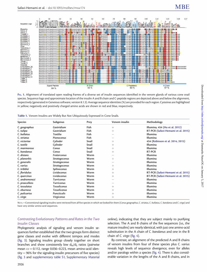
Contrasting Evolutionary Patterns and Rates in the TwoInsulin ClassesPhylogenetic analysis of signaling and venom insulin se-quences further established that the two groups form distinctgene classes and evolve with different tempos and modes(fig. 3). Signaling insulins group closely together on shortbranches and show consistently low dN/dS ratios (pairwisemean x¼ 0.112, range 0.036–0.223, mean amino-acid iden-tity¼ 96% for the signaling-insulin precursors of five species)(fig. 3 and supplementary table S1, Supplementary Material
online), indicating that they are subject mainly to purifyingselection. The A and B chains of the five sequences (i.e., themature insulins) are nearly identical, with just one amino-acidsubstitution in the A chain of C. bandanus and one in the Bchain of C. virgo (fig. 4).
By contrast, an alignment of the predicted A and B chainsof venom insulins from four of these species plus C. variusreveals high levels of sequence divergence, even for allelesand/or paralogs within a species (fig. 4). There is also consid-erable variation in the lengths of the A and B chains, and in
FIG. 1. Alignment of translated open reading frames of a diverse set of insulin sequences identified in the venom glands of various cone snailspecies. Sequence logo and approximate location of the insulin A and B chain and C-peptide regions are depicted above and below the alignment,respectively (generated in Geneious software, version 8.1.3). Average sequence identities (%) are provided for each region. Cysteines are highlightedin yellow; negatively and positively charged amino acids are shown in red and blue, respectively.
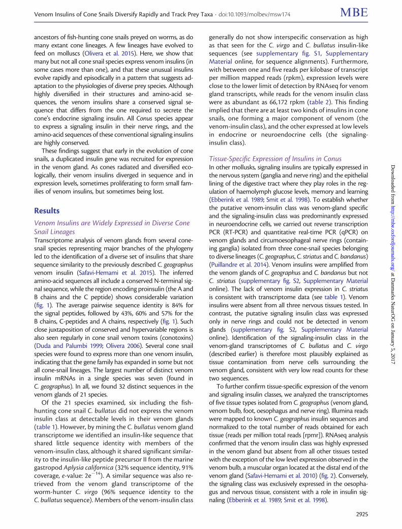
Table 1. Venom Insulins are Widely But Not Ubiquitously Expressed in Cone Snails.
Species Subgenus Prey Venom insulin Methodology
C. geographus Gastridium Fish þ Illumina, 454 (Hu et al. 2012)C. tulipa Gastridium Fish þ RT-PCR (Safavi-Hemami et al. 2015)C. bullatus Textilia Fish � IlluminaC. striatus Pionoconus Fish � IlluminaC. victoriae Cylinder Snail þ 454 (Robinson et al. 2014, 2015)C. textile Cylinder Snail þ IlluminaC. marmoreus Conus Snail þ IlluminaC. bandanus Conus Snail þ RT-PCRC. distans Fraterconus Worm � IlluminaC. planorbis Strategoconus Worm þ IlluminaC. generalis Strategoconus Worm � IlluminaC. varius Strategoconus Worm þ IlluminaC. tribblei Splinoconus Worm þ IlluminaC. floridulus Lividoconus Worm þ RT-PCR (Safavi-Hemami et al. 2015)C. quercinus Lividoconus Worm þ RT-PCR (Safavi-Hemami et al. 2015)C. andremenezi Turriconus Worm � IlluminaC. praecellens Turriconus Worm � IlluminaC. tessulatus Tesseliconus Worm þ IlluminaC. eburneus Tesseliconus Worm þ IlluminaC. pulicarius Puncticulis Worm þ IlluminaC. virgo Virgiconus Worm þ Illumina
NOTE.—Conventional signaling insulins were retrieved from all five species in which we looked for them (Conus geographus, C. striatus, C. bullatus, C. bandanus and C. virgo) andhave very similar amino-acid sequences.
Safavi-Hemami et al. . doi:10.1093/molbev/msw174 MBE
2926
at Danm
arks NaturO
G on January 5, 2017
http://mbe.oxfordjournals.org/
Dow
nloaded from

the numbers and placements of cysteine residues.Phylogenetic analysis of amino-acid substitutions in thevenom insulins of 15 species yielded an overall branchingpattern congruent with recently published phylogenies ofthe genus Conus (Puillandre et al. 2014), with distinct groupsobserved for worm-, snail- and fish-hunters. Within theworm-hunting group, the venom insulins also generally sortinto clades that reflect the species’ phylogenetic relationshipsas determined by Puillandre and co-workers (fig. 3).
Venom insulins show spectacularly elevated dN/dS ratios,implying that their amino-acid sequences often have evolvedunder positive selection. For example, in pairwise compari-sons among 17 representative sequences from the 15 speciesshown in figure 3, the mean dN/dS (x) over 73 codons presentin the proinsulins of all 17 sequences is 2.30, and 71% of the136 pairwise estimates are >1.0 (supplementary table S2,Supplementary Material online). A tree-structured test forpositive selection (comparing M8 to M7 in PAML) stronglyfavors a model in which 36% of the 73 codons evolve with anaverage x of 3.09 (M8, lnL¼�2053.87, 36 parameters), incomparison to the otherwise equivalent model where x isconstrained to be <1.0 at all sites (M7, lnL¼�2082.08, 34parameters, 2DlnL¼ 56.42, df¼ 2, P¼ 5.6e-13).
Branch-specific estimates of x on the phylogeny aremostly >1.0 (fig. 5), but they are much more variable thanthe pairwise estimates, suggesting that adaptive evolution ofthe amino-acid sequences may have been highly episodic.This inference is supported by the much higher likelihoodof a model with independent estimates of x for each branch(lnL¼�2085.94, 63 parameters) than of a model with onecommon value of x (¼1.23) for all branches (lnL¼�2113.61, 33 parameters, 2DlnL¼ 55.34, df¼ 30,P¼ 0.003). Although this estimate of the overall x for venominsulins suggests that dN may have exceeded dS on average,the 95% confidence interval (0.77–1.69) includes x¼ 1.
Even within the hypervariable A and B chains, someamino-acid positions are highly conserved in some species(e.g., the cysteines that make disulfide bridges). This site-to-
site rate heterogeneity is reflected visually in the sequencelogo displayed along the top of figure 1, and is consistent withthe result of the M8/M7 test described earlier. We used themethod of Murrell et al. (2015) to test simultaneously for rateheterogeneity among sites and among branches in the tree,especially those branches following shifts from worm-huntingto mollusk-hunting, and from worm-hunting to fish-hunting.A model with all branches in the foreground estimated a veryhigh rate of amino-acid substitution (x3¼8.94) at 21% ofbranch-site combinations, and yielded a significantly higher
Table 2. Expression Levels of Insulins in the Venom Glands of SeveralConus Species.
Prey Species rpkmVenom insulins
Fish-hunter Geographus 1 (fish-like) 20,472Geographus 3 (fish-like) 8,981Geographus 2 (mollusk-like) 6,249
Snail-hunter Marmoreus 66,172Textile 5,773
Worm-hunter Varius 2,273Planorbis 441Tribblei 2,478Pulicarius 117Virgo 170Tessulatus 71Eburneus 305
Nerve-ring insulinsFish-hunter Bullatus 1Worm-hunter Virgo 5
NOTE.—Values are expressed as reads per million of mapped reads (rpkm).
FIG. 2. Tissue-specific expression of the venom and nerve ring insulinclasses in different organs of Conus geographus. (A) Transcriptomeanalysis of five tissue types (venom gland, venom bulb, foot, oesoph-agus and nerve ring) demonstrated that venom insulins are exclu-sively expressed in the venom gland and venom bulb (orange bars)whereas expression of endogenous insulins is restricted to the nervering (including ganglia) and oesophagus (blue bars). Mapped readswere normalized to total reads obtained for each tissue and are ex-pressed as reads per million total reads (rprm). (B) Schematic repre-sentation of the anatomy of a fixed specimen of C. tessulatus(anesthetized with isotonic magnesium chloride and preserved in80% ethanol) showing equivalent tissues dissected and analyzedfrom C. geographus in this study. (B1–B3) Pictures were taken at dif-ferent stages of dissections and converted into schematic drawingsusing a graphic tablet. Image editing was performed in AdobePhotoshop. (B1) Mantle removed. Position of the shell is shown fororientation. (B2) Proboscis cavity and body haemocoel dissected.Venom gland and nerve ring are depicted in orange and blue, respec-tively. (B3) Venom gland, radular diverticulum and salivary glandsremoved. (B4) Close-up of venom gland, venom bulb and nervering. In a hunting animal, a radula tooth would be positioned atthe tip of the proboscis.
Venom Insulins of Cone Snails Diversify Rapidly and Track Prey Taxa . doi:10.1093/molbev/msw174 MBE
2927
at Danm
arks NaturO
G on January 5, 2017
http://mbe.oxfordjournals.org/
Dow
nloaded from

likelihood than the null model of no positive selection(P< 0.00001). Shifts from worm-hunting to mollusk-hunting and fish-hunting are also associated with strong sig-nals of positive selection, on all five branches that we tested(fig. 5).
The three venom-insulin paralogs in C. geographus and C.tulipa also have evolved episodically. The paralogGeographus_2b has evolved relatively slowly since the speci-ation of C. geographus and the worm-hunting C. pulicarius,and it has retained its ancestral “snail-like” structure and
chemical character (figs. 4 and 6). However, the duplicatedcopy ancestral to the “fish-like” venom insulins(Geographus_3, Geographus_1b and Tulipa; fig. 5) evolvedvery rapidly (x¼ 12 over all sites, and x3¼21.8 at 53% ofbranch-site combinations) and on this branch it also acquireda dramatically different structure, with shortened A and Bchains and a different pattern of cysteines (fig. 4). Then fol-lowing a second duplication, one paralog hardly changed(Geographus_3) while the other (Geographus_1b/Tulipa) ex-perienced a burst of amino-acid substitution (x3¼40 at 27%
FIG. 3. Bayesian protein tree of nerve-ring and venom insulin sequences from diverse Conus species. Branches for venom insulins are colored toindicate prey preferences, with worm, snail and fish-hunting denoted by magenta, blue and yellow, respectively. Insulins from fish-hunting speciesare divided into mollusk- and fish-like sequences. Branch lengths are drawn proportional to estimated amino-acid change, highlighting the speedwith which venom insulins have diverged, as compared with nerve-ring insulins. Tree was rooted with Aplysia californica (Californian sea hare)insulin as an outgroup. Multiple sequences from a given species are probably alleles in most cases, but clearly represent at least three paralogs inC. geographus. Posterior probabilities (Bayesian tree) and bootstrap values (ML tree) are provided for each branch. Codon deletions are depicted asdashes on branches and codon insertions are shown as boxes.
Safavi-Hemami et al. . doi:10.1093/molbev/msw174 MBE
2928
at Danm
arks NaturO
G on January 5, 2017
http://mbe.oxfordjournals.org/
Dow
nloaded from
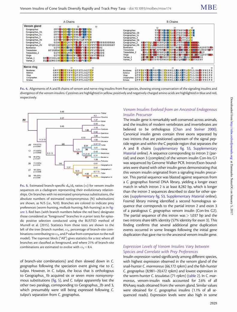
of branch-site combinations) and then slowed down in C.geographus following the speciation event giving rise to C.tulipa. However, in C. tulipa, the locus that is orthologousto Geographus_1b acquired six or seven more nonsynony-mous substitutions (fig. 5), and C. tulipa apparently lost theother two paralogs, corresponding to Geographus_2b and 3,which presumably were still being expressed following C.tulipa’s separation from C. geographus.
Venom Insulins Evolved from an Ancestral EndogenousInsulin PrecursorThe insulin gene is remarkably well conserved across animals,and the insulins of modern vertebrates and invertebrates arebelieved to be orthologous (Chan and Steiner 2000).Canonical insulin genes contain three exons separated bytwo introns that are positioned upstream of the signal pep-tide region and within the C peptide region that separates theA and B chains (supplementary fig. S3, SupplementaryMaterial online). A sequence corresponding to intron 2 (par-tial) and exon 3 (complete) of the venom insulin Con-Ins G1was sequenced by Genome Walker PCR. Intron/Exon bound-aries were shared with other insulin genes demonstrating thatthis venom insulin originated from a signaling insulin precur-sor. This partial sequence was blasted against sequences froma C. geographus fosmid DNA library, yielding a longer exactmatch in which intron 2 is at least 8,282 bp, which is longerthan the intron 2 sequences described to date for other spe-cies (supplementary fig. S3, Supplementary Material online).Fosmid library mining identified a second homologous se-quence that corresponds to the partial intron 2 and exon 3of a paralogous C. geographus venom insulin (Con-Ins G2).The partial sequence of this intron was> 1,037 bp and thetwo introns share 68% identity (57% identity for exon 3). Thisfinding confirms that several additional gene duplicationevents occurred in some lineages following the initial geneduplication that gave rise to the ancestral venom insulin gene.
Expression Levels of Venom Insulins Vary betweenSpecies and Correlate with Prey PreferencesInsulin expression varied significantly among different species,with highest expression observed in the venom gland of thesnail-hunter C. marmoreus (66,172 rpkm) and the fish-hunterC. geographus (8,981–20,472 rpkm) and lowest expression inthe worm-hunter C. tessulatus (71 rpkm) (table 2). In C. mar-moreus, venom-insulin reads accounted for 2.6% of allRNAseq reads obtained from the venom gland. Similar valueswere obtained for C. geographus insulins (1.1% of all se-quenced reads). Expression levels were also high in some
FIG. 4. Alignments of A and B chains of venom and nerve-ring insulins from five species, showing strong conservation of the signaling insulins anddivergence of the venom insulins. Cysteines are highlighted in yellow; positively and negatively charged amino acids are highlighted in blue and red,respectively.
FIG. 5. Estimated branch-specific dN/dS ratios (x) for venom insulinsequences on a cladogram representing their evolutionary relation-ships. On branches with no estimated synonymous substitutions, theabsolute numbers of estimated nonsynonymous (N) substitutionsare shown, as N/S (i.e., N/0). Branches are colored to indicate preypreferences (worm-hunting, mollusk-hunting, fish-hunting) as in fig-ure 3. Red bars (with branch numbers below the red bars) designatethose considered as “foreground” branches in a priori tests for episo-dic positive selection conducted using the BUSTED method ofMurrell et al. (2015). Statistics from those tests are shown to theleft of the tree (branch number, x3, percentage of branch-site com-binations contributing to x3, and P value from comparison to the nullmodel). The topmost block (“All”) gives statistics for a test where allbranches are classified as foreground, and where 21% of branch-sitecombinations are estimated to evolve with x3¼8.4.
Venom Insulins of Cone Snails Diversify Rapidly and Track Prey Taxa . doi:10.1093/molbev/msw174 MBE
2929
at Danm
arks NaturO
G on January 5, 2017
http://mbe.oxfordjournals.org/
Dow
nloaded from
worm-hunters (0.1% in C. tribblei and C. varius), but the av-erage expression was lower in worm-hunters than in snail-and fish-hunters.
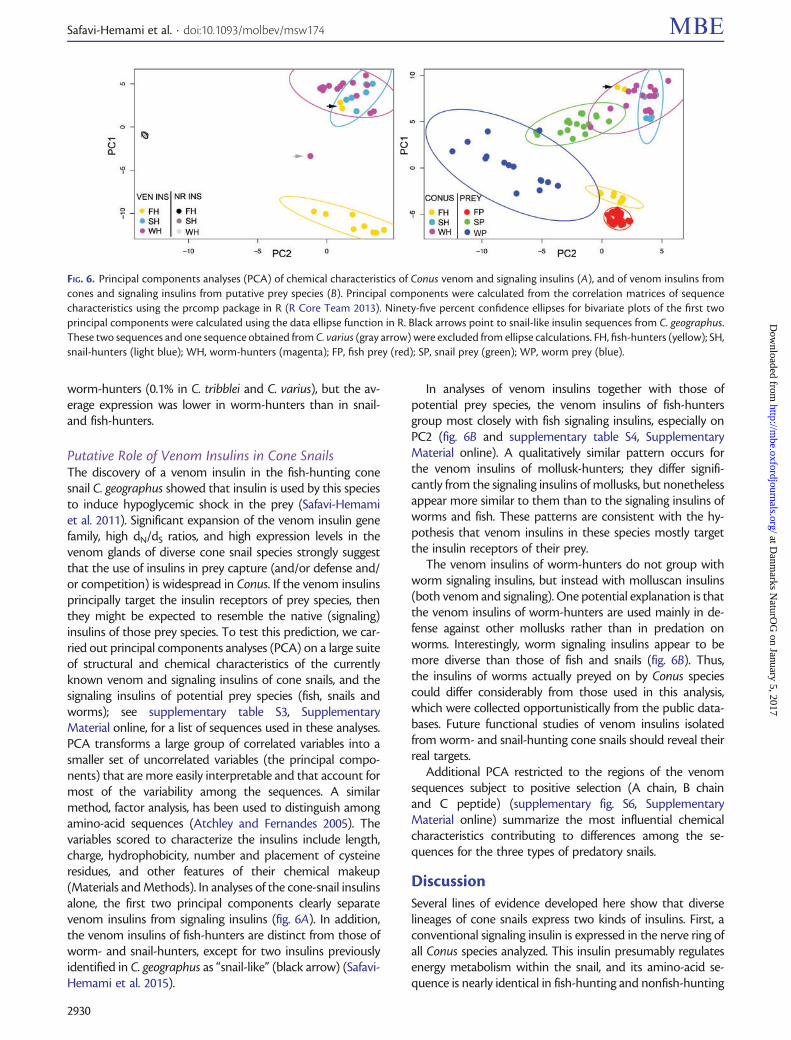
Putative Role of Venom Insulins in Cone SnailsThe discovery of a venom insulin in the fish-hunting conesnail C. geographus showed that insulin is used by this speciesto induce hypoglycemic shock in the prey (Safavi-Hemamiet al. 2011). Significant expansion of the venom insulin genefamily, high dN/dS ratios, and high expression levels in thevenom glands of diverse cone snail species strongly suggestthat the use of insulins in prey capture (and/or defense and/or competition) is widespread in Conus. If the venom insulinsprincipally target the insulin receptors of prey species, thenthey might be expected to resemble the native (signaling)insulins of those prey species. To test this prediction, we car-ried out principal components analyses (PCA) on a large suiteof structural and chemical characteristics of the currentlyknown venom and signaling insulins of cone snails, and thesignaling insulins of potential prey species (fish, snails andworms); see supplementary table S3, SupplementaryMaterial online, for a list of sequences used in these analyses.PCA transforms a large group of correlated variables into asmaller set of uncorrelated variables (the principal compo-nents) that are more easily interpretable and that account formost of the variability among the sequences. A similarmethod, factor analysis, has been used to distinguish amongamino-acid sequences (Atchley and Fernandes 2005). Thevariables scored to characterize the insulins include length,charge, hydrophobicity, number and placement of cysteineresidues, and other features of their chemical makeup(Materials and Methods). In analyses of the cone-snail insulinsalone, the first two principal components clearly separatevenom insulins from signaling insulins (fig. 6A). In addition,the venom insulins of fish-hunters are distinct from those ofworm- and snail-hunters, except for two insulins previouslyidentified in C. geographus as “snail-like” (black arrow) (Safavi-Hemami et al. 2015).
In analyses of venom insulins together with those ofpotential prey species, the venom insulins of fish-huntersgroup most closely with fish signaling insulins, especially onPC2 (fig. 6B and supplementary table S4, SupplementaryMaterial online). A qualitatively similar pattern occurs forthe venom insulins of mollusk-hunters; they differ signifi-cantly from the signaling insulins of mollusks, but nonethelessappear more similar to them than to the signaling insulins ofworms and fish. These patterns are consistent with the hy-pothesis that venom insulins in these species mostly targetthe insulin receptors of their prey.
The venom insulins of worm-hunters do not group withworm signaling insulins, but instead with molluscan insulins(both venom and signaling). One potential explanation is thatthe venom insulins of worm-hunters are used mainly in de-fense against other mollusks rather than in predation onworms. Interestingly, worm signaling insulins appear to bemore diverse than those of fish and snails (fig. 6B). Thus,the insulins of worms actually preyed on by Conus speciescould differ considerably from those used in this analysis,which were collected opportunistically from the public data-bases. Future functional studies of venom insulins isolatedfrom worm- and snail-hunting cone snails should reveal theirreal targets.
Additional PCA restricted to the regions of the venomsequences subject to positive selection (A chain, B chainand C peptide) (supplementary fig. S6, SupplementaryMaterial online) summarize the most influential chemicalcharacteristics contributing to differences among the se-quences for the three types of predatory snails.
DiscussionSeveral lines of evidence developed here show that diverselineages of cone snails express two kinds of insulins. First, aconventional signaling insulin is expressed in the nerve ring ofall Conus species analyzed. This insulin presumably regulatesenergy metabolism within the snail, and its amino-acid se-quence is nearly identical in fish-hunting and nonfish-hunting
FIG. 6. Principal components analyses (PCA) of chemical characteristics of Conus venom and signaling insulins (A), and of venom insulins fromcones and signaling insulins from putative prey species (B). Principal components were calculated from the correlation matrices of sequencecharacteristics using the prcomp package in R (R Core Team 2013). Ninety-five percent confidence ellipses for bivariate plots of the first twoprincipal components were calculated using the data ellipse function in R. Black arrows point to snail-like insulin sequences from C. geographus.These two sequences and one sequence obtained from C. varius (gray arrow) were excluded from ellipse calculations. FH, fish-hunters (yellow); SH,snail-hunters (light blue); WH, worm-hunters (magenta); FP, fish prey (red); SP, snail prey (green); WP, worm prey (blue).
Safavi-Hemami et al. . doi:10.1093/molbev/msw174 MBE
2930
at Danm
arks NaturO
G on January 5, 2017
http://mbe.oxfordjournals.org/
Dow
nloaded from

species. Second, in several (but not all) species, one or morederived insulins are expressed in the venom gland. Some ofthese venom-insulin genes are transcribed at extremely highlevels, and the encoded insulins are abundant components ofthe snail’s venom (as previously documented for the Con-InsG1 insulin from C. geographus). The venom insulins differdramatically from the signaling insulins and from each other.
In this and other respects, the venom insulins appear tofunction and to evolve like other components of Conus ven-oms. Like the well-known conotoxins that target neurotrans-mitter receptors, venom insulins appear to interact not withendogenous macromolecules but with molecular targets inother species (Duda and Palumbi 1999, 2000; Olivera 2006;Puillandre et al. 2010; Sunagar and Moran 2015). In addition,they evolve extremely rapidly like conotoxins (Puillandre et al.2010; Chang and Duda 2012; Wu et al. 2013).
The ancestral prey of cone snails were polychaete worms.Some cone-snail lineages later shifted to mollusks and fish,and diversified extensively (Kohn 1956, 1966; Duda et al. 2001;Puillandre et al. 2014; Olivera et al. 2015). A venom insulinthat efficiently targeted the insulin receptor of a fish wouldpresumably differ from one that efficiently targeted the insu-lin receptor of a mollusk or a worm. Consistent with thisexpectation, the venom insulins of fish-hunting Conus tendto resemble the signaling insulins of fish (see fig. 6). Likewise,the venom insulins expressed in the mollusk-hunting cladestend to be relatively similar to gastropod insulins. This broadpattern is consistent with the hypothesis that at least many ofthese venom insulins are used in prey capture, and that as aconsequence they are strongly selected to interact with theinsulin receptors of prey that appear regularly in a cone’s diet.
The venom-insulin genes are clearly homologous tosignaling-insulin genes, implying that they probably arose byduplication and divergence from an ancestral signaling insu-lin, early in the history of Conus. And apparently, in somelineages, subsequent duplications have given rise to multipleparalogs, as in C. geographus, where there are at least threedistinct venom insulins. While two of these are fish-like invarious chemical properties including the cysteine framework,the third differs considerably and more closely resembles mol-luscan insulins. One intriguing possibility is that this venominsulin may function mainly in defensive or competitive in-teractions with other gastropod molluscs, rather than in preycapture.
However, not all lineages that inherit one or more venominsulins seem to retain them. Among fish-hunting lineages,the subgenus Gastridium (which includes C. geographus andC. tulipa) does express venom insulins, but two other sub-genera, Pionoconus (C. striatus) and Textilia (C. bullatus) donot appear to express a venom-insulin gene. Differing preycapture strategies may explain this difference. Conus geogra-phus appears to release venom insulins into the water tomake an entire school of small fish hypoglycemic, therebyenhancing the snail’s ability to engulf multiple fish. In con-trast, fish-hunting species in the other subgenera capture fishby causing hyper-excitability of the nervous system and rapidonset of a tetanic paralysis. There may be no role for a venominsulin in this prey-capture strategy (Olivera et al. 2015).
Conus tulipa is a close relative of C. geographus and presum-ably inherited the three or more paralogs found in that spe-cies, but it appears to express only one of them (fig. 5).Similarly, we found both mollusk-hunting and worm-hunting lineages that express a venom insulin, but twoworm-hunting lineages that apparently do not.Additionally, several worm-hunting species exhibit low levelsof venom insulin expression suggesting that insulins havebeen inherited but may have ceased to be important in thesespecies.
Selection should strongly favor resistance to Conus ven-oms. Do prey insulin receptors ever acquire amino-acid sub-stitutions that allow them to escape their predator’s venominsulin? And if so, do venom insulins then acquire compen-sating substitutions that restore binding to recently escapedreceptors, and so on, in the dynamic often referred to as “RedQueen” coevolution (Van Valen 1973)? A prey species thatmodified its insulin receptor to escape a venom insulin mightsuffer large costs arising from interference with its own insulinsignaling pathways, which would then need to be readjustedin other ways. For the resulting benefits to be large enough tooffset such costs, the prey species would probably need toexperience a particular Conus species as a major, persistentsource of mortality. Dietary breadth varies greatly amongcone-snail lineages, from highly specialized feeding to omni-vory (Duda et al. 2001). Conus imperialis feeds exclusively onfire worms (Nybakken 1970) and might therefore present agood system in which to look for effects of predation on themolecular adaptation of venom targets in prey. Whether spe-cialized Conus species loom large in the fears of their prey, andactively coevolve with those prey, are questions about whichwe currently know little.
Materials and Methods
Transcriptome AnalysisAssembled and annotated venom-gland transcriptomes ofcone snails belonging to 13 different subgenera, includingones that prey on worms (12 species), mollusks (3 species)and fish (3 species), were mined for transcripts encodinginsulin-like peptides (see table 1 for species and platformsused in this analysis). Briefly, total RNA was isolated fromvenom glands using TRIzol Reagent (Life Technologies) orthe RNeasy kit (Qiagen) following the manufacturers’ instruc-tions. RNA integrity, quantity and purity were determined ona 2100 Bioanalyzer (Agilent Technologies). cDNA librarieswere prepared and sequenced on an Illumina HiSeq 2000instrument (Sanger/Illumina 1.9 reads, 101- or 125-bppaired-end). Adapter clipping and quality trimming of rawreads were performed using fqtrim software (version 0.9.4,http://ccb.jhu.edu/software/fqtrim/) and PRINSEQ (version0.20.4; Schmieder and Edwards 2011), by calculating the com-plexity using the DUST method with a maximum allowedscore of 7. Poly-A/T tails longer than 3 bases were trimmed,and sequences shorter than 70 bps and those containing>5% ambiguous bases (Ns) were discarded. De novo tran-scriptome assembly was performed using Trinity version 2.0.5(Grabherr et al. 2011) with a kmer size for building De Bruijn
Venom Insulins of Cone Snails Diversify Rapidly and Track Prey Taxa . doi:10.1093/molbev/msw174 MBE
2931
at Danm
arks NaturO
G on January 5, 2017
http://mbe.oxfordjournals.org/
Dow
nloaded from
Graphs of 32, a minimum percent identity for two paths to bemerged of 99, a maximum allowed difference between com-bined path sequences of 1, a maximum internal gap lengthallowed for combining paths of 3 and a minimum outputcontig length of 75. Assembled transcripts were annotatedusing BLASTx (ncbi-blast-2.2.28þ; Altschul et al. 1990) againstthe UniProt database (2015), and insulin transcripts extractedbased on annotation. To check fidelity of assembled insulintranscripts, raw reads were mapped back using the map-to-reference tool in Geneious version 8.1.3 (Kearse et al. 2012)and manually examined.
To determine tissue-specific insulin expression, several dif-ferent tissues were dissected from C. geographus (foot, venombulb, oesophagus, nerve ring and venom gland) and totalRNA extracted using Direct-zol RNA MiniPrep Plus (ZymoResearch) following the manufacturer’s instructions. RNA in-tegrity, quantity and purity were determined on a 2100Bioanalyzer (Agilent Technologies). Libraries were preparedusing the TruSeq Stranded mRNA Sample Prep withpoly(A) selection (Illumina) and sequenced on a HiSeq instru-ment (Illumina, 125-bp paired-end). Reads were mappedonto the open reading frame of venom and nerve ring insulinsequences using Geneious (version 8.1.3; Kearse et al. 2012)and normalized to the total number of reads obtained foreach RNAseq data set.
Expression LevelsRead counts for transcripts were determined by mappingRNAseq data sets onto contigs using Bowtie2 software withdefault settings (Langmead and Salzberg 2012). Expressionlevels were obtained by calculating the number of reads perkilobase of transcript per million total mapped reads (rpkm).Average rpkm values were taken for allelic variants becausehigh sequence similarities do not allow for unambiguous as-signment of reads. Read counts could not be determined forsequences obtained by PCR and for insulin from C. victoriaebecause the transcriptome library was normalized prior tosequencing (Robinson et al. 2014).
Comparative RT-PCR and qPCRVenom glands and nerve rings were dissected from C. geo-graphus, C. striatus and C. bandanus and total RNA extractedusing the Ambion Purelink RNA Mini Kit according to themanufacturer’s instructions, with on-column DNase treat-ment. cDNA was prepared using SmartScribe reverse tran-scriptase (Clontech) with a 1:1 mixture of random hexamerand oligo-dT primers. RT-PCR was performed using theClontech Advantage 2 PCR kit (30 cycles using an annealingtemperature of 51�C). Venom insulin primers were designedagainst known C. geographus sequences (Safavi-Hemami et al.2015) and nerve ring insulin primers were designed based onsequences obtained from the venom gland transcriptomes ofC. bullatus and C. virgo (see supplementary table S5,Supplementary Material online, for all primers used). PCRamplicons were gel purified (Qiagen gel extraction kit), ligatedinto the pGEM-T Easy vector (Promega) and transformedinto competent NB 10-beta Escherichia coli (New EnglandBiolabs). Plasmid DNA was isolated from overnight cultures
using the Qiagen plasmid mini kit and sequenced at theUniversity of Utah Sequencing and Genomics core facilityusing Sanger sequencing. Accession numbers are providedin supplementary table S3, Supplementary Material online.qPCR was performed using the SsoFast EvaGreen Supermixwith 20 ng cDNA template for nerve ring and venom glandon a CFX96 instrument (Bio-Rad) and analyzed in CFXManager software (Bio-Rad). The TMX1 and mitochondrialNADH dehydrogenase gene were used for normalization.
Phylogenetic AnalysisMultiple protein sequence alignment of nerve-ring andvenom insulin sequences was performed using MAFFTv7.017 with the slow, iterative refinement setting (FFT-NS-i)(Katoh et al. 2005). Signaling insulin from the sea hare Aplysiacalifornica was used as an outgroup (GenBank:NP_001191503). Alignment gaps were removed prior to phy-logenetic analysis. A bayesian tree was estimated by MrBayes3.2.2 (Huelsenbeck and Ronquist 2001) with two runs each offour Markov chains sampling every 200 generations. The like-lihood score stabilized after 1,100,000 generations. The con-sensus tree was calculated after omitting the first 25% of thesamples as burn-in. Bootstrap branch support values wereobtained for an equivalent ML tree estimated by PhyML(Guindon and Gascuel 2003) (WAG substitution model, 4rate categories, 100 bootstraps).
Selection AnalysisFor selection analysis, one venom insulin-coding sequencewas randomly selected from each of the 15 species with theexception of C. geographus for which one sequence was ran-domly selected from each of the three clearly distinct paral-ogous loci. The purpose of this sampling scheme was toensure that selection analysis was carried out on differentloci (fig. 3), not on alleles. The program codeml from PAML4.7 (Yang 2007) was used to estimate synonymous and non-synonymous substitutions under the F3x4 codon frequencymodel, over all codon positions without gaps in an alignmentof the proinsulin sequences (i.e., the A and B chains plus Cpeptide, omitting the highly conserved signal peptide).Analyses were conducted under several different models ofsequence evolution: (1) pairwise comparisons of the 17 se-quences; (2) a beta distribution of dN/dS ratios (0<x< 1)over sites (M7); (3) a beta distribution over some sites, with asingle higher value of x at others (M8); (4) a single value of xfor all branches (M0); and (5) independent estimates of x forall branches in the phylogeny (M1) (Yang et al. 2000). Similaranalyses were carried out for an alignment of the five nerve-ring insulin sequences. Conventional likelihood-ratio testswere used to compare models that form nested pairs.
Using the same venom-insulin alignment, we carried outbranch-site statistical tests for episodic selection in HyPhy(Kosakovsky Pond et al. 2005) using the BUSTED algorithm(Murrell et al. (2015). BUSTED is an extension of the Nielsenand Yang (1998) episodic model that averages x values acrosssites within the foreground (the branches included in an apriori specified hypothesis of positive selection) and withinthe background (all other branches). The averaging tends to
Safavi-Hemami et al. . doi:10.1093/molbev/msw174 MBE
2932
at Danm
arks NaturO
G on January 5, 2017
http://mbe.oxfordjournals.org/
Dow
nloaded from
wash out the effect of localized selection pressures. In con-trast, BUSTED models site-wise variable rates for each branchof the tree to more sensitively detect local episodic selection.The test accounts for stochastically variable rates across sitesand across branches by estimating three categories of selec-tion, under the alternative constraints thatx1�x2�1�x3, for the foreground and the background.The constraints for the null hypothesis of no episodic positiveselection are x1�x2�x3�1.00. The proportions ofbranch-site combinations in each category of x are esti-mated. We tested a model with all branches in the foreground(unrestricted positive selection), and five different a priorimodels motivated by the phylogenetically inferred shifts inprey preferences.
Statistical Sequence ComparisonPrincipal components analysis was performed on two sets ofamino-acid sequences: (1) venom and nerve-ring (signaling)insulins from cone snails, and (2) venom insulins from conesnails and signaling insulins in potential prey species (seesupplementary table S3, Supplementary Material online, forall sequences used in this analysis). The list of variables used inthe PCA analyses (sequence length, number of cysteine res-idues, molecular weight, isoelectric point, and percentage ofpositive amino acids) is given in supplementary table S4,Supplementary Material online. Variables were determinedfor intact insulin precursors, predicted A and B chains, anddistinct regions within the A and B chains. These regionsincluded cysteine loops and the N- and C-terminal tails fol-lowing the first and last cysteine of each chain. This is illus-trated in supplementary figure S5, Supplementary Materialonline. Principal components were calculated using theprcomp package in R (R Core Team 2013). Values for a givensequence feature were centered to zero and scaled to haveunit variance prior to analysis. The two components with thebest resolving power were chosen to generate plots.
Supplementary MaterialSupplementary tables S1–S5 and figures S1–S3, S5 and S6 areavailable at Molecular Biology and Evolution online (http://www.mbe.oxfordjournals.org/).
AcknowledgmentsWe thank Kevin Chase for help with the PCA analysis, and thestaff of the sequencing and genomics core at the University ofUtah for fast and excellent DNA sequencing and for helpfuladvice. This work was supported in part by National Institutesof Health Grants GM 48677 (to B.M.O.) and GM 099939 (toM.Y.), and by an International Outgoing Fellowship Grantfrom the European Commission (CONBIOS 330486, toH.S.-H.).
ReferencesAdams MJ, Blundell TL, Dodson EJ, Dodson GG, Vijayan M, Baker EN,
Harding MM, Hodkin DC, Rimmer B, Sheat S. 1969. Structure ofrhombohedral 2 zinc insulin crystals. Nature 224:491–495.
Altschul SF, Gish W, Miller W, Myers EW, Lipman DJ. 1990. Basic localalignment search tool. J Mol Biol. 215:403–410.
Atchley WR, Fernandes AD. 2005. Sequence signatures and the proba-bilistic identification of proteins in the Myc-Max-Mad network. ProcNatl Acad Sci U S A. 102:6401–6406.
Blumenthal S. 2010. From insulin and insulin-like activity to the insulinsuperfamily of growth-promoting peptides: a 20th-century odyssey.Perspect Biol Med. 53:491–508.
Chan SJ, Steiner DF. 2000. Insulin through the ages: phylogeny of agrowth promoting and metabolic regulatory hormone. Am Zool.40:213–222.
Chang D, Duda TFJ. 2012. Extensive and continuous duplication facili-tates rapid evolution and diversification of gene families. Mol BiolEvol. 29:2019–2029.
Consortium U. 2015. UniProt: a hub for protein information. NucleicAcids Res. 43:D204–D212.
Duda TF, Jr., Kohn AJ, Palumbi S. 2001. Origins of diverse feeding ecol-ogies within Conus, a genus of venomous marine gastropods. Biol JLinnean Soc. 73:391–409.
Duda TF, Palumbi SR. 2000. Evolutionary diversification of multigenefamilies: allelic selection of toxins in predatory cone snails. Mol BiolEvol. 17:1286–1293.
Duda TF, Palumbi SR. 1999. Molecular genetics of ecological di-versification: duplication and rapid evolution of toxin genes of thevenomous gastropod Conus. Proc Natl Acad Sci U S A. 96:6820–6823.
Ebberink RHM, Smit AB, Van Minnen J. 1989. The insulin family: evolu-tion of structure and function in vertebrates and invertebrates. BiolBull. 177:176–182.
Floyd PD, Li L, Rubakhin SS, Sweedler JV, Horn CC, Kupfermann I,Alexeeva VY, Ellis TA, Dembrow NC, Weiss KR, et al. 1999. Insulinprohormone processing, distribution, and relation to metabolism inAplysia californica. J Neurosci. 19:7732–7741.
Grabherr MG, Haas BJ, Yassour M, Levin JZ, Thompson DA, Amit I,Adiconis X, Fan L, Raychowdhury R, Zeng Q, et al. 2011. Full-lengthtranscriptome assembly from RNA-Seq data without a referencegenome. Nat Biotechnol. 29:644–652.
Guindon S, Gascuel O. 2003. A simple, fast, and accurate algorithm toestimate large phylogenies by maximum likelihood. Syst Biol.52:696–704.
Hu H, Bandyopadhyay PK, Olivera BM, Yandell M. 2012. Elucidation ofthe molecular envenomation strategy of the cone snail Conus geo-graphus through transcriptome sequencing of its venom duct. BMCGenomics 13:1–12.
Huelsenbeck JP, Ronquist F. 2001. MRBAYES: Bayesian inference of phy-logenetic trees. Bioinformatics 17:754–755.
Katoh K, Kuma K, Toh H, Miyata T. 2005. MAFFT version 5: improve-ment in accuracy of multiple sequence alignment. Nucleic Acids Res.20:511–518.
Kearse M, Moir R, Wilson A, Stones-Havas S, Cheung M, Sturrock S,Buxton S, Cooper A, Markowitz S, Duran C, et al. 2012. GeneiousBasic: an integrated and extendable desktop software platform forthe organization and analysis of sequence data. Bioinformatics28:1647–1649.
Kohn AJ. 1956. Piscivorous gastropods of the genus Conus. Proc NatlAcad Sci U S A. 42:168–171.
Kohn AJ. 1966. Food specialization in Conus in Hawaii and California.Ecology 47:1041–1043.
Kosakovsky Pond SL, Frost SD, Muse SV. 2005. HyPhy: hypothesis testingusing phylogenies. Bioinformatics 21:676–679.
Langmead B, Salzberg SL. 2012. Fast gapped-read alignment with Bowtie2. Nat Methods 9:357–359.
Murrell B, Weaver S, Smith MD, Wertheim JO, Murrell S, Aylward A, ErenK, Pollner T, Martin DP, Smith DM, et al. 2015. Gene-wide identi-fication of episodic selection. Mol Biol Evol 32:1365–1371.
Nielsen R, Yang Z. 1998. Likelihood models for detecting positivelyselected amino acid sites and applications to the HIV-1 envelopegene. Genetics 148:929–936.
Nybakken J. 1970. Correlation of radula tooth structure and food habitsof three vermivorous species of Conus. Veliger 12:316–318.
Olivera BM. 2006. Conus peptides: biodiversity-based discovery and exo-genomics. J Biol Chem. 281:31173–31177.
Venom Insulins of Cone Snails Diversify Rapidly and Track Prey Taxa . doi:10.1093/molbev/msw174 MBE
2933
at Danm
arks NaturO
G on January 5, 2017
http://mbe.oxfordjournals.org/
Dow
nloaded from

Olivera BM, Seger J, Horvath MP, Fedosov AE. 2015. Prey-capture strat-egies of fish-hunting cone snails: behavior, neurobiology and evolu-tion. Brain Behav Evol. 86:58–74.
Puillandre N, Bouchet P, Duda TF, Jr., Kauferstein S, Kohn AJ, Olivera BM,Watkins M, Meyer C. 2014. Molecular phylogeny and evolution ofthe cone snails (Gastropoda, Conoidea). Mol Phylogenet Evol.78:290–303.
Puillandre N, Watkins M, Olivera BM. 2010. Evolution of Conus peptidegenes: duplication and positive selection in the A-superfamily. J MolEvol. 70:190–202.
R Core Team. 2013. R: A language and environment for statistical com-puting. Vienna (Austria): R Foundation for Statistical Computing.Available from: http://www.R-project.org/.
Robinson SD, Li Q, Bandyopadhyay PK, Gajewiak J, Yandell M, PapenfussAT, Purcell AW, Norton RS, Safavi-Hemami H. 2015. Hormone-likepeptides in the venoms of marine cone snails. Gen Comp Endocrinol.Advance Access published August 25, 2015, doi: 10.1016/j.ygcen.2015.07.012.
Robinson SD, Safavi-Hemami H, McIntosh LD, Purcell AW, Norton RS,Papenfuss AT. 2014. Diversity of conotoxin gene superfamilies in thevenomous snail, Conus victoriae. PLoS One 9:e87648.
Safavi-Hemami H, Gajewiak J, Karanth S, Robinson SD, Ueberheide B,Douglass AD, Schlegel A, Imperial JS, Watkins M, Bandyopadhyay PK,et al. 2015. Specialized insulin is used for chemical warfare by fish-hunting cone snails. Proc Natl Acad Sci U S A. 112:1743–1748.
Safavi-Hemami H, Siero WA, Gorasia DG, Young ND, MacMillan D,Williamson NA, Purcell AW. 2011. Specialisation of the venomgland proteome in predatory cone snails reveals functional
diversification of the conotoxin biosynthetic pathway. J ProteomeRes. 10:3904–3919.
Safavi-Hemami H, Young ND, Williamson NA, Purcell AW. 2010.Proteomic interrogation of venom delivery in marine cone snails– novel insights into the role of the venom bulb. J Proteome Res.9:5610–5619.
Schmieder R, Edwards R. 2011. Quality control and preprocessing ofmetagenomic datasets. Bioinformatics 27:863–864.
Shabanpoor F, Separovic F, Wade JD. 2009. The human insulin super-family of polypeptide hormones. Vitam Horm. 80:1–31.
Smit AB, van Kesteren RE, Li KW, Van Minnen J, Spijker S,Van Heerikhuizen H, Geraerts WP. 1998. Towards under-standing the role of insulin in the brain: lessons from insulin-relatedsignaling systems in the invertebrate brain. Prog Neurobiol. 54:35–54.
Sunagar K, Moran Y. 2015. The rise and fall of an evolutionary innova-tion: contrasting strategies of venom evolution in ancient and younganimals. PLoS Genet. 11:e1005596.
Van Valen L. 1973. A new evolutionary law. Evol. Theory 1:1–30.Wu Y, Wang L, Zhou M, You Y, Zhu X, Qiang Y, Qin M, Luo S, Ren Z, Xu
A. 2013. Molecular evolution and diversity of Conus peptide toxins,as revealed by gene structure and intron sequence analyses. PLoSOne 8:e82495.
Yang Z. 2007. PAML 4: phylogenetic analysis by maximum likelihood.Mol Biol Evol. 24:1586–1591.
Yang Z, Nielsen R, Goldman N, Pedersen AM. 2000. Codon-substitutionmodels for heterogeneous selection pressure at amino acid sites.Genetics 155:431–449.
Safavi-Hemami et al. . doi:10.1093/molbev/msw174 MBE
2934
at Danm
arks NaturO
G on January 5, 2017
http://mbe.oxfordjournals.org/
Dow
nloaded from