stat-3 activates nf-kb in chronic lymphocytic leukemia cellschronic lymphocytic leukemia (cll) is...
TRANSCRIPT

Signaling and Regulation
STAT-3 Activates NF-kB in Chronic LymphocyticLeukemia Cells
Zhiming Liu, Inbal Hazan-Halevy, David M. Harris, Ping Li, Alessandra Ferrajoli, Stefan Faderl,Michael J. Keating, and Zeev Estrov
AbstractNF-kB plays a major role in the pathogenesis of B-cell neoplasms. A broad array of mostly extracellular
stimuli has been reported to activate NF-kB, to various degrees, in chronic lymphocytic leukemia (CLL) cells.Because CLL cells harbor high levels of unphosphorylated STAT-3 (USTAT-3) and USTAT-3 was reported toactivate NF-kB, we sought to determine whether USTAT-3 activates NF-kB in CLL. Using the electro-phoretic mobility shift assay (EMSA), we studied peripheral blood low-density cells from 15 patients with CLLand found that CLL cell nuclear extracts from all the samples bound to an NF-kB DNA probe, suggesting thatNF-kB is constitutively activated in CLL. Immunoprecipitation studies showed that STAT-3 bound NF-kBp65, and confocal microscopy studies detected USTAT-3/NF-kB complexes in the nuclei of CLL cells, therebyconfirming these findings. Furthermore, infection of CLL cells with retroviral STAT-3-short hairpin RNAattenuated the binding of NF-kB to DNA, as assessed by EMSA, and downregulated mRNA levels of NF-kB–regulated genes, as assessed by quantitative PCR. Taken together, our data suggest that USTAT-3 binds to theNF-kB p50/p65 dimers and that the USTAT-3/NF-kB complexes bind to DNA and activate NF-kB–regulated genes in CLL cells. Mol Cancer Res; 9(4); 507–15. �2011 AACR.
Introduction
Chronic lymphocytic leukemia (CLL) is the most com-mon adult leukemia in the Western Hemisphere. CLL ischaracterized by a dynamic imbalance between the prolif-eration and apoptosis of neoplastic B-lymphocytes coex-pressing CD5 and CD19 antigens, leading to theaccumulation of these cells in the peripheral blood, bonemarrow, and lymphatic tissues (1). Several mechanismshave been reported to provide CLL cells with a survivaladvantage. One such mechanism involves the activation ofNF-kB (2–8). NF-kB plays an important role in thesurvival and proliferation of normal and neoplastic B cells.
In CLL, NF-kB has been found to be activated, to a variabledegree, regardless of disease stage or treatment status (2–8),suggesting that NF-kB might be a target for therapy in CLL(9–11).The NF-kB family of transcription factors consists
of p50, p52, p65 (RelA), c-Rel, and RelB, which sharean N-terminal Rel homology domain responsible for DNAbinding and homo- and heterodimerization. NF-kB dimersbind to kB binding sites within the promoters/enhancers ofNF-kB target genes and regulate transcription. Typically,NF-kB dimers are associated with 1 of 3 IkB proteins,IkBa, IkBb, or IkBe, or the precursor proteins p100 andp105, which maintain NF-kB dimers in the cytoplasm in aninactive state. The activation of NF-kB can be mediated byeither the canonical pathway or the alternative pathway.The canonical pathway is mainly activated by extracellularfactors such as ligands, whose interaction with their corre-sponding cellular receptors results in activation of the bsubunit of the IkB kinase (IKK) complex (IKKb) thatinduces the phosphorylation and degradation of the NF-kB inhibitor IkBa. NF-kB activation in response to anti-gen receptor ligation is mediated by the IKK enzymaticsubunits, IKKa, IKKb, and IKKg (NEMO). FollowingIkBa degradation, NF-kB heterodimers (composed of p50,p65, and/or c-Rel) translocate to the nucleus and bind toDNA. Activation of the alternative NF-kB pathway resultsfrom the transformation of NF-kB2/p100 to p52, which istriggered by the phosphorylation of NF-kB2/p100 by thea-subunit of the IKK complex (IKKa). This allows for
Authors' Affiliation: Department of Leukemia, The University of TexasMDAnderson Cancer Center, Houston, Texas
Note: Z. Liu and I. Hazan-Halevy performed the experiments, and analyzedthe data. D.M. Harris performed retroviral infection studies, confocalmicroscopy studies, and analyzed the data. P. Li performed quantitativePCR analysis and analyzed the data. A. Ferrajoli, S. Faderl, and M.J.Keating treated the studied patients, provided clinical samples, andanalyzed the data. Z. Estrov initiated, designed, and supervised theresearch, and wrote the manuscript
Corresponding Author: Zeev Estrov, Department of Leukemia, Unit 402,MD Anderson Cancer Center, 1515 Holcombe Blvd., Houston, TX 77030;Phone: 713-794-1675; Fax: 713-745-2374; E-mail:[email protected]
doi: 10.1158/1541-7786.MCR-10-0559
�2011 American Association for Cancer Research.
MolecularCancer
Research
www.aacrjournals.org 507
Research. on January 21, 2020. © 2011 American Association for Cancermcr.aacrjournals.org Downloaded from
Published OnlineFirst March 1, 2011; DOI: 10.1158/1541-7786.MCR-10-0559

nuclear translocation of p52, along with RelB, and induc-tion of NF-kB-regulated genes (12).NF-kB is activated in CLL by several different mechan-
isms: interaction with stroma cells (13), activation of thetumor necrosis factor receptor (TNFR) family members(14–17), activation of the cell surface receptor CD40 by itsligand CD154 (4, 18, 19), activation of the B-cell receptor(20), activation of Notch signaling (21), induction of nitricoxide synthase (22), deregulation of the caspase-recruitmentdomain membrane-associated guanylate kinase protein 1(23), deregulation of the T-cell leukemia/lymphoma-1(TCL1) oncogene (24), induction of glycogen synthasekinase-3b (GSK-3b; ref. 25), and modulation of epigeneticregulators (26).In 2005, Yang and colleagues (27) described another,
previously unknown,mechanismofNF-kB activation.Theyfound that unphosphorylated STAT-3 (USTAT-3) binds tothe NF-kB dimers p65/p50 in competition with IkB. TheUSTAT-3/NF-kB complex translocates to the nucleus,binds to DNA, and activates NF-kB–regulated genes. We
recently found that STAT-3, constitutively phosphorylatedon serine 727 residues in CLL cells, induces the productionof STAT-3 protein, and that CLL cells harbor high levels ofUSTAT-3 (28). Therefore, we sought to determine whether,as in other cellular systems (29), USTAT-3 activates NF-kBin CLL cells.
Materials and Methods
Cell fractionationAfter obtaining Institutional Review Board–approved
informed consent, we collected peripheral blood (PB) cellsfrom healthy donors and from 22 patients with CLL whowere treated at The University of Texas MD AndersonCancer Center Leukemia Clinic during the years 2006 to2010. The clinical characteristics of the patients with CLLwhose PB samples were used in the current study arepresented in Table 1. To isolate low-density cells, PB cellswere fractionated using Ficoll Hypaque 1077 (Sigma-Aldrich). More than 90% of the CLL PB cells were
Table 1. Patient characteristics
CLLPt.no.
Sex/age(y)
WBC(109/L)
Lymph.%
Hb.(g/dL)
Plts.(109/L)
Raistage
CD38þ/CD19þ%
b2M(mg/dL)
VH
mutationZAP-70%
Status Cytogenetics Previoustreatment
1 M/58 73.7 90 13.1 128 1 NA 4.1 Y 80.9 Alive t 122 F/74 102.2 88 12.3 236 1 NA 1.9 N NA Alive NA3 F/88 49.1 85 12.2 186 0 NA 3 ND NA Alive D134 M/61 52.9 84 14.8 129 1 0.9 2.7 N Pos. Alive del 11q FCR5 M/63 143 82 13 164 1 97.5 3 Y 33.3 Alive del 11q FCR6 F/70 118 94 12.5 178 1 62.5 3.2 Y 0 Alive t 12 Revlimid7 M/60 133.5 96 13.1 113 0 NA 2.8 N 0 Alive del 13q8 F/56 95.5 97 13 334 0 2.1 2.4 Y 0 Alive Dip.9 F/74 66.7 94 12.5 231 1 NA 1.8 ND NA Alive NA10 F/75 160.7 90 12.1 146 1 1 4 Y 44.6 Alive del 13q FCR11 F/72 136.6 91 11.8 244 2 2 3.6 Y Pos. Alive t 12 Revlimid12 M/63 96 92 14.7 155 0 0.3 3.5 Y 0 Alive del 13q13 M/60 90.6 91 14.7 167 2 NA 4.8 Y 10.8 Alive del 13q FCR14 F/70 26.4 93 10.1 101 3 0.2 3.2 Y 3.4 Alive del 13q Revlimid15 F/58 172.6 98 13.3 212 1 NA 3.3 Y 0 Alive NA16 F/72 46.9 79 9.8 272 3 2 4.7 Y Pos. Alive t 12 Revlimid17 F/60 111.8 92 12.7 219 0 NA 3.5 Y 1.8 Alive NA Ritux. GM18 F/75 60.9 89 12.5 179 0 NA 3.5 Y NA Alive NA19 M/47 130.9 88 13 258 1 98.8 3.3 Y 46.2 Alive NA FCR20 M/48 8.7 20 15.2 173 0 NA NA N Pos. Alive NA CFAR21 F/58 184.4 88 13.6 216 1 NA 2.7 N Neg. Alive NA22 F/75 258.9 94 11.8 140 1 1.1 4.2 Y 44.6 Alive del 13q
Abbreviations: WBC, white blood cells; lymph., lymphocytes; Hb., hemoglobin; Plts., platelets; b2M, b2 microglobulin; M, male; F,female; NA, not available; VH mutation, hypermutation of the immunoglobulin heavy chain gene presented as N (negative; if %derivation from the germline sequence is �2%) or Y (positive, if % derivation from germline sequence is >2%); Ritux., Rituxan; GM,granulocyte-macrophage colony-stimulating factor; FCR, cyclophosphamide, fludarabine, and rituxan; CFAR, cyclophosphamide,fludarabine, alemtuzumab, and rituxan; Pos., positive (analyzed by immunohistochemistry only); Neg., negative; t, translocation; del,deletion.
Liu et al.
Mol Cancer Res; 9(4) April 2011 Molecular Cancer Research508
Research. on January 21, 2020. © 2011 American Association for Cancermcr.aacrjournals.org Downloaded from
Published OnlineFirst March 1, 2011; DOI: 10.1158/1541-7786.MCR-10-0559

CD19þ/CD5þ lymphocytes. To isolate healthy volunteers'CD19þ cells, low-density PB cells were fractionated usingmicroimmunomagnetic beads (Miltenyi Biotec) in accor-dance with the manufacturer's instructions. Flow cytometryanalysis confirmed that 95% or more of the fractionatedcells were CD19þ.
Electrophoretic mobility shift assayNondenatured nuclear extracts were prepared using the
NE-PER extraction kit (Thermo Scientific). Two micro-grams of nuclear protein was incubated with a biotin-labeled NF-kB p65 binding-site DNA probe (50-AGTT-GAGGGGACTTTCCCAGGC-30; synthesized by SigmaGenosys) in binding buffer for 30 minutes on ice. Followingincubation, the samples were separated on a 5% polyacry-lamide gel in Tris-borate EDTA, transferred onto a nylonmembrane, and fixed on the membrane by ultraviolet cross-linking. The biotin-labeled probe was detected with strep-tavidin-horseradish peroxidase (Gel–Shift Kit; Panomics).As a negative control, we used a probe lacking nuclearextracts. The competition control consisted of up to 7-foldexcess unlabeled cold probe combined with the biotin-labeled probes.
ImmunoprecipitationCell pellets were lysed in ice-cold radioimmunopreci-
pitation (RIPA) assay buffer containing 1 mmol/L sodiumorthovanadate, 10 mmol/L sodium fluoride, 100 mmol/LEDTA, 5 mmol/L b-glycerol phosphate, 1 mg/mL apro-tinin, 0.4 mmol/L benzamidine, 1 mg/mL antipain, 1mmol/L phenylmethylsulphonyl fluoride, 1 mg/mL leu-peptin, and 1 mg/mL trypsin inhibitor soybean. The lysedcell pellets were incubated on ice for 5 minutes andvortexed for 1 minute. Then, cell debris was eliminatedby centrifugation at 14,000 rpm for 15 minutes, super-natants were harvested, and the protein concentration wasdetermined using the Micro BCA protein assay reagent kit(Thermo Scientific, Pierce). Whole-cell lysates were incu-bated with 4 mg of polyclonal rabbit anti-human STAT-3antibodies (Upstate Cell Signaling Solutions/Millipore)for 16 hours at 4�C. Protein A agarose beads (Upstate CellSignaling Solutions) were added for 2 hours at 4�C. As anegative control, whole-cell lysates were incubated witheither rabbit serum and protein A agarose beads or proteinA agarose beads only. After 3 washes with RIPA assaybuffer, the beads were resuspended in SDS sample buffer,boiled for 5 minutes, and removed by centrifugation. Thesupernatant proteins were separated by SDS-PAGE (asdescribed in Western blot analysis) and subsequentlyprobed with mouse anti-human NF-kB P65 antibodies(Cell Signaling Technology).
Western blot analysisWestern immunoblotting was performed as previously
described (28). Briefly, cell pellets were lysed in ice-coldRIPA assay buffer, incubated on ice for 5 minutes, andvortexed for 1 minute. Then, cell debris was eliminated bycentrifugation at 14,000 rpm for 15 minutes, supernatants
were harvested, and the protein concentration was deter-mined using the Micro BCA protein assay reagent kit(Thermo Scientific). Supernatant proteins were denaturedby boiling for 5 minutes in SDS, separated by SDS-PAGEusing either 5% or 10% density gels, and transferred to anitrocellulose membrane. After transfer, equal loading wasverified by Ponceau staining.The membranes were blocked with 5% skimmilk in Tris-
buffered saline and incubated with the following antibodies:monoclonal mouse anti-human STAT-3 (BD Biosciences);monoclonal mouse anti-human phosphotyrosine STAT-3and polyclonal rabbit anti-human phosphoserine STAT-3(Cell Signaling Technology); and monoclonal mouse anti-human NF-kB p50 (Pierce Biotechnology) and p65(Sigma-Aldrich). After binding with horseradish peroxi-dase-conjugated secondary antibodies, blots were visualizedwith an enhanced chemiluminescence detection system (GEHealthcare), and densitometry analysis was performed usingan Epson Expression 1680 scanner (Epson America). Den-sitometry values were normalized by dividing the numericalvalue of each sample signal by the numerical value of thesignal from the corresponding loading control. In someexperiments, the membranes were stripped by incubationwith stripping buffer (62.5 mmol/L Tris-HCl, pH 6.7, 2%SDS, 100 mmol/L b-mercaptoethanol) for 30 minutes at50�C, washed, and reprobed with an antibody.
Confocal microscopyCLL low-density cells were cytospun on poly-L-lysine-
coated slides and fixed in 3.7% formaldehyde for 15minutes at room temperature on a shaker. The slides werethen washed 3 times with PBS, incubated with 1% TritonX-100 for 5 minutes at room temperature, and washed 3more times with PBS before blocking with mouse serum for1 hour. After blocking, the slides were washed in PBS andincubated overnight with PE-labeled mouse anti-humanSTAT-3 antibody (BD Biosciences) and AlexaFluor 488-labeled mouse anti-human NF-kB (BioLegend). Afterincubation, the slides were washed 3 times in PBS andthen mounted with Vectashield hard set (Vector Labora-tories). The mounted slides were viewed using an OlympusFluoView 500 laser scanning confocal microscope (Olym-pus America), and the images were analyzed using FluoViewsoftware (Olympus).
Generation of green fluorescence protein-lentivirusSTAT-3–small hairpin RNA and infection of cells293T cells (American type culture collection) were
cotransfected with green fluorescence protein (GFP)-lenti-virus STAT-3 small hairpin RNA (shRNA) or GFP-lenti-virus empty vector and the packaging vectors pCMVdR8.2and pMDG (generously provided by Dr. G. Inghirami,Torino, Italy) using the superfect transfection reagent(Qiagen, Inc.). 293T cell culture medium was changedafter 16 hours and collected after 48 hours. The culturemedium was filtered through a 45-mm syringe filter toremove floating cells; the lentivirus was then concentratedby filtration through an Amicon ultra centrifugal filter
STAT-3 Activates NF-kB in CLL
www.aacrjournals.org Mol Cancer Res; 9(4) April 2011 509
Research. on January 21, 2020. © 2011 American Association for Cancermcr.aacrjournals.org Downloaded from
Published OnlineFirst March 1, 2011; DOI: 10.1158/1541-7786.MCR-10-0559

device (Milipore), and the concentrated supernatant wasused to infect CLL cells.CLL cells (5 � 106/mL) were incubated in 6-well plates
(Becton Dickinson) in 2 mL DMEM supplemented with10% FCS and transfected with 100 mL of the viral super-natant. Polybrene (10 ng/mL) was added to the viralsupernatant at a ratio of 1:1,000 (v/v). Transfection effi-ciency was measured after 48 to 72 hours and was found torange between 30% and 60% [calculated on the basis of theratio of propidium iodide (PI)-negative/GFP-positive cells].These experiments were conducted by using the FACS-Calibur flow cytometer (BectonDickinson Immunocytome-try Systems). Data analysis was performed using CellQuestPro software (Becton Dickinson). A shift from the controlcurve was calculated as percent shift beyond control.
Chromatin immunoprecipitationThe enzymatic chromatin immunoprecipitation (ChIP)
Kit (Cell Signaling Technology) was used. FractionatedCLL cells were cross-linked with 1% formaldehyde for 10minutes at room temperature, a glycine solution wasadded to arrest the reaction, and the cells were washedwith ice-cold PBS. The cells were pooled, pelleted, andincubated on ice for 10 minutes in cell lysis buffersupplemented with 100 mmol/L phenylmethylsulfonylfluoride, dithiothreitol (DTT), and a protease cocktailinhibitor mix. Nuclei were pelleted and resuspended inbuffer supplemented with DTT, digested by micrococcalnuclease, and homogenized on ice. Following sonication(35 pulses, 20 s/pulse at 25% to 30% power) andcentrifugation, 12 mg of sheared chromatin was incubatedwith anti-STAT-3 or rabbit serum (negative control)overnight at 4�C. Then, magnetic-coupled protein Gbeads were added and the chromatin was incubated for2 hours in rotation. Antibody-bound protein/DNA com-plexes were washed, eluted, treated with proteinase digestproteins, and subjected to PCR analyses. An aliquot ofchromatin that was not incubated with an antibody wasused as the input control sample. The primers to amplifythe human STAT-3 promoter were F: 30-CCG AAC GAGCTG GCC TTT CAT-50 and R: 50-GGA TTG GCTGAA GGG GCT GTA-30; to amplify the VEGF Cpromoter: F: 30-CCA GAA AGG ATG TGT AGCATC-50 and R: 50-TGG TCC TCT GTA ACC TGCTCA-30; to amplify the CXCR5 promoter: F: 30-CCTGCC TCA CAA CTC ATC ACT-50 and R: 0-GTT GAGACA ATT ATT GCC GGG-30; and to amplify the CCL5promoter: F: 30-CTCACACTGTAAATTGAGGCA-50and R: 50-AGG TCG CTT AGC AAG TAA ATG-30. The human RPL30 gene primers were provided by CellSignaling Technologies. PCR products were resolved on1.8% agarose gels containing ethidium bromide.
RNA purification and quantitative real-time PCRRNAwas isolated using an RNeasy purification procedure
(Qiagen, Inc.). RNA quality and concentration were ana-lyzed with a NanoDrop spectrophotometer (ND-1000;NanoDrop Technologies). Ten micrograms of total RNA
was used for one-step RT-PCR (Applied Biosystems) withthe sequence detection system ABI Prism 7700 (AppliedBiosystems) using the TaqMan gene expression assay forSTAT-3, VEGF C, CCL5, and CXCR5 (Hs00374200_m1,Hs01099203_m1, Hs00174575_m1, Hs00173527_m1,respectively), according to the manufacturer's instructions.Samples were run in triplicate, and relative quantificationwas performed by comparing the values obtained at thefractional cycle number at which the amount of amplifiedtarget reaches a fixed (CT) threshold.
Results
NF-kB is constitutively activated in CLL cellsSeveral investigators have reported that NF-kB is acti-
vated, to various degrees, in CLL cells (2–8). To confirmthese observations, we obtained PB CLL cells and, by usingelectrophoretic mobility shift assay (EMSA), assessed NF-kB-DNA binding. We studied randomly chosen PB sam-ples from 15 patients with CLL who had favorable orunfavorable prognostic factors and different stages of disease(Table 1). As shown in Figure 1, we found that nuclear
NF-κB-DNA Complexes
1 2 1 2 1 2 1 2 1 2
p
Lane
CLL 1 CLL 2 CLL 3 CLL 4 CLL 5
NF-κB-DNA Complexes
Lane 1 2 1 2 1 2 1 2 1 2
Complexes
CLL 8 CLL 9 CLL 10CLL 6 CLL 7
NF-κB-DNA
L 1 2 1 2 1 2 1 2 1 2
Complexes
CLL 13 CLL 14 CLL 15CLL 11 CLL 12
Lane 1 2 1 2 1 2 1 2 1 2
Figure 1. NF-kB is constitutively activated in CLL cells. EMSA studies ofPB low-density cell nuclear extracts from 15 different patients with CLL arepresented. Lane 1 of each sample depicts binding of CLL cell nuclearextract to a biotin-labeled (hot) NF-kB DNA probe, whereas lane 2 depictsa reduction in binding when an excess of unlabeled probe was added tothe biotin-labeled (hot) kB DNA probe (hot þ cold). In all 15 samples, NF-kB-DNA (p50/p65) complexes were detected and NF-kB-DNA bindingwas almost completely abolished by the cold probe.
Liu et al.
Mol Cancer Res; 9(4) April 2011 Molecular Cancer Research510
Research. on January 21, 2020. © 2011 American Association for Cancermcr.aacrjournals.org Downloaded from
Published OnlineFirst March 1, 2011; DOI: 10.1158/1541-7786.MCR-10-0559

extracts from the PB cells of all 15 patients formed com-plexes with the NF-kB DNA probe, and the addition ofexcess (5-fold) unlabeled (cold) probe inhibited the binding.These data suggest that NF-kB was constitutively activatedin the CLL cells of all studied patients.
STAT-3 binds NF-kB in CLL cells, and the USTAT-3/NF-kB complex translocates to the nucleusThe p65/p50 heterodimer is the prototype NF-kB
transcription factor that is crucial for the expression ofgenes encoding several cytokines and pro-inflammatorymediators. To determine whether STAT-3 binds NF-kBin CLL cells, as previously reported in another cellularsystem (29), we immunoprecipitated STAT-3 proteinwith rabbit anti-human STAT-3 antibodies, and detectedNF-kB p65 and p50, and STAT-3 proteins in the immu-noprecipitate by Western blotting using mouse anti-human p65, p50, and STAT-3 antibodies, respectively.As shown in Figure 2A, the NF-kB p65 and p50 proteinswere detected in CLL cell STAT-3 immunopreciptateobtained from 4 different PB CLL samples. To furtherdelineate this observation, p65 protein was immunopre-cipitated with mouse anti-human p65 antibodies, andSTAT-3, serine pSTAT-3, p65, and p50 proteins weredetected by Western blot analysis. As shown in Figure 2B,p65, p50, STAT-3, but not serine pSTAT-3 proteins weredetected in CLL cell p50 immunopreciptate obtainedfrom 4 different PB CLL samples, suggesting thatUSTAT-3 binds NF-kB p50/p65 dimers in CLL cells.To confirm this observation, we conducted confocal
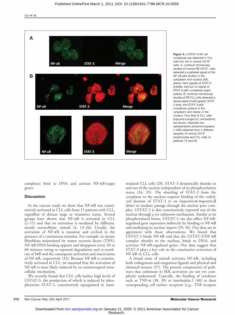
microscopy studies. In normal PB CD19þ cells, faint signalsof STAT-3 were detected, as we previously reported (28).Scattered signals of the NF-kB p65 protein were detected inthe cytoplasm and the nucleus in these control cells(Fig. 3A), likely as a result of the constant shuttling ofIkBa/NF-kB complexes between the nucleus and thecytoplasm (30). In contrast, in CLL cells [characterizedby an overwhelmingly big nucleus and a thin cytoplasm(28)], dense signals of STAT-3 and of the p65 form of NF-kB, as well as STAT-3/p65 complexes, were detectedmainly in the nucleus and, to a lesser extent, in thecytoplasm (Fig. 3B).
USTAT-3/NF-kB complexes bind to DNA and activateNF-kB-regulated genesTo determine whether USTAT-3/NF-kB complexes
bind to DNA we isolated nuclear extracts from CLLcells and, using EMSA, assessed binding of CLL cellnuclear proteins to the kB binding site. As shown inFigure 4A, we found that both anti-p65 and anti-STAT-3antibodies, added to the DNA biotinylated probe,induced a supershift, suggesting that the bands detectedby EMSA consisted not only of NF-kB subunits but ofSTAT-3 protein as well. To further delineate this finding,we infected CLL cells with retroviral STAT-3-shRNAand, using EMSA examined the binding of CLL cellnuclear extracts to the biotinylated NF-kB p65 binding-site DNA probe. As shown in Figure 4B, infection of CLL
cells with retroviral STAT-3-shRNA [previously found todownregulate STAT-3 mRNA and protein levels (28)],but not with empty virus, significantly attenuated thebinding of NF-kB heterodimers to DNA. To determinewhether USTAT-3/NF-kB complexes bind to the pro-moters of NF-kB genes we used ChIP. As shown inFigure 5A, anti-STAT-3 antibodies coimmunoprecipi-tated DNA of STAT-3, and the NF-kB-regulated genesVEGF C, CCL5 and CXCR5. Then, we asked whetherUSTAT-3/NF-kB complexes activate NF-kB-regulatedgenes. As shown in Figure 5B, STAT-3-shRNA down-regulated mRNA levels of the NF-kB-regulated genesVEGF C, CCL5, and CXCR5 [which are not regulatedby STAT-3 (31, 32)] and, as previously reported(28), STAT-3-shRNA downregulated STAT-3 mRNA.Taken together, these data suggest that USTAT-3/NF-kB
A CLL 16 CLL 17 CLL 18 CLL 19
p65
p50
STAT-3
HelaIP B IP B IP B IP B
HelaIP B IP B IP B IP B
B CLL 16 CLL 17 CLL 18 CLL 19
STAT-3
SerinepSTAT-3
p65
p50
p65
Figure 2. U-STAT-3 binds NF-kB p50/p65 dimers. A, PB low-densitycell-protein extracts from 4 different patients with CLL wereimmunoprecipitated with anti-STAT-3 antibodies. As shown, STAT-3 andthe NF-kB p65 and p50 proteins were detected in the immunoprecipitatesof all 3 PB samples. B, PB low-density cell-protein extracts from the samepatients with CLL were immunoprecipitated with anti-p65 antibodies. Helacell protein extract was used as a positive control. As shown, p65, p50,STAT-3, but not serine pSTAT-3 proteins were detected in allimmunopreciptates. IP, immunoprecipitate; B, beads (control).
STAT-3 Activates NF-kB in CLL
www.aacrjournals.org Mol Cancer Res; 9(4) April 2011 511
Research. on January 21, 2020. © 2011 American Association for Cancermcr.aacrjournals.org Downloaded from
Published OnlineFirst March 1, 2011; DOI: 10.1158/1541-7786.MCR-10-0559
complexes bind to DNA and activate NF-kB-targetgenes.
Discussion
In the current study we show that NF-kB was consti-tutively activated in CLL cells from 15 patients with CLL,regardless of disease stage or treatment status. Severalgroups have shown that NF-kB is activated in CLL(2–11) and that its activation is mediated by different,mostly extracellular, stimuli (4, 13–26). Usually, theactivation of NF-kB is transient and cyclical in thepresence of a continuous stimulus. For example, in mousefibroblasts maintained by tumor necrosis factor (TNF),NF-kB-DNA binding appears and disappears every 30 to60 minutes owing to repeated degradation and re-synth-esis of IkB and the consequent activation and inactivationof NF-kB, respectively (33). Because NF-kB is constitu-tively activated in CLL, we assumed that the activation ofNF-kB is most likely induced by an uninterrupted intra-cellular mechanism.We recently found that CLL cells harbor high levels of
USTAT-3, the production of which is induced by phos-phoserine STAT-3, constitutively upregulated in unsti-
mulated CLL cells (28). STAT-3 dynamically shuttles inand out of the nucleus independent of its phosphorylationstatus (34, 35). The shuttling of STAT-3 from thecytoplasm to the nucleus requires binding of the coiled-coil domain of STAT-3 to an importin-a-importin-bdimer to mediate passage through the nuclear pore com-plex. USTAT-3 is also constitutively exported out of thenucleus through a yet unknown mechanism. Similar to itsphosphorylated forms, USTAT-3 can also affect NF-kB-regulated gene expression indirectly by binding to NF-kBand mediating its nuclear import (29, 36). Our data are inagreement with those observations. We found thatUSTAT-3 binds NF-kB and that the USTAT-3/NF-kBcomplex shuttles to the nucleus, binds to DNA, andactivates NF-kB-regulated genes. Our data suggest thatSTAT-3 plays a key role in the constitutive activation ofNF-kB in CLL cells.A broad array of stimuli activates NF-kB, including
both endogenous and exogenous ligands and physical andchemical stresses (37). The protein components of path-ways that culminate in IKK activation are not yet com-pletely understood. Typically, the binding of cytokinessuch as TNF-a (38, 39) or interleukin-1 (40) to theircorresponding cell surface receptors [e.g., TNF-receptor
A
NF-κB MergeSTAT-3
B
NF-κB STAT-3 Merge
NF-κB STAT-3 Merge
Figure 3. U-STAT-3-NF-kBcomplexes are detected in CLLcells but not in normal CD19þ
cells. A, confocal microscopystudies of normal PB CD19þ cellsdetected a scattered signal of theNF-kB p65 protein in thecytoplasm and nucleus (left;green), faint signals of STAT-3(middle; red) but no signal ofSTAT-3-p65 complexes (right;yellow). B, confocal microscopystudies of PB CLL cells detected adense signal of p65 (green), STAT-3 (red), and STAT-3-p65complexes (yellow) in thecytoplasm and mainly in thenucleus. One field of CLL cells(top) and a single CLL cell (bottom)are shown. Depicted arerepresentative photomicrographs(�400) obtained from 2 differentsamples of normal CD19þ
lymphocytes and CLL cells ofpatients 19 and 20.
Liu et al.
Mol Cancer Res; 9(4) April 2011 Molecular Cancer Research512
Research. on January 21, 2020. © 2011 American Association for Cancermcr.aacrjournals.org Downloaded from
Published OnlineFirst March 1, 2011; DOI: 10.1158/1541-7786.MCR-10-0559

(TNFR) or a Toll-like receptor] recruits adaptor proteins[e.g., TNFR-associated factors (TRAFs) and receptor-interacting proteins] to the receptor's cytoplasmicdomain, and these molecules, clustered at the receptor,activate the IKK complex. IKK then phosphorylates IkBat two serine residues, which leads to its ubiquitinationand degradation by the proteasome; NF-kB, free of itsinhibitor, then enters the nucleus to turn on target genes(12). This established model appears to be more complexthan initially thought. The crystal structure of IkBabound to the p65/p50 heterodimer reveals that the IkBaprotein masks only the nuclear localization sequence(NLS) of p65, whereas the NLS of p50 remains exposed.The exposed NLS of p50 coupled with nuclear exportsequences in IkBa and p65 leads to constant shuttling ofIkBa/NF-kB (p50/p65) complexes between the nucleusand the cytoplasm, despite steady state localization thatappears almost exclusively cytosolic (30). Our confocalmicroscopy data showing NF-kB in the nucleus of normalB lymphocytes agree with this observation. Degradation ofIkBa drastically alters the dynamic balance betweencytosolic and nuclear localization signals to favor nuclearlocalization of NF-kB. Apparently, NF-kB might beactivated without the degradation of IkB (29, 36), aswe now show in CLL cells.Several investigators have suggested that NF-kB should
be considered a target for therapy in CLL (9–11). We haverecently reported that STAT-3-shRNA reduced STAT-3mRNA and protein levels and induced apoptosis in CLLcells (28). In the current study, we show that STAT-3-shRNA inhibits the activity of NF-kB. Whether the STAT-3 inhibitors currently being studied in clinical trials inCLL would also inhibit NF-kB activity remains to bedetermined.
CLL 18 CLL 19A
NF-κ B-DNA complexes
Supershi�
CLL 21 CLL 22B
NF-κB-DNA complexes
Figure 4. The U-STAT-3/NF-kB complex binds to DNA. A, CLL cell nuclearproteins p65 and STAT-3 bind to a biotinylated kB-DNA probe. EMSA ofCLL cell nuclear extract is depicted. As shown, the addition of anti-p65 oranti-STAT-3 antibodies to the kB DNA biotinylated probe induced asupershift, suggesting that p65 and STAT-3 bind to DNA. B, STAT-3-shRNA attenuates the binding of CLL cell nuclear extract to a biotin-labeled DNA probe. CLL cells were infected either with lentiviral STAT-3-shRNA or empty virus. Nuclear protein was extracted and EMSA wasperformed. As shown, NF-kB-DNA complexes were detected, and excessunlabeled (cold) DNA probe abolished the binding. Furthermore, infectionswith STAT-3-shRNA, but not with empty virus, significantly attenuatedNF-kB-DNA binding.
AInput IgGSTAT-3p g
STAT-3
VEGF C
CXCL5
VEGF C
CCL5
RPL30
2
B
VEGF C CCL5 CXCR5STAT3
EMPTY
-2
0
e
Control
STAT-3 VEGF C CCL5 CXCR5
-8
-6
-4
Fold
cha
nge
-12
-10
-8F
-14
Figure 5. U-STAT-3/NF-kB complexes activate NF-kB-target genes. A, STAT-3-shRNA downregulated mRNA levels of specific NF-kB-regulated genes. Asshown, infection with lentiviral STAT-3-shRNA downregulated mRNA levels of VEGF C, CCL5, and CXCR5. As expected, lentiviral STAT-3-shRNAdownregulated the level of STAT-3 mRNA as well. In this experiment we used PB cells from patient 21. B, STAT-3 coimmunoprecipitates with DNA of NF-kB-regulated genes. CLL cell-derived chromatin was immunoprecipitated by anti-STAT-3 antibodies or rabbit serum (IgG control). The coimmunoprecipitatedDNA was amplified by PCR using promoter constructs of STAT-3, VEGF C, CCL5, CXCR5, or the control gene RPL30. As shown, DNA of STAT-3, VEGF C,CCL5, andCXCR5 but not the control RPL30 gene, coimmunoprecipitatedwith STAT-3 protein. STAT-3, VEGFC, CCL5, or RPL30 DNAwas detected in wholecell chromatin-extracted DNA (Input).
STAT-3 Activates NF-kB in CLL
www.aacrjournals.org Mol Cancer Res; 9(4) April 2011 513
Research. on January 21, 2020. © 2011 American Association for Cancermcr.aacrjournals.org Downloaded from
Published OnlineFirst March 1, 2011; DOI: 10.1158/1541-7786.MCR-10-0559

Disclosure of Potential Conflicts of Interest
No potential conflict of interests were disclosed.
Acknowledgments
The authors thank Susan Lerner and Susan C. Smith for retrieving the patients'clinical data and Dawn Chalaire for editing the article.
Grant Support
This study was supported by a grant from the CLL Global ResearchFoundation.
The costs of publication of this article were defrayed in part by the payment of pagecharges. This article must therefore be hereby marked advertisement in accordancewith 18 U.S.C. Section 1734 solely to indicate this fact.
Received December 8, 2010; revised January 20, 2011; accepted February 14,2011; published OnlineFirst March 1, 2011.
References1. Yee KW, O' , Brien SM. Chronic lymphocytic leukemia: diagnosis and
treatment. Mayo Clin Proc 2006;81:1105–29.2. Schuh K, Avots A, Tony HP, Serfling E, Kneitz C. Nuclear NF-ATp is a
hallmark of unstimulated B cells from B-CLL patients. Leuk Lym-phoma 1996;23:583–92.
3. Sembries S, Pahl H, Stilgenbauer S, Dohner H, Schriever F. Reducedexpression of adhesion molecules and cell signaling receptors bychronic lymphocytic leukemia cells with 11q deletion. Blood1999;93:624–31.
4. Furman RR, Asgary Z, Mascarenhas JO, Liou HC, Schattner EJ.Modulation of NF-kappa B activity and apoptosis in chronic lympho-cytic leukemia B cells. J Immunol 2000;164:2200–6.
5. Bernal A, Pastore RD, Asgary Z, Keller SA, Cesarman E, Liou HC, et al.Survival of leukemic B cells promoted by engagement of the antigenreceptor. Blood 2001;98:3050–7.
6. Cuní S, P�erez-Aciego P, P�erez-Chac�on G, Vargas JA, S�anchez A,Martín-Saavedra FM, et al. A sustained activation of PI3K/NF-kappaBpathway is critical for the survival of chronic lymphocytic leukemia Bcells. Leukemia 2004;18:1391–400.
7. Rodriguez A, Martinez N, Camacho FI, Ruíz-Ballesteros E, Algara P,García JF, et al. Variability in the degree of expression of phosphory-lated IkappaBalpha in chronic lymphocytic leukemia cases with nodalinvolvement. Clin Cancer Res 2004;10:6796–806.
8. Hewamana S, Alghazal S, Lin TT, Clement M, Jenkins C, GuzmanML, et al. The NF-kappaB subunit Rel A is associated with in vitrosurvival and clinical disease progression in chronic lymphocyticleukemia and represents a promising therapeutic target. Blood2008;111:4681–9.
9. Pickering BM, de Mel S, Lee M, Howell M, Habens F, Dallman CL,et al. Pharmacological inhibitors of NF-kappaB accelerate apopto-sis in chronic lymphocytic leukaemia cells. Oncogene 2007;26:1166–77.
10. Lopez-Guerra M, Colomer D. NF-kappaB as a therapeutic target inchronic lymphocytic leukemia. Expert Opin Ther Targets 2010;14:275–88.
11. Hertlein E, Wagner AJ, Jones J, Lin TS, Maddocks KJ, TownsWH 3rd,et al. 17-DMAG targets the NF-{kappa}B family of proteins to induceapoptosis in CLL: clinical implications of HSP90 inhibition. Blood2010;116:45–53.
12. Hayden MS, Ghosh S. Shared principles in NF-kappaB signaling. Cell2008;132:344–62.
13. Edelmann J, Klein-Hitpass L, Carpinteiro A, F€uhrer A, Sellmann L,Stilgenbauer S, et al. Bone marrow fibroblasts induce expression ofPI3K/NF-kappaB pathway genes and a pro-angiogenic phenotype inCLL cells. Leuk Res 2008;32:1565–72.
14. Munzert G, Kirchner D, Stobbe H, Bergmann L, Schmid RM, D€ohnerH, et al. Tumor necrosis factor receptor-associated factor 1 geneoverexpression in B-cell chronic lymphocytic leukemia: analysis ofNF-kappa B/Rel-regulated inhibitors of apoptosis. Blood 2002;100:3749–56.
15. Kern C, Cornuel JF, Billard C, Tang R, Rouillard D, Stenou V, et al.Involvement of BAFF and APRIL in the resistance to apoptosis of B-CLL through an autocrine pathway. Blood 2004;103:679–88.
16. Endo T, Nishio M, Enzler T, Cottam HB, Fukuda T, James DF, et al.BAFF and APRIL support chronic lymphocytic leukemia B-cell survival
through activation of the canonical NF-kappaB pathway. Blood2007;109:703–10.
17. Nishio M, Endo T, Tsukada N, Ohata J, Kitada S, Reed JC, et al.Nurselike cells express BAFF and APRIL, which can promote survivalof chronic lymphocytic leukemia cells via a paracrine pathway distinctfrom that of SDF-1alpha. Blood 2005;106:1012–20.
18. Karin M, Lin A. NF-kappaB at the crossroads of life and death. NatImmunol 2002;3:221–7.
19. RomanoMF, Lamberti A, Tassone P, Alfinito F, Costantini S, ChiurazziF, et al. Triggering of CD40 antigen inhibits fludarabine-inducedapoptosis in B chronic lymphocytic leukemia cells. Blood 1998;92:990–5.
20. Muzio M, Scielzo C, Bertilaccio MT, Frenquelli M, Ghia P, Caligaris-Cappio F. Expression and function of toll like receptors in chroniclymphocytic leukaemia cells. Br J Haematol 2009;144:507–16.
21. Rosati E, Sabatini R, Rampino G, Tabilio A, Di Ianni M, Fettucciari K,et al. Constitutively activated Notch signaling is involved insurvival and apoptosis resistance of B-CLL cells. Blood 2009;113:856–65.
22. Hammadi A, Billard C, Faussat AM, Kolb JP. Stimulation of iNOSexpression and apoptosis resistance in B-cell chronic lymphocyticleukemia (B-CLL) cells through engagement of Toll-like receptor 7(TLR-7) and NF-kappaB activation. Nitric Oxide 2008;19:138–45.
23. Blonska M, Lin X. CARMA1-mediated NF-kappaB and JNK activationin lymphocytes. Immunol Rev 2009;228:199–211.
24. Pekarsky Y, Palamarchuk A, Maximov V, Efanov A, Nazaryan N,Santanam U, et al. Tcl1 functions as a transcriptional regulator andis directly involved in the pathogenesis of CLL. Proc Natl Acad SciU S A 2008;105:19643–8.
25. Ougolkov AV, BoneND, Fernandez-ZapicoME, Kay NE, Billadeau DD.Inhibition of glycogen synthase kinase-3 activity leads to epigeneticsilencing of nuclear factor kappaB target genes and induction ofapoptosis in chronic lymphocytic leukemia B cells. Blood 2007;110:735–42.
26. Chen SS, Raval A, Johnson AJ, Hertlein E, Liu TH, Jin VX, et al.Epigenetic changes during disease progression in a murine model ofhuman chronic lymphocytic leukemia. Proc Natl Acad Sci U S A2009;106:13433–8.
27. Yang J, Liao X, Agarwal MK, Barnes L, Auron PE, Stark GR. Unpho-sphorylated STAT-3 accumulates in response to IL-6 and activatestranscription by binding to NFkappaB. Genes Dev. 2007;21:1396–408.
28. Hazan-Halevy I, Harris D, Liu Z, Liu J, Li P, Chen X, et al. STAT-3 isconstitutively phosphorylated on serine 727 residues, binds DNA, andactivates transcription in CLL cells. Blood. 2010;115:2852–63.
29. Yang J, Chatterjee-Kishore M, Staugaitis SM, Nguyen H, Schles-singer K, Levy DE, et al. Novel roles of unphosphorylated STAT-3 inoncogenesis and transcriptional regulation. Cancer Res 2005;65:939–47.
30. Ghosh S, Karin M. Missing pieces in the NF-kappaB puzzle. Cell2002;109Suppl: S81–96.
31. Pahl HL. Activators and target genes of Rel/NF-kappaB transcriptionfactors. Oncogene 1999;18:6853–66.
32. Bourillot PY, Aksoy I, Schreiber V, Wianny F, Schulz H, Hummel O,et al. Novel STAT-3 target genes exert distinct roles in the inhibition of
Liu et al.
Mol Cancer Res; 9(4) April 2011 Molecular Cancer Research514
Research. on January 21, 2020. © 2011 American Association for Cancermcr.aacrjournals.org Downloaded from
Published OnlineFirst March 1, 2011; DOI: 10.1158/1541-7786.MCR-10-0559
mesoderm and endoderm differentiation in cooperation with Nanog.Stem Cells 2009;27:1760–71.
33. Hoffmann A, Natoli G, Ghosh G. Transcriptional regulation via the NF-kappaB signaling module. Oncogene 2006;25:6706–16.
34. Reich NC, Liu L. Tracking STAT nuclear traffic. Nat Rev Immunol2006;6:602–12.
35. Liu L, McBride KM, Reich NC. STAT-3 nuclear import is independentof tyrosine phosphorylation and mediated by importin-alpha3. ProcNatl Acad Sci U S A 2005;102:8150–5.
36. Yang J, Stark GR. Roles of unphosphorylated STATs in signaling. CellRes 2008;18:443–51.
37. Gilmore TD. Introduction to NF-kappaB: players, pathways, perspec-tives. Oncogene 2006;25:6680–4.
38. Brown K, Gerstberger S, Carlson L, Franzoso G, Siebenlist U. Controlof I kappa B-alpha proteolysis by site-specific, signal-induced phos-phorylation. Science 1995;267:1485–8.
39. Chen Z, Hagler J, Palombella VJ, Melandri F, Scherer D, Ballard D,et al. Signal-induced site-specific phosphorylation targets I kappa Balpha to the ubiquitin-proteasome pathway. Genes Dev 1995;9:1586–97.
40. Estrov Z, Shishodia S, Faderl S, Harris D, Van Q, KantarjianHM, et al. Resveratrol blocks interleukin-1beta-induced acti-vation of the nuclear transcription factor NF-kappaB, inhibitsproliferation, causes S-phase arrest, and induces apoptosisof acute myeloid leukemia cells. Blood 2003;102:987–95.
STAT-3 Activates NF-kB in CLL
www.aacrjournals.org Mol Cancer Res; 9(4) April 2011 515
Research. on January 21, 2020. © 2011 American Association for Cancermcr.aacrjournals.org Downloaded from
Published OnlineFirst March 1, 2011; DOI: 10.1158/1541-7786.MCR-10-0559
2011;9:507-515. Published OnlineFirst March 1, 2011.Mol Cancer Res Zhiming Liu, Inbal Hazan-Halevy, David M. Harris, et al. Cells
B in Chronic Lymphocytic LeukemiaκSTAT-3 Activates NF-
Updated version
10.1158/1541-7786.MCR-10-0559doi:
Access the most recent version of this article at:
Cited articles
http://mcr.aacrjournals.org/content/9/4/507.full#ref-list-1
This article cites 40 articles, 22 of which you can access for free at:
Citing articles
http://mcr.aacrjournals.org/content/9/4/507.full#related-urls
This article has been cited by 6 HighWire-hosted articles. Access the articles at:
E-mail alerts related to this article or journal.Sign up to receive free email-alerts
SubscriptionsReprints and
To order reprints of this article or to subscribe to the journal, contact the AACR Publications
Permissions
Rightslink site. (CCC)Click on "Request Permissions" which will take you to the Copyright Clearance Center's
.http://mcr.aacrjournals.org/content/9/4/507To request permission to re-use all or part of this article, use this link
Research. on January 21, 2020. © 2011 American Association for Cancermcr.aacrjournals.org Downloaded from
Published OnlineFirst March 1, 2011; DOI: 10.1158/1541-7786.MCR-10-0559