standards for integrated reporting in cellular pathology
TRANSCRIPT

CEff 090117 1 V9 Final
Standards for integrated reporting in cellular pathology
January 2017
Authors: Professor Tim Helliwell, Royal Liverpool University Hospital
Dr Stefan Dojcinov, University Hospital of Wales
Professor Ian Cree, University Hospitals Coventry and Warwickshire
Dr Brian Rous, Cambridge University Hospitals NHS Foundation Trust
Dr Michael Eden, Cambridge University Hospitals NHS Foundation Trust
Professor Simon Cross, Sheffield Teaching Hospitals NHS Foundation Trust
Unique document number G155
Document name Standards for integrated reporting in cellular pathology
Version number 1
Produced by TH, SD, IC, BR, ME and SC are consultant histopathologists with interests in cancer datasets and guidelines. At The Royal College of Pathologists: TH is Vice-President for Learning; BR is Chair of the Working Group on Cancer Services; SC is clinical lead for eLearning; IC is Chair of the Research Committee.
Date active January 2017
Date for full review January 2019
Comments In accordance with the College’s pre-publications policy, this document was on the College website for consultation from 30 June to 30 July 2016. Thirty-one items of feedback were received and the document was amended as necessary. Please email [email protected] if you wish to see the responses and comments.
Dr Lorna Williamson
Director of Publishing and Engagement
The Royal College of Pathologists Fourth Floor, 21 Prescot Street, London, E1 8BB Tel: 020 7451 6700 Web: www.rcpath.org
Registered charity in England and Wales, no. 261035 © 2017, The Royal College of Pathologists This work is copyright. You may download, display, print and reproduce this document for your personal, non-commercial use. Apart from any use as permitted under the Copyright Act 1968 or as set out above, all other rights are reserved. Requests and inquiries concerning reproduction and rights should be addressed to The Royal College of Pathologists at the above address. First published: 2017
CEff 090117 2 V9 Final
Contents Foreword ........................................................................................................................................ 3
1 Introduction ............................................................................................................................ 4
2 Guidance on the provision of integrated reports for cellular pathology samples ..................... 6
3 Laboratory multidisciplinary team meeting ............................................................................. 8
4 Organisational aspects of laboratory services for integrated reporting ................................... 9
5 IT integration ........................................................................................................................ 12
6 Criteria for audit ................................................................................................................... 12
7 References .......................................................................................................................... 12
Appendix A Example of a generic integrated report .............................................................. 14
Appendix B Example of a haemato-oncology integrated report ............................................. 15
Appendix C Summary table – Explanation of grades of evidence ......................................... 16
Appendix D AGREE compliance monitoring sheet ................................................................ 17

CEff 090117 3 V9 Final
Foreword
Guidelines published by The Royal College of Pathologists are documents that should assist pathologists in providing a high standard of care for patients. Guidelines are systematically developed statements to assist the decisions of practitioners and patients regarding appropriate healthcare for specific clinical circumstances and are based on the best available evidence at the time the document was prepared. In this guideline, we emphasise the general principles involved in the integrated reporting of cancer cases, which may be adapted by individual laboratories to meet the contextual needs of their multidisciplinary teams. This document has been produced through a consultation process with members of a short-term Integrated Reporting Working Group of The Royal College of Pathologists (RCPath), the Specialty Advisory Committee on Cellular Pathology and the Interspecialty Committee for Molecular Pathology, and has reported to College Council. The following stakeholders, external to the College, have been asked for comments:
Genomics England
UKNEQAS
British In Vitro Diagnostics Association (BIVDA)
Association of the British Pharmaceutical Industry (ABPI)
The Royal Colleges of Surgeons
The Royal College of Physicians. The recommendations rely on published evidence, existing National Institute for Health and Clinical Excellence (NICE) guidance and other UK and international guidance. Evidence evaluation was carried out as per the recommendations of The Royal College of Pathologists utilising the SIGN guidance (Appendix C). No major organisational changes or cost implications have been identified that would hinder the implementation of the principles of the guideline. The authors recognise that suppliers of laboratory information management systems (LIMS) need to be involved in facilitating any changes to practice that enhance good patient management. A formal revision for all guidelines normally takes place on a five-year cycle. The authors recommend that this guidance is reviewed two years after publication, so that the impact of changes in practice and NHS organisations can be evaluated. The College will ask the authors of the guideline to consider whether or not the guideline needs to be revised. A full consultation process will be undertaken if major revisions are required. If minor revisions or changes are required, a short note of the proposed changes will be placed on the College website for two weeks for members’ attention. If members do not object to the changes, the short notice of change will be incorporated into the guideline and the full revised version (incorporating the changes) will replace the existing version on the College website. The guideline has been reviewed by the RCPath’s Clinical Effectiveness Department and Publishing Department and was placed on the College website for consultation with the membership from 30 June to 30 July 2016. All comments received from the membership have been addressed by the authors, to the satisfaction of the Director of Publishing and Engagement. This guideline was developed without external funding to the writing group. The College requires the authors of guidelines to provide a list of potential conflicts of interest; these are monitored by the Clinical Effectiveness Department and are available on request. The authors of this document have declared that there are no conflicts of interest.
CEff 090117 4 V9 Final
1 Introduction
Microscopic assessment of morphology in haematoxylin and eosin-stained sections remains the cornerstone of cellular pathology diagnosis and prognosis. The work of cellular pathologists is increasingly influenced by techniques such as immunocytochemistry that identify specific tissue constituents. The rapid advances in molecular diagnostic techniques, including cytogenetic and molecular genetic assessments of histological or cytological samples, provide important additional diagnostic, prognostic and predictive information. This is the foundation for personalised management, variously referred to as individualised, stratified or precision medicine, whereby treatments for many diseases, especially cancers, are linked to specific genetic profiles. For other acquired and inherited conditions, although there may not yet be specific therapies, cytogenetic or molecular genetic results inform the more precise diagnosis and prognostication that is of value in shared decision-making with patients. Cellular pathologists have been at the forefront of understanding the molecular basis of cancers and other diseases and incorporating this information for patient benefit. In some areas, such as haematopathology, these investigations have been led by cellular pathology. In other areas, implementation has been achieved through the skills of other laboratory disciplines. There is now significant variation in the way results of certain investigations, particularly molecular diagnostic tests performed on cytology/histology specimens, are interpreted and integrated into a definitive pathological diagnosis, presented to diagnostic and treating clinicians, and used for patient management. Cellular pathologists, more than any other group of diagnostic clinical staff, are essential in understanding such information and interpreting it in conjunction with morphological assessments, including immunocytochemistry. It is therefore likely that in most UK hospital environments cellular pathologists will normally be the most appropriate diagnosticians to receive advice from other professionals in order to provide definitive, meaningful, safe and clinically useful integration of molecular diagnostic data into cytology and histology reports, and present this information to the clinical multidisciplinary team (MDT) to guide patient management. At present, there is no accepted international or UK national standard for the integration of multiple facets of diagnostic information into cellular pathology reports. In some European countries, regulation has been implemented to require mandatory incorporation of all diagnostic laboratory results into a single ‘integrated report’. The diagnostic utility and patient safety aspects of good integration of pathological information have been recognised and formalised in some subspecialties. The Royal College of Pathologists (RCPath) datasets for reporting of lymphomas, soft tissue tumours and tumours of the central nervous system provide a comprehensive guide for multi-disciplinary laboratory integration and integrated reporting.1,2,3 This is also addressed in the NICE Improved Outcomes Guidance for Haematological Cancers.4 The College of American Pathologists, in its series of Cancer Protocol Templates, indicates the requirement for integration of molecular data into pathology reports in certain subspecialties.5 Diagnostic experience and best practice have been researched and documented in published literature, but not translated into recognised, implementable and auditable guidelines.6 The variable approach to integration and interpretation of all relevant diagnostic information obtained from cytology and histology samples into definitive pathology reports6 has wide-reaching and potentially adverse consequences.
Pieces of important diagnostic information are not managed uniformly or in a standardised way.
Interpretation of certain elements of pathological diagnosis is provided by professionals with a variety of qualifications.
Interpretation of some diagnostic tests on cytology/histology samples may be performed without reference to all available diagnostic information. There is therefore a risk of misinterpreting results.

CEff 090117 5 V9 Final
Organisation of multidisciplinary laboratory services does not uniformly take into account requirements to manage samples in a structured way and to integrate the results of all investigations.
LIMS and reporting interfaces do not uniformly provide functionality to integrate data from a variety of sources into a single definitive report.
Education of biomedical scientists and other clinical staff in cellular pathology has not kept pace with the requirement to interpret diagnostic data produced by other laboratory disciplines.
Fragmentation of diagnostic information as a result of a lack of integration into a single summary adversely affects accurate disease registration.
The lack of a standard requirement to integrate complex diagnostic information from a variety of sources to create integrated reports can significantly impact upon the commissioning, funding and organisation of diagnostic services.
Problems arising from the variable approach to reporting investigations on cytology/histology samples are exacerbated by the different organisational structures that have developed in UK laboratories. Most cellular pathology departments do not have facilities to provide the full spectrum of laboratory investigations required for precision medicine. With the exception of some highly specialised centralised services, molecular diagnostics services are usually provided in separate, often managerially independent, units that offer services for local and geographically separated pathology departments, generally without dedicated IT integration. The Specialty Advisory Committee on Cellular Pathology and the Interspecialty Committee for Molecular Pathology of the RCPath recognise the need for general guidance on integration of molecular genetics and other relevant investigations into cellular pathology reports to maximise diagnostic value and maintain a clear, auditable link between these data and the primary tissue sample. We recognise that data may be available from diverse professions or several laboratories and will vary considerably in complexity; any changes in clinical practice should therefore be proportionate to the specific clinical context. The aim of this document is to:
review developments in integrated reporting, including those in other health systems, and identify areas for its application to cancer and non-cancer diagnosis
advise on the systematic integration of results from all investigations undertaken on cytology/histology specimens
provide a strategic view and immediate guidance in the form of standards and a framework for the provision of integrated reports incorporating macroscopic, morphological, immunohistochemical, molecular genetics and other investigations
address wider issues regarding the organisation of laboratory services, communication
between professionals, IT integration, education and training.
1.1 Target users of this guideline
The primary users of this guideline are likely to be practising cellular pathologists. The recommendations will also be of value to trainees and other laboratory professionals.
CEff 090117 6 V9 Final
2 Guidance on the provision of integrated reports for cellular pathology
(histology and cytology) samples
2.1 Key points
The integrated report is a single accessible document that contains information from and interpretation of all relevant investigations undertaken on a cell or tissue sample in a given diagnostic episode.
All diagnostic information is amalgamated into a concise interpretation and diagnostic summary (incorporating any areas of uncertainty), authorised by a qualified medical practitioner or consultant clinical scientist.
Final responsibility for the integrated report and overall interpretation of results should be accepted by the cellular pathologist or other clinical laboratory specialist who oversees the diagnostic process (e.g. a consultant haematologist overseeing the diagnosis of a bone marrow trephine specimen). This individual may be advised by other laboratory staff, including clinical scientists and, for clarity, the responsible clinician would normally be the person who liaises with the clinical MDT.
Individual results from different investigations obtained from one tissue sample (or complementary samples, such as bone marrow aspirate and trephine specimens) should not normally be circulated or made available via hospital information systems to managing physicians as stand-alone reports. Their contribution should be as a component of an integrated report, interpreted alongside morphological and immunophenotypic information. This principle works well in some contexts, such as haemato-oncology, but is overly restrictive in other areas where a morphological diagnosis is sufficient to determine the initial phase of patient management. Flexibility may also be needed when reporting predictive rather than diagnostic markers.
Where comparative testing is undertaken on samples taken at different times, e.g. primary and recurrent or metastatic sites, every effort should be made to ensure that there is a record on the LIMS against each sample of the fact (but not necessarily the detail) of testing. In practice, given the limitations of LIMS, the most recent sample would be the most useful one against which to record comparative results.
Parts of individual reports contributing to the integrated report that relate to quality control (QC) may be omitted from the integrated report but must remain as an essential record in respective laboratories (e.g. within their internal reports). If individual results need to be interpreted with caution for technical reasons (sample quality or quantity), this information should be included in the integrated report.
Integrated reports may be supplemented by successive, newly available results; cellular pathologists or equivalent laboratory medicine specialists are responsible for ensuring that supplementary information is interpreted in conjunction with the existing data and that conclusions are appropriately amended if necessary.
Integrated reports have a data format aligned with relevant RCPath datasets, and with international datasets where these have been adopted by RCPath.
2.2 Practical issues 2.2.1 Cellular pathologists should be proactive in embracing new technologies and incorporating
them into their reports. This should streamline the diagnostic process, facilitate accurate interpretation of results for treating clinicians and prevent errors arising from fragmentary presentation of complex diagnostic data.
2.2.2 The aim of integrating the results of multiple investigations on cytology/histology samples is to create a clinically relevant report that amalgamates and interprets all diagnostic and prognostic

CEff 090117 7 V9 Final
parameters as a single dataset. The subsequent integrated report should provide the patient and their treating clinician with a summary document that can be used as a basis for shared decision-making.6
2.2.3 All laboratory investigations on a cell/tissue sample should be undertaken in a systematic and consistent way to provide the necessary information for accurate diagnosis, prognostication and choice of treatment. The investigations should normally be instigated by cellular pathologists according to agreed diagnostic pathways. The interpretation of each result must be assessed in the context of the entirety of results available at a particular point in time. The interpretations should be meaningful, concordant (or justified, with evidence, if discrepant) and in keeping with the pathological process observed. Cellular pathologists, in communication and consultation with other professionals who undertake the analyses and provide results (see section 3, ‘Laboratory multidisciplinary team’), will normally be best placed to synthesise all relevant laboratory investigations into the integrated report and present this to the clinical MDT. If the final integrated report follows a consensus or multidisciplinary meeting, those present at the meeting and contributing to the conclusion should be recorded, with one person taking responsibility for the final report.
2.2.4 Co-location of the results of all investigations undertaken on a cell/tissue sample in a unified report promotes patient safety by minimising the risk of confusion inherent in attempting to reconcile multiple individual reports or interpreting them out with the context of the full spectrum of investigations undertaken or without awareness of potential diagnostic pitfalls.7–11 It is therefore recommended that individual tests, undertaken as part of a suite of investigations on a cytology/histology specimen, are not normally circulated to clinical users in isolation and outside the integrated pathology report.
[Level of evidence C – Integrated pathology reports reduce risk of result misinterpretation and facilitate patient safety.]
2.2.5 Where the results of assessments may only become available over a period of time, it is appropriate for a preliminary/provisional report to be issued, e.g. on the basis of morphology and immunocytochemistry. The status of the report should be clearly indicated, as well as the nature of the assessments still pending completion.
2.2.6 An integrated report takes relevant data from original ‘individual test’ records, so there should be a standard procedure for linking back to the original reports, including test details, authorisation trail and quality control measures. Quality control (within the laboratory) for each individual test is stringently applied in the NHS and is regulated by UK National External Quality Service (NEQAS). Where multiple investigations are integrated, this information is excessive for most clinicians. It is therefore anticipated that the contributing laboratories will keep internal records for the individual tests they provide, including information regarding quality control. These reports may be circulated internally among participants of the integrated report process but not externally to end-user clinicians.
2.2.7 The reporting format is likely to be governed by the relevant RCPath recommendations and datasets. Cellular pathologists should be mindful that extensive and wordy reports are seldom read in full, and concise, proforma-based reporting is recommended. The tests performed and the results, including their significant limitations, should be clearly presented. Extending current practice, integrated reports should be organised into logical sections including (as appropriate): clinical information and indications for investigation; specimen type; macroscopic description; microscopic features (proforma-based or concise freestyle); immunohistochemistry; flow cytometry; cytogenetics; interphase FISH; PCR/mutational analysis/sequencing; gene expression profile; interpretation; conclusion and diagnosis; SNOMED coding.5 Appendices A and B provide examples of possible styles for a generic IR and an IR for a haematopathology specimen.
[Level of evidence D – Integrated pathology reports facilitate provision of optimal clinical information for precision medicine management.]
CEff 090117 8 V9 Final
3 Laboratory multidisciplinary team meeting 3.1 Key points
All laboratory results obtained from a cytology/histology sample should be discussed in a multi-professional setting before they are formally incorporated into an integrated report to ensure appropriate interpretation and maintain patient safety. This guidance can be adapted to suit the local context and the potential complexity of the reports so that, for example, if molecular testing is only performed to confirm a morphological diagnosis, rediscussion at the MDT may not be necessary.
This is likely to be best achieved by a laboratory multidisciplinary team (LMDT) to enable formal communication between the professionals involved in producing the individual components of the integrated report. The individual taking responsibility for the accurate collation of information and authorisation of the integrated report should be clearly identified.
3.2 Practical issues 3.2.1 The generation of an integrated report should not simply ‘cut and paste’ outputs from a
spectrum of investigations into a single report. The formulation of an integrated report is an orderly and organised process, utilising the professional expertise and experience of all participating scientists and clinicians including, for example, molecular geneticists, cytogeneticists, cellular pathologists and flow cytometrists. An example of a diagnostic algorithm indicating the role of the LMDT is shown in Figure 1. The pathway should be adapted to suit the local context and the potential complexity of the reports so that, for example, if molecular testing is only performed to confirm a morphological diagnosis, rediscussion at the MDT may not be necessary. Conversely, for malignancies of unknown primary site, a LMDT may assist in defining the optimal investigatory strategy. The team must have a documented and auditable process to follow, which facilitates the production of an integrated report in a timely manner. Communication may include face-to-face meetings, teleconferencing and/or electronic communication, depending on local factors such as job plans and geography.
3.2.2 Multidisciplinary teams facilitate good communication and contribute to patient safety. They
ensure that all the results are contextual and appropriately interpreted. In addition, exchange of information between different professional groups is an important learning exercise and an opportunity to widen experience.
[Level of evidence GPP – LMDTs enable good professional communication between all diagnosticians involved in integrated reporting, accurate interpretation of results and optimal diagnosis.]

CEff 090117 9 V9 Final
Figure 1: Exemplar diagnostic algorithm, laboratory integration and role of LMDT in integrated reporting
4 Organisational aspects of laboratory services for integrated reporting
4.1 Key points
Services contributing to integrated reporting should aspire to be included in a unified management structure, with defined roles and responsibilities in performing stratified laboratory investigations, data acquisition and final data interpretation. Organisational and geographic constraints may make the full realisation of this goal impossible. Cellular pathologists would normally be best placed to provide leadership in this process, and this responsibility should be included in their job plan. This should encompass the generation of integrated report and participation in the entire laboratory organisation and decision-making process.
Centralised molecular and other diagnostic laboratory services need to ensure a suitable profile of staff and management to meet the diagnostic requirements of users of their services. ‘Users’ in this context include other staff in all of the contributing departments.

CEff 090117 10 V9 Final
Appropriate management arrangements and defined responsibilities should be established between departments of cellular pathology, molecular diagnostic services and other contributing laboratories to facilitate a unified process for integrated reporting.
Robust and dedicated lines of communication and information exchange must be established between the different laboratory services contributing to integrated reporting.
Within cellular pathology, protocols for handling of cytology and histology specimens, test requesting and multidisciplinary interpretation and reporting should optimise and facilitate the various contributing laboratories’ needs, agreed collaboratively with molecular and other specialist laboratory services.
4.2 Practical issues 4.2.1 There is highly variable delivery of multidisciplinary laboratory services in the UK in the context
of integrated data generation for the diagnosis of cancers and other conditions where molecular testing is essential, with different models of regional centralisation and specialisation. Molecular diagnostic services are becoming increasingly centralised across the country as part of strategic commissioning. Centralised ‘molecular hubs’ provide services for their hosting institutions, within which there is typically a high level of communication and integration with other laboratories. However, in many instances, the access for cellular pathologists to such services is in a ‘hub and spoke’ relationship, with geographically separate providers. This does not preclude clear management arrangements, nor should it interfere with the involvement of cellular pathologists in ‘spoke’ laboratories being full members of the multidisciplinary team, participating in person or by video conferencing. All parties should collaborate to improve these arrangements to support effective and safe integrated reporting on all sites.
4.2.2 Regardless of local variations, the principle of the integration of laboratory results for the
diagnosis of cancer and other relevant pathological processes should be the same. For this to be successful, it is necessary that policies and protocols at different levels of involvement (from generalist to specialist) are clearly defined and coordinated within local practices. All participants in the diagnostic process must have clearly defined roles and appropriate management relationships. Dedicated management to achieve functional and operational integration between contributing laboratories is highly desirable.
4.2.3 Figure 2 shows a possible model for the functional and managerial integration of diagnostic
services in the process of integrated reporting. This model assumes dedicated subspecialist diagnostic teams, both in cellular pathology and molecular diagnostic facilities. Formal arrangements should facilitate cooperative daily working and strategy development through management structures affiliated to centralised molecular laboratories (‘user interface groups’). A user interface group involves key practitioners representing all laboratories contributing to integrated reporting and all responsible subspecialties in cellular pathology. Examples of such high levels of integration have proven to be functionally and financially viable.4,12,13
4.2.4 Protocols for requesting tests, specimen transfer between laboratories and diagnostic
algorithms must be developed to fit best with local arrangements and availability of skills. This approach will facilitate the undertaking of appropriate investigations on cytology/histology specimens, without excess and achieving the greatest synergy between the individual tests.
4.2.5 Dedicated and robust lines of communication must be in place to enable transfer of specimens,
cross-referencing of different (internal and external) laboratory accession numbers, electronic data communication and integration. With this in mind, commissioning and organisation of services should include requirements for dedicated clerical support.
[Level of evidence B – Functional integration of cellular pathology, molecular and other laboratory services for provision of integrated reporting is supported by ‘user interface groups’.]

CEff 090117 11 V9 Final
Figure 2: An example of a model for the integration of cellular pathology and molecular
diagnostic services for integrated reporting

CEff 090117 12 V9 Final
5 IT integration 5.1 Dedicated LIMS will need to be developed to provide solutions to integrating information across
different laboratories into the single integrated report expected by patients and clinical teams. 5.2 The volume and complexity of results provided from a wide range of laboratory services in the
process of integrated reporting can only be appropriately handled through IT integration. It is an essential requirement for LIMS to facilitate easy and robust access to all tests generated from a cytology/histology sample and place them into a single reporting environment accessible by all participants in the process. This assumes access by a range of professionals who may be based in laboratories separated by institutional or administrative boundaries. There is also a requirement for the longitudinal accumulation of results from sequential patient episodes while maintaining the integrated reporting discipline.
5.3 Ongoing modernisation of LIMS occurs across the UK in a piecemeal fashion. NHS managers
and others involved in this process should be mindful that LIMS provision for integrated reporting represents a crucial requirement for a modern molecular and cellular pathology service. Technical and programming solutions should therefore be regarded as a priority for LIMS development teams, including scope for integrating an individual patient’s sequential and other linked specimens.
5.4 In the absence of a comprehensive LIMS solution to integration, laboratory staff should
consider how they may develop an interim solution using coordinated rather than integrated reporting. As a minimum, reports might indicate that the results of molecular testing are available and state that they have not been interpreted in the context of the morphological findings. Free text and appropriate laboratory cross-reference numbers might be helpful.
6 Criteria for audit
Audits of the availability of integrated pathology reports and data at MDT meetings:
each MDT should agree the expected reporting time for provisional and integrated reports to optimise patient management pathways. The complexity of integrated reports means that the standard will be highly context-specific but, as a guide, 90% of cases should meet the locally agreed standard turnaround times.
7 References 1. Dojcinov S, Wilkins B, Calaminici M. Standards for specialist laboratory integration and
dataset for the histopathological reporting of lymphomas. London: The Royal College of Pathologists, 2015. www.rcpath.org/resourceLibrary/dataset-for-the-histopathological-reporting-of-lymphomas.html
2. Fisher C. Dataset for histopathology reporting of soft tissue sarcomas (3rd edition). London: The Royal College of Pathologists, 2014. www.rcpath.org/resourceLibrary/g094_datasetsofttissue_mar14-pdf.html
3. Wharton SB, Hilton D, Ironside JW, Grant, R, Collins VP, Brandner S et al. Dataset for tumours of the central nervous system, including the pituitary gland (4th edition). London: The Royal College of Pathologists, 2016. www.rcpath.org/resourceLibrary/g069-cnsdataset-jan16-pdf.html

CEff 090117 13 V9 Final
4. National Collaborating Centre for Cancer. Addendum to Haematological Cancers: Improving Outcomes (update); Service guidance addendum – Methods, evidence and recommendations (Consultation document). National Institute for Health and Care Excellence [in press].
5. College of American Pathologists. Cancer Protocol Templates. 2016. www.cap.org/web/oracle/webcenter/portalapp/pagehierarchy/cancer_protocol_templates.jspx?_adf.ctrl-state=2ub6hrwfq_4&_afrLoop=742983826197457
6. Cree IA, Deans Z, Ligtenberg MJ, Normanno N, Edsjö A, Rouleau E et al. Guidance for laboratories performing molecular pathology for cancer patients. J Clin Pathol 2014;67: 923–931.
7. Tuff-Lacey A, Shaw E, Cummings R, Walker I, Johnson PW. A collaborative approach to enabling stratified cancer medicine in the UK. Drug Discov Today 2015;20:1414–1418.
8. Deans ZC, Wallace A, O'Sullivan B, Purvis A, Camus S, Fairley JA et al. External quality assessment of BRAF molecular analysis in melanoma. J Clin Pathol 2014;67:120–124.
9. Deans ZC, Tull J, Beighton G, Abbs S, Robinson DO, Butler R. Molecular genetics external quality assessment pilot scheme for KRAS analysis in metastatic colorectal cancer. Genet Test Mol Biomarkers 2011;15:777–783.
10. Wong NA, Deans ZC, Ramsden SC. The UK NEQAS for Molecular Genetics scheme for gastrointestinal stromal tumour: findings and recommendations following four rounds of circulation. J Clin Pathol 2012;65:786–790.
11. Deans ZC, Bilbe N, O'Sullivan B, Lazarou LP, de Castro DG, Parry S et al. Improvement in the quality of molecular analysis of EGFR in non-small-cell lung cancer detected by three rounds of external quality assessment. J Clin Pathol 2013;66:319–325.
12. Salto-Tellez M, Kennedy RD. Integrated molecular pathology: the Belfast model. Drug Discov Today 2015;20:1451–1454.
13. Jack A. Organisation of neoplastic haematopathology services: a UK perspective. Pathology 2005;37:479–492.

CEff 090117 14 V9 Final
Appendix A Example of a generic integrated report
The intention is that this type of layout can be used flexibly according to the diagnostic context.
The most important aspect is to clearly indicate the most recent integrated summary.
Patient ID
Location/responsible clinical consultant
Site and nature of specimen
Date of collection/receipt
Laboratory number
Investigation (A)
Data inserted here (For complex investigations this might be a summary statement, e.g. no mutations identified using ADFRE panel, other tests may be reported in full – context dependent and would need to link to place where full report available on request)
Reported by ………………………………………………………… Date………………………………
Investigation (B)
Data inserted here
Reported by ………………………………………………………… Date………………………………
Investigation (C)
Data inserted here
Testing provided at XYZ laboratories (ISO15189:2012 accredited). Their reference: …....................
Reported by ………………………………………………………… Date………………………………
Integrated conclusions This patient was discussed at the multidisciplinary team meeting on [date]. Morphological assessment of this material, in conjunction with the FISH and PCR assessments indicates a diagnosis of XYZ disease (grading and staging, as appropriate). This is a provisional report. The results of some investigations are pending (see below). Signed ………………………………….…………………. Date: …………………............ Investigations for which results are pending xxx xxx
CEff 090117 15 V9 Final
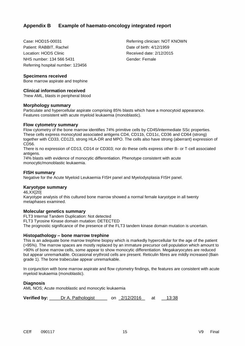
Appendix B Example of haemato-oncology integrated report Case: HOD15-00031
Patient: RABBIT, Rachel
Location: HODS Clinic
NHS number: 134 566 5431
Referring hospital number: 123456
Referring clinician: NOT KNOWN
Date of birth: 4/12/1959
Received date: 2/12/2015
Gender: Female
Specimens received Bone marrow aspirate and trephine
Clinical information received ?new AML, blasts in peripheral blood
Morphology summary Particulate and hypercellular aspirate comprising 85% blasts which have a monocytoid appearance. Features consistent with acute myeloid leukaemia (monoblastic).
Flow cytometry summary Flow cytometry of the bone marrow identifies 74% primitive cells by CD45/intermediate SSc properties. These cells express monocytoid associated antigens CD4, CD11b, CD11c, CD36 and CD64 (strong) together with CD33, CD123, strong HLA-DR and MPO. The cells also have strong (aberrant) expression of CD56. There is no expression of CD13, CD14 or CD303; nor do these cells express other B- or T-cell associated antigens. 74% blasts with evidence of monocytic differentiation. Phenotype consistent with acute monocytic/monoblastic leukaemia.
FISH summary Negative for the Acute Myeloid Leukaemia FISH panel and Myelodysplasia FISH panel.
Karyotype summary 46,XX[20] Karyotype analysis of this cultured bone marrow showed a normal female karyotype in all twenty metaphases examined.
Molecular genetics summary FLT3 Internal Tandem Duplication: Not detected FLT3 Tyrosine Kinase domain mutation: DETECTED The prognostic significance of the presence of the FLT3 tandem kinase domain mutation is uncertain.
Histopathology – bone marrow trephine This is an adequate bone marrow trephine biopsy which is markedly hypercellular for the age of the patient (>95%). The marrow spaces are mostly replaced by an immature precursor cell population which amount to >90% of bone marrow cells, some appear to show monocytic differentiation. Megakaryocytes are reduced but appear unremarkable. Occasional erythroid cells are present. Reticulin fibres are mildly increased (Bain grade 1). The bone trabeculae appear unremarkable. In conjunction with bone marrow aspirate and flow cytometry findings, the features are consistent with acute myeloid leukaemia (monoblastic).
Diagnosis AML NOS; Acute monoblastic and monocytic leukaemia
Verified by: Dr A. Pathologist on 2/12/2016 at 13:38

CEff 090117 16 V9 Final
Appendix C Summary table – Explanation of grades of evidence
(modified from Palmer K et al. BMJ 2008;337:1832)
Grade (level) of evidence Nature of evidence
Grade A At least one high-quality meta-analysis, systematic review of randomised controlled trials or a randomised controlled trial with a very low risk of bias and directly attributable to the target type
or
A body of evidence demonstrating consistency of results and comprising mainly well-conducted meta-analyses, systematic reviews of randomised controlled trials or randomised controlled trials with a low risk of bias, and directly applicable to the target type.
Grade B A body of evidence demonstrating consistency of results and comprising mainly high-quality systematic reviews of case-control or cohort studies and high-quality case-control or cohort studies with a very low risk of confounding or bias and a high probability that the relation is causal and which are directly applicable to the target type
or
Extrapolation of evidence from studies described in A.
Grade C A body of evidence demonstrating consistency of results and including well-conducted case-control or cohort studies and high-quality case-control or cohort studies with a low risk of confounding or bias and a moderate probability that the relation is causal and which are directly applicable to the target type
or
Extrapolation of evidence from studies described in B.
Grade D Non-analytic studies such as case reports, case series or expert opinion
or
Extrapolation of evidence from studies described in C.
Good practice point (GPP) Recommended best practice based on the clinical experience of the authors of the writing group.

CEff 090117 17 V9 Final
Appendix D AGREE compliance monitoring sheet
The guidelines of The Royal College of Pathologists comply with the AGREE II standards for good quality clinical guidelines (www.agreetrust.org). The sections of this guideline that indicate compliance with each of the AGREE II standards are indicated in the table.
AGREE standard Section of guideline
Scope and purpose
1 The overall objective(s) of the guideline is (are) specifically described 1
2 The health question(s) covered by the guideline is (are)specifically described
1
3 The population (patients, public, etc.) to whom the guideline is meant to apply is specifically described
Foreword, 1
Stakeholder involvement
4 The guideline development group includes individuals from all the relevant professional groups
Foreword, 1
5 The views and preferences of the target population (patients, public, etc.) have been sought
n/a
6 The target users of the guideline are clearly defined 1
Rigour of development
7 Systematic methods were used to search for evidence Foreword
8 The criteria for selecting the evidence are clearly described Foreword
9 The strengths and limitations of the body of evidence are clearly described Foreword
10 The methods for formulating the recommendations are clearly described Foreword
11 The health benefits, side effects and risks have been considered in formulating the recommendations
Foreword
12 There is an explicit link between the recommendations and the supporting evidence
2,3,4
13 The guideline has been externally reviewed by experts prior to its publication
Foreword
14 A procedure for updating the guideline is provided Foreword
Clarity of presentation
15 The recommendations are specific and unambiguous 2,3,4
16 The different options for management of the condition or health issue are clearly presented
2,3,4
17 Key recommendations are easily identifiable 2,3,4
Applicability
18 The guideline describes facilitators and barriers to its application Foreword
19 The guideline provides advice and/or tools on how the recommendations can be put into practice
2,3,4
20 The potential resource implications of applying the recommendations have been considered
Foreword
21 The guideline presents monitoring and/or auditing criteria 6
Editorial independence
22 The views of the funding body have not influenced the content of the guideline
Foreword
23 Competing interest of guideline development group members have been recorded and addressed
Foreword