stabilization and mobility of the head, neck and trunk in ... · touchdown of the limbs...
TRANSCRIPT
3889
INTRODUCTIONLocomotion, which requires stability, progression and adaptation(Das and McCollum, 1988; Patla, 1991; Patla, 1997; Shumway-Cook and Woollacott, 2001), depends upon visual, vestibular andsomatosensory inputs to provide continually updated informationon whole-body orientation relative to space (gravito-inertialacceleration vector) and on how to navigate through complex andcluttered natural environments without falling down or getting lost.All three types of sensory input contribute to balance and spatialorientation, and a loss of one of these inputs can be partially, if notlargely, compensated through sensory re-weighting (Peterka, 2002).The visual and vestibular systems, nevertheless, take on predominantroles in locomotion. Vision is central to adapting step patterns onuneven terrain, for avoiding obstacles and arriving at pre-planneddestinations. The vestibular system, responding to gravito-inertialaccelerations of head rotations and translations, provides informationabout head orientation in space. It also, however, contributes toposture (e.g. neck and trunk orientation) and locomotion throughvestibulo-collic reflexes (VCR) and other vestibulospinalmechanisms (Kleine et al., 2004; Peterson and Boyle, 2004; Wilsonand Peterson, 1981), and to gaze stabilization through angular(semicircular canals), and linear (otolith organ) vestibulo-ocularreflexes (VOR) (Fuchs, 1981; Liao et al., 2008; Moore et al., 1999;Paige, 1989; Pozzo et al., 1990; Schwarz and Miles, 1991).Furthermore, vestibular (but not visual) inputs are critical to
discharge patterns of forebrain–midbrain head direction cells(Stackman and Taube, 1997; Stackman et al., 2002) andhippocampal place cells (Russell et al., 2003; Stackman et al., 2002),thereby providing the nervous system with a means to continuallyupdate its internal two-dimensional surface map for navigation (Dayand Fitzpatrick, 2005).
During locomotion, the head rotates and translates through space,requiring compensatory eye movements that stabilize the image onthe retina for clear vision. Much of this stabilization is accomplishedby eye-in-orbit rotations using VOR (Fuchs, 1981; Grossman et al.,1988; Liao et al., 2008; Moore et al., 1999; Paige, 1989; Schwarzand Miles, 1991). Laboratory studies of humans (Cromwell et al.,2001; Cromwell et al., 2004; Pozzo et al., 1990), monkeys and agibbon (Hirasaki and Kumakura, 2004) walking bipedallyoverground, humans walking bipedally on a treadmill (Moore etal., 1999; Pozzo et al., 1990) and humans seated on a verticallymovable apparatus (Paige, 1989), demonstrate that anothermechanism is a compensatory (approximately 180deg. out-of-phase)angular head rotation accompanying vertical head translations.Closer examination of one of these studies (Pozzo et al., 1990),which presents graphs depicting typical head vertical translationsoverlapped with simultaneous head pitch-plane rotations, however,reveals that head pitch rotations are compensatory in some locomotortasks more than others. Whereas, head pitch and vertical translationare, with few exceptions, approximately 180deg. out of phase during
The Journal of Experimental Biology 211, 3889-3907Published by The Company of Biologists 2008doi:10.1242/jeb.020578
Stabilization and mobility of the head, neck and trunk in horses during overgroundlocomotion: comparisons with humans and other primates
Donald C. Dunbar1,*, Jane M. Macpherson2, Roger W. Simmons3 and Athina Zarcades3
1Department of Anatomy and Neurobiology, and Caribbean Primate Research Center, University of Puerto Rico School of Medicine,PO Box 365067, San Juan, PR 00936, USA, 2Neurological Sciences Institute, Oregon Health and Science University, 505 NW 185thAvenue, Beaverton, OR 97006, USA and 3School of Exercise and Nutritional Sciences, San Diego State University, 5500 Campanile
Drive, San Diego, CA 92182, USA*Author for correspondence (e-mail: [email protected])
Accepted 13 October 2008
SUMMARYSegmental kinematics were investigated in horses during overground locomotion and compared with published reports onhumans and other primates to determine the impact of a large neck on rotational mobility (>20deg.) and stability (≤20deg.) of thehead and trunk. Three adult horses (Equus caballus) performing walks, trots and canters were videotaped in lateral view. Dataanalysis included locomotor velocity, segmental positions, pitch and linear displacements and velocities, and head displacementfrequencies. Equine, human and monkey skulls and cervical spines were measured to estimate eye and vestibular arc lengthduring head pitch displacements. Horses stabilized all three segments in all planes during all three gaits, unlike monkeys andhumans who make large head pitch and yaw rotations during walks, and monkeys that make large trunk pitch rotations duringgallops. Equine head angular displacements and velocities, with some exceptions during walks, were smaller than in humans andother primates. Nevertheless, owing to greater off-axis distances, orbital and vestibular arc lengths remained larger in horses,with the exception of head–neck axial pitch during trots, in which equine arc lengths were smaller than in running humans. Unlikemonkeys and humans, equine head peak-frequency ranges fell within the estimated range in which inertia has a compensatorystabilizing effect. This inertial effect was typically over-ridden, however, by muscular or ligamentous intervention. Thus, equinehead pitch was not consistently compensatory, as reported in humans. The equine neck isolated the head from the trunk enablingboth segments to provide a spatial reference frame.
Key words: quadrupedal, bipedal, walk, trot, canter, kinematics, sensorimotor control, vision, vestibular, spatial orientation, navigation, referenceframes, inertia, pitch rotation, linear displacement, velocity, Equus caballus, Homo, Hylobates, Cercopithecus, Macaca, Semnopithecus.
THE JOURNAL OF EXPERIMENTAL BIOLOGY
3890
bipedal runs and hops, phase shifts appear during walks, bothoverground and treadmill. Thus out-of-phase compensatory headpitch rotations, while common, are not essential in all forms oflocomotion.
Pozzo and colleagues, based upon laboratory investigations ofhuman bipedal walks and a variety of other bipedal locomotor tasks(e.g. runs, hops), have proposed that the head is rotationallystabilized (≤20deg. rotation) to provide the brain with a gravito-inertial reference for whole-body spatial orientation and gazeaccommodation (Pozzo et al., 1990). Bipedal walking studies byothers on humans (Cromwell and Wellmon, 2001), monkeys andgibbons (Hirasaki and Kumakura, 2004) found the head to besimilarly stabilized, lending support to this proposal. From our ownreal-world experiences, however, we know that large head rotations(>20deg.), especially head turns in the yaw plane, are indeed avoidedwhen we run or hop. When we walk, by contrast, our headfrequently rotates through much more than 20deg. in the yaw andpitch planes, as when we visually scan our surroundings (e.g. sight-seeing). Thus, if the brain depends upon the head to provide a criticalstable platform for spatial orientation (Pozzo et al., 1990), how thencan we rotate our head during walks and not disturb balance?
The answer may lie with the trunk. Although not explicitly takeninto consideration by Pozzo and colleagues (Pozzo et al., 1990),another study revealed that the trunk remains stabilized (≤20deg.rotation) during bipedal walks (Cromwell et al., 2001). Based on astick figure drawing (Pozzo et al., 1990), the trunk is likely to bealso stabilized during bipedal runs. This finding raises the possibilitythat the head may be free to move without detriment to thelocomotor performance because the stabilized trunk provides thespatial reference frame for postural vertical. Indeed, whereas thevestibular apparatus and eyes provide the essential information fornavigation (position, heading, velocity), trunk receptors makeimportant contributions to spatial orientation for posture and balance.Experimental evidence indicates that the nervous system can usethe trunk as the body-to-space reference in two ways: (1) relativeto gravity-vertical using information from vestibular signals and neckproprioceptors in combination (Kleine et al., 2004; Mergner et al.,1983; Mergner et al., 1991; Mergner et al., 1992), and (2) relativeto gravity-vertical using proprioceptive (Jakobs et al., 1985;Mittelstaedt, 1988; Taylor and McCloskey, 1990) and non-proprioceptive receptors (Mittelstaedt, 1995; Mittelstaedt, 1996;Mittelstaedt, 1997; Mittelstaedt, 1998; Vaitl et al., 1997; Vaitl etal., 2002) in the trunk itself.
The above studies focus on humans and other primates performingbipedally, raising the possibility that the observed segmentalmovement patterns are specific to two-point support. To addressthe issue for four-point support, Dunbar and colleagues conducteda study on two quadrupedal monkey species (Macaca radiata andSemnopithecus entellus) walking and galloping in the wild (Dunbaret al., 2004). Quadrupedal walks were revealed to be comparablewith bipedal walks in that the trunk remains stabilized (≤20deg.rotation). In addition, although commonly stabilized, the headfrequently rotates in the yaw and pitch planes through more than20deg. as the monkeys view their surroundings. Laboratory studiesof head and trunk movements by Macaca fuscata during overgroundlocomotion (Hirasaki and Kumakura, 2004), and Macaca mulatta,Macaca fascicularis (Xiang et al., 2008) and Cercopithecus aethiops(Dunbar, 2004) during treadmill locomotion, also found the monkeytrunk to be stabilized. By contrast, during overground quadrupedalgallops, a fast running gait characterized by unequal timing betweentouchdown of the limbs (Hildebrand, 1977), the trunk is notstabilized (Dunbar et al., 2004). Rather, it rotates in the pitch plane
up to 50deg., while the head remains stabilized in all planes. Basedon these findings, the hypothesis was presented that either the heador the trunk will be rotationally stabilized (≤20deg. rotation) in orderto provide the brain with a reference frame for spatial orientationrelative to the gravito-inertial acceleration vector (Dunbar et al.,2004). The only known exception to the hypothesis is treadmillquadrupedal locomotion, during which the head (pitch and yaw)and trunk (pitch) frequently rotate simultaneously through more than20deg. (Dunbar, 2004). Under these unnatural conditions, the stablevisual surround and dependably smooth treadmill belt surface, andpossibly sensory re-weighting of vestibular and proprioceptiveinputs, may provide novel spatial reference frames that allowsimultaneous head and trunk rotations to occur without balancedetriment. Furthermore, head pitch-plane rotations are rarely180deg. out-of-phase with head vertical translations. Rather, thephase relationship shifts throughout the cycle during quadrupedalwalks, as revealed in an illustration of human bipedal walks (Pozzoet al., 1990), and are primarily in-phase during gallops. Regardless,under real-world conditions, the basic proposal of Pozzo andcolleagues (Pozzo et al., 1990) that the brain requires a stabilizedsegment for determining whole-body spatial orientation remainssupported for both bipedal and quadrupedal locomotion, with theamendment that either the head or the trunk may meet thatrequirement (Dunbar et al., 2004).
Neither the human study by Pozzo and colleagues (Pozzo et al.,1990) nor the monkey studies (Dunbar, 2004; Dunbar et al., 2004)considered the role of the neck in stabilization. Cromwell andcolleagues, however, found that the human neck rotates in the pitchplane more than either the head or trunk during bipedal walking inthe laboratory but that all three segments nevertheless remainedstabilized (≤20 deg. rotation) (Cromwell et al., 2001). Neckmovement patterns in monkeys during natural overgroundlocomotion remain unknown because the neck is obscured byshoulder and arm movements in lateral view, thus prohibitingreliable kinematic measurements (Dunbar et al., 2004).Anatomically, however, monkeys are similar to humans in that theyhave round heads and short necks. The cervical column of monkeysand humans also articulates with the inferior (ventral) surface orbase of the skull, as opposed to the typical non-primate mammaliancolumn, which has a more posterior (caudal) articulation with theskull (Ankel-Simons, 2000). In addition, whereas they are similarto other quadrupeds in allowing large ranges of flexion(ventroflexion) and extension (dorsiflexion) at the cervicothoracicjoints (between the 6th cervical and 1st thoracic vertebrae), bothmonkeys and humans differ from other quadrupeds in that therotational range in the pitch plane at the atlanto–occipital joint isquite restricted (Graf et al., 1995a; Graf et al., 1995b). Thus, theanatomical similarities reported above lead us to predict that themonkey neck will be rotationally stabilized in a manner similar tothe human neck.
A stabilized neck could theoretically provide the spatial referenceframe through a combination of signals from its proprioceptors withsignals from the vestibular apparatus. At least for humans andmonkeys, however, we do not believe the neck, in itself, is servingin this role because no overground locomotor pattern is known toincorporate simultaneous head and trunk rotations of more than20deg. in these species. The human and monkey necks may simplybe too short and of too little mass compared with the head and trunkto serve in this role.
If, then, a segment must be large to adequately serve as a referenceframe, what is the impact of a long and heavy neck on head andtrunk movements during locomotion? In the present study, we
D. C. Dunbar and others
THE JOURNAL OF EXPERIMENTAL BIOLOGY
3891Equine axial segment stabilization
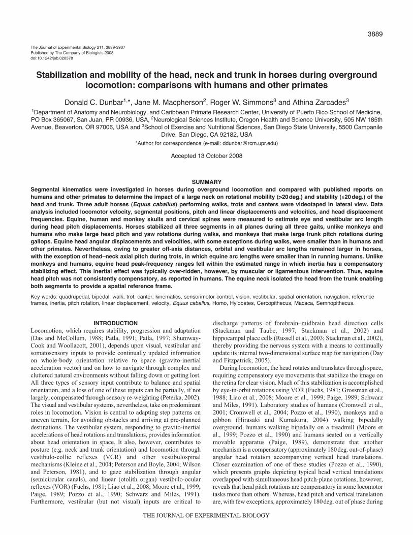
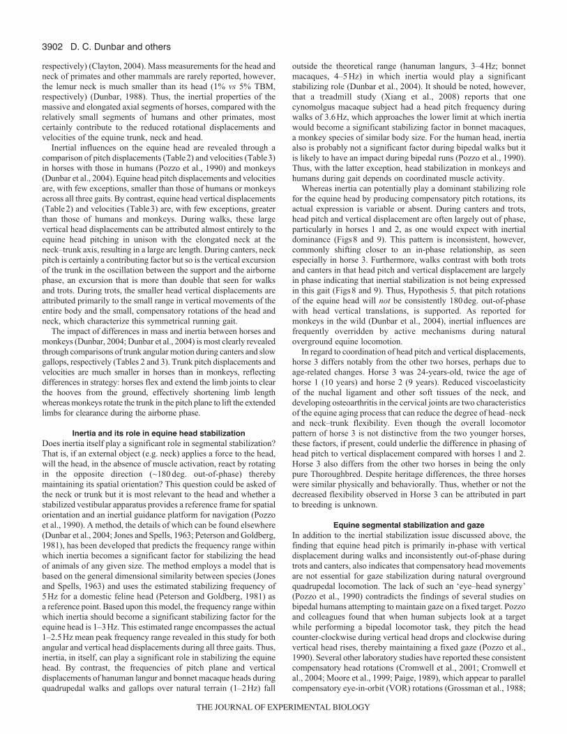
examine this issue by investigating the kinematics of head, neckand trunk movements in horses during natural overgroundlocomotion. We address the following basic question. Are equinehead, neck or trunk movements limited to 20deg. or less in the pitchplane relative to space (i.e. gravity vertical, earth horizontal) and,if so, does the stabilized segment change with a change in gait? Ourfirst two hypotheses are based on the overground patterns observedin quadrupedal monkeys (Dunbar et al., 2004). Hypothesis 1 statesthat the equine head is free to rotate in the pitch and yaw planes ona stabilized trunk during walks. Hypothesis 2, by contrast, statesthat the equine trunk will pitch through more than 20deg. on astabilized head during canters, which are slow gallops. Hypothesis3 states that the equine head and trunk will remain rotationallystabilized during trots. Unlike gallops, trots are characterized by thesymmetrical coordination (equal timing) of contralateral hindlimb–forelimb pairs and vertical trunk movements (Clayton, 2004).The monkey study did not analyze the trot, which is a slowerquadrupedal running gait than the gallop, because these animalsrarely practice it. In addition, large head rotations that are comparablewith those seen during walks are not practiced during monkeyquadrupedal gallops (Dunbar et al., 2004) or human bipedal runs(Pozzo et al., 1990), and head yaw-plane rotations during trots andother running gaits may induce unwanted lateral body sway due toa sudden increase in optic flow (Dunbar, 2004; Schubert et al., 2003).Hypothesis 4 states that the neck will not be rotationally stabilizedin the pitch plane because it must make large (>20 deg.)compensatory movements to isolate the head from the rotationalinfluences of the trunk, thereby enabling independent headstabilization relative to space. That the human neck pitches throughmore degrees than the head or trunk during bipedal walks (Cromwelland Wellmon, 2001) supports this hypothesis. Fig.1 illustratesHypotheses 1–4. Lastly, Hypothesis 5 states that, based on what isknown for quadrupedal monkeys (Dunbar et al., 2004), pitch planerotations of the equine head will not be consistently 180deg. out-of-phase with head vertical translation. To lend context andsignificance to our investigation, the Discussion will includecomparisons of the equine data with those published on humans(Cromwell and Wellmon, 2001; Cromwell et al., 2004; Liebermanet al., 2006; Pozzo et al., 1990) and other primates (Dunbar, 2004;Dunbar et al., 2004; Hirasaki and Kumakura, 2004; Xiang et al.,2008).
MATERIALS AND METHODSAnimals, training and videotaping
Three gelded domestic horses (Equus caballus L. 1758), 14.3±8.3years of age, 164.6±2.7cm in height and 536.2±16.8kg in mass,were the subjects of this study. The horses were a Thoroughbred,a Warm Blood and a Thoroughbred-Warm Blood cross. TheThoroughbred is a true Hot Blood breed that is characterized byspeed, stamina and athleticism but also tends to be nervous andtemperamental. The Warm Blood, by contrast, is a group of horsebreeds and types that were developed by crossbreeding Hot Bloodbreeds with large and powerful, but relatively even-tempered (ColdBlood), draft horses in an effort to produce athletic horses ofreasonable temperament. Hot Bloods are commonly crossbred withWarm Bloods in order to further improve the latter for competitivesports (Edwards, 2001). Despite their different heritages, however,all three horses were of similar build, temperament, and behavior.The horse owners provided written permission to use the animals.All experimental procedures were approved by the InstitutionalAnimal Care and Use Committee (IACUC) and conducted accordingto the National Institutes of Health (NIH) guidelines (USA).
The horses were videotaped while walking, trotting and cantering(slow galloping) in an outdoor arena, the support surface of whichconsisted of an 8cm sand mixture on a firm base. Gaits wererecorded using a Sony Digital 8, 60 Hz video camera (SonyElectronics, San Diego, CA, USA) positioned approximately 28mfrom the plane of action. Small squares of white masking tape(5cm�5cm) were placed on the camera side of the horse inalignment with the withers (spinous process of the 3rd thoracicvertebra) and the ear (external auditory meatus) to assist data
�20 deg.
>20 deg.
H2
>20 deg.
H4
>20 deg.
�20 deg.>20 deg.
H1
�20 deg.
H3
�20 deg.
Fig. 1. Graphic depictions of Hypotheses 1–4. Although the hypothesesrefer to different gaits, a standard walking figure is used for all illustrationsin order to emphasize the differences in head, neck and trunk pitchpredicted by each hypothesis. Hypothesis 1 (H1); the head is free to rotatemore than 20 deg. in the pitch and yaw planes on a stabilized trunk duringwalks. Hypothesis 2 (H2); the trunk will pitch through more than 20 deg. ona stabilized head during canters. Hypothesis 3 (H3); the head and trunk willremain rotationally stabilized (≤20 deg.) during trots. Hypothesis 4 (H4); theneck will not be rotationally stabilized in the pitch plane because it mustmake large (>20 deg.) compensatory movements to isolate the head fromthe rotational influences of the trunk, thereby enabling independent headstabilization relative to space. Arrows indicate the direction and relativemagnitude of pitch rotation.
THE JOURNAL OF EXPERIMENTAL BIOLOGY
3892
reduction. The camera was mounted on a tripod and leveled withrespect to earth horizontal. The built-in telephoto lens was used toreduce parallax and to provide data capture for a minimum of onecomplete stride cycle. Before and after each recording session, a457cm (15ft) wooden pole was placed parallel to, and in line with,the plane of gait and also leveled. Videotape of this pole provideda horizontal reference for measurement of head, neck, and trunkposition and a length calibration. Thus, measurement error wasminimized during data collection by using clear landmarks,minimizing parallax with the zoom lens, and placing the beam ofknown length in the locomotor path. The 60Hz sampling rate provedsufficient because the segments of interest are massive and displacedat rates slow enough to avoid or minimize blurring, even at the fastestgallops sampled.
Data were collected while the horses completed their usualexercise regimen. Following a warm-up period, each horseperformed eight trials of each gait in the following sequence: walks,trots and canters. All trials were videotaped in a single exercisesession. During walks and trots, an experienced handler used a looseshank to lead the horses through the plane of action. The loosenessof the shank allowed unrestricted head movement, and the handlerwas always in front of the horses to ensure the video lens anunobstructed view. During canters, the halter and shank wereremoved and the horses were free to move at will. The handlerencouraged cantering through handclaps and verbal prompts. At theconclusion of the exercise session, the horse was recaptured, cooledoff and returned to the stable.
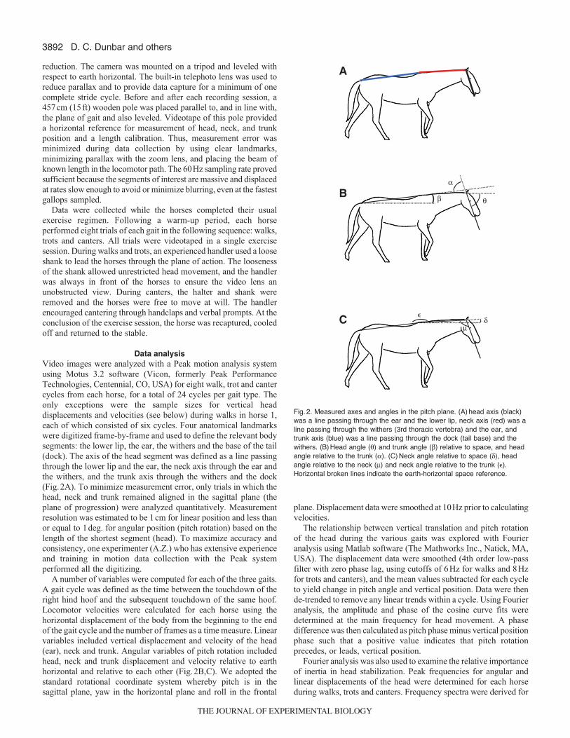
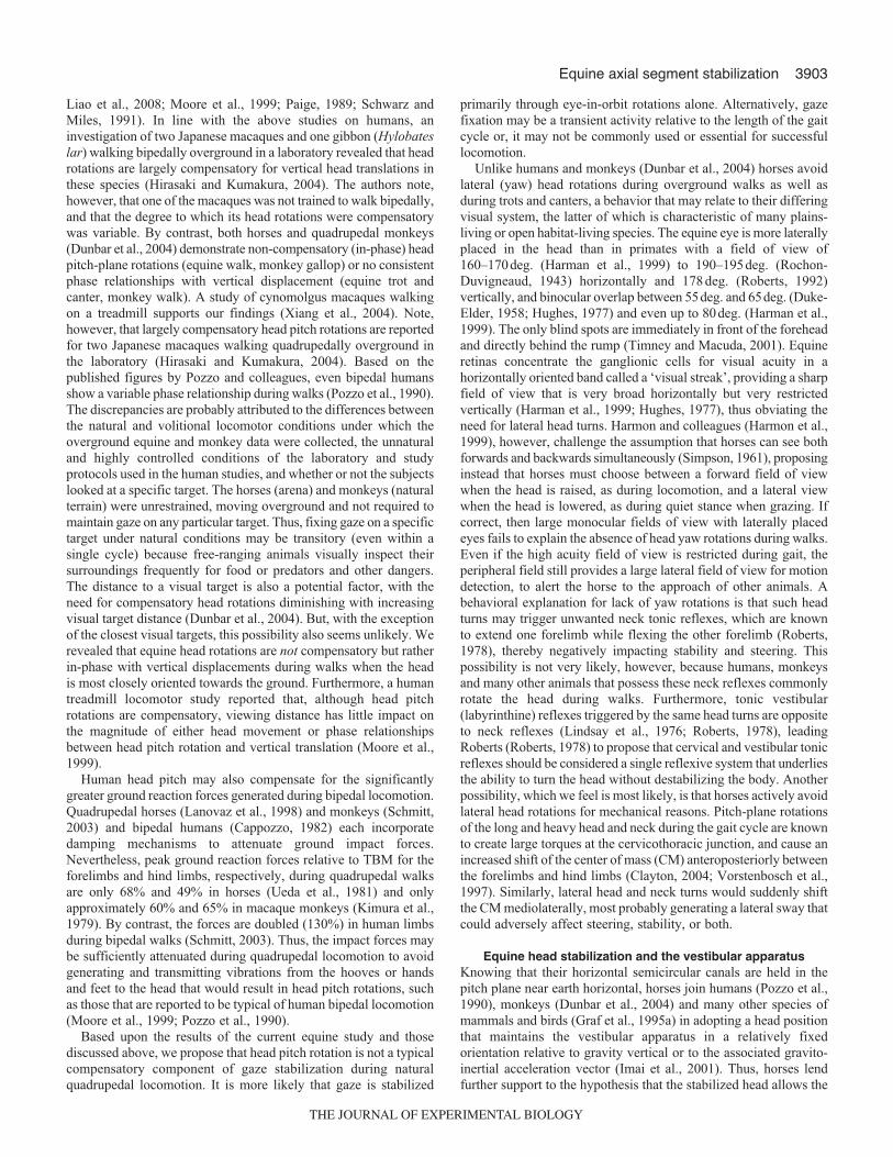
Data analysisVideo images were analyzed with a Peak motion analysis systemusing Motus 3.2 software (Vicon, formerly Peak PerformanceTechnologies, Centennial, CO, USA) for eight walk, trot and cantercycles from each horse, for a total of 24 cycles per gait type. Theonly exceptions were the sample sizes for vertical headdisplacements and velocities (see below) during walks in horse 1,each of which consisted of six cycles. Four anatomical landmarkswere digitized frame-by-frame and used to define the relevant bodysegments: the lower lip, the ear, the withers and the base of the tail(dock). The axis of the head segment was defined as a line passingthrough the lower lip and the ear, the neck axis through the ear andthe withers, and the trunk axis through the withers and the dock(Fig.2A). To minimize measurement error, only trials in which thehead, neck and trunk remained aligned in the sagittal plane (theplane of progression) were analyzed quantitatively. Measurementresolution was estimated to be 1cm for linear position and less thanor equal to 1deg. for angular position (pitch rotation) based on thelength of the shortest segment (head). To maximize accuracy andconsistency, one experimenter (A.Z.) who has extensive experienceand training in motion data collection with the Peak systemperformed all the digitizing.
A number of variables were computed for each of the three gaits.A gait cycle was defined as the time between the touchdown of theright hind hoof and the subsequent touchdown of the same hoof.Locomotor velocities were calculated for each horse using thehorizontal displacement of the body from the beginning to the endof the gait cycle and the number of frames as a time measure. Linearvariables included vertical displacement and velocity of the head(ear), neck and trunk. Angular variables of pitch rotation includedhead, neck and trunk displacement and velocity relative to earthhorizontal and relative to each other (Fig.2B,C). We adopted thestandard rotational coordinate system whereby pitch is in thesagittal plane, yaw in the horizontal plane and roll in the frontal
plane. Displacement data were smoothed at 10Hz prior to calculatingvelocities.
The relationship between vertical translation and pitch rotationof the head during the various gaits was explored with Fourieranalysis using Matlab software (The Mathworks Inc., Natick, MA,USA). The displacement data were smoothed (4th order low-passfilter with zero phase lag, using cutoffs of 6Hz for walks and 8Hzfor trots and canters), and the mean values subtracted for each cycleto yield change in pitch angle and vertical position. Data were thende-trended to remove any linear trends within a cycle. Using Fourieranalysis, the amplitude and phase of the cosine curve fits weredetermined at the main frequency for head movement. A phasedifference was then calculated as pitch phase minus vertical positionphase such that a positive value indicates that pitch rotationprecedes, or leads, vertical position.
Fourier analysis was also used to examine the relative importanceof inertia in head stabilization. Peak frequencies for angular andlinear displacements of the head were determined for each horseduring walks, trots and canters. Frequency spectra were derived for
D. C. Dunbar and others
θ
α
β
δμ
B
C
A
�
Fig. 2. Measured axes and angles in the pitch plane. (A) head axis (black)was a line passing through the ear and the lower lip, neck axis (red) was aline passing through the withers (3rd thoracic vertebra) and the ear, andtrunk axis (blue) was a line passing through the dock (tail base) and thewithers. (B) Head angle (θ) and trunk angle (β) relative to space, and headangle relative to the trunk (α). (C) Neck angle relative to space (δ), headangle relative to the neck (μ) and neck angle relative to the trunk (�).Horizontal broken lines indicate the earth-horizontal space reference.
THE JOURNAL OF EXPERIMENTAL BIOLOGY

3893Equine axial segment stabilization
each individual cycle, and then averaged across cycles. Thistechnique, which was applied previously to monkeys (Dunbar etal., 2004), produced clear mean peak frequencies. Harmonics,however, were not clearly produced and, thus, were not includedin the analysis. Owing to camera orientation, rotations in otherplanes, if present, were analyzed qualitatively.
To aid discussion of the effects of head motion during the variousgaits on the signaling capacity of the visual and vestibular systems,various anatomical measures were made. Orientation of thehorizontal semicircular canals in the head was estimated through acombination of horse skull illustrations depicting the canal’salignment in the sagittal plane (DeBeer, 1947) and specific externalcranial landmarks (Goody, 1983), and found to be pitched upwardby 60deg. relative to the head axis used in the present study.
Both the eyes and the vestibular organs of the horse are at somedistance from the main axes of head rotation, the atlanto–occipital(head–neck) and cervicothoracic (neck–trunk) joints, and willtherefore experience curvilinear sagittal plane displacement duringpitch rotation of the head. To aid in estimating this motion, wemeasured the distance of the orbit and the external auditory meatus(approximated vestibular apparatus location) from a line passingthrough the mid-sagittal anterior (rostral) border of the foramenmagnum between the occipital condyles (approximated pitch axis)on the adult skull of one horse. For comparison, we made similarmeasurements on adult skulls of one human and one monkey(Macaca mulatta Zimmermann 1780). We also estimated the lengthof the cervical vertebral column from an anatomical drawing of anadult horse skeleton (Goody, 1983) scaled to the length of the horseskull that was used for cranial measurements. Cervical spine lengthwas measured directly from the mounted adult skeletons of onehuman and one monkey (M. mulatta). For all three species, thecervical spine was measured with the skeletons in a quiet stanceposture. The impact of off-axis locations of both end organs in ahead rotating in pitch at the head–neck axis alone and neck–trunkaxis alone were then estimated by calculating the arc length at thefollowing measured distances: (1) linear distance to the orbit fromthe head–neck axis of pitch rotation – between the pitch axis andthe medial border of the orbital rim; (2) linear distance of the externalauditory meatus from the head–neck axis of pitch rotation – betweenthe pitch axis and the posterior (horse) or superior (monkey, human)border of the external auditory meatus. (3) Cervical spine length –linear distance between the 1st thoracic and 1st cervical or atlasvertebrae. The cervical spine length was added to the cranial lineardistances to approximate arc lengths generated by pitch at theneck–trunk axis alone.
Statistical analysesVariables were presented as means ± standard deviation (s.d.) ormeans ± standard error (s.e.m.). The exception was the phaserelationships between change in head angle and change in headvertical position, which were presented as means ± angulardispersion (a.d.), a measure of variability in the circular domain.The majority of variables were analyzed using two-way analysis ofvariance (ANOVA). Head angular and vertical mean peakfrequencies, however, were analyzed using one-way ANOVA.Kruskal–Wallis ANOVA on Ranks was used if normality failed.Post hoc analyses were performed on pair-wise comparisons forsignificant results. Angular-linear correlation, a parametric circularstatistical procedure (Johnson and Wehrly, 1977; Mardia, 1976),was employed to determine how strongly change in head pitchrelated to change in head vertical position in each horse for eachgait. The accepted P-value for significance was equal to or less than
0.05 for both the main analyses and all post hoc pair-wisecomparisons.
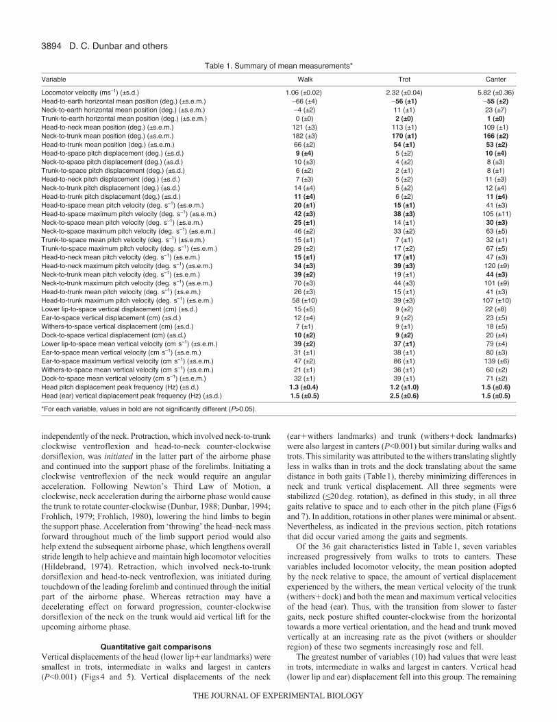
RESULTSDescriptions of rotational direction below assume that the horsesare moving from left to right. Thus, ‘clockwise’ rotations are forwardand downward whereas ‘counter-clockwise’ rotations are backwardand upward. The means and standard deviations or standard errorsof the measured variables are summarized in Table1. For ease ofcomparison, those values within each variable that did not differsignificantly (P-value greater than 0.05) among the three gaits havebeen highlighted in bold type.
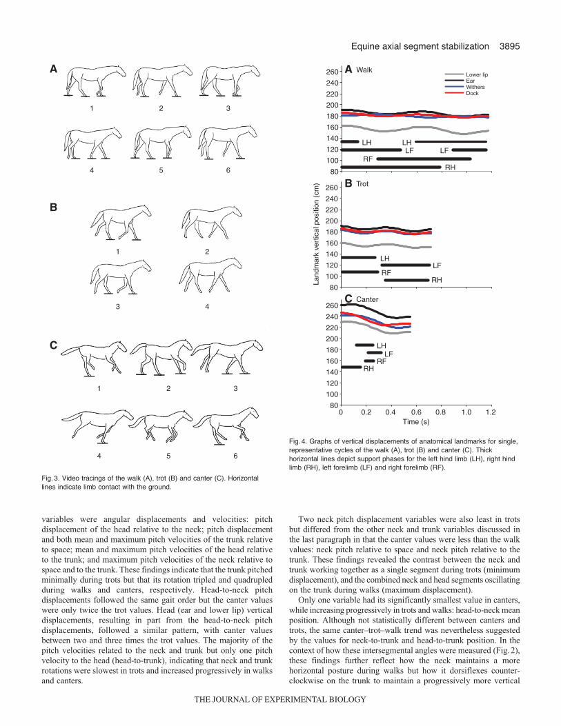
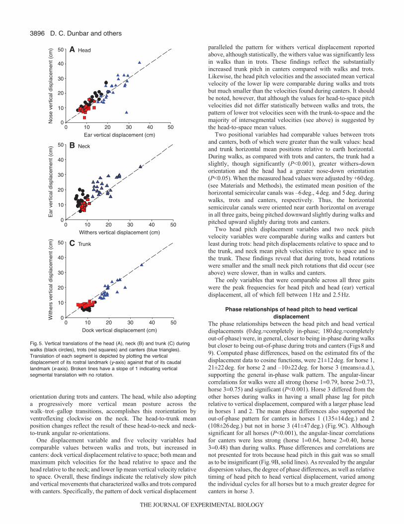
Qualitative segmental coordination patternsThe overall characteristics of the three quadrupedal gaits analyzedin this study, in terms of the pattern and timing of footfalls,corresponded to the combined definitions and descriptions providedby others (Alexander, 1982; Hildebrand, 1966; Hildebrand, 1977;Howell, 1944). All three gaits are illustrated in Fig.3. In walks, thetiming between footfalls was nearly equal, and the support phaseduration of each limb exceeded 50% of the total cycle duration. Thehorses used a lateral sequence walk pattern in that a hind limbtouchdown was followed by touchdown of the forelimb on the same(ipsilateral) side of the body. In trots, both touchdown and liftoffof a hind limb and its opposite (contralateral) forelimb were nearlysimultaneous, and the timing between footfalls of the forelimb–hindlimb pairs was nearly equal. Each limb pair contacted the supportfor 50% or less of the total cycle duration, and airborne phases,when present, occurred between touchdowns of forelimb–hind limbpairs. In canters, the timing between footfalls was unequal, and thesupport phase duration of each limb was less than 50% of the totalcycle duration. Sequential touchdowns of the hind limbs werefollowed by sequential touchdowns of the forelimbs. Two types ofcanters were observed in the horses: a ‘rotary’ canter, in which theipsilateral forelimb touched down following touchdown of theleading (second) hind limb, and a ‘transverse’ canter, in which thecontralateral forelimb touched down following touchdown of theleading hind limb. Head, neck and trunk movements, however, didnot distinguish one canter type from the other. The cantersinvestigated in this study occasionally lacked an airborne phase nearthe end of the cycle following liftoff of the leading forelimb.Whereas it is typically present in most gallops, an airborne phaseis commonly absent in slow canters (Howell, 1944).
Head, neck and trunk rotations appeared small in all three gaitsbut the coordination pattern of the segmental rotations that did occurdiffered among the three gaits. During walks, the head and neckpitched on the trunk as a single, combined segment. Counter-clockwise rotations in the latter portion of the support phase of eachforelimb enabled the combined head and neck to assist in liftingthe forelimbs off the ground at the beginning of swing phase.Oscillating the cantilevered head–neck mass in this manner alsomost likely assisted forward propulsion through stretch and recoilof the nuchal ligament (particularly in the cervicothoracic region)for storage and release, respectively, of potential energy (Gellmanand Bertram, 2002a; Gellman and Bertram, 2002b). A second patternoccurred during trots, in which the neck and trunk formed a single,combined segment on which the head rotated (bobbed) slightly, butrapidly, in the pitch plane as the body rose and fell vertically. Yeta third pattern occurred during canters, in that all segments rotatedrelative to space and to their adjacent segments. This pattern allowedthe head and neck to both protract and retract on the trunk duringthe gait cycle but for the head to also maintain its spatial orientation
THE JOURNAL OF EXPERIMENTAL BIOLOGY
3894
independently of the neck. Protraction, which involved neck-to-trunkclockwise ventroflexion and head-to-neck counter-clockwisedorsiflexion, was initiated in the latter part of the airborne phaseand continued into the support phase of the forelimbs. Initiating aclockwise ventroflexion of the neck would require an angularacceleration. Following Newton’s Third Law of Motion, aclockwise, neck acceleration during the airborne phase would causethe trunk to rotate counter-clockwise (Dunbar, 1988; Dunbar, 1994;Frohlich, 1979; Frohlich, 1980), lowering the hind limbs to beginthe support phase. Acceleration from ‘throwing’ the head–neck massforward throughout much of the limb support period would alsohelp extend the subsequent airborne phase, which lengthens overallstride length to help achieve and maintain high locomotor velocities(Hildebrand, 1974). Retraction, which involved neck-to-trunkdorsiflexion and head-to-neck ventroflexion, was initiated duringtouchdown of the leading forelimb and continued through the initialpart of the airborne phase. Whereas retraction may have adecelerating effect on forward progression, counter-clockwisedorsiflexion of the neck on the trunk would aid vertical lift for theupcoming airborne phase.
Quantitative gait comparisonsVertical displacements of the head (lower lip�ear landmarks) weresmallest in trots, intermediate in walks and largest in canters(P<0.001) (Figs 4 and 5). Vertical displacements of the neck
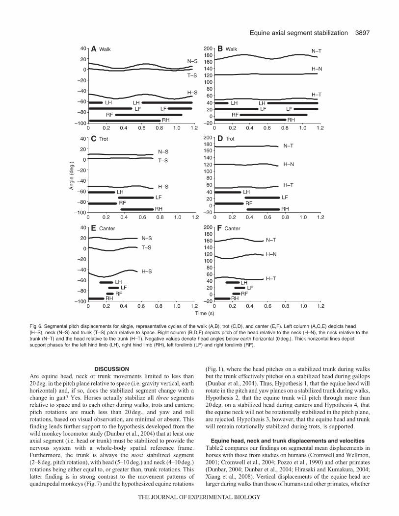
(ear�withers landmarks) and trunk (withers�dock landmarks)were also largest in canters (P<0.001) but similar during walks andtrots. This similarity was attributed to the withers translating slightlyless in walks than in trots and the dock translating about the samedistance in both gaits (Table1), thereby minimizing differences inneck and trunk vertical displacement. All three segments werestabilized (≤20deg. rotation), as defined in this study, in all threegaits relative to space and to each other in the pitch plane (Figs6and 7). In addition, rotations in other planes were minimal or absent.Nevertheless, as indicated in the previous section, pitch rotationsthat did occur varied among the gaits and segments.
Of the 36 gait characteristics listed in Table1, seven variablesincreased progressively from walks to trots to canters. Thesevariables included locomotor velocity, the mean position adoptedby the neck relative to space, the amount of vertical displacementexperienced by the withers, the mean vertical velocity of the trunk(withers�dock) and both the mean and maximum vertical velocitiesof the head (ear). Thus, with the transition from slower to fastergaits, neck posture shifted counter-clockwise from the horizontaltowards a more vertical orientation, and the head and trunk movedvertically at an increasing rate as the pivot (withers or shoulderregion) of these two segments increasingly rose and fell.
The greatest number of variables (10) had values that were leastin trots, intermediate in walks and largest in canters. Vertical head(lower lip and ear) displacement fell into this group. The remaining
D. C. Dunbar and others
Table 1. Summary of mean measurements*
Variable Walk Trot Canter
Locomotor velocity (ms–1) (±s.d.) 1.06 (±0.02) 2.32 (±0.04) 5.82 (±0.36) Head-to-earth horizontal mean position (deg.) (±s.e.m.) –66 (±4) –56 (±1) –55 (±2)Neck-to-earth horizontal mean position (deg.) (±s.e.m.) –4 (±2) 11 (±1) 23 (±7)Trunk-to-earth horizontal mean position (deg.) (±s.e.m.) 0 (±0) 2 (±0) 1 (±0)Head-to-neck mean position (deg.) (±s.e.m.) 121 (±3) 113 (±1) 109 (±1)Neck-to-trunk mean position (deg.) (±s.e.m.) 182 (±3) 170 (±1) 166 (±2)Head-to-trunk mean position (deg.) (±s.e.m.) 66 (±2) 54 (±1) 53 (±2)Head-to-space pitch displacement (deg.) (±s.d.) 9 (±4) 5 (±2) 10 (±4) Neck-to-space pitch displacement (deg.) (±s.d.) 10 (±3) 4 (±2) 8 (±3) Trunk-to-space pitch displacement (deg.) (±s.d.) 6 (±2) 2 (±1) 8 (±1) Head-to-neck pitch displacement (deg.) (±s.d.) 7 (±3) 5 (±2) 11 (±3) Neck-to-trunk pitch displacement (deg.) (±s.d.) 14 (±4) 5 (±2) 12 (±4) Head-to-trunk pitch displacement (deg.) (±s.d.) 11 (±4) 6 (±2) 11 (±4)Head-to-space mean pitch velocity (deg. s–1) (±s.e.m.) 20 (±1) 15 (±1) 41 (±3) Head-to-space maximum pitch velocity (deg. s–1) (±s.e.m.) 42 (±3) 38 (±3) 105 (±11)Neck-to-space mean pitch velocity (deg. s–1) (±s.e.m.) 25 (±1) 14 (±1) 30 (±3)Neck-to-space maximum pitch velocity (deg. s–1) (±s.e.m.) 46 (±2) 33 (±2) 63 (±5)Trunk-to-space mean pitch velocity (deg. s–1) (±s.e.m.) 15 (±1) 7 (±1) 32 (±1)Trunk-to-space maximum pitch velocity (deg. s–1) (±s.e.m.) 29 (±2) 17 (±2) 67 (±5)Head-to-neck mean pitch velocity (deg. s–1) (±s.e.m.) 15 (±1) 17 (±1) 47 (±3)Head-to-neck maximum pitch velocity (deg. s–1) (±s.e.m.) 34 (±3) 39 (±3) 120 (±9)Neck-to-trunk mean pitch velocity (deg. s–1) (±s.e.m.) 39 (±2) 19 (±1) 44 (±3) Neck-to-trunk maximum pitch velocity (deg. s–1) (±s.e.m.) 70 (±3) 44 (±3) 101 (±9)Head-to-trunk mean pitch velocity (deg. s–1) (±s.e.m.) 26 (±3) 15 (±1) 41 (±3)Head-to-trunk maximum pitch velocity (deg. s–1) (±s.e.m.) 58 (±10) 39 (±3) 107 (±10)Lower lip-to-space vertical displacement (cm) (±s.d.) 15 (±5) 9 (±2) 22 (±8)Ear-to-space vertical displacement (cm) (±s.d.) 12 (±4) 9 (±2) 23 (±5)Withers-to-space vertical displacement (cm) (±s.d.) 7 (±1) 9 (±1) 18 (±5)Dock-to-space vertical displacement (cm) (±s.d.) 10 (±2) 9 (±2) 20 (±4)Lower lip-to-space mean vertical velocity (cm s–1) (±s.e.m.) 39 (±2) 37 (±1) 79 (±4)Ear-to-space mean vertical velocity (cm s–1) (±s.e.m.) 31 (±1) 38 (±1) 80 (±3)Ear-to-space maximum vertical velocity (cm s–1) (±s.e.m.) 47 (±2) 86 (±1) 139 (±6)Withers-to-space mean vertical velocity (cm s–1) (±s.e.m.) 21 (±1) 36 (±1) 60 (±2) Dock-to-space mean vertical velocity (cm s–1) (±s.e.m.) 32 (±1) 39 (±1) 71 (±2) Head pitch displacement peak frequency (Hz) (±s.d.) 1.3 (±0.4) 1.2 (±1.0) 1.5 (±0.6)Head (ear) vertical displacement peak frequency (Hz) (±s.d.) 1.5 (±0.5) 2.5 (±0.6) 1.5 (±0.5)
*For each variable, values in bold are not significantly different (P>0.05).
THE JOURNAL OF EXPERIMENTAL BIOLOGY
3895Equine axial segment stabilization
variables were angular displacements and velocities: pitchdisplacement of the head relative to the neck; pitch displacementand both mean and maximum pitch velocities of the trunk relativeto space; mean and maximum pitch velocities of the head relativeto the trunk; and maximum pitch velocities of the neck relative tospace and to the trunk. These findings indicate that the trunk pitchedminimally during trots but that its rotation tripled and quadrupledduring walks and canters, respectively. Head-to-neck pitchdisplacements followed the same gait order but the canter valueswere only twice the trot values. Head (ear and lower lip) verticaldisplacements, resulting in part from the head-to-neck pitchdisplacements, followed a similar pattern, with canter valuesbetween two and three times the trot values. The majority of thepitch velocities related to the neck and trunk but only one pitchvelocity to the head (head-to-trunk), indicating that neck and trunkrotations were slowest in trots and increased progressively in walksand canters.
Two neck pitch displacement variables were also least in trotsbut differed from the other neck and trunk variables discussed inthe last paragraph in that the canter values were less than the walkvalues: neck pitch relative to space and neck pitch relative to thetrunk. These findings revealed the contrast between the neck andtrunk working together as a single segment during trots (minimumdisplacement), and the combined neck and head segments oscillatingon the trunk during walks (maximum displacement).
Only one variable had its significantly smallest value in canters,while increasing progressively in trots and walks: head-to-neck meanposition. Although not statistically different between canters andtrots, the same canter–trot–walk trend was nevertheless suggestedby the values for neck-to-trunk and head-to-trunk position. In thecontext of how these intersegmental angles were measured (Fig.2),these findings further reflect how the neck maintains a morehorizontal posture during walks but how it dorsiflexes counter-clockwise on the trunk to maintain a progressively more vertical
4 5 6
1 2 3
21
43
A
B
C
4 5 6
1 2 3
Fig. 3. Video tracings of the walk (A), trot (B) and canter (C). Horizontallines indicate limb contact with the ground.
Land
mar
k ve
rtic
al p
ositi
on (
cm)
80
100
120
140
160
180
200
220
240
260
80
100
120
140
160
180
200
220
240
260
A Walk
Trot
Canter
LH LH
RFRH
LFLF
Lower lipEarWithersDock
LHLF
RFRH
B
C
Time (s)0 0.2 0.4 0.6 0.8 1.0 1.2
LHLF
RFRH
80
100
120
140
160
180
200
220
240
260
Fig. 4. Graphs of vertical displacements of anatomical landmarks for single,representative cycles of the walk (A), trot (B) and canter (C). Thickhorizontal lines depict support phases for the left hind limb (LH), right hindlimb (RH), left forelimb (LF) and right forelimb (RF).
THE JOURNAL OF EXPERIMENTAL BIOLOGY
3896
orientation during trots and canters. The head, while also adoptinga progressively more vertical mean posture across thewalk–trot–gallop transitions, accomplishes this reorientation byventroflexing clockwise on the neck. The head-to-trunk meanposition changes reflect the result of these head-to-neck and neck-to-trunk angular re-orientations.
One displacement variable and five velocity variables hadcomparable values between walks and trots, but increased incanters: dock vertical displacement relative to space; both mean andmaximum pitch velocities for the head relative to space and thehead relative to the neck; and lower lip mean vertical velocity relativeto space. Overall, these findings indicate the relatively slow pitchand vertical movements that characterized walks and trots comparedwith canters. Specifically, the pattern of dock vertical displacement
paralleled the pattern for withers vertical displacement reportedabove, although statistically, the withers value was significantly lessin walks than in trots. These findings reflect the substantiallyincreased trunk pitch in canters compared with walks and trots.Likewise, the head pitch velocities and the associated mean verticalvelocity of the lower lip were comparable during walks and trotsbut much smaller than the velocities found during canters. It shouldbe noted, however, that although the values for head-to-space pitchvelocities did not differ statistically between walks and trots, thepattern of lower trot velocities seen with the trunk-to-space and themajority of intersegmental velocities (see above) is suggested bythe head-to-space mean values.
Two positional variables had comparable values between trotsand canters, both of which were greater than the walk values: headand trunk horizontal mean positions relative to earth horizontal.During walks, as compared with trots and canters, the trunk had aslightly, though significantly (P<0.001), greater withers-downorientation and the head had a greater nose-down orientation(P<0.05). When the measured head values were adjusted by +60deg.(see Materials and Methods), the estimated mean position of thehorizontal semicircular canals was –6deg., 4deg. and 5deg. duringwalks, trots and canters, respectively. Thus, the horizontalsemicircular canals were oriented near earth horizontal on averagein all three gaits, being pitched downward slightly during walks andpitched upward slightly during trots and canters.
Two head pitch displacement variables and two neck pitchvelocity variables were comparable during walks and canters butleast during trots: head pitch displacements relative to space and tothe trunk, and neck mean pitch velocities relative to space and tothe trunk. These findings reveal that during trots, head rotationswere smaller and the small neck pitch rotations that did occur (seeabove) were slower, than in walks and canters.
The only variables that were comparable across all three gaitswere the peak frequencies for head pitch and head (ear) verticaldisplacement, all of which fell between 1Hz and 2.5Hz.
Phase relationships of head pitch to head verticaldisplacement
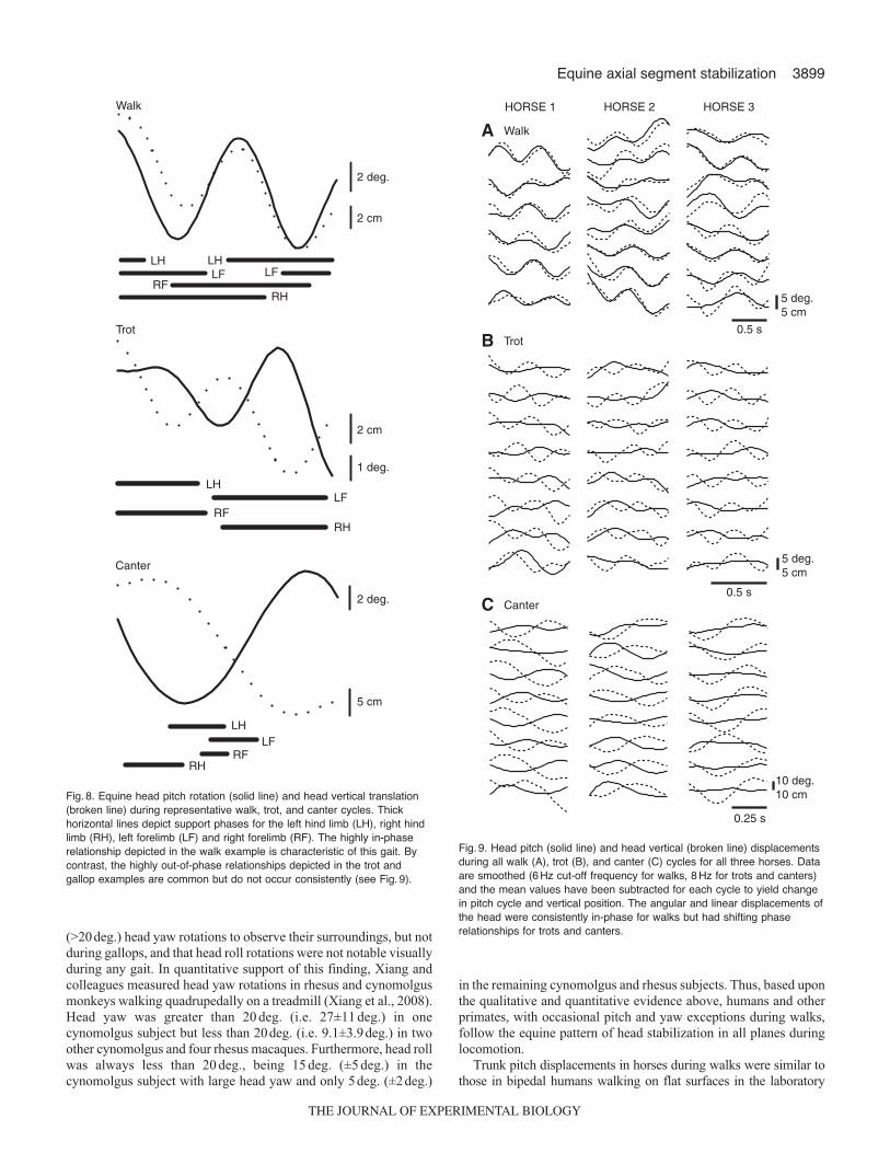
The phase relationships between the head pitch and head verticaldisplacements (0deg.=completely in-phase; 180deg.=completelyout-of-phase) were, in general, closer to being in-phase during walksbut closer to being out-of-phase during trots and canters (Figs8 and9). Computed phase differences, based on the estimated fits of thedisplacement data to cosine functions, were 21±12deg. for horse 1,21±22deg. for horse 2 and –10±22deg. for horse 3 (means±a.d.),supporting the general in-phase walk pattern. The angular-linearcorrelations for walks were all strong (horse 1=0.79, horse 2=0.73,horse 3=0.75) and significant (P<0.001). Horse 3 differed from theother horses during walks in having a small phase lag for pitchrelative to vertical displacement, compared with a larger phase leadin horses 1 and 2. The mean phase differences also supported theout-of-phase pattern for canters in horses 1 (135±14deg.) and 2(108±26deg.) but not in horse 3 (41±47deg.) (Fig.9C). Althoughsignificant for all horses (P<0.001), the angular-linear correlationsfor canters were less strong (horse 1=0.64, horse 2=0.40, horse3=0.48) than during walks. Phase differences and correlations arenot presented for trots because head pitch in this gait was so smallas to be insignificant (Fig.9B, solid lines). As revealed by the angulardispersion values, the degree of phase differences, as well as relativetiming of head pitch to head vertical displacement, varied amongthe individual cycles for all horses but to a much greater degree forcanters in horse 3.
D. C. Dunbar and others
Dock vertical displacement (cm)
0 10 20 30 40 50
With
ers
vert
ical
dis
plac
emen
t (cm
)
0
10
20
30
40
50 C Trunk
Withers vertical displacement (cm)
0 10 20 30 40 50
Ear
ver
tical
dis
plac
emen
t (cm
)
0
10
20
30
40
50 B Neck
Ear vertical displacement (cm)
0 10 20 30 40 50
Nos
e ve
rtic
al d
ispl
acem
ent (
cm)
0
10
20
30
40
50 A Head
Fig. 5. Vertical translations of the head (A), neck (B) and trunk (C) duringwalks (black circles), trots (red squares) and canters (blue triangles).Translation of each segment is depicted by plotting the verticaldisplacement of its rostral landmark (y-axis) against that of its caudallandmark (x-axis). Broken lines have a slope of 1 indicating verticalsegmental translation with no rotation.
THE JOURNAL OF EXPERIMENTAL BIOLOGY
3897Equine axial segment stabilization
DISCUSSIONAre equine head, neck or trunk movements limited to less than20deg. in the pitch plane relative to space (i.e. gravity vertical, earthhorizontal) and, if so, does the stabilized segment change with achange in gait? Yes. Horses actually stabilize all three segmentsrelative to space and to each other during walks, trots and canters;pitch rotations are much less than 20deg., and yaw and rollrotations, based on visual observation, are minimal or absent. Thisfinding lends further support to the hypothesis developed from thewild monkey locomotor study (Dunbar et al., 2004) that at least oneaxial segment (i.e. head or trunk) must be stabilized to provide thenervous system with a whole-body spatial reference frame.Furthermore, the trunk is always the most stabilized segment(2–8deg. pitch rotation), with head (5–10deg.) and neck (4–10deg.)rotations being either equal to, or greater than, trunk rotations. Thislatter finding is in strong contrast to the movement patterns ofquadrupedal monkeys (Fig.7) and the hypothesized equine rotations
(Fig.1), where the head pitches on a stabilized trunk during walksbut the trunk effectively pitches on a stabilized head during gallops(Dunbar et al., 2004). Thus, Hypothesis 1, that the equine head willrotate in the pitch and yaw planes on a stabilized trunk during walks,Hypothesis 2, that the equine trunk will pitch through more than20deg. on a stabilized head during canters and Hypothesis 4, thatthe equine neck will not be rotationally stabilized in the pitch plane,are rejected. Hypothesis 3, however, that the equine head and trunkwill remain rotationally stabilized during trots, is supported.
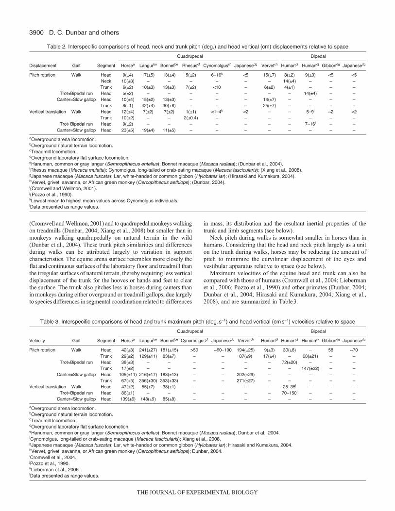
Equine head, neck and trunk displacements and velocitiesTable2 compares our findings on segmental mean displacements inhorses with those from studies on humans (Cromwell and Wellmon,2001; Cromwell et al., 2004; Pozzo et al., 1990) and other primates(Dunbar, 2004; Dunbar et al., 2004; Hirasaki and Kumakura, 2004;Xiang et al., 2008). Vertical displacements of the equine head arelarger during walks than those of humans and other primates, whether
Ang
le (
deg.
)
N–T
H–N
H–T
H–N
N–T
H–T
–200
20406080
100120140160180200
N–T
H–N
H–T
–100
–80
–60
–40
–20
0
20
40
H–S
N–S
T–S
H–S
N–S
T–S
LHLF
LHLF
RFRH
H–S
N–S
T–S
LHLF
RFRH
LHLF
RFRH
LHLF
RFRH
LHLF
LHLF
RFRH
LHLF
RFRH
A Walk
Trot
Canter
C
B Walk
TrotD
E
Time (s)0 0.2 0.4 0.6 0.8 1.0 1.2
CanterF
0 0.2 0.4 0.6 0.8 1.0 1.2
–200
20406080
100120140160180200
–100
–80
–60
–40
–20
0
20
40
0 0.2 0.4 0.6 0.8 1.0 1.2 0 0.2 0.4 0.6 0.8 1.0 1.2
–200
20406080
100120140160180200
–100
–80
–60
–40
–20
0
20
40
0 0.2 0.4 0.6 0.8 1.0 1.2 0 0.2 0.4 0.6 0.8 1.0 1.2
Fig. 6. Segmental pitch displacements for single, representative cycles of the walk (A,B), trot (C,D), and canter (E,F). Left column (A,C,E) depicts head(H–S), neck (N–S) and trunk (T–S) pitch relative to space. Right column (B,D,F) depicts pitch of the head relative to the neck (H–N), the neck relative to thetrunk (N–T) and the head relative to the trunk (H–T). Negative values denote head angles below earth horizontal (0 deg.). Thick horizontal lines depictsupport phases for the left hind limb (LH), right hind limb (RH), left forelimb (LF) and right forelimb (RF).
THE JOURNAL OF EXPERIMENTAL BIOLOGY
3898
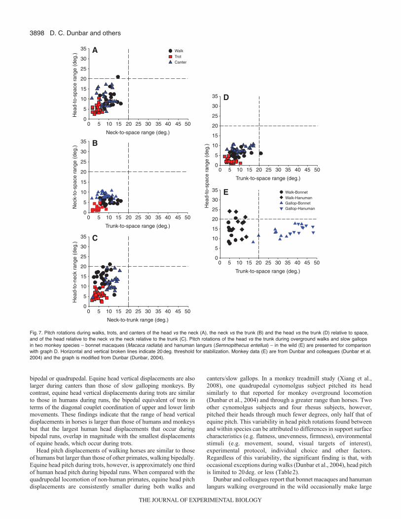
bipedal or quadrupedal. Equine head vertical displacements are alsolarger during canters than those of slow galloping monkeys. Bycontrast, equine head vertical displacements during trots are similarto those in humans during runs, the bipedal equivalent of trots interms of the diagonal couplet coordination of upper and lower limbmovements. These findings indicate that the range of head verticaldisplacements in horses is larger than those of humans and monkeysbut that the largest human head displacements that occur duringbipedal runs, overlap in magnitude with the smallest displacementsof equine heads, which occur during trots.
Head pitch displacements of walking horses are similar to thoseof humans but larger than those of other primates, walking bipedally.Equine head pitch during trots, however, is approximately one thirdof human head pitch during bipedal runs. When compared with thequadrupedal locomotion of non-human primates, equine head pitchdisplacements are consistently smaller during both walks and
canters/slow gallops. In a monkey treadmill study (Xiang et al.,2008), one quadrupedal cynomolgus subject pitched its headsimilarly to that reported for monkey overground locomotion(Dunbar et al., 2004) and through a greater range than horses. Twoother cynomolgus subjects and four rhesus subjects, however,pitched their heads through much fewer degrees, only half that ofequine pitch. This variability in head pitch rotations found betweenand within species can be attributed to differences in support surfacecharacteristics (e.g. flatness, unevenness, firmness), environmentalstimuli (e.g. movement, sound, visual targets of interest),experimental protocol, individual choice and other factors.Regardless of this variability, the significant finding is that, withoccasional exceptions during walks (Dunbar et al., 2004), head pitchis limited to 20deg. or less (Table2).
Dunbar and colleagues report that bonnet macaques and hanumanlangurs walking overground in the wild occasionally make large
D. C. Dunbar and others
E
Trunk-to-space range (deg.)
0 5 10 15 20 25 30 35 40 45 500
5
10
15
20
25
30
35 Walk-BonnetWalk-HanumanGallop-BonnetGallop-Hanuman
D
0
5
10
15
20
25
30
35
A
0
5
10
15
20
25
30
35 Walk
TrotCanter
B
0
5
10
15
20
25
30
35
C
0
5
10
15
20
25
30
35
Trunk-to-space range (deg.)
0 5 10 15 20 25 30 35 40 45 50
Hea
d-to
-spa
ce r
ange
(de
g.)
Neck-to-trunk range (deg.)
0 5 10 15 20 25 30 35 40 45 50
Trunk-to-space range (deg.)
0 5 10 15 20 25 30 35 40 45 50
Neck-to-space range (deg.)
0 5 10 15 20 25 30 35 40 45 50
Hea
d-to
-spa
ce r
ange
(de
g.)
Nec
k-to
-spa
ce r
ange
(de
g.)
Hea
d-to
-nec
k ra
nge
(deg
.)
Fig. 7. Pitch rotations during walks, trots, and canters of the head vs the neck (A), the neck vs the trunk (B) and the head vs the trunk (D) relative to space,and of the head relative to the neck vs the neck relative to the trunk (C). Pitch rotations of the head vs the trunk during overground walks and slow gallopsin two monkey species – bonnet macaques (Macaca radiata) and hanuman langurs (Semnopithecus entellus) – in the wild (E) are presented for comparisonwith graph D. Horizontal and vertical broken lines indicate 20 deg. threshold for stabilization. Monkey data (E) are from Dunbar and colleagues (Dunbar et al.2004) and the graph is modified from Dunbar (Dunbar, 2004).
THE JOURNAL OF EXPERIMENTAL BIOLOGY
3899Equine axial segment stabilization
(>20deg.) head yaw rotations to observe their surroundings, but notduring gallops, and that head roll rotations were not notable visuallyduring any gait. In quantitative support of this finding, Xiang andcolleagues measured head yaw rotations in rhesus and cynomolgusmonkeys walking quadrupedally on a treadmill (Xiang et al., 2008).Head yaw was greater than 20 deg. (i.e. 27±11 deg.) in onecynomolgus subject but less than 20deg. (i.e. 9.1±3.9deg.) in twoother cynomolgus and four rhesus macaques. Furthermore, head rollwas always less than 20 deg., being 15 deg. (±5 deg.) in thecynomolgus subject with large head yaw and only 5deg. (±2deg.)
in the remaining cynomolgus and rhesus subjects. Thus, based uponthe qualitative and quantitative evidence above, humans and otherprimates, with occasional pitch and yaw exceptions during walks,follow the equine pattern of head stabilization in all planes duringlocomotion.
Trunk pitch displacements in horses during walks were similar tothose in bipedal humans walking on flat surfaces in the laboratory
2 deg.
2 cm
LHLF
LHLF
RFRH
Walk
Trot
LHLF
RFRH
1 deg.
2 cm
Canter
2 deg.
5 cm
LHLF
RFRH
Fig. 8. Equine head pitch rotation (solid line) and head vertical translation(broken line) during representative walk, trot, and canter cycles. Thickhorizontal lines depict support phases for the left hind limb (LH), right hindlimb (RH), left forelimb (LF) and right forelimb (RF). The highly in-phaserelationship depicted in the walk example is characteristic of this gait. Bycontrast, the highly out-of-phase relationships depicted in the trot andgallop examples are common but do not occur consistently (see Fig. 9).
HORSE 1 HORSE 2 HORSE 3
5 deg.5 cm
Walk
5 deg.5 cm
Trot
Canter
10 deg.10 cm
0.5 s
0.5 s
0.25 s
A
B
C
Fig. 9. Head pitch (solid line) and head vertical (broken line) displacementsduring all walk (A), trot (B), and canter (C) cycles for all three horses. Dataare smoothed (6 Hz cut-off frequency for walks, 8 Hz for trots and canters)and the mean values have been subtracted for each cycle to yield changein pitch cycle and vertical position. The angular and linear displacements ofthe head were consistently in-phase for walks but had shifting phaserelationships for trots and canters.
THE JOURNAL OF EXPERIMENTAL BIOLOGY
3900
(Cromwell and Wellmon, 2001) and to quadrupedal monkeys walkingon treadmills (Dunbar, 2004; Xiang et al., 2008) but smaller than inmonkeys walking quadrupedally on natural terrain in the wild(Dunbar et al., 2004). These trunk pitch similarities and differencesduring walks can be attributed largely to variation in supportcharacteristics. The equine arena surface resembles more closely theflat and continuous surfaces of the laboratory floor and treadmill thanthe irregular surfaces of natural terrain, thereby requiring less verticaldisplacement of the trunk for the hooves or hands and feet to clearthe surface. The trunk also pitches less in horses during canters thanin monkeys during either overground or treadmill gallops, due largelyto species differences in segmental coordination related to differences
in mass, its distribution and the resultant inertial properties of thetrunk and limb segments (see below).
Neck pitch during walks is somewhat smaller in horses than inhumans. Considering that the head and neck pitch largely as a uniton the trunk during walks, horses may be reducing the amount ofpitch to minimize the curvilinear displacement of the eyes andvestibular apparatus relative to space (see below).
Maximum velocities of the equine head and trunk can also becompared with those of humans (Cromwell et al., 2004; Liebermanet al., 2006; Pozzo et al., 1990) and other primates (Dunbar, 2004;Dunbar et al., 2004; Hirasaki and Kumakura, 2004; Xiang et al.,2008), and are summarized in Table3.
D. C. Dunbar and others
Table 2. Interspecific comparisons of head, neck and trunk pitch (deg.) and head vertical (cm) displacements relative to space
Quadrupedal Bipedal
Displacement Gait Segment Horsea Langurbe Bonnetbe Rhesuscf Cynomolguscf Japanesedg Vervetch Humandi Humandj Gibbondg Japanesedg
Pitch rotation Walk Head 9(±4) 17(±5) 13(±4) 5(±2) 6–16k <5 15(±7) 8(±2) 9(±3) <5 <5Neck 10(±3) – – – – – – 14(±4) – – –Trunk 6(±2) 10(±3) 13(±3) 7(±2) <10 – 6(±2) 4(±1) – – –
Trot≈Bipedal run Head 5(±2) – – – – – – – 14(±4) – –Canter=Slow gallop Head 10(±4) 15(±2) 13(±3) – – – 14(±7) – – – –
Trunk 8(+1) 42(+4) 30(+8) – – – 25(±7) – – – –Vertical translation Walk Head 12(±4) 7(±2) 7(±2) 1(±1) <1–4k <2 – – 5–9l ~2 <2
Trunk 10(±2) – – 2(±0.4) – – – – – – –Trot≈Bipedal run Head 9(±2) – – – – – – – 7–16l – –
Canter=Slow gallop Head 23(±5) 19(±4) 11(±5) – – – – – – – –
aOverground arena locomotion.bOverground natural terrain locomotion.cTreadmill locomotion.dOverground laboratory flat surface locomotion.eHanuman, common or gray langur (Semnopithecus entellus); Bonnet macaque (Macaca radiata); (Dunbar et al., 2004).fRhesus macaque (Macaca mulatta); Cynomolgus, long-tailed or crab-eating macaque (Macaca fascicularis); (Xiang et al., 2008).gJapanese macaque (Macaca fuscata); Lar, white-handed or common gibbon (Hylobates lar); (Hirasaki and Kumakura, 2004).hVervet, grivet, savanna, or African green monkey (Cercopithecus aethiops); (Dunbar, 2004).i(Cromwell and Wellmon, 2001).j(Pozzo et al., 1990).kLowest mean to highest mean values across Cynomolgus individuals.lData presented as range values.
Table 3. Interspecific comparisons of head and trunk maximum pitch (deg.s–1) and head vertical (cms–1) velocities relative to space
Quadrupedal Bipedal
Velocity Gait Segment Horsea Langurbe Bonnetbe Cynomolguscf Japanesedg Vervetch Humandi Humandj Humanck Gibbondg Japanesedg
Pitch rotation Walk Head 42(±3) 241(±27) 181(±15) >50 ~60–100 194(±25) 9(±3) 30(±8) – 58 ~70Trunk 29(±2) 129(±11) 83(±7) – – 87(±9) 17(±4) – 68(±21) – –
Trot≈Bipedal run Head 38(±3) – – – – – – 72(±20) – – –Trunk 17(±2) – – – – – – – 147(±22) – –
Canter=Slow gallop Head 105(±11) 216(±17) 183(±13) – – 202(±29) – – – – –Trunk 67(+5) 356(+30) 353(+33) – – 271(±27) – – – – –
Vertical translation Walk Head 47(±2) 55(±7) 38(±1) – – – – 25–35l – – –Trot≈Bipedal run Head 86(±1) – – – – – – 70–150l – – –
Canter=Slow gallop Head 139(±6) 148(±9) 85(±8) – – – – – – – –
aOverground arena locomotion.bOverground natural terrain locomotion.cTreadmill locomotion.dOverground laboratory flat surface locomotion.eHanuman, common or gray langur (Semnopithecus entellus); Bonnet macaque (Macaca radiata); Dunbar et al., 2004.fCynomolgus, long-tailed or crab-eating macaque (Macaca fascicularis); Xiang et al., 2008.gJapanese macaque (Macaca fuscata); Lar, white-handed or common gibbon (Hylobates lar); Hirasaki and Kumakura, 2004.hVervet, grivet, savanna, or African green monkey (Cercopithecus aethiops); Dunbar, 2004.iCromwell et al., 2004.jPozzo et al., 1990.kLieberman et al., 2006.lData presented as range values.
THE JOURNAL OF EXPERIMENTAL BIOLOGY
3901Equine axial segment stabilization
Maximum vertical velocities of the equine head are up to twotimes larger than those of humans during walks but fall within thehuman bipedal running range during trots and parallel the findingsfor equine and human head displacements. During both walks andcanters/slow gallops, equine head vertical velocities are consistentlylarger than those of bonnet macaques despite the greater inertia ofthe equine head. This is most probably attributed to the contributionsof the equine nuchal ligament to head and neck pitch on the trunkduring walks and, along with overall body movements, duringgallops. By contrast, horses have smaller head vertical velocitiesthan hanuman langurs, probably due to the relatively large vertical(and angular) displacements of the hanuman’s body segments duringboth walks and gallops, giving this and other langur species bounce-like gaits that distinguish them from macaques and most othermonkeys (Dunbar et al., 2004).
Maximum pitch velocities of the equine head during walks are1.5–5 times greater than those of bipedal humans but less than thoseof bipedal and quadrupedal monkeys. Head pitch velocities are alsoless in trotting horses than in humans running bipedally, and lessin cantering horses than in monkeys galloping overground or on atreadmill. Thus, with the exception of humans during walks,maximum pitch velocities of the equine head are less than those ofhumans and other primates. These low pitch velocities are mostlikely attributed to the greater mass and inertial properties of theequine head and neck. The larger equine head pitch velocitiescompared with those in humans during walks, despite comparableranges of head pitch displacement between the species, are mostlikely attributed to the equine head and neck bob during walks thatstores and releases kinetic energy in the nuchal ligament to assistforward progression (Gellman and Bertram, 2002a; Gellman andBertram, 2002b).
Maximum pitch velocities for the equine trunk are 1.7 times largerthan those of humans walking overground (Cromwell et al., 2004).This finding is not surprising considering the sequential footfall ofthe four equine limbs, combined with the head and neck bobbingon the trunk. Humans on a treadmill (Lieberman et al., 2006), showmuch higher maximum pitch velocities than horses during walkingand running/trotting. These findings for the faster gaits areunderstandable given the mechanical conditions of each gait.Humans rapidly swing their upper and lower limbs but support bodymass alternately with the lower limbs, which would certainly resultin rapid pitch (and probably yaw and roll) movements of the verticaltrunk. Horses, by contrast, alternate diagonal hind limb–forelimbpairs to support the cranial and caudal ends of the horizontal trunk,resulting in small and slow pitch rotations of the trunk. Maximumpitch velocities of the trunk are much smaller in horses than inmonkeys during both walks and canters/slow gallops, primarily dueto the significantly greater mass and inertia of the equine trunk.
Curvilinear displacement of equine eyes and vestibularapparatus during pitch-plane rotations
The strategy of horses to limit the angular excursion of the headand neck segments relative to primates may relate to the particularmorphology of the horse head and head-based sensors. During headpitch, the eyes and vestibular apparatus experience curvilineardisplacements because both end organs are located at a distancefrom the head–neck and neck–trunk rotational axes. Our equine skullmeasurements reveal an occipital condyle-to-orbit distance of 22cmand a pitch axis-to-external auditory meatus distance of 6cm, bothof which are two times larger than similar measurements in thehuman skull (10cm and 3.5cm, respectively) and three times largerthan those in the monkey (rhesus macaque) skull (7cm and 2cm,
respectively). Moreover, the equine cervical spine measured 55cm,which is five times the measured length of the human spine(11.5cm) and nine times that of the monkey (rhesus macaque) spine(6.5cm). Thus, for 20deg. head pitch, for example, the eye andvestibular arc lengths due to rotation at the head–neck(atlanto–occipital joint) axis alone would be greatest in horses(7.7cm and 2.1cm, respectively), intermediate in humans (3.5cmand 1.2cm, respectively) and least in monkeys (2.4cm and 0.7cm,respectively). These estimated head pitch arc lengths are mostrelevant to horse trots, in which the majority of rotation occurs atthe head–neck axis. The same 20deg. pitch of the head and necktogether about the cervicothoracic joint dramatically increases theeye and vestibular arc lengths to 26.9cm and 21.3cm, respectively,as compared with the much smaller arc lengths in humans (7.5cmand 5.2cm) and monkeys (4.7cm and 3.0cm), respectively. Theseestimated neck pitch arc lengths are most relevant to equine andhuman walking. Considering the limited range of mobility at thebase of the primate skull (Graf et al., 1995b) compared with horses(Clayton and Townsend, 1989), these combined head/neck pitcharc lengths are probably most relevant to human and monkey gaitsin general. Nevertheless, for the same rotation at either the head–neckaxis or neck–trunk axis alone, eye and vestibular arc lengths willbe much greater in horses than in humans or monkeys. Thesemorphological differences alone have a significant impact on howthe eyes and vestibular apparatus in each species are spatiallydisplaced during comparable gaits and most probably on thequalitative and quantitative characteristics of the sensory signalstransmitted to the brain.
Horses appear to compensate in part for this morphological impactby reducing the range of pitch rotation compared to humans andmonkeys. This reduction is accomplished by isolating the relativelysmall degree of pitch primarily to a single joint (e.g. neck–trunkduring walks, head–neck during trots), or by adjusting thedorsiflexion–plantar flexion phase relationships between thehead–neck and neck-trunk joints to counteract one another (e.g.canters). During walks and canters, the equine arc lengths, thoughreduced, nevertheless remain greater than in humans and monkeysperforming comparable gaits when measured from either the skullbase (head–neck axis) or cervical base (neck–trunk axis). Duringtrots, by contrast, whereas the arc lengths from the cervical basealso remain greater in horses, the equine eye (1.92cm) and vestibular(0.52cm) arc lengths from the skull base are actually smaller thanthose in running humans (2.44cm and 0.86cm, respectively).
Equine body mass, its distribution and segmental mobilityand stability
The head, neck and trunk movements characterizing equinelocomotion can be attributed, in large part, to morphologicalfeatures and adaptations. Owing to the enormous stresses generatedin muscles and other supportive tissues to propel such massiveanimals (Hill, 1950), the movement of the large and heavy axialsegments is minimized, allowing the smaller and lighter distal limbsegments to move rapidly through several degrees of rotation(Hildebrand, 1959; Hildebrand, 1974). The head and neck togethercontribute approximately 10% of total body mass (TBM) in horses(Clayton, 2004), compared with 8% in humans (Dempster, 1955)and chimpanzees (Crompton et al., 1996) and 6% in prosimianlemurs (Dunbar, 1988). Although the 2%–4% difference may seemsmall in absolute terms, it is large given the horse’s TBM of roughly500kg, a value that is 10, 11 and 250 times greater than that ofhumans, chimpanzees and lemurs, respectively. Furthermore, theequine neck is larger in mass than the head (6% vs 4% TBM,
THE JOURNAL OF EXPERIMENTAL BIOLOGY
3902
respectively) (Clayton, 2004). Mass measurements for the head andneck of primates and other mammals are rarely reported, however,the lemur neck is much smaller than its head (1% vs 5% TBM,respectively) (Dunbar, 1988). Thus, the inertial properties of themassive and elongated axial segments of horses, compared with therelatively small segments of humans and other primates, mostcertainly contribute to the reduced rotational displacements andvelocities of the equine trunk, neck and head.
Inertial influences on the equine head are revealed through acomparison of pitch displacements (Table2) and velocities (Table3)in horses with those in humans (Pozzo et al., 1990) and monkeys(Dunbar et al., 2004). Equine head pitch displacements and velocitiesare, with few exceptions, smaller than those of humans or monkeysacross all three gaits. By contrast, equine head vertical displacements(Table2) and velocities (Table3) are, with few exceptions, greaterthan those of humans and monkeys. During walks, these largevertical head displacements can be attributed almost entirely to theequine head pitching in unison with the elongated neck at theneck–trunk axis, resulting in a large arc length. During canters, neckpitch is certainly a contributing factor but so is the vertical excursionof the trunk in the oscillation between the support and the airbornephase, an excursion that is more than double that seen for walksand trots. During trots, the smaller head vertical displacements areattributed primarily to the small range in vertical movements of theentire body and the small, compensatory rotations of the head andneck, which characterize this symmetrical running gait.
The impact of differences in mass and inertia between horses andmonkeys (Dunbar, 2004; Dunbar et al., 2004) is most clearly revealedthrough comparisons of trunk angular motion during canters and slowgallops, respectively (Tables 2 and 3). Trunk pitch displacements andvelocities are much smaller in horses than in monkeys, reflectingdifferences in strategy: horses flex and extend the limb joints to clearthe hooves from the ground, effectively shortening limb lengthwhereas monkeys rotate the trunk in the pitch plane to lift the extendedlimbs for clearance during the airborne phase.
Inertia and its role in equine head stabilizationDoes inertia itself play a significant role in segmental stabilization?That is, if an external object (e.g. neck) applies a force to the head,will the head, in the absence of muscle activation, react by rotatingin the opposite direction (~180 deg. out-of-phase) therebymaintaining its spatial orientation? This question could be asked ofthe neck or trunk but it is most relevant to the head and whether astabilized vestibular apparatus provides a reference frame for spatialorientation and an inertial guidance platform for navigation (Pozzoet al., 1990). A method, the details of which can be found elsewhere(Dunbar et al., 2004; Jones and Spells, 1963; Peterson and Goldberg,1981), has been developed that predicts the frequency range withinwhich inertia becomes a significant factor for stabilizing the headof animals of any given size. The method employs a model that isbased on the general dimensional similarity between species (Jonesand Spells, 1963) and uses the estimated stabilizing frequency of5Hz for a domestic feline head (Peterson and Goldberg, 1981) asa reference point. Based upon this model, the frequency range withinwhich inertia should become a significant stabilizing factor for theequine head is 1–3Hz. This estimated range encompasses the actual1–2.5Hz mean peak frequency range revealed in this study for bothangular and vertical head displacements during all three gaits. Thus,inertia, in itself, can play a significant role in stabilizing the equinehead. By contrast, the frequencies of pitch plane and verticaldisplacements of hanuman langur and bonnet macaque heads duringquadrupedal walks and gallops over natural terrain (1–2Hz) fall
outside the theoretical range (hanuman langurs, 3–4Hz; bonnetmacaques, 4–5 Hz) in which inertia would play a significantstabilizing role (Dunbar et al., 2004). It should be noted, however,that a treadmill study (Xiang et al., 2008) reports that onecynomolgus macaque subject had a head pitch frequency duringwalks of 3.6Hz, which approaches the lower limit at which inertiawould become a significant stabilizing factor in bonnet macaques,a monkey species of similar body size. For the human head, inertiaalso is probably not a significant factor during bipedal walks but itis likely to have an impact during bipedal runs (Pozzo et al., 1990).Thus, with the latter exception, head stabilization in monkeys andhumans during gait depends on coordinated muscle activity.
Whereas inertia can potentially play a dominant stabilizing rolefor the equine head by producing compensatory pitch rotations, itsactual expression is variable or absent. During canters and trots,head pitch and vertical displacement are often largely out of phase,particularly in horses 1 and 2, as one would expect with inertialdominance (Figs8 and 9). This pattern is inconsistent, however,commonly shifting closer to an in-phase relationship, as seenespecially in horse 3. Furthermore, walks contrast with both trotsand canters in that head pitch and vertical displacement are largelyin phase indicating that inertial stabilization is not being expressedin this gait (Figs8 and 9). Thus, Hypothesis 5, that pitch rotationsof the equine head will not be consistently 180deg. out-of-phasewith head vertical translations, is supported. As reported formonkeys in the wild (Dunbar et al., 2004), inertial influences arefrequently overridden by active mechanisms during naturaloverground equine locomotion.
In regard to coordination of head pitch and vertical displacements,horse 3 differs notably from the other two horses, perhaps due toage-related changes. Horse 3 was 24-years-old, twice the age ofhorse 1 (10 years) and horse 2 (9 years). Reduced viscoelasticityof the nuchal ligament and other soft tissues of the neck, anddeveloping osteoarthritis in the cervical joints are two characteristicsof the equine aging process that can reduce the degree of head–neckand neck–trunk flexibility. Even though the overall locomotorpattern of horse 3 is not distinctive from the two younger horses,these factors, if present, could underlie the difference in phasing ofhead pitch to vertical displacement compared with horses 1 and 2.Horse 3 also differs from the other two horses in being the onlypure Thoroughbred. Despite heritage differences, the three horseswere similar physically and behaviorally. Thus, whether or not thedecreased flexibility observed in Horse 3 can be attributed in partto breeding is unknown.
Equine segmental stabilization and gazeIn addition to the inertial stabilization issue discussed above, thefinding that equine head pitch is primarily in-phase with verticaldisplacement during walks and inconsistently out-of-phase duringtrots and canters, also indicates that compensatory head movementsare not essential for gaze stabilization during natural overgroundquadrupedal locomotion. The lack of such an ‘eye–head synergy’(Pozzo et al., 1990) contradicts the findings of several studies onbipedal humans attempting to maintain gaze on a fixed target. Pozzoand colleagues found that when human subjects look at a targetwhile performing a bipedal locomotor task, they pitch the headcounter-clockwise during vertical head drops and clockwise duringvertical head rises, thereby maintaining a fixed gaze (Pozzo et al.,1990). Several other laboratory studies have reported these consistentcompensatory head rotations (Cromwell et al., 2001; Cromwell etal., 2004; Moore et al., 1999; Paige, 1989), which appear to parallelcompensatory eye-in-orbit (VOR) rotations (Grossman et al., 1988;
D. C. Dunbar and others
THE JOURNAL OF EXPERIMENTAL BIOLOGY
3903Equine axial segment stabilization
Liao et al., 2008; Moore et al., 1999; Paige, 1989; Schwarz andMiles, 1991). In line with the above studies on humans, aninvestigation of two Japanese macaques and one gibbon (Hylobateslar) walking bipedally overground in a laboratory revealed that headrotations are largely compensatory for vertical head translations inthese species (Hirasaki and Kumakura, 2004). The authors note,however, that one of the macaques was not trained to walk bipedally,and that the degree to which its head rotations were compensatorywas variable. By contrast, both horses and quadrupedal monkeys(Dunbar et al., 2004) demonstrate non-compensatory (in-phase) headpitch-plane rotations (equine walk, monkey gallop) or no consistentphase relationships with vertical displacement (equine trot andcanter, monkey walk). A study of cynomolgus macaques walkingon a treadmill supports our findings (Xiang et al., 2004). Note,however, that largely compensatory head pitch rotations are reportedfor two Japanese macaques walking quadrupedally overground inthe laboratory (Hirasaki and Kumakura, 2004). Based on thepublished figures by Pozzo and colleagues, even bipedal humansshow a variable phase relationship during walks (Pozzo et al., 1990).The discrepancies are probably attributed to the differences betweenthe natural and volitional locomotor conditions under which theoverground equine and monkey data were collected, the unnaturaland highly controlled conditions of the laboratory and studyprotocols used in the human studies, and whether or not the subjectslooked at a specific target. The horses (arena) and monkeys (naturalterrain) were unrestrained, moving overground and not required tomaintain gaze on any particular target. Thus, fixing gaze on a specifictarget under natural conditions may be transitory (even within asingle cycle) because free-ranging animals visually inspect theirsurroundings frequently for food or predators and other dangers.The distance to a visual target is also a potential factor, with theneed for compensatory head rotations diminishing with increasingvisual target distance (Dunbar et al., 2004). But, with the exceptionof the closest visual targets, this possibility also seems unlikely. Werevealed that equine head rotations are not compensatory but ratherin-phase with vertical displacements during walks when the headis most closely oriented towards the ground. Furthermore, a humantreadmill locomotor study reported that, although head pitchrotations are compensatory, viewing distance has little impact onthe magnitude of either head movement or phase relationshipsbetween head pitch rotation and vertical translation (Moore et al.,1999).
Human head pitch may also compensate for the significantlygreater ground reaction forces generated during bipedal locomotion.Quadrupedal horses (Lanovaz et al., 1998) and monkeys (Schmitt,2003) and bipedal humans (Cappozzo, 1982) each incorporatedamping mechanisms to attenuate ground impact forces.Nevertheless, peak ground reaction forces relative to TBM for theforelimbs and hind limbs, respectively, during quadrupedal walksare only 68% and 49% in horses (Ueda et al., 1981) and onlyapproximately 60% and 65% in macaque monkeys (Kimura et al.,1979). By contrast, the forces are doubled (130%) in human limbsduring bipedal walks (Schmitt, 2003). Thus, the impact forces maybe sufficiently attenuated during quadrupedal locomotion to avoidgenerating and transmitting vibrations from the hooves or handsand feet to the head that would result in head pitch rotations, suchas those that are reported to be typical of human bipedal locomotion(Moore et al., 1999; Pozzo et al., 1990).
Based upon the results of the current equine study and thosediscussed above, we propose that head pitch rotation is not a typicalcompensatory component of gaze stabilization during naturalquadrupedal locomotion. It is more likely that gaze is stabilized
primarily through eye-in-orbit rotations alone. Alternatively, gazefixation may be a transient activity relative to the length of the gaitcycle or, it may not be commonly used or essential for successfullocomotion.
Unlike humans and monkeys (Dunbar et al., 2004) horses avoidlateral (yaw) head rotations during overground walks as well asduring trots and canters, a behavior that may relate to their differingvisual system, the latter of which is characteristic of many plains-living or open habitat-living species. The equine eye is more laterallyplaced in the head than in primates with a field of view of160–170deg. (Harman et al., 1999) to 190–195deg. (Rochon-Duvigneaud, 1943) horizontally and 178 deg. (Roberts, 1992)vertically, and binocular overlap between 55deg. and 65deg. (Duke-Elder, 1958; Hughes, 1977) and even up to 80deg. (Harman et al.,1999). The only blind spots are immediately in front of the foreheadand directly behind the rump (Timney and Macuda, 2001). Equineretinas concentrate the ganglionic cells for visual acuity in ahorizontally oriented band called a ‘visual streak’, providing a sharpfield of view that is very broad horizontally but very restrictedvertically (Harman et al., 1999; Hughes, 1977), thus obviating theneed for lateral head turns. Harmon and colleagues (Harmon et al.,1999), however, challenge the assumption that horses can see bothforwards and backwards simultaneously (Simpson, 1961), proposinginstead that horses must choose between a forward field of viewwhen the head is raised, as during locomotion, and a lateral viewwhen the head is lowered, as during quiet stance when grazing. Ifcorrect, then large monocular fields of view with laterally placedeyes fails to explain the absence of head yaw rotations during walks.Even if the high acuity field of view is restricted during gait, theperipheral field still provides a large lateral field of view for motiondetection, to alert the horse to the approach of other animals. Abehavioral explanation for lack of yaw rotations is that such headturns may trigger unwanted neck tonic reflexes, which are knownto extend one forelimb while flexing the other forelimb (Roberts,1978), thereby negatively impacting stability and steering. Thispossibility is not very likely, however, because humans, monkeysand many other animals that possess these neck reflexes commonlyrotate the head during walks. Furthermore, tonic vestibular(labyrinthine) reflexes triggered by the same head turns are oppositeto neck reflexes (Lindsay et al., 1976; Roberts, 1978), leadingRoberts (Roberts, 1978) to propose that cervical and vestibular tonicreflexes should be considered a single reflexive system that underliesthe ability to turn the head without destabilizing the body. Anotherpossibility, which we feel is most likely, is that horses actively avoidlateral head rotations for mechanical reasons. Pitch-plane rotationsof the long and heavy head and neck during the gait cycle are knownto create large torques at the cervicothoracic junction, and cause anincreased shift of the center of mass (CM) anteroposteriorly betweenthe forelimbs and hind limbs (Clayton, 2004; Vorstenbosch et al.,1997). Similarly, lateral head and neck turns would suddenly shiftthe CM mediolaterally, most probably generating a lateral sway thatcould adversely affect steering, stability, or both.
Equine head stabilization and the vestibular apparatusKnowing that their horizontal semicircular canals are held in thepitch plane near earth horizontal, horses join humans (Pozzo et al.,1990), monkeys (Dunbar et al., 2004) and many other species ofmammals and birds (Graf et al., 1995a) in adopting a head positionthat maintains the vestibular apparatus in a relatively fixedorientation relative to gravity vertical or to the associated gravito-inertial acceleration vector (Imai et al., 2001). Thus, horses lendfurther support to the hypothesis that the stabilized head allows the
THE JOURNAL OF EXPERIMENTAL BIOLOGY
3904
vestibular apparatus to provide information about whole-bodyspatial orientation and to function as an inertial guidance systemfor navigation (Pozzo et al., 1990). Horses, however, hold anadvantage in fulfilling these functions. Compared with humans andmonkeys, the large ranges of pitch rotation permitted between thehead and the neck, and the neck and the trunk, enable the equinehead to adopt an angular spatial orientation that is much moreindependent of neck and trunk orientation. In addition, the greaterinertial properties of the equine head, along with viscoelasticproperties of the nuchal ligament and other neck tissues, provide amuch greater passive, energy efficient mechanism for maintaininghead orientation.
The vestibular apparatus, like the eyes, experiences curvilineardisplacements during head rotations because these sensory organsare located a distance from the cranial rotational axes. Unlike theeyes, however, which can rotate independently within the orbits,the skull-fixed vestibular apparatus must faithfully follow headrotations. Thus, placing the vestibular apparatus close to the cranialrotational axes dramatically reduces the magnitude of curvilineardisplacement, which is experienced during rotations.
Owing to morphological differences in the skull, the position ofthe equine vestibular apparatus relative to the axes (pitch and yaw)of cranial rotation on the neck differs from that in humans andmonkeys. During natural posture and locomotion, the vestibularapparatus is located anteriorly (rostrally) in horses whereas it islocated superiorly (dorsally) in humans and other primates. Giventhe same degree of head-on-neck pitch, the vertical vectorcomponent of curvilinear displacement is larger than the horizontalcomponent in horses but the horizontal component is larger thanthe vertical component in primates (Fig.10). Pitch rotation withinthe normal range for horse locomotion (±5deg.) would produce avery small horizontal displacement in the posterior direction in thehorse but a significant motion in the primate, in both the anterior(�pitch) and posterior (–pitch) directions. Thus, for comparable headpitch rotations, the pattern of saccular and utricular stimulationdiffers for horses and primates. This morphological difference maybe an advantage in the horse in that linear accelerations due toforward progression will be minimally confounded by the linearcomponents of pitch rotations, unlike primates in who this confoundis a significant issue.
This leads to a fundamental problem of how the nervous systemcan distinguish among and interpret the complex sensory signalsgenerated by the vestibular apparatus. During natural behaviors suchas locomotion, the otolith signal encodes not only the linearacceleration of the head in space, but also that due to the off-axisrotations, as discussed above, and the gravitational accelerationinduced by head tilts. Following Einstein’s equivalence principleof gravity and inertial acceleration (Einstein, 1908), the otolithorgans do not distinguish between inertial acceleration due totranslation (linear head movements) and that due to gravitationalacceleration (head tilt) (Angelaki et al., 1999; Angelaki et al., 2004;Hess, 2007; Merfeld et al., 1999). How, then, does the nervoussystem successfully interpret vestibular signals that arise from themultiple segmental and whole body movements that characterizelocomotion?
Experimental studies over the last decade have largely resolvedthis problem. Firstly, studies of VOR, which are based on eyemovement measurements in humans (Merfeld et al., 1999) and singleunit recordings in rhesus monkeys (Angelaki et al., 1999; Angelakiet al., 2004), reveal that the nervous system resolves the ambiguityof head tilt vs linear translation through a computation that combinesotolith and semicircular canal signals. This computed signal enables
the nervous system to determine head-to-gravity orientationregardless of how the head rotates or translates in space (Macphersonet al., 2007). Secondly, a psychophysical study exposed seatedhuman subjects (no vision, diminished proprioceptive and hearinginformation) to constant velocity yaw rotations with and withoutgalvanically induced illusory rotations while the subject held thehead in various tilt positions in the pitch plane, in order to examineperception of the route traveled (Day and Fitzpatrick, 2005). Theyconcluded that the nervous system combines the otolith gravitationalsignal with the net canal signal to accurately compute the distancetraveled in the yaw plane. Thus, using the above mechanisms, thenervous system is able to parse out the various components of thevestibular signal to determine segmental and whole-body orientationand to successfully navigate through space. Furthermore, theproblem of differing patterns of saccular and utricular stimulationin horses and primates is most likely moot because the meaning ofthe different resultant signals to the nervous system of each speciesmost certainly will be comparable for the same segmental or wholebody movements.
Equine neckThe long neck, which enables the horse to graze while standing, isalso of central importance to equine locomotion and headstabilization. Powerful ligaments and muscles stabilize and movethe head and neck. The well-developed and structurally complexnuchal ligament, which is poorly developed or absent in primatesand carnivores, extends from the elongated spinous processes of thecranial thoracic vertebrae across the cervical vertebrae to theoccipital region of the skull. Gellman and Bertram (Gellman andBertram, 2002a; Gellman and Bertram, 2002b) found that the equinenuchal ligament, which consists of a cord-like dorsal (funicular)part and a sheet-like ventral (lamellar) part, does more than simplysupport head and neck position. The cranial portion of the lamellarpart and caudal portion of the funicular part, in particular, store andrelease elastic strain energy generated by head and neck oscillationsduring locomotion (Gellman and Bertram, 2002a), thereby helpingto reduce neck pitch excursions to the ranges that we present in thecurrent study (Gellman and Bertram, 2002b). Indeed, the nuchal
D. C. Dunbar and others
Primate
Caudal(posterior)
Ventral(inferior)
Horse
Pitchaxis
Hh
VhRh
Hp
VpRp
Fig. 10. Schematic drawing comparing vector components of equine andprimate (human and monkey) vestibular acceleration in response toclockwise pitch rotation. Rectangle in the 3 oʼclock position representsequine vestibular apparatus whereas rectangle in the 12 oʼclock positionrepresents primate vestibular apparatus. Circle with small arrowheadsdepicts direction of rotation about the pitch axis. Resultant vectors (Rh, Rp)and their horizontal (Hh, Hp) and vertical (Vh, Vp) components are depictedfor horses and primates, respectively.
THE JOURNAL OF EXPERIMENTAL BIOLOGY
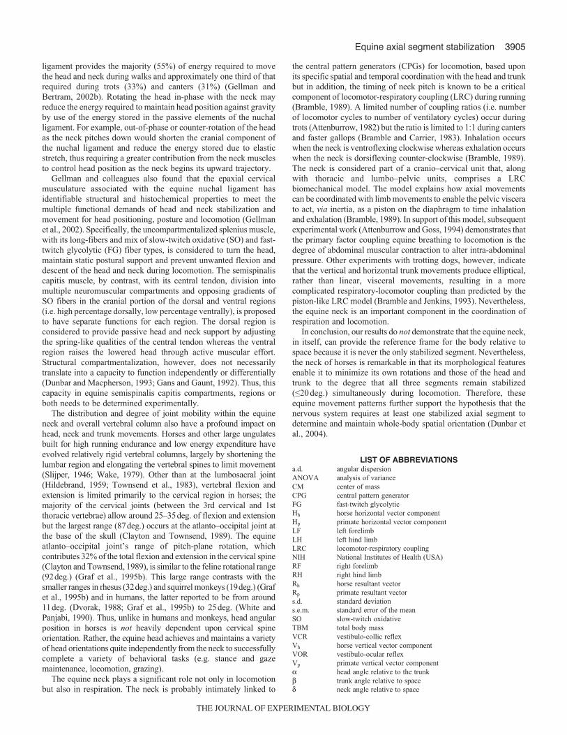
3905Equine axial segment stabilization
ligament provides the majority (55%) of energy required to movethe head and neck during walks and approximately one third of thatrequired during trots (33%) and canters (31%) (Gellman andBertram, 2002b). Rotating the head in-phase with the neck mayreduce the energy required to maintain head position against gravityby use of the energy stored in the passive elements of the nuchalligament. For example, out-of-phase or counter-rotation of the headas the neck pitches down would shorten the cranial component ofthe nuchal ligament and reduce the energy stored due to elasticstretch, thus requiring a greater contribution from the neck musclesto control head position as the neck begins its upward trajectory.
Gellman and colleagues also found that the epaxial cervicalmusculature associated with the equine nuchal ligament hasidentifiable structural and histochemical properties to meet themultiple functional demands of head and neck stabilization andmovement for head positioning, posture and locomotion (Gellmanet al., 2002). Specifically, the uncompartmentalized splenius muscle,with its long-fibers and mix of slow-twitch oxidative (SO) and fast-twitch glycolytic (FG) fiber types, is considered to turn the head,maintain static postural support and prevent unwanted flexion anddescent of the head and neck during locomotion. The semispinaliscapitis muscle, by contrast, with its central tendon, division intomultiple neuromuscular compartments and opposing gradients ofSO fibers in the cranial portion of the dorsal and ventral regions(i.e. high percentage dorsally, low percentage ventrally), is proposedto have separate functions for each region. The dorsal region isconsidered to provide passive head and neck support by adjustingthe spring-like qualities of the central tendon whereas the ventralregion raises the lowered head through active muscular effort.Structural compartmentalization, however, does not necessarilytranslate into a capacity to function independently or differentially(Dunbar and Macpherson, 1993; Gans and Gaunt, 1992). Thus, thiscapacity in equine semispinalis capitis compartments, regions orboth needs to be determined experimentally.
The distribution and degree of joint mobility within the equineneck and overall vertebral column also have a profound impact onhead, neck and trunk movements. Horses and other large ungulatesbuilt for high running endurance and low energy expenditure haveevolved relatively rigid vertebral columns, largely by shortening thelumbar region and elongating the vertebral spines to limit movement(Slijper, 1946; Wake, 1979). Other than at the lumbosacral joint(Hildebrand, 1959; Townsend et al., 1983), vertebral flexion andextension is limited primarily to the cervical region in horses; themajority of the cervical joints (between the 3rd cervical and 1stthoracic vertebrae) allow around 25–35deg. of flexion and extensionbut the largest range (87deg.) occurs at the atlanto–occipital joint atthe base of the skull (Clayton and Townsend, 1989). The equineatlanto–occipital joint’s range of pitch-plane rotation, whichcontributes 32% of the total flexion and extension in the cervical spine(Clayton and Townsend, 1989), is similar to the feline rotational range(92deg.) (Graf et al., 1995b). This large range contrasts with thesmaller ranges in rhesus (32deg.) and squirrel monkeys (19deg.) (Grafet al., 1995b) and in humans, the latter reported to be from around11deg. (Dvorak, 1988; Graf et al., 1995b) to 25deg. (White andPanjabi, 1990). Thus, unlike in humans and monkeys, head angularposition in horses is not heavily dependent upon cervical spineorientation. Rather, the equine head achieves and maintains a varietyof head orientations quite independently from the neck to successfullycomplete a variety of behavioral tasks (e.g. stance and gazemaintenance, locomotion, grazing).
The equine neck plays a significant role not only in locomotionbut also in respiration. The neck is probably intimately linked to
the central pattern generators (CPGs) for locomotion, based uponits specific spatial and temporal coordination with the head and trunkbut in addition, the timing of neck pitch is known to be a criticalcomponent of locomotor-respiratory coupling (LRC) during running(Bramble, 1989). A limited number of coupling ratios (i.e. numberof locomotor cycles to number of ventilatory cycles) occur duringtrots (Attenburrow, 1982) but the ratio is limited to 1:1 during cantersand faster gallops (Bramble and Carrier, 1983). Inhalation occurswhen the neck is ventroflexing clockwise whereas exhalation occurswhen the neck is dorsiflexing counter-clockwise (Bramble, 1989).The neck is considered part of a cranio–cervical unit that, alongwith thoracic and lumbo–pelvic units, comprises a LRCbiomechanical model. The model explains how axial movementscan be coordinated with limb movements to enable the pelvic viscerato act, via inertia, as a piston on the diaphragm to time inhalationand exhalation (Bramble, 1989). In support of this model, subsequentexperimental work (Attenburrow and Goss, 1994) demonstrates thatthe primary factor coupling equine breathing to locomotion is thedegree of abdominal muscular contraction to alter intra-abdominalpressure. Other experiments with trotting dogs, however, indicatethat the vertical and horizontal trunk movements produce elliptical,rather than linear, visceral movements, resulting in a morecomplicated respiratory-locomotor coupling than predicted by thepiston-like LRC model (Bramble and Jenkins, 1993). Nevertheless,the equine neck is an important component in the coordination ofrespiration and locomotion.
In conclusion, our results do not demonstrate that the equine neck,in itself, can provide the reference frame for the body relative tospace because it is never the only stabilized segment. Nevertheless,the neck of horses is remarkable in that its morphological featuresenable it to minimize its own rotations and those of the head andtrunk to the degree that all three segments remain stabilized(≤20 deg.) simultaneously during locomotion. Therefore, theseequine movement patterns further support the hypothesis that thenervous system requires at least one stabilized axial segment todetermine and maintain whole-body spatial orientation (Dunbar etal., 2004).
LIST OF ABBREVIATIONSa.d. angular dispersionANOVA analysis of varianceCM center of massCPG central pattern generatorFG fast-twitch glycolyticHh horse horizontal vector componentHp primate horizontal vector componentLF left forelimbLH left hind limbLRC locomotor-respiratory couplingNIH National Institutes of Health (USA)RF right forelimbRH right hind limbRh horse resultant vectorRp primate resultant vectors.d. standard deviations.e.m. standard error of the meanSO slow-twitch oxidativeTBM total body massVCR vestibulo-collic reflexVh horse vertical vector componentVOR vestibulo-ocular reflexVp primate vertical vector componentα head angle relative to the trunkβ trunk angle relative to spaceδ neck angle relative to space
THE JOURNAL OF EXPERIMENTAL BIOLOGY
3906
� neck angle relative to the trunkμ head angle relative to the neckθ head angle relative to space
The authors wish to thank the following individuals and institutions: Ms Anderson,Mrs Coit and Ms Hadden (Trainer), who own the horses, and Mr B. Milligan, whomanages the Rancho Riding Club in Rancho Santa Fe, California, kindly providedus with the equine subjects, location and facilities for video collection; skull andcervical spine measurements were made possible at the University of Puerto Ricoby Dr Jean E. Turnquist (horse) and the Department of Anatomy andNeurobiology (human) in the School of Medicine, and by the Laboratory ofPrimate Morphology and Genetics (rhesus monkey) in the Caribbean PrimateResearch Center; Dr Stéphane Vieilledent (Université de Bretagne Occidentale,Brest, France) contributed to the head frequency analyses; and two anonymousreviewers provided helpful comments on the manuscript. Funded by the ResearchCenters in Minority Institutions Program (NIH RR-03051), the Caribbean PrimateResearch Center (NIH P40 RR003640), and the University of Puerto Rico Schoolof Medicine.
REFERENCESAlexander, R. M. (1982). Locomotion in Animals. Glasgow: Blackie.Angelaki, D. E., McHenry, M. Q., Dickman, J. D., Newlands, S. D. and Hess, B. J.
M. (1999). Computation of inertial motion: neural strategies to resolve ambiguousotolith information. J. Neurosci. 19, 316-327.
Angelaki, D. E., Shaikh, A. G., Green, A. M. and Dickman, J. D. (2004). Neuronscompute internal models of the physical laws of motion. Nature 430, 560-564.
Ankel-Simons, F. (2000). Primate Anatomy: An Introduction. 2nd edn. San Diego:Academic Press.
Attenburrow, D. P. (1982). Time relationships between the respiratory cycle and thelimb cycle in the horse. Equine Vet. J. 14, 69-72.
Attenburrow, D. P. and Goss, V. A. (1994). The mechanical coupling of lungventilation to locomotion in the horse. Med. Eng. Phys. 16, 188-192.
Bramble, D. M. (1989). Axial-appendicular dynamics and the integration of breathingand gait in mammals. Am. Zool. 29, 171-186.
Bramble, D. M. and Carrier, D. R. (1983). Running and breathing in mammals.Science 219, 251-256.
Bramble, D. M. and Jenkins, F. A., Jr (1993). Mammalian locomotor-respiratoryintegration: implications for diaphragmatic and pulmonary design. Science 262, 235-240.
Cappozzo, A. (1982). Low frequency self-generated vibration during ambulation innormal men. J. Biomech. 8, 599-609.
Clayton, H. M. (2004). The Dynamic Horse. Mason, MI: Sport Horse.Clayton, H. M. and Townsend, H. G. G. (1989). Kinematics of the cervical spine of
the adult horse. Equine Vet. J. 21, 189-192.Crompton, R. H., Li, Y., Alexander, R. M., Wang, W. and Gunther, M. M. (1996).
Segmental inertial properties of primates: new techniques for laboratory and fieldstudies of locomotion. Am. J. Phys. Anthropol. 99, 547-570.
Cromwell, R. and Wellmon, R. (2001). Sagittal plane head stabilization during levelwalking and ambulation on stairs. Physiother. Res. Int. 6, 179-192.
Cromwell, R. L., Aadland-Monahan, T. K., Nelson, A. T., Stern-Sylvestre, S. M.and Seder, B. (2001). Sagittal plane analysis of head, neck, and trunk kinematicsand electromyographic activity during locomotion. J. Orthop. Sports Phys. Ther. 31,255-262.
Cromwell, R. L., Pidcoe, P. E., Griffin, L. A., Sotillo, T., Ganninger, D. and Feagin,M. (2004). Adaptations in horizontal head stabilization in response to altered visionand gaze during natural walking. J. Vestib. Res. 14, 367-373.
Das, P. and McCollum, G. (1988). Invariant structure in locomotion. Neuroscience 25,1023-1034.
Day, B. L. and Fitzpatrick, R. C. (2005). Virtual head rotation reveals a process ofroute reconstruction from human vestibular signals. J. Physiol. (Lond.) 567, 591-597.
DeBeer, G. R. (1947). How animals hold their heads. Proc. Linn. Soc. Lond. 159, 125-139.
Dempster, W. T. (1955). Space requirements of the seated operator. Wright AirDevelopment Center (WADC) Technical Report 55-159. Ohio: Wright-Patterson AirForce Base.
Duke-Elder, S. S. (1958). System of Ophthalmology. Vol. 1, The Eye in Evolution.London: Henry Kimpton.
Dunbar, D. C. (1988). Aerial maneuvers of leaping lemurs: the physics of whole-bodyrotations while airborne. Am. J. Primatol. 16, 291-303.
Dunbar, D. C. (1994). The influence of segmental movements and design on whole-body rotations during the airborne phase of primate leaps. Z. Morphol. Anthropol. 80,109-124.
Dunbar, D. C. (2004). Stabilization and mobility of the head and trunk in vervetmonkeys (Cercopithecus aethiops) during treadmill walks and gallops. J. Exp. Biol.207, 4427-4438.
Dunbar, D. C. and Macpherson, J. M. (1993). Activity of neuromuscularcompartments in lateral gastrocnemius evoked by postural corrections during stance.J. Neurophysiol. 70, 2337-2349.
Dunbar, D. C., Badam, G. L., Hallgrímsson, B. and Vieilledent, S. (2004).Stabilization and mobility of the head and trunk in wild monkeys during terrestrialand flat-surface walks and gallops. J. Exp. Biol. 207, 1027-1042.
Dvorak, J. (1988). Funktionelle Anatomie der oberen Halswirbelsäule unter besondererBerückersichtigung des Bandapparates. In Die Sonderstellung desKopfgelenkbereichs (ed. H. D. Wolff), pp. 19-46. Berlin: Springer.
Edwards, E. H. (2001). The New Encyclopedia of the Horse. New York: DorlingKindersley.
Einstein, A. (1908). Über das Relativitätsprinzip und die aus demselben gezogenenFolgerungen. Jahrb. Radioakt. 4, 411-462.
Frohlich, C. (1979). Do springboard divers violate angular momentum conservation?Am. J. Phys. 47, 583-592.
Frohlich, C. (1980). The physics of somersaulting and twisting. Sci. Am. 242, 154-164.Fuchs, A. F. (1981). Eye-head coordination. In Handbook of Behavioral Neurobiology,
Vol. 5: Motor Coordination (ed. A. L. Towe and E. S. Luschei), pp. 303-366. NewYork: Plenum Press.
Gans, C. and Gaunt, A. S. (1992). Muscle architecture and control demands. BrainBehav. Evol. 40, 70-81.
Gellman, K. S. and Bertram, J. E. A. (2002a). The equine nuchal ligament 1,structural and material properties. Vet. Comp. Orthop. Traumatol. 15, 1-6.
Gellman, K. S. and Bertram, J. E. A. (2002b). The equine nuchal ligament 2, passivedynamic energy exchange in locomotion. Vet. Comp. Orthop. Traumatol. 15, 7-14.
Gellman, K. S., Bertram, J. E. A. and Hermanson, J. W. (2002). Morphology,histochemistry, and function of epaxial cervical musculature in the horse (Equuscaballus). J. Morphol. 251, 182-194.
Goody, P. C. (1983). Horse Anatomy: A Pictorial Approach to Equine Structure.London: J. A. Allen.
Graf, W., de Waele, C. and Vidal, P. P. (1995a). Functional anatomy of the head-neck system of quadrupedal and bipedal mammals. J. Anat. 186, 55-74.
Graf, W., de Waele, C., Vidal, P. P., Wang, D. H. and Evinger, C. (1995b). Theorientation of the cervical vertebral column in unrestrained awake animals. II:movement strategies. Brain Behav. Evol. 45, 209-231.
Grossman, G. E., Leigh, R. J., Abel, L. A., Lanska, D. J. and Thurston, S. E.(1988). Frequency and velocity of rotational head perturbation during locomotion.Exp. Brain Res. 70, 470-476.
Harman, A. M., Moore, S., Hoskins, R. and Keller, P. (1999). Horse vision and anexplanation for the visual behaviour originally explained by the “ramp retina”. EquineVet. J. 31, 384-390.
Hess, B. J. M. (2007). Sensorimotor transformations in spatial orientation relative togravity. In Spatial Processing in Navigation, Imagery and Perception (ed. F. W. Mastand L. Jäncke), pp. 281-300. New York: Springer.
Hildebrand, M. (1959). Motions of the running cheetah and horse. J. Mammal. 40,481-495.
Hildebrand, M. (1966). Analysis of symmetrical gaits of tetrapods. Folia Biotheoret. 13,9-22.
Hildebrand, M. (1974). Analysis of Vertebrate Structure. New York: John Wiley.Hildebrand, M. (1977). Analysis of asymmetrical gaits. J. Mammal. 58, 131-156.Hill, A. V. (1950). The dimensions of animals and their muscular dynamics. Sci. Prog.
38, 209-230.Hirasaki, E. and Kumakura, H. (2004). Head movements during locomotion in a
gibbon and Japanese macaques. NeuroReport 15, 643-647.Howell, A. B. (1944). Speed in Animals. Chicago: University of Chicago Press.Hughes, A. (1977). The topography of vision in mammals of contrasting lifestyle:
comparative optics and retinal organization. In Handbook of Sensory Physiology: theVisual System in Vertebrates, Vol. VII/5 (ed. F. Crescitelli), pp. 615-756. Berlin:Springer Verlag.
Imai, T., Moore, S. T., Raphan, T. and Cohen, B. (2001). Interaction of the body,head, and eyes during walking and turning. Exp. Brain Res. 136, 1-18.
Jakobs, T., Miller, J. A. A. and Schultz, A. B. (1985). Trunk position sense in thefrontal plane. Exp. Neurol. 90, 129-138.
Johnson, R. A. and Wehrly, T. (1977). Measures and models for angular correlationand angular-linear correlation. J. R. Stat. Soc. Ser. B Methodol. 39, 222-229.
Jones, G. M. and Spells, K. E. (1963). A theoretical and comparative study offunctional dependence of the semicircular canal upon physical dimensions. Proc. R.Soc. Lond., B, Biol. Sci. 157, 403-419.
Kimura, T., Okada, M. and Ishida, H. (1979). Kinesiological characteristics of primatewalking: its significance in human walking. In Environment, Behavior, andMorphology: Dynamic Interactions in Primates (ed. M. E. Morbeck, H. Preuschoftand N. Gomberg), pp. 297-311. New York: Gustav Fischer.
Kleine, J. F., Guan, Y., Kipiani, E. L., Glonti, L., Hoshi, M. and Büttner, U. (2004).Trunk position influences vestibular responses of fastigial nucleus neurons in thealert monkey. J. Neurophysiol. 91, 2090-2100.
Lanovaz, J. L., Clayton, H. M. and Watson, L. G. (1998). In vitro attenuation ofimpact shock in equine digits. Equine Vet. J. Suppl. 26, 96-102.
Liao, K., Walker, M. F., Joshi, A., Reschke, M. and Leigh, R. J. (2008). Vestibulo-ocular responses to vertical translation in normal human subjects. Exp. Brain Res.185, 553-562.
Lieberman, D. E., Raichlen, D. A., Pontzer, H., Bramble, D. M. and Cutright-Smith,E. (2006). The human gluteus maximus and its role in running. J. Exp. Biol. 209,2143-2155.
Lindsay, K. W., Roberts, T. D. M. and Rosenberg, J. R. (1976). Asymmetrical toniclabyrinth reflexes and their interaction with neck reflexes in the decerebrate cat. J.Physiol. (Lond.) 261, 583-601.
Macpherson, J. M., Everaert, D. G., Stapley, P. J. and Ting, L. H. (2007). Bilateralvestibular loss in cats leads to active destabilization of balance during pitch and rollrotations of the support surface. J. Neurophysiol. 97, 4357-4367.
Mardia, K. V. (1976). Linear-circular correlation coefficients and rhythmometry.Biometrika 63, 403-405.
Merfeld, D. M., Zupan, L. and Peterka, R. J. (1999). Humans use internal models toestimate gravity and linear acceleration. Nature 398, 615-618.
Mergner, T., Nardi, G. L., Becker, W. and Keeke, L. (1983). The role of canal-neckinteraction for the perception of horizontal trunk and head rotation. Exp. Brain Res.49, 198-208.
Mergner, T., Siebold, C., Schweigart, G. and Becker, W. (1991). Human perceptionof horizontal trunk and head rotation in space during vestibular and neck stimulation.Exp. Brain Res. 85, 389-404.
D. C. Dunbar and others
THE JOURNAL OF EXPERIMENTAL BIOLOGY
3907Equine axial segment stabilization
Mergner, T., Rottler, G., Kimmig, H. and Becker, W. (1992). Role of vestibular andneck inputs for the perception of object motion in space. Exp. Brain Res. 89, 655-668.
Mittelstaedt, H. (1988). The information processing structure of the subjective vertical:a cybernetic bridge between its psychophysics and its neurobiology. In ProcessingStructures For Perception and Action (ed. H. Marko, G. Hauske and A. Struppler),pp. 217-263. Weinheim: V. C. H. Verlagsgesellschaft.
Mittelstaedt, H. (1995). Evidence of somatic graviception from new and classicalinvestigations. Acta Otolaryngol. Suppl. 520, 186-187.
Mittelstaedt, H. (1996). Somatic graviception. Biol. Psychol. 42, 53-74.Mittelstaedt, H. (1997). Interaction of eye-, head-, and trunk-bound information in
spatial perception and control. J. Vestib. Res. 7, 283-302.Mittelstaedt, H. (1998). Origin and processing of postural information. Neurosci.
Biobehav. Rev. 22, 473-478.Moore, S. T., Hirasaki, E., Cohen, B. and Raphan, T. (1999). Effect of viewing
distance on the generation of vertical eye movements during locomotion. Exp. BrainRes. 129, 374-361.
Paige, G. D. (1989). The influence of target distance on eye movement responsesduring vertical linear motion. Exp. Brain Res. 77, 585-593.
Patla, A. E. (1991). Understanding the control of human locomotion: a prologue. InAdaptability of Human Gait (ed. A. E. Patla), pp. 3-17. Amsterdam: North-Holland.
Patla, A. E. (1997). Understanding the roles of vision in the control of humanlocomotion. Gait Posture 5, 54-69.
Peterka, R. J. (2002). Sensorimotor integration in human posture control. J.Neurophysiol. 88, 1097-1118.
Peterson, B. W. and Boyle, R. D. (2004). Vestibulocollic reflexes. In The VestibularSystem (ed. S. M. Highstein, R. R. Fay, and A. N. Popper), pp. 343-374. New York:Springer.
Peterson, B. W. and Goldberg, J. (1981). Role of vestibular and neck reflexes incontrolling eye and head position. In Physiological and Pathological Aspects of EyeMovements (ed. A. Roucoux and M. Crommelinck), pp. 352-364. The Hague: Dr W.Junk.
Pozzo, T., Berthoz, A. and Lefort, L. (1990). Head stabilization during variouslocomotor tasks in humans. I: Normal subjects. Exp. Brain Res. 82, 97-106.
Roberts, S. M. (1992). Equine vision and optics. Vet. Clin. North. Am. Equine Pract. 8,451-457.
Roberts, T. D. M. (1978). Neurophysiology of Postural Mechanisms. 2nd edn. London:Butterworths.
Rochon-Duvigneaud, A. (1943). Les Yeaux et la Vision des Vértebrés. Paris:Mason.
Russell, N. A., Horii, A., Smith, P. F., Darlington, C. L. and Bilkey, D. K. (2003).Long-term effects of permanent vestibular lesions on hippocampal spatial firing. J.Neurosci. 23, 6490-6498.
Schmitt, D. (2003). Insights into the evolution of human bipedalism from experimentalstudies of humans and other primates. J. Exp. Biol. 206, 1437-1448.
Schubert, M., Bohner, C., Berger, W. V., Sprundel, M. and Duysens, J. E. J.(2003). The role of vision in maintaining heading direction: effects of changing gazeand optic flow on human gait. Exp. Brain Res. 150, 163-173.
Schwarz, U. and Miles, F. A. (1991). Ocular responses to translation and theirdependence on viewing distance. I: Motion of the observer. J. Neurophysiol. 66,851-864.
Shumway-Cook, A. and Woollacott, M. H. (2001). Motor Control. 2nd edn.Philadelphia: Lippincott Williams and Wilkins.
Simpson, G. G. (1961). Horses. New York: Doubleday Anchor.Slijper, E. J. (1946). Comparative biologic-anatomical investigations on the vertebral
column and spinal musculature of mammals. Verh. Kon. Nederl. Akad. Wetensch.Amsterdam II 42, 1-128.
Stackman, R. W. and Taube, J. S. (1997). Firing properties of head direction cells inthe rat anterior thalamic nucleus: dependence on vestibular input. J. Neurosci. 17,4349-4358.
Stackman, R. W., Clark, A. S. and Taube, J. S. (2002). Hippocampal spatialrepresentations require vestibular input. Hippocampus 12, 291-303.
Taylor, J. L. and McCloskey, D. I. (1990). Proprioceptive sensation in rotation of thetrunk. Exp. Brain Res. 81, 413-416.
Timney, B. and Macuda, T. (2001). Vision and hearing in horses. J. Am. Vet. Med.Assoc. 218, 1567-1574.
Townsend, H. G., Leach, D. H. and Fretz, P. B. (1983). Kinematics of the equinethoracolumbar spine. Equine Vet. J. 15, 117-122.
Ueda, Y., Niki, Y., Yoshida, K. and Masumitsu, H. (1981). Force plate study ofequine biomechanics. Floor reaction force of normal walking and trotting horses.Bull. Equine Res. Inst. 18, 28-41.
Vaitl, D., Mittelstaedt, H. and Baisch, F. (1997). Shifts in blood volume alter theperception of posture. Int. J. Psychophysiol. 27, 99-105.
Vaitl, D., Mittelstaedt, H., Saborowski, R., Stark, R. and Baisch, F. (2002). Shifts inblood volume alter the perception of posture: further evidence for somaticgraviception. Int. J. Psychophysiol. 44, 1-11.
Vorstenbosch, M. A. T. M., Buchner, H. H. F., Savelberg, H. H. C. M., Schamhardt,H. C. and Barneveld, A. (1997). Modeling study of compensatory head movementsin lame horses. Am. J. Vet. Res. 58, 713-718.
Wake, D. B. (1979). The endoskeleton: the comparative anatomy of the vertebralcolumn and ribs. In Hymanʼs Comparative Vertebrate Anatomy. 3rd Edn (ed. by M.H. Wake), pp. 192-237. Chicago: Univerisity of Chicago Press.
White, A. A. and Panjabi, M. M. (1990). Clinical Biomechanics of the Spine. 2nd edn.Philadelphia: Lippincott.
Wilson, V. J. and Peterson, B. W. (1981). Vestibulospinal and reticulospinal systems.In Handbook of Physiology, Sec. 1, Vol. II: Motor Control, Part 1 (ed. V. B. Brooks),pp. 667-702. Bethesda, MD: American Physiological Society.
Xiang, Y., Yakushin, S. B., Kunin, M., Raphan, T. and Cohen, B. (2008). Headstabilization by vestibulocollic reflexes during quadrupedal locomotion in monkey. J.Neurophysiol. 100, 763-780.
THE JOURNAL OF EXPERIMENTAL BIOLOGY