spontaneous uterine rupture and surgical repair at 21
TRANSCRIPT
CASE REPORT Open Access
Spontaneous uterine rupture and surgicalrepair at 21 weeks gestation withprogression to live birth: a case reportLesley Hawkins1, Deborah Robertson1,2, Helena Frecker1,3, Howard Berger1,2 and Abheha Satkunaratnam1,2*
Abstract
Background: Uterine rupture in the non-laboring uterus is a rare occurrence, which can lead to significant morbidityand mortality for the mother and fetus. Management of this presentation is complex at pre-viable gestations.
Case presentation: A 35 year old primigravid woman with multiple previous myomectomies presented withspontaneous complete thickness uterine rupture at 21 weeks gestation. A 10 cm myometrial defect and iatrogenicamniotomy were surgically corrected with fetal preservation. This led to pregnancy continuation to 32 weeks gestationwhen elective cesarean delivery resulted in excellent neonatal outcome.
Conclusions: Early surgical diagnosis, multidisciplinary team approach, iatrogenic amniotomy and continuous two-layermyometrial closure were factors that contributed to pregnancy prolongation in this large myometrial rupture.
Keywords: Uterine rupture, Spontaneous uterine rupture, Uterine wall defect, Second trimester
BackgroundUterine rupture is defined as complete separation of themyometrium [1, 2]. It can occur in the laboring or non-laboring uterus, the latter known as spontaneous uterinerupture. Spontaneous uterine rupture is a rare occur-rence which can lead to maternal hemorrhage, placentalabruption and extrusion of the amniotic sac and fetalparts through the uterine defect. This can result insignificant consequences for both the mother and fetus,including blood transfusion, hysterectomy, urologic in-jury, neonatal respiratory distress, perinatal asphyxia andmaternal or fetal death [3–5]. A history of uterine surgeryhas been identified as the most common risk factor forspontaneous uterine rupture in a small cohort of cases [6].Less common causes include iatrogenic uterine perfor-ation, invasive placenta, congenital anomalies, trauma andsacculation of the entrapped retroverted uterus [7].Management of this rare pregnancy complication re-
quires several considerations. Cesarean delivery with either
uterine repair or hysterectomy may be appropriate at fetalviability. However, when the fetus is previable or extremelypremature, management decisions are complex. Termin-ation of the pregnancy with uterine repair or hysterectomywas the traditional approach [8]. In recent years, repair ofuterine rupture in the second and early third trimesters hasbeen reported, with successful delay of delivery [9–11]. Wedescribe a rare case of spontaneous uterine rupture in themid-second trimester and successful surgical repair includ-ing inadvertent iatrogenic amniotomy with continuation ofpregnancy to 32 weeks gestation.
Case presentationA 35-year old primigravid woman presented to hospitalat 21 weeks 2 days gestation. She described a two-dayhistory of periumbilical abdominal discomfort that sud-denly became severe, waking her from sleep. There wasno history of trauma. She was afebrile and had no othergastrointestinal symptoms. She had previously under-gone four hysteroscopic myomectomies and one openmyomectomy, during which 5 uterine incisions weremade, including a fundal incision, and over 100fibroids were resected. The indication for thesesurgeries was primary infertility, abnormal uterinebleeding and anemia. The interval between open
* Correspondence: [email protected] of Obstetrics and Gynaecology, University of Toronto, 123Edward St, 12th Floor, Toronto, ON M5G1E2, Canada2Department of Obstetrics and Gynaecology, St Michael’s Hospital, 308-55Queen St East, Toronto, ON M5C1R6, CanadaFull list of author information is available at the end of the article
© The Author(s). 2018 Open Access This article is distributed under the terms of the Creative Commons Attribution 4.0International License (http://creativecommons.org/licenses/by/4.0/), which permits unrestricted use, distribution, andreproduction in any medium, provided you give appropriate credit to the original author(s) and the source, provide a link tothe Creative Commons license, and indicate if changes were made. The Creative Commons Public Domain Dedication waiver(http://creativecommons.org/publicdomain/zero/1.0/) applies to the data made available in this article, unless otherwise stated.
Hawkins et al. BMC Pregnancy and Childbirth (2018) 18:132 https://doi.org/10.1186/s12884-018-1761-x

myomectomy and conception was 26 months. Thecurrent pregnancy was spontaneously conceived andhad been progressing normally.At presentation, blood pressure was 99/54, heart rate
84 beats per minute, respiratory rate 20, temperature 36degrees Celsius (96.8 degrees Farenheit) and fetal heartrate 160 beats per minute. Abdominal examination re-vealed diffuse peritonitis and significant tenderness at theuterine fundus. Hemoglobin at presentation was 87 g/Land white blood cell count was 10.89 E9/L (referencerange 120-160 g/L and 4.00 to 11.00 E9/L, respectively).Ultrasound examination showed a live singleton fetus withnormal amniotic fluid volume. The placental position wasleft posterofundal. Moderate volume hemoperitoneumwas seen. Ovaries appeared normal bilaterally; the appen-dix was not visualized. Evaluation of the uterine wall dem-onstrated no focal uterine disruption or thinning, but waslimited due to patient tenderness and bowel gas, therefore,a Magnetic Resonance Imaging (MRI) study was recom-mended. Urgent unenhanced abdominal MRI, completedthat evening, 6 h following ultrasound examination,showed a sentinel clot at the left aspect of the uterinefundus, where the myometrium was markedly thinned,suspected to be the source of hemorrhage. During thediagnostic work-up, vital signs were stable and hemoglobinreached a stable nadir of 71 g/L.Differential diagnosis included fibroid degeneration or
torsion, placenta percreta, concealed placental abruption,uterine rupture and non-obstetrical causes. A patientcare conference and complex consent process was under-taken to include several potential scenarios and outcomes.The patient was counselled extensively around possiblecomplications of surgical repair, including prolongation ofthe pregnancy leading to severe prematurity, preterm rup-ture of membranes, and risks of unsuccessful repair, in-cluding hysterectomy and fetal loss. With a demonstrationof an understanding of these risks, she consented to adiagnostic laparoscopy, uterine repair with possible lapar-otomy, possible hysterotomy and evacuation of the fetusand possible hysterectomy.The following day, on the second day of admission, at
21 weeks 3 days gestation, the patient was taken to theoperating room. The procedure was initiated laparoscopic-ally via a left upper quadrant veress needle entry. A 10 cmcomplete thickness uterine fundus rupture was diagnosed(Fig. 1). Active bleeding was visualized from the edge of therupture, and the chorioamniotic membranes were visiblybulging through the defect. Large hemoperitoneum (esti-mated 1000 mL) was present. Given the extent and locationof the defect, the size of the gravid uterus and visualizationlaparoscopically, we converted to midline laparotomy.Closure of the defect was then undertaken. Uninten-tional iatrogenic amniotomy occurred due to the sutureneedle shaft scratching the membranes. This resulted in
two adjacent 2 mm defects in the amniotic sac and par-tial loss of amniotic fluid volume. Two large vascular clipswere applied to each defect for repair. We introduced a 22French foley catheter into the myometrial defect and in-flated the balloon to displace the amniotic sac to facilitateuterine repair. With the membranes reduced, a two-layeruterine closure was successfully completed using fullthickness continuous 0 polyglactin 910 (Vicryl) and im-bricating 2–0 Polydioxanone (PDS, Ethicon, USA) su-tures, respectively (Fig. 2). EVICEL and SURGICELSnoW (Ethicon, USA) were applied to the area of repairto affect hemostasis. Intraoperatively, the patient requiredtransfusion of 7 units packed erythrocytes, 2 units freshfrozen plasma and 1 unit cryoprecipitate; estimated totalblood loss was 2500 mL.The patient’s postoperative course was uncomplicated.
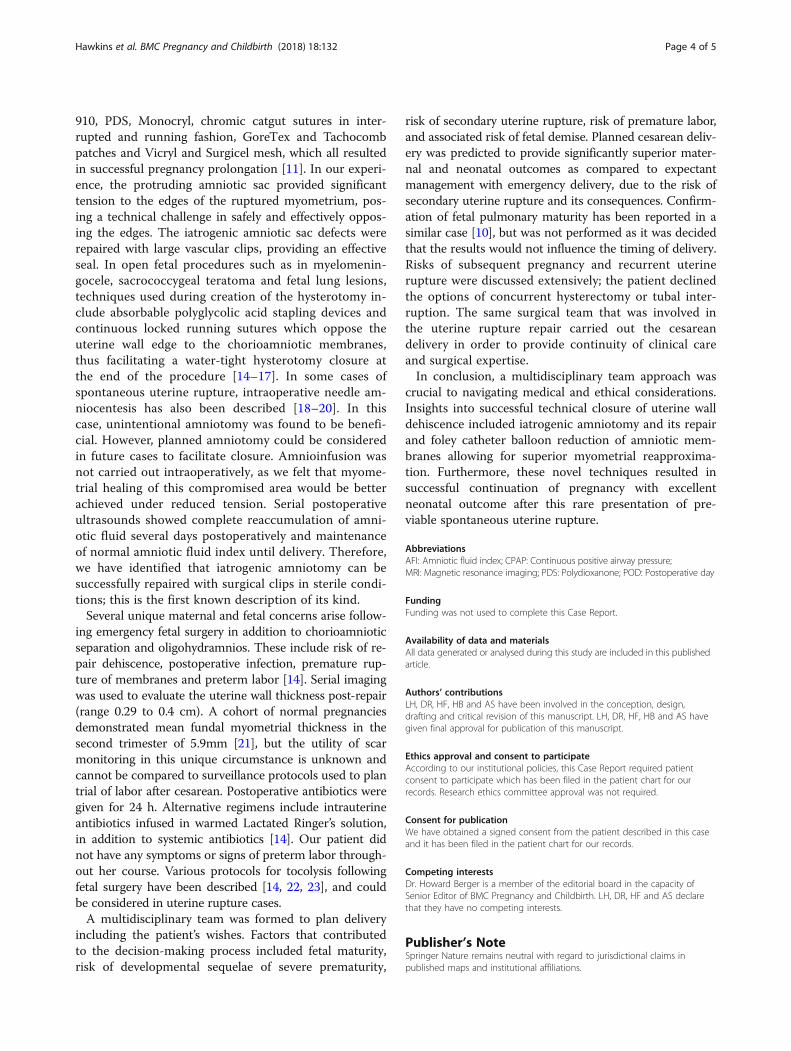
Indomethacin was administered for tocolysis per thestandard of care at our centre (100 mg per rectum then25 mg per os every 6 h for 4 doses). Postoperative intra-venous cefazolin was administered for 24 h. Fetal sur-veillance demonstrated amniotic fluid re-accumulation,appropriate growth parameters (estimated fetal weight60–85%ile) and normal fetal well-being. Amniotic fluidindex (AFI) was 6.5 cm on postoperative day (POD) 3,9 cm on POD4, 8.9 cm on POD5 and 12.5 cm onPOD12. Myometrial thickness at the repair site was eval-uated at several time points by ultrasound and rangedfrom 0.29 to 0.4 cm. Ultrasound and MRI at 27 weeksgestation showed a 0.7 cm by 5 cm area of chorion-amnion separation at the site of repair which remainedstable throughout our patient’s course (Fig. 3). Thepatient was transferred to a centre with Level 3 neonatalcare at 24 weeks gestation, for continued inpatientobservation, then transferred back to our Level 2.5centre at 30 weeks gestation. A course of antenatalcorticosteroids was administered at 25 weeks gestation.
Fig. 1 At laparotomy, the 10 cm fundal complete uterine wall defectwith protruding chorioamniotic membrane
Hawkins et al. BMC Pregnancy and Childbirth (2018) 18:132 Page 2 of 5
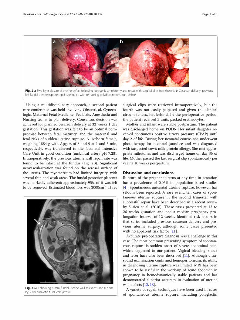
Using a multidisciplinary approach, a second patientcare conference was held involving Obstetrical, Gyneco-logic, Maternal Fetal Medicine, Pediatric, Anesthesia andNursing teams to plan delivery. Consensus decision wasachieved for planned cesarean delivery at 32 weeks 1 daygestation. This gestation was felt to be an optimal com-promise between fetal maturity, and the maternal andfetal risks of sudden uterine rupture. A liveborn female,weighing 1884 g with Apgars of 8 and 9 at 1 and 5 min,respectively, was transferred to the Neonatal IntensiveCare Unit in good condition (umbilical artery pH 7.28).Intraoperatively, the previous uterine wall repair site wasfound to be intact at the fundus (Fig. 2B). Significantneovascularization was found on the serosal surface ofthe uterus. The myometrium had limited integrity, withseveral thin and weak areas. The fundal posterior placentawas markedly adherent; approximately 95% of it was feltto be removed. Estimated blood loss was 2000cm3. Three
surgical clips were retrieved intraoperatively, but thefourth was not easily palpated and given the clinicalcircumstances, left behind. In the perioperative period,the patient received 3 units packed erythrocytes.Mother and infant were stable postpartum. The patient
was discharged home on POD6. Her infant daughter re-ceived continuous positive airway pressure (CPAP) untilday 2 of life. During her neonatal course, she underwentphototherapy for neonatal jaundice and was diagnosedwith suspected cow’s milk protein allergy. She met appro-priate milestones and was discharged home on day 36 oflife. Mother passed the last surgical clip spontaneously pervagina 10 weeks postpartum.
Discussion and conclusionsRupture of the pregnant uterus at any time in gestationhas a prevalence of 0.05% in population-based studies[4]. Spontaneous antenatal uterine rupture, however, hasseldom been reported. A rare event, ten cases of spon-taneous uterine rupture in the second trimester withsuccessful repair have been described in a recent reviewby Surico et al. (2016). These cases presented at 13 to26 weeks gestation and had a median pregnancy pro-longation interval of 12 weeks. Identified risk factors inthat series included previous cesarean delivery and pre-vious uterine surgery, although some cases presentedwith no apparent risk factor [11].Accurate pre-operative diagnosis was a challenge in this
case. The most common presenting symptom of spontan-eous rupture is sudden onset of severe abdominal pain,which happened to our patient. Vaginal bleeding, shockand fever have also been described [11]. Although ultra-sound examination confirmed hemoperitoneum, its utilityin diagnosing uterine rupture was limited. MRI has beenshown to be useful in the work-up of acute abdomen inpregnancy in hemodynamically stable patients and hasdemonstrated superior accuracy in evaluation of uterinewall defects [12, 13].A variety of repair techniques have been used in cases
of spontaneous uterine rupture, including polyglactin
Fig. 2 a Two-layer closure of uterine defect following iatrogenic amniotomy and repair with surgical clips (not shown). b. Cesarean delivery: previousleft fundal uterine rupture repair site intact, with remaining polydioxanone suture visible
Fig. 3 MRI showing 4 mm fundal uterine wall thickness and 0.7 cmby 5 cm amniotic fluid leak (arrow)
Hawkins et al. BMC Pregnancy and Childbirth (2018) 18:132 Page 3 of 5
910, PDS, Monocryl, chromic catgut sutures in inter-rupted and running fashion, GoreTex and Tachocombpatches and Vicryl and Surgicel mesh, which all resultedin successful pregnancy prolongation [11]. In our experi-ence, the protruding amniotic sac provided significanttension to the edges of the ruptured myometrium, pos-ing a technical challenge in safely and effectively oppos-ing the edges. The iatrogenic amniotic sac defects wererepaired with large vascular clips, providing an effectiveseal. In open fetal procedures such as in myelomenin-gocele, sacrococcygeal teratoma and fetal lung lesions,techniques used during creation of the hysterotomy in-clude absorbable polyglycolic acid stapling devices andcontinuous locked running sutures which oppose theuterine wall edge to the chorioamniotic membranes,thus facilitating a water-tight hysterotomy closure atthe end of the procedure [14–17]. In some cases ofspontaneous uterine rupture, intraoperative needle am-niocentesis has also been described [18–20]. In thiscase, unintentional amniotomy was found to be benefi-cial. However, planned amniotomy could be consideredin future cases to facilitate closure. Amnioinfusion wasnot carried out intraoperatively, as we felt that myome-trial healing of this compromised area would be betterachieved under reduced tension. Serial postoperativeultrasounds showed complete reaccumulation of amni-otic fluid several days postoperatively and maintenanceof normal amniotic fluid index until delivery. Therefore,we have identified that iatrogenic amniotomy can besuccessfully repaired with surgical clips in sterile condi-tions; this is the first known description of its kind.Several unique maternal and fetal concerns arise follow-
ing emergency fetal surgery in addition to chorioamnioticseparation and oligohydramnios. These include risk of re-pair dehiscence, postoperative infection, premature rup-ture of membranes and preterm labor [14]. Serial imagingwas used to evaluate the uterine wall thickness post-repair(range 0.29 to 0.4 cm). A cohort of normal pregnanciesdemonstrated mean fundal myometrial thickness in thesecond trimester of 5.9mm [21], but the utility of scarmonitoring in this unique circumstance is unknown andcannot be compared to surveillance protocols used to plantrial of labor after cesarean. Postoperative antibiotics weregiven for 24 h. Alternative regimens include intrauterineantibiotics infused in warmed Lactated Ringer’s solution,in addition to systemic antibiotics [14]. Our patient didnot have any symptoms or signs of preterm labor through-out her course. Various protocols for tocolysis followingfetal surgery have been described [14, 22, 23], and couldbe considered in uterine rupture cases.A multidisciplinary team was formed to plan delivery
including the patient’s wishes. Factors that contributedto the decision-making process included fetal maturity,risk of developmental sequelae of severe prematurity,
risk of secondary uterine rupture, risk of premature labor,and associated risk of fetal demise. Planned cesarean deliv-ery was predicted to provide significantly superior mater-nal and neonatal outcomes as compared to expectantmanagement with emergency delivery, due to the risk ofsecondary uterine rupture and its consequences. Confirm-ation of fetal pulmonary maturity has been reported in asimilar case [10], but was not performed as it was decidedthat the results would not influence the timing of delivery.Risks of subsequent pregnancy and recurrent uterinerupture were discussed extensively; the patient declinedthe options of concurrent hysterectomy or tubal inter-ruption. The same surgical team that was involved inthe uterine rupture repair carried out the cesareandelivery in order to provide continuity of clinical careand surgical expertise.In conclusion, a multidisciplinary team approach was
crucial to navigating medical and ethical considerations.Insights into successful technical closure of uterine walldehiscence included iatrogenic amniotomy and its repairand foley catheter balloon reduction of amniotic mem-branes allowing for superior myometrial reapproxima-tion. Furthermore, these novel techniques resulted insuccessful continuation of pregnancy with excellentneonatal outcome after this rare presentation of pre-viable spontaneous uterine rupture.
AbbreviationsAFI: Amniotic fluid index; CPAP: Continuous positive airway pressure;MRI: Magnetic resonance imaging; PDS: Polydioxanone; POD: Postoperative day
FundingFunding was not used to complete this Case Report.
Availability of data and materialsAll data generated or analysed during this study are included in this publishedarticle.
Authors’ contributionsLH, DR, HF, HB and AS have been involved in the conception, design,drafting and critical revision of this manuscript. LH, DR, HF, HB and AS havegiven final approval for publication of this manuscript.
Ethics approval and consent to participateAccording to our institutional policies, this Case Report required patientconsent to participate which has been filed in the patient chart for ourrecords. Research ethics committee approval was not required.
Consent for publicationWe have obtained a signed consent from the patient described in this caseand it has been filed in the patient chart for our records.
Competing interestsDr. Howard Berger is a member of the editorial board in the capacity ofSenior Editor of BMC Pregnancy and Childbirth. LH, DR, HF and AS declarethat they have no competing interests.
Publisher’s NoteSpringer Nature remains neutral with regard to jurisdictional claims inpublished maps and institutional affiliations.
Hawkins et al. BMC Pregnancy and Childbirth (2018) 18:132 Page 4 of 5
Author details1Department of Obstetrics and Gynaecology, University of Toronto, 123Edward St, 12th Floor, Toronto, ON M5G1E2, Canada. 2Department ofObstetrics and Gynaecology, St Michael’s Hospital, 308-55 Queen St East,Toronto, ON M5C1R6, Canada. 3Department of Obstetrics and Gynaecology,Michael Garron Hospital, Suite 311, 658 Danforth Avenue, M4J5B9 Toronto,Ontario, Canada.
Received: 15 May 2017 Accepted: 23 April 2018
References1. Scott JR. Avoiding labor problems during vaginal birth after cesarean
delivery. Clin Obstet Gynecol. 1997;40:533–41.2. Bucklin BA. Vaginal birth after cesarean delivery. Anesthesiology. 2003;99:1444–8.3. Chauhan SP, Martin JN, Jr., Henrichs CE, Morrison JC, Magann EF. Maternal
and perinatal complications with uterine rupture in 142,075 patients whoattempted vaginal birth after cesarean delivery: a review of the literature.Am J Obstet Gynecol 2003;189:408–417.
4. Hofmeyr GJ, Say L, Gulmezoglu AM. WHO Systematic review of maternalmortality and morbidity: the prevalence of uterine rupture. BJOG : aninternational journal of obstetrics and gynaecology. 2005;112:1221–8.
5. Leung AS, Leung EK, Paul RH. Uterine rupture after previous cesarean delivery:maternal and fetal consequences. Am J Obstet Gynecol. 1993;169:945–50.
6. Vaknin Z, Maymon R, Mendlovic S, Barel O, Herman A, Sherman D. Clinical,sonographic, and epidemiologic features of second- and early third-trimester spontaneous antepartum uterine rupture: a cohort study. PrenatDiagn. 2008;28:478–84.
7. Arbab F, Boulieu D, Bied V, Payan F, Lornage J, Guerin JF. Uterine rupture infirst or second trimester of pregnancy after in-vitro fertilization and embryotransfer. Hum Reprod. 1996;11:1120–2.
8. Martin JN Jr, Brewer DW, Rush LV Jr, Martin RW, Hess LW, Morrison JC.Successful pregnancy outcome following mid-gestational uterine ruptureand repair using gore-Tex soft tissue patch. Obstet Gynecol. 1990;75:518–21.
9. Sugawara T, Ogawa M, Tanaka T. Repair of uterine rupture during secondtrimester leading to successful pregnancy outcome: case study andLiterature's review. AJP reports. 2014;4:9–12.
10. Matsunaga JS, Daly CB, Bochner CJ, Agnew CL. Repair of uterine dehiscencewith continuation of pregnancy. Obstet Gynecol. 2004;104:1211–2.
11. Surico D, Amadori R, Vigone A, D'Agostino C, Dessole M, Surico N.Successful delivery after surgical repair of uterine rupture at 15 weeks ofgestation: case report and brief review. Eur J Obstet Gynecol Reprod Biol.2016;204:5–8.
12. Furey EA, Bailey AA, Pedrosa I. Magnetic resonance imaging of acuteabdominal and pelvic pain in pregnancy. Top Magn Reson Imaging. 2014;23:225–42.
13. Casciani E, De Vincentiis C, Mazzei MA, et al. Errors in imaging the pregnantpatient with acute abdomen. Abdom Imaging. 2015;40:2112–26.
14. Adzick NS, Thom EA, Spong CY, et al. A randomized trial of prenatal versuspostnatal repair of myelomeningocele. N Engl J Med. 2011;364:993–1004.
15. Bennett KA, Carroll MA, Shannon CN, et al. Reducing perinatalcomplications and preterm delivery for patients undergoing in uteroclosure of fetal myelomeningocele: further modifications to themultidisciplinary surgical technique. J Neurosurg Pediatr. 2014;14:108–14.
16. Adzick NS. Open fetal surgery for life-threatening fetal anomalies. SeminFetal Neonatal Med. 2010;15:1–8.
17. Wenstrom KD, Carr SR. Fetal surgery: principles, indications, and evidence.Obstet Gynecol. 2014;124:817–35.
18. Fujii T, Kozuma S, Unno N, Kuwabara Y, Taketani Y. Successful pregnancyfollowing antenatal closure of uterine wall defect. Int J Gynaecol Obstet.2000;68:261–2.
19. Gorthi S, Simpson NA, Lodge V, Dunham RJ, Lane G. Management ofasymptomatic mid-trimester lower segment scar dehiscence. Eur J ObstetGynecol Reprod Biol. 2009;147(2):241.
20. Liao CY, Ding DC. Repair of uterine rupture in twin gestation afterlaparoscopic cornual resection. J Minim Invasive Gynecol. 2009;16:493–5.
21. Durnwald CP, Mercer BM. Myometrial thickness according to uterine site,gestational age and prior cesarean delivery. J Matern Fetal Neonatal Med.2008;21:247–50.
22. Flake AW, Crombleholme TM, Johnson MP, Howell LJ, Adzick NS. Treatmentof severe congenital diaphragmatic hernia by fetal tracheal occlusion: clinicalexperience with fifteen cases. Am J Obstet Gynecol. 2000;183:1059–66.
23. Hedrick HL, Flake AW, Crombleholme TM, et al. Sacrococcygeal teratoma:prenatal assessment, fetal intervention, and outcome. J Pediatr Surg.2004;39:430–8.
Hawkins et al. BMC Pregnancy and Childbirth (2018) 18:132 Page 5 of 5