spiroplasma arp sequences: relationships with
TRANSCRIPT

SPIROPLASMA ARP SEQUENCES: RELATIONSHIPS
WITH EXTRACHROMOSOMAL ELEMENTS
By
BHARAT DILIP JOSHI
Bachelor of Science University of Pune, India
1995
Master of Science University of Pune, India
1997
Submitted to the Faculty of the
Graduate College of the Oklahoma State University
in partial fulfillment of the requirements for
the Degree of DOCTOR OF PHILOSOPHY
July, 2006
brought to you by COREView metadata, citation and similar papers at core.ac.uk
provided by SHAREOK repository

ii
SPIROPLASMA ARP SEQUENCES: RELATIONSHIPS
WITH EXTRACHROMOSOMAL ELEMENTS
Dissertation Approved:
Dr. Ulrich K. Melcher
Dissertation Adviser Dr. Andrew J. Mort
Dr. Robert L. Matts
Dr. Richard C. Essenberg
________________________________________________
Dr. Jacqueline Fletcher
Dr. A. Gordon Emslie
Dean of the Graduate College

iii
TABLE OF CONTENTS Chapter Page I. LITERATURE REVIEW ............................................................................... 1 Background ........................................................................................... 1 Economic Importance ............................................................................ 1 Classification ......................................................................................... 2 Transmission in Nature ......................................................................... 3 Molecular Mollicute-host Interactions .................................................. 4 Molecular Spiroplasma-host Interactions ............................................. 4 Mollicute Extrachromosomal DNAs .................................................... 7 Objectives of the Present Study ............................................................ 8 II. SPIROPLASMA CITRI BR3-3X PLASMID pBJS-O: ISOLATION,
DISTRIBUTION, SEQUENCING AND EVOLUTION ............................... 10 Abstract ................................................................................................. 10 Background ........................................................................................... 11 Results ................................................................................................... 13 Detection and Analysis of arp2 ................................................. 13 Isolation and Distribution of Spiroplasma Extrachromosomal DNA..................................................... 14 arp1 and arp2 Locations in S. citri BR3-3X ............................. 18 Complete pBJS-O Sequencing and Analysis ............................ 20 Discussion ............................................................................................. 27 Conservation of arp and pBJS-O Sequences in Spiroplasma..................................................................... 27 pBJS-O Genes ........................................................................... 30 pBJS-O Gene Organization and Evolution ............................... 31 Future Directions ...................................................................... 32 Conclusions ............................................................................... 32 Methods ................................................................................................. 33 Spiroplasmas ............................................................................. 33 Purification of Chromosomal and Extrachromosomal ds DNAs from Spiroplasmas............................................... 34 PCR and Sequencing Using S. citri BR3-3X Plasmid and Chromosomal DNAs ................................................... 34 Southern Blotting ...................................................................... 35 Complete Nucleotide Sequencing of pBJS-O .......................... 36 Authors’ Contributions ............................................................. 37

iv
Chapter Page III. THE GENE ENCODING THE SPIROPLASMA ADHESION
RELATED PROTEIN 2 (SARP2): DISTRIBUTION IN THE MEMBERS OF CLASS MOLLICUTES ..................................... 38
Introduction ........................................................................................... 39 Methods ................................................................................................. 40 Spiroplasmas and Mycoplasmas ............................................... 40 DNA Extraction ........................................................................ 42 Southern Blotting ...................................................................... 42 Results ................................................................................................... 43 Occurrence of arp2 Among Selected Mollicutes ..................... 43 Discussion ............................................................................................. 44 IV. SPIROPLASMA CITRI ASP-1 AND R8A2 PLASMIDS: ISOLATION, CHARACTERIZATION AND EVOLUTION .............................................. 49 Background ........................................................................................... 49 Materials and Methods .......................................................................... 50 Spiroplasmas ............................................................................. 50 Purification of Extrachromosomal DNAs ................................ 50 Cloning of S. citri ASP-1 and R8A2 Plasmids ......................... 50 Sequencing of the Recombinant Clones ................................... 52 Results and Discussion .......................................................................... 54 Sequence Analysis of S. citri ASP-1 and R8A2 Clones ........... 55 V. DISCUSSION AND FUTURE WORK ......................................................... 57 REFERENCES .................................................................................................................60

v
LIST OF TABLES Table Page I. Results of the Southern Hybridizations of Undigested Plasmid Preparations From Various Spiroplasma Species and S. citri Strains To Either An I-Derived Probe Or Whole pBJS-O Probe ........................................................................................... 17 II. Descriptions of ORFs Present on pBJS-O....................................................... 25 III. Spiroplasma and Mycoplasma Isolates .......................................................... 41 IV. Results of Southern Blot Analysis of DNA from Spiroplasmas and Mycoplasmas Digested with Restriction Enzyme EcoRI and Hybridized with
a Probe Derived from arp1 ............................................................................. 46

vi
LIST OF FIGURES Figure Page 1. Restriction Digests of pBJS-O DNA with MboI, BglII and NdeI. ................. 15 2. Southern Blotting Hybridization of (A) S. citri BR3-3X, BR3-G, BR3–T, and (B) S. citri BR3-M and BR3-P Plasmid Preparations to an arp1-derived Probe ..................................................... 16 3. Southern Blotting Hybridization of Undigested Plasmid Preparations from Different S. citri Strains and Spiroplasma Species to (A) an arp1-derived Probe and (B) the Whole pBJS-O Probe ............................ 19 4. Comparison of Partial Nucleotide Sequences of S. citri BR3-3X Chromosomal (BR3-3X_chr) and Plasmid (BR3-3X_pl) Sequences to Those of Available BR3-T arp1 and arp2 Genes (BR3-T_arp1 and BR3-T_arp2), Respectively .............................................................. 21 5. An Unrooted Phylogenetic Tree Representing the Nucleotide Sequence Alignment Shown in Figure 4A ............................................................... 22 6. The ORF and Restriction Map of pBJS-O ..................................................... 24 7. Predicted Locations of Transmembrane Helices and Intervening Loops in the Putative Protein Encoded by ORF5 (traE) of pBJS-O ................... 26 8. Southern Blot Hybridizations of the Genomic DNAs of BR3-derived S. citri Strains, Corn Stunt Spiroplasma [CSS] and CSS-CR2, S. phoeniceum, and S. citri SPA-T and Beni Mellal with an arp Probe .................................................................................................. 45 9. Undigested S. citri ASP-1 and R8A2 Plasmid Preparations .......................... 51 10. TaqI Restriction Patterns of pBJS-O, S. citri ASP-1 ExtrachromosomalDNA and
S. citri R8A2 Extrachromosomal DNA ................................................... 53

vii
ACKNOWLEDGEMENTS
I am deeply indebted to my thesis advisor, Dr. Ulrich Melcher, and the co-
advisor, Dr. Jacqueline Fletcher, for their constant and unwavering support and
inspiration. I could not have gone through my Ph.D. without all of their help. I am
grateful to the other members of my advisory committee, viz. Dr. R. C. Essenberg, Dr. A.
J. Mort and Dr. R. L. Matts, for their valued advice and assistance from time to time.
Staff members of the Recombinant DNA/Protein Resource Facility at OSU are thanked
for oligonucleotide synthesis, DNA sequencing, and for their precious technical
assistance and advice.
I can never thank enough all of my friends, in and out of the Department and town alike,
for their continued motivational support and for standing by me all the time throughout
the course of my degree. And last, but by no means the least, I have absolutely no words
to thank my parents, deceased grandparents and brother and his family for all the
strength, support and help a person can get. It was simply impossible to get to the point in
my life where I am today without them.

1
CHAPTER I
LITERATURE REVIEW
Background
Spiroplasmas are wall-less, helical prokaryotes that belong to the family
Spiroplasmataceae of the order Entomoplasmatales in the class Mollicutes. Mollicutes,
including spiroplasmas, mycoplasmas and acholeplasmas, evolved by genome reduction
from ancestral Gram-positive bacteria, such as Clostridium innocuum and C. ramosum,
that have low G+C contents (~ 24 to 26 %) [1, 2]. Their genome sizes range from 0.8 to
2.2 Mb. Mycoplasmas, mollicutes distantly related to spiroplasmas, have genomes of ~
0.5 Mb. In some mollicutes, the UGA codon can be translated into tryptophan rather than
being a translation termination codon [3], which makes the expression of spiroplasma
genes in E. coli difficult. This problem may be overcome by, among other approaches,
using spiroplasmas themselves as expression hosts [4].
Economic Importance
Spiroplasmas, initially termed mycoplasma-like organisms (MLOs), were
originally isolated from citrus with stubborn disease and maize with corn stunt disease [5,
6]. To date, there are more than 50 spiroplasma species from various hosts including
ticks, wasps, beetles, bees, flies, mammals and from plants [7-9]. Of the spiroplasmas

2
that are pathogenic, so far only three species are known to cause diseases on plants. S.
citri [10-12] is the causative agent of stubborn disease of citrus and brittle root disease of
horseradish; S. kunkelii [13-15] is the etiological agent of corn stunt; and S. phoeniceum
[16] causes periwinkle yellows. The phytopathogenic spiroplasmas, and the distantly
related phytopathogenic phytoplasmas, inflict millions of dollars of economic losses
worldwide every year [17, 2].
Classification
Spiroplasmas are classified based on 16S rDNA sequences, DNA-DNA
homology, G+C %, serological tests and biochemical properties into different groups [18-
19]. S. citri, S. kunkelii, S. phoeniceum and S. melliferum belong to group I [20]. 16S
rDNA analysis of several spiroplasmas and other representative mollicutes by various
phylogenetic analysis methods revealed four main clusters within the Class Mollicutes.
Within the S. citri cluster, there are three clades, including group I (containing the
phytopathogenic spiroplasmas) and group II spiroplasmas, viz. the citri clade. The two
others are the chrysopicola and mirum clades, containing pathogens distributed over very
wide host and geographical ranges. Spiroplasmas from group I are further classified into
eight subgroups based on serological data, with S. citri forming subgroup I-1, S. kunkelii
being in subgroup I-3 and S. phoeniceum in subgroup I-8 [20]. Phylogenetically, S. citri
is closer to S. melliferum than any other spiroplasma in the group, whereas S. kunkelii is
more related to S. phoeniceum than any other member of the group.

3
Transmission in Nature
Phytopathogenic spiroplasmas are transmitted from plant to plant by insect
vectors. Phloem-feeding insects, predominantly leafhoppers, transmit these pathogens in
nature [21]. The beet leafhopper, Circulifer tenellus, transmits S. citri within the U.S. and
the related species, C. haematoceps, transmits S. citri in the Eastern Hemisphere [22],
whereas the leafhopper Dalbulus maidis transmits S. kunkelii. For S. phoeniceum, only
experimental transmission by the leafhopper Macrosteles fascifrons is known so far [23].
S. citri is deposited into, and multiplies within, the phloem sieve tube cells of the plant
host and is translocated to various parts of the plant through phloem. For successful
transmission to the plant host, S. citri must cross the gut wall within the leafhopper after
ingestion, enter the haemocoel and multiply in the haemolymph. It also must evade the
insect’s immune system, cross the salivary gland barrier and enter and multiply in the
glands before exiting through the saliva during the feeding of the insect. There are three
physical barriers in the leafhopper gut and salivary glands: the apical plasmalemma, the
basal plasmalemma and the basal lamina [25]. The leafhopper basal lamina is devoid of
pores and channels. How spiroplasmas cross this barrier is unclear.
Structures on the surface of intact S. kunkelii cells interpreted as similar to pili and
fimbriae have been reported [26], and were speculated to have a role in conjugation [27].
Certain spiroplasma viruses, similar in size and shape to the surface structures reported,
associate with the host plasma membrane and can appear similar to those structures [28].
The leafhopper haemolymph may serve as a supportive environment for
spiroplasma maintenance and multiplication within the vector. S. citri and S. kunkelii
have been shown to cross the gut wall of their natural vectors, C. tenellus and D. maidis,

4
respectively. Kwon et al. [29], in their electron micrographs, showed entry of S. citri into
the cytoplasm of leafhopper midgut and salivary gland cells. Similarly, Ozbek et al. [26]
reported S. kunkelii crossing the midgut and Malpighian tubule barriers in D. maidis.
Further, S. citri is known to lose its helical morphology and become pleiomorphic a week
after microinjection into its experimental vector Euscelidius variegatus [30]. It can be
speculated that spiroplasma transmission by insect vectors is mediated by interactions
between specific pathogen surface factors and insect cell surface receptors. Receptor-
mediated endocytosis may be involved in the process [31].
Molecular Mollicute-host Interactions
Many animal- and human-infecting mycoplasmas initiate their infection cycle by
the attachment of specific surface proteins (adhesins) to the host receptors [32]. The P1
adhesin of Mycoplasma pneumoniae and the adhesin MgPa of M. genitalium [33-35] are
among the well characterized mollicute adhesins. The P1 protein forms the tip of a
structure in M. pneumoniae called “the attachment organelle”, which is necessary for the
adherence of the pathogen to the host cells. In nonpathogenic M. pneumoniae strains,
however, P1 is present uniformly over the pathogen surface [31].
Molecular Spiroplasma-host Interactions
The role of adhesins in S. citri transmission by leafhoppers is unclear, although
the presence of proteins on its surface is known [36]. Unlike phytoplasmas, spiroplasmas
can be cultured in vitro and, thus, have become models in studying the molecular
mollicute–vector interactions. When S. citri BR3, isolated in Illinois [12], was
experimentally maintained in different regimes [25], S. citri lines descending from it

5
developed genetic and phenotypic variations. BR3 was first triply cloned to get a
homogeneous BR3 stock culture that was frozen. The derivative line was named BR3-
3X. When BR3-3X was maintained, for many generations, in periwinkle plants by graft
transmission rather than by insect transmission, the resulting line, BR3-G, lost its insect
transmissibility. BR3-T, the line resulting after long-term maintenance of BR3-3X in
turnip plants by leafhopper transmission, however retained its transmissibility. The
chromosomal gene organization of BR3-G differed from that of BR3-3X, but those of
BR3-T and BR3-3X remained similar to each other. BR3-G had sustained a large
chromosomal inversion associated with a deletion of about 10 kb at each of the two
borders of the inversion [37]. Within one of the deleted regions, an open reading frame
(ORF) encoding a protein of ~58 kDa, P58, was observed. P58 (designated P58A) is
distantly similar in sequence to two mycoplasma adhesins [38] and contains a putative
transmembrane helix, suggesting that it is a surface protein. Recent work [J. Comer et al.,
manuscript submitted] showed that P58A is a member of a multigene family in S. citri
BR3-3X. A homologous family of P58 genes was also found in S. kunkelii CR2-3X,
whose genome sequence is nearly complete and is publicly available
(www.genome.ou.edu/spiro.html). Moreover, recent data also suggest that, in both S. citri
and S. kunkelii, the P58 multigene family arose by recombination between genes
resembling ancestral adhesin and bacteriophage terminase genes [J. Comer et al.,
manuscript submitted]. However, the exact functions of P58 proteins in S. citri are not yet
known.
Yu et al. [32] successfully developed enzyme-linked immunosorbent assay
(ELISA)-based and radioisotope-based microtiter plate assays to investigate the binding

6
of S. citri cells to tissue-cultured C. tenellus cells. Using such an assay, they observed
significant reduction in the adherence by prior treatment of the spiroplasmas with
proteinase K or pronase. Electrophoresis and western blotting of spiroplasma membrane
proteins, before and after exposure of intact spiroplasmas to proteases, revealed the
concomitant disappearance of a major S. citri membrane protein (P89) and appearance of
a new polypeptide of ~46-kDa (P46) in the protease-treated preparations. Labeling of P46
with anti-P89 serum suggested that it may be a breakdown product of P89. Moreover,
regeneration of P89 after proteinase K treatment of spiroplasmas (after their transient
incubation in broth medium) was directly associated with restoration of the pathogen’s
ability to attach to the vector cells. In addition, a protein of approximately the same size
as P89 (92-kDa) was differentially expressed in transmissible and non-transmissible S.
citri BR3-derived lines [39]. These results showed that P89 (designated SARP1 for
spiroplasma adhesion related protein 1) is a surface protein and suggested that it has a
role in S. citri adherence to C. tenellus cells [32]. It is therefore hypothesized that SARP1
is an adhesin involved in spiroplasma attachment to the leafhopper tissues. Further, recent
work in S. citri GII-3 [40] revealed a surface protein highly similar to SARP1, P80,
which is absent from the non-transmissible strains and present in transmissible ones and
is one of eight Scarp (S. citri adhesion-related protein) proteins reported to be present in
the pathogen.
The gene encoding SARP1 (arp1) was isolated, cloned and characterized
(GenBank Accession number AJ297706) [41]. The mature SARP1 protein has a domain,
named sarpin, at its N-terminus that is made of six repeats of 39-42 amino acids each,
which belong to a novel family of amino acid repeats. The six repeats in the sarpin

7
domain were predicted to form a propeller-like structure rich in �-sheets followed by a
long shaft. Near the C-terminus, SARP1 has a predicted transmembrane helix with a
short C-terminal tail [41]. Anti-SARP1 antibodies were used to localize SARP1 on intact
spiroplasma cells by immuno-gold labeling [Wayadande and Berg, unpublished data].
When tested with a polyclonal antibody, a dense decoration on the surface of intact S.
citri cells and a moderate decoration on intact S. kunkelii cells were seen, substantiating
that SARP1 is a surface protein and, therefore, a putative adhesin.
Recent work [40] in S. citri GII-3, a phytopathogen from the Eastern Hemisphere,
showed that spiralin, an abundant surface protein, is necessary for transmission of the
spiroplasmas by the insects, but is not involved in plant pathogenicity of the microbes.
Moreover, another protein, P32, encoded by a large plasmid in S. citri GII-3 and
expressed only in the transmissible strains, likely plays a role in the spiroplasma
attachment to the leafhopper salivary gland cells [42]. However, since functional
complementation of the gene encoding P32 in the non-transmissible strains did not
restore insect transmissibility, P32 probably isn’t the only protein involved in the
transmission of the spiroplasmas.
Mollicute Extrachromosomal DNAs
There have been numerous reports of extrachromosomal DNAs in mollicutes [43-
48]. Among the phytopathogenic spiroplasmas, only S. citri and S. kunkelii have been
shown to harbor plasmids. Davis et al. [49] recently isolated and sequenced a plasmid,
pSKU146, from S. kunkelii CR2-3X that encodes the S. kunkelii homolog of SARP1, Sk-
ARP1. Moreover, from S. citri GII-3, five arp-containing plasmids (pSci1-5), ranging
from 13–28 kb in size, have been sequenced and their sequences deposited in GenBank

8
(Foissac et al., unpublished data). A total of eight arp-related genes are scattered over
these plasmids, with pSci5 containing three arp genes and pSci6 having none. Different
S. citri strains contain extrachromosomal DNAs with different restriction patterns [43-
45]. There also are replicative forms (RFs) of different viruses, such as SpV1 through
SpV4 and SVTS2, and other uncharacterized extrachromosomal DNAs in S. citri [47].
Integrated virus-like sequences cause extensive genome rearrangements in spiroplasmas
[47]. Among the phytopathogenic phytoplasmas, the beet leafhopper-transmitted
virescence agent (BLTVA) and the onion yellows phytoplasma have been shown to
contain plasmids [48 and 50]. Among mycoplasmas, Mycoplasma mycoides subsp.
mycoides harbors a 1.7 kb plasmid, pADB201, encoding a staphylococcal repF homolog
[51].
Objectives of the Present Study
Davis and colleagues [49] reported the complete sequence of the S. kunkelii CR2-
3X plasmid, pSKU146, which encodes a homolog of SARP1, SkARP1. Hence, in the
first part of the work performed it was of interest to isolate and characterize
extrachromosomal DNA from S. citri BR3-3X. the publication describing the isolation,
distribution and characterization of pBJS-O, a novel, SARP1-encoding plasmid from S.
citri BR3-3X [52], also reported the presence of arp2, a homolog of arp1, on the BR3-3X
chromosome, but distribution of the gene in various mollicutes was not presented.
Therefore, analysis of distribution of arp2 was undertaken as a second study. Joshi and
co-workers [52] also showed that the extrachromosomal DNAs of S. citri R8A2 and
ASP-1 lack arp1, but share sequences with that of pBJS-O in other regions. Therefore, it

9
was of interest to determine the extent of sequences absent from their plasmids with
respect to pBJS-O.
Thus, in Chapter II isolation, distribution and sequence characterization of a
SARP1-encoding plasmid, pBJS-O, from S. citri BR3-3X are shown. Those findings
were published in BMC Genomics in December 2005. Chapter III presents the
distribution of arp2 in various spiroplasmas and select mycoplasmas. Chapter IV
highlights isolation and sequence characterization of plasmids from S. citri ASP-1 and
R8A2. Finally, the overall understanding of Spiroplasma arp sequences, their
relationships with extrachromosomal elements and with spiroplasma transmission by
leafhoppers and some of the proposed directions of future work are presented in Chapter
V.

10
CHAPTER II
SPIROPLASMA CITRI BR3-3X PLASMID pBJS-O:
ISOLATION, DISTRIBUTION, SEQUENCING
AND EVOLUTION
Abstract
Spiroplasma citri BR3-3X and S. kunkelii CR2-3X cause serious diseases
worldwide on citrus and maize species, respectively. S. citri BR3-3X harbors a plasmid,
pBJS-Original (pBJS-O), that encodes the spiroplasma adhesion related protein 1
(SARP1), a protein implicated in binding of the pathogen to cells of its leafhopper vector,
Circulifer tenellus. The S. kunkelii CR2-3X plasmid, pSKU146, encodes a homolog of
SARP1, SkARP1. Due to the close phylogenetic relationship of the two pathogens, we
hypothesized that the two plasmids are closely related as well. The nucleotide sequence
of pBJS-O was determined and compared to the sequences of a plasmid from BR3-T
(pBJS-T), which is a multiply passaged leafhopper transmissible derivative of BR3-3X,
and to known plasmid sequences including that of pSKU146. In addition to arp1, the
13,374 bp pBJS-O sequence putatively contains nine genes, recognized as open reading
frames (ORFs). Several pBJS-O ORFs have homologs on pSKU146. However, the
sequences flanking soj-like genes on both plasmids were found to be more distant from
one another than sequences in any other region. Further, unlike pSKU146, pBJS-O lacks
the conserved oriT region characteristic of the IncP group of bacterial plasmids. We
were unable to identify a region in pBJS-O resembling a known plasmid origin of

11
transfer. In regions where sequence was available for pBJS from both BR3-3X and BR3-
T, the pBJS-T sequence had a 0.4 kb deletion relative to its progenitor, pBJS-O. Southern
blot hybridization of extrachromosomal DNA from various S. citri strains and
spiroplasma species to an arp-specific probe and a probe made from the entire plasmid
DNA of BR3-3X revealed limited conservation of both sequences in the genus
Spiroplasma. Finally, we also report the presence on the BR3-3X chromosome of arp2,
an S. citri homolog of arp1 that encodes the predicted protein SARP2. The C-terminal
domain of SARP2 is homologous to that of SARP1, but its N-terminal domain is distinct.
Our data suggest that pBJS is a novel S. citri plasmid that does not belong to any known
plasmid incompatibility group. The differences between pBJS-O and pSKU146 suggest
that one or more events of recombination have contributed to the divergence of the
plasmids of the two sister Spiroplasma species; pBJS itself has diverged slightly during
the derivation of S. citri BR3-T from BR3-3X. Our data also show that pBJS encodes the
putative adhesin SARP1. The presence of traE and mob on pBJS suggests a role for the
plasmid in spiroplasmal conjugation.
Background
The phytopathogenic spiroplasmas and phytoplasmas, which cause serious
diseases of economically important plant species worldwide [17 and 2], are wall-less
prokaryotes phylogenetically related to Gram-positive eubacteria with low G+C content
[1]. They are transmitted in nature by phloem-feeding insects, predominantly
leafhoppers, in a propagative manner [21]. Even though there are close to forty
recognized spiroplasma species, only three plant pathogenic spiroplasmas have been
identified and characterized to date. S. citri [10, 11 and 12] is the causative agent of

12
stubborn disease of citrus and brittle root disease of horseradish; S. kunkelii [13, 14 and
15] is the etiological agent of corn stunt; and S. phoeniceum [16] causes periwinkle
yellows. Unlike phytoplasmas, spiroplasmas can be cultured in vitro. Therefore, the
relationships between S. citri and its insect vectors, the beet leafhopper, Circulifer
tenellus, and the related species, C. haematoceps [22], have been investigated
extensively, serving as models for investigating the molecular aspects of mollicute–
vector interactions.
Spiroplasma binding to insect host and non-host cells, both in tissue-culture and
within the intact insect, has been reported [24]. The loss and restoration of the ability of
S. citri to adhere to tissue-cultured C. tenellus cells was associated with degradation and
restoration of P89 (designated SARP1), a spiroplasma membrane protein [32]. Due to the
possible direct involvement of SARP1 in the spiroplasma-leafhopper interaction, it was
hypothesized that SARP1 is an adhesin. Later, Berg et al [41] reported cloning and
characterization of arp1, the gene encoding SARP1, from S. citri BR3-T. They also
reported that mature SARP1 protein contains a novel domain at the N-terminus, called
“sarpin”, made of six repeats of 39-42 amino acids each.
S. citri harbors several extrachromosomal DNAs with unique restriction patterns
[44, 45, 46 and 43]. S. citri lines, derived from a clone, and sister clones of the same lines
showed differences in their extrachromosomal DNAs [39]. In addition to known
plasmids, there are replicative forms (RFs) of several viruses and other uncharacterized
circular extrachromosomal DNAs in S. citri [47].
Plasmids have also been noted in strains of S. kunkelii [27]. Recently, Davis and
colleagues [49] reported the complete sequence of the S. kunkelii CR2-3X plasmid

13
pSKU146, which encodes a homolog of SARP1, SkARP1. In the present study, we
isolated and characterized a related indigenous plasmid, designated pBJS-Original (pBJS-
O), from S. citri BR3-3X. This is a report of the discovery, distribution and
characterization of that plasmid. Among other genes, pBJS-O contains arp1. The
significance of the discovery of pBJS-O in relation to our current understanding of the S.
citri-leafhopper interactions and potential genetic manipulations in mollicutes is
discussed. Implications for the evolution of both pBJS-O and pSKU146 are also
presented.
Results
Detection and Analysis of arp2
SARP1 has been characterized previously and the gene encoding it, arp1, has
been cloned and sequenced [GenBank:AJ297706] from S. citri BR3-T [41]. In the
process, an RsaI restriction fragment was cloned and sequenced from BR3-T genomic
DNA; the alignment of this fragment with AJ297706 revealed 92% similarity in the 3’
660 nucleotides of the former sequence. However in the 5’ 55 bases of the total 715 bp,
upstream from position 2370 in AJ297706, the new fragment was not similar to the
known sequence. We designated this gene, which resembles but is not identical to arp1,
as arp2 and its putative protein product as SARP2. As also noted by Bai et al. [27], the S.
kunkelii CR2-3X genome (URL: http://www.genome.ou.edu/spiro.html) contains two
sequences similar to those of S. citri BR3-T arp genes. The predicted protein, Sk-ARP1
(for S. kunkelii adhesion related protein 1), encoded by the first sequence, Sk-arp1,
contains seven rather than six sarpin repeats and has C-terminal domains resembling

14
those of SARP1 [41]. The second sequence encodes a putative protein whose C-terminus
is homologous to that of SARP1, but has an unrelated N-terminus. This protein is
designated Sk-ARP2 (S. kunkelii adhesion related protein 2) and the corresponding gene
is named Sk-arp2. SARP1 has sequence similarity with known adhesins. Fleury et al.
[53] have shown that the predicted amino acid sequence of P40, a Mycoplasma
agalactiae cytadhesin, is similar not only to that of SARP1 but also to the one of P50, an
adhesin of M. hominis.
Isolation and Distribution of Spiroplasma
Extrachromosomal DNA
We isolated extrachromosomal DNA from S. citri BR3-3X to test the hypothesis
that this DNA contains an arp-like gene as in S. kunkelii. Restriction of the DNA with
single enzymes, including BglII and NdeI, converted a DNA migrating with 9 kb into a
fragment migrating close to 7 kb (Figure 1). These results were consistent with the
presence of a single major plasmid. We designated the plasmid pBJS-O. By nucleotide
sequencing, we determined that the actual size of the plasmid was 13,374 bp and
deposited the sequence in the EMBL Nucleotide Sequence Database [EMBL:AJ972409]
To test the conservation of pBJS-O in S. citri strains derived from S. citri BR3,
plasmid preparations from S. citri BR3-3X and from BR3-G, BR3-T, BR3-M and BR3-P,
lines derived from BR3-3X, were probed with a DNA fragment derived from arp1
(Figure 2 and Table I). All hybridized with the probe, producing two or more bands. To

15
Figure 1. Restriction Digests of pBJS-O DNA with MboI, BglII and NdeI. The marker used was the High Mass Ladder (Invitrogen Corp., Carlsbad, CA, USA). Sizes of the fragments are denoted in kb.
10
kb Mbo
I
BglII
Nde
I
Und
iges
ted
Mar
ker
4

16
A B
Figure 2. Southern Blotting Hybridization of (A) S. citri BR3-3X, BR3-G, BR3–T, and
(B) S. citri BR3-M and BR3-P Plasmid Preparations to an arp1-derived Probe. EcoRI-digested S. citri BR3-3X chromosomal DNA and EcoRI-digested and undigested plasmid preparations from BR3-3X, BR3-G, BR3-T, BR3-M and BR3-P are shown. D, digested with EcoRI; U, undigested. Hybridization in the marker lane is due to presence of short pBluescript vector sequences in the probe.
B
R3-
3X
D U D
D U UD
10
4
B
R3-
G
B
R3-
T
B
R3-
3X
B
R3-
G
B
R3-
T
BR
3-3X
chr
omos
ome
BR
3-3X
chr
omos
ome
B
R3-
G
B
R3-
T
BR
3-M
BR
3-P
BR
3-M
BR
3-P
kb
Mar
ker
Mar
ker
10
4
kb

17
TABLE I
RESULTS OF THE SOUTHERN HYBRIDIZATIONS OF UNDIGESTED PLASMID PREPARATIONS FROM
VARIOUS SPIROPLASMA SPECIES AND S. CITRI STRAINS TO EITHER AN
ARP1-DERIVED PROBE OR WHOLE pBJS-O PROBE
Spiroplasma
Species* Strain
Probe
arp1 pBJS-O
Biological features
Transmissibility Pathogenicity
BR3-3X
BR3-G
BR3-T
S. citri BR3-M
BR3-P
ASP-1
R8A2
Beni Mellal
+ +
+ +
+ +
+ +
+ +
- +
- +
- -
+ +
- -
+ +
+ + Very low -
Unknown Unknown
- Unknown
- Unknown
S. kunkelii CR2-3X + + + +
S. phoeniceum P40 - - +** +
S. melliferum TS2 + + Unknown -
S. floricola 23-6 - - Unknown -
*S. citri, S. kunkelii, S. phoeniceum and S. melliferum belong to serogroup I, whereas S. floricola belongs to serogroup III. The biological features of the spiroplasmas, except for S. phoeniceum, are taken from references 20 and 25. **Only experimental transmission to the plant host is known for this spiroplasma. It is unclear whether it can be naturally transmitted by leafhoppers. + and – denote positive and negative hybridizations, respectively.

18
test the conservation of pBJS-O in other S. citri strains, other plant-associated
spiroplasmas and the closest relative of S. citri, S. melliferum [20], the plasmids of S.
kunkelii CR2-3X, S. melliferum, S. citri strains R8A2, ASP-1 and Beni Mellal, S.
floricola and S. phoeniceum also were probed with the arp1-derived probe (Figure 3A).
Only S. kunkelii CR2-3X and S. melliferum reacted in the hybridization. However, when
the same plasmids were probed with the whole pBJS-O plasmid as a probe (Figure 3B),
all the sample preparations, except those from S. citri Beni Mellal, S. floricola and S.
phoeniceum, hybridized with the probe. All of the above Southern hybridization
experiments revealed multiple reactive species in the plasmid preparations and the
hybridization patterns of EcoRI-digested and undigested plasmid samples were very
similar to each other. For comparison, the blots included EcoRI-digested chromosomal
DNA of S. citri BR3-3X. A single hybridization signal distinct from those of plasmid
preparations was observed (Figure 2).
arp1 and arp2 Locations in S. citri BR3-3X
The Southern blot hybridization results suggest that arp-related sequences are
present on both a plasmid and the chromosome. arp1 and arp2 from BR3-T are nearly
identical over a considerable portion of their nucleotide sequence. Hence, using a probe
containing this conserved region should detect both genes. Nevertheless, arp1 and arp2
differ at several positions in those regions. To determine whether the BR3-3X plasmid
and chromosomal sequences represented arp1 or arp2 genes, we determined parts of the
sequences of BR3-3X plasmid and chromosomal DNAs by direct sequencing and by
sequencing amplified PCR products. Comparison of the BR3-3X arp sequences with

19
Figure 3. Southern blotting hybridization of undigested plasmid preparations from different S. citri strains and spiroplasma species to (A) an arp1-derived probe and (B) the whole pBJS-O probe. Plasmids from S. citri ASP-1, R8A2 and Beni Mellal, and from S. floricola, S. melliferum, S. phoeniceum and S. kunkelii CR2-3X were used. The blot shown in panel A was stripped and rehybridized using the whole pBJS-O probe, shown in panel B.
kb
ASP
-1
S
. flo
rico
la
S.
mel
lifer
um
S
. pho
enic
eum
CR
2-3X
B
eni M
ella
l
R
8A2
10
4
10
4
A
B

20
those of BR3-T revealed that the BR3-3X arp2 sequence had diverged more from the
other three sequences than the latter had from each other (Figures 4 and 5). At positions
where the two BR3-T genes differed from one another, the chromosomal BR3-3X
sequence had arp2 residues in 21 positions and arp1 residues in only 3 positions (Figures
4A and 4B). Conversely, at arp1- and arp2-specific positions, the BR3-3X plasmid DNA
had no arp2 residues and 28 arp1 residues. Further, at all 57 positions at which
chromosomal and plasmid sequences differed, the BR3-3X plasmid and arp1 nucleotides
were identical. Hence, we conclude that, in S. citri BR3-3X, the arp1 gene resides on a
plasmid and that the arp2 gene most likely resides on the chromosome. The newly
determined arp2 sequences from BR3-3X and BR3-T were deposited in the EMBL
Nucleotide Sequence Database [EMBL:AM040506 and EMBL:AM040505,
respectively].
Complete pBJS-O Sequencing and Analysis
The 4273 bp sequence [GenBank:AJ297706] originally cloned and characterized
from S. citri BR3-T [41] contains a partial ORF soj, followed by ORF2, P89 (arp1) and
another partial ORF, ORF4. AJ297706 was used to design primers and initiate primer
walking to determine the complete pBJS-O plasmid sequence and allow its
characterization. During sequencing, a segment (from nucleotide 1-80) of the assembled
sequence proved particularly difficult to sequence. It contained three of the six
oligopurine/oligopyrimidine tracts of 12 or more bp in the entire plasmid sequence. That
the sequence of the tracts was consistent with triple-helix formation suggests that this
region of the plasmid may readily form triple-helical structures interfering with
sequencing.

21
Figure 4. Comparison of Partial Nucleotide Sequences of S. citri BR3-3X Chromosomal (BR3-3X_chr) and Plasmid (BR3-3X_pl) Sequences to Those of Available BR3-T arp1 and arp2 Genes (BR3-T_arp1 and BR3-T_arp2, Respectively). Only the regions containing polymorphic positions are shown. (A) region of arp1 positions 3572 to 3800 (AJ297706). (B) region of arp1 positions 4118 to 4177 and (C) region of arp1 positions 4658 to 4717). S. citri BR3-T arp2 is not available for the last sequence alignment. Gaps are denoted by dashed lines, whereas dots denote identical bases.

22
Figure 5. An Unrooted Phylogenetic Tree Representing the Nucleotide Sequence Alignment Shown in Figure 4A. The tree was generated using algorithms ClustalW and PHYLIP from the Biology Workbench using the neighbour-joining method.

23
The total plasmid sequence is 13,374 bp in length and contains ten predicted
ORFs (Figure 6 and Table II), of which orf2 (S. citri ORF2) has no homologs, and orf9
and orf10 appear to have distant relatives (E values 0.34 and 0.024, respectively). Of the
ten, six putative pBJS-O-ORFs have homologs in pSKU146, the recently characterized S.
kunkelii CR2-3X plasmid [49]: arp1 (adhesin protein; E value 0.0), orf4 (hypothetical
protein pSKU146_11; E value 0.0), traE (conjugation ATPase; E value 0.0), orf6
(hypothetical protein pSKU146_13; E value 9 x 10-45), mob (mobilization protein; E
value 0.0) and orf8 (hypothetical protein pSKU146_17; E value 1 x 10-103). Predicted
products of traE and mob are similar to proteins involved in conjugative DNA transfer in
other bacterial genera. In the regions where the plasmid sequence was available from
both BR3-3X and BR3-T, pBJS-T (the plasmid from S. citri BR3-T) sequence had a 0.4
kb deletion relative to pBJS-O, bringing the orf4 gene close to arp1 and traE. In BR3-3X,
however, arp1 and orf4 are separated by 281 bp. The nucleotide sequence variations
between pBJS-O and pBJS-T were found to be clustered. Two regions of enhanced
variation were observed over a 200 bp stretch in the ORF2-arp1 intergenic region
(positions 2700 to 2900 in pBJS-O). In a comparable stretch from position 5262 to 5544
in the arp1-ORF4 intergenic region, a single stretch of dissimilarity was found.
Algorithm TMHMM v. 2.0 was used to predict the locations of transmembrane
helices and intervening loops in the putative products of traE (Figure 7), mob and orf4.
Although the TraE polypeptide was predicted to contain three transmembrane helices, the
third helix was predicted at a lower probability than were the other two. Assuming the
presence of three transmembrane helices, the protein was predicted to have the N-
terminal region (about 10% of the length of the polypeptide) in the cytosol and almost all

24
pBJS-O13374 bp
soj
orf2
arp1
orf4
traE
orf6
mob
orf8
orf9
orf10
AvaI (9165)
ClaI (2318)
ClaI (2383)
EcoRI (2837)
EcoRI (9401)
Figure 6. The ORF and Restriction Map of pBJS-O.

25
TABLE II
DESCRIPTIONS OF ORFS PRESENT ON pBJS-O
ORF # Map Position
Length
(bp) Closest homolog (from BLASTP search) E value
1 1114-1896 783 Soj-like protein [S. citri]* 1 x 10-116
2 1916-2434 519 hypothetical protein [S. citri]* 1 x 10-100
3 2859-5255 2397 putative adhesin P89 [S. citri]* 0
4 5536-7101 1566 hypothetical protein [S. kunkelii] 0
5 7091-9613 2523 conjugation ATPase [S. kunkelii] 0
6 9617-9955 339 hypothetical protein [S. kunkelii] 9 x 10-45
7 10047-11558 1512 mobilization protein [S. kunkelii] 0
8 12338-12988 651 hypothetical protein [S. kunkelii] 1 x 10-103
9 270-581 312 Unknown 0.34
10 831-1127 297 Unknown 0.024
* AJ297706, the sequence originally characterized from S. citri BR3-T, is the source of
these hits.

26
Amino acid sequence position
Figure 7. Predicted Locations of Transmembrane Helices and Intervening Loops in the Putative Protein Encoded by ORF5 (traE) of pBJS-O. The sequential amino acid positions in the primary sequence of the polypeptide are on the X-axis, while the probability score of each residue for being in a transmembrane helix is on the Y-axis in red. The blue and pink curves denote the probability of each amino acid in the sequence to be cytosolic or extracellular, respectively. In the schematic representation of the protein domains at the top, blue lines show the cytosolic portions, purple ones denote the extracellular portions and the thick horizontal bars denote the predicted transmembrane portions of the polypeptide, respectively.

27
of the rest of the protein extracellular.
Plasmid pSKU146 from S. kunkelii CR2-3X encodes the S. kunkelii homolog of
SARP1, SkARP1. In addition to skarp1, pSKU146 contains 17 ORFs. The pSKU146-
ORFs having counterparts on pBJS-O were listed above. However, although both
plasmids contain genes encoding the ParA-like protein, Soj, sequences surrounding those
genes are more distant from one another than are sequences in any other regions.
Further, unlike pSKU146, pBJS-O lacks the conserved oriT region characteristic of the
IncP group of bacterial plasmids. Also, we were unable to identify a region in pBJS-O
resembling a known plasmid origin of transfer.
Discussion
In the present study we report isolation, distribution and structural
characterization of pBJS-O, an indigenous S. citri BR3-3X plasmid. We also present
evidence that pBJS-O harbors arp1, the gene encoding SARP1, and describe the presence
on the BR3-3X chromosome of arp2, an S. citri homolog of arp1. Finally, the sequences
of pBJS-O, pBJS-T and the S. kunkelii CR2-3X plasmid, pSKU146, in relation to plasmid
evolution are discussed.
Conservation of arp and pBJS-O Sequences
in Spiroplasma
In Southern hybridizations, the similarity in the hybridization patterns of EcoRI
digested versus undigested pBJS-O preparations, despite the presence of two GAATTC
recognition sequences, may be due to an adenine methylation system in S. citri.
Restriction site modification in S. citri has been reported elsewhere. Rascoe et al. [54]

28
detected multiple bands of S. citri extrachromosomal DNA by Southern blotting, which
they attributed to incomplete restriction due to variable restriction site modification in the
DNA, and Ye et al. [55] reported protection of an EcoRI site in the S. citri 16S rDNA.
Moreover, differential methylation of restriction sites in the RF of the spiroplasma virus,
SVTS2, allowed Sha et al. [56] to clone the full-length DNA.
S. citri BR3-3X showed probe-reactive sequences in both the chromosomal and
extrachromosomal DNA fractions. However, that the patterns of hybridization of the two
fractions differed significantly from each other demonstrates that the two fractions of
BR3-3X DNA were not appreciably cross-contaminated. Sequence analyses of DNA
from the two fractions showed that, in BR3-3X, arp1 resides on pBJS-O and arp2 on the
chromosome. Hybridization of S. citri ASP-1 and R8A2 plasmid preparations with the
pBJS-O probe (Figure 3B), but not with the arp1 probe (Figure 3A), indicates that each
of these two strains contained a plasmid related to pBJS-O, which differed from pBJS-O
in lacking arp1. Although S. citri ASP-1 and R8A2 were originally derived from the
same parent strain, both have undergone extensive cultivation in vitro since their first
isolation, which may have contributed to the differences between their plasmids and
pBJS-O. Moreover, the differences in the maintenance regimes of the various
spiroplasmas tested may have contributed to the evolution of their plasmids. In this paper
we could not correlate pBJS-O and pBJS-O like sequences with either transmissibility or
phytopathogenicity of the spiroplasmas tested. However, it is still hypothesized that
SARP1 is involved in S. citri transmission by the insect vector.
Frequent chromosomal rearrangements such as inversions and deletions, leading
to genome instability, have been reported in spiroplasmas, such as in the lines derived

29
from S. citri strain BR3 [57 and 37]. In the present study, we detected a 0.4 kbp deletion
in pBJS-T relative to pBJS-O. Unlike BR3-3X, which was stored frozen, S. citri BR3-T
was maintained for several years in turnip plants via transmission by the natural insect
vector C. tenellus, possibly leading to the sequence differences between pBJS-T and
pBJS-O. A recombinational chromosomal rearrangement is indicated by the 5’-sequence
differences between arp1 and arp2 reported above.
Recombination likely also played a role in the generation of pBJS-O like
plasmids. The gene organization on pBJS-O is similar to that of the recently characterized
IncP-like S. kunkelii CR2-3X plasmid, pSKU146. Yet, the two plasmids have
substantially different sequences in the region including the soj-like gene in both
plasmids and the IncP oriT sequence in pSKU146. Highly similar sequences in the
remainder of the two plasmids suggest that recombination events have occurred during
the generation of one or both plasmids.
Phage sequences have been implicated in many recombination events in
spiroplasmas. Only a short region with similarity to a phage gene was found in pBJS-O.
However, the observation of strong stops to sequencing reactions in the region of
nucleotides 1 to 80 is reminiscent of a strong stop encountered during the sequencing of
the SVTS2 phage [60]. This strong stop was attributed to potential secondary structure
putatively involved in phage packaging. It is, thus, possible that pBJS-O has some
phage-like properties.

30
pBJS-O Genes
As mentioned above, ORF3, encoding SARP1, and adjacent ORFs
[GenBank:AJ297706], had been cloned and characterized from S. citri BR3-T [41].
ORF3 was flanked downstream by a partial ORF (ORF4) having no known homologs.
Upstream, ORF3 was flanked by ORF2, encoding a hypothetical protein with no
similarity to any known protein, and ORF1, a partial ORF encoding a putative homolog
of a ParA-like protein, Soj, which oscillates from pole to pole [58] and is important for
chromosome partitioning in Bacillus subtilis [59]. In this study, the putative protein
product of orf4 was predicted to contain eight transmembrane helices. Due to a 0.4 kb
deletion in the derivation of pBJS-T, orf4 is possibly a part of the same transcription unit
as arp1 and traE in this strain. In BR3-3X, arp1 and orf4 are separated by 281 bp,
suggesting that they are transcribed separately. Consistent with different translational
constraints on this region in BR3-T and BR3-3X, this region contains a large proportion
of the differences between the lines. The translation start site of traE was predicted to be
ten nucleotides upstream of the orf4 translation stop site.
Consistent with the observation of Bai et al. [27], putative products of the other
pBJS-O ORFs, traE [61] and mob [62], are homologous to proteins that are components
of the bacterial type IV secretion system involved in conjugative DNA transfer. Members
of the TraE family of proteins are thought to form pili that, in addition to conjugation, are
involved in processes like virus infection and biofilm formation. Bai et al. [27] reported

31
the presence of three conserved transmembrane helices in four TraE homologs that they
characterized from S. kunkelii M2. Ozbek et al. [26], in their transmission electron
micrographs, reported the presence of structures resembling fimbriae and pili in S.
kunkelii and Bai et al. [27] considered whether the structures may be involved in
conjugation. Bové [28] reported that rod-shaped spiroplasma viruses, approximately 230-
280 by 10-15 µm in size, can also be surface-associated. Because they can attach
perpendicularly to the host membrane at their tips [30 and 63], they might resemble the
structures reported as pili/fimbriae. In the putative TraE homolog reported here, unlike its
S. kunkelii counterpart, two transmembrane helices were predicted at high probability and
a third one at moderate probability. Should the third not actually be a transmembrane
helix, the ATP binding site would be located intracellularly rather than extracellularly.
pBJS-O Gene Organization and Evolution
Unlike pSKU146, pBJS-O was found to lack the conserved oriT region
characteristic of the IncP group of plasmids. We were also unable to identify a region in
pBJS-O resembling any other known plasmid origins of transfer, suggesting that pBJS-O
belongs to a hitherto unidentified group of plasmids. Horizontal transfer of a
promiscuous plasmid, such as an IncP plasmid, between phylogenetically related and
unrelated bacteria would help the hosts quickly adapt to different niches [64]. It is
possible that an IncP-like plasmid was acquired by the ancestor of S. citri and S. kunkelii.
The plasmid may have co-evolved with the host chromosomes after the divergence of the
two species, leading to the emergence of pBJS-O and pSKU146, respectively, and to the
adaptation of the pathogens to phylogenetically distinct leafhopper vectors and plant
hosts.

32
Future Directions
Molecular genetic tools such as cloning and transposon-mediated mutagenesis are
available for the study of mollicutes [65]. Cloned genes were expressed in S. citri GII-3
using artificial plasmids based on the S. citri chromosomal oriC [66-68]. However, those
plasmids tend to integrate into the S. citri chromosome. When pCJ32, a derivative of the
oriC plasmid pBOT1, containing an internal fragment of the gene scm1 (a motility-
related S. citri gene), was transformed into S. citri GII-3 cells it successfully integrated
into the host chromosome by homologous recombination and disrupted scm1, resulting in
non-motile S. citri GII-3 mutants [69]. However, attempts to use pBOT1 in S. citri BR3-
3X have been unsuccessful (F. Ye, unpublished data), possibly due to the incompatibility
of the plasmid with the host. The indigenous S. citri BR-3X plasmid, pBJS-O, will help
us develop a better vector for genetic manipulation not only in S. citri BR3-3X but also in
other spiroplasmas.
Conclusions
We have shown that the S. citri BR3-3X plasmid, pBJS-O, encodes the putative
adhesin SARP1. This is the first report of an S. citri plasmid encoding a putative adhesin.
We have further shown that the arp1-like gene, arp2, resides on the BR3-3X
chromosome. The indigenous S. citri BR3-3X plasmid, pBJS-O, will be useful for the
development of a better vector for genetic manipulation not only in S. citri BR3-3X but
also in other spiroplasmas. Our data also suggest that pBJS-O is a novel S. citri plasmid
that does not belong to any known plasmid incompatibility group. The differences

33
between pBJS-O and pSKU146 suggest that recombination has contributed to the
divergence of the two plasmids.
Methods
Spiroplasmas
S. citri BR3 was isolated from horseradish plants with brittle root disease [12]. S.
citri BR3-T, derived from the triply cloned parental isolate (BR3-3X) by repeated
transmission in turnips via its insect vector C. tenellus, is insect-transmissible. BR3-M,
derived by passage in liquid medium 43 times, is also a transmissible line. The lines
BR3-G (maintained in periwinkle plants by graft transmission) and BR3-P (passed in
liquid medium more than 130 times) are insect non-transmissible [25]. S. citri R8A2,
isolated from citrus in Morocco [70], and its non-helical derivative ASP-1 (both obtained
from R.E. Davis, USDA/ARS, Beltsville, MD), are non-transmissible. Also provided by
R.E. Davis were S. citri Beni Mellal, originally isolated from C. haematoceps collected in
Morocco; S. melliferum TS2, isolated from honeybees and S. floricola 23-6, isolated from
a flower surface [71]. S. phoeniceum P40, a gift from G. Gasparich (Towson University,
Towson, MD), was originally isolated from periwinkle in Syria [16]. S. kunkelii CR2-3X
was isolated by one of us (J. Fletcher) from stunt-diseased corn collected in Costa Rica
[72]. All spiroplasmas, except S. kunkelii CR2-3X, were grown in LD8 broth medium
[73] at 310C. The latter was grown in LD8A3 broth medium [74] at 280C.

34
Purification of Chromosomal and Extrachromosomal
ds DNAs from Spiroplasmas
For Southern blot hybridization and PCR, extrachromosomal double-stranded (ds)
DNA of spiroplasma strains was isolated using the QIAprep Spin Miniprep and the
QIAGEN Plasmid Mini Kits (Qiagen, Santa Clarita, CA), following the manufacturer’s
protocols. For primer walking, S. citri BR3-3X extrachromosomal DNA was isolated
using a previously published procedure [56]. The isolation of chromosomal DNA from S.
citri BR3-3X cells was performed according to Murray and Thompson [75] and using 1.4
M NaCl, 2.5% cetyltrimethylammonium bromide [CTAB], 100 mM Tris-HCl, pH 8.0,
and 20 mM EDTA in the extraction buffer.
PCR and Sequencing Using S. citri BR3-3X Plasmid
and Chromosomal DNAs
To amplify the 3’- and flanking regions of arp genes from S. citri BR3-3X
plasmid and chromosomal DNAs, two oligonucleotides were designed, forward (#7686)
5’-AACACTATTTTCACTGCGG-3’, from the S. citri BR3-T arp1 sequence (GenBank
accession number AJ297706), and reverse (#7960) 5’-TTTTCCATTGTTTTTGTCTCC-
3’, from the sequence homologous to ORF4 from the plasmid pSKU146 (pSKU146_11;
accession number NC_006400). The PCR was carried out in a DNA thermal cycler (MJ
Research, Waltham, MA) performing 35 cycles, each of 30 sec at 94°C, 1 min at 42°C
and 3 min at 72°C. Reactions were performed separately in a volume of 50 µl containing
2.5 Units Taq polymerase (Promega), 0.20 µM primers, 200 µM of each dNTP, 1.5 mM
MgCl2, and 100-150 ng BR3-3X plasmid and ~3.5 µg chromosomal DNA. The

35
amplicons were sequenced using ~100 ng of each of the PCR products, 10 µM of the
same primers used in the PCR in separate reactions by the ABI PRISM BigDye
Terminator Cycle Sequencing method (version 1.0, Applied Biosystems, Foster City,
CA) with an ABI PRISM 3700 Automated DNA Analyzer (Perkin Elmer Biosystems,
Foster City, CA).
Southern Blotting
Extrachromosomal DNA of each spiroplasma strain was digested with EcoRI
(Life Technologies, Inc.) for 4 h at 37ºC. The fragments were separated by
electrophoresis on a 0.75% (w/v) agarose gel in 1X TAE running buffer and transferred
to Hybond-N+ nylon membranes (Amersham Biosciences, Uppsala, Sweden) according
to standard procedures. The blots were subsequently hybridized to Dig-11-UTP-labeled
arp1-derived and whole-plasmid probes, labeled using a DIG DNA Labeling Kit (Roche
Molecular Biochemicals, Indianapolis, IN), following the manufacturer’s instructions.
The arp1-derived probe was obtained by PCR, using clone pP89B (an RsaI fragment of
S. citri BR3-T genomic DNA; [41]) as template, and primer pair T7 and #7483 (5’-
TTTAACATCAACCGAACCC–3’). The probe comprised 657 bp of a DNA segment
from S. citri BR3-T (AJ297706; positions 2315-2989) and 72 bp derived from the cloning
vector (pBluescript). PCR was carried out in a DNA thermal cycler performing 34 cycles,
each of 30 sec at 94°C, 30 sec at 54°C and 1 min at 72°C. Reactions were performed in a
volume of 50 µl containing 1 Unit Taq polymerase, 0.25 µM primers, 250 µM of each
dNTP, 50 - 100 ng template DNA, and 2.5 mM MgCl2. Hybridizations were performed
at 55°C in Church buffer (0.5 M sodium phosphate buffer, pH 7.2, 7% SDS, and 1 mM

36
EDTA) overnight followed by four washes, each of 20 min, at 55°C in washing buffer
(40 mM sodium phosphate buffer, pH 7.2, containing 0.1% SDS). Detection of the DIG-
labeled probes was performed using a DIG Luminescent Detection Kit (Roche) following
the manufacturer’s protocol.
Complete Nucleotide Sequencing of pBJS-O
The sequence AJ297706 was used to design primers to initiate primer walking to
completely sequence and characterize the unknown portion of pBJS-O. The sequencing
reactions were performed using ~1.2 µg of pBJS-O DNA and 40 µM of primers with the
ABI PRISM BigDye Terminator Cycle Sequencing method and the ABI PRISM 3700
Automated DNA Analyzer, as mentioned above. The total 134 sequence reads with an
average length of 600 bases gave us about 6X coverage of the entire plasmid sequence.
The fragments were assembled from the trace files using the software package
PipeOnline 2.0 [76]. Physical gaps in the sequence were closed by PCR and cloning of
the products into vector pGEM-T (Promega). The clones were sequenced using primers
T7 and SP6. The consensus sequence of the final assembly was annotated using the
BLASTX search program [77] and the ORF Finder tool at NCBI (URL:
http://www.ncbi.nlm.nih.gov/gorf/gorf.html), in which a minimum length of 100 bases
was used for the nucleotide sequence of a putative ORF. The nucleotide and amino acid
sequence analysis tools offered by the Biology Workbench at the San Diego
Supercomputer Center (URL: http://workbench.sdsc.edu), such as ClustalW and PHYLIP
for generating the unrooted phylogenetic tree of the S. citri arp sequences, were used to
further analyze the plasmid and the polypeptide sequences. BLASTN and BLASTP

37
searches were carried out to find out relationships with the closest homologs. S. kunkelii
CR2-3X genome sequence data were accessed and BLAST searches were performed at
the Spiroplasma Genome Sequencing Project Web site mentioned above.
Authors’ Contributions
BDJ performed isolation, distribution and sequence characterization of pBJS-O.
JR carried out pBJS-O sequencing and assisted BDJ in primer design and sequence
assembly. MB performed S. citri BR3-T arp2 gene cloning and sequencing, and also
assisted BDJ in pBJS-O distribution experiments. BDJ, MB, UM and JF planned the
research, BDJ and UM wrote the manuscript and MB and JF reviewed it.
Acknowledgements
We are indebted to Dr. Robert E. Davis of USDA/ARS, Beltsville, MD for
personally communicating his work on the plasmid related to pBJS-O from S. kunkelii
CR2-3X and sharing with us some of the unpublished data. Staff members of the
Recombinant DNA/Protein Resource Facility at OSU are thanked for oligonucleotide
synthesis, DNA sequencing, and valuable technical assistance and advice. Dr. Samir
Gunjan is thanked for providing technical help in cloning pBJS-O PCR products while
filling the gaps in the sequence. The people involved in the S. kunkelii CR2-3X genome
sequencing project [B.A. Roe, S.P. Lin, H.G. Jia, H.M. Wu, D. Kupfer, and R.E. Davis]
are thanked for making the sequence data publicly available.
We are grateful to Drs. Moses N. Vijaykumar, Richard C. Essenberg and Astri
Wayadande for critically reviewing the manuscript. This work was supported by grants

38
from the United States Department of Agriculture, the Robert J. Sirny Professorship to
UM, and the Oklahoma Agricultural Experiment Station, whose Director has approved
the manuscript for publication.

39
CHAPTER III
THE GENE ENCODING THE SPIROPLASMA ADHESION
RELATED PROTEIN 2 (SARP2): DISTRIBUTION
IN THE MEMBERS OF CLASS MOLLICUTES
Introduction
Yu et al. [32] found that loss and restoration of the ability of S. citri to adhere to a
monolayer of cultured C. tenellus cells is clearly associated with degradation and
restoration of a specific spiroplasma membrane protein, P89. Their results suggested that
P89 (designated SARP1, for spiroplasma adhesion related protein 1) is directly involved
in spiroplasma-insect cell interaction. Hence, the gene encoding SARP1 (arp1) from S.
citri BR3-T, was isolated and characterized [Gen-Bank:AJ297706] [41]. During
characterization, a BR3-T genomic DNA fragment was sequenced, whose 3' 660
nucleotides were 92% similar to AJ297706. However, upstream from position 2370 in
AJ297706 the new fragment was not similar to the known sequence. This gene,
resembling arp1, was named arp2. The corresponding protein was called SARP2. The S.
kunkelii CR2-3X genome revealed two sequences like the S. citri BR3-T arp sequences,
as was also noted by Bai and colleagues [27]. Sk-ARP1 (for S. kunkelii adhesion related
protein 1), the predicted product of the first sequence (Sk-arp1), has seven sarpin repeats
as opposed to six in S. citri and its C-terminal domains are similar to those of SARP1
[41]. The putative protein product of the second sequence has its C-terminal portion like
that of SARP1, with a different N-terminal region. This second protein was named Sk-
ARP2 (S. kunkelii adhesion related protein 2) and its gene was called Sk-arp2.

40
Recently, Joshi and colleagues [52] reported the presence of arp1 on a plasmid,
pBJS-O, in S. citri BR3-3X. They also reported the presence of arp2 on the BR3-3X
chromosome but did not present distribution of the gene in mollicutes. It was important to
determine whether the arp1-containing spiroplasmas possess other arp-related sequences
on their chromosomal DNAs. Further, the possible evolutionary relationships of all the
arp sequences with leafhopper transmission of the spiroplasmas were explored. Thus,
here we report the distribution of arp2, which encodes the SARP1-homolog, SARP2,
among spiroplasmas and select mycoplasmas.
Methods
Spiroplasmas and Mycoplasmas
The mollicute isolates used in this study are shown in Table III. S. phoeniceum
P40 was grown in LD8 broth medium [73] at 310C. S. kunkelii CR2-3X was grown in
LD8A3 broth medium [74] at 280C. All the other spiroplasmas and the mycoplasmas
were grown by A. Chandrasekaran [78]. All the S. citri strains, S. floricola and S.
melliferum were grown in LD8. Initially, 5 ml cultures were maintained at 320C and
grown to the titer of 108 cells/ml. After checking for contamination by dark-field
microscopy the entire cultures were inoculated

41
TABLE III
SPIROPLASMA AND MYCOPLASMA ISOLATES
Organism Host Comment(s)
S. citri BR3-3X BR3-T BR3-G
Beni Mellal SPA-T
MDHR3 GO4
Aceratagallia R8A2
M200H ASP-1
Horseradish, IL
C. haematoceps, Morocco
Horseradish, MD Orange, CA
Aceratagallia leafhoppers Citrus, Morocco
Derived from S. citri BR3
Derived from BR3-3X Derived from BR3-3X
Obtained from R.E. Davis, MD Obtained by JF and AW
Obtained from R.E. Davis, MD Obtained from George Oldfield, CA
Obtained from R. Whitcomb Obtained from R.E. Davis, MD
Derived from R8A2 Derived from R8A2
S. kunkelii (CR2-3X) S. kunkelii (CSS-Mex)
Corn Corn
Cultured by JF; is from Costa Rica Obtained from R.E. Davis, MD; is from Mexico
S. melliferum (TS2) Honeybees Obtained from R.E. Davis, MD
S. phoeniceum (P40) Periwinkle Obtained from G. Gasparich, MD
S. floricola (23-6) Flower surfaces Obtained from R.E. Davis, MD
S. apis Honeybees Obtained from G. Gasparich, MD
S. syrphidicola Syrphid flies Obtained from G. Gasparich, MD
Mycoplasma bovis Cattle Obtained from R. Welsh, OK
Mycoplasma felis Cats Obtained from R. Welsh, OK
into 100 ml broth medium and grown as mentioned above for 5 ml cultures. Pellets of S.
apis and S. syrphidicola cells were provided by G. Gasparich (Towson University, MD).
The mycoplasmas were grown in Friis liquid medium [79] at 37ºC under reduced oxygen
or oxygen-free environments until the pH indicator in the medium turned pink to pale

42
orange and were used for further analyses before the medium turned yellow to avoid cell
death.
DNA Extraction
DNA of spiroplasma strains S. kunkelii CR2-3X and S. phoeniceum P40, cultured
in broth, was isolated using a DNeasyTM Tissue Kit (Qiagen, Santa Clarita, CA, USA)
following the manufacturer’s protocol. The isolation of DNA from all other spiroplasmas
was performed according to Murray and Thompson [75] with slight modifications of the
extraction buffer (1.4 M NaCl, 2.5% cetryltrimethylammoniumbromide (CTAB), 100
mM Tris-HCl, pH 8, and 20 mM EDTA). The mycoplasma DNAs were isolated by A.
Chandrasekaran [78].
Southern Blotting
About 1.0 µg spiroplasma DNA of each strain was digested with restriction
enzyme EcoRI (20 units) following the manufacturer’s (Life Technologies Inc.,
Rockville, MD, USA) recommendations. The DNA fragments were separated by
electrophoresis in a 0.75% agarose gel and transferred to nylon membrane (Hybond-N+,
Amersham Pharmacia Biotech, Uppsala, Sweden) according to Sambrook et al. [80]. The
blot was subsequently hybridized with a Dig-11-UTP DNA probe labeled with a DIG
DNA Labeling Kit (Roche Molecular Biochemicals, Indianapolis, IN, USA) following
the manufacturer’s protocol. The probe used for hybridization was obtained by PCR
amplification, using clone cP89b [41] as template and primer pair T7 (5'-TAA TAC GAC
TCA CTA TAG GG-3') and 7483 (5’-TTTAACATCAACCGAACCC–3’). The probe
comprises 657 bp of a DNA sequence from S. citri BR3-T conserved between arp1 and

43
arp2 (pos. 2315-2989, accession number AJ297706) and 72 bp derived from the cloning
vector (pBluescriptTM). PCR was carried out in a DNA thermal cycler (MJ Research Inc.,
Waltham, MA) performing 34 cycles, each of 30 sec at 94°C, 30 sec at 54°C and 1 min at
72°C. Reactions were performed in a volume of 50 µl containing 1 Unit Taq polymerase
(Promega, Madison, WI), 0.25µM primers, 250 µM of each dNTP, 50 - 100 ng template
DNA, and 2.5 mM MgCl2.
Hybridizations were at 55°C in Church buffer (0.5 M sodium phosphate buffer,
pH 7.2, 7% SDS, and 1 mM EDTA) overnight followed by four washes, each 20 min, at
55°C in washing buffer (40 mM sodium phosphate buffer, pH 7.2, containing 0.1%
SDS). Detection of the DIG-labeled probe was performed using a DIG Luminescent
Detection Kit following the manufacturer’s protocol (Roche Molecular Biochemicals,
Indianapolis, IN).
Results
Occurrence of arp2 Among Selected Mollicutes
Chromosomal DNAs of S. citri strains, other spiroplasma species and selected
mycoplasmas were tested for the occurrence of arp-related sequences by Southern blot
hybridization (Figure 8 and Table IV) using a probe derived from BR3-T arp1. DNA
fragments sharing similarities with the arp1-probe were detected in all S. citri lines
derived from strain BR3 and in most other S. citri strains tested. However, their patterns
of hybridization differed considerably. S. citri strains BR3-3X, BR3-T and Beni Mellal
showed one major band each, but the one from BR3-3X was larger in size than the other

44
two, which were similar in size to each other. BR3-G showed two bands, the larger one
was similar in size to the one from BR3-X and the other one was smaller than the ones
from the rest of the three strains. The bands from the two corn stunt spiroplasma (CSS)
strains were larger than the ones from all the S. citri strains and the one from S. citri SPA-
T was the largest of all. S. citri strain R8A2 and its derivative lines, ASP-1 and M200H,
on the other hand, did not react with the probe (data not shown). Of the DNA from other
tested spiroplasmas, only that from S. kunkelii and S. melliferum, which belong to the
same serogroup as S. citri, hybridized with the probe, whereas DNAs of Mycoplasma
bovis and M. felis did not react.
Discussion
The probe used in the Southern blot hybridizations in this study was designed
from a region in S. citri BR3-T DNA that is conserved between arp1 and arp2, as
mentioned above. Hence, if any plasmid DNA contamination existed in the genomic
DNA preparations used here, then low molecular weight bands would have been detected
with the probe. Since we did not see any such bands, we conclude that our genomic DNA
preparations were adequately pure for this study.

45
Figure 8. Southern Blot Hybridizations of the Genomic DNAs of BR3-derived S. citri Strains, CSS-Mex and CSS-CR2-3X, S. phoeniceum, and S. citri SPA-T and Beni Mellal with an arp Probe. The band sizes are in Kb.
10
4 S
. pho
enic
eum
B
R3-
3X
B
R3-
G
B
R3-
T
CSS
-Mex
CSS
-CR
2-3X
SP
A-T
B
eni M
ella
l
46
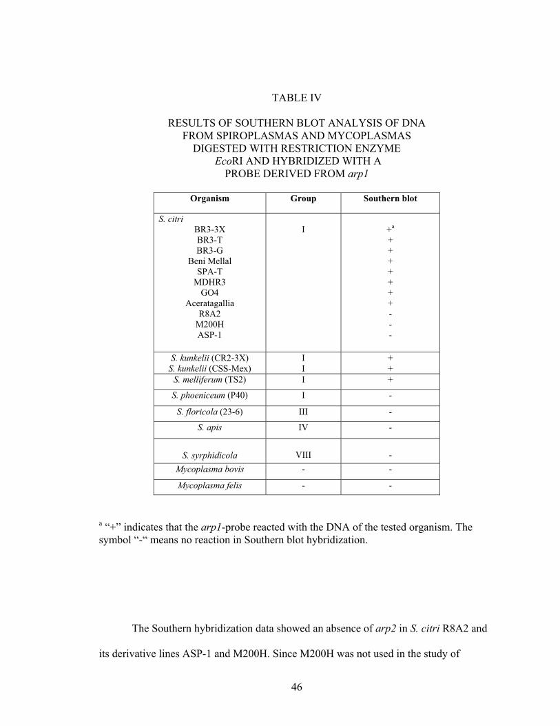
TABLE IV
RESULTS OF SOUTHERN BLOT ANALYSIS OF DNA FROM SPIROPLASMAS AND MYCOPLASMAS
DIGESTED WITH RESTRICTION ENZYME EcoRI AND HYBRIDIZED WITH A
PROBE DERIVED FROM arp1
Organism Group Southern blot
S. citri BR3-3X BR3-T BR3-G
Beni Mellal SPA-T
MDHR3 GO4
Aceratagallia R8A2
M200H ASP-1
I
+a + + + + + + + - - -
S. kunkelii (CR2-3X) S. kunkelii (CSS-Mex)
I I
+ +
S. melliferum (TS2) I +
S. phoeniceum (P40) I -
S. floricola (23-6) III -
S. apis IV -
S. syrphidicola
VIII -
Mycoplasma bovis - -
Mycoplasma felis - -
a “+” indicates that the arp1-probe reacted with the DNA of the tested organism. The symbol “-“ means no reaction in Southern blot hybridization.
The Southern hybridization data showed an absence of arp2 in S. citri R8A2 and
its derivative lines ASP-1 and M200H. Since M200H was not used in the study of

47
distribution of arp1, we do not know whether a plasmid with an arp-like sequence is
absent from that strain also. Chromosomal DNA of other S. citri strains and of other
Group I spiroplasmas, S. kunkelii and S. melliferum, did react with the probe. Out of the
several non-BR3-derived S. citri strains tested in this study, only Beni Mellal was tested
for the presence of both a plasmid and a chromosomal arp sequence. Previously [52] it
was shown that arp1 as well as any plasmid related to pBJS-O is absent from its
extrachromosomal DNA. In contrast, here we find that it has arp-related sequences in its
genome. S. phoeniceum, one of the three plant pathogenic spiroplasmas, did not react
with the probe in this study. S. apis and S. syrphidicola, two other insect-associated
spiroplasmas that belong to groups other than S. citri, also did not react with the probe in
this study and neither did the two mycoplasma isolates used here.
The non-transmissible S. citri strain, BR3-G, not only possesses arp2 in its
genome, but carries pBJS-O, the plasmid containing arp1 [52]. In contrast, S.
phoeniceum neither possesses arp1, arp2 nor any of the other regions related to the
plasmid pBJS-O. These findings suggest that genes other than arp1 and arp2 may be
involved in the transmission of these phytopathogens. Interestingly, Berho and colleagues
[40] found a putative adhesin highly similar in sequence with SARP1, P80 (designated,
Scarp4a), from S. citri GII-3 differentially expressed between the wild type and non-
transmissible strains. They had made the comparison, however, using only three non-
transmissible strains. The group also reported the presence of eight Scarp genes on six
different plasmids in S. citri GII-3. Gene duplication and recombination may have played
roles in giving rise to those genes. Furthermore, in this study S. citri Beni Mellal was
found to lack arp1-like sequences, but was found to possess arp-like sequences on its

48
chromosome, suggesting that it probably lost the extrachromosomal arp-related
sequences during its adaptation to the insect vector C. haematoceps. The differences in
the long-term maintenance regimes of the S. citri BR3-derived strains may have led to the
differences among their hybridization patterns, like the differences in protein profiles
between transmissible and non-transmissible S. citri BR3-derived lines caused by the
differences in the way S. citri BR3 was maintained [39]. Similarly, the differences in the
hybridization patterns among the S. citri and the CSS strains may reflect adaptation to
different vector and plant environments. As far as the absence of arp-related sequences
from mycoplasmas is concerned, more conclusive results may be obtained if a different
arp probe is used. Taken together, these results suggest that arp2 may be sufficient but
may not be necessary for the leafhopper transmissibility of the spiroplasmas.

49
CHAPTER IV
SPIROPLASMA CITRI ASP-1 AND R8A2 PLASMIDS: ISOLATION,
CHARACTERIZATION AND EVOLUTION
Background
There have been several reports of extrachromosomal DNAs from S. citri as well
as other spiroplasmas from ticks, insects and plants and from phytoplasmas [43; 44; 45;
46; 39; 47, 48 and 40]. Recently Joshi and coworkers [52] reported sequencing and
structural characterization of pBJS-O, an indigenous, SARP1-encoding plasmid from S.
citri BR3-3X. Plasmids have also been noted in S. kunkelii M2 [27] and Davis and
colleagues [49] completely sequenced and characterized the S. kunkelii CR2-3X plasmid,
pSKU146, which encodes an S. kunkelii homolog of SARP1, Sk-ARP1.
As mentioned in CHAPTER II, the extrachromosomal DNAs of S. citri R8A2, an
S. citri strain isolated from citrus from Morocco, and S. citri ASP-1, a non-helical
derivative of R8A2, lack arp1, the gene encoding SARP1, but possibly share sequences
with that of pBJS-O in other regions [52]. SARP1 is hypothesized to be involved in the
attachment of S. citri to the cells of its leafhopper vector C. tenellus. We hypothesized
that arp1-related sequences were deleted from a pBJS-O-like ancestral plasmid present in
S. citri R8A2 and ASP-1 during the evolution of the strains. To test this hypothesis and to
determine the extent of sequences absent from ASP-1 and R8A2 plasmids with respect to
pBJS-O, we sequenced and characterized extrachromosomal DNAs of the two strains.

50
Materials and Methods
Spiroplasmas
S. citri BR3-3X was originally isolated from brittle root diseased horseradish
plants in Illinois [12], triply cloned [25], and frozen. Leafhopper-transmissibility and
plant pathogenicity of BR3-3X was confirmed [25]. S. citri R8A2, isolated from citrus
from Morocco [70], and its non-helical derivative, ASP-1, both obtained from R.E.
Davis, USDA/ARS, Beltsville, MD, are non-transmissible. All the three S. citri strains
were grown in LD8 broth medium [73] at 310C.
Purification of Extrachromosomal DNAs
S. citri ASP-1 and R8A2 extrachromosomal double-stranded (ds) DNAs were
isolated (Figure 9) using the QIAprep Spin Miniprep Kit (QIAGEN Sciences, MD),
following the manufacturer’s protocol.
Cloning of S. citri ASP-1 and R8A2 Plasmids
Approximately 1 µg each of S. citri ASP-1 and R8A2 extrachromosomal DNAs
were digested with 20 units of TaqI (Invitrogen Corp., Carlsbad, CA) at 650C for 4 hrs

51
Figure 9. Undigested S. citri ASP-1 and R8A2 Plasmid Preparations. The marker used was High Mass Ladder (Invitrogen) and the band sizes are in Kb.
(Figure 10) and the reactions were stopped by heating at 800C for 20 min. About 15 µg of
pBluescriptTM (Stratagene, Cedar Creek, TX) vector DNA was digested with 20 units of
ClaI (GIBCOBRL, Bethesda, MD) at 37ºC for 2h and the enzyme was inactivated by
heating at 650C for 10 min. About 10 ng of the vector DNA was mixed with ~ 200 ng of
the insert DNAs and the ligations were carried out with 0.1 units of T4 DNA Ligase
10
4
A
SP-1
R8A
2
M
arke
r

52
(Promega) at 140C for about 16 hrs. The reactions were stopped by heating at 700C for 10
min. Two microliters of each reaction was mixed with 50 µl of XL2-Blue Ultracompetent
E. coli cells (Stratagene). Transformations were carried out by first keeping the tubes on
ice for 30 min, followed by a heat shock of 45 seconds at 420C and keeping the tubes
again on ice for 2 min [81]. Fresh LB broth (950 µl) was added to each tube. The
transformants were grown at 37ºC for 90 min and 50 µl of each culture was plated onto
separate LB-agar plates supplemented with 50 µg/ml ampicillin, 33 µg/ml X-gal and 8.1
µg/ml IPTG. The plates were incubated at 37ºC for 17 hrs. Individual white colonies
were streaked onto separate LB-agar plates containing 50 µg/ml ampicillin, 33 µg/ml X-
gal and 8.1 µg/ml IPTG as above, incubated at 37ºC for 17 hrs and 5 ml mid log phase
LB broth cultures, containing 50 µg/ml ampicillin, from each of the colonies were grown
at 37ºC for 13 hrs.
Sequencing of the Recombinant Clones
Cells from 3 ml of each of the LB broth cultures were harvested by centrifugation
at 15, 800 x g for 5 min at 40C and the pellets were used to prepare plasmids using the
QIAprep Spin Miniprep Kit (QIAGEN). Approximately 1 µg of each of the plasmids
were digested with 20 units of PvuII (Promega) at 37ºC for 4 hrs and electrophoresed on

53
Figure 10. TaqI restriction patterns of pBJS-O, S. citri ASP-1 extrachromosomal DNA and S. citri R8A2 extrachromosomal DNA. The marker used was High Mass Ladder (Invitrogen) and the band sizes are in Kb.
10
4
p
BJS
-O
ASP
-1
R8A
2
M
arke
r

54
a 0.75% (w/v) agarose gel with 1X TAE as running buffer to confirm the presence of
inserts. ~ 1 µg of the recombinant plasmids containing inserts of different sizes were end-
sequenced in both directions using 5 pmoles each of T3 (5'-ATT AAC CCT CAC TAA
AGG GA-3') and T7 (5'-TAA TAC GAC TCA CTA TAG GG-3') primers by the ABI
PRISM BigDye Terminator Cycle Sequencing method (version 1.0, Applied Biosystems,
Foster City, CA) and an ABI PRISM 3700 Automated DNA Analyzer (Perkin Elmer
Biosystems, Foster City, CA). The raw sequences were screened for vector contamination
using the software tool VecScreen from NCBI and the vector-free sequences were
aligned with known sequences by BLAST searches [77] at the NCBI server and by using
software tool AlignX provided by Vector NTI Suite 6.0 (Invitrogen).
Results and Discussion
Since the plasmids of S. citri ASP-1 and R8A2 do not contain arp1, we wanted to
determine the precise extent of pBJS-O sequences missing from ASP-1 and R8A2
extrachromosomal DNA. As seen before in Southern blot hybridizations of S. citri ASP-1
and R8A2 extrachromosomal DNA with pBJS-O probe [52], multiple forms of native
plasmids from the two strains were observed in this study (Figure 9). When the
extrachromosomal DNAs were treated with TaqI, their restriction patterns differed
completely from that of pBJS-O. Out of the seven restriction fragments observable from
pBJS-O, none were conserved in ASP-1 and R8A2 (Figure 10).

55
Sequence Analysis of S. citri ASP-1 and R8A2 Clones
Alignment of the preliminary nucleotide sequences of S. citri ASP-1 plasmid
clones by BLAST searches at the NCBI server revealed strong similarity with different
regions of plasmid pSciA, an approximately 8.0 kb plasmid from S. citri GII-3
[EMBL:AJ966734]. BLASTX search using one of the ASP-1 sequences also revealed
limited similarity with ORF 10 of pBJS-O and ORF 17 of pSKU146.
A dot plot analysis revealed no significant similarity between pBJS-O and pSciA
nucleotide sequences. We recently reported [52] the detection of pBJS-O-related
sequences in S. citri ASP-1 extrachromosomal DNA by Southern blot hybridization using
pBJS-O itself as a probe, but not with an arp-related probe. It is possible that some
pSciA-related sequences were also present along with pBJS-O in the probe. Furthermore,
it appears that soj-related nucleotide sequences on pBJS-O and pSciA are not similar
enough to be detected by a dot plot, although their protein products are 25% identical to
each other in sequence (E value: 2e-14). The SOJ-like protein encoded by pSciA is more
similar to its counterparts encoded by pSKU146 and by the five other pSci plasmids of S.
citri GII-3 than to the one encoded by pBJS-O. Moreover, a 35 bp GC-rich sequence was
found in pSciA upstream of the soj gene. A BLASTN search, using that sequence as a
query, detected almost all of the sequence, with more than 95% identity, in all of the five
arp-containing plasmids of S. citri GII-3 [40] and in pSKU146, in which the sequence
was found about 30 bases upstream of the palindromic region, palSK1 [49]. Interestingly,
this sequence was not detected in pBJS-O at all.
Analysis of the preliminary sequence data of S. citri R8A2 clones revealed strong
matches with a spiroplasmavirus, SVTS2, from S. citri GII-3 and S. melliferum [60 and

56
X. Foissac, unpublished data] and with plasmids pBJS-O and pSKU146 of S. citri BR3-
3X and S. kunkelii CR2-3X, indicating the presence of multiple plasmids in the
extrachromosomal DNA. The sequence from primer T3 was similar to SVTS2 of S.
melliferum, whereas the one from primer T7 was similar to the S. citri GII-3 version of
the virus. In both versions of the virus sequence, however, the R8A2 sequence was
similar to that of an open reading frame (ORF) C. In this study, multiple plasmids were
also detected in the extrachromosomal DNA of S. citri BR3-3X; the major one being a
related to plasmid pSciA of S. citri GII-3 [X. Foissac, unpublished data] and pBJS-O
being in the minority.
Spiroplasmas contain numerous virus-related sequences integrated within their genomes,
leading to extensive genome rearrangements [47]. Such events have significant impacts
on the evolution of genomes of this important group of pathogens. Four types of
spiroplasmaviruses, SpV1-4, have been identified. One genus of viruses, plectroviruses,
that are rod-shaped and have circular single-stranded DNA genomes, has its members
scattered in the S. citri genome [60]. One such virus, SpV1-C74, for instance, was found
to be a member of the insertion sequence 3 (IS3) family whose insertion had inactivated a
DNA adenine methylase gene in the host chromosome. Another one, SpV1-R8A2 B,
contained several deletions due to multiple homologous recombination events with
related sequences within the S. citri chromosome [47]. Further, we detected an SpV1-
R8A2 B-like sequence in pBJS-O [52], which may have hybridized with a related
sequence on the S. citri R8A2 extrachromosomal DNA in the Southern blot
hybridization. Thus, virus-like sequences in S. citri R8A2 extrachromosomal DNA may
have arisen by recombination with a similar sequence of the chromosomal DNA.

57
CHAPTER V
DISCUSSION AND FUTURE WORK
The relationships of chromosomal and extrachromosomal arp sequences of the
genus Spiroplasma with insect transmissibility of phytopathogenic members of the genus
are presented in this dissertation. The overall research comprises three parts that are
reported here in three separate chapters, viz. Chapter II, Chapter III and Chapter IV.
The first study was of the isolation, distribution and sequence characterization of
a novel SARP1-encoding plasmid, pBJS-O, from S. citri BR3-3X, which is reported in
CHAPTER 2. To date, pBJS-O does not belong to any known incompatibility group of
bacterial plasmids. Those findings were published in BMC Genomics in December 2005.
The second was of the distribution of an arp sequence from the chromosome of S.
citri BR3-3X, arp2, a homolog of the plasmid-borne gene, arp1, in various mollicutes. It
revealed that Spiroplasma arp sequences can be located on the chromosomal DNA and
may not be present on the extrachromosomal DNA alone. Since the arp sequences can be
present on plasmids, it would make a genetic exchange and/or a loss of the sequences
possible through horizontal DNA transfer, depending upon the selection pressures
encountered by the pathogens within the leafhopper and in the plant host. This might
explain the absence of arp-like sequences from certain phytopathogens such as S.
phoeniceum. It must be noted here that certain spiroplasma isolates used in these studies,
such as S. citri ASP-1, R8A2 and Beni Mellal, were passaged in artificial media to a high
degree here at OSU, which may have led to the loss of arp sequences from their DNAs
and to the differences in their insect transmissibilities and phytopathogenicities, as

58
compared to the original isolates. Furthermore, two separate plasmids, with sequences
similar to SVTS2 and to pBJS-O and pSKU146, were recently found in the
extrachromosomal DNA of S. citri R8A2 [Joshi et al., unpublished data], which may
have also arisen by horizontal DNA transfer and recombination. Such a mixture of
plasmids was also recently found in the extrachromosomal DNA of S. citri BR3-3X.
Contrary to the published report [52], majority of the extrachromosomal DNA is
represented by a 7 Kb plasmid related to pSciA of S. citri GII-3 and only a small, but
considerable, portion contains pBJS-O. This fact explains the observed disparities
between the in silico and in vitro restriction digestion patterns of the extrachromosomal
DNA obtained with BglII, NdeI, EcoRI and EcoRV. The exact extent of sequence
conservation among pSciA, pBJS-O, pSKU146 and the related plasmids from S. citri
BR3-3X and R8A2 is not yet known.
Finally, isolation and sequence characterization of plasmids from S. citri ASP-1
and R8A2, which lack arp1, is presented. Our data suggest that the extrachromosomal
DNAs of both ASP-1 and R8A2 have little similarity with pBJS-O. Previous data suggest
that ASP-1 does not bind the tissue-cultured cells of the leafhopper C. tenellus [F. Ye,
unpublished data]. Hence, it could be used as a host for the functional complementation
of adherence-related genes using its indigenous plasmid, pSciA, as a molecular vector.
This vector might be better than the one made from pBJS-O and the vectors currently
available for gene manipulation in S. citri. Random transposon mutagenesis of pBJS-O
was carried out to develop it into a molecular vector for molecular genetic applications in
S. citri, first using pMOD-5 (Epicentre Biotechnologies, Madison, WI), a cloning vector
containing a Tn5-derived transposon having an oriC and a multiple cloning site (MCS),

59
for amplification in Escherichia coli and then using R6Κγοri/KAN-2 (Epicentre), another
Tn5-derived transposon, but containing a kanamycin-resistance marker gene, used for in
vitro insertion into target DNA. With the first approach, insertion of the transposon into
pBJS-O failed, possibly because of damage to the transposase recognition sequences due
to overexposure to UV during gel-extraction of the free transposon, whereas unusual
topologies of pBJS-O molecules inhibited transposon insertion with the other approach.
Thus, overall the data presented in this dissertation suggest that Spiroplasma arp
genes can be distributed over chromosomal as well as extrachromosomal DNAs and
SARP1 may be sufficient, but not necessary, for S. citri transmission by C. tenellus. None
of the genes, however, is solely necessary for the transmission of the pathogens by
leafhoppers. Genes other than arp1 and arp2 may also be involved in the process and
more work needs to be done to utilize spiroplasma plasmids as vectors for gene
manipulations in mollicutes. Finally, it can be said that differences in hosts, geographical
niches and maintenance regimes of spiroplasmas can lead to differences in their plasmid
content.

60
REFERENCES
1. Woese CR: Bacterial evolution. Microbiol Rev 1987, 51:221-271.
2. Garnier M, Foissac X, Gaurivaud P, Laigret F, Renaudin J, Saillard C, Bové JM:
Mycoplasmas, plants, insect vectors: a matrimonial triangle. C R Acad Sci
Paris 2001, 324:923-928.
3. Yamao F, Muto A, Kawauchi Y, Iwami M, Iwagami S, Azumi Y, Osawa S: UGA is
read as tryptophan in Mycoplasma capricolum. Proc Natl Acad Sci U S A 1985,
82:2306-2309.
4. Stamburski C, Renaudin J, Bové JM: First step toward a virus-derived vector for
gene cloning and expression in spiroplasmas, organisms which read UGA as a
tryptophan codon: synthesis of chloramphenicol acetyltransferase in Spiroplasma
citri. J Bacteriol 1991, 173:2225-2230.
5. Davis RE, Worley JF: Spiroplasma: Motile, helical microorganisms associated with
corn stunt disease. Phytopathology 1973, 63:403-408.
6. Fudl-Allah A. E-SA, Calavan EC: Effect of temperature and pH on growth in vitro
of a mycoplasmalike organism associated with stubborn disease of citrus.
Phytopathology 1973, 63:256-259.
7. Tully JG, Whitcomb RF, Clark HF, Williamson DL: Pathogenic mycoplasmas:
cultivation and vertebrate pathogenicity of a new spiroplasma. Science 1977,
195:892-894.
8. McGarrity GJ, Megraud F, Gamon L: Rabbit lens cell cultures in the
characterization of Spiroplasma mirum pathogenicity. Ann Microbiol (Paris) 1984,
135A:249-254.

61
9. Chastel C, Le Goff F, Humphery-Smith I: Multiplication and persistence of
Spiroplasma melliferum strain A56 in experimentally infected suckling mice. Res
Microbiol 1991, 142:411-417.
10. Saglio P, Lhospital M, Lafleche D, Dupont G, Bové JM, Tully JG, Freundt EA:
Spiroplasma citri gen. and sp. n.: a Mycoplasma-like Organism Associated
with “Stubborn” Disease of Citrus. Int J Syst Bacteriol 1973, 23:191-204.
11. Calavan EC, Bové JM: Ecology of Spiroplasma citri. In The Mycoplasmas.
Volume 5. Edited by Whitcomb RF and Tully JG. New York: Academic Press;
1989:425-485.
12. Fletcher J, Schultz GA, Davis RE, Eastman CE, Goodman RE: Brittle root
disease of horseradish: Evidence for an etiological role of Spiroplasma citri.
Phytopathology 1981, 71:1073–1080.
13. Chen TA, Liao CH: Corn stunt spiroplasma: isolation, cultivation, and proof
of pathogenicity. Science 1975, 188:1015-1017.
14. Williamson DL, Whitcomb RF: Plant mycoplasmas: a cultivable spiroplasma
causes corn stunt disease. Science 1975, 188:1018-1020.
15. Whitcomb RF, Chen TA, Williamson DL, Liao C, Tully JG, Bové JM, Mouches
C, Rose DL, Coan ME, Clark TB: Spiroplasma kunkelii sp. nov.:
Characterization of the Etiological Agent of Corn Stunt Disease. Int J Syst
Bact 1986, 36:170-178.
16. Saillard C, Vignault JC, Gadeau A, Carle P, Garnier M, Fos A, Bové JM, Tully
JG, Whitcomb RF: Discovery of a new plant-pathogenic spiroplasma. Isr J
Med Sci 1984, 20:1013-1015.

62
17. McCoy RE, Caudwell A, Chang CJ, Chen TA, Chiykowski LN, Cousin MT, Dale
JL, deLeeuw GTN, Golino DA, Hackett KJ, Kirkpatrick BC, Marwitz R, Petzold
H, Sinha RC, Sugiura M, Whitcomb RF, Yang IL, Zhu BM and Seemûller E:
Plant diseases associated with mycoplasma-like organisms. In The
Mycoplasmas. Volume 5. Edited by Whitcomb RF and Tully JG. New York:
Academic Press; 1989:545-640.
18. Wayne LG, Brenner DJ, Colwell RR, Grimont AD, Kandler O, Krichevsky MI,
Moore LH, Moore WEC, Murray RGE, Stackebrant E, Starr MP, Truper HG: Report of
the ad hoc committee on reconciliation of approaches to bacterial systematics. Int J
Syst Bact 1987, 37:463-464.
19. Williamson DL, Whitcomb RF, Tully JG, Gasparich GE, Rose DL, Carle P, Bové
JM, Hackett KJ, Adams JR, Henegar RB, Konai M, Chastel C, French FE: Revised
group classification of the genus Spiroplasma. Int J Syst Bact 1998, 48:1-12.
20. Gasparich GE: Spiroplasmas: evolution, adaptation and diversity. Front Biosci
2002, 7:d619-640.
21. Whitcomb RF, Tully JG, Bové JM, Saglio P: Spiroplasmas and acholeplasmas:
multiplication in insects. Science 1973, 182:1251-1253.
22. Bové JM, Renaudin J, Saillard C, Foissac X, Garnier M: Spiroplasma citri, a
plant pathogenic molligute: relationships with its two hosts, the plant and the
leafhopper vector. Annu Rev Phytopathol 2003, 41:483-500.
23. Williamson DL, Tully JG, Whitcomb RF: The genus Spiroplasma. In The
Mycoplasmas. Edited by Whitcomb RF and Tully JG. New York: Academic Press;
1989:71-112.

63
24. Steiner T, McGarrity GJ, Bové JM, Phillips DM, Garnier M: Insect cell cultures
in the study of attachment and pathogenicity of spiroplasmas and
mycoplasmas. Ann Microbiol (Paris) 1984, 135A:47-53.
25. Wayadande AC, Fletcher J: Transmission of Spiroplasma citri lines and their
ability to cross gut and salivary gland barriers within the leafhopper vector
Circulifer tenellus. Phytopathology 1995, 85:1256–1259.
26. Ozbek E, Miller SA, Meulia T, Hogenhout SA: Infection and replication sites of
Spiroplasma kunkelii (Class: Mollicutes) in midgut and Malpighian tubules of the
leafhopper Dalbulus maidis. J Invertebr Pathol 2003, 82:167-175.
27. Bai X, Fazzolari T, Hogenhout SA: Identification and characterization of traE
genes of Spiroplasma kunkelii. Gene 2004, 336:81-91.
28. Bové JM: Wall-less prokaryotes of plants. Ann Rev Phytopathol 1984, 22:361-
396.
29. Kwon M.-O, Wayadande AC, Fletcher J: Spiroplasma citri movement into the
intestines and salivary glands of its leafhopper vector, Circulifer tenellus.
Phytopathology 1999, 89:1144–1151.
30. Townsend R, Markham PG, Plaskitt KA: Multiplication and morphology of
Spiroplasma citri in the leafhopper Euscelidius variegatus. Ann Appl Biol 1977,
87:307-313.
31. Fletcher J, Wayadande A, Melcher U, Ye F: The phytopathogenic mollicute-insect
vector interface: a closer look. Phytopathology 1998, 88:1351–1358.
32. Yu J, Wayadande AC, Fletcher J: Spiroplasma citri surface protein P89
implicated in adhesion to cells of the vector Circulifer tenellus. Phytopathology

64
2000, 90:716–722.
33. Razin S, Jacobs E: Mycoplasma adhesion. J Gen Microbiol 1992, 138:407–422.
34. Baseman JB, Reddy SP, Dallo SF: Interplay between mycoplasma surface
proteins, airway cells, and the protean manifestations of mycoplasma-mediated
human infections. Am J Respir Crit Care Med 1996, 154:137-144.
35. Krause DC: Mycoplasma pneumoniae cytadherence: unravelling the tie that
binds. Mol Microbiol 1996, 20:247-253.
36. Fletcher J, Wills JW, Denman SE: Identification of surface proteins of
Spiroplasma citri with protease and antibody probes. Curr Microbiol 1989, 19:383-
391.
37. Ye F, Melcher U, Rascoe JE, Fletcher J: Extensive chromosome aberrations in
Spiroplasma citri strain BR3. Biochem Genet 1996, 34:269–286.
38. Ye F, Melcher U, Fletcher J: Molecular characterization of a gene encoding a
membrane protein of Spiroplasma citri. Gene 1997, 189:95-100.
39. Fletcher J, Shaw ME, Baker GR, Dugan KJ, Ye F, Sha Y, Zuck PD, Myers GD:
Molecular characterization of Spiroplasma citri BR3 lines that differ in
transmissibility by the leafhopper Circulifer tenellus. Can J Microbiol 1996,
42:124-131.
40. Berho N, Duret S, Renaudin J: Absence of plasmids encoding adhesion-related
proteins in non-insect-transmissible strains of Spiroplasma citri. Microbiology 2006,
152:873-886.
41. Berg M, Melcher U, Fletcher J: Characterization of Spiroplasma citri adhesion
related protein SARP1, which contains a domain of a novel family designated

65
sarpin. Gene 2001, 275:57-64.
42. Killiny N, Batailler B, Foissac X, Saillard C: Identification of a Spiroplasma citri
hydrophilic protein associated with insect transmissibility. Microbiology 2006,
152:1221-1230.
43. Archer DB, Best J, Barber C: Isolation and Restriction Mapping of a
Spiroplasma Plasmid. J Gen Microbiol 1981, 126:511-514.
44. Barber CE, Archer, DB, Daniels MJ: Molecular biology of spiroplasma
plasmids. Yale J Biol Med 1983, 56:777-781.
45. Mouches C, Barroso G, Gadeau A, Bové JM: Characterization of two cryptic
plasmids from Spiroplasma citri and occurrence of their DNA sequences
among various spiroplasmas. Ann Microbiol (Paris) 1984, 135A:17-24.
46. Gasparich GE, Hackett KJ, Clark EA, Renaudin J, Whitcomb RF: Occurrence of
extrachromosomal deoxyribonucleic acids in spiroplasmas associated with plants,
insects, and ticks. Plasmid 1993, 29:81-93.
47. Melcher U, Fletcher J: Genetic variation in Spiroplasma citri. Eur J Plant
Pathol 1999, 105:519-533.
48. Liefting LW, Shaw ME, Kirkpatrick BC: Sequence analysis of two plasmids from
the phytoplasma beet leafhopper-transmitted virescence agent. Microbiology 2004,
150:1809-17.
49. Davis RE, Dally EL, Jomantiene R, Zhao Y, Roe B, Lin SP, Shao J: Cryptic
plasmid pSKU146 from the wall-less plant pathogen Spiroplasma kunkelii
encodes an adhesin and components of a type IV translocation-related
conjugation system. Plasmid 2005, 53:179-190.

66
50. Nishigawa H, Oshima K, Kakizawa S, Jung HY, Kuboyama T, Miyata S, Ugaki M,
Namba S: A plasmid from a non-insect-transmissible line of a phytoplasma lacks two
open reading frames that exist in the plasmid from the wild-type line. Gene 2002,
298:195-201.
51. Bergemann AD, Whitley JC, Finch LR: Homology of mycoplasma plasmid
pADB201 and staphylococcal plasmid pE194. J Bacteriol 1989, 171:593-5.
52. Joshi BD, Berg M, Rogers J, Fletcher J, Melcher U: Sequence comparisons of
plasmids pBJS-O of Spiroplasma citri and pSKU146 of S. kunkelii: implications for
plasmid evolution. BMC Genomics 2005, 6:175.
53. Fleury B, Bergonier D, Berthelot X, Peterhans E, Frey J, Vilei EM:
Characterization of P40, a cytadhesin of Mycoplasma agalactiae. Infect Immun
2002, 70:5612-5621.
54. Rascoe J, Melcher U, Fletcher J: Evaluation of variations in gene presence and
expression among lines of Spiroplasma citri using arbitrarily-primed PCR of
cDNA. Phytopathology 1996, 86:S96.
55. Ye F, Laigret F, Carle P, Bové JM: Chromosomal heterogeneity among various
strains of Spiroplasma citri. Int J Syst Bact 1995, 45:729-734.
56. Sha Y, Melcher U, Davis RE, Fletcher J: Resistance of Spiroplasma citri lines to
the virus SVTS2 is associated with integration of viral DNA sequences into
host chromosomal and extrachromosomal DNA. Appl Environ Microbiol 1995,
61:3950-3959.
57. Melcher U, Sha Y, Ye F, Fletcher J: Mechanisms of spiroplasma genome
variation associated with SpV1-like viral DNA inferred from sequence

67
comparisons. Microb Comp Genomics 1999, 4:29–46.
58. Quisel JD, Lin DC, Grossman AD: Control of development by altered
localization of a transcription factor in B. subtilis. Mol Cell 1999, 4:665-672.
59. Ireton K, Gunther IV NW, Grossman AD: spoOJ is required for normal
chromosome segregation as well as the initiation of sporulation in Bacillus
subtilis. J Bacteriol 1994, 176:5320-5329.
60. Sha Y, Melcher U, Davis RE, Fletcher J: Common elements of spiroplasma
plectroviruses revealed by nucleotide sequence of SVTS2. Virus Genes 2000,
20:47-56.
61. Winans SC, Walker GC: Conjugal transfer system of the N incompatibility
plasmid pKM101. J Bacteriol 1985, 161:402–410.
62. Clewell D, Helinski D: Supercoiled circular DNA-protein complex in
Escherichia coli: purification and induced conversion to an open circular
DNA form. Proc Natl Acad Sci U S A 1969, 62:1159-1166.
63. Sha Y.-S: Molecular characterization of spiroplasma viruses and the
mechanism of resistance of Spiroplasma citri lines to infection by the virus
SVTS2. PhD thesis. Oklahoma State University, Entomology and Plant Pathology
Department; 1993.
64. Nojiri H, Shintani M, Omori T: Divergence of mobile genetic elements involved
in the distribution of xenobiotic-catabolic capacity. Appl Microbiol Biotechnol
2004, 64:154-174.
65. Renaudin J: Extrachromosomal elements and gene transfer. In Molecular
Biology and Pathogenicity of Mycoplasmas. Edited by Razin S and Herrmann R.

68
New York: Kluwer Academic Publishers/Plenum Press; 2002:347-370.
66. Renaudin J, Bové JM: Plasmid and viral vectors for gene cloning and
expression in Spiroplasma citri. In Molecular and Diagnostic Procedures in
Mycoplasmology. Edited by Razin S and Tully JG. New York: Academic Press;
1995:167–178.
67. Renaudin J, Marais A, Verdin E, Duret S, Foissac X, Laigret F, Bové JM:
Integrative and free Spiroplasma citri oriC plasmids: expression of the
Spiroplasma phoeniceum spiralin in Spiroplasma citri. J Bacteriol 1995,
177:2800–2877
68. Ye F, Renaudin J, Bové JM, Laigret F: Cloning and sequencing of the
replication origin (oriC) of the Spiroplasma citri chromosome and
construction of autonomously replicating artificial plasmids. Curr Microbiol
1994, 29:23–29.
69. Duret S, Danet JL, Garnier M, Renaudin J: Gene disruption through
homologous recombination in Spiroplasma citri: an scm1-disrupted motility
mutant is pathogenic. J Bacteriol 1999, 181:7449-7456.
70. Saglio P, Lafleche D, Bonissol C, Bové JM: Culture in vitro des mycoplasmes
associés au stubborn des agrumes et leur observation au microscope
électronique. C R Acad Sci Paris 1971, 272:1387–1390.
71. Davis RE, Lee I.-M, Basciano LK: Spiroplasmas: serological grouping of strains
associated with plants and insects. Can J Microbiol 1979, 25:861-866.
72. Zhao Y, Hammond RW, Jomantiene R, Dally EL, Lee I.-M, Jia H, Wu H, Lin S,
Zhang P, Kenton S, Najar FZ, Hua A, Roe BA, Fletcher J, Davis RE: Gene

69
content and organization of an 85-kbp DNA segment from the genome of the
phytopathogenic mollicute Spiroplasma kunkelii. Molec Genet Genom 2003,
269: 592-602.
73. Chen TA, Davis RE: Cultivation of spiroplasmas. In The Mycoplasmas. Edited
by Whitcomb RF and Tully JG. New York: Academic Press; 1979:65–79.
74. Lee I.-M, Davis RE: Serum-free media for cultivation of spiroplasmas. Can J
Microbiol 1989, 35:1092-1099.
75. Murray MG, Thompson WF: Rapid isolation of high molecular weight plant
DNA. Nucleic Acids Res 1980, 8:4321-4325.
76. Ayoubi P, Jin X, Leite S, Liu X, Martajaja J, Abduraham A, Wan Q, Yan W,
Misawa E, Prade RA: PipeOnline 2.0: automated EST processing and
functional data sorting. Nucleic Acids Res 2002, 30:4761-4769.
77. Altschul SF, Gish W, Miller W, Myers EW, Lipman DJ: Basic local alignment
search tool. J Mol Biol 1990, 215:403-410.
78. Chandrasekaran A: Distribution of P58-like genes among mollicutes. MS thesis.
Oklahoma State University, Biochemistry and Molecular Biology Department; 2000.
79. Friis NF: Some recommendations concerning primary isolation of Mycoplasma
suipneumoniae and Mycoplasma flocculare a survey. Nord Vet Med 1975, 27:337-339.
80. Sambrook J, Fritsch EF, Maniatis T: Molecular Cloning: A Laboratory Manual.
Cold Spring Harbor: Cold Spring Harbor Laboratory Press; 1989.
81. Hanahan D: Studies on transformation of Escherichia coli with plasmids. J Mol
Biol 1983, 166:557-580.
S. kunkelii Genome Project [http://www.genome.ou.edu/spiro.html]

70
ORF Finder [http://www.ncbi.nlm.nih.gov/gorf/gorf.html]
Biology Workbench [http://workbench.sdsc.edu/]

VITA
Bharat Dilip Joshi
Candidate for the Degree of
Doctor of Philosophy Thesis: SPIROPLASMA ARP SEQUENCES: RELATIONSHIPS WITH
EXTRACHROMOSOMAL ELEMENTS Major Field: Biochemistry and Molecular Biology Biographical: Personal Data: Born in Pune, Maharashtra, India on October 21, 1974, the son of Dr. Mrs. Mangala Dilip Joshi and Dr. Dilip Avadhoot Joshi. Education: Received the Bachelor of Science Degree in Zoology from the University of Pune, India, in 1995; received the Masters in Science Degree in Zoology from the University of Pune, India, in 1997; completed requirements for the Doctor of Philosophy Degree at Oklahoma State University in July, 2006.
Professional Experience:
Spring 2000-Summer 2006 Graduate Research Assistant, department of biochemistry and molecular biology. Spring and Summer 2005 Supervisor, freshman research scholar working on restriction mapping of the plasmid pBJS-O. Fall 2003, 2004 and 2005 Teaching Assistant, graduate microbiology and molecular genetics laboratory course. Summer 2003 Supervisor, undergraduate intern carrying out biochemical techniques such as plasmid isolation, agarose gel electrophoresis, bacterial transformation by electroporation, in vitro cultivation of bacteria, Quick- Check method to test sizes of plasmids, PCR and ELISA. Fall 1999 Graduate Teaching Assistant, departmental undergraduate biochemistry and molecular biology laboratory course.
Honors and Organizations: Member of the American Association for the
Advancement of Science, the American Society for Microbiology, and the American Phytopathological Society

Name: Bharat D. Joshi Date of Degree: July, 2006
Institution: Oklahoma State University Location: Stillwater, OK
Title of Study: SPIROPLASMA ARP SEQUENCES: RELATIONSHIPS WITH
EXTRACHROMOSOMAL ELEMENTS
Pages in Study: 70 Candidate for the Degree of Doctor of Philosophy
Major Field: Biochemistry and Molecular Biology
Scope and Method of Study: Spiroplasmas, members of Class Mollicutes, are scattered
over wide host and geographic niches. Those which are plant pathogenic, such as
Spiroplasma citri, are transmitted from plant to plant by homopteran insect vectors,
leafhoppers, and cause devastating damages to several crops world-wide. A variety of
model systems are available to study the transmission of phytopathogenic spiroplasmas
by leafhoppers, their predominant vectors. Spiroplasma citri-Circulifer tenellus is one
such system. Because spiroplasmas can be cultured in artificial media, they offer an
excellent means to investigate the complex molecular interactions underlying the
transmission of S. citri by the insect vector C. tenellus. Evidence suggests that an S. citri
membrane protein, spiroplasma adhesion related protein 1 (SARP1), is involved in the
adhesion of S. citri cells to tissue-cultured C. tenellus cells and, thus, is thought to be a
putative adhesin. The gene encoding SARP1 (arp1) has been isolated, cloned and
characterized. In this study, Southern blot hybridizations, PCR, cloning, sequencing and
sequence comparisons of S. citri chromosomal and extrachromosomal DNAs were
performed to investigate the relationships of the arp genes present on them with insect
transmissibility and plant pathogenicity of various members of Class Mollicutes.

Findings and Conclusions: In this dissertation, arp1 is shown to be present on a novel,
indigenous plasmid, pBJS-O, in S. citri BR3-3X. Furthermore, evidence of arp1-related
sequences on the chromosomal DNA in certain spiroplasmas and the partial
characterization of plasmids from two S. citri strains that lack arp1, S. citri ASP-1 and
R8A2, is presented. The presence of multiple plasmids in the extrachromosomal DNAs of
S. citri BR3-3X and R8A2 is also shown. Taken together, the data presented suggest that
SARP1 may be sufficient, but not necessary, for S. citri transmission by C. tenellus and
the differences in host species, geographical niches and maintenance regimes of
spiroplasmas can lead to differences in their plasmid content.
ADVISER’S APPROVAL: Dr. Ulrich Melcher