spinal disorders pfn: somool08 - jsomtcslides.jsomtc.org/somool08/somool08.pdf · spinal tap (lp)...
TRANSCRIPT
1
Slide 1JSOMTC, SWMG(A)
Spinal DisordersPFN: SOMOOL08
Hours: 2.5
Slide 2JSOMTC, SWMG(A)
Terminal Learning Objective
Action: Communicate knowledge of “Spinal Disorders”
Condition: Given a lecture in a classroom environment
Standard: Received a minimum score of 75% on the written exam IAW course standards
Slide 3JSOMTC, SWMG(A)
References
Pathophysiology for the Health Professions, 2011, 4th edition
Merck Manual, 20011, 19th edition
Current Medical Diagnosis & Treatment, 2012, 51st Edition
Special Operations Forces Medical Handbook, 2008 edition
Sanford Guide to Antimicrobial Therapy
2
Slide 4JSOMTC, SWMG(A)
Reason
Slide 5JSOMTC, SWMG(A)
Agenda
Communicate common disorders of the spine
Communicate referral decisions and red flags
Recall the gross anatomy of the spine and related terminology
Slide 6JSOMTC, SWMG(A)
Agenda
Communicate the signs and symptoms, physical exam findings, diagnostic tests, and management of cauda equinasyndrome
Communicate the signs and symptoms, physical exam findings, and management of cervical spondylosis

3
Slide 7JSOMTC, SWMG(A)
Agenda
Communicate the signs and symptoms, physical exam findings, diagnostic tests, and management of traumatic cervical spine disorders, to include sprain, radiculopathy, and fractures
Slide 8JSOMTC, SWMG(A)
Agenda
Communicate the signs and symptoms, physical exam findings, diagnostic tests, and management of spinal cord shock
Communicate the management of fractures to the thoracic and lumbar spine
Communicate the management of low back pain
Slide 9JSOMTC, SWMG(A)
Agenda
Communicate the management of lumbar degenerative disk disease and chronic low back pain
Communicate the management of lumbar herniated disk
Communicate the signs and symptoms, physical exam findings, diagnostic tests, and management of spondylolisthesis
4
Slide 10JSOMTC, SWMG(A)
Agenda
Communicate the management of acute and chronic infections of the spine
Identify common metabolic causes of back pain
Communicate the relationship between metastatic disease and back pain
Communicate common disorders associated with abnormal curvature of the spine
Slide 11JSOMTC, SWMG(A)
Common Disorders of the Spine
Slide 12JSOMTC, SWMG(A)
Common Spine Disorders
Overview
Common causes of back pain
• Strains and sprains
• Degenerative causes
• Herniated disk
• Spondylolisthesis
• Fractures
• Spinal deformities
What else could it be?
5
Slide 13JSOMTC, SWMG(A)
Common Spine Disorders
The diagnostic challenge – red flags
History
Physical exam
Diagnostic tests
Treatment options
Slide 14JSOMTC, SWMG(A)
History
Start with a good history
•Mechanism of injury
• Historical factors
• Certain qualities of back pain suggest the diagnosis
• Associated symptoms are important!
OPQRST of the pain/duration of symptoms
Any problems with bowel or bladder?
Common Spine Disorders
Slide 15JSOMTC, SWMG(A)
Common Spine Disorders
Type of pain/pattern
Location of pain
Deformity
Trauma
Gender
6
Slide 16JSOMTC, SWMG(A)
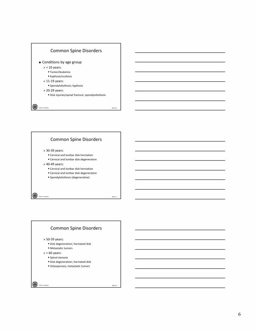
Common Spine Disorders
Conditions by age group
< 10 years:
• Tumor/leukemia
• Kyphosis/scoliosis
11‐19 years:
• Spondylolisthesis; kyphosis
20‐29 years:
• Disk injuries/spinal fracture; spondylolisthesis
Slide 17JSOMTC, SWMG(A)
Common Spine Disorders
30‐39 years:
• Cervical and lumbar disk herniation
• Cervical and lumbar disk degeneration
40‐49 years:
• Cervical and lumbar disk herniation
• Cervical and lumbar disk degeneration
• Spondylolisthesis (degenerative)
Slide 18JSOMTC, SWMG(A)
Common Spine Disorders
50‐59 years:
• Disk degeneration; herniated disk
•Metastatic tumors
> 60 years:
• Spinal stenosis
• Disk degeneration; herniated disk
• Osteoporosis; metastatic tumors
7
Slide 19JSOMTC, SWMG(A)
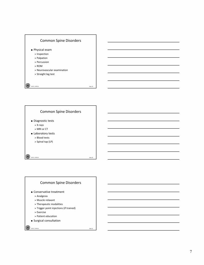
Common Spine Disorders
Physical exam
Inspection
Palpation
Percussion
ROM
Neurovascular examination
Straight leg test
Slide 20JSOMTC, SWMG(A)
Common Spine Disorders
Diagnostic tests
X‐rays
MRI or CT
Laboratory tests
Blood tests
Spinal tap (LP)
Slide 21JSOMTC, SWMG(A)
Common Spine Disorders
Conservative treatment
Analgesia
Muscle relaxant
Therapeutic modalities
Trigger point injections (if trained)
Exercise
Patient education
Surgical consultation
8
Slide 22JSOMTC, SWMG(A)
Referral Decisions and Red Flags
Slide 23JSOMTC, SWMG(A)
Referral Decisions / Red Flags
Age under 17 or over 55
Violent trauma (e.g., fall from twice height of patient)
Significant nighttime pain or pain @ rest
Bilateral or progressive neurologic deficit
Bowel or bladder control problems
Slide 24JSOMTC, SWMG(A)
Referral Decisions / Red Flags
PMH – Ca, steroids, HIV or drug abuse
Unwell: fever, chills, unexplained wt. loss
Nerve root pain that is not resolving after 6 weeks
Patients whose symptoms are getting worse despite treatment

9
Slide 25JSOMTC, SWMG(A)
The Gross Anatomy of the Spine and Related Terminology
Slide 26JSOMTC, SWMG(A)
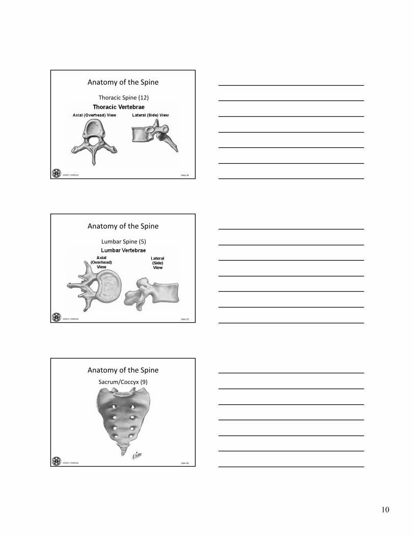
Anatomy of the Spine
Slide 27JSOMTC, SWMG(A)
Anatomy of the Spine
Cervical Spine (7)
10
Slide 28JSOMTC, SWMG(A)
Anatomy of the Spine
Thoracic Spine (12)
Slide 29JSOMTC, SWMG(A)
Anatomy of the Spine
Lumbar Spine (5)
Slide 30JSOMTC, SWMG(A)
Sacrum/Coccyx (9)
Anatomy of the Spine
11
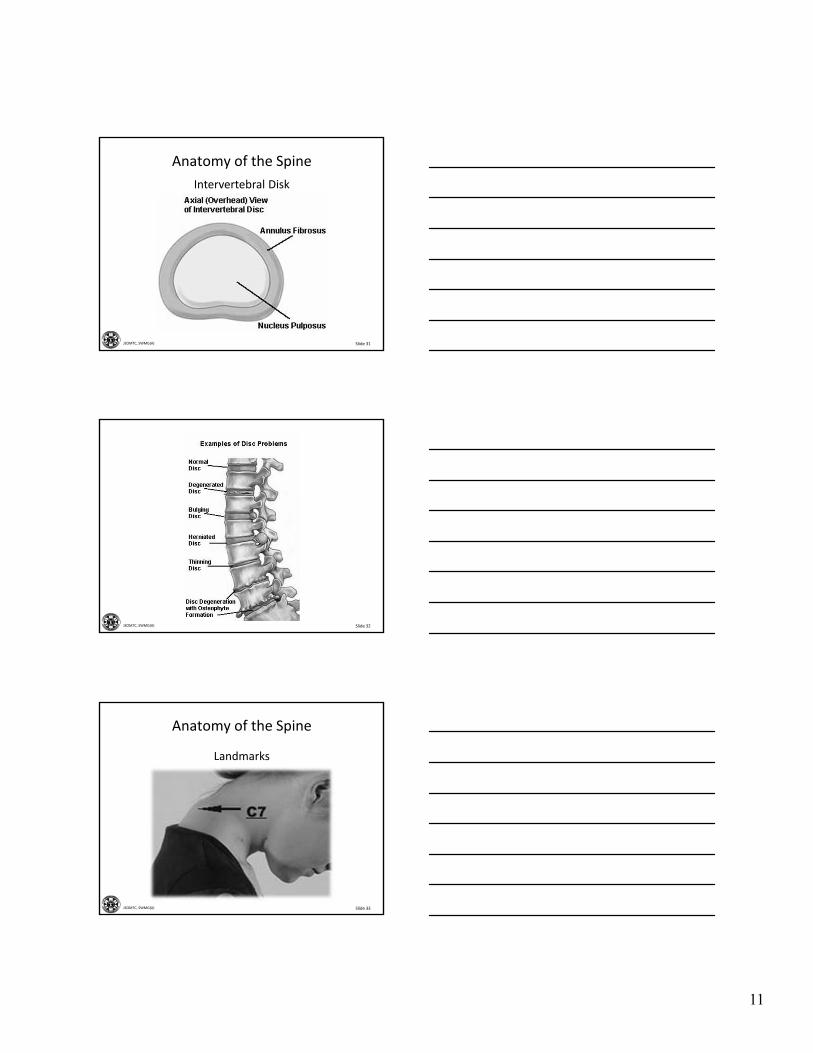
Slide 31JSOMTC, SWMG(A)
Intervertebral Disk
Anatomy of the Spine
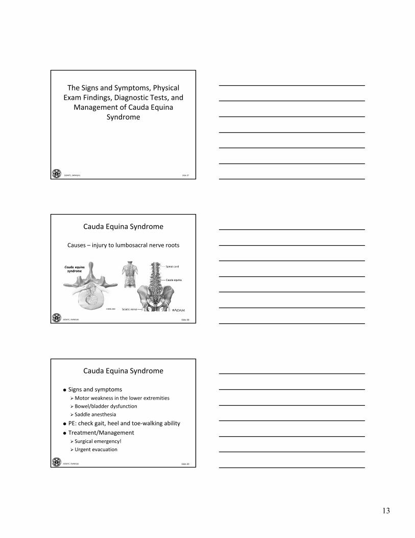
Slide 32JSOMTC, SWMG(A)
Slide 33JSOMTC, SWMG(A)
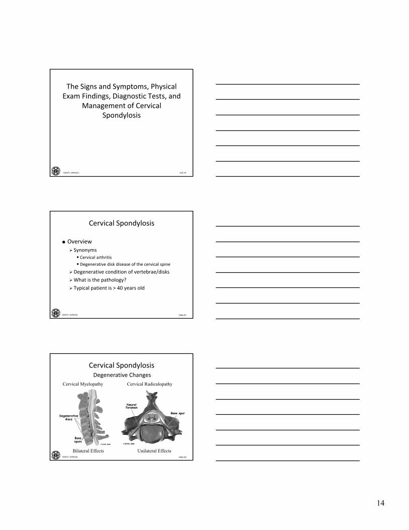
Anatomy of the Spine
Landmarks

12
Slide 34JSOMTC, SWMG(A)
Anatomy of the Spine
Slide 35JSOMTC, SWMG(A)
Terminology
Paresthesias – abnormal sensations
Paralysis – loss of sensation, anesthesia, loss of purposefulmovement
Paresis – partial or incomplete paralysis
Paraplegia – paralysis of the lower portion of the body and of both legs
Quadriplegia – paralysis of four extremities
Radiculopathy – irritation of a spinal nerve root; produces pain, weakness, numbness
Slide 36JSOMTC, SWMG(A)
Terminology
Spondylosis – degenerative arthritis, osteoarthritis, of the cervical or lumbar vertebrae
Spondylolysis – stress fracture, in one of the vertebra (usually @ L5
Spondylolisthesis – weakness in the bone so that it is unable to maintain its proper position and may slip out of place
13
Slide 37JSOMTC, SWMG(A)
The Signs and Symptoms, Physical Exam Findings, Diagnostic Tests, and
Management of Cauda EquinaSyndrome
Slide 38JSOMTC, SWMG(A)
Cauda Equina Syndrome
Causes – injury to lumbosacral nerve roots
Slide 39JSOMTC, SWMG(A)
Cauda Equina Syndrome
Signs and symptoms
Motor weakness in the lower extremities
Bowel/bladder dysfunction
Saddle anesthesia
PE: check gait, heel and toe‐walking ability
Treatment/Management
Surgical emergency!
Urgent evacuation
14
Slide 40JSOMTC, SWMG(A)
The Signs and Symptoms, Physical Exam Findings, Diagnostic Tests, and
Management of Cervical Spondylosis
Slide 41JSOMTC, SWMG(A)
Cervical Spondylosis
Overview
Synonyms
• Cervical arthritis
• Degenerative disk disease of the cervical spine
Degenerative condition of vertebrae/disks
What is the pathology?
Typical patient is > 40 years old
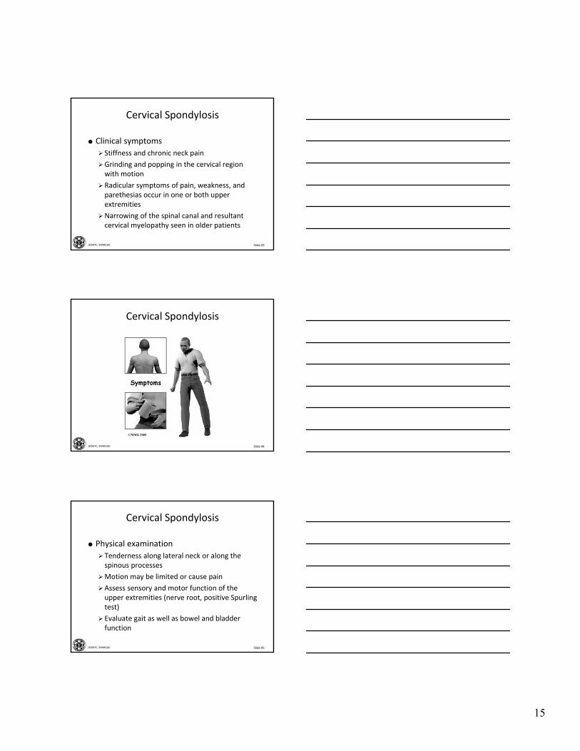
Slide 42JSOMTC, SWMG(A)
Degenerative Changes
Cervical Spondylosis
Bilateral Effects Unilateral Effects
Cervical Myelopathy Cervical Radiculopathy
15
Slide 43JSOMTC, SWMG(A)
Cervical Spondylosis
Clinical symptoms
Stiffness and chronic neck pain
Grinding and popping in the cervical region with motion
Radicular symptoms of pain, weakness, and parethesias occur in one or both upper extremities
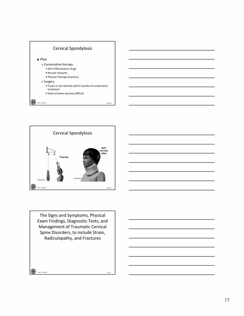
Narrowing of the spinal canal and resultant cervical myelopathy seen in older patients
Slide 44JSOMTC, SWMG(A)
Cervical Spondylosis
Slide 45JSOMTC, SWMG(A)
Cervical Spondylosis
Physical examination
Tenderness along lateral neck or along the spinous processes
Motion may be limited or cause pain
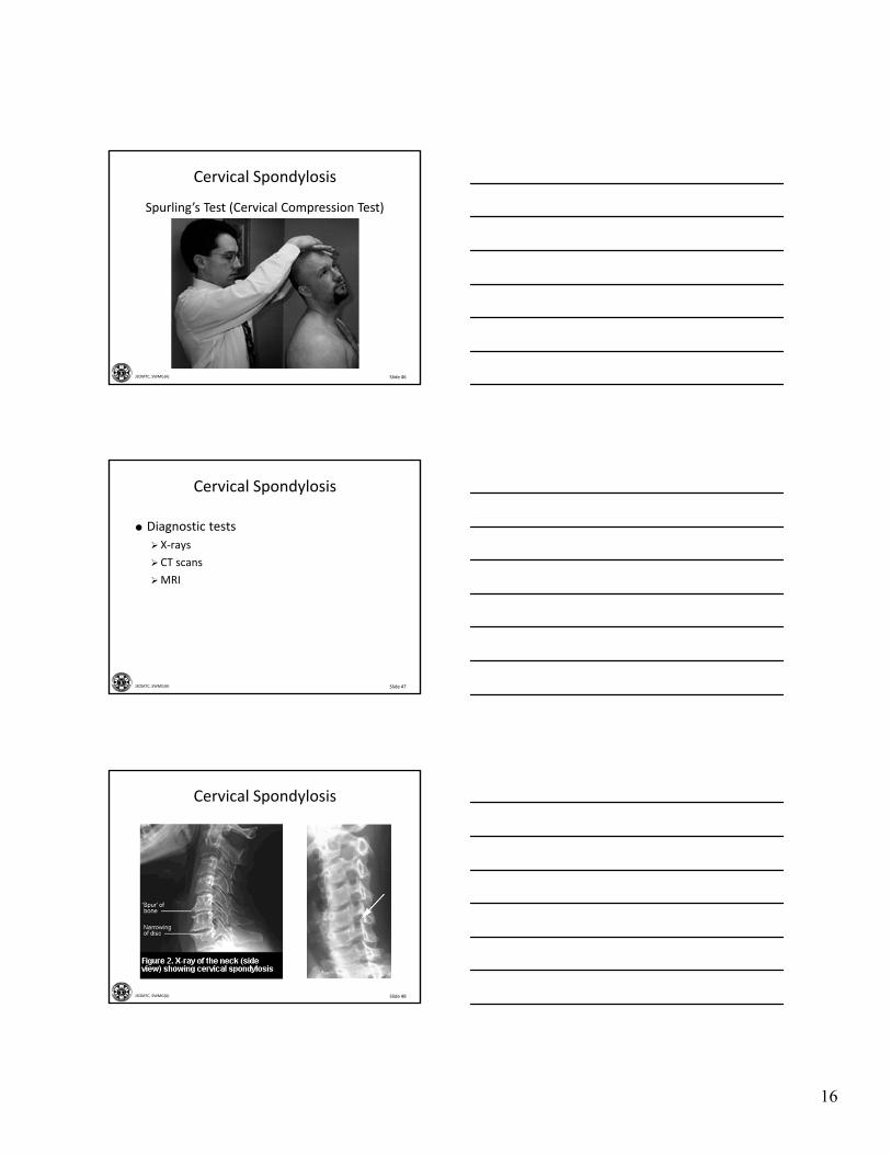
Assess sensory and motor function of the upper extremities (nerve root, positive Spurlingtest)
Evaluate gait as well as bowel and bladder function
16
Slide 46JSOMTC, SWMG(A)
Cervical Spondylosis
Spurling’s Test (Cervical Compression Test)
Slide 47JSOMTC, SWMG(A)
Cervical Spondylosis
Diagnostic tests
X‐rays
CT scans
MRI
Slide 48JSOMTC, SWMG(A)
Cervical Spondylosis
17
Slide 49JSOMTC, SWMG(A)
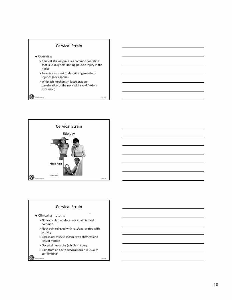
Cervical Spondylosis
Plan
Conservative therapy
• Anti‐inflammatory drugs
•Muscle relaxants
• Physical Therapy (traction)
Surgery
• If pain is not relieved with 6 months of conservative treatment
• Daily activities become difficult
Slide 50JSOMTC, SWMG(A)
Cervical Spondylosis
Slide 51JSOMTC, SWMG(A)
The Signs and Symptoms, Physical Exam Findings, Diagnostic Tests, and Management of Traumatic Cervical Spine Disorders, to include Strain, Radiculopathy, and Fractures
18
Slide 52JSOMTC, SWMG(A)
Cervical Strain
Overview
Cervical strain/sprain is a common condition that is usually self‐limiting (muscle injury in the neck)
Term is also used to describe ligamentous injuries (neck sprain)
Whiplash mechanism (acceleration‐deceleration of the neck with rapid flexion‐extension)
Slide 53JSOMTC, SWMG(A)
Cervical Strain
Etiology
Slide 54JSOMTC, SWMG(A)
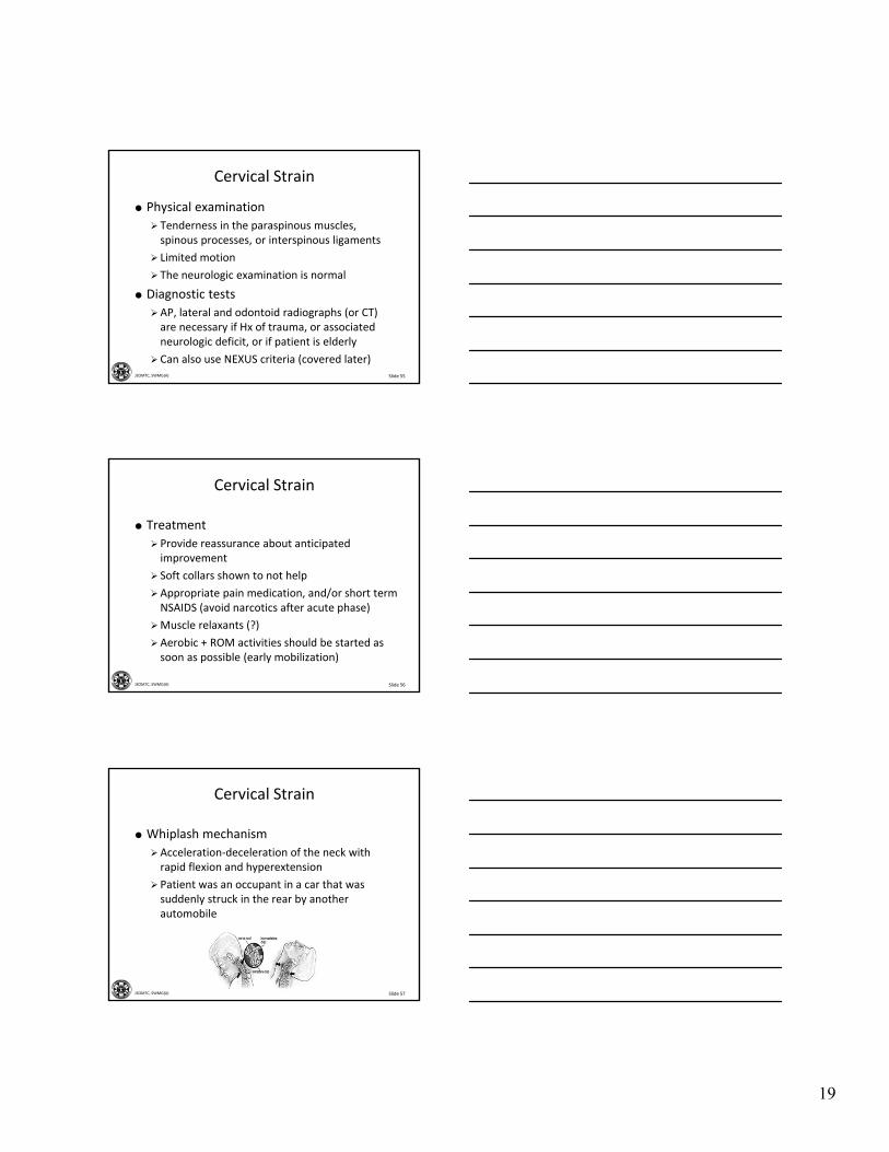
Cervical Strain
Clinical symptoms
Nonradicular, nonfocal neck pain is most common
Neck pain relieved with rest/aggravated with activity
Paraspinal muscle spasm, with stiffness and loss of motion
Occipital headache (whiplash injury)
Pain from an acute cervical sprain is usually self‐limiting*
19
Slide 55JSOMTC, SWMG(A)
Cervical Strain
Physical examination
Tenderness in the paraspinous muscles, spinous processes, or interspinous ligaments
Limited motion
The neurologic examination is normal
Diagnostic tests
AP, lateral and odontoid radiographs (or CT) are necessary if Hx of trauma, or associated neurologic deficit, or if patient is elderly
Can also use NEXUS criteria (covered later)
Slide 56JSOMTC, SWMG(A)
Cervical Strain
Treatment
Provide reassurance about anticipated improvement
Soft collars shown to not help
Appropriate pain medication, and/or short term NSAIDS (avoid narcotics after acute phase)
Muscle relaxants (?)
Aerobic + ROM activities should be started as soon as possible (early mobilization)
Slide 57JSOMTC, SWMG(A)
Cervical Strain
Whiplash mechanism
Acceleration‐deceleration of the neck with rapid flexion and hyperextension
Patient was an occupant in a car that was suddenly struck in the rear by another automobile
20
Slide 58JSOMTC, SWMG(A)
Cervical Strain
Pearls of management
Is a cervical collar and spine board needed?
X‐rays (NEXUS criteria)
Documentation is important
Delayed recovery related to secondary gain
Therapeutic modalities
Rehabilitation
Slide 59JSOMTC, SWMG(A)
The Signs and Symptoms, Physical Exam Findings, Diagnostic Tests, and
Management of Cervical Radiculopathy
Slide 60JSOMTC, SWMG(A)
Cervical Radiculopathy
Herniated Cervical Disk
21
Slide 61JSOMTC, SWMG(A)
Cervical Radiculopathy
Overview
Cause – lateral nerve root entrapment
< 40 y/o is herniation of a cervical disk due to trauma
> 40 y/o is a combination of foraminalnarrowing and degenerative changes to the disk
Slide 62JSOMTC, SWMG(A)
Slide 63JSOMTC, SWMG(A)
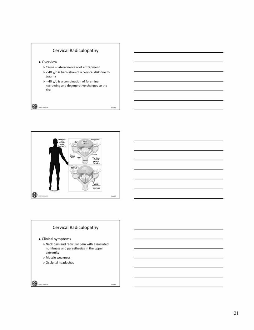
Cervical Radiculopathy
Clinical symptoms
Neck pain and radicular pain with associated numbness and paresthesias in the upper extremity
Muscle weakness
Occipital headaches
22
Slide 64JSOMTC, SWMG(A)
Cervical Radiculopathy
Physical examination
Cervical lordosis may be reduced
Cervical ROM may be restricted
Extension and axial rotation (Spurling’s test) will often cause pain in the arm and shoulder
Diagnostic tests
Plain radiographs
MRI or CT
Slide 65JSOMTC, SWMG(A)
Cervical Radiculopathy
Plan
Acute phase – modalities like heat or ice
Immobilization – soft cervical collar
Analgesia – non‐narcotic and muscle relaxants
Physical therapy and exercise
Good PO hydration
Cervical traction in a head halter
Slide 66JSOMTC, SWMG(A)
The Signs and Symptoms, Physical Exam Findings, Diagnostic Tests, and Management of Cervical Fractures
23
Slide 67JSOMTC, SWMG(A)
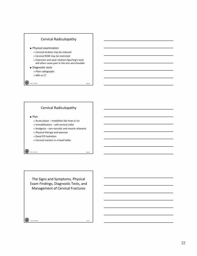
Fractures of the Cervical Spine
Slide 68JSOMTC, SWMG(A)
Fractures of the Cervical Spine
Overview
Result of high‐energy trauma
Identified or ruled out in all trauma patients who report neck pain, or blows above clavicles
Radiographs are required for all unconscious or intoxicated patients involved in an accident
Generally classified as flexion, extension, compression, or multiple/complex
Slide 69JSOMTC, SWMG(A)
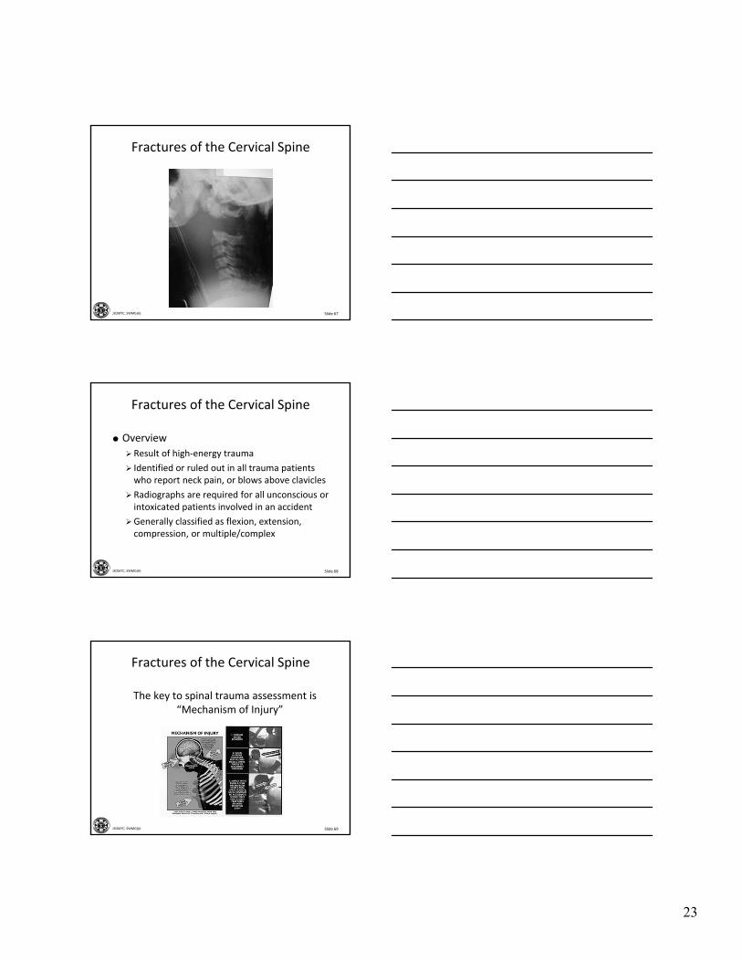
The key to spinal trauma assessment is “Mechanism of Injury”
Fractures of the Cervical Spine

24
Slide 70JSOMTC, SWMG(A)
Fractures of the Cervical Spine
Clinical symptoms
Severe neck pain, paraspinous muscle spasm, and/or point tenderness
Pain that radiates into the shoulder or arm suggests nerve root impingement
Global sensory or motor deficits suggest spinal cord injury
Multiple‐trauma patients: the absence of neck pain does not “clear” a cervical injury
NEXUS criteria (for x‐ray)
Slide 71JSOMTC, SWMG(A)
Fractures of the Cervical Spine
Physical exam
Inspect for swelling and contusions
Palpate for tenderness and paraspinal spasm
Evaluate motor, sensory function in both upper and lower extremities
Perianal sensation, sphincter tone, priapism, and bulbocavernosus reflex
Urine retention
Slide 72JSOMTC, SWMG(A)
Fractures of the Cervical Spine
Rectal Examination

25
Slide 73JSOMTC, SWMG(A)
Fractures of the Cervical Spine
Diagnostic tests
X‐rays
• Anterior/posterior, lateral, and odontoid views
• Who needs them?
• Who can be cleared without?
MRI
CT
Slide 74JSOMTC, SWMG(A)
Fractures of the Cervical Spine First X‐ray of the C‐spine
Cross‐Table Lateral
Slide 75JSOMTC, SWMG(A)
Fractures of the Cervical Spine
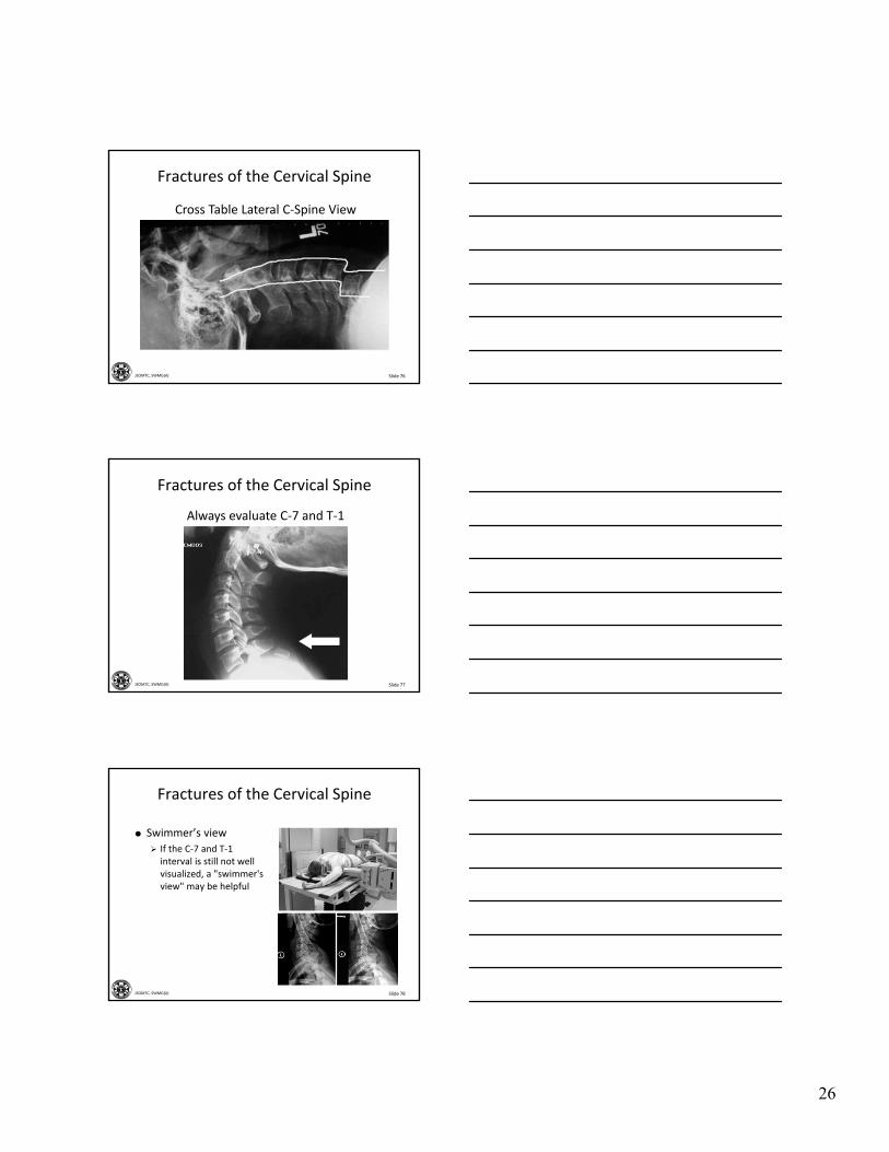
Reading c‐spine x‐rays
A ‐ Alignment
• Anterior contour line
• Posterior contour line
• Spinolaminal line
• Spinous process tips
B ‐ Bony contour
C ‐ Cartilage/disk
S ‐ Soft tissues
26
Slide 76JSOMTC, SWMG(A)
Cross Table Lateral C‐Spine View
Fractures of the Cervical Spine
Slide 77JSOMTC, SWMG(A)
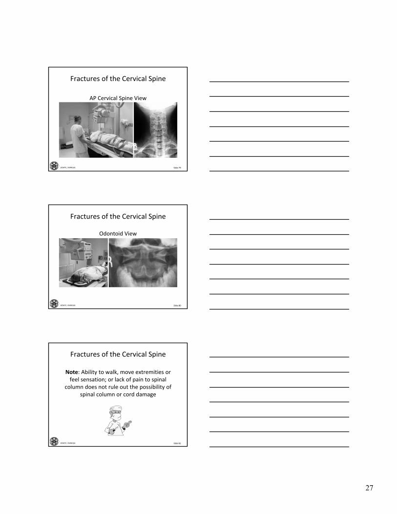
Fractures of the Cervical Spine
Always evaluate C‐7 and T‐1
Slide 78JSOMTC, SWMG(A)
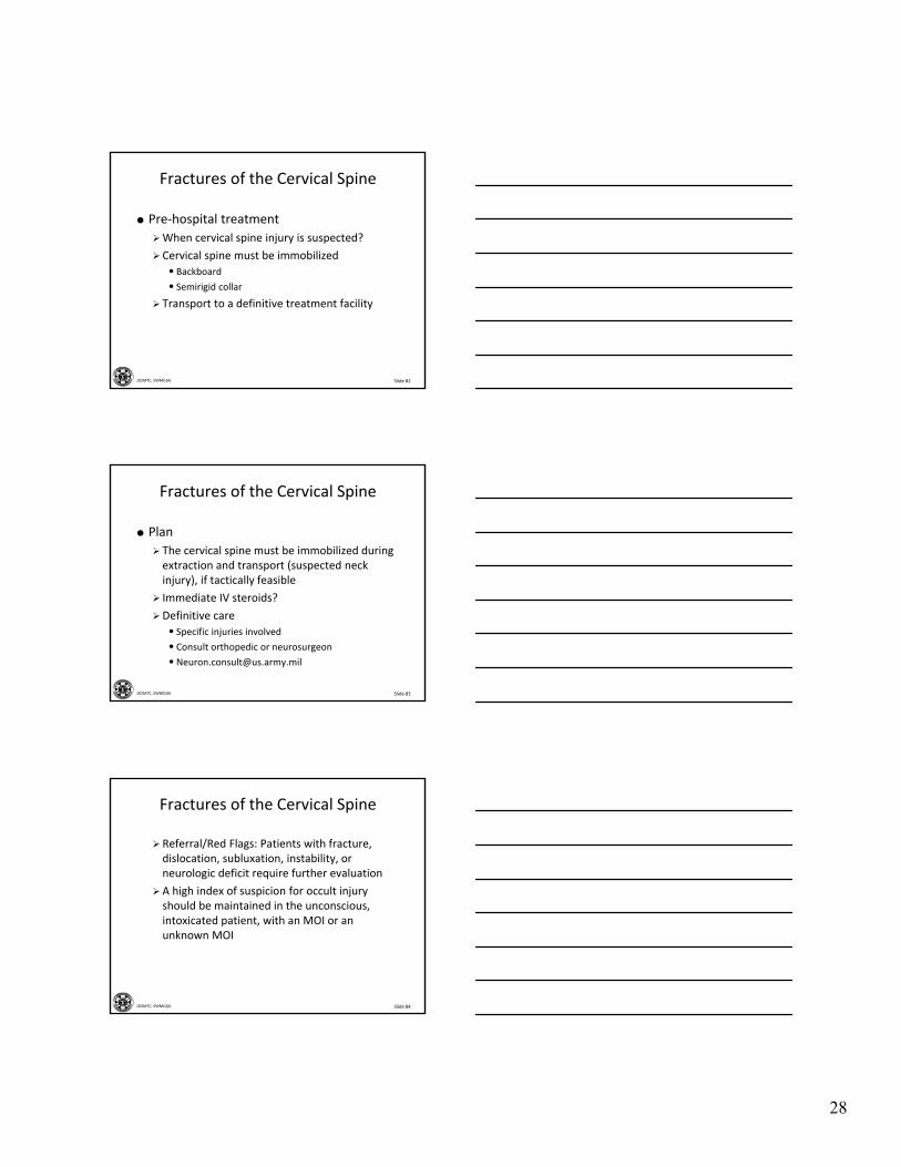
Fractures of the Cervical Spine
Swimmer’s view
If the C‐7 and T‐1 interval is still not well visualized, a "swimmer's view" may be helpful
27
Slide 79JSOMTC, SWMG(A)
AP Cervical Spine View
Fractures of the Cervical Spine
Slide 80JSOMTC, SWMG(A)
Odontoid View
Fractures of the Cervical Spine
Slide 81JSOMTC, SWMG(A)
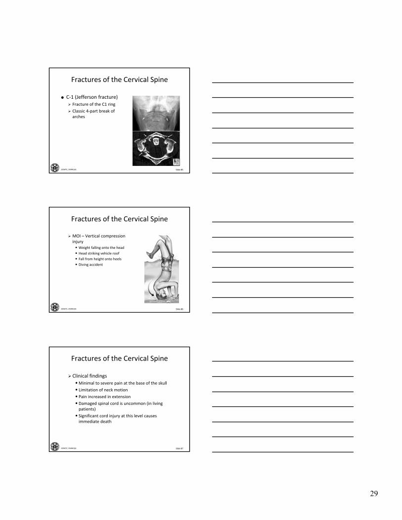
Note: Ability to walk, move extremities or feel sensation; or lack of pain to spinal
column does not rule out the possibility of spinal column or cord damage
Fractures of the Cervical Spine
28
Slide 82JSOMTC, SWMG(A)
Fractures of the Cervical Spine
Pre‐hospital treatment
When cervical spine injury is suspected?
Cervical spine must be immobilized
• Backboard
• Semirigid collar
Transport to a definitive treatment facility
Slide 83JSOMTC, SWMG(A)
Fractures of the Cervical Spine
Plan
The cervical spine must be immobilized during extraction and transport (suspected neck injury), if tactically feasible
Immediate IV steroids?
Definitive care
• Specific injuries involved
• Consult orthopedic or neurosurgeon
Slide 84JSOMTC, SWMG(A)
Fractures of the Cervical Spine
Referral/Red Flags: Patients with fracture, dislocation, subluxation, instability, or neurologic deficit require further evaluation
A high index of suspicion for occult injury should be maintained in the unconscious, intoxicated patient, with an MOI or an unknown MOI
29
Slide 85JSOMTC, SWMG(A)
Fractures of the Cervical Spine
C‐1 (Jefferson fracture)
Fracture of the C1 ring
Classic 4‐part break of arches
Slide 86JSOMTC, SWMG(A)
Fractures of the Cervical Spine
MOI – Vertical compression injury• Weight falling onto the head
• Head striking vehicle roof
• Fall from height onto heels
• Diving accident
Slide 87JSOMTC, SWMG(A)
Fractures of the Cervical Spine
Clinical findings
•Minimal to severe pain at the base of the skull
• Limitation of neck motion
• Pain increased in extension
• Damaged spinal cord is uncommon (in living patients)
• Significant cord injury at this level causes immediate death
30
Slide 88JSOMTC, SWMG(A)
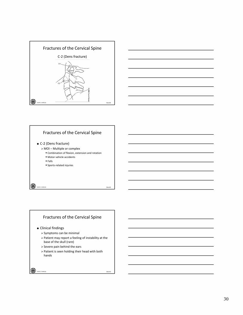
C‐2 (Dens fracture)
Fractures of the Cervical Spine
Slide 89JSOMTC, SWMG(A)
Fractures of the Cervical Spine
C‐2 (Dens fracture)
MOI – Multiple or complex
• Combination of flexion, extension and rotation
•Motor vehicle accidents
• Falls
• Sports‐related injuries
Slide 90JSOMTC, SWMG(A)
Fractures of the Cervical Spine
Clinical findings
Symptoms can be minimal
Patient may report a feeling of instability at the base of the skull (rare)
Severe pain behind the ears
Patient is seen holding their head with both hands
31
Slide 91JSOMTC, SWMG(A)
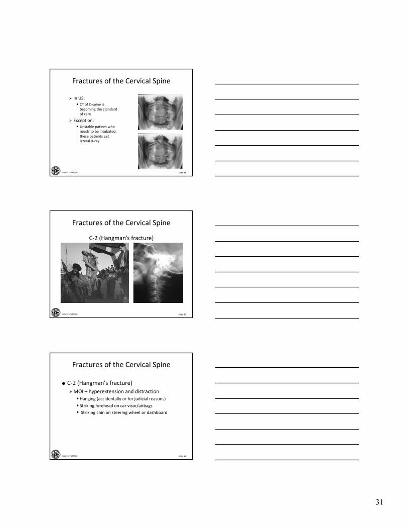
Fractures of the Cervical Spine
In US:• CT of C‐spine is becoming the standard of care
Exception:
• Unstable patient who needs to be intubated, these patients get lateral X‐ray
Slide 92JSOMTC, SWMG(A)
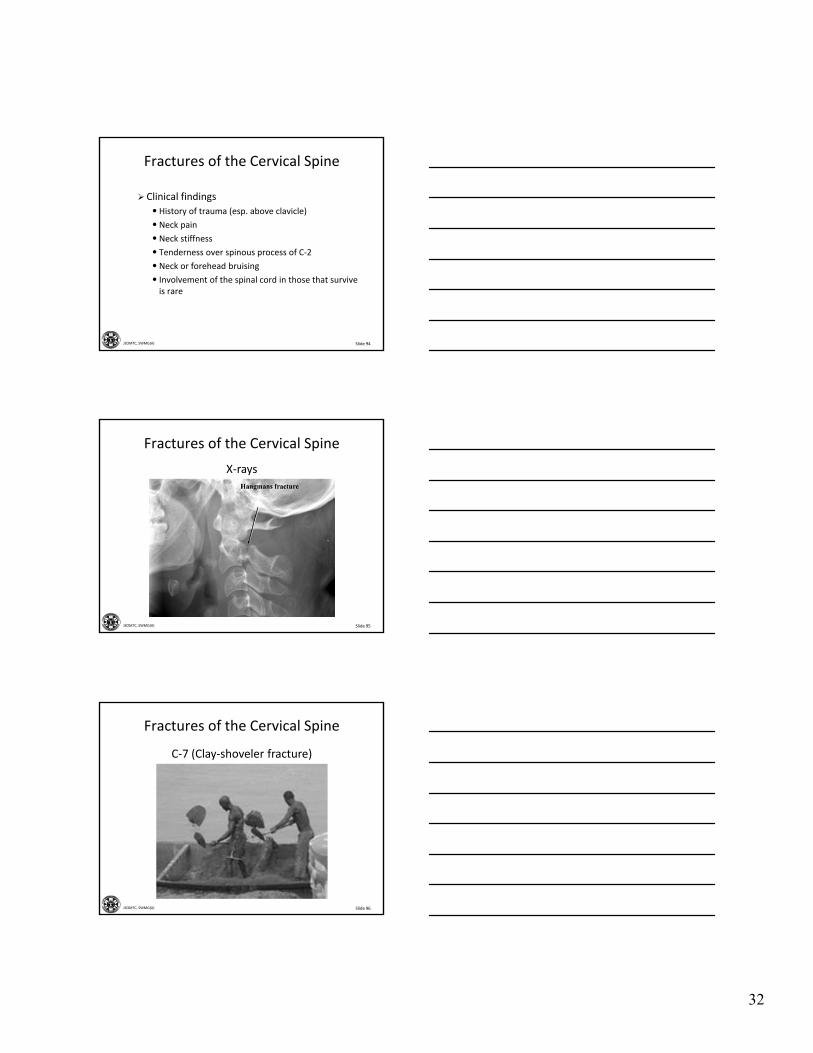
C‐2 (Hangman’s fracture)
Fractures of the Cervical Spine
Slide 93JSOMTC, SWMG(A)
Fractures of the Cervical Spine
C‐2 (Hangman’s fracture)
MOI – hyperextension and distraction
• Hanging (accidentally or for judicial reasons)
• Striking forehead on car visor/airbags
• Striking chin on steering wheel or dashboard
32
Slide 94JSOMTC, SWMG(A)
Fractures of the Cervical Spine
Clinical findings
• History of trauma (esp. above clavicle)
• Neck pain
• Neck stiffness
• Tenderness over spinous process of C‐2
• Neck or forehead bruising
• Involvement of the spinal cord in those that survive is rare
Slide 95JSOMTC, SWMG(A)
Fractures of the Cervical Spine
X‐rays
Slide 96JSOMTC, SWMG(A)
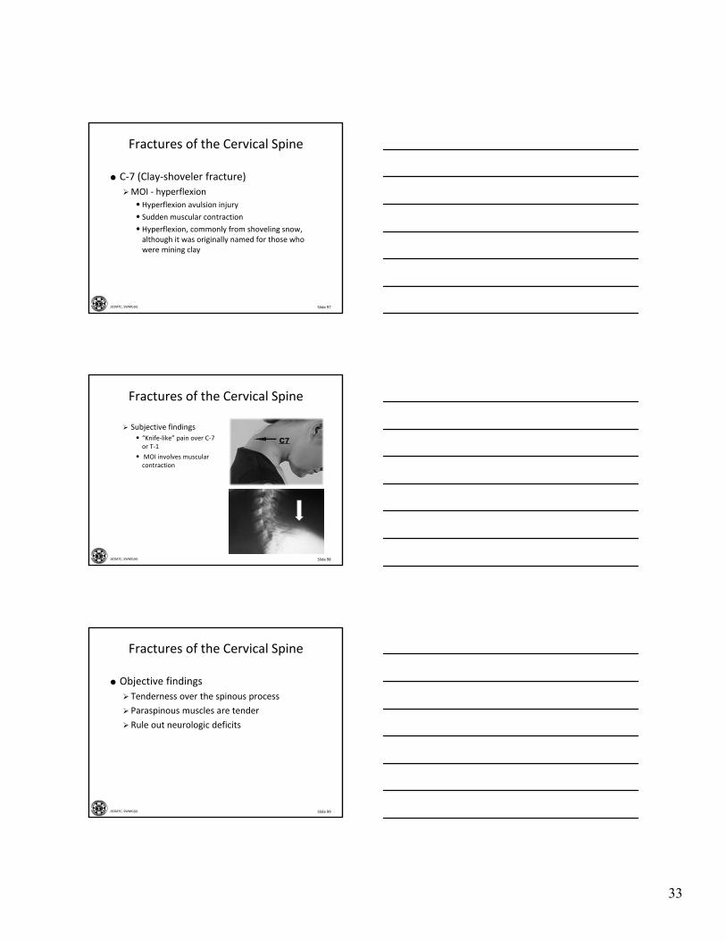
Fractures of the Cervical Spine
C‐7 (Clay‐shoveler fracture)
33
Slide 97JSOMTC, SWMG(A)
Fractures of the Cervical Spine
C‐7 (Clay‐shoveler fracture)
MOI ‐ hyperflexion
• Hyperflexion avulsion injury
• Sudden muscular contraction
• Hyperflexion, commonly from shoveling snow, although it was originally named for those who were mining clay
Slide 98JSOMTC, SWMG(A)
Fractures of the Cervical Spine
Subjective findings• “Knife‐like” pain over C‐7 or T‐1
• MOI involves muscular contraction
Slide 99JSOMTC, SWMG(A)
Fractures of the Cervical Spine
Objective findings
Tenderness over the spinous process
Paraspinous muscles are tender
Rule out neurologic deficits
34
Slide 100JSOMTC, SWMG(A)
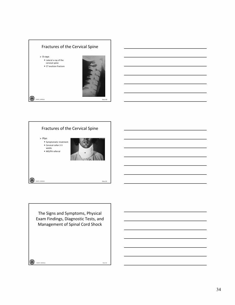
Fractures of the Cervical Spine
X‐rays• Lateral x‐ray of the cervical spine
• C7 avulsion fracture
Slide 101JSOMTC, SWMG(A)
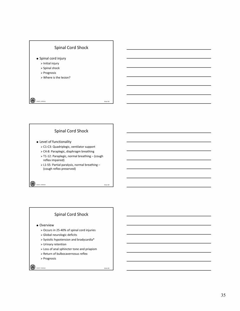
Fractures of the Cervical Spine
Plan• Symptomatic treatment
• Cervical collar 2‐3 weeks
• MD/PA referral
Slide 102JSOMTC, SWMG(A)
The Signs and Symptoms, Physical Exam Findings, Diagnostic Tests, and Management of Spinal Cord Shock
35
Slide 103JSOMTC, SWMG(A)
Spinal Cord Shock
Spinal cord injury
Initial injury
Spinal shock
Prognosis
Where is the lesion?
Slide 104JSOMTC, SWMG(A)
Spinal Cord Shock
Level of functionality
C1‐C3: Quadriplegic, ventilator support
C4‐8: Paraplegic, diaphragm breathing
T1‐12: Paraplegic, normal breathing – (cough reflex impaired)
L1‐S5: Partial paralysis, normal breathing –(cough reflex preserved)
Slide 105JSOMTC, SWMG(A)
Spinal Cord Shock
Overview
Occurs in 25‐40% of spinal cord injuries
Global neurologic deficits
Systolic hypotension and bradycardia*
Urinary retention
Loss of anal sphincter tone and priapism
Return of bulbocavernosus reflex
Prognosis
36
Slide 106JSOMTC, SWMG(A)
The Management of Fractures to the Thoracic and Lumbar Spine
Slide 107JSOMTC, SWMG(A)
Thoracic and Lumbar Spine Fractures
Introduction
High‐energy trauma such as motor vehicle accidents or fall from a height
Minor trauma: osteoporosis, tumors, infections
Long‐term steroid use
Slide 108JSOMTC, SWMG(A)
Thoracic and Lumbar Spine Fractures
37
Slide 109JSOMTC, SWMG(A)
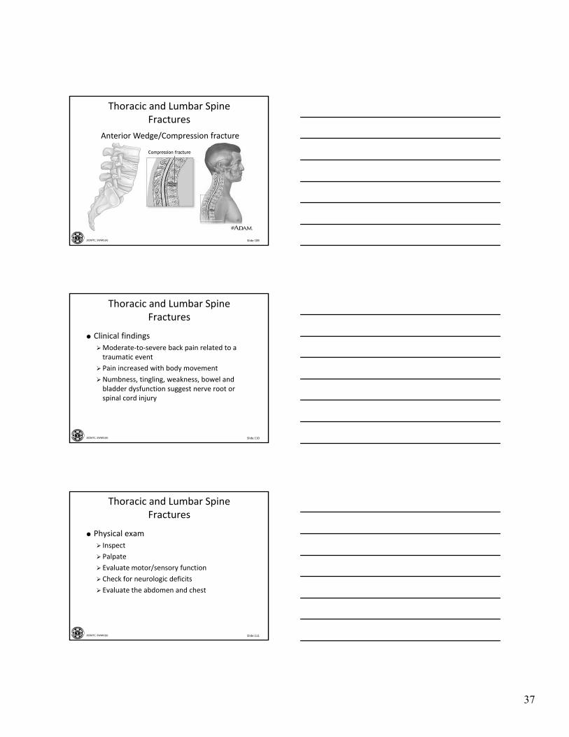
Anterior Wedge/Compression fracture
Thoracic and Lumbar Spine Fractures
Slide 110JSOMTC, SWMG(A)
Thoracic and Lumbar Spine Fractures
Clinical findings
Moderate‐to‐severe back pain related to a traumatic event
Pain increased with body movement
Numbness, tingling, weakness, bowel and bladder dysfunction suggest nerve root or spinal cord injury
Slide 111JSOMTC, SWMG(A)
Thoracic and Lumbar Spine Fractures
Physical exam
Inspect
Palpate
Evaluate motor/sensory function
Check for neurologic deficits
Evaluate the abdomen and chest
38
Slide 112JSOMTC, SWMG(A)
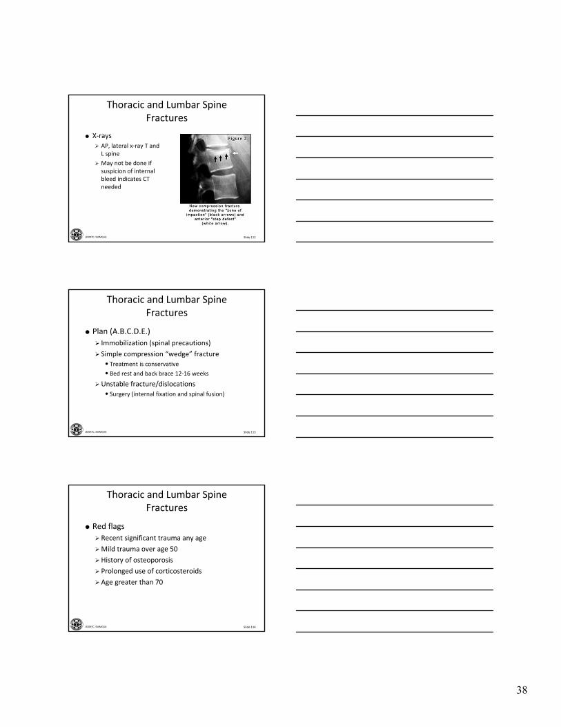
Thoracic and Lumbar Spine Fractures
X‐rays
AP, lateral x‐ray T and L spine
May not be done if suspicion of internal bleed indicates CT needed
Slide 113JSOMTC, SWMG(A)
Thoracic and Lumbar Spine Fractures
Plan (A.B.C.D.E.)
Immobilization (spinal precautions)
Simple compression “wedge” fracture
• Treatment is conservative
• Bed rest and back brace 12‐16 weeks
Unstable fracture/dislocations
• Surgery (internal fixation and spinal fusion)
Slide 114JSOMTC, SWMG(A)
Thoracic and Lumbar Spine Fractures
Red flags
Recent significant trauma any age
Mild trauma over age 50
History of osteoporosis
Prolonged use of corticosteroids
Age greater than 70
39
Slide 115JSOMTC, SWMG(A)
The Management of Low Back Pain
Slide 116JSOMTC, SWMG(A)
Low Back Pain
Synonyms
Low back pain (LBP)
Pulled low back
Lumbar strain/sprain
History
Slide 117JSOMTC, SWMG(A)
Low Back Pain
Clinical symptoms
Acute onset of low back pain, often following a lifting episode (may be trivial)
The pain often radiates into the buttocks and posterior thighs; muscle spasms
Patients have difficulty standing erect
Exaggerated responses: generalized hypersensitivity to light touch (or facial grimacing) with minimal loading or movement of spine
40
Slide 118JSOMTC, SWMG(A)
Low Back Pain
Physical exam
Inspection
Diffuse tenderness in the low back
ROM is reduced and elicits pain
The motor and sensory function of the lumbosacral nerve roots and lower extremity reflexes are normal
Slide 119JSOMTC, SWMG(A)
Low Back Pain
Diagnostic tests
When are plain radiographs needed?
• Infection
• Cancer
• Fractures (and no CT required)
MRI
• Reserved for patients considering surgery
• Cauda equina
• Evidence of a systemic disease
• Not available on emergency basis
Slide 120JSOMTC, SWMG(A)
Low Back Pain
Treatment
Rest in bed or remain active?
NSAIDs and/or other non‐narcotic pain medications (7‐14 days)
Muscle relaxants may be helpful initially
Avoid narcotic analgesics and sedatives
Reassurance
Patient education
41
Slide 121JSOMTC, SWMG(A)
The Management of Lumbar Degenerative Disk Disease and
Chronic Low Back Pain
Slide 122JSOMTC, SWMG(A)
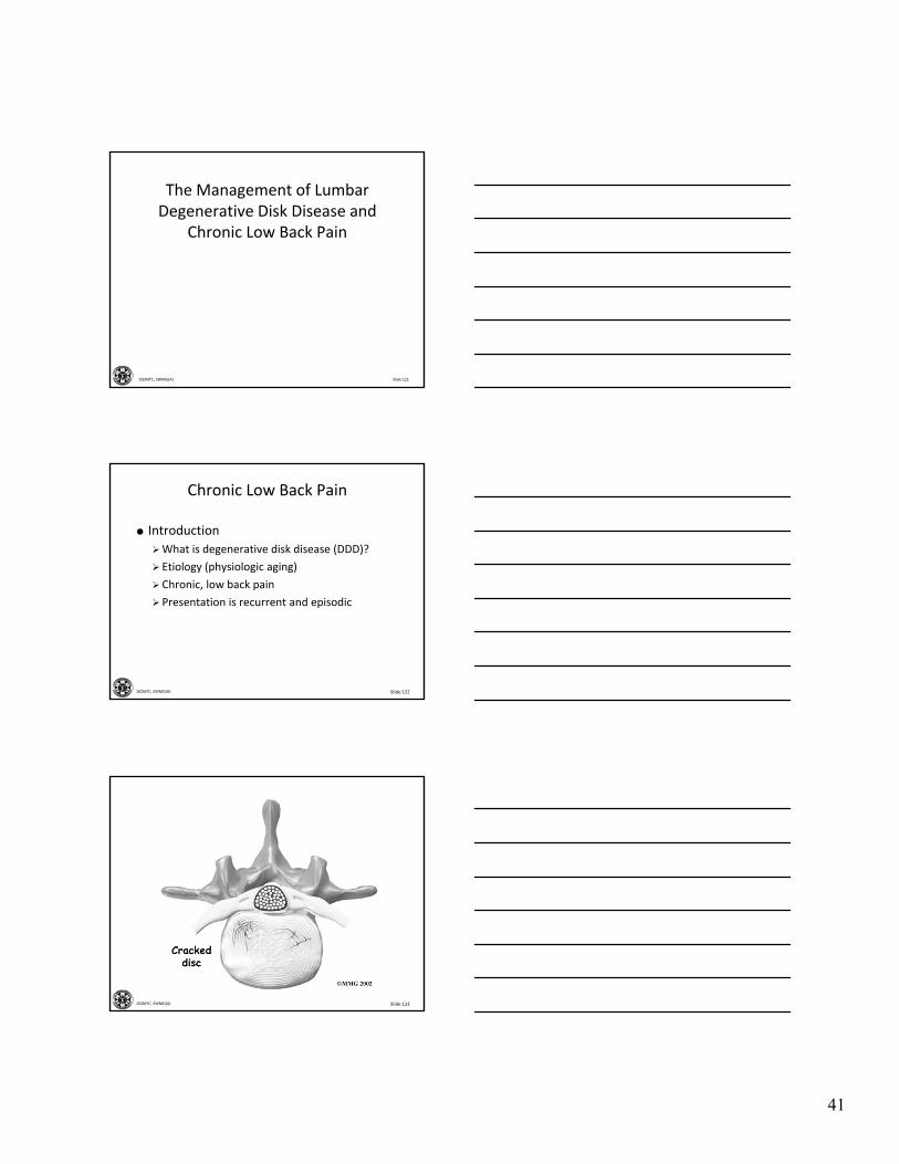
Chronic Low Back Pain
Introduction
What is degenerative disk disease (DDD)?
Etiology (physiologic aging)
Chronic, low back pain
Presentation is recurrent and episodic
Slide 123JSOMTC, SWMG(A)
42
Slide 124JSOMTC, SWMG(A)
Chronic Low Back Pain
Clinical findings
Pain is centered on the lower back, may radiate to hips and legs
Aggravated by activities such as bending, lifting, stooping, or twisting
Relieved with lying down or a night’s rest
Motor, sensory, reflexes normal
Muscle spasm causing a side or forward list
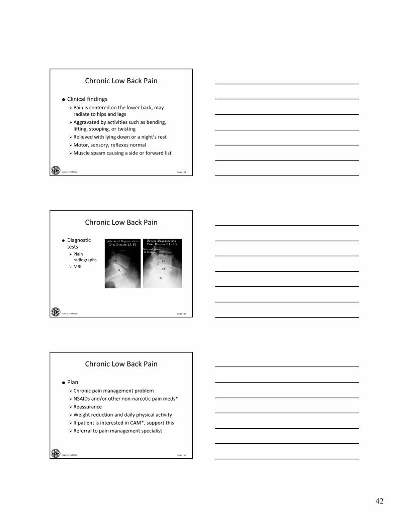
Slide 125JSOMTC, SWMG(A)
Chronic Low Back Pain
Diagnostic tests
Plain radiographs
MRI
Slide 126JSOMTC, SWMG(A)
Chronic Low Back Pain
Plan
Chronic pain management problem
NSAIDs and/or other non‐narcotic pain meds*
Reassurance
Weight reduction and daily physical activity
If patient is interested in CAM*, support this
Referral to pain management specialist

43
Slide 127JSOMTC, SWMG(A)
The Management of Lumbar Herniated Disk
Slide 128JSOMTC, SWMG(A)
Lumbar Herniated Disk
Synonyms
Sciatica
Lumbar radiculopathy
Slide 129JSOMTC, SWMG(A)
Lumbar Herniated Disk
Introduction
Herniated disk syndrome (commonly called “sciatica”)
Mechanism of injury
• Traumatic injury
• Progressive degeneration of the disc
Low back pain that radiates to the leg
Commonly occurs at level L‐4 /L 5, or L‐5/ S‐1
44
Slide 130JSOMTC, SWMG(A)
Lumbar Herniated Disk
Subjective findings
O‐onset – when and how did it start? Abrupt or insidious
P‐provocation – exaggerated by sitting, walking, standing, coughing, and sneezing
Q‐quality – sharp or dull, burning, stabbing, numbness or tingling
R‐radiation – unilateral, to the posterior leg
S‐severity – mild to severe
T‐time – history, change in symptoms
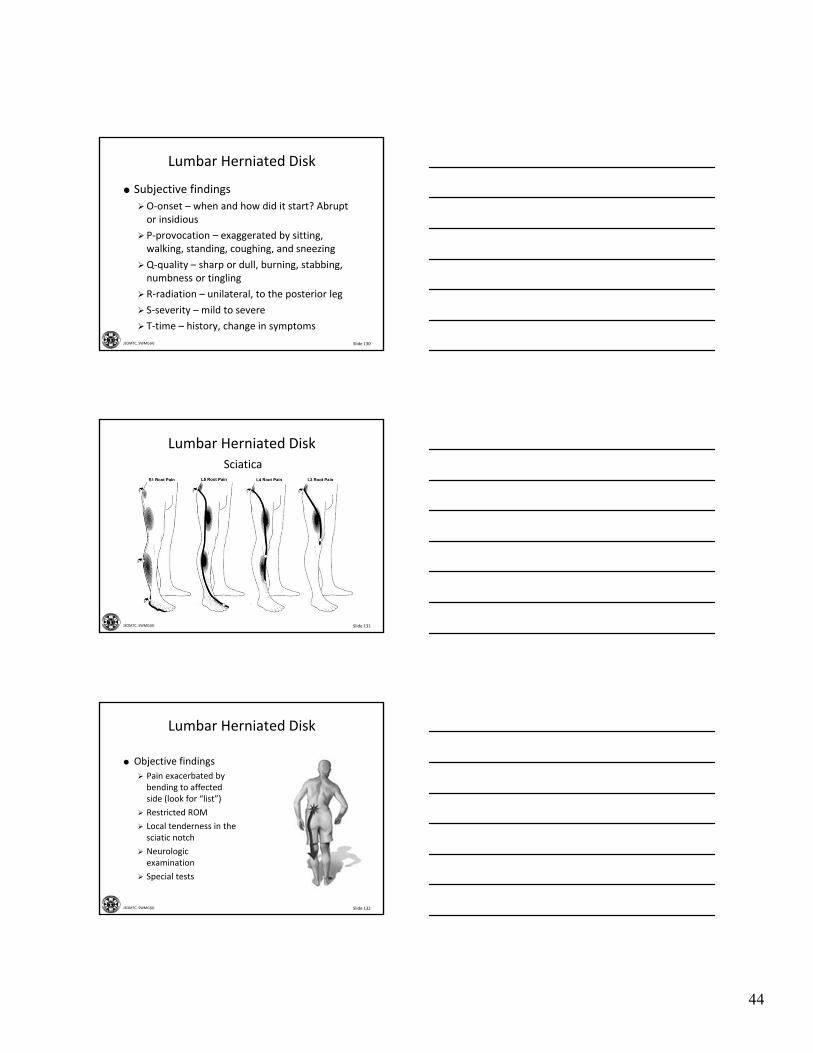
Slide 131JSOMTC, SWMG(A)
Sciatica
Lumbar Herniated Disk
Slide 132JSOMTC, SWMG(A)
Lumbar Herniated Disk
Objective findings
Pain exacerbated by bending to affected side (look for “list”)
Restricted ROM
Local tenderness in the sciatic notch
Neurologic examination
Special tests
45
Slide 133JSOMTC, SWMG(A)
Sciatica
Lumbar Herniated Disk
Slide 134JSOMTC, SWMG(A)
Sciatica
Lumbar Herniated Disk
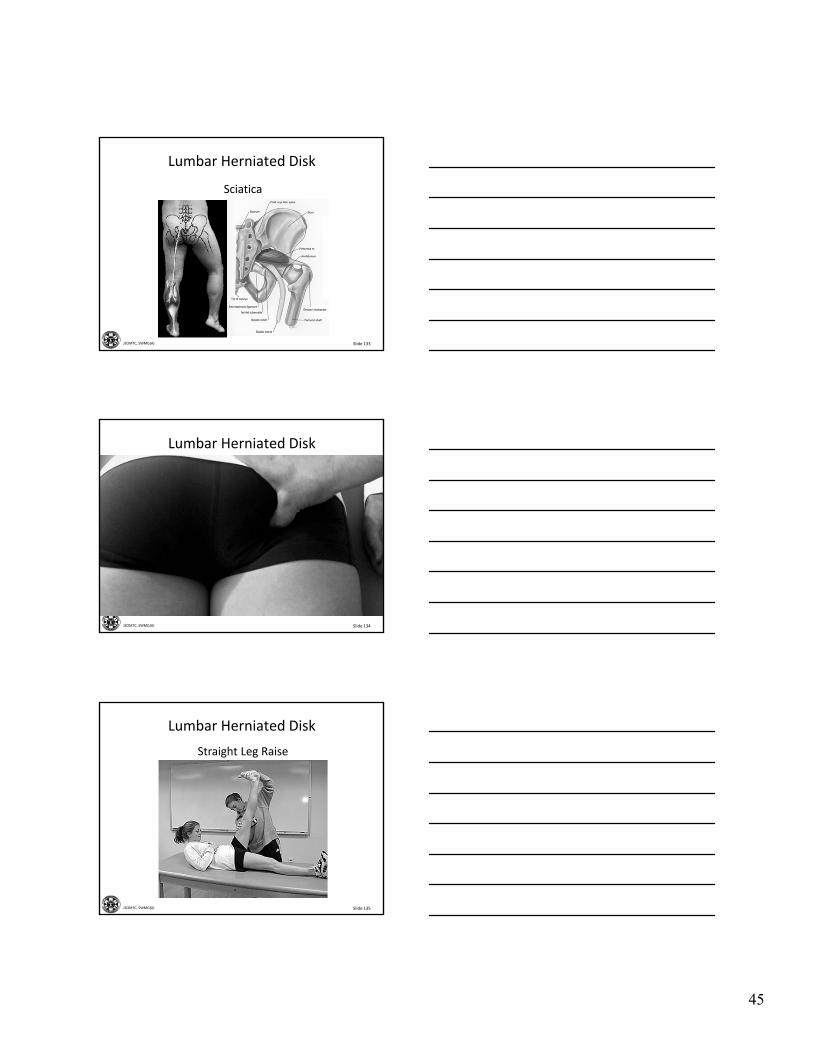
Slide 135JSOMTC, SWMG(A)
Straight Leg Raise
Lumbar Herniated Disk
46
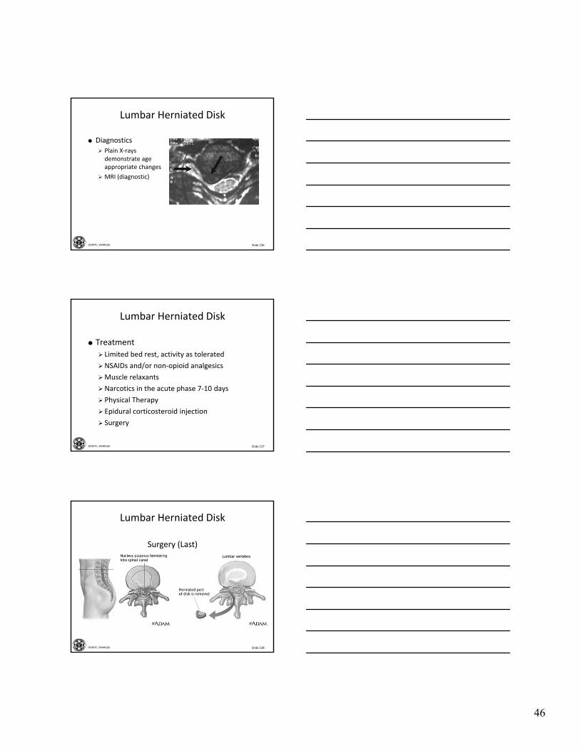
Slide 136JSOMTC, SWMG(A)
Lumbar Herniated Disk
Diagnostics
Plain X‐rays demonstrate age appropriate changes
MRI (diagnostic)
Slide 137JSOMTC, SWMG(A)
Lumbar Herniated Disk
Treatment
Limited bed rest, activity as tolerated
NSAIDs and/or non‐opioid analgesics
Muscle relaxants
Narcotics in the acute phase 7‐10 days
Physical Therapy
Epidural corticosteroid injection
Surgery
Slide 138JSOMTC, SWMG(A)
Surgery (Last)
Lumbar Herniated Disk
47
Slide 139JSOMTC, SWMG(A)
The Signs and Symptoms, Physical Exam Findings, Diagnostic Tests, and Management of Spondylolisthesis
Slide 140JSOMTC, SWMG(A)
Spondylolisthesis
Slide 141JSOMTC, SWMG(A)
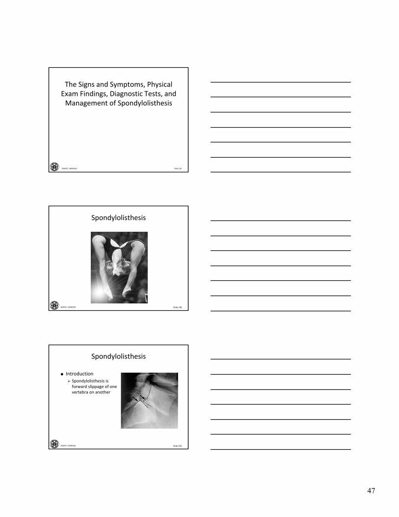
Spondylolisthesis
Introduction
Spondylolisthesis is forward slippage of one vertebra on another
48
Slide 142JSOMTC, SWMG(A)
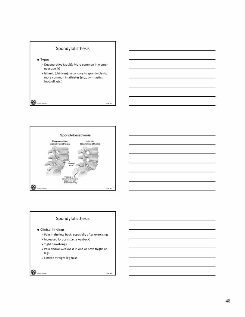
Types
Degenerative (adult): More common in women over age 40
Isthmic (children): secondary to spondylolysis; more common in athletes (e.g., gymnastics, football, etc.)
Spondylolisthesis
Slide 143JSOMTC, SWMG(A)
Slide 144JSOMTC, SWMG(A)
Clinical findings
Pain in the low back, especially after exercising
Increased lordosis (i.e., swayback)
Tight hamstrings
Pain and/or weakness in one or both thighs or legs
Limited straight leg raise
Spondylolisthesis
49
Slide 145JSOMTC, SWMG(A)
Spondylolisthesis
Diagnostics
Lateral x‐ray: forward translation of L‐5 relative to S‐1
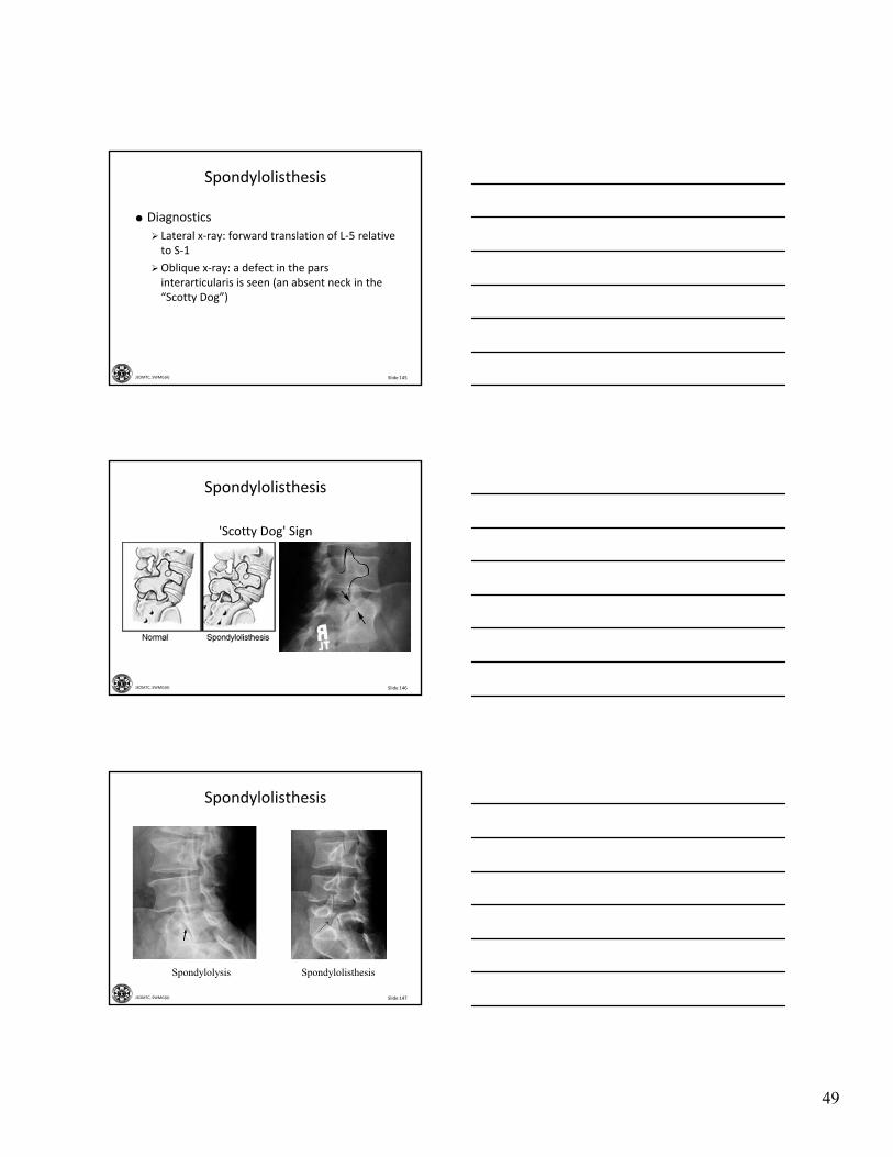
Oblique x‐ray: a defect in the pars interarticularis is seen (an absent neck in the “Scotty Dog”)
Slide 146JSOMTC, SWMG(A)
'Scotty Dog' Sign
Spondylolisthesis
Slide 147JSOMTC, SWMG(A)
Spondylolisthesis
Spondylolysis Spondylolisthesis
50
Slide 148JSOMTC, SWMG(A)
Spondylolisthesis
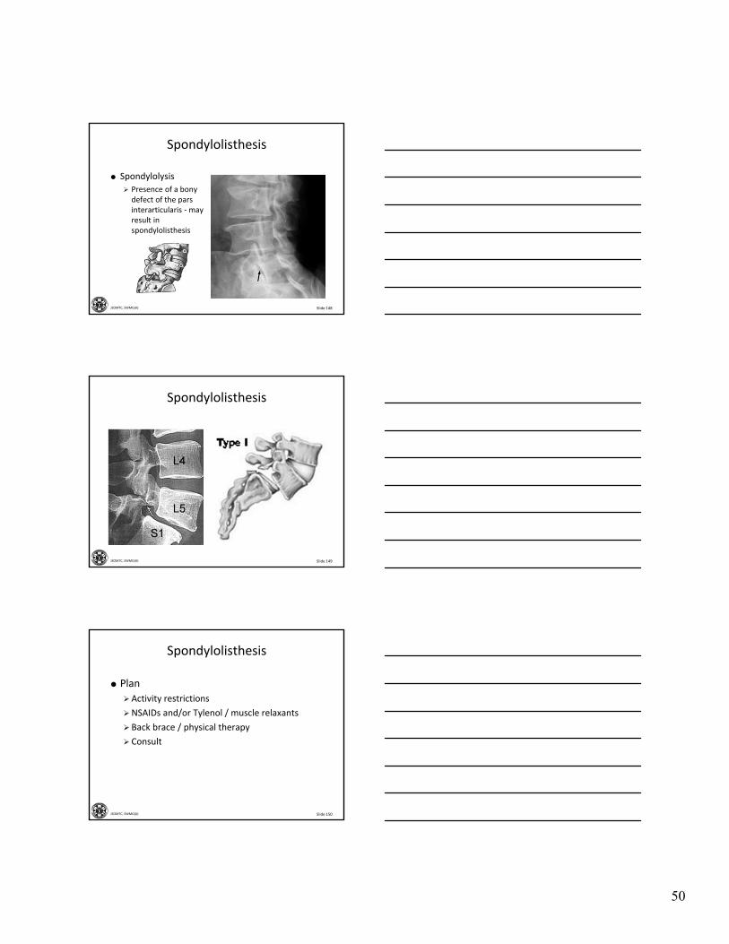
Spondylolysis
Presence of a bony defect of the pars interarticularis ‐ may result in spondylolisthesis
Slide 149JSOMTC, SWMG(A)
Spondylolisthesis
Slide 150JSOMTC, SWMG(A)
Spondylolisthesis
Plan
Activity restrictions
NSAIDs and/or Tylenol / muscle relaxants
Back brace / physical therapy
Consult
51
Slide 151JSOMTC, SWMG(A)
The Management of Acute and Chronic Infections of the Spine
Slide 152JSOMTC, SWMG(A)
Osteomyelitis
What is it?
Acute infection:
• Staphylococcus aureus (common)
Chronic infection:
• Tuberculosis (Pott’s Disease)
Symptoms
Diagnosis
Spine Infections
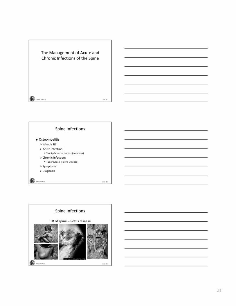
Slide 153JSOMTC, SWMG(A)
TB of spine – Pott’s disease
Spine Infections
52
Slide 154JSOMTC, SWMG(A)
Red flags
Persistent fever (temp over 100.4oF)
History of intravenous drug abuse
Recent bacterial infection (GU, cellulitis, pneumonia)
Immunocompromised states (e.g., DM, HIV, etc.)
Night pain, not relieved with rest
Patient usually feels sick
Spine Infections
Slide 155JSOMTC, SWMG(A)
Spine Infections
Plan
Hospital admit
Antibiotics
• 6 weeks of IV
• 6 weeks oral
Analgesics
Surgery consult
Slide 156JSOMTC, SWMG(A)
Common Metabolic Causes of Back Pain
53
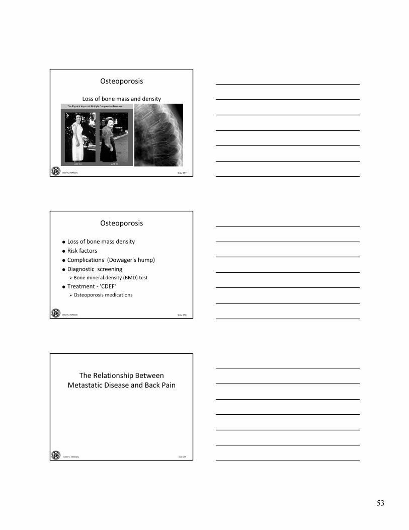
Slide 157JSOMTC, SWMG(A)
Osteoporosis
Loss of bone mass and density
Slide 158JSOMTC, SWMG(A)
Osteoporosis
Loss of bone mass density
Risk factors
Complications (Dowager's hump)
Diagnostic screening
Bone mineral density (BMD) test
Treatment ‐ 'CDEF'
Osteoporosis medications
Slide 159JSOMTC, SWMG(A)
The Relationship Between Metastatic Disease and Back Pain
54
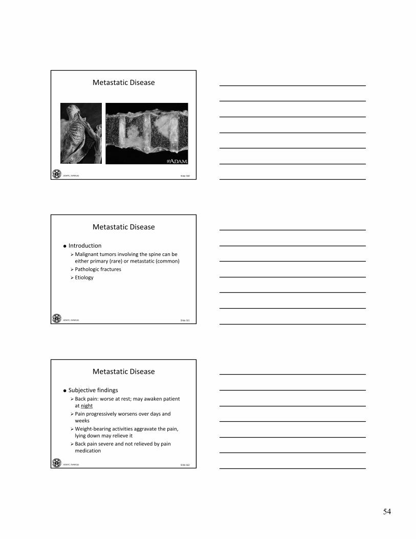
Slide 160JSOMTC, SWMG(A)
Metastatic Disease
Slide 161JSOMTC, SWMG(A)
Metastatic Disease
Introduction
Malignant tumors involving the spine can be either primary (rare) or metastatic (common)
Pathologic fractures
Etiology
Slide 162JSOMTC, SWMG(A)
Metastatic Disease
Subjective findings
Back pain: worse at rest; may awaken patient at night
Pain progressively worsens over days and weeks
Weight‐bearing activities aggravate the pain, lying down may relieve it
Back pain severe and not relieved by pain medication
55
Slide 163JSOMTC, SWMG(A)
Metastatic Disease
Objective findings
Tenderness to palpation or percussion along the spinous processes
Assess motor and sensory function
Check deep tendon reflexes (DTRs)
Evaluation of metastaic cause of tumor
Slide 164JSOMTC, SWMG(A)
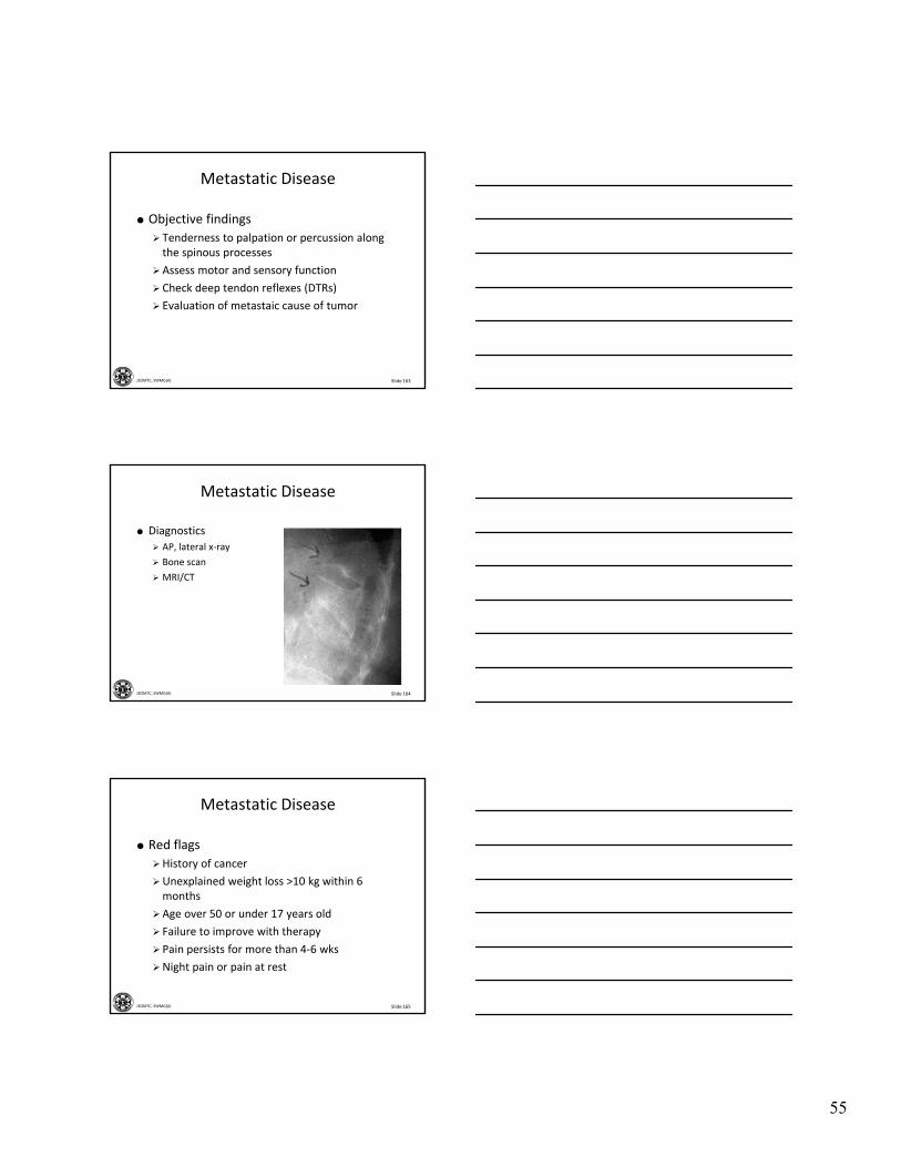
Metastatic Disease
Diagnostics
AP, lateral x‐ray
Bone scan
MRI/CT
Slide 165JSOMTC, SWMG(A)
Metastatic Disease
Red flags
History of cancer
Unexplained weight loss >10 kg within 6 months
Age over 50 or under 17 years old
Failure to improve with therapy
Pain persists for more than 4‐6 wks
Night pain or pain at rest
56
Slide 166JSOMTC, SWMG(A)
Plan
Immediate consultation
Chemotherapy
Radiation therapy
Surgery
Metastatic Disease
Slide 167JSOMTC, SWMG(A)
Common Disorders Associated with Abnormal Curvatures of the Spine
Slide 168JSOMTC, SWMG(A)
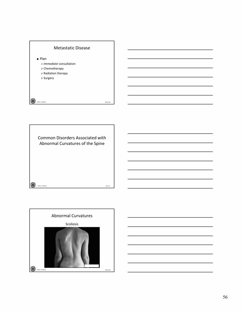
Abnormal Curvatures
Scoliosis
57
Slide 169JSOMTC, SWMG(A)
Abnormal Curvatures
Scoliosis
A lateral curvature of the spine
In adolescence the most common cause is unknown
More common in girls than boys
Changes occur with aging, such as osteoporosis, degenerative disk disease, spinal stenosis, and spondylolisthesis
Slide 170JSOMTC, SWMG(A)
Abnormal Curvatures
Subjective findings
Pain localized in the region of deformity
Progressive spinal deformity
"getting shorter"
Slide 171JSOMTC, SWMG(A)
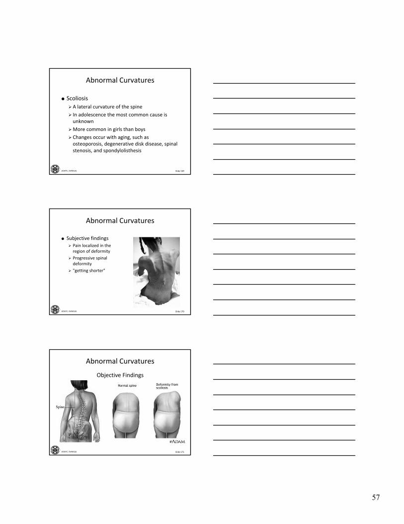
Objective Findings
Abnormal Curvatures
58
Slide 172JSOMTC, SWMG(A)
Abnormal Curvatures
Plan• Non‐operatively
• Stabilization (fusion)
Slide 173JSOMTC, SWMG(A)
Abnormal Curvatures
Kyphosis
Synonyms
• Postural round back
• Dowager’s hump
• Adolescent kyphosis
Slide 174JSOMTC, SWMG(A)
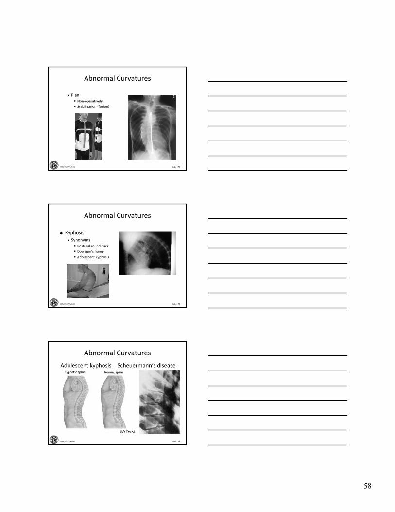
Adolescent kyphosis – Scheuermann’s disease
Abnormal Curvatures
59
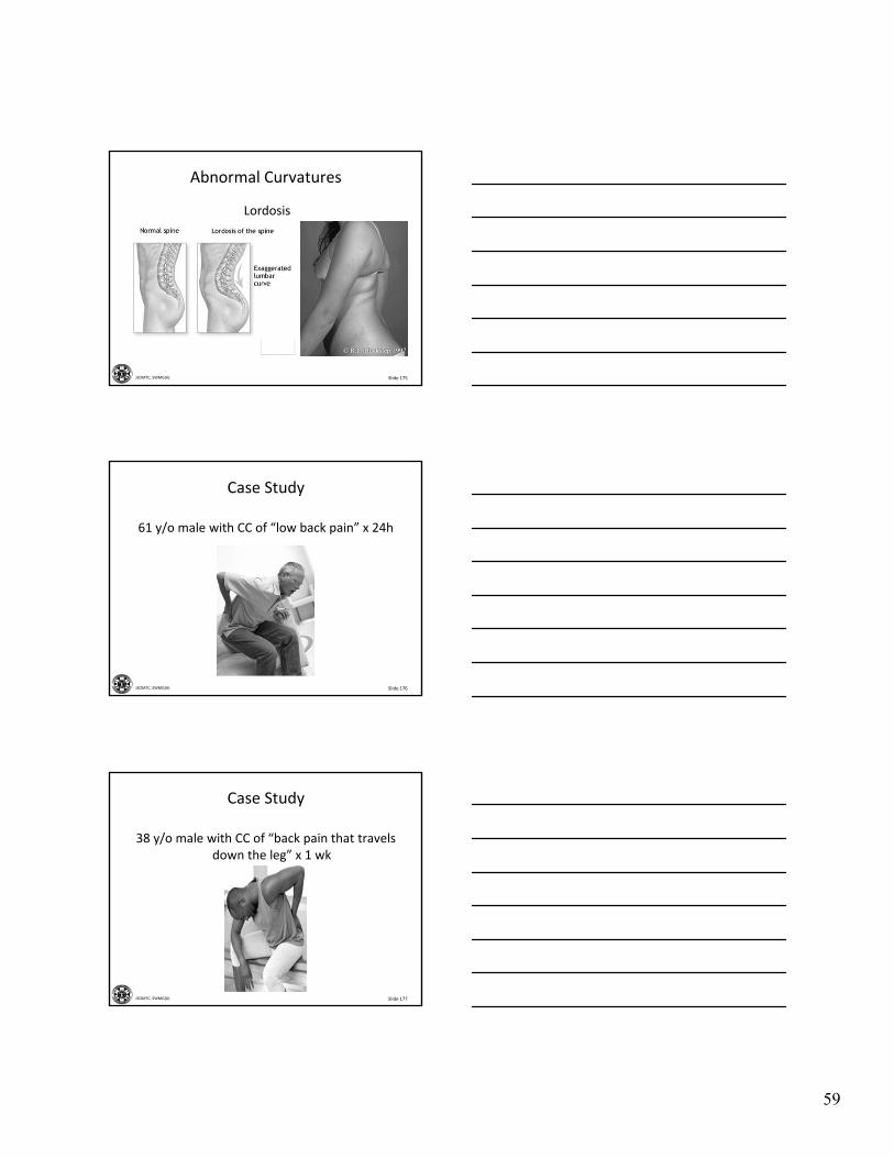
Slide 175JSOMTC, SWMG(A)
Lordosis
Abnormal Curvatures
Slide 176JSOMTC, SWMG(A)
Case Study
61 y/o male with CC of “low back pain” x 24h
Slide 177JSOMTC, SWMG(A)
Case Study
38 y/o male with CC of “back pain that travels down the leg” x 1 wk
60
Slide 178JSOMTC, SWMG(A)
Case Study
26 y/o male with CC of back pain x 24h
Slide 179JSOMTC, SWMG(A)
Case Study
66 y/o male with CC of low back pain x 2 wks
Slide 180JSOMTC, SWMG(A)
Questions?
61
Slide 181JSOMTC, SWMG(A)
Terminal Learning Objective
Action: Communicate knowledge of “Spinal Disorders”
Condition: Given a lecture in a classroom environment
Standard: Received a minimum score of 75% on the written exam IAW course standards
Slide 182JSOMTC, SWMG(A)
Agenda
Communicate common disorders of the spine
Communicate referral decisions and red flags
Recall the gross anatomy of the spine and related terminology
Slide 183JSOMTC, SWMG(A)
Agenda
Communicate the signs and symptoms, physical exam findings, diagnostic tests, and management of cauda equinasyndrome
Communicate the signs and symptoms, physical exam findings, and management of cervical spondylosis

62
Slide 184JSOMTC, SWMG(A)
Agenda
Communicate the signs and symptoms, physical exam findings, diagnostic tests, and management of traumatic cervical spine disorders, to include strain, radiculopathy, and fractures
Slide 185JSOMTC, SWMG(A)
Agenda
Communicate the signs and symptoms, physical exam findings, diagnostic tests, and management of spinal cord shock
Communicate the management of fractures to the thoracic and lumbar spine
Communicate the management of low back pain
Slide 186JSOMTC, SWMG(A)
Agenda
Communicate the management of lumbar degenerative disk disease and chronic low back pain
Communicate the management of lumbar herniated disk
Communicate the signs and symptoms, physical exam findings, diagnostic tests, and management of spondylolisthesis
63
Slide 187JSOMTC, SWMG(A)
Agenda
Communicate the management of acute and chronic infections of the spine
Identify common metabolic causes of back pain
Communicate the relationship between metastatic disease and back pain
Communicate common disorders associated with abnormal curvature of the spine
Slide 188JSOMTC, SWMG(A)
Reason
Slide 189JSOMTC, SWMG(A)
Break
64
Slide 190JSOMTC, SWMG(A)
Check on Learning
Your team sergeant comes to you with a complaint of back pain x 2 weeks. Which of the following will help you the MOST to make the correct assessment?
A. History
B. Range of motion, looking for limitations
C. X‐rays of the spine
D. Magnetic resonance imaging (MRI)
Slide 191JSOMTC, SWMG(A)
Check on Learning
Which of the following patients do you feel requires further evaluation?
A. 17‐year‐old taking Motrin as advised for back pain, now complains of pain at rest
B. Soldier who complains of back pain after a hard PLF
C. Injection drug user with back pain and no history of trauma
D. Elderly woman who tripped over her cat complains of moderate back discomfort
Slide 192JSOMTC, SWMG(A)
Check on Learning
Which part of the spine bears the most weight and as a result is prone to degradation and injury?
A. The first two cervical vertebrae (“Atlas and Axis”)
B. The twelve thoracic vertebrae and ribs
C. The lumbar spine
D. The fused sacrum
65
Slide 193JSOMTC, SWMG(A)
Check on Learning
Which of the following clinical findings is or are consistent with a cervical radiculopathy?
A. The usual cause in young adults (< 40 y/o) is herniation of a cervical disk
B. May cause painful burning, tingling, or numbness in the neck, shoulder, arm or hand
C. Most cases of cervical radiculopathy resolve in 4‐6 weeks with conservative treatment
D. Many patients state that they can relieve pain by placing the hand of effected side on top of their head
Slide 194JSOMTC, SWMG(A)
Check on Learning
Which of the following statement(s) is/are true about cervical fractures?
A. Commonly the result of high‐energy trauma
B. Must be identified or ruled out in all trauma patients who report neck pain
C. Radiographs are required for all unconscious or intoxicated patients involved in an accident
D. Generally classified as flexion, extension, compression, or multiple/complex
Slide 195JSOMTC, SWMG(A)
Check on Learning
Which of the following statements is incorrect about spinal cord shock?
A. Expect hypotension with tachycardia
B. Complete loss of all neurologic function, including reflexes, and rectal tone below level of injury.
C. Spinal shock can last from several days to several weeks
D. Return of bulbocavernosus reflex signifies the end of spinal shock
66
Slide 196JSOMTC, SWMG(A)
Check on Learning
Preventing neurologic damage is one of the goals of treatment for spinal fracture. How can the medic best accomplish this?
A. Immobilization and spinal precautions during extraction and transport
B. Internal fixation and spinal fusion
C. Steroids should be started immediately
D. Early return to activities as tolerated
Slide 197JSOMTC, SWMG(A)
Check on Learning
Which of the following choices are consistent with simple low back pain?
A. Acute onset of low back pain without neurologic deficits
B. Night pain that is not relieved with rest
C. Positive smoking history with unexplained weight loss
D. Pain followed post‐traumatic event such as a hard PLF landing
Slide 198JSOMTC, SWMG(A)
Check on Learning
Which of the following are consistent with degenerative disk disease/chronic low back pain?
A. Age of patient between 30‐50 years old
B. Symptoms are typically recurrent and episodic
C. Symptoms have persisted over 3 months
D. Symptoms are aggravated with activity and alleviated with rest
67
Slide 199JSOMTC, SWMG(A)
Check on Learning
Which of the following is not consistent with a lumbar radiculopathy “Sciatica”?
A. Typically radiating leg pain is bilateral
B. Herniated disk is most commonly found at the L‐4,5 and L‐5 ‐ S‐1 level
C. Neurologic evidence of irritated S‐1 nerve root is inability to toe walk
D. Clinical findings include a positive straight leg raise test
Slide 200JSOMTC, SWMG(A)
Check on Learning
Which x‐ray view would be used to show a stress fracture in spondylolysis?
A. Lateral view
B. Anterior‐posterior view
C. Oblique view
D. Inlet‐outlet view
Slide 201JSOMTC, SWMG(A)
Check on Learning
The usual cause of acute osteomyelitis of the spine?
A. Tuberculosis
B. Gonorrhea
C. Neisseria meningitidis
D. Staphylococcus aureus
68
Slide 202JSOMTC, SWMG(A)
Check on Learning
Which diagnostic test is the most useful to identify osteoporosis?
A. X‐ray
B. Bone scan
C. Bone mineral density
D. MRI
Slide 203JSOMTC, SWMG(A)
Check on Learning
The majority of low back complaints will resolve in 1‐4 weeks. Which of the following questions should be asked in a history?
A. Any MOI to suggest fracture
B. Any signs or symptoms to suggest tumor/infection
C. Any bowel/bladder problems or neurologic deficits
D. ROS for referred pain from abdomen or pelvis