sperm cell driven microrobots—emerging ... - atlas
TRANSCRIPT
micromachines
Review
Sperm Cell Driven Microrobots—EmergingOpportunities and Challenges for BiologicallyInspired Robotic Design
Ajay Vikram Singh 1,2,*,† , Mohammad Hasan Dad Ansari 3,4,†, Mihir Mahajan 5,Shubhangi Srivastava 6, Shubham Kashyap 7, Prajjwal Dwivedi 8, Vaibhav Pandit 9 andUma Katha 10
1 Physical Intelligence Department, Max Planck Institute for Intelligent Systems, 70569 Stuttgart, Germany2 Department of Chemical and Product Safety, German Federal Institute for Risk Assessment (BfR),
Max-Dohrn-Strasse 8-10, 10589 Berlin, Germany3 The BioRobotics Institute, Scuola Superiore Sant’Anna, Via Rinaldo Piaggio 34, 56025 Pontedera, Italy;
[email protected] Department of Excellence in Robotics & AI, Scuola Superiore Sant’Anna, Via Rinaldo Piaggio 34,
56025 Pontedera, Italy5 Königin-Olga-Stift Gymnasium, Johannesstraße 18, 70176 Stuttgart, Germany;
[email protected] Department of Zoology, Institute of Science, Banaras Hindu University, Varanasi 221005, India;
[email protected] Dr. A.P.J. Abdul Kalam Technical University, Lucknow 226031, India; [email protected] Department of Physics, Shri Ramswaroop Memorial University, Lucknow 226007, India;
[email protected] Dynex Technologies, 14340 Sullyfield Circle, Chantilly, VA 20151-1621 USA; [email protected] BioPharma Division, GALAB Laboratories GmbH, 21029 Hamburg, Germany; [email protected]* Correspondence: [email protected] or [email protected]† The authors contributed equally to this work.
Received: 25 March 2020; Accepted: 22 April 2020; Published: 23 April 2020�����������������
Abstract: With the advent of small-scale robotics, several exciting new applications like Targeted DrugDelivery, single cell manipulation and so forth, are being discussed. However, some challenges remainto be overcome before any such technology becomes medically usable; among which propulsion andbiocompatibility are the main challenges. Propulsion at micro-scale where the Reynolds number isvery low is difficult. To overcome this, nature has developed flagella which have evolved over millionsof years to work as a micromotor. Among the microscopic cells that exhibit this mode of propulsion,sperm cells are considered to be fast paced. Here, we give a brief review of the state-of-the-artof Spermbots—a new class of microrobots created by coupling sperm cells to mechanical loads.Spermbots utilize the flagellar movement of the sperm cells for propulsion and as such do not requireany toxic fuel in their environment. They are also naturally biocompatible and show considerablespeed of motion thereby giving us an option to overcome the two challenges of propulsion andbiocompatibility. The coupling mechanisms of physical load to the sperm cells are discussed alongwith the advantages and challenges associated with the spermbot. A few most promising applicationsof spermbots are also discussed in detail. A brief discussion of the future outlook of this extremelypromising category of microrobots is given at the end.
Keywords: biohybrid; spermbot; microrobot; in vitro fertilization (IVF); targeted drug delivery
Micromachines 2020, 11, 448; doi:10.3390/mi11040448 www.mdpi.com/journal/micromachines

Micromachines 2020, 11, 448 2 of 17
1. Introduction
Nature becomes an excellent teacher when we seek solutions to complex problems that cannotbe solved using contemporary engineering principles [1]. At times, instead of mimicking nature,it can be directly used to our advantage [2]. When bioengineers translate fundamental biologicalprinciples into intricate designs of micro/nanorobots for Targeted Drug Delivery (TDD), they adopt thefunctionality of living systems. Soft-robotic systems have been introduced for reducing the difficultiesassociated with complex surgical procedures and for extending the capabilities of human clinico-surgicalinterventions [3–7]. Such soft robot-assisted surgery is a rapidly evolving field that allows doctorsto perform a variety of minimally invasive procedures with high precision, flexibility and control [8].Scaling down these devices to micron scale, tiny robots, unlike their large robotic counterparts, canpotentially navigate throughout the complex human body, operate in many hard-to-reach tissuelocations and hence target many specific health problems [9–11]. Therefore, micro/nanorobotics designand materials choice is also seeing a shift from rigid/hard to flexible/soft robots, the latter beingmore compliant to pass/squeeze through biological systems [12]. Miniaturized soft components withviscoelasticity that match with biological cells are developed as joints, hinges, sensors, actuators andreservoirs to create soft and pliable micro/nanorobots. Often, biological cell machinery is used asmicro-engines to drive such pliable synthetic carriers developed via organic structures such as softpolymers and supramolecular ensembles. Any system developed for TDD must work at small scales,where its propulsion occurs at low Reynold’s number. For comparison, it would be like movingin a highly viscous liquid at macro scale like honey. Interestingly, nature has developed motilecells over millions of years for efficient actuation and motion in low Reynolds number regime [13].In nature, for propulsion at the microscale in low Reynolds number regime, a very versatile micromotor operated flagellum is found in microswimmers like in bacteria, microalgae, spermatozoa and soforth [14–16]. Learning from nature, we can directly couple biological microswimmers like bacteria,sperm and algae to propel tiny synthetic robots, leading to this special class of micro/nanorobotscalled as biohybrid microrobots (schematic Figure 1 and Table 1). When the cargo is drug loadednanoparticles, such a system is designated as nanorobot [17]. Various approaches which use differentmotile microorganisms [14,18–22] or contractile cells [13,23,24] as actuators of biohybrid micromotorshave been suggested in the literature. The advantage of using certain biological cells is the on-boardpower source to propel the robots, harnessing the energy from surrounding non-toxic medium.
Table 1. Timeline for biohybrid sperm development detailing the motor type used and the load carried,if any (Further details can be found in schematic time line in Figure 1).
Year Authors Motor Type Load Type Reference
2005 Dreyfus et al. Artificial flagella Red blood cells [25]2005 Weibel et al. Algae Polystyrene beads [26]2013 Magdanz, Sanchez & Schmidt Spermatozoa Magnetic microtubes [27]
2014 Khalil et al. Sperm shaped syntheticmagnetic microbot - [28,29]
2016 Medina-Sanchez et al. Magnetic microhelices Low motilityspermatozoa [30]
2018 Xu et al. SpermatozoaDoxorubicin
hydrochloride(Anti-cancer drug)
[31]
2019 Magdanz et al.Khalil et al. Spermatozoa - [32–35]
2020 Ridzewski et al. Spermatozoa Gelatin microtubes [36]
2020 Xu et al. Spermatozoa with astreamlined-horned cap
Heparin-loadedliposomes
(Anticoagulant)[37]
2020 Khalil et al. Spermatozoa Magnetic nanoparticles [38]
Micromachines 2020, 11, 448 3 of 17
The avid development of biohybrid microrobots harnessing biological power sources forphysiologically compatible nano/microdevices has recently caught the attention of the internationalresearch community that is looking for a solution for the actuation and locomotion on the microscale.Any drug-delivery microrobots need to be powered and operated in a physiologically compatiblemanner. Biological cells such as bacteria have inbuilt stimuli responsive systems against low oxygen(hypoxia), temperature (thermotaxis), magnetic field (magnetotaxis), pH (chemotaxis), glucose(glucotaxis), whereas microalgae show phototaxis and so forth [39]. In fact, the strain MC-1 ofthe magnetotactic bacterium Magnetococcus marinus has been successfully shown to sense the oxygendepleted hypoxic regions within the center of a tumor. It offers the advantage to deliver the drugloaded nanoliposomes directly into the hard-to-reach necrotic tumor core in presence of an externalmagnetic field [40]. The MC-1 cells contain a chain of magnetic iron-oxide nanocrystals, which wereused for directionality in presence of external magnetic resonance imaging (MRI) coils to guide theMC-1 cells towards the hypoxic region via unique magneto-aerotaxis of TDD using micro/nanorobots.Our lab also introduced a bacteria-driven microswimmer lately that combines the sensing capabilitiesof bacteria for active locomotion with the desirable encapsulation. This biohybrid microsystem showsmammalian cell like viscoelastic properties of a soft double-micelle microemulsion for active transportand delivery of cargo (e.g., imaging agents, genes and drugs) to live macrophages and cancer cells [9,41].It is beneficial to couple biological propulsion methods with magnetic loads because of the advantagesin tracking and the controllability. It has been demonstrated that magnetic resonance imaging (MRI)can be used to perform the dual function of tracking and controlling magnetic micro-/nanoparticlesinside living tissue [42–45]. Further, magnetic load by itself can be therapeutic [46]. However, it is stillbest to remove any magnetic particles from the bloodstream/body after their intended use to avoidany unnecessary complications [47,48]. On the other hand, effects of long-term biotransformation ofmagnetic nanoparticles in the living tissue is also being studied [49,50].
2. Concept of Spermbots with Undulatory Locomotion
Among microswimmers, sperm cells are known to perform snake like undulatory locomotion forthe swimming cells [51]. Such a biohybrid microrobot that uses spermatozoa for propulsion is called asspermbots (Figure 1). There are certain conditions that any drug delivery system, especially the biohybridsystems like the spermbots based micro-/nanorobots, should meet to be effective—(i) Biocompatibility–Suchsystems should not trigger the immune system and not produce any unwanted complications. (ii) Controlmechanisms–The ability to guide/direct the spermbot to a highly targeted location. (iii) Efficient locomotionin the low Reynolds number regime–Considering that locomotion under low Reynolds number is difficult;the spermbot should show time- and energy-efficiency in locomotion when propelled by the cell itself.(iv) Drug carrying capability–Ultimately, the spermbot should be able to do what it is intended for–carryand deliver the drug to the targeted site. Various applications of sperm driven micro-bio-robots in thefield of biomedical research areas are emerging that aim at achieving locomotion on the microscale suchas drug delivery and single cell manipulation [31,52–55] (Figure 2). Taking cue from this impressivestructure, there has been considerable interest in developing artificial structures mimicking the motionof these flagella using external magnetic fields [20,25,56].
Micromachines 2020, 11, 448 4 of 17Micromachines 2020, 11, x 4 of 17
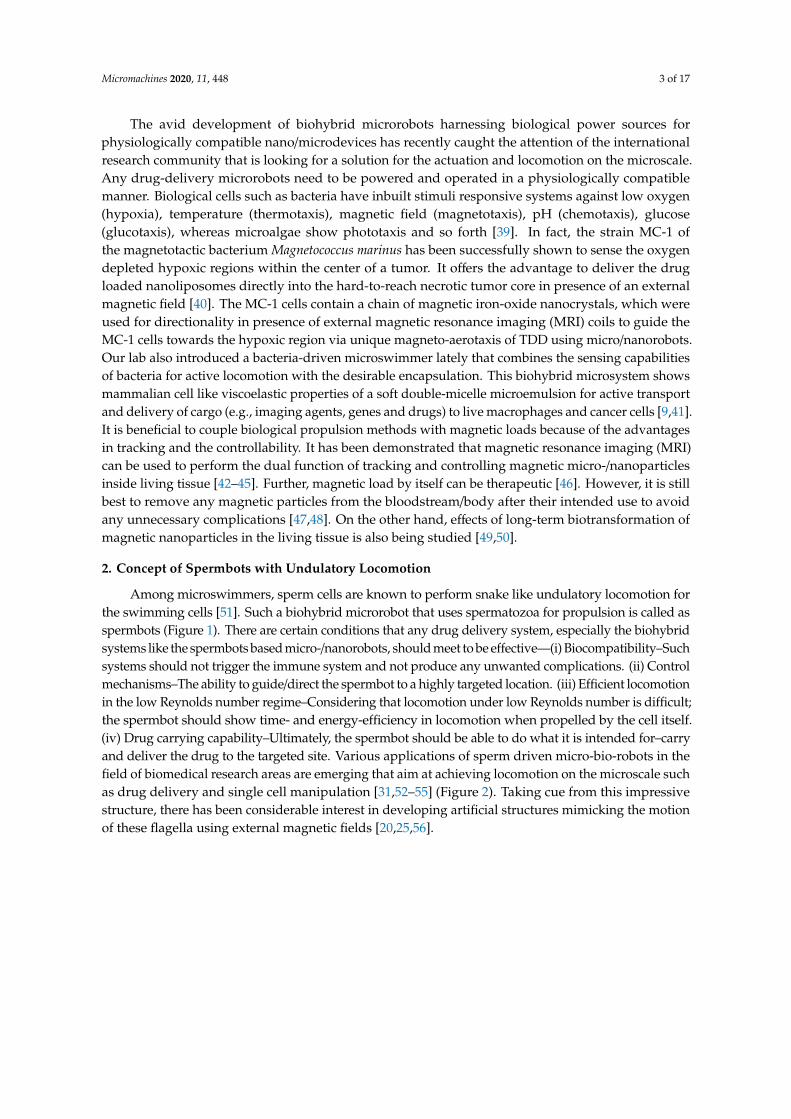
Figure 1. Timeline for biohybrid sperm development, starting with the first biohybrid synthetic bead. Details can be found in Table 1.
Figure 2. Futurist development of spermbots for target drug delivery and assisted fertilizations. Taking advantage of external guidance and actuation, several spermbots can be actuated towards a specific target, either for drug delivery or for assisted fertilization.
2. Creating Biohybrid and Synthetic Spermbots—Technical Challenges and Solutions
Creating spermbots requires coupling sperm cells with a load. Researchers [55,57,58] have used magnetic nanoparticles coupled with sperm cells for drug delivery and tracking cells in-vivo. However, nanoparticle toxicity is still a contested topic [59,60]. Therefore, the pioneer group for spermbots [27] decided to instead use microtubes to encapsulate the sperm cells. In fact, they were the first group to use real sperm cells to propel a microrobot. The microtubes used are fabricated utilizing microfabrication techniques by rolled-up nanotechnology on photoresist [61,62]. The method followed by the Dresden group, as described in References [22,27,63], is as follows (Figure 3)—A photoresist structure (sacrificial layer) is patterned on a glass slide as squares with 50 µm each dimensions. On these patterns, nanometer thick layers of two different metals are deposited via electron-beam evaporation at an angle. It is stated that the difference in deposition rates and the tilt angle creates a strained bilayer. When the sacrificial layer is dissolved, this strained bilayer naturally rolls into microtubes of 50 µm length and nanometer-scale thin walls. When these microtubes, with the diameter just slightly bigger than the head of the sperm, are immersed in a spermatozoa solution, the sperm cells enter the microtubes, get trapped and start propelling the microtubes along with them. However, this coupling is purely physical and random too. As such, the coupling efficiency is not too high. To increase, the efficiency of coupling, specific molecular binders can be used. There are several biomolecules that can be used to bind the sperm cells to the inner tube
Figure 1. Timeline for biohybrid sperm development, starting with the first biohybrid synthetic bead.
Micromachines 2020, 11, x 4 of 17
Figure 1. Timeline for biohybrid sperm development, starting with the first biohybrid synthetic bead. Details can be found in Table 1.
Figure 2. Futurist development of spermbots for target drug delivery and assisted fertilizations. Taking advantage of external guidance and actuation, several spermbots can be actuated towards a specific target, either for drug delivery or for assisted fertilization.
2. Creating Biohybrid and Synthetic Spermbots—Technical Challenges and Solutions
Creating spermbots requires coupling sperm cells with a load. Researchers [55,57,58] have used magnetic nanoparticles coupled with sperm cells for drug delivery and tracking cells in-vivo. However, nanoparticle toxicity is still a contested topic [59,60]. Therefore, the pioneer group for spermbots [27] decided to instead use microtubes to encapsulate the sperm cells. In fact, they were the first group to use real sperm cells to propel a microrobot. The microtubes used are fabricated utilizing microfabrication techniques by rolled-up nanotechnology on photoresist [61,62]. The method followed by the Dresden group, as described in References [22,27,63], is as follows (Figure 3)—A photoresist structure (sacrificial layer) is patterned on a glass slide as squares with 50 µm each dimensions. On these patterns, nanometer thick layers of two different metals are deposited via electron-beam evaporation at an angle. It is stated that the difference in deposition rates and the tilt angle creates a strained bilayer. When the sacrificial layer is dissolved, this strained bilayer naturally rolls into microtubes of 50 µm length and nanometer-scale thin walls. When these microtubes, with the diameter just slightly bigger than the head of the sperm, are immersed in a spermatozoa solution, the sperm cells enter the microtubes, get trapped and start propelling the microtubes along with them. However, this coupling is purely physical and random too. As such, the coupling efficiency is not too high. To increase, the efficiency of coupling, specific molecular binders can be used. There are several biomolecules that can be used to bind the sperm cells to the inner tube
Figure 2. Futurist development of spermbots for target drug delivery and assisted fertilizations. Takingadvantage of external guidance and actuation, several spermbots can be actuated towards a specifictarget, either for drug delivery or for assisted fertilization.
3. Creating Biohybrid and Synthetic Spermbots—Technical Challenges and Solutions
Creating spermbots requires coupling sperm cells with a load. Researchers [55,57,58] have usedmagnetic nanoparticles coupled with sperm cells for drug delivery and tracking cells in-vivo. However,nanoparticle toxicity is still a contested topic [59,60]. Therefore, the pioneer group for spermbots [27]decided to instead use microtubes to encapsulate the sperm cells. In fact, they were the first group to usereal sperm cells to propel a microrobot. The microtubes used are fabricated utilizing microfabricationtechniques by rolled-up nanotechnology on photoresist [61,62]. The method followed by the Dresdengroup, as described in References [22,27,63], is as follows (Figure 3)—A photoresist structure (sacrificiallayer) is patterned on a glass slide as squares with 50 µm each dimensions. On these patterns,nanometer thick layers of two different metals are deposited via electron-beam evaporation at an angle.It is stated that the difference in deposition rates and the tilt angle creates a strained bilayer. Whenthe sacrificial layer is dissolved, this strained bilayer naturally rolls into microtubes of 50 µm lengthand nanometer-scale thin walls. When these microtubes, with the diameter just slightly bigger thanthe head of the sperm, are immersed in a spermatozoa solution, the sperm cells enter the microtubes,get trapped and start propelling the microtubes along with them. However, this coupling is purelyphysical and random too. As such, the coupling efficiency is not too high. To increase, the efficiency ofcoupling, specific molecular binders can be used. There are several biomolecules that can be used tobind the sperm cells to the inner tube surface [64–72]. In order to functionalize the inner surface ofthe microtubes for better entrapment of sperm cells, two attachment methods are utilized—surfacelinker chemistry and microcontact printing technology. While both the methods improve the trappingof sperm cells, the coupling still relies on random events and is not controlled. Therefore, It will behelpful to develop a method that attaches previously selected single sperm cells to the microtubes in acontrolled manner [73].
Micromachines 2020, 11, 448 5 of 17
In the case of artificial spermbots, they can be fabricated using two photon lithography (NanoscribeGmbH, Eggenstein-Leopoldshafen, Germany). When a photosensitive polymer is coated on a substrateand exposed to a pulse laser of appropriate wavelength, duration and intensity, it gets locally cured dueto two photon absorption. When the process is repeated in 3D space while controlled by a computer,it can generate 3D structures with very high resolutions (100 nm) [74]. For the purpose of mimickingthe screw like motion of the flagella, microhelices corresponding to the size of sperm cells can beprinted (Figure 3). To make these artificial flagella responsive to magnetic fields, they are coatedwith a magnetic material like Nickel or Iron. To ensure biocompatibility, Titanium layer may alsobe coated. Such magnetic microhelices demonstrate a screw-like forward/backward motion underrotating magnetic fields. After their release from the substrate, they are transferred to a chambercontaining sperm cell solution for coupling. Unlike random coupling in case of spermbots, in this case,the coupling is manual. The microhelices are magnetically driven to an immotile sperm and capturedtail-first [30]. With the advent of bottom-up self-assembly, 3D and 4D bioprinting [75–78], it may bepossible to print artificial spermbots in one step in the future.
Micromachines 2020, 11, x 5 of 17
surface [64–72]. In order to functionalize the inner surface of the microtubes for better entrapment of sperm cells, two attachment methods are utilized—surface linker chemistry and microcontact printing technology. While both the methods improve the trapping of sperm cells, the coupling still relies on random events and is not controlled. Therefore, It will be helpful to develop a method that attaches previously selected single sperm cells to the microtubes in a controlled manner [73].
In the case of artificial spermbots, they can be fabricated using two photon lithography (Nanoscribe GmbH, Eggenstein-Leopoldshafen, Germany). When a photosensitive polymer is coated on a substrate and exposed to a pulse laser of appropriate wavelength, duration and intensity, it gets locally cured due to two photon absorption. When the process is repeated in 3D space while controlled by a computer, it can generate 3D structures with very high resolutions (100 nm) [74]. For the purpose of mimicking the screw like motion of the flagella, microhelices corresponding to the size of sperm cells can be printed (Figure 3). To make these artificial flagella responsive to magnetic fields, they are coated with a magnetic material like Nickel or Iron. To ensure biocompatibility, Titanium layer may also be coated. Such magnetic microhelices demonstrate a screw-like forward/backward motion under rotating magnetic fields. After their release from the substrate, they are transferred to a chamber containing sperm cell solution for coupling. Unlike random coupling in case of spermbots, in this case, the coupling is manual. The microhelices are magnetically driven to an immotile sperm and captured tail-first [30]. With the advent of bottom-up self-assembly, 3D and 4D bioprinting [75–78], it may be possible to print artificial spermbots in one step in the future.
Figure 3. Fabrication routes for tubular (A and B) and helical (C) spermbots. A‐i) A schematic of the rolled‐up nanotechnology to fabricate nanomembranes into microtubes. A‐ii) The concept of the tubular spermbots A‐iii) An array of 50 µm‐long microtubes. Scale bar 50 µm. A‐iv) An optical image of a tubular spermbot. Scale bar 20 µm. B‐i) The fabrication route for laser‐written SU8 microtubes. B‐ii) The control of microtubes length B‐iii) A scanning electron microscopy image of an array of laser‐written microtubes. Scale bar 40 µm. B‐iv) The SU8 tube with a trapped spermatozoon. The red arrow indicates the sperm tail. Scale bar 10 µm. C‐i) Fabrication route of 3D nanoprinted helices by
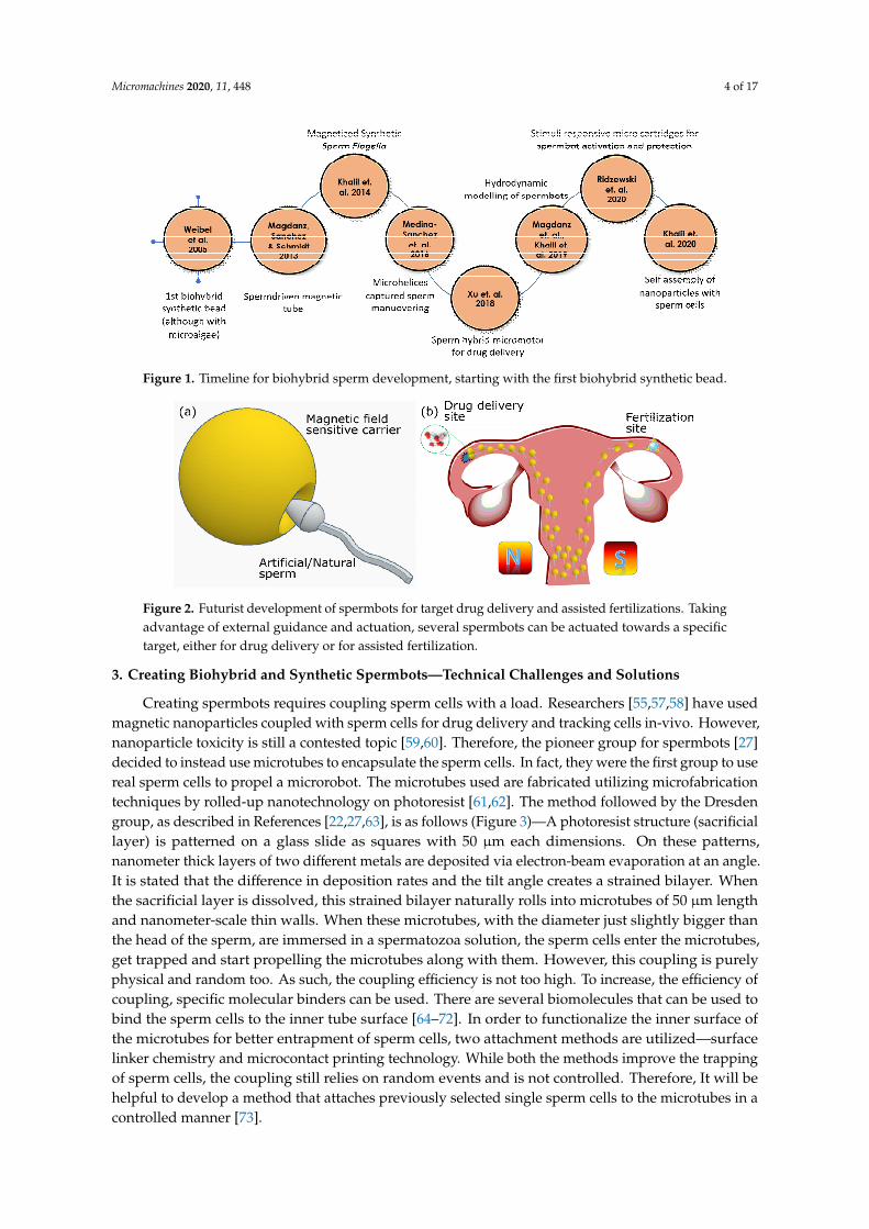
Figure 3. Fabrication routes for tubular (A,B) and helical (C) spermbots. A-(i) A schematic of therolled-up nanotechnology to fabricate nanomembranes into microtubes. A-(ii) The concept of thetubular spermbots A-(iii) An array of 50 µm-long microtubes. Scale bar 50 µm. A-(iv) An optical imageof a tubular spermbot. Scale bar 20 µm. B-(i) The fabrication route for laser-written SU8 microtubes.B-(ii) The control of microtubes length B-(iii) A scanning electron microscopy image of an array oflaser-written microtubes. Scale bar 40 µm. B-(iv) The SU8 tube with a trapped spermatozoon. The redarrow indicates the sperm tail. Scale bar 10 µm. C-(i) Fabrication route of 3D nanoprinted helicesby two-photon absorption lithography. C-(ii) The concept of helical spermbots to transport immotilesperm cells. C-(iii) A scanning electron microscopy image of the fabricated helices. Scale bar 2 µm.C-(iv) A helical spermbot that is carrying a bovine sperm cell. Scale bar 10 µm. (Reprinted fromReference [22] with permission from John Wiley & Sons, Inc.).
Micromachines 2020, 11, 448 6 of 17
4. Spermbot Assisted Targeted Delivery—Proof of Concept Examples with Assisted Fertilizationand Drug Delivery
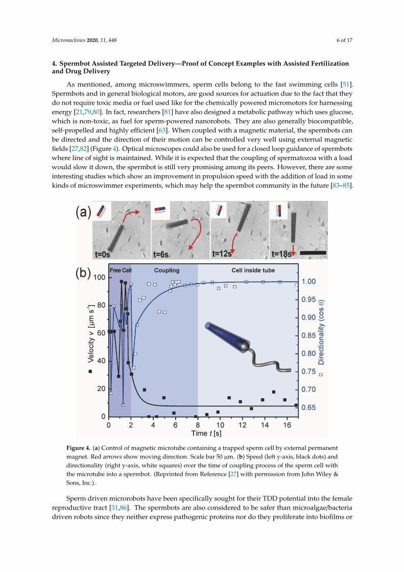
As mentioned, among microswimmers, sperm cells belong to the fast swimming cells [51].Spermbots and in general biological motors, are good sources for actuation due to the fact that theydo not require toxic media or fuel used like for the chemically powered micromotors for harnessingenergy [21,79,80]. In fact, researchers [81] have also designed a metabolic pathway which uses glucose,which is non-toxic, as fuel for sperm-powered nanorobots. They are also generally biocompatible,self-propelled and highly efficient [63]. When coupled with a magnetic material, the spermbots canbe directed and the direction of their motion can be controlled very well using external magneticfields [27,82] (Figure 4). Optical microscopes could also be used for a closed loop guidance of spermbotswhere line of sight is maintained. While it is expected that the coupling of spermatozoa with a loadwould slow it down, the spermbot is still very promising among its peers. However, there are someinteresting studies which show an improvement in propulsion speed with the addition of load in somekinds of microswimmer experiments, which may help the spermbot community in the future [83–85].
Micromachines 2020, 11, x 6 of 17
two‐photon absorption lithography. C‐ii) The concept of helical spermbots to transport immotile sperm cells. C‐iii) A scanning electron microscopy image of the fabricated helices. Scale bar 2 µm. C‐iv) A helical spermbot that is carrying a bovine sperm cell. Scale bar 10 µm. (Reprinted from Reference [22] with permission from John Wiley & Sons, Inc.).
3. Spermbot Assisted Targeted Delivery—Proof of Concept Examples with Assisted Fertilization and Drug Delivery
As mentioned, among microswimmers, sperm cells belong to the fast swimming cells [51]. Spermbots and in general biological motors, are good sources for actuation due to the fact that they do not require toxic media or fuel used like for the chemically powered micromotors for harnessing energy [21,79,80]. In fact, researchers [81] have also designed a metabolic pathway which uses glucose, which is non-toxic, as fuel for sperm-powered nanorobots. They are also generally biocompatible, self-propelled and highly efficient [63]. When coupled with a magnetic material, the spermbots can be directed and the direction of their motion can be controlled very well using external magnetic fields [27,82] (Figure 4). Optical microscopes could also be used for a closed loop guidance of spermbots where line of sight is maintained. While it is expected that the coupling of spermatozoa with a load would slow it down, the spermbot is still very promising among its peers. However, there are some interesting studies which show an improvement in propulsion speed with the addition of load in some kinds of microswimmer experiments, which may help the spermbot community in the future [83–85].
Figure 4. (a) Control of magnetic microtube containing a trapped sperm cell by external permanent magnet. Red arrows show moving direction. Scale bar 50 µm. (b) Speed (left y‐axis, black dots) and directionality (right y‐axis, white squares) over the time of coupling process of the sperm cell with the microtube into a spermbot. (Reprinted from Reference [27] with permission from John Wiley & Sons, Inc.).
Figure 4. (a) Control of magnetic microtube containing a trapped sperm cell by external permanentmagnet. Red arrows show moving direction. Scale bar 50 µm. (b) Speed (left y-axis, black dots) anddirectionality (right y-axis, white squares) over the time of coupling process of the sperm cell withthe microtube into a spermbot. (Reprinted from Reference [27] with permission from John Wiley &Sons, Inc.).
Sperm driven microrobots have been specifically sought for their TDD potential into the femalereproductive tract [31,86]. The spermbots are also considered to be safer than microalgae/bacteriadriven robots since they neither express pathogenic proteins nor do they proliferate into biofilms or
Micromachines 2020, 11, 448 7 of 17
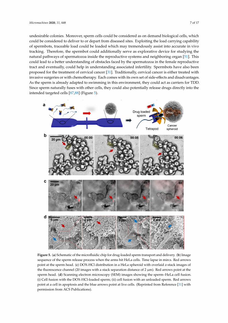
undesirable colonies. Moreover, sperm cells could be considered as on demand biological cells, whichcould be considered to deliver to or depart from diseased sites. Exploiting the load carrying capabilityof spermbots, traceable load could be loaded which may tremendously assist into accurate in vivotracking. Therefore, the spermbot could additionally serve as explorative device for studying thenatural pathways of spermatozoa inside the reproductive systems and neighboring organ [51]. Thiscould lead to a better understanding of obstacles faced by the spermatozoa in the female reproductivetract and eventually, could help in understanding associated infertility. Spermbots have also beenproposed for the treatment of cervical cancer [31]. Traditionally, cervical cancer is either treated withinvasive surgeries or with chemotherapy. Each comes with its own set of side-effects and disadvantages.As the sperm is already adapted to swimming in this environment, they could act as carriers for TDD.Since sperm naturally fuses with other cells, they could also potentially release drugs directly into theintended targeted cells [87,88] (Figure 5).
Micromachines 2020, 11, x 7 of 17
Sperm driven microrobots have been specifically sought for their TDD potential into the female reproductive tract [31,86]. The spermbots are also considered to be safer than microalgae/bacteria driven robots since they neither express pathogenic proteins nor do they proliferate into biofilms or undesirable colonies. Moreover, sperm cells could be considered as on demand biological cells, which could be considered to deliver to or depart from diseased sites. Exploiting the load carrying capability of spermbots, traceable load could be loaded which may tremendously assist into accurate in vivo tracking. Therefore, the spermbot could additionally serve as explorative device for studying the natural pathways of spermatozoa inside the reproductive systems and neighboring organ [51]. This could lead to a better understanding of obstacles faced by the spermatozoa in the female reproductive tract and eventually, could help in understanding associated infertility. Spermbots have also been proposed for the treatment of cervical cancer [31]. Traditionally, cervical cancer is either treated with invasive surgeries or with chemotherapy. Each comes with its own set of side-effects and disadvantages. As the sperm is already adapted to swimming in this environment, they could act as carriers for TDD. Since sperm naturally fuses with other cells, they could also potentially release drugs directly into the intended targeted cells [87,88] (Figure 5).
Figure 5. (a) Schematic of the microfluidic chip for drug-loaded sperm transport and delivery. (b) Image sequence of the sperm release process when the arms hit HeLa cells. Time lapse in min:s. Red arrows point at the sperm head. (c) DOX-HCl distribution in a HeLa spheroid with overlaid z-stack images of the fluorescence channel (20 images with a stack separation distance of 2 µm). Red arrows point at the sperm head. (d) Scanning electron microscopy (SEM) images showing the sperm–HeLa cell fusion. (i) Cell fusion with the DOX-HCl-loaded sperm; (ii) cell fusion with an unloaded sperm. Red arrows point at a cell in apoptosis and the blue arrows point at live cells. (Reprinted from Reference [31] with permission from ACS Publications).
Figure 5. (a) Schematic of the microfluidic chip for drug-loaded sperm transport and delivery. (b) Imagesequence of the sperm release process when the arms hit HeLa cells. Time lapse in min:s. Red arrowspoint at the sperm head. (c) DOX-HCl distribution in a HeLa spheroid with overlaid z-stack images ofthe fluorescence channel (20 images with a stack separation distance of 2 µm). Red arrows point at thesperm head. (d) Scanning electron microscopy (SEM) images showing the sperm–HeLa cell fusion.(i) Cell fusion with the DOX-HCl-loaded sperm; (ii) cell fusion with an unloaded sperm. Red arrowspoint at a cell in apoptosis and the blue arrows point at live cells. (Reprinted from Reference [31] withpermission from ACS Publications).
Micromachines 2020, 11, 448 8 of 17
Another specific potential of the sperm-driven micro-biorobot is described [51] that might haveimpact on the development of assisted reproductive technologies. It is stated that the success rate ofstate-of-the-art assisted reproduction techniques is still low. These techniques involve the removal ofthe oocyte from the body, fertilization in the petri dish, cultivation of embryos and reimplantation ofthe embryo into the uterus. This is where the spermbots can be helpful in bypassing these lengthyand cost associated steps and by guiding the spermbots to the target oocytes inside the human bodyitself. Schmidt et al. [27,82] used a 50 µm long microtube with iron membrane to encapsulate bovinespermatozoa. The flagellum of the sperm cell serves to propel the microtube forward while the ironmembrane is used to steer using untethered magnetic fields. Further, electromagnetic coils withfeedback from optical microscope can be used for closed loop control of the spermbots for the targeteddelivery to a selected reference point. Schmidt et al. [27] also studied the effect of microtube radius,extent of sperm cell penetration inside the microtube and temperature on the speed of spermbots.With an increase in microtube radius, the extent of spermatozoa penetration also increased. It wasfound that spermbots with higher penetration percentage have decreased speed because of increasedconfinement of the flagella. An increase in speed with increase in temperature was also observed.It is reported that, in general, the speed of spermbots is considerably decreased to around 10% ofthe speed of initial spermatozoa speed. In this case, the coupling of spermatozoa and microtubesalso occurs randomly which causes low coupling efficiency. To improve the performance, the samegroup [73] proposes shortening the length of the microtube to 20 µm from 50 µm. The average speedof the spermbots goes up from about 20% bodylength per second to about 65% bodylength per second.To further enhance the performance, binding the sperm cells to the hollow space of microtubes usingFibronectin (Fn) protein and adding caffeine to the environment to boost the motility of the cells wasproposed. As discussed above, there are several biomolecules which can be used to bind the spermcells to the inner tube surface [64–72]. Because the microtube is ferromagnetic, its orientation can becontrolled and maintained using an external magnetic field–like a compass needle. Therefore, while thepropulsion of an uncoupled sperm cell is random, the direction of propulsion of a coupled spermbotcan be highly controlled using an external magnetic field generated either by permanent magnets orby electromagnets.
Artificial Spermbots
One of the main causes of infertility in men is sometimes called low sperm motility, a conditionwhere the sperm is healthy but unable to swim effectively to make it to an egg for fertilization. Amongthe couples who struggle to have a baby, the male partner plays a role in the infertility about 40 percentof the time according to the American Society for Reproductive Medicine [89]. Techniques like artificialinsemination or in vitro fertilization (IVF) can help but they tend to be not very reliable or complexand expensive [51]. Researchers have come up with a possible solution to help sperm to swim morequickly and effectively with the motorized ‘spermbot.’ Taking cue from the spermatozoa flagella itself,researchers have prepared a metallic helix that can wrap around the flagella of the spermatozoa andpropel it using external magnetic field [30]. Once the sperm has entered the egg, the metal casing canreverse direction to slip-off the spermbot with the help of the externally controlled magnetic field.
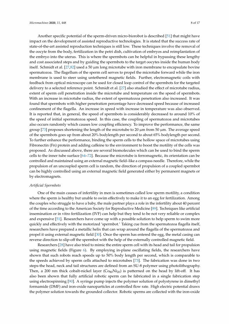
Researchers [28] have also tried to mimic the entire sperm cell with its head and tail for propulsionusing magnetic fields (Figure 6). By employing in-plane oscillating fields, the researchers haveshown that such robots reach speeds up to 50% body length per second, which is comparable tothe speeds achieved by sperm cells attached to microtubes [73]. The fabrication was done in twosteps-the head, neck and tail structures are defined from an SU-8 polymer using photolithography.Then, a 200 nm thick cobalt-nickel layer (Co80Ni20) is patterned on the head by lift-off. It hasalso been shown that fully artificial robotic sperm can be fabricated in a single fabrication stepusing electrospinning [90]. A syringe pump injects the polymer solution of polystyrene in dimethylformamide (DMF) and iron-oxide nanoparticles at controlled flow rate. High electric potential drawsthe polymer solution towards the grounded collector. Robotic sperms are collected with the iron-oxide
Micromachines 2020, 11, 448 9 of 17
nanoparticles contained within their beads. Such robotic sperms are actuated by applying appropriatemagnetic torques on the robotic sperm heads which propagates travelling waves along its flexible tail.Out-of-plane wobbling of the head results in helical wave propagation along the flagellum, whereasin-plane wobbling achieves planar wave propagation. Controllable switching between planar andhelical flagellar propulsion has also been shown [91]. Modifying the same process, artificial roboticsperms with two collinear, unequal and opposite tails have also been fabricated which are able topropel back and forth in bi-direction without a U-turn trajectory [92].
Micromachines 2020, 11, x 9 of 17
iron-oxide nanoparticles contained within their beads. Such robotic sperms are actuated by applying appropriate magnetic torques on the robotic sperm heads which propagates travelling waves along its flexible tail. Out-of-plane wobbling of the head results in helical wave propagation along the flagellum, whereas in-plane wobbling achieves planar wave propagation. Controllable switching between planar and helical flagellar propulsion has also been shown [91]. Modifying the same process, artificial robotic sperms with two collinear, unequal and opposite tails have also been fabricated which are able to propel back and forth in bi-direction without a U-turn trajectory [92].
Figure 6. Another kind of Spermbot (MagnetoSperm) moving under the influence of the oscillating (25 Hz) weak magnetic fields (∼5 mT). At t = 1 s, oscillating weak magnetic fields are applied which allows MagnetoSperm to swim at a speed of 53 µm/s. (Reprinted from Reference [28] with permission from AIP Publishing).
4. Challenges
Spermbots are not without their own disadvantages in the female reproductive tract because of the hostile environment and natural defense mechanisms. About 30 min after entering the body, < 1% of living sperm cells remain in the female reproductive tract due to vaginal flowback, the acidic pH and phagocytosis by leukocytes [51,93]. However, with the microtube enveloping the sperm cell in the spermbot, it could be equipped in ways to prevent the leukocytosis in the same fashion that bacterial pathogens are able to overcome the phagocytic engulfment and killing by appropriate blocking methods[63].
There are several challenges that need to be addressed before spermbots can truly be translated into medical applications:
1. Sperm cells do not all have the same motility. Their motility varies from individual to individual and from cell to cell even from the same individual. This is a cause for concern because we need sperm cells that are highly motile, to be as efficient as possible for actuation. While there is considerable interest in this particular field [94–104], a standardized method and protocol is highly crucial and is the need of the hour.
2. The load/microtubes by itself should not be toxic and should be able to pass through any barriers that it may encounter on the way to the targeted site.
3. The attachment of load or microtubes to the sperm cell is random and a very low-yielding process [27]. Therefore, robust methods to increase this coupling efficiency are needed [38,73]. Electrostatic-based self-assembly helps in partial coating of all sperm heads with magnetic nanoparticle aggregates [38].
Figure 6. Another kind of Spermbot (MagnetoSperm) moving under the influence of the oscillating(25 Hz) weak magnetic fields (~5 mT). At t = 1 s, oscillating weak magnetic fields are applied whichallows MagnetoSperm to swim at a speed of 53 µm/s. (Reprinted from Reference [28] with permissionfrom AIP Publishing).
5. Challenges
Spermbots are not without their own disadvantages in the female reproductive tract becauseof the hostile environment and natural defense mechanisms. About 30 min after entering the body,<1% of living sperm cells remain in the female reproductive tract due to vaginal flowback, the acidicpH and phagocytosis by leukocytes [51,93]. However, with the microtube enveloping the spermcell in the spermbot, it could be equipped in ways to prevent the leukocytosis in the same fashionthat bacterial pathogens are able to overcome the phagocytic engulfment and killing by appropriateblocking methods [63].
There are several challenges that need to be addressed before spermbots can truly be translatedinto medical applications:
1. Sperm cells do not all have the same motility. Their motility varies from individual to individualand from cell to cell even from the same individual. This is a cause for concern because weneed sperm cells that are highly motile, to be as efficient as possible for actuation. While thereis considerable interest in this particular field [94–104], a standardized method and protocol ishighly crucial and is the need of the hour.
2. The load/microtubes by itself should not be toxic and should be able to pass through any barriersthat it may encounter on the way to the targeted site.
3. The attachment of load or microtubes to the sperm cell is random and a very low-yieldingprocess [27]. Therefore, robust methods to increase this coupling efficiency are needed [38,73].
Micromachines 2020, 11, 448 10 of 17
Electrostatic-based self-assembly helps in partial coating of all sperm heads with magneticnanoparticle aggregates [38].
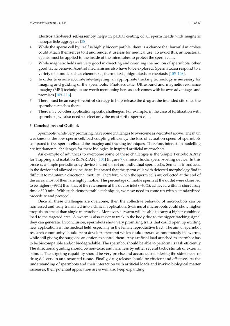
4. While the sperm cell by itself is highly biocompatible, there is a chance that harmful microbescould attach themselves to it and render it useless for medical use. To avoid this, antibacterialagents must be applied to the inside of the microtubes to protect the sperm cells.
5. While magnetic fields are very good in directing and orienting the motion of spermbots, othergood tactic behavior/control mechanisms also have to be explored. Spermatozoa respond to avariety of stimuli, such as chemotaxis, thermotaxis, thigmotaxis or rheotaxis [105–108].
6. In order to ensure accurate site-targeting, an appropriate tracking technology is necessary forimaging and guiding of the spermbots. Photoacoustic, Ultrasound and magnetic resonanceimaging (MRI) techniques are worth mentioning here as each comes with its own advantages andpromises [109–116].
7. There must be an easy-to-control strategy to help release the drug at the intended site once thespermbots reaches there.
8. There may be other application specific challenges. For example, in the case of fertilization withspermbots, we also need to select only the most fertile sperm cells.
6. Conclusions and Outlook
Spermbots, while very promising, have some challenges to overcome as described above. The mainweakness is the low sperm cell/load coupling efficiency, the loss of actuation speed of spermbotscompared to free sperm cells and the imaging and tracking techniques. Therefore, interaction modellingare fundamental challenges for these biologically inspired artificial microrobots.
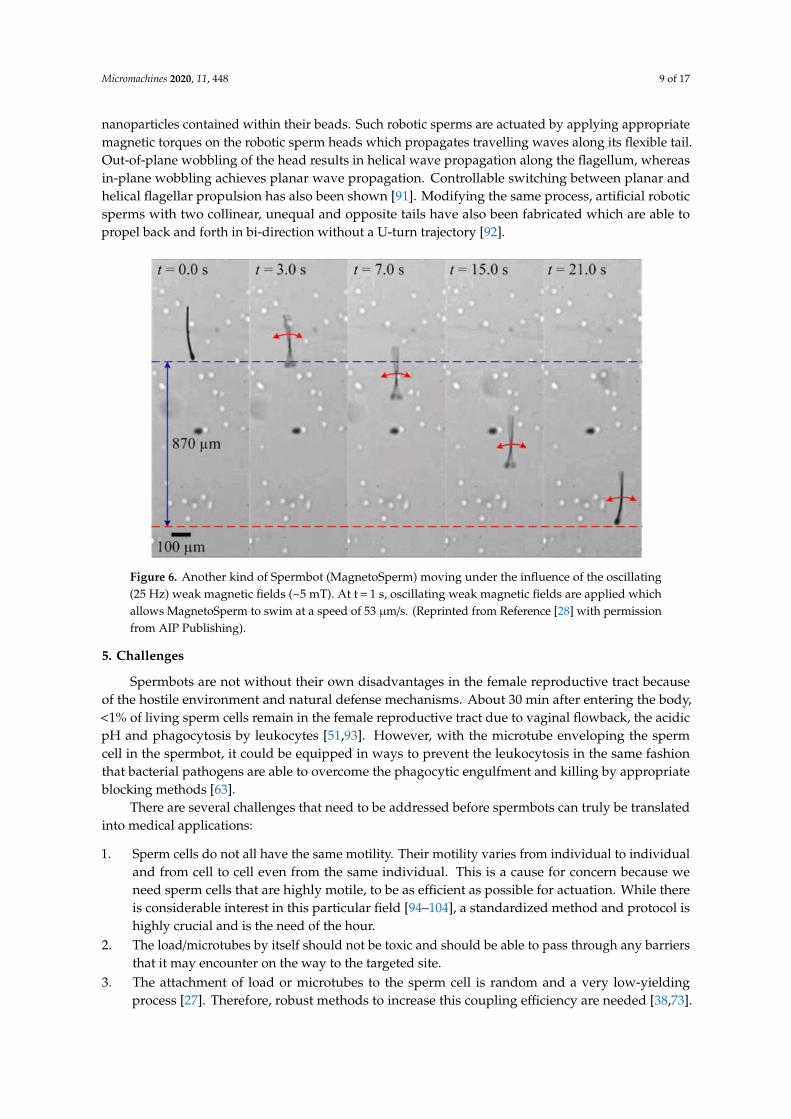
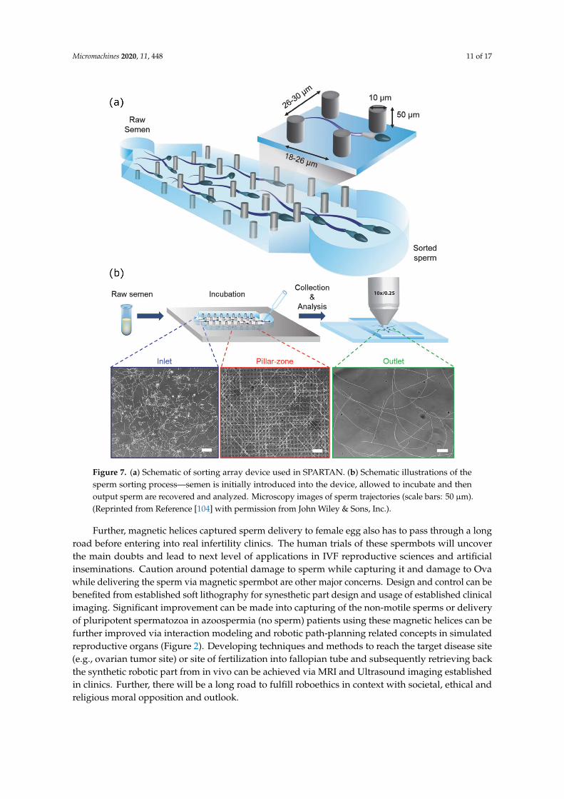
An example of advances to overcome some of these challenges is the Simple Periodic ARrayfor Trapping and isolation (SPARTAN) [104] (Figure 7), a microfluidic sperm-sorting device. In thisprocess, a simple periodic array device is used to sort out individual sperm cells. Semen is introducedin the device and allowed to incubate. It is stated that the sperm cells with defected morphology find itdifficult to maintain a directional motility. Therefore, when the sperm cells are collected at the end ofthe array, most of them are highly motile. The percentage of motile sperm at the outlet were observedto be higher (~99%) than that of the raw semen at the device inlet (~60%), achieved within a short assaytime of 10 min. With such demonstrable techniques, we now need to come up with a standardizedprocedure and protocol.
Once all these challenges are overcome, then the collective behavior of microrobots can beharnessed and truly translated into a clinical application. Swarms of microrobots could show higherpropulsion speed than single microrobots. Moreover, a swarm will be able to carry a higher combinedload to the targeted area. A swarm is also easier to track in the body due to the bigger tracking signalthey can generate. In conclusion, spermbots show very promising traits that could open up excitingnew applications in the medical field, especially in the female reproductive tract. The aim of spermbotresearch community should be to develop spermbot which could operate autonomously in swarms,while still giving the surgeons an option to control them. Any artificial load attached to spermbot hasto be biocompatible and/or biodegradable. The spermbot should be able to perform its task efficiently.The directional guiding should be non-toxic and harmless by either several tactic stimuli or externalstimuli. The targeting capability should be very precise and accurate, considering the side-effects ofdrug delivery in an unwanted tissue. Finally, drug release should be efficient and effective. As theunderstanding of spermbots and their interaction with artificial loads and in-vivo biological matterincreases, their potential application areas will also keep expanding.
Micromachines 2020, 11, 448 11 of 17Micromachines 2020, 11, x 11 of 17
Figure 7. (a) Schematic of sorting array device used in SPARTAN. (b) Schematic illustrations of the sperm sorting process—semen is initially introduced into the device, allowed to incubate and then output sperm are recovered and analyzed. Microscopy images of sperm trajectories (scale bars: 50 µm). (Reprinted from Reference [104] with permission from John Wiley & Sons, Inc.).
Once all these challenges are overcome, then the collective behavior of microrobots can be harnessed and truly translated into a clinical application. Swarms of microrobots could show higher propulsion speed than single microrobots. Moreover, a swarm will be able to carry a higher combined load to the targeted area. A swarm is also easier to track in the body due to the bigger tracking signal they can generate. In conclusion, spermbots show very promising traits that could open up exciting new applications in the medical field, especially in the female reproductive tract. The aim of spermbot research community should be to develop spermbot which could operate autonomously in swarms, while still giving the surgeons an option to control them. Any artificial load attached to spermbot has to be biocompatible and/or biodegradable. The spermbot should be able to perform its task efficiently. The directional guiding should be non-toxic and harmless by either several tactic stimuli or external stimuli. The targeting capability should be very precise and accurate, considering the side-effects of drug delivery in an unwanted tissue. Finally, drug release should be efficient and effective. As the understanding of spermbots and their interaction with artificial loads and in-vivo biological matter increases, their potential application areas will also keep expanding.
Figure 7. (a) Schematic of sorting array device used in SPARTAN. (b) Schematic illustrations of thesperm sorting process—semen is initially introduced into the device, allowed to incubate and thenoutput sperm are recovered and analyzed. Microscopy images of sperm trajectories (scale bars: 50 µm).(Reprinted from Reference [104] with permission from John Wiley & Sons, Inc.).
Further, magnetic helices captured sperm delivery to female egg also has to pass through a longroad before entering into real infertility clinics. The human trials of these spermbots will uncoverthe main doubts and lead to next level of applications in IVF reproductive sciences and artificialinseminations. Caution around potential damage to sperm while capturing it and damage to Ovawhile delivering the sperm via magnetic spermbot are other major concerns. Design and control can bebenefited from established soft lithography for synesthetic part design and usage of established clinicalimaging. Significant improvement can be made into capturing of the non-motile sperms or deliveryof pluripotent spermatozoa in azoospermia (no sperm) patients using these magnetic helices can befurther improved via interaction modeling and robotic path-planning related concepts in simulatedreproductive organs (Figure 2). Developing techniques and methods to reach the target disease site(e.g., ovarian tumor site) or site of fertilization into fallopian tube and subsequently retrieving backthe synthetic robotic part from in vivo can be achieved via MRI and Ultrasound imaging establishedin clinics. Further, there will be a long road to fulfill roboethics in context with societal, ethical andreligious moral opposition and outlook.

Micromachines 2020, 11, 448 12 of 17
Author Contributions: Conceptualization, A.V.S. and M.H.D.A.; formal analysis, A.V.S. and P.D.; investigation,A.V.S., data curation, A.V.S. and M.H.D.A.; writing—original draft preparation, A.V.S., M.H.D.A., M.M. and S.S.;writing—review and editing, A.V.S., M.H.D.A., V.P. and U.K.; visualization, S.S., S.K. and P.D.; supervision, A.V.S.;project administration, A.V.S.; funding acquisition, A.V.S. All authors have read and agreed to the publishedversion of the manuscript.
Funding: The Max Planck Society funded this work.
Acknowledgments: A.V.S. thanks Max Planck Institute for Intelligent Systems for the Grassroots project grantsM10335 and M10338. M.H.D.A. is part of the ATLAS project that received funding from the European Union’sHorizon 2020 research and innovation program under the Marie Sklodowska-Curie grant agreement No 813782.
Conflicts of Interest: The authors declare no conflict of interest.
References
1. Singh, A.V.; Rahman, A.; Sudhir Kumar, N.V.G.; Aditi, A.S.; Galluzzi, M.; Bovio, S.; Barozzi, S.; Montani, E.;Parazzoli, D. Bio-inspired approaches to design smart fabrics. Mater. Des. (1980–2015) 2012, 36, 829–839.[CrossRef]
2. Singh, A.V.; Dad Ansari, M.H.; Dayan, C.B.; Giltinan, J.; Wang, S.; Yu, Y.; Kishore, V.; Laux, P.; Luch, A.;Sitti, M. Multifunctional magnetic hairbot for untethered osteogenesis, ultrasound contrast imaging anddrug delivery. Biomaterials 2019, 219, 119394. [CrossRef] [PubMed]
3. Kim, S.; Laschi, C.; Trimmer, B. Soft robotics: A bioinspired evolution in robotics. Trends Biotechnol. 2013, 31,287–294. [CrossRef] [PubMed]
4. Albu-Schaffer, A.; Eiberger, O.; Grebenstein, M.; Haddadin, S.; Ott, C.; Wimbock, T.; Wolf, S.; Hirzinger, G.Soft robotics. IEEE Robot. Autom. Mag. 2008, 15, 20–30. [CrossRef]
5. Cianchetti, M.; Laschi, C.; Menciassi, A.; Dario, P. Biomedical applications of soft robotics. Nat. Rev. Mater.2018, 3, 143–153. [CrossRef]
6. Abidi, H.; Gerboni, G.; Brancadoro, M.; Fras, J.; Diodato, A.; Cianchetti, M.; Wurdemann, H.; Althoefer, K.;Menciassi, A. Highly dexterous 2-module soft robot for intra-organ navigation in minimally invasive surgery.Int. J. Med. Robot. Comput. Assist. Surg. 2018, 14, e1875. [CrossRef]
7. Cianchetti, M.; Ranzani, T.; Gerboni, G.; Falco, I.D.; Laschi, C.; Menciassi, A. STIFF-FLOP surgical manipulator:Mechanical design and experimental characterization of the single module. In Proceedings of the 2013IEEE/RSJ International Conference on Intelligent Robots and Systems, Tokyo, Japan, 3–7 November 2013;pp. 3576–3581.
8. Runciman, M.; Darzi, A.; Mylonas, G.P. Soft Robotics in Minimally Invasive Surgery. Soft Robot. 2019, 6,423–443. [CrossRef]
9. Singh, A.V.; Hosseinidoust, Z.; Park, B.-W.; Yasa, O.; Sitti, M. Microemulsion-Based Soft Bacteria-DrivenMicroswimmers for Active Cargo Delivery. ACS Nano 2017, 11, 9759–9769. [CrossRef]
10. Iacovacci, V.; Blanc, A.; Huang, H.; Ricotti, L.; Schibli, R.; Menciassi, A.; Behe, M.; Pané, S.; Nelson, B.J.High-Resolution SPECT Imaging of Stimuli-Responsive Soft Microrobots. Small 2019, 15, 1900709. [CrossRef]
11. Palagi, S.; Singh, D.P.; Fischer, P. Light-Controlled Micromotors and Soft Microrobots. Adv. Opt. Mater. 2019,7, 1900370. [CrossRef]
12. Hu, C.; Pané, S.; Nelson, B.J. Soft Micro- and Nanorobotics. Annu. Rev. Control Robot. Auton. Syst. 2018, 1,53–75. [CrossRef]
13. Carlsen, R.W.; Sitti, M. Bio-Hybrid Cell-Based Actuators for Microsystems. Small 2014, 10, 3831–3851.[CrossRef] [PubMed]
14. Macnab, R.M. The bacterial flagellum: Reversible rotary propellor and type III export apparatus. J. Bacteriol.1999, 181, 7149–7153. [CrossRef] [PubMed]
15. Baccetti, B.; Afzelius, B.A. The Biology of the Sperm Cell; Karger AG: Basel, Switzerland, 1976.16. Astbury, W.T.; Saha, N.N. Structure of Algal Flagella. Nature 1953, 171, 280–283. [CrossRef]17. Singh, A.V.; Ansari, M.H.D.; Laux, P.; Luch, A. Micro-nanorobots: Important considerations when developing
novel drug delivery platforms. Expert Opin. Drug Deliv. 2019, 16, 1259–1275. [CrossRef] [PubMed]18. Hosseinidoust, Z.; Mostaghaci, B.; Yasa, O.; Park, B.-W.; Singh, A.V.; Sitti, M. Bioengineered and biohybrid
bacteria-based systems for drug delivery. Adv. Drug Deliv. Rev. 2016, 106, 27–44. [CrossRef]
Micromachines 2020, 11, 448 13 of 17
19. Singh, A.V.; Jahnke, T.; Xiao, Y.; Wang, S.; Yu, Y.; David, H.; Richter, G.; Laux, P.; Luch, A.; Srivastava, A.; et al.Peptide-Induced Biomineralization of Tin Oxide (SnO2) Nanoparticles for Antibacterial Applications.J. Nanosci. Nanotechnol. 2019, 19, 5674–5686. [CrossRef]
20. Zhang, L.; Abbott, J.J.; Dong, L.; Kratochvil, B.E.; Bell, D.; Nelson, B.J. Artificial bacterial flagella: Fabricationand magnetic control. Appl. Phys. Lett. 2009, 94, 064107. [CrossRef]
21. Behkam, B.; Sitti, M. Bacterial flagella-based propulsion and on/off motion control of microscale objects.Appl. Phys. Lett. 2007, 90, 023902. [CrossRef]
22. Magdanz, V.; Medina-Sánchez, M.; Schwarz, L.; Xu, H.; Elgeti, J.; Schmidt, O.G. Spermatozoa as FunctionalComponents of Robotic Microswimmers. Adv. Mater. 2017, 29, 1606301. [CrossRef]
23. Ricotti, L.; Menciassi, A. Bio-hybrid muscle cell-based actuators. Biomed. Microdevices 2012, 14, 987–998.[CrossRef] [PubMed]
24. Williams, B.J.; Anand, S.V.; Rajagopalan, J.; Saif, M.T.A. A self-propelled biohybrid swimmer at low Reynoldsnumber. Nat. Commun. 2014, 5, 3081. [CrossRef] [PubMed]
25. Dreyfus, R.; Baudry, J.; Roper, M.L.; Fermigier, M.; Stone, H.A.; Bibette, J. Microscopic artificial swimmers.Nature 2005, 437, 862–865. [CrossRef] [PubMed]
26. Weibel, D.B.; Garstecki, P.; Ryan, D.; DiLuzio, W.R.; Mayer, M.; Seto, J.E.; Whitesides, G.M. Microoxen:Microorganisms to move microscale loads. Proc. Natl. Acad. Sci. USA 2005, 102, 11963. [CrossRef] [PubMed]
27. Magdanz, V.; Sanchez, S.; Schmidt, O.G. Development of a Sperm-Flagella Driven Micro-Bio-Robot. Adv. Mater.2013, 25, 6581–6588. [CrossRef]
28. Khalil, I.S.M.; Dijkslag, H.C.; Abelmann, L.; Misra, S. MagnetoSperm: A microrobot that navigates usingweak magnetic fields. Appl. Phys. Lett. 2014, 104, 223701. [CrossRef]
29. Khalil, I.S.M.; Youakim, K.; Sánchez, A.; Misra, S. Magnetic-based motion control of sperm-shaped microrobotsusing weak oscillating magnetic fields. In Proceedings of the 2014 IEEE/RSJ International Conference onIntelligent Robots and Systems, Chicago, IL, USA, 14–18 September 2014; pp. 4686–4691.
30. Medina-Sánchez, M.; Schwarz, L.; Meyer, A.K.; Hebenstreit, F.; Schmidt, O.G. Cellular Cargo Delivery:Toward Assisted Fertilization by Sperm-Carrying Micromotors. Nano Lett. 2016, 16, 555–561. [CrossRef]
31. Xu, H.; Medina-Sánchez, M.; Magdanz, V.; Schwarz, L.; Hebenstreit, F.; Schmidt, O.G. Sperm-HybridMicromotor for Targeted Drug Delivery. ACS Nano 2018, 12, 327–337. [CrossRef] [PubMed]
32. Magdanz, V.; Boryshpolets, S.; Ridzewski, C.; Eckel, B.; Reinhardt, K. The motility-based swim-up techniqueseparates bull sperm based on differences in metabolic rates and tail length. PLoS ONE 2019, 14, e0223576.[CrossRef]
33. Khalil, I.S.M.; Klingner, A.; Magdanz, V.; Striggow, F.; Medina-Sánchez, M.; Schmidt, O.G.; Misra, S. Modelingof Spermbots in a Viscous Colloidal Suspension. Adv. Theory Simul. 2019, 2, 1900072. [CrossRef]
34. Magdanz, V.; Gebauer, J.; Sharan, P.; Eltoukhy, S.; Voigt, D.; Simmchen, J. Sperm–Particle Interactions andTheir Prospects for Charge Mapping. Adv. Biosyst. 2019, 3, 1900061. [CrossRef]
35. Khalil, I.S.M.; Klingner, A.; Hamed, Y.; Magdanz, V.; Toubar, M.; Misra, S. Characterization of FlagellarPropulsion of Soft Microrobotic Sperm in a Viscous Heterogeneous Medium. Front. Robot. AI 2019, 6, 65.[CrossRef]
36. Ridzewski, C.; Li, M.; Dong, B.; Magdanz, V. Gelatin Microcartridges for Onboard Activation and AntioxidantProtection of Sperm. ACS Appl. Bio Mater. 2020. [CrossRef]
37. Xu, H.; Medina-Sánchez, M.; Maitz, M.F.; Werner, C.; Schmidt, O.G. Sperm-Micromotors for Cargo-Deliverythrough Flowing Blood. ACS Nano 2020. [CrossRef] [PubMed]
38. Khalil, I.S.M.; Magdanz, V.; Simmchen, J.; Klingner, A.; Misra, S. Resemblance between motile andmagnetically actuated sperm cells. Appl. Phys. Lett. 2020, 116, 063702. [CrossRef]
39. Mura, S.; Nicolas, J.; Couvreur, P. Stimuli-responsive nanocarriers for drug delivery. Nat. Mater. 2013, 12,991–1003. [CrossRef]
40. Felfoul, O.; Mohammadi, M.; Taherkhani, S.; de Lanauze, D.; Xu, Y.Z.; Loghin, D.; Essa, S.; Jancik, S.;Houle, D.; Lafleur, M.; et al. Magneto-aerotactic bacteria deliver drug-containing nanoliposomes to tumourhypoxic regions. Nat. Nanotechnol. 2016, 11, 941–947. [CrossRef]
41. Park, B.-W.; Zhuang, J.; Yasa, O.; Sitti, M. Multifunctional Bacteria-Driven Microswimmers for TargetedActive Drug Delivery. ACS Nano 2017, 11, 8910–8923. [CrossRef]
Micromachines 2020, 11, 448 14 of 17
42. Martel, S.; Mathieu, J.-B.; Felfoul, O.; Chanu, A.; Aboussouan, E.; Tamaz, S.; Pouponneau, P.; Yahia, L.H.;Beaudoin, G.; Soulez, G.; et al. Automatic navigation of an untethered device in the artery of a livinganimal using a conventional clinical magnetic resonance imaging system. Appl. Phys. Lett. 2007, 90, 114105.[CrossRef]
43. Pouponneau, P.; Leroux, J.-C.; Soulez, G.; Gaboury, L.; Martel, S. Co-encapsulation of magnetic nanoparticlesand doxorubicin into biodegradable microcarriers for deep tissue targeting by vascular MRI navigation.Biomaterials 2011, 32, 3481–3486. [CrossRef]
44. Folio, D.; Ferreira, A. Two-Dimensional Robust Magnetic Resonance Navigation of a FerromagneticMicrorobot Using Pareto Optimality. IEEE Trans. Robot. 2017, 33, 583–593. [CrossRef]
45. Felfoul, O.; Becker, A.T.; Fagogenis, G.; Dupont, P.E. Simultaneous steering and imaging of magnetic particlesusing MRI toward delivery of therapeutics. Sci. Rep. 2016, 6, 33567. [CrossRef]
46. Egolf, P.W.; Shamsudhin, N.; Pané, S.; Vuarnoz, D.; Pokki, J.; Pawlowski, A.-G.; Tsague, P.; de Marco, B.;Bovy, W.; Tucev, S.; et al. Hyperthermia with rotating magnetic nanowires inducing heat into tumor by fluidfriction. J. Appl. Phys. 2016, 120, 064304. [CrossRef]
47. Iacovacci, V.; Ricotti, L.; Sinibaldi, E.; Signore, G.; Vistoli, F.; Menciassi, A. An Intravascular Magnetic CatheterEnables the Retrieval of Nanoagents from the Bloodstream. Adv. Sci. 2018, 5, 1800807. [CrossRef]
48. Iacovacci, V.; Ricotti, L.; Signore, G.; Vistoli, F.; Sinibaldi, E.; Menciassi, A. Retrieval of magnetic medicalmicrorobots from the bloodstream. In Proceedings of the 2019 International Conference on Robotics andAutomation (ICRA), Montreal, QC, Canada, 20–24 May 2019; pp. 2495–2501.
49. Levy, M.; Luciani, N.; Alloyeau, D.; Elgrabli, D.; Deveaux, V.; Pechoux, C.; Chat, S.; Wang, G.; Vats, N.;Gendron, F.; et al. Long term in vivo biotransformation of iron oxide nanoparticles. Biomaterials 2011, 32,3988–3999. [CrossRef] [PubMed]
50. Mejías, R.; Gutiérrez, L.; Salas, G.; Pérez-Yagüe, S.; Zotes, T.M.; Lázaro, F.J.; Morales, M.P.; Barber, D.F. Longterm biotransformation and toxicity of dimercaptosuccinic acid-coated magnetic nanoparticles support theiruse in biomedical applications. J. Control. Release 2013, 171, 225–233. [CrossRef]
51. Magdanz, V.; Schmidt, O.G. Spermbots: Potential impact for drug delivery and assisted reproductivetechnologies. Expert Opin. Drug Deliv. 2014, 11, 1125–1129. [CrossRef]
52. Magdanz, V.; Gebauer, J.; Mahdy, D.; Simmchen, J.; Khalil, I.S.M. Sperm-templated magnetic microrobots.In Proceedings of the 2019 International Conference on Manipulation, Automation and Robotics at SmallScales (MARSS), Helsinki, Finland, 1–5 July 2019; pp. 1–6.
53. Jang, D.; Jeong, J.; Song, H.; Chung, S.K. Targeted drug delivery technology using untethered microrobots:A review. J. Micromech. Microeng. 2019, 29, 053002. [CrossRef]
54. Medina-Sánchez, M.; Xu, H.; Schmidt, O.G. Micro- and nano-motors: The new generation of drug carriers.Ther. Deliv. 2018, 9, 303–316. [CrossRef]
55. Pan, Y.; Du, X.; Zhao, F.; Xu, B. Magnetic nanoparticles for the manipulation of proteins and cells. Chem. Soc.Rev. 2012, 41, 2912–2942. [CrossRef]
56. Gao, W.; Sattayasamitsathit, S.; Manesh, K.M.; Weihs, D.; Wang, J. Magnetically Powered Flexible MetalNanowire Motors. J. Am. Chem. Soc. 2010, 132, 14403–14405. [CrossRef] [PubMed]
57. Dobson, J. Gene therapy progress and prospects: Magnetic nanoparticle-based gene delivery. Gene Ther.2006, 13, 283–287. [CrossRef] [PubMed]
58. Scarberry, K.E.; Dickerson, E.B.; McDonald, J.F.; Zhang, Z.J. Magnetic Nanoparticle−Peptide Conjugates forin Vitro and in Vivo Targeting and Extraction of Cancer Cells. J. Am. Chem. Soc. 2008, 130, 10258–10262.[CrossRef] [PubMed]
59. Khlebtsov, N.; Dykman, L. Biodistribution and toxicity of engineered gold nanoparticles: A review of in vitroand in vivo studies. Chem. Soc. Rev. 2011, 40, 1647–1671. [CrossRef]
60. Ansari, M.H.D.; Lavhale, S.; Kalunke, R.M.; Srivastava, P.L.; Pandit, V.; Gade, S.; Yadav, S.; Laux, P.; Luch, A.;Gemmati, D. Recent Advances in Plant Nanobionics and Nanobiosensors for Toxicology Applications.Curr. Nanosci. 2019, 16, 27–41. [CrossRef]
61. Schmidt, O.G.; Eberl, K. Thin solid films roll up into nanotubes. Nature 2001, 410, 168. [CrossRef]62. Mei, Y.; Huang, G.; Solovev, A.A.; Ureña, E.B.; Mönch, I.; Ding, F.; Reindl, T.; Fu, R.K.Y.; Chu, P.K.;
Schmidt, O.G. Versatile Approach for Integrative and Functionalized Tubes by Strain Engineering ofNanomembranes on Polymers. Adv. Mater. 2008, 20, 4085–4090. [CrossRef]
Micromachines 2020, 11, 448 15 of 17
63. Magdanz, V.; Guix, M.; Schmidt, O.G. Tubular micromotors: From microjets to spermbots. Robot. Biomim.2014, 1, 11. [CrossRef]
64. Bansal, P.; Gupta, S.K. Binding characteristics of sperm with recombinant human zona pellucida glycoprotein-3coated beads. Indian J. Med. Res. 2009, 130, 37.
65. Diaz, E.S.; Kong, M.; Morales, P. Effect of fibronectin on proteasome activity, acrosome reaction, tyrosinephosphorylation and intracellular calcium concentrations of human sperm. Hum. Reprod. 2007, 22, 1420–1430.[CrossRef]
66. Wennemuth, G.; Schiemann, P.J.; Krause, W.; Gressner, A.M.; AumÜLler, G. Influence of fibronectin on themotility of human spermatozoa. Int. J. Androl. 1997, 20, 10–16. [CrossRef] [PubMed]
67. Frimat, J.P.; Bronkhorst, M.; de Wagenaar, B.; Bomer, J.G.; van der Heijden, F.; van den Berg, A.; Segerink, L.I.Make it spin: Individual trapping of sperm for analysis and recovery using micro-contact printing. Lab Chip2014, 14, 2635–2641. [CrossRef]
68. Huszar, G.; Ozenci, C.C.; Cayli, S.; Zavaczki, Z.; Hansch, E.; Vigue, L. Hyaluronic acid binding by humansperm indicates cellular maturity, viability and unreacted acrosomal status. Fertil. Steril. 2003, 79, 1616–1624.[CrossRef]
69. Yagci, A.; Murk, W.; Stronk, J.; Huszar, G. Spermatozoa Bound to Solid State Hyaluronic Acid ShowChromatin Structure with High DNA Chain Integrity: An Acridine Orange Fluorescence Study. J. Androl.2010, 31, 566–572. [CrossRef]
70. Parmegiani, L.; Cognigni, G.E.; Bernardi, S.; Troilo, E.; Taraborrelli, S.; Arnone, A.; Maccarini, A.M.; Filicori, M.Comparison of two ready-to-use systems designed for sperm–hyaluronic acid binding selection beforeintracytoplasmic sperm injection: PICSI vs. Sperm Slow: A prospective, randomized trial. Fertil. Steril. 2012,98, 632–637. [CrossRef]
71. Campos, L.B.; Peixoto, G.C.X.; da Silva, A.M.; Souza, A.L.P.; de Souza Castelo, T.; Maia, K.M.; Pereira, A.F.;Silva, A.R. Estimating the binding ability of collared peccary (Pecari tajacu Linnaeus, 1758) sperm usingheterologous substrates. Theriogenology 2017, 92, 57–62. [CrossRef]
72. Tecle, E.; Reynoso, H.S.; Wang, R.; Gagneux, P. The female reproductive tract contains multiple innate sialicacid-binding immunoglobulin-like lectins (Siglecs) that facilitate sperm survival. J. Biol. Chem. 2019, 294,11910–11919. [CrossRef]
73. Magdanz, V.; Medina-Sánchez, M.; Chen, Y.; Guix, M.; Schmidt, O.G. How to Improve Spermbot Performance.Adv. Funct. Mater. 2015, 25, 2763–2770. [CrossRef]
74. Singh, A.V.; Patil, R.; Thombre, D.K.; Gade, W.N. Micro-nanopatterning as tool to study the role ofphysicochemical properties on cell–surface interactions. J. Biomed. Mater. Res. Part A 2013, 101, 3019–3032.[CrossRef] [PubMed]
75. Spiegel, C.A.; Hippler, M.; Münchinger, A.; Bastmeyer, M.; Barner-Kowollik, C.; Wegener, M.; Blasco, E. 4DPrinting at the Microscale. Adv. Funct. Mater. 2019, 1907615. [CrossRef]
76. Vikram Singh, A.; Hasan Dad Ansari, M.; Wang, S.; Laux, P.; Luch, A.; Kumar, A.; Patil, R.; Nussberger, S.The adoption of three-dimensional additive manufacturing from biomedical material design to 3d organprinting. Appl. Sci. 2019, 9, 811. [CrossRef]
77. Singh, A.V.; Laux, P.; Luch, A.; Balkrishnan, S.; Dakua, S.P. Bottom-UP assembly of nanorobots: Extendingsynthetic biology to complex material design. Front. Nanosci. Nanotechnol. 2019, 5, 1–2.
78. Singh, A. Top-Down Versus Bottom-Up Nanoengineering Routes to Design Advanced OropharmacologicalProducts. Curr. Pharm. Des. 2016, 22, 1534–1545. [CrossRef] [PubMed]
79. Martel, S. Bacterial microsystems and microrobots. Biomed. Microdevices 2012, 14, 1033–1045. [CrossRef][PubMed]
80. Soong, R.K.; Bachand, G.D.; Neves, H.P.; Olkhovets, A.G.; Craighead, H.G.; Montemagno, C.D. Powering anInorganic Nanodevice with a Biomolecular Motor. Science 2000, 290, 1555. [CrossRef] [PubMed]
81. Mukai, C.; Bergkvist, M.; Nelson, J.L.; Travis, A.J. Sequential Reactions of Surface- Tethered GlycolyticEnzymes. Chem. Biol. 2009, 16, 1013–1020. [CrossRef] [PubMed]
82. Khalil, I.S.M.; Magdanz, V.; Sanchez, S.; Schmidt, O.G.; Misra, S. Biocompatible, accurate and fullyautonomous: A sperm-driven micro-bio-robot. J. Micro-Bio Robot. 2014, 9, 79–86. [CrossRef]
83. Raz, O.; Leshansky, A.M. Efficiency of cargo towing by a microswimmer. Phys. Rev. E 2008, 77, 055305.[CrossRef]
Micromachines 2020, 11, 448 16 of 17
84. Grosjean, G.; Hubert, M.; Vandewalle, N. Magnetocapillary self-assemblies: Locomotion andmicromanipulation along a liquid interface. Adv. Colloid Interface Sci. 2018, 255, 84–93. [CrossRef]
85. Gao, W.; Kagan, D.; Pak, O.S.; Clawson, C.; Campuzano, S.; Chuluun-Erdene, E.; Shipton, E.; Fullerton, E.E.;Zhang, L.; Lauga, E.; et al. Cargo-Towing Fuel-Free Magnetic Nanoswimmers for Targeted Drug Delivery.Small 2012, 8, 460–467. [CrossRef]
86. Luo, M.; Feng, Y.; Wang, T.; Guan, J. Micro-/Nanorobots at Work in Active Drug Delivery. Adv. Funct. Mater.2018, 28, 1706100. [CrossRef]
87. Yan, W. Toward Better Treatment for Women’s Reproductive Health: New Devices, Nanoparticles and EvenRobotic Sperm May Hold the Key to Preventing a Range of Health Conditions. IEEE Pulse 2018, 9, 21–24.[CrossRef] [PubMed]
88. Bendich, A.; Borenfreund, E.; Sternberg, S.S. Penetration of Somatic Mammalian Cells by Sperm. Science1974, 183, 857. [CrossRef]
89. Lindsay, T.J.; Vitrikas, K.R. Evaluation and treatment of infertility. Am. Fam. Physician 2015, 91, 308–314.[PubMed]
90. Khalil, I.S.M.; Fatih Tabak, A.; Klingner, A.; Sitti, M. Magnetic propulsion of robotic sperms at low-Reynoldsnumber. Appl. Phys. Lett. 2016, 109, 033701. [CrossRef]
91. Khalil, I.S.M.; Tabak, A.F.; Abou Seif, M.; Klingner, A.; Sitti, M. Controllable switching between planar andhelical flagellar swimming of a soft robotic sperm. PLoS ONE 2018, 13, e0206456. [CrossRef] [PubMed]
92. Khalil, I.S.M.; Tabak, A.F.; Hamed, Y.; Mitwally, M.E.; Tawakol, M.; Klingner, A.; Sitti, M. Swimming Back andForth Using Planar Flagellar Propulsion at Low Reynolds Numbers. Adv. Sci. 2018, 5, 1700461. [CrossRef][PubMed]
93. Suarez, S.S.; Pacey, A.A. Sperm transport in the female reproductive tract. Hum. Reprod. Update 2005, 12,23–37. [CrossRef] [PubMed]
94. Schuster, T.G.; Cho, B.; Keller, L.M.; Takayama, S.; Smith, G.D. Isolation of motile spermatozoa from semensamples using microfluidics. Reprod. Biomed. Online 2003, 7, 75–81. [CrossRef]
95. Cho, B.S.; Schuster, T.G.; Zhu, X.; Chang, D.; Smith, G.D.; Takayama, S. Passively Driven IntegratedMicrofluidic System for Separation of Motile Sperm. Anal. Chem. 2003, 75, 1671–1675. [CrossRef]
96. Zhang, B.; Yin, T.L.; Yang, J. A novel microfluidic device for selecting human sperm to increase the proportionof morphologically normal, motile sperm with uncompromised DNA integrity. Anal. Methods 2015, 7,5981–5988. [CrossRef]
97. Zhang, Y.; Xiao, R.-R.; Yin, T.; Zou, W.; Tang, Y.; Ding, J.; Yang, J. Generation of Gradients on a MicrofluidicDevice: Toward a High-Throughput Investigation of Spermatozoa Chemotaxis. PLoS ONE 2015, 10, e0142555.[CrossRef] [PubMed]
98. Nosrati, R.; Vollmer, M.; Eamer, L.; San Gabriel, M.C.; Zeidan, K.; Zini, A.; Sinton, D. Rapid selection ofsperm with high DNA integrity. Lab Chip 2014, 14, 1142–1150. [CrossRef]
99. Eamer, L.; Vollmer, M.; Nosrati, R.; San Gabriel, M.C.; Zeidan, K.; Zini, A.; Sinton, D. Turning the cornerin fertility: High DNA integrity of boundary-following sperm. Lab Chip 2016, 16, 2418–2422. [CrossRef][PubMed]
100. Nosrati, R.; Driouchi, A.; Yip, C.M.; Sinton, D. Two-dimensional slither swimming of sperm within amicrometre of a surface. Nat. Commun. 2015, 6, 8703. [CrossRef] [PubMed]
101. Nosrati, R.; Graham, P.J.; Liu, Q.; Sinton, D. Predominance of sperm motion in corners. Sci. Rep. 2016, 6, 1–9.[CrossRef]
102. Tasoglu, S.; Safaee, H.; Zhang, X.; Kingsley, J.L.; Catalano, P.N.; Gurkan, U.A.; Nureddin, A.; Kayaalp, E.;Anchan, R.M.; Maas, R.L.; et al. Exhaustion of Racing Sperm in Nature-Mimicking Microfluidic ChannelsDuring Sorting. Small 2013, 9, 3374–3384. [CrossRef]
103. Asghar, W.; Velasco, V.; Kingsley, J.L.; Shoukat, M.S.; Shafiee, H.; Anchan, R.M.; Mutter, G.L.; Tüzel, E.;Demirci, U. Selection of Functional Human Sperm with Higher DNA Integrity and Fewer Reactive OxygenSpecies. Adv. Healthc. Mater. 2014, 3, 1671–1679. [CrossRef]
104. Chinnasamy, T.; Kingsley, J.L.; Inci, F.; Turek, P.J.; Rosen, M.P.; Behr, B.; Tüzel, E.; Demirci, U. Guidance andSelf-Sorting of Active Swimmers: 3D Periodic Arrays Increase Persistence Length of Human Sperm Selectingfor the Fittest. Adv. Sci. 2018, 5, 1700531. [CrossRef]
105. Kantsler, V.; Dunkel, J.; Goldstein, R.E. Surface Interactions in Suspensions of Swimming Cells. Biophys. J.2014, 106, 210a. [CrossRef]
Micromachines 2020, 11, 448 17 of 17
106. Kantsler, V.; Dunkel, J.; Blayney, M.; Goldstein, R.E. Rheotaxis facilitates upstream navigation of mammaliansperm cells. eLife 2014, 3, e02403. [CrossRef]
107. Zhang, Z.; Liu, J.; Meriano, J.; Ru, C.; Xie, S.; Luo, J.; Sun, Y. Human sperm rheotaxis: A passive physicalprocess. Sci. Rep. 2016, 6, 23553. [CrossRef] [PubMed]
108. Woolley, D.M. Motility of spermatozoa at surfaces. Reproduction 2003, 126, 259–270. [CrossRef] [PubMed]109. Kruger, R.A. Photoacoustic ultrasound. Med. Phys. 1994, 21, 127–131. [CrossRef] [PubMed]110. Omar, M.; Schwarz, M.; Soliman, D.; Symvoulidis, P.; Ntziachristos, V. Pushing the Optical Imaging Limits
of Cancer with Multi-Frequency-Band Raster-Scan Optoacoustic Mesoscopy (RSOM). Neoplasia 2015, 17,208–214. [CrossRef] [PubMed]
111. Neuschmelting, V.; Lockau, H.; Ntziachristos, V.; Grimm, J.; Kircher, M.F. Lymph Node Micrometastasesand In-Transit Metastases from Melanoma: In Vivo Detection with Multispectral Optoacoustic Imaging in aMouse Model. Radiology 2016, 280, 137–150. [CrossRef] [PubMed]
112. Khalil, I.S.M.; Ferreira, P.; Eleutério, R.; Korte, C.L.d.; Misra, S. Magnetic-based closed-loop control ofparamagnetic microparticles using ultrasound feedback. In Proceedings of the 2014 IEEE InternationalConference on Robotics and Automation (ICRA), Hong Kong, China, 31 May–7 June 2014; pp. 3807–3812.
113. Sánchez, A.; Magdanz, V.; Schmidt, O.G.; Misra, S. Magnetic control of self-propelled microjets underultrasound image guidance. In Proceedings of the 5th IEEE RAS/EMBS International Conference onBiomedical Robotics and Biomechatronics, Sao Paulo, Brazil, 12–15 August 2014; pp. 169–174.
114. Weissleder, R.; Moore, A.; Mahmood, U.; Bhorade, R.; Benveniste, H.; Chiocca, E.A.; Basilion, J.P. In vivomagnetic resonance imaging of transgene expression. Nat. Med. 2000, 6, 351–354. [CrossRef]
115. Lee, S.-C.; Kim, K.; Kim, J.; Lee, S.; Han Yi, J.; Woo Kim, S.; Ha, K.-S.; Cheong, C. One Micrometer ResolutionNMR Microscopy. J. Magn. Reson. 2001, 150, 207–213. [CrossRef]
116. Uecker, M.; Zhang, S.; Voit, D.; Karaus, A.; Merboldt, K.-D.; Frahm, J. Real-time MRI at a resolution of 20 ms.NMR Biomed. 2010, 23, 986–994. [CrossRef]
© 2020 by the authors. Licensee MDPI, Basel, Switzerland. This article is an open accessarticle distributed under the terms and conditions of the Creative Commons Attribution(CC BY) license (http://creativecommons.org/licenses/by/4.0/).